Embed Size (px)
Citation preview

A
NGa
Hb
Ac
d
a
ARRAA
KNSLCP
1
fialoi
h0
Journal of Pharmaceutical and Biomedical Analysis 96 (2014) 1–9
Contents lists available at ScienceDirect
Journal of Pharmaceutical and Biomedical Analysis
j o ur na l ho mepage: www.elsev ier .com/ locate / jpba
nalytical characterization of NOTA-modified somatropins�
athalie Brackea, Evelien Wynendaelea, Matthias D’Hondta, Rob Haselbergb,overt W. Somsenb, Ewald Pauwelsc, Christoph Van de Wieled, Bart De Spiegeleera,∗
Drug Quality and Registration (DruQuaR) group, Department of Pharmaceutical Analysis, Faculty of Pharmaceutical Sciences, Ghent University,arelbekestraat 72, 9000 Ghent, BelgiumDivision of BioAnalytical Chemistry, AIMMS research group BioMolecular Analysis, Faculty of Sciences, VU University, De Boelelaan 1083, 1081 HVmsterdam, The NetherlandsCenter for Molecular Modeling, Ghent University, Technologiepark 903, 9052 Zwijnaarde, BelgiumDepartment of Nuclear Medicine, Ghent University Hospital, 9000 Ghent, Belgium
r t i c l e i n f o
rticle history:eceived 27 November 2013eceived in revised form 26 February 2014ccepted 11 March 2014vailable online 20 March 2014
eywords:OTA modificationomatropinC–MSE–MSeptide mapping
a b s t r a c t
Chemical modification of biomolecules like the introduction of metal-chelators into proteins can lead toheterogeneous product formation. The nature and extend of the modification is important in interpre-ting the biological properties of the bioconjugate, given their possible influence on the pharmacokineticsas well as on the binding affinity to the target. The present study describes the synthesis and analyti-cal characterization of somatropin modified on its lysine’s �-amino groups with the acylating chelatorS-2-(4-isothiocyanatobenzyl)-1,4,7-triazacyclononane-1,4,7-triacetic acid (p-SCN-Bn-NOTA). Direct sep-aration and identification techniques (i.e. RP–MS and CE–MS) and peptide mapping after trypsin andchymotrypsin digestion demonstrated that the use of higher amounts of p-SCN-Bn-NOTA during syn-thesis leads to a complex product composition with higher order substitution degrees (i.e. multipleNOTA-moieties per somatropin molecule), as well as the presence of different position isomers. Fromthe nine lysine (Lys) residues in somatropin, Lys-70 was experimentally found to be the modificationhotspot under our synthesis conditions (pH = 9.0). This was supported by the in silico calculated lowestpKa value of 8.3 for Lys-70. Based on the crystal structure of somatropin in complex with the extracellu-lar parts of the growth hormone receptor, the Lys-70 residue is positioned outside the binding pockets
and will therefore not directly interfere with receptor binding. Gallium chelation by NOTA-somatropinresulted in a 100% complexation. The synthesis of NOTA-somatropin using p-SCN-Bn-NOTA and soma-tropin under our operational conditions is therefore a suitable synthesis procedure for the productionof a target-specific radiopharmaceutical for further investigation toward treatment and visualization ofgrowth hormone-specific cancers.© 2014 Elsevier B.V. All rights reserved.
. Introduction
The recent successes of biopharmaceuticals are changing theocus of the pharmaceutical industry. The US Food and Drug Admin-stration (FDA)’s Center for Drug Evaluation and Research (CDER)pproved 39 new drugs in 2012, whereof six represent biologicsicense applications (BLAs) [1]. By 2015, it is even expected that 50%
f the newly approved drugs will be biologics [2], which furtherllustrates the importance of the development of new biological� Selected Paper presented at PBA 2013, Bologna, Italy, 30 June–3 July 2013.∗ Corresponding author. Tel.: +32 9 264 81 00; fax: +32 9 264 81 93.
E-mail address: [email protected] (B. De Spiegeleer).
ttp://dx.doi.org/10.1016/j.jpba.2014.03.014731-7085/© 2014 Elsevier B.V. All rights reserved.
entities (NBEs) and bioanalytical tools for the characterization ofNBEs and biosimilars toward registration [3,4].
Cancer is still the largest therapeutic area nowadays [1,5]. Tumortargeting in nuclear medicine is based on a target-specific radio-pharmaceutical ligand for selective receptor binding in the diseasetissue [6–8]. The targeting biomolecules can be used as therapeu-tics to deliver a toxic radioactive payload selectively to a tumorsite (e.g. I-131 tositumomab or Y-90 ibritumomab), as well as diag-nostic agents for non-invasive single photon emission computedtomography (SPECT) or positron emission tomography (PET) imag-ing (e.g. In-111 Capromab pendetide or In-111 pentetreotide) [9,10]
and playing an important role in cancer management as a form ofpersonalized medicine [9,11].One of the biomolecules of current interest is human growthhormone (hGH). In the late 1950s, severe growth hormone
2 tical a
dhrCtruSlMs[a
ihwpatrott
ltrpnisoapoc
2
2
(EBpa(rGuGaSbg
dcLTwaa
N. Bracke et al. / Journal of Pharmaceu
eficiencies (GHD) in children were treated by cadaveric pituitaryGH or somatotropin extracts. However, several cases wereeported which correlated the use of cadaveric somatotropin withreutzfeldt–Jakob disease, leading to an abrupt stop of hGH extractreatment [12]. This led to the worldwide regulatory approval ofecombinant hGH. Recombinant hGH or somatropin is nowadayssed for the treatment of GHD, as well as the treatment of Turneryndrome and AIDS associated catabolism [13]. Recently, biosimi-ar somatropin formulations were also globally approved [14–16].
oreover, somatropin and analogues are often encountered aspurious/falsely-labeled/falsified/counterfeit (SFFC) medicines17–19] and drug abuses in sports [20,21], agriculture [22] andnti-aging [23] have been reported.
Somatropin can perform its actions by binding with high affin-ty to the extracellular domains of two identical molecules ofuman growth hormone receptors (hGHR) [24–27], which areidely expressed in liver tissue, but are also aberrantly overex-ressed in numerous cancers such as prostate [28,29], breast [30]nd colon cancer [31]. The potential involvement of the GH sys-em in tumor promotion and progression, which has been criticallyeviewed in references [32] and [33], as well as the internalizationf the receptor–ligand complex [34], makes hGHR a potential tumorarget for the development of somatropin-based radiopharmaceu-icals.
Modifications with bifunctional chelating agents (BFCA)ike S-2-(4-isothiocyanatobenzyl)-1,4,7-triazacyclononane-1,4,7-riacetic acid (p-SCN-Bn-NOTA) allow the incorporation ofadiometals for SPECT/PET-diagnostic (67Ga, 68Ga, 111In) or thera-eutic (90Y) purposes [9,35,36]. However, because somatropin hasine potential lysine-amino binding sites for p-SCN-Bn-NOTA, it
s important to characterize the obtained product under differentynthesis procedures. We present the analytical characterizationf NOTA-modified somatropins with special attention to the oper-tional strategy and procedure which are widely applicable to otherrotein systems containing multiple modification sites towardther bifunctional chelators (e.g. DOTA, DTPA, MAMA) or fluores-ent labels (e.g. fluorescein) [9].
. Materials and methods
.1. Materials and equipment
The p-SCN-Bn-NOTA was purchased from Macrocyclics Inc.Dallas, TX, USA). Zomacton® 4 mg, (Ferring, somatropin Ph.ur.) was purchased at the Ghent University Hospital (Ghent,elgium). The enzymes for peptide mapping, L-1-tosylamide-2-henylethyl chloromethyl ketone (TPCK)-treated trypsin solutionnd immobilized chymotrypsin solution, were purchased at PierceErembodegem, Belgium) and Sigma Aldrich (Diegem, Belgium),espectively. PD-10 sephadex G-25M columns were obtained fromE healthcare (Diegem, Belgium). Water was purified in-housesing an Arium 611 purification system (Sartorius, Göttingen,ermany), yielding ≥18.2 M� cm quality water. Other chemicalsnd solvents were purchased from Merck (Overijse, Belgium),igma Aldrich (Diegem, Belgium) or Fischer Scientific (Erem-odegem, Belgium), all high quality (>98% purity) and/or HPLC/MSrade.
Freeze-drying was done using a Christ gamma 1-16 LSC freezeryer (Qlab, Vilvoorde, Belgium). The HPLC–UV–MS apparatusonsisted of a SpectraSystem separations module, a FinniganCQ Classic ion trap mass spectrometer in positive ion mode (all
hermo, San José, CA, USA) equipped with a Waters 2487 dualavelength absorbance UV detector (Waters, Milford, MA, USA)nd Xcalibur 2.0 software (Thermo, San José, CA, USA) for datacquisition. For CE–MS, a P/ACE MDQ capillary electrophoresis
nd Biomedical Analysis 96 (2014) 1–9
instrument (Beckman Coulter Inc., Brea, CA, USA) was usedfor separations, whereas a micrOTOFQ orthogonal acceleratedtime-of-flight (TOF) mass spectrometer (Bruker Daltonics, Bremen,Germany) was used for detection and identification.
2.2. Synthesis
1 mg of somatropin (eq. to 45 nmol) was dissolved in 200 �L car-bonate buffer (pH 9.0; 0.1 M), added to different volumes of 20 mMp-SCN-Bn-NOTA in carbonate buffer (pH 9.0; 0.1 M) and dilutedto 350 �L with carbonate buffer (pH 9.0; 0.1 M) to obtain molarequivalents of 1:1 NOTA:somatropin, 3:1 NOTA:somatropin and10:1 NOTA:somatropin. The mixtures were incubated for 20 h at20 ◦C in the dark, while shaking at 300 rpm. The sample was loadedonto a PD-10 sephadex G-25 M column (equilibrated with diges-tion buffer consisting of 10 mM ACES, 20 mM CaCl2, pH 7.0). Aftercollection of the protein fraction, the samples were freeze driedawaiting analytical characterization.
2.3. Direct analysis of NOTA-somatropins
NOTA-labeling of the 3:1 sample was monitored during the20 h incubation period, by taking 30 �L of sample after 1, 3, 5,8, 10, 12.5, 17, 20 and 22 h. The samples were diluted to 100 �Lusing carbonate buffer (pH 9.0; 0.1 M) and characterized usingLC–MS. For the analysis of the different NOTA-somatropins, 0.1 mgof lyophilized 1:1 NOTA:somatropin, 3:1 NOTA:somatropin and10:1 NOTA:somatropin (4.5 nmol protein) was dissolved in 100 �Lwater. Samples were analyzed via LC–MS. A Vydac Everest MSRP-C4 (250 mm × 4.6 mm I.D., 5 �m particle size, 300 A) column(Grace Vydac, Hesperia, CA, USA) was used and temperature con-trolled during analysis (35 ◦C). The injection volume was 20 �L. Theflow rate was set to 0.5 mL/min and the following gradient wasused for separation of different somatropin derivatives (A: 50 mMammonium bicarbonate pH 7.5 and B: 1-propanol): for 60 min, alinear gradient was applied from 70% A (v/v) + 30% B (v/v) to 50% A(v/v) + 50% B (v/v), followed by a 10 min isocratic section (50:50A:B (v/v)). The method also included a rinsing step out of 60%1-propanol, followed by returning to the initial conditions and re-equilibration. ESI was conducted with a capillary voltage of 4.5 kV.Nitrogen was used as the sheath and auxiliary gas; the tempera-ture of the heated capillary was set to 280 ◦C. MS/MS spectra wereused for identification and obtained by collision induced dissocia-tion (CID) of the parent m/z, with the relative collision energy set to35%. UV detection and quantification were performed at 220 nm.
The CE–MS analysis was performed using fused-silica capillaries(800 mm × 50 �m I.D., Polymicro Technologies, Phoenix, AZ, USA)coated with a bilayer of polybrene and poly(vinyl sulfonic acid)as described previously [37]. Sample injections were performedat 1 psi for 12 s. The separation voltage was 30 kV and the cap-illary temperature was 20 ◦C. The background electrolyte (BGE)was 75 mM ammonium hydroxide adjusted to pH 8.5 with 1%(v/v) formic acid in deionized water. As sheath liquid, a mixtureof isopropanol–water–acetic acid (75/22/3 v/v/v) was employedat 4 �L/min. ESI was conducted in positive ionization mode witha capillary voltage of 4.5 kV. To assure proper ion transfer, theanalysis of somatropin was performed with a capillary exit andskimmer voltage of 250 and 83 V, respectively. CE–MS data wereanalyzed using Bruker Daltonics Data Analysis software. Molecu-lar weight determinations of proteins were performed using the“charge deconvolution” utility of the data analysis software. Quan-tification of the conjugates was performed using the construction
of an extracted-ion electropherogram. Each detected conjugateshowed the same charge state distribution profile, with (M + 9H)9+and (M + 10H)10+ as most abundant ions. The relative abundanceof each conjugate in the analyzed preparation was established by

tical a
ca
2
sgaStilwwpwsi1biwwtL
uaoilA34itieataPmstodt
L
N
is a well-established hexadentate BFCA, forming an exceedinglystable complex with Ga(III) (log K = 30.98) [48]. The isothiocyanate
N. Bracke et al. / Journal of Pharmaceu
alculating the ratio of the conjugate peak area to the total peakrea.
.4. Peptide mapping
The unmodified somatropin (i.e. control) and NOTA-modifiedomatropins (45 nmol protein) were dissolved in 1000 �L 6 Muanidine HCl, 35 mM Tris, 20 mM dl-dithiothreitol at pH 7.5nd incubated for 30 min at 37 ◦C, while shaking at 300 rpm.-carboxymethylation of cysteine residues was performed by addi-ion of 20 �L of iodoacetate (pH 7.2; 58 mM) and subsequentncubation for 30 min at 37 ◦C (at 300 rpm). One hundred micro-iters of dl-dithiothreitol (1 M) was added and mixed. The sample
as loaded onto a PD-10 column sephadex G-25M (equilibratedith digestion buffer consisting of 10 mM ACES, 20 mM CaCl2,H 7.0). After collection of the protein fraction in 2.0 mL, 1.0 mLas transferred into 100 �L of immobilized TPCK-treated trypsin
olution (20 TAME units) and mixed. The reaction mixture wasncubated for 4 h at 37 ◦C (300 rpm). For the 3:1 protein sample,.0 mL of the protein fraction was transferred to 200 �L of immo-ilized chymotrypsin solution (8.3 ATEE units) as well, mixed, and
ncubated for 24 h at 37 ◦C (300 rpm). After incubation, the solutionsere centrifuged at 100g for 10 s. One milliliter of the supernatantas transferred to 10 �L of formic acid (10% v/v), mixed and cen-
rifuged at 20,000g for 10 min. The supernatant was analyzed byC–MS.
ESI was conducted using a needle voltage of 3 kV. Nitrogen wassed as the sheath and auxiliary gas with the heated capillary sett 250 ◦C. Positive mode mass spectra were obtained in the rangef m/z from 100 to 2000. MS/MS spectra were obtained by collisionnduced dissociation (CID) of the parent m/z, with the relative col-ision energy set to 35%. UV detection was performed at 195 nm.
Vydac Everest RP-C18 (250 mm × 2.1 mm I.D., 5 �m particle size,00 A) column (Grace Vydac, Hesperia, CA, USA) in an oven set at5 ◦C, with a mobile phase consisting of (A) 0.1% (w/v) formic acid
n water and (B) 0.1% (w/v) formic acid in acetonitrile was used inhis experiment. The linear gradient program started with a 5 minsocratic hold at 96% (v/v) A and 4% (v/v) B, followed by a lin-ar gradient to 60% (v/v) A + 40% (v/v) B at 113 min. The methodlso included a rinsing step at 60% B, followed by returning tohe initial conditions and re-equilibration. The flow rate was sett 0.2 mL/min and a fixed injection volume of 20 �L was applied.rediction of peak identity was performed upon comparison of/z values with the SEQUEST algorithm of the Thermo BioWorks
oftware (San José, CA, USA). Peptides with probability of morehan 95% were reported and individually verified. Quantificationccurred via ion-extracted chromatography (IEC) of the most abun-ant peptide charge responses. Peptide responses were normalizedo the sum of all responses. The following calculations were made:
Sequence coverage (%)
= Number of amino acid residues found
Total number of amino acid residues in somatropin (= 191)× 100
Lysine coverage (%)
= Number of lysine residues found
Total number of lysine residues in somatropin (= 9)× 100
ysX coverage (%) = Number of peptides containing LysX
Number of peptides containing lysine residue× 100
OTA − LysX modification yield (%) = Area of NOTA − LysX
Sum area of all LysX× 100
NOTA − LysX distribution yield (%)
= Area of NOTA − LysX
Sum area of all NOTA modified lysines× 100
nd Biomedical Analysis 96 (2014) 1–9 3
2.5. In silico pKa calculations
Structure-based pKa calculations were performed using theAdaptive Poisson–Boltzmann Solver (APBS version 1.1.0) [38], inwhich the pKa per titratable residue is determined as the sum of anunperturbed model value [39] and a perturbational shift reflectingthe transfer of the amino acid from solution to the protein envi-ronment. The latter is calculated through a rigorous free energycycle and numerical solution of the linearized Poisson–Boltzmannequation [40]. All calculations were carried out at 298.15 K with asolvent dielectric constant of 78.54 and a protein dielectric constantof 20. pKa calculations were performed on the protein structureonly, taken from the last frame of a molecular dynamics (MD)simulation. Appropriate PQR files were generated with the aid ofPDB2PQR version 1.4.0 [41,42], employing the CHARMM [43,44]parameterization.
Prior to the pKa calculations, structural calculations were per-formed using NAMD version 2.6 [45] and the CHARMM forcefield[43,44], starting from the 1HGU crystal structure of the humansomatotropin [46]. Optimal protonation states were assigned andmissing atoms were added. In an ensuing 5000-step conjugate-gradient energy minimization only these atoms were allowed tomove, while constraining all other atoms. Twelve amino acidswere then mutated in accordance with the somatropin sequence(Q11D, E29Q, A47N, A50T, Q66E, A67T, Q75E, G91Q, D109N, A138I,A144S, A148T) followed by 5000 steps of conjugate-gradient mini-mization of the atoms of the mutated residues only. The resultingstructure was solvated with 20.442 water molecules in an ortho-gonal box of size 83.1 × 86.5 × 95.1 A3 and made charge neutralby adding six sodium ions. The entire structure was subject toenergy minimization (5000 conjugate gradient steps) with con-straints on all protein atoms. This was followed by an unconstrainedMD equilibration run of 50 ps (1 fs time step) in the NVT ensembleat 300 K, employing Langevin dynamics with a damping coeffi-cient of 1 ps−1 to control temperature. Electrostatics were treatedwith particle-mesh Ewald (PME) [47]. A short-range cutoff of 14 Awas maintained, and electrostatic and van der Waals’ interactionswere gradually switched off starting from 10 A. Neighbor lists wereupdated every 2 fs using a cutoff of 16.5 A. The final production MDsimulation totaled 1 ns with identical parameters.
2.6. Gallium labeling and quality control
For the labeling of 3:1 NOTA-somatropin with gallium, 45 nmollyophilized protein sample was dissolved in 450 �L of 0.6 mMGaCl3, 0.1 M HCl to obtain a 2:1 molar excess compared to the initialp-SCN-Bn-NOTA concentration. Then, 20 �L of 2 M NaOH and 40 �Lof 0.1 M ammonium acetate, 0.2 mM acetylacetone buffer (pH 5.5)were added and the solution was mixed and incubated for 1 h at20 ◦C protected from light, while shaking at 300 rpm. The chela-tion efficiency with gallium was analyzed via peptide mapping asdiscussed in Section 2.4.
3. Results and discussion
3.1. Production of NOTA-somatropin
Successful clinical use demands that a bifunctional chelatingagent (BFCA) is both capable of maintaining a stable complex with aradionuclidic metal in vivo, e.g. Ga(III), while possessing a functionalgroup which can be used for protein attachment. p-SCN-Bn-NOTA
function (R-NCS) allows formation of stable thiourea bonds at alka-line pH with free amines (Fig. 1). Somatropin has nine potential

4 N. Bracke et al. / Journal of Pharmaceutical and Biomedical Analysis 96 (2014) 1–9
beled
rBvacce
Fd
Fig. 1. Synthesis of gallium la
eaction sites (lysine’s �-amino residues) for the addition of p-SCN-n-NOTA. We have used a pH of 9.0 during synthesis: higher pHalues will tend to accelerate the degradation of somatropin [49]nd the R-NCS reagent, while lower pH values will lower the con-
entration of the free base form of the amines. These nine sitesan lead to heterogeneous product formation consisting of differ-nt substitution degrees (i.e. the amount of bound NOTA-moleculesig. 2. NOTA-modified somatropins product composition. (A) Scheme of the possible pegree of the 3:1 NOTA:somatropin preparation during 22 h of incubation, product comp
NOTA-modified somatropin.
per somatropin protein). Moreover, for a somatropin entity thathas a single NOTA-molecule attached, the NOTA-moiety may beattached at different amine sites. This creates the potential fora large number of position isomers as the degree of substitution
increases (Fig. 2A) [50]. Analytical characterization of the hetero-geneous production is very important as special attention mustbe paid to the lysine residues that are modified, since chemicalroducts with different substitution degrees and position isomers. (B) Substitutionosition after 20 h of incubation indicated in gray.

N. Bracke et al. / Journal of Pharmaceutical and Biomedical Analysis 96 (2014) 1–9 5
F obtainb /v) + 3N
moseN(N
3
iosftM
TP
Py
ig. 3. Analytical results of the substitution degree of NOTA-modified somatropin
icarbonate pH 7.5 and (B) n-propanol: a 60 min linear gradient from 70% (A) (vOTA-somatropin sample are given.
odification can influence receptor binding and hence, theutcome of biological assays [51]. We applied three differentynthesis ratios of p-SCN-Bn-NOTA based on references [52,53]:quimolar amounts of p-SCN-Bn-NOTA and somatropin (1:1OTA:somatropin), three times molar excess of p-SCN-Bn-NOTA
3:1 NOTA:somatropin) and 10 times molar excess of p-SCN-Bn-OTA (10:1 NOTA:somatropin) were applied.
.2. Direct analytical characterization of the products
A single analytical technique for the characterization of biolog-cals is generally not sufficient [54,55]. Therefore, a combination
f LC–MS and CE–MS was applied to investigate the NOTA-omatropin products. In the biopharmaceutical field, LC is usedor both the assessment of protein batch purity, protein modifica-ion (e.g. glycosylation) and aggregation [4,56]. In combination withS, precise and complementary information is generated. In this
able 1eptide mapping results (tryptic digest) of the individual peptides with NOTA modificatio
Product Sequence Th. mass (Da) (z) Exp. m
1:1 EETQQK70SNLELLR + NOTA 680.07 (3) 680.13:1 EETQQK70SNLELLR + NOTA 680.07 (3) 680.2
FDTNSHNDDALLK158NYGLLY + NOTA 888.61 (3) 888.6DTNSHNDDALLK158NY + NOTA 690.71 (3) 691.0DMDK172VETFLR + NOTA 568.64 (3) 568.7TGQIFK140QTY + NOTA 768.36 (2) 768.4
10:1 EETQQK70SNLELLR + NOTA 680.07 (3) 680.2DTNSHNDDALLK158NY + NOTA 690.71 (3) 691.1TGQIFK140QTY + NOTA 768.36 (2) 768.5
roduct: molar ratio of NOTA over somatropin. NOTA-LysX distribution yield: percentageield: percentage of LysX that is NOTA modified (i.e. lysine site depicted in bold in the seq
ed with RP-C4 (A) and CE (B). A typical LC chromatogram ((A) 50 mM ammonium0% (B) (v/v) to 50% (A) (v/v) 50% (B) (v/v)) and CE electropherogram of the 3:1
direct analytical approach, intact protein molecular ions generatedby electrospray (ESI) or matrix-assisted laser desorption (MALDI)are introduced into the mass analyzer [57].
We have initially based our method on the related protein testdescribed in the European Pharmacopoeia for somatropin (Ph. Eur.8.0: 01/2008:0951) [58]: an isocratic LC-method using a mixtureof 1-propanol and 0.05 M tris–hydrochloride buffer solution pH 7.5(29:71 (v/v)) as the mobile phase. As the tris–hydrochloride bufferis not MS compatible, an ammonium bicarbonate buffer pH 7.5 wasused. During pilot development, we have used isocratic methodswith different mobile phase compositions (i.e. 20%, 30% and 35%organic mobile phase) and studied the somatropin retention time(RT: 49, 11 and 7.5 min, respectively). Based on the retention time ofsomatropin in the isocratic methods and the fact that the hydropho-
bicity of somatropin decreases upon NOTA labeling, we have useda 60 min linear gradient going from 30% to 50% 1-propanol. Thismethod enabled us to characterize the NOTA:somatropin substitu-tion degrees.n.
ass (Da) RT (min) NOTA-LysXdistribution yield (%)
NOTA-LysXmodification yield (%)
3 56.16 100 100 (n = 1)8 52.95 64 94 (n = 4)8 72.37 30 24 (n = 4)3 77.371 67.53 4 7 (n = 4)5 59.69 2 4 (n = 4)5 55.61 80 100 (n = 1)
80.02 13 100 (n = 1)2 56.75 7 8 (n = 1)
NOTA of LysX among all NOTA modified lysine residues. NOTA-LysX modificationuence).

6 tical a
2(fio
Fc
N. Bracke et al. / Journal of Pharmaceu
The NOTA-labeling was quantitatively monitored over time for
2 h for the 3:1 NOTA:somatropin sample using our RP-C4 methodFig. 2B). The complexity of the product composition was con-rmed by a decrease of unmodified somatropin and an increasef higher order substitution degrees over time. Steady state wasig. 4. LC chromatogram of the tryptic digest and MS2 spectra of the peptide EETQQKSNLircle. Inset: MS spectra, with the selected precursor ion indicated by a black circle.
nd Biomedical Analysis 96 (2014) 1–9
reached after 20 h of incubation. Using this incubation period dur-
ing synthesis, the unmodified somatropin was completely absentin the 10:1 NOTA:somatropin sample (Fig. 3A). The highest yield ofthe desired mono- and di-NOTA-somatropin (substitution degree+1 and +2) were obtained in the 3:1 sample (58%). Higher orderELLR (A) with NOTA-modification (B) and gallium labeling (C), indicated by a black
tical a
mrdea
amicodtfi1deCwsh
3
“mdotfmaitts
Fp
N. Bracke et al. / Journal of Pharmaceu
odifications (substitution degree of more than 2) were heavilyepresented in sample 10:1 (98%). For some substitution degrees,ifferent position isomers could be detected. The total peak recov-ry (peak balance) was found between 90–110%, confirming thenalytical characterizing capacity of the method.
Our findings were confirmed by CE–MS (Fig. 3B), a techniquelso included in the somatropin monograph of the European Phar-acopoeia (Ph. Eur. 8.0: 01/2008:0951) [58]. In CE, the separation
s based on charge differences [59], in our case a loss of one positiveharge of lysine with the simultaneous addition of negative chargesf NOTA, whereas in RP HPLC mainly the decrease in hydrophobicityue to the attachment of NOTA leads to the separation [56]. Overall,he 1:1 and 3:1 samples show similar degrees of somatropin modi-cation as measured with CE–MS compared to LC–MS (Fig. 3). In the0:1 sample, CE–MS revealed somatropin with NOTA substitutionegrees ≥5 (amounted for 3%). As these highly polar compoundsnd up in the LC dead time they were not detected, whereas inE they migrate later and, therefore, could be detected. In general,e can conclude that both CE–MS and LC–MS techniques lead to
imilar conclusions regarding the extent of modification. However,igher order substitution degrees are detectable using CE–MS.
.3. Peptide mapping of the NOTA-modified somatropins
The current gold standard in protein characterization is thebottom-up approach”. This method relies on the digestion of aixture of proteins of interest and subsequent analysis of the
igested peptides by LC–MS. All peptides were identified basedn their peptide mass fingerprint (m/z value) and CID fragmen-ation pattern, thereby establishing the validity of these patternsor peptide identification and structural elucidation of the protein
odification (Fig. 4). Peptides containing the NOTA-label are char-cterized by a mass increase of 449.52 Da and specific NOTA-losses
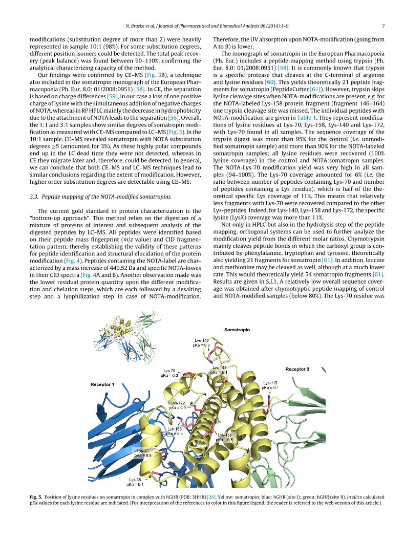
n their CID spectra (Fig. 4A and B). Another observation made washe lower residual protein quantity upon the different modifica-ion and chelation steps, which are each followed by a desaltingtep and a lyophilization step in case of NOTA-modification.ig. 5. Position of lysine residues on somatropin in complex with hGHR (PDB: 3HHR) [26Ka values for each lysine residue are indicated. (For interpretation of the references to c
nd Biomedical Analysis 96 (2014) 1–9 7
Therefore, the UV absorption upon NOTA-modification (going fromA to B) is lower.
The monograph of somatropin in the European Pharmacopoeia(Ph. Eur.) includes a peptide mapping method using trypsin (Ph.Eur. 8.0: 01/2008:0951) [58]. It is commonly known that trypsinis a specific protease that cleaves at the C-terminal of arginineand lysine residues [60]. This yields theoretically 21 peptide frag-ments for somatropin (PeptideCutter [61]). However, trypsin skipslysine cleavage sites when NOTA-modifications are present, e.g. forthe NOTA-labeled Lys-158 protein fragment (fragment 146–164)one trypsin cleavage site was missed. The individual peptides withNOTA-modification are given in Table 1. They represent modifica-tions of lysine residues at Lys-70, Lys-158, Lys-140 and Lys-172,with Lys-70 found in all samples. The sequence coverage of thetrypsin digest was more than 95% for the control (i.e. unmodi-fied somatropin sample) and more than 90% for the NOTA-labeledsomatropin samples; all lysine residues were recovered (100%lysine coverage) in the control and NOTA:somatropin samples.The NOTA-Lys-70 modification yield was very high in all sam-ples (94–100%). The Lys-70 coverage amounted for 6% (i.e. theratio between number of peptides containing Lys-70 and numberof peptides containing a Lys residue), which is half of the the-oretical specific Lys coverage of 11%. This means that relativelyless fragments with Lys-70 were recovered compared to the otherLys-peptides. Indeed, for Lys-140, Lys-158 and Lys-172, the specificlysine (LysX) coverage was more than 11%.
Not only in HPLC but also in the hydrolysis step of the peptidemapping, orthogonal systems can be used to further analyze themodification yield from the different molar ratios. Chymotrypsinmainly cleaves peptide bonds in which the carboxyl group is con-tributed by phenylalanine, tryptophan and tyrosine, theoreticallyalso yielding 21 fragments for somatropin [61]. In addition, leucineand methionine may be cleaved as well, although at a much lower
rate. This would theoretically yield 54 somatropin fragments [61].Results are given in S.I.1. A relatively low overall sequence cover-age was obtained after chymotryptic peptide mapping of controland NOTA-modified samples (below 80%). The Lys-70 residue was]. Yellow: somatropin; blue: hGHR (site I); green: hGHR (site II). In silico calculatedolor in this figure legend, the reader is referred to the web version of this article.)

8 tical a
fhpNaNumcamflc
(tiha(or(a1Lar
3
eSaowlwc
4
cgapafandp(LBrlrstir
[
[
[
[
[
[
[
[
[
[
[
[
[
[
N. Bracke et al. / Journal of Pharmaceu
ound in 37% of the Lys containing peptide fragments, i.e. a muchigher Lys-70 coverage compared to trypsin-cleaved peptide map-ing, but at the expense of Lys-158 with a LysX coverage of 0%. TheOTA-LysX distribution yield in sample 3:1 NOTA:somatropin waslso found mostly on Lys-70 (86%) followed by Lys-140 (14%), withOTA-LysX modification yields of 50% and 67%, respectively. These of chymotrypsine therefore gives another perspective on theodification yield compared to trypsine (94% vs 50% for Lys-70),
onfirming the structural information. Similar conclusions werelso obtained with an enzyme combination of trypsin and chy-otrypsin (see S.I.2). The use of S. aureus V8 protease was not suited
or peptide mapping: the sequence coverage was below 40% and theysine coverage amounted 22% for NOTA-labeled somatropin andontrol (see S.I.3).
Our data indicate that Lys-70 is a hotspot for NOTA-modificationTable 1), which was also suggested by Sakal et al. after modifica-ion of somatropin with fluorescein isothiocyanate (FITC) [62]. Ourn silico ab initio pKa calculations revealed that the Lys-70 residueas a lower pKa value (pKa = 8.3) than the other eight Lys-residuesnd is therefore more reactive under our modification conditionsFig. 5). In addition, Lys-70 is positioned outside the binding pocketf the somatropin:hGHR interaction: modification of this lysineesidue will therefore not directly interfere with receptor bindingFig. 5). According to our data, i.e. the NOTA-LysX distribution yieldnd modification yield, we conclude that Lys-70 followed by Lys-58 are most reactive toward p-SCN-Bn-NOTA and Lys-140 andys-172 are the less reactive lysine residues. Except for Lys-172,ll found lysine residues lie outside the binding pocket with theeceptor.
.4. Analysis of gallium labeled NOTA-somatropin
For the complexation of gallium in NOTA, two times molarxcess of gallium was used compared to the used amount of p-CN-Bn-NOTA during NOTA:somatropin synthesis. Similar resultss for NOTA-modified somatropin (Section 3.2) were obtained: theverall sequence coverage after tryptic peptide mapping was 96%,ith 100% lysine coverage. Tryptic peptide mapping of gallium
abeled NOTA-somatropin demonstrated that all NOTA moleculesere complexed with gallium (Fig. 4C), resulting in labeling effi-
iencies of 100%.
. Conclusion
The presence of multiple reactive sites on biomolecules towardhemical modifications during conjugation reactions can have areat impact on the product composition, and hence, the biologicalctivity. Therefore, it is important to analytically characterize theroducts originating from different synthesis procedures. Directnalytical and bottom-up approaches were used to profile the dif-erent modified somatropin proteins and demonstrated that highermounts of p-SCN-Bn-NOTA during synthesis led to a heteroge-eous product with higher order substitution degrees, as well asifferent position isomers. The 1:1 NOTA:somatropin synthesisrocedure yielded the highest mono-NOTA-somatropin fraction42%) with less higher order substitution degrees (≥2 NOTA, 12%);ys-70 was found to be the modification hotspot toward p-SCN-n-NOTA. We conclude that Lys-70 followed by Lys-158 are mosteactive toward p-SCN-Bn-NOTA and Lys-140 and Lys-172 are theess reactive lysine residues. Except for Lys-172, all found lysineesidues lie outside the binding pocket with the receptor. The
ynthesis of NOTA-somatropin is a suited synthesis procedure forhe production of target-specific radiopharmaceuticals for furthernvestigation of the treatment and visualization of growth hormoneeceptor overexpressing cancers.[
[
nd Biomedical Analysis 96 (2014) 1–9
Acknowledgments
This research project was supported by grants from Ghent Uni-versity (BOF special research fund 01J22510) to BDS and EW andthe Institute for the Promotion of Innovation through Scienceand Technology in Flanders (IWT-Vlaanderen) to MD (101529).We thank Dr. Valentijn Vergote for his operational help in theinitial experiments. The computational resources (Stevin Super-computer Infrastructure) and services used in this work wereprovided by Ghent University, the Hercules Foundation and theFlemish Government—department EWI.
Appendix A. Supplementary data
Supplementary data associated with this article can be found, inthe online version, at http://dx.doi.org/10.1016/j.jpba.2014.03.014.
References
[1] A. Mullard, 2012 FDA drug approvals, Nat. Rev. Drug Discovery 12 (2013) 87–90.[2] S. Lawrence, Billion dollar babies—biotech drugs as blockbusters, Nat. Bio-
technol. 25 (2007) 380–382.[3] C.K. Schneider, C. Vleminckx, I. Gravanis, F. Ehmann, J.H. Trouvin, M. Weise,
S. Thirstrup, Setting the stage for biosimilar monoclonal antibodies, Nat. Bio-technol. 30 (2012) 1179–1185.
[4] S.A. Berkowitz, J.R. Engen, J.R. Mazzeo, G.B. Jones, Analytical tools for character-izing biopharmaceuticals and the implications for biosimilars, Nat. Rev. DrugDiscovery 11 (2012) 527–540.
[5] J. Arrowsmith, A decade of change, Nat. Rev. Drug Discovery 11 (2012) 17–18.[6] A. Signore, A. Annovazzi, M. Chianelli, F. Corsetti, C. Van de Wiele, R.N. Wather-
house, Peptide radiopharmaceuticals for diagnosis and therapy, Eur. J. Nucl.Med. 28 (2001) 1555–1565.
[7] I.U. Khan, A.G. Beck-Sickinger, Targeted tumor diagnosis and therapy withpeptide hormones as radiopharmaceuticals, Anticancer Agents Med. Chem. 8(2008) 186–199.
[8] S.M. Okarvi, Peptide-based radiopharmaceuticals and cytotoxic conjugates:potential tools against cancer, Cancer Treat. Rev. 34 (2008) 13–26.
[9] E. Wynendaele, N. Bracke, S. Stalmans, B.D. Spiegeleer, Development of peptideand protein based radiopharmaceuticals, Curr. Pharm. Des. (2013) (in press).
10] C.S. Cutler, H.M. Hennkens, N. Sisay, S. Huclier-Markai, S.S. Jurisson, Radiomet-als for combined imaging and therapy, Chem. Rev. 113 (2013) 858–883.
11] J. Buscombe, Radiolabeled probes targeting G-protein-coupled receptors forpersonalized medicine, Curr. Pharm. Des. (2013) (in press).
12] T.K. Koch, B.O. Berg, S.J. Dearmond, R.F. Gravina, Creutzfeldt–Jakob disease in ayoung-adult with idiopathic hypopituitarism—possible relation to the admin-istration of cadaveric human growth-hormone, N. Engl. J. Med. 313 (1985)731–733.
13] G. Walsh, Biopharmaceutical benchmarks 2010, Nat. Biotechnol. 28 (2010)917–924.
14] A. Dubois, S. Gsteiger, S. Balser, E. Pigeolet, J.L. Steimer, G. Pillai, F. Mentre,Pharmacokinetic similarity of biologics: analysis using nonlinear mixed-effectsmodeling, Clin. Pharmacol. Ther. 91 (2012) 234–242.
15] H.K. Heim, Similar biological medicinal products currently licensed in the Euro-pean Union—overview of non-clinical study programs, Biologicals 39 (2011)284–288.
16] P. Saenger, Current status of biosimilar growth hormone, Int. J. Pediatr.Endocrinol. 2009 (2009) 370329.
17] H.T. Jiang, S.L. Wu, B.L. Karger, W.S. Hancock, Mass spectrometric analysis ofinnovator, counterfeit, and follow-on recombinant human growth hormone,Biotechnol. Prog. 25 (2009) 207–218.
18] M. Thevis, M. Bredehoft, M. Kohler, W. Schanzer, Mass spectrometry-basedanalysis of IGF-1 and hGH, Handb. Exp. Pharmacol. (2010) 201–207.
19] M. Bidlingmaier, C.J. Strasburger, Growth hormone, Handb. Exp. Pharmacol.(2010) 187–200.
20] C. Ehrnborg, B.A. Bengtsson, T. Rosen, Growth hormone abuse, Baillieres BestPract. Res. Clin. Endocrinol. Metab. 14 (2000) 71–77.
21] R.I.G. Holt, I. Erotokritou-Mulligan, P.H. Sonksen, The history of doping andgrowth hormone abuse in sport, Growth Horm. IGF Res. 19 (2009) 320–326.
22] D. Courtheyn, B. Le Bizec, G. Brambilla, H.F. De Brabander, E. Cobbaert, A.V. deWiele, J. Vercammen, K. De Wasch, Recent developments in the use and abuseof growth promoters, Anal. Chim. Acta 473 (2002) 71–82.
23] S.J. Olshansky, T.T. Perls, New developments in the illegal provision of growthhormone for anti-aging and bodybuilding, JAMA 299 (2008) 2792–2794.
24] B.C. Cunningham, J.A. Wells, Comparison of a structural and a functional epi-tope, J. Mol. Biol. 234 (1993) 554–563.
25] T. Amit, H. Hacham, O. Daily, P. Hertz, R.J. Barkey, Z. Hochberg, The Hep G2cell line in the study of growth hormone receptor/binding protein, Mol. Cell.Endocrinol. 101 (1994) 29–36.

tical a
[
[
[
[
[
[
[
[
[
[
[
[
[
[
[
[
[
[
[
[
[
[
[
[
[
[
[
[
[
[
[
[
[
[
[
[
N. Bracke et al. / Journal of Pharmaceu
26] A.M. de Vos, M. Ultsch, A.A. Kossiakoff, Human growth hormone and extra-cellular domain of its receptor: crystal structure of the complex, Science 255(1992) 306–312.
27] D. Poger, A.E. Mark, Turning the growth hormone receptor on: evidence thathormone binding induces subunit rotation, Proteins 78 (2010) 1163–1174.
28] M. Bidosee, R. Karry, E. Weiss-Messer, R.J. Barkey, Regulation of growth hor-mone receptors in human prostate cancer cell lines, Mol. Cell. Endocrinol. 309(2009) 82–92.
29] E. Weiss-Messer, O. Merom, A. Adi, R. Karry, M. Bidosee, R. Ber, A. Kaploun,A. Stein, R.J. Barkey, Growth hormone (GH) receptors in prostate cancer: geneexpression in human tissues and cell lines and characterization, GH signalingand androgen receptor regulation in LNCaP cells, Mol. Cell. Endocrinol. 220(2004) 109–123.
30] M. Gebre-Medhin, L.G. Kindblom, H. Wennbo, J. Tornell, J.M. Meis-Kindblom,Growth hormone receptor is expressed in human breast cancer, Am. J. Pathol.158 (2001) 1217–1222.
31] X.D. Yang, F.K. Liu, Z. Xu, C. Chen, G. Li, X.Y. Wu, J.S. Li, Growth hormone receptorexpression in human colorectal cancer, Dig. Dis. Sci. 49 (2004) 1493–1498.
32] P.E. Clayton, I. Banerjee, P.G. Murray, A.G. Renehan, Growth hormone, theinsulin-like growth factor axis, insulin and cancer risk, Nat. Rev. Endocrinol.7 (2011) 11–24.
33] J.K. Perry, D.X. Liu, Z.S. Wu, T. Zhu, P.E. Lobie, Growth hormone and can-cer: an update on progress, Curr. Opin. Endocrinol. Diabetes Obes. 20 (2013)307–313.
34] M. Maamra, J. Finidori, S. Von Laue, S. Simon, S. Justice, J. Webster, S. Dower, R.Ross, Studies with a growth hormone antagonist and dual-fluorescent confocalmicroscopy demonstrate that the full-length human growth hormone receptor,but not the truncated isoform, is very rapidly internalized independent of Jak2-Stat5 signaling, J. Biol. Chem. 274 (1999) 14791–14798.
35] M.W. Brechbiel, Bifunctional chelates for metal nuclides, Q. J. Nucl. Med. Mol.Imaging 52 (2008) 166–173.
36] E.W. Price, C. Orvig, Matching chelators to radiometals for radiopharmaceuti-cals, Chem. Soc. Rev. (2013) (in press).
37] R. Haselberg, V. Brinks, A. Hawe, G.J. de Jong, G.W. Somsen, Capillaryelectrophoresis–mass spectrometry using noncovalently coated capillaries forthe analysis of biopharmaceuticals, Anal. Bioanal. Chem. 400 (2011) 295–303.
38] N.A. Baker, D. Sept, S. Joseph, M.J. Holst, J.A. McCammon, Electrostatics ofnanosystems: application to microtubules and the ribosome, Proc. Nat. Acad.Sci. U.S.A. 98 (2001) 10037–10041.
39] J.E. Nielsen, G. Vriend, Optimizing the hydrogen-bond network inPoisson–Boltzmann equation-based pK(a) calculations, Proteins 43 (2001)403–412.
40] R.E. Bank, M. Holst, A new paradigm for parallel adaptive meshing algorithms,Siam J. Sci. Comput. 22 (2000) 1411–1443.
41] T.J. Dolinsky, J.E. Nielsen, J.A. McCammon, N.A. Baker, PDB2PQR: an automatedpipeline for the setup of Poisson–Boltzmann electrostatics calculations, NucleicAcids Res. 32 (2004) W665–W667.
42] T.J. Dolinsky, P. Czodrowski, H. Li, J.E. Nielsen, J.H. Jensen, G. Klebe, N.A. Baker,PDB2PQR: expanding and upgrading automated preparation of biomolec-ular structures for molecular simulations, Nucleic Acids Res. 35 (2007)W522–W525.
43] A.D. MacKerell, D. Bashford, M. Bellott, R.L. Dunbrack, J.D. Evanseck, M.J. Field,S. Fischer, J. Gao, H. Guo, S. Ha, D. Joseph-McCarthy, L. Kuchnir, K. Kucz-era, F.T.K. Lau, C. Mattos, S. Michnick, T. Ngo, D.T. Nguyen, B. Prodhom, W.E.Reiher, B. Roux, M. Schlenkrich, J.C. Smith, R. Stote, J. Straub, M. Watanabe,J. Wiorkiewicz-Kuczera, D. Yin, M. Karplus, All-atom empirical potential for
[
nd Biomedical Analysis 96 (2014) 1–9 9
molecular modeling and dynamics studies of proteins, J. Phys. Chem. B 102(1998) 3586–3616.
44] A.D. Mackerell, M. Feig, C.L. Brooks, Extending the treatment of backbone ener-getics in protein force fields: limitations of gas-phase quantum mechanics inreproducing protein conformational distributions in molecular dynamics sim-ulations, J. Comput. Chem. 25 (2004) 1400–1415.
45] J.C. Phillips, R. Braun, W. Wang, J. Gumbart, E. Tajkhorshid, E. Villa, C. Chipot, R.D.Skeel, L. Kale, K. Schulten, Scalable molecular dynamics with NAMD, J. Comput.Chem. 26 (2005) 1781–1802.
46] L. Chantalat, N.D. Jones, F. Korber, J. Navaza, A.G. Pavlovsky, The crystal-structure of wild-type growth-hormone at 2.5 Angstrom resolution, ProteinPept. Lett. 2 (1995) 333–340.
47] U. Essmann, L. Perera, M.L. Berkowitz, T. Darden, H. Lee, L.G. Pedersen, A smoothparticle mesh Ewald method, J. Chem. Phys. 103 (1995) 8577–8593.
48] E.T. Clarke, A.E. Martell, Stabilities of the Fe(III), Ga(III) and In(III) chelatesof N,N′ ,N-triazacyclononanetriacetic acid, Inorg. Chim. Acta 181 (1991)273–280.
49] M.C. Manning, K. Patel, R.T. Borchardt, Stability of protein pharmaceuticals,Pharm. Res. 6 (1989) 903–918.
50] W.J. Zhang, X. Luo, Y.L. Liu, X.X. Shao, J.D. Wade, R.A.D. Bathgate, Z.Y. Guo,Site-specific DOTA/europium-labeling of recombinant human relaxin-3 forreceptor–ligand interaction studies, Amino Acids 43 (2012) 983–992.
51] A.E. Loot, A. van Buiten, A.J. Roks, R.H. Henning, The suitability of iodinatedAngiotensin-(1–7) peptides as pharmacological tools, J. Pharmacol. Toxicol.Methods 51 (2005) 51–55.
52] J.M. Jeong, M.K. Hong, Y.S. Chang, Y.S. Lee, Y.J. Kim, G.J. Cheon, D.S.Lee, J.K. Chung, M.C. Lee, Preparation of a promising angiogenesisPET imaging agent: Ga-68-labeled c(RGDyK)-isothiocyanatobenzyl-1,4,7-triazacyclononane-1,4,7-triacetic acid and feasibility studies in mice, J. Nucl.Med. 49 (2008) 830–836.
53] Z.B. Li, K. Chen, X. Chen, Ga-68-labeled multimeric RGD peptides for microPETimaging of integrin alpha(V)beta(3) expression, Eur. J. Nucl. Med. Mol. Imaging35 (2008) 1100–1108.
54] A. Staub, D. Guillarme, J. Schappler, J.L. Veuthey, S. Rudaz, Intact protein analysisin the biopharmaceutical field, J. Pharm. Biomed. Anal. 55 (2011) 810–822.
55] R. Haselberg, G.J. de Jong, G.W. Somsen, Capillary electrophoresis–mass spec-trometry for the analysis of intact proteins 2007–2010, Electrophoresis 32(2011) 66–82.
56] S. Fekete, J.L. Veuthey, D. Guillarme, New trends in reversed-phase liquid chro-matographic separations of therapeutic peptides and proteins: theory andapplications, J. Pharm. Biomed. Anal. 69 (2012) 9–27.
57] B.A. Garcia, What does the future hold for top down mass spectrometry? J. Am.Soc. Mass Spectrom. 21 (2010) 193–202.
58] European Directorate for the Quality of Medicines and HealthCare, EuropeanPharmacopoeia 8.0, Council of Europe, Strassbourg, 2014.
59] R. Haselberg, G.J. de Jong, G.W. Somsen, Capillary electrophoresis–mass spec-trometry for the analysis of intact proteins, J. Chromatogr. A 1159 (2007)81–109.
60] J.V. Olsen, S.E. Ong, M. Mann, Trypsin cleaves exclusively C-terminal to arginineand lysine residues, Mol. Cell. Proteomics 3 (2004) 608–614.
61] M.R. Wilkins, E. Gasteiger, A. Bairoch, J.C. Sanchez, K.L. Williams, R.D. Appel,
D.F. Hochstrasser, Protein identification and analysis tools in the ExPASy server,Methods Mol. Biol. 112 (1999) 531–552.62] E. Sakal, A. Gertler, Y. Shechter, Biological-activity of a fluorescein humangrowth-hormone derivative prepared by specific covalent labeling of Lysine-70, Biochemistry (Mosc.) 30 (1991) 8899–8904.


