Embed Size (px)
Citation preview

ANATOMY OF ANAL CANAL AND RECTUM
THE SURGICAL ANATOMY OF THE ANAL CANALAND RECTUM.
By C. NAUNTON MORGAN, M.B., F.R.C.S.(Senior Assistant Surgeon, St. Mark's Hospital, etc.)
EMBRYOLOGY.
In the early embryo the allantois, which lies in the body stalk, is situated atthe hind end of the foetus and is at first meirely a continuation of the hind gut. Asgrowth proceeds, the hind end of the embryo assumes a curvature and the allan-tois and body stalk are displaced ventrally. The differentiation of the allantoisand hind gut is now easy to see and the sharp bend between the two becomesdilated to form the cloaca.
During the backward growth of the embryo, its dorsal portion grows more quicklythan the ventral so that there is now a portion of gut lying behind the junctionof the allantois and hind gut. This is known as the post-allantoic gut. The hindgut and post-allantoic gut soon become shut off from the allantoic portion ofthe cloaca by the uro-rectal septum which grows down from the junction of theallantois and the hind gut; thus the uro-genital passages are separated from thealimentary tract.
On the ventral aspect of the post-allantoic gut, a thickening in the epiblastappears, which breaks down forming a depression called the proctodeum. It willbe noted therefore that the proctodeal membrane invaginates the ventral aspectof the post-allantoic gut. This proctodeal membrane finally breaks down and itsremnants represent in the adult, the free edges of the valves of Morgagni. Sincethe proctodeal membrane invaginates the post-allantoic gut on its ventral aspect,the sinuses of Morgagni must be deeper and more prominent on the posterior aspectof the anal canal. This is exactly what is found in the adult, and thus thereappears to be no doubt that these valves mark the site of junction between thepost-allantoic gut and the proctodeum.
The junction of the allantois with the hind gut is situated at the posterior limitof the body cavity: this point, in the fully developed individual, being the reflec-tion of the peritoneum off the anterior wall of the rectum on to the bladder orvagina. The peritoneal reflection therefore, is the dividing line between the hindgut and the post-allantoic gut, and the sub-peritoneal portion of the rectum repre-sents the post-allantoic gut of the embryo.
The commonest site for termination of the abnormal rectum is at the junctionof the hind gut with the post-allantoic gut.
From the study of development, the rectum and anal canal are divisible intothree parts:--
(a) The Pelvic Rectum or Hind Gut Portion. This is the portion of therectum situated above the peritoneal reflection between the first and thirdvalves of Houston. It has no mesentery, no appendices epiploica, andthe taenia begin to form a uniform muscle coat, and the superior haemorr-hoidal artery divides into branches which run on to the gut along thelines of the peritoneal reflection.
287August, 1936
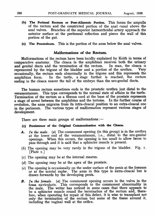
(b) The Perineal Rectum or Post-Allantoic Portion. This forms the ampullaof the rectum and the constricted portion of the anal canal above theanal valves. Branches of the superior haemorrhoidal artery approach theanterior surface at the peritoneal reflection and pierce the wall of thisportion of the gut.
(c) The Proctodeum. This is the portion of the anus below the anal valves.
Malformations of the Rectum.
Malformations of the rectum have been lucidly explained by Keith in terms ofcomparative anatomy. The cloaca in the amphibian receives both the urinaryand genital ducts and the termination of the rectum. In man, the cloaca isrepresented by the trigone of the bladder and a portion of the urethra. Veryoccasionally, the rectum ends abnormally in the trigone and this represents theamphibian form. In the turtle, a stage further is reached, the rectumending in the cloaca nearer the tail of the embryo than the uro-genital ducts.
The human rectum sometimes ends in the prostatic urethra just distal to theveramontanum. This type corresponds to the normal state of affairs in the turtle.Termination of the rectum as a fibrous cord at the base of the prostate representsa stage of arrest between the amphibian and the tortoise. In the further course ofevolution, the anus migrates from its intra-cloacal position to an extra-cloacal onein the perineum. The various types of malformation represent various stages ofdevelopment.
There are three main groups of malformations:-
(x) Persistence of the Original Communication with the Cloaca.
A. In the male. (a) The commonest opening (in this group) is in the urethraat the lower end of the veramontanum, i.e., distal to the uro-genitalopenings. When this occurs, the opening is too small to allow faeces topass through and it is said that a sphincter muscle is present.
(b) The opening may be very rarely in the trigone of the bladder. Fig. I.[Plate I.]
(c) The opening may be at the internal meatus.
(d) The opening may be at the apex of the prostate.(e) The opening is occasionally on the under surface of the penis at the fraenum
or at the scrotal raphe. The anus in this type is extra-cloacal but isdrawn forwards by the developing penis.
B. In the female. (a) The commonest opening occurs in the vulva in thefossa navicularis. This corresponds to the commonest abnormality inthe male. The writer has noticed in some cases that there appears tobe a sphincter muscle round the termination of the rectum and, there-fore, when operating for repair of this defect, it is wise to transplant notonly the termination of the rectum but some of the tissue around it,including the vaginal wall at the orifice.
288 POST-GRADUATE MEDICAL JOURNAL August, 1936

ANATOMY OF ANAL CANAL AND RECTUM
(b) The rectum has been found opening into the vagina and it is said thatsuch cases are associated with divided vagina because the rectum pre-vented the fusion of the Mullerian ducts.
(2) Abnormalities of the Post-Allantoic Gut. (a) This portion of the gut maybe entirely absent, the rectum ending at the peritoneal reflection, i.e. atthe base of the prostate or posterior fornix of the vagina. There may bea fibrous cord joining the termination of the gut to the uro-genital organs.
(b) The gut may end blindly at the same level but be displaced posteriorlyby the vagina or prostate. In such cases there is a fibrous cord passingdown to the anus. These facts must be remembered when exploring theperineum in such cases.
(3) Abnormalities of the Proctodeum. (a) The commonest abnormality is thepersistence of all or part of the proctodeal membrane producing on theone hand a stricture or on the other hand an imperforate anus.
(b) The proctodeum may be absent or represented only by a small eminence.It is noteworthy that an external sphincter may be present when thereis no evidence of a proctodeum. This fact definitely disproves the theorythat the external sphincter is formed from the proctodeum.
The rectum ends in the uro-genital passages in 26% of cases of imperforateanus. As a rule, however, the proctodeum is absent when the rectum so ends,but the presence or absence of the proctodeum is no indication of the degree ofdevelopment of the rectum. It has been said that the nearer the rectum is to theperineum, the better the development of the proctodeum, but this does notnecessarily follow.
THE ANAL CANAL.Though the anal canal commences embryologically at the level of the valves
of Morgagni, for practical purposes it is more convenient to regard the anal canalas starting at the place where the rectum passes through the pelvic diaphragm.At this point, the pubo-rectalis portion of the levator ani muscles fuses with thelongitudinal muscle coat of the rectum and, together with the deepest portion ofthe external sphincter and the internal sphincter, forms a prominent fibro-muscularring. This ring is called the ano-rectal ring.
The anal canal exists as an antero-posterior slit surrounded by a strong mus-cular tube. It passes downwards and backwards from its commencement at theano-rectal ring, forming almost a right-angle with the termination of the rectum.It is I-in. to Il-in. in length, depending upon the degree of contraction of thesurrounding muscles. Contraction of the muscles, especially the subcutaneousportion of the external sphincter, increases its length.
The Musculature of the Anal Canal.The muscles of the Anal Canal are:
(i) The external sphincter.(2) The pubo-rectalis portion of the levator ani muscles.(3) The longitudinal muscle of the rectum.
(4) The internal sphincter.
August, 1936 289
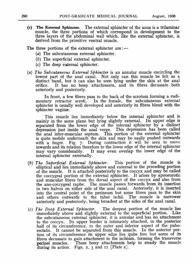
(x) The External Sphincter. The external sphincter of the anus is a trilaminarmuscle, the three portions of which correspond in development to thethree layers of the abdominal wall which, like the external sphincter, isderived from the primitive ventral muscle.
The three portions of the external sphincter are:-(a) The subcutaneous external sphincter.(b) The superficial external sphincter.(c) The deep external sphincter.
(a) The Subcutaneous External Sphincter is an annular muscle encircling thelowest part of the anal canal. Not only can this muscle be felt as adistinct band, but it can also be seen lying under the skin at the analorifice. It has no bony attachments, and its fibres decussate bothanteriorly and posteriorly.
In front, a few fibres pass to the back of the scrotum forming a rudi-mentary retractor scroti. In the female, the subcutaneous externalsphincter is usually well developed and anteriorly its fibres blend with thesphincter vaginae.
This muscle lies immediately below the internal sphincter and ismainly in the same plane but lying slightly external. Its upper edge isseparated from the lower edge of the internal sphincter by a distinctdepression just inside the anal verge. This depression has been calledthe anal inter-muscular septum. This portion of the external sphincteris quite mobile underneath the skin and may be easily pushed outwardswith a finger. Fig. 7. During contraction it will be seen to moveinwards and its relation therefore to the lower edge of the internal sphinctermay vary considerably. It may even overlap the lower edge of theinternal sphincter externally.
(b) The Superficial External Sphincter. This portion of the muscle iselliptical and lies immediately above and external to the preceding portionof the muscle. It is attached posteriorly to the coccyx and may be calledthe coccygeal portion of the external sphincter. It arises by aponeuroticand muscular fibres from the dorsal aspect of the coccyx and also fromthe ano-coccygeal raphe. The muscle passes forwards from its insertionin two halves on either side of the anal canal. Anteriorly, it is insertedinto the central point of the perineum but some fibres pass to the skinand others outwards to the tuber ischii. The muscle is narroweranteriorly and posteriorly, being broadest at the sides of the anal canal.
(c) The Deep External Sphincter. The deepest portion of the muscle liesimmediately above and slightly external to the superficial portion. Likethe subcutaneous external sphincter, it is annular and has no attachmentto the coccyx. Its upper border is intimately attached, in the posteriorhalf of its circumference, to the outer and inferior aspect of the pubo-rectalis. It cannot be separated from this muscle. In the anterior por-tion of its circumference its upper edge lies quite free but some of itsfibres decussate to gain attachment to the ischium, forming the transverseperinei muscles. These bony attachments help to steady the muscleduring its action. Figs. 2, 3 and II [Plate 2].
290 POST-GRADUATE MEDICAL JOURNAL August, 1936

Auut 96 AAOYO NLCNLADRCU 9
FIG. 2.
Diagrammatic representation ofthe portions of external sphincterani. Note:--the vertical levels ofthe various parts cannot beshown.
.:._:.. :'. .[
LEELOFAORMAL "RING--- -
'EXTSPHM-. 3uS' .
EXT. SPH. SUPERFICIALIS
EXT. m...u...wc
iE. !,S ia.: , oz-wa
EXT. 5PH. USSUEFCAI
* - . ·Z. ':"'-... ; "-
PaWTERWA IMMORM40AL RPLS..m ? OF PBO-
tu COAT" SEPT·'
.:.L.-.. ...:.:
.: ..- :
FIG. 3.
Diagram of sagittal section of anal canal, shewing relationship of external sphincter,internal sphincter, pubo-rectalis, Longitudinal muscle, hemorrhoidal plexuses and
lining of anal canal.
(2) The Pubo-rectalis Muscle. This portion of the levator ani muscles has animportant r6le in the control of continence. It forms a sling round thetermination of the rectum and its junction with the anal canal. It arisesin front from the lowest portion of the symphysis pubis and the adjacentpart of the pubis and also from the deep layer of the triangular ligament.From this origin, its passes downwards and backwards on either sideof the prostate (or vagina) and rectum. The fibres of either side are
August, 1936 ANATOMY OF ANAL CANAL AND RECTUM 291
a

292 PS-RDAEMDCLJUNL Ags,13
continuous behind the rectum, so forming a strong muscle sling. Thepubo-rectalis is quite a thick band of muscle which is closely adherentto the gut. In excision of the rectum, these stout fibres must be cutthrough before the rectum can be stripped off the posterior aspect ofthe prostate. Their close contact with the rectal wall must be remembered.
It will be noted that the pubo-rectalis is only related to the ano-rectalwall posteriorly and laterally, the anterior wall of the ano-rectal junctionbeing unsupported by this muscle. The inferior aspect of the muscle lies inrelation to the deep portion of the external sphincter, to which it is firmlyadherent and from which it cannot be separated by dissection. Fig. 4.
;~~.'. ... ......"...................................... ..
??. .......
.............. . . .......... .. ...2^...
FIG. 4.
Diagram of levator ani(sphincter muscles not
shown).
At the level of the ano-rectal ring, the fibres of the muscle blendwith those of the longitudinal muscle coat of the rectum and form a con-joined fibro-muscular band which has been called the longitudinal muscleof the anal canal. Figs 3 and II. [Plate 2].
The Nerve Supply of the superficial or coccygeal portion of the externalsphincter is derived from perineal branches of the fourth sacral nerves which passforwards into the muscle at the sides of the coccyx. The two remaining portionsof the external sphincter are supplied by the inferior haemorrhoidal nerves fromthe third and fourth sacral nerves. The inferior haemorrhoidal nerve passesinwards from its origin from the internal pudic nerve, across the ischio-rectal fossa,where it soon divides into fine filaments which enter the two portions of theexternal sphincter. These fibres enter the muscles over a wide area, both in thantero-posterior and lateral planes, so that incisions near the anus do not, asa rule, interfere with the nerve supply of the sphincters. The pubo-rectalis por-tion of the levator ani muscle is supplied by the third and fourth sacral nervesfrom the internal pudic, representing the sphincteric portion. The remainder ortail portion of the levator ani is supplied by the fourth sacral nerve on its pelvicsurface.
(3) The Longitudinal Muscle of the Rectum. This fibro-muscular structureis of varying thickness but can always be easily demonstrated. It appears tobe better developed on the anterior and posterior aspects of the anal
292 POST-GRADUATE MEDICAL JOURNAL August, 1936

canal. It passes downwards from the level of the ano-rectal ring,enveloping the internal sphincter and lying between it and the deep andsuperficial portions of the external sphincter externally. Between thelower border of the internal sphincter and the upper border of the sub-cutaneous external sphincter, it is firmly attached to the lining of theanal canal. Its attachment is broad and somewhat fan-shaped. Thelining of the anal canal is held down tightly by this insertion and thedepression called the anal inter-muscular septum is produced. Mostfistulae enter the anal canal at this inter-muscular insertion of the longi-tudinal muscle. Some of the fibres of the longitudinal muscle passoutwards between the deep and superficial portions, between the super-ficial and subcutaneous portions and also between the subcutaneousportion and the skin at the anal verge, ending in the ischio-rectal fossa orperi-anal subcutaneous fat. Spread of infection from the anal canalprobably occurs along these inter-muscular planes. The insertion of thelongitudinal muscle holds in place non-prolapsing internal haemorrhoids.As internal hemorrhoids increase in size and prolapse through the anus,the longitudinal muscle is elongated but its insertion remains intact. Thisexplains the presence of a distinct sulcus in cases of intero-externalhaemorrhoids. The sulcus is much more pronounced in thrombosed pro-lapsed piles. In the modified Salmon operation for internal hemorrhoids,these longitudinal fibres are divided, so exposing the sub-mucous layer.Figs. 3, 5, and 6.
AB -.
C-...D
- E
FIG. 5. FIG. 6.
Relationships and terminal attachmentsof longitudinal muscles of anal canal.
[E. T. C. Milligan].
A. Subcutaneous external sphincter.B. Intermuscular septum.C. Lower border of internal sphincter.D. Longitudinal muscle.E. Prolapsed thrombosed intero-external pile.
Relationships of longitudinal muscleof anal canal to haemorrhoids and
sphincters. [E. T. C. Milligan].
A.B.C.D.E.
Subcutaneous external sphincter.Submucosa with vessels.Internal sphincter.Longitudinal muscle cut and retracted.Prolapsed pile.
August, 1936 ANATOMY OF ANAL CANAL AND RECTUM 293
::,.,
... -j
- C

294 POST-GRADUATE MEDICAL
(4) The Internal Sphincter. The internal sphincter is a continuation of thecircular muscle coat of the rectum which becomes markedly thickened.It commences just above. the level of the ano-rectal ring and is tubular,completely encircling almost the whole length of the anal canal. It extendsdownwards almost to the lower end of the anal canal and at its loweredge is just above the level of the anal inter-muscular depression. It doesnot end in the upper part of the anal canal as described in most textbooksof anatomy. Figs. 3, 5, 6, 7, and II. [Plate 2].
-:.'-.".:'.:;:'::: :~ ..-l l FIG. 7.
-I- .....· Showing lower border of internalsphincter. Subcutaneous sphincterani externus retracted outwards
* with finger.
The Nerve Supply of the internal sphincter is by means of the sympatheticand para-sympathetic systems. Its action is similar to that of the circular musclecoat of the colon and rectum. By its relaxation and contraction it helps to pushfaeces through the anal canal. Its action differs considerably from that of theexternal sphincter which, together with the pubo-rectalis, enables the act of de-faecation to be controlled by the will under normal circumstances. There mustexist, therefore, a very nice co-ordination between these two distinct types of muscleand it is suggested that derangements in this balance produce hypertrophy andpossibly fibrous degeneration of the internal sphincter. From the study of morethan forty dissections of the muscles of this region by Pallares, we have noticedthat the size of the internal sphincter varies very considerably, being especiallylarge in the aged and in cases which have suffered from prolonged constipation.
By pushing outwards the subcutaneous external sphincter and incising thelining of the anal canal, the lower edge of the internal sphincter is seen presentinga white glistening appearance. Occasionally it can be felt tightly contracted,under an anesthetic. The upper part of a fissure-in-ano lies over the lower endof the internal sphincter and also across the annular subcutaneous externalsphincter. This latter muscle is in spasm in such cases and its division is anecessary part of the operation for cure of a chronic fissure. Fibrosis alsooccurs in the subcutaneous sphincter and its division produces relaxation of theanus and relief of symptoms.
From the foregoing description of the anal musculature, it is possible topalpate with precision certain landmarks.
294 POST-GRADUATE MEDICAL JOURNAL August, 1936

The Ano-Rectal Ring.This ring, as already stated, lies at the junction of the anal canal and rectum,
and is composed of the pubo-rectalis sling (posteriorly and laterally only), thedeep portion of the external sphincter, the longitudinal muscle of the anal canal,and the internal sphincter. It is, of course, impossible to palpate these structuresseparately but anteriorly the deep portion of the external sphincter can be felt asit forms the upper edge of the ano-rectal ring. The diverging fibres of the pubo-rectalis may be followed at the sides of the rectum as they pass downwards fromthe symphysis pubis. Figs. 8 and 9.
.... - 4
FIG. 8.
Palpation of posterior part ofano-rectal ring. Proximal inter-phalangeal joint at level of anus.
I. Posterior portion of ano-rectal ring.
FIG. 9.
Palpation of anterior part ofano-rectal ring. Middle of secondphalanx at level of anus.
I. Anterior part of ano-rectal ring.2. Longitudinal muscle.3. Internal sphincter.4. Pubo-rectalis.5. Deep external sphincter.6. Superficial external sphincter.7. Subcutaneous external sphincter.
The surgical problem in the radical cure of fistulae in ano is not the positionand number of the external openings but the position of the internal opening inrelation to the ano-rectal ring. If this ring is not destroyed there will not be in-continence. In front, where the ano-rectal ring is less prominent and narrower,division of the anal muscles must be done with care, since the ano-rectal ring ismuch more easily destroyed in this situation. The ano-rectal ring may be seenby slowly withdrawing a 2-in. proctoscope after its full introduction.
The highest part of the internal haemorrhoidal plexus lies immediately abovethe level of the ring and in the high sub-mucous method of injection for haemorr-hoids, this is the correct site for injection. Fig. 3.
ANATOMY OF ANAL CANAL AND RECTUM 295August, 1936
:::

POST-GRADUATE MEDICAL JOURNAL
The Anal Inter-Muscular Septum.The formation of this depression has already been described. It lies just
inside the anal verge and if the finger be placed at the depression, the lower edgeof the internal sphincter is palpated immediately above and the subcutaneoussphincter below and externally. Fig. IO.
:.. n..ul ...S .
:.....'Zq.,..
.....
FIG. 10.
Palpation of intermuscular septum.Middle of distal phalanx at level of
anus.
I.S. Intermuscular Septum.
The Lining of the Anal Canal.Prom the study of sections prepared by Dr. Cuthbert Dukes at St. Mark's
Hospital, the following description has been obtained. Just above the level ofthe ano-rectal ring, where the circular muscle coat of the bowel commences tobecome thicker to form the internal sphincter, the columnar epithelium of therectum becomes thinner and so also does the muscularis mucosm. The columnarglandular epithelium continues downwards over the ano-rectal ring into the upperpart of the anal canal for about half an inch or slightly less. It is dull red incolour. Below this, the columnar epithelium changes gradually to cuboid epithe-lium of several layers thickness. This type of epithelium extends down over thecolumns of Morgagni to the level of the anal valves, becoming thinner as itdescends. The columns of Morgagni are formed by vessels running longitudin-ally in the mucosa, the mucous membrane being thrown into folds. The colourof this cuboid lining is still red but much paler than the dull red of the columnarmucous membrane.
The valves of Morgagni are situated about three-quarters of an inch belowthe level of the ano-rectal ring. The epithelium changes abruptly to modifiedsquamous or a transitional type of epithelium at the level of the valves and justabove this point the muscularis mucose disappears. This type of epitheliumcovers the lowest portion of the internal sphincter and is more firmly adherent tothe subjacent tissues than the cuboid and columnar epithelium above. Immedi-ately below the insertion of the longitudinal muscle of the rectum it becomesgradually more definitely squamous. Its extent is about three-eighths of an inchand its colour distinct, being pale and slightly bluish.
296 August, 1936

ANATOMY OF ANAL CANAL AND RECTUM
Below the anal inter-muscular septumn, true skin is encountered and cutaneousglandular elements are present. It is at this site that Hilton's white line occurs,but this much described line is only rarely seen. The line of the valves, however,can readily be seen through a proctoscope. The subcutaneous external sphincteris covered by true skin on its anal as well as its inferior aspect.. Fig. 3.
The position of the internal and external haemorrhoidal plexuses and the con-necting vessels lying in the columns of Morgagni are seen in the diagram. In-ternal haemorrhoids are due to dilatation of the radicles of the superior hemorrhoidvein lying in the internal haemorrhoidal plexus and external hemorrhoids are dueto dilatation of the external haemorrhoidal plexus which lies superficial to the sub-cutaneous external sphincter. When the connecting vessels lying in the columnsof Morgagni become dilated, an intero-external hemorrhoid is produced.
The valves and sinuses of Morgagni are more numerous and prominent on theposterior wall of the anal canal, as already stated. Lying under the epithelium inthis region and also extending outwards into the substance of the internal sphincterare glandular structures described by Johnson, Dukes and others, named intra-muscular glands. These glands may open into the columns but more commonlyinto the sinuses of Morgagni. They are of two types, simple tubular, ending inthe sub-mucosa and branched glands ending in the underlying muscles. Gordon-Watson has described several cases, in which there seems to be no doubt that afistula-in-ano originated in these glandular structures. Tucker and Hellwig, in aninvestigation into the condition of cryptitis, have noted that the mucosa of thiesinuses of Morgagni are not as a rule the site of infection but that the infectioncommences in these intramuscular glands.
The mucous membrane above the level of the valves is either insensitive topain or its sensation is markedly diminished. On the other hand, the epitheliumat this level and below it, is highly sensitive, more so than the true skin. It issupplied by branches from the inferior haemorrhoidal nerve. It has been pointedout by Pennington that about 85 per cent. of all proctological diseases occur inthis region.
THE RECTUM.The rectum commences opposite the third piece of the sacrum and ends at a
point z-in. in front and I-in. below the tip of the coccyx at the level of the apexof the prostate. It is about 5-in. in length and follows the sacral curve. Inaddition, there are two important lateral flexures-one convex to the right oppositeto the junction of the 3rd and 4th pieces of the sacrum and another to theleft at the level of the sacro-coccygeal articulation. These curves must be remem-bered when passing a sigmoidoscope. The mucous membrane is loosely attachedto the muscle coat, especially in children, so favouring prolapse. Contraction ofthe bowel throws the mucous membrane into folds which disappear when therectum is distended with air. The perineal portion of the rectum is dilated toform the ampulla which, in a normal state, is empty.
There are two constant transverse folds of mucous membrane and muscleseen in the rectum, namely the valves of Houston. The proximal valve is situatedon the right side at the junction of the sigmoid colon and pelvic rectum. Theother, which may be called the main valve of Houston, is situated at the levelof the peritoneal reflection (the junction of the hind gut and post-allantoic gut)on the anterior aspect of the bowel. Two other folds may be present, one in thepelvic rectum on the left side and the other in the perineal rectum on the leftposterior aspect I-in. above the anal canal.
August, 1936 297

When the rectum is examined with a sigmoidoscope, the opening into thesigmoid colon is usually found on the right anterior aspect of the bowel, thoughthis is variable. Pulsation of the left internal iliac artery can usually be seenat this level.
The relations of the rectum are well known and need no repetition. The fol-lowing facts, however, are important from a surgical point of view. The rectumis surrounded by its tube of visceral pelvic fascia, which becomes fused to theparietal pelvic fascia covering the sacrum. Between this composite fascia pos-teriorly and the rectum anteriorly there is a large quantity of fat in which lies theperi-rectal lymph sinus described by Miles, the retro-rectal lymphatic glands andthe vessels and nerves. The lymphatic glands lie along the course of the mainvessels which run near the wall of the gut. One inch above the levator ani musclesthe parietal pelvic fascia becomes condensed to form two stout fascial supportsto the rectum. These have been termed the lateral ligaments of the rectum orthe rectal stalks and pass from the level of the third piece of the sacrum to therectal wall. In them run the middle haemorrhoidal arteries and the nervi erigentes.These structures must be divided when the rectum is being excised. The parietalpelvic fascia covering the sacrum becomes continuous with the periosteum in itslower part. When the coccyx has been excised, this dense fascia is immediatelyseen and it must be incised before the posterior borders of the levators can bedefined and before the rectum can be separated from the front of the sacrum.
Anteriorly, lying between the rectum and the posterior surface of the prostateand seminal vessels, is a tough fascia consisting of two layers, with a potentialspace between them. This is attached firmly to the peritoneum above, and belowto the triangular ligament in the region of the apex of the prostate. The fasciais the embryonic remains of a peritoneal pouch which extended downwards behindthe prostate and has been called the prostato-peritoneal fascia of Denonvilliers.The anterior layer of this fascia is firmly attached to the posterior aspect of theprostate but its posterior layer is loosely attached to the rectal wall. The spacebetween these two layers is opened when the rectum is dissected off the prostate.In perineal excision of the rectum, some difficulty may be experienced in findingthis level at the apex of the prostate, since in this region the anterior rectal wallis held forward to the base of the triangular ligament and the apex of the prostateby two bundles of muscle derived from the longitudinal muscle coat of the rectum.This is the recto-urethralis muscle of Roux. These muscle bundles are usuallyquite distinct structures and if their presence is not remembered, the rectal wallis easily damaged at this point. These muscles actually angulate the rectum for-wards towards the apex of the prostate. The anterior layer of the fascia ofDenonvilliers above the base of the prostate is well developed, and may completelyhide the seminal vesicles. It may have to be incised to verify the position of thesestructures.
From below, the lowest end of the peritoneal pouch is not readily foundbetween the vesicles and the anterior rectal wall. It is more easily located bycarefully cutting backwards towards the rectum. When the peritoneum has beenopened, care must be taken not to continue its division too far laterally from thesulci at the sides of the rectum, since the ureters may be damaged.
298 POST-GRADUATE MEDICAL JOURNAL August, 1936

The Blood Vessels of the Rectum.The superior hemorrhoidal artery is a continuation of the inferior mesenteric
and lies behind the rectum. It divides into two branches at the level of the thirdpiece of the sacrum and these branches pass on to the lateral aspects of therectum following the reflection of the peritoneum.
The site and mode of division of this vessel has been studied from X-rayphotographs of injected specimens and it has been found to be extremely variable,so much so that no accurate anatomical description is possible. About 4-in. abovethe anus, the main branches further divide and pierce the muscle coat, runningdown in the sub-mucous layer as straight regularly spaced vessels to end as aseries of loops at the level of the internal sphincter. The superior hemorrhoidalsupplies the mucosa of the rectum and the musculature of its upper portion. Ithas been stated by Miles that the right main branch of the artery divides into twofurther branches, a right anterior and right posterior. The left main branch doesnot divide. These three arteries mark the position of the three primary piles.The right posterior branch and the left main branch further divide into two terminalbranches indicating the site of the secondary piles. Very occasionally the leftmain branch again divides giving rise to an additional secondary pile anteriorly.
The middle hemorrhoidal artery lies, as already stated, in the lateral liga-ments and supplies mainly the muscle coat of the upper part of the rectum. Theinferior hemorrhoidal arteries supply the anal canal.
The Veins.The venous return from the rectum is by means of the superior, middle and
inferior hemorrhoidal veins. The main veins follow the same course as theartenries.
The external hemorrhoidal plexus, around the anal canal, drains mainly intothe inferior haemorrhoidal veins but also communicates with the middle andsuperior hemorrhoidal veins through the internal hemorrhoidal plexus.
The superior hemorrhoidal vein is formed by the junction of about six vesselsof considerable size which run upwards in the sub-mucosa of the rectum for three tofive inches, being more or less parallel to one another and devoid of valves. Theythen pass through the muscle coats of the bowel and join to form, as a rule, asingle venous trunk.
Since these tributaries of the superior haemorrhoidal vein are large and runin the sub-mucosa, carcinoma cells, as they extend through the rectal wall, mayeasily erode a venous channel in this situation at a comparatively early stage andgive rise to hepatic metastases. This may occur with quite a small carcinoma,especially when placed on the posterior wall and when it is "button-like."
It has been further noted that during the extra-rectal spread of carcinoma,growth extends along the peri-vascular lymphatics; the carcinoma spreadingupwards around the walls of the vessels. Malignant cells may conceivably passinto the venous stream from the extra-rectal growth, especially in the region of therecto-sigmoid junction.
The Lymphatics.The lymphatics of the anal canal below the level of the valves of Morgagni
pass to the Inguinal Lymph Nodes.
August, 1936 ANATOMY OF ANAL CANAL AND RECTUM 299

300 POST-GRADUATE MEDICAL JOURNAL August, 1936
The lymphatics of the rectum above this level are divisible into intramuraland extra-mural. The intra-mural lymphatic vessels communicate with the lymphsinus which lies between the rectal wall and the surrounding fat. It is imperativetherefore to remove with the rectum, all the fat between it and the sacrum. Theextra-mural lymphatics extend in the ischio-rectal fossa to the internal iliac glands,along the upper surface of the levators to the internal iliac glands and posteriorlyto the glands behind the rectum. They also extend upwards along the superiorhemorrhoidal vessels to the pelvic meso-colon and to glands at the bifurcationof the left common iliac artery. Spread of growth along all these zones is pos-sible, but very careful dissection and histological investigation of 250 specimensof carcinoma of the rectum at St. Mark's Hospital shows that spread into theretro-rectal lymphatic glands and upwards along the main vessels, is by far themost constant. Extension into the ischio-rectal fossa or along the levators is veryuncommon and only occurs in advanced inoperable growths where the mainupward lymphatic channel has been blocked. In only seven cases was thereevidence of downward spread and it was noted that these occurred only in veryadvanced cases with extensive permeation of the peri-vascular lymphatics. It isalso very unusual to find involvment of the para-colic glands secondary to acarcinoma of the rectum. In this series only one case showed these glands to beinvolved.
I am indebted to Dr. Vicente Pallares who is my Clinical Assistant at St.Mark's Hospital, for his help in verifying, by extensive dissection, the arrangementof the muscles in this region, and to H. Bussey, B.Sc., for the preparation ofdiagrams etc.
REFERENCES:
Milligan, E.T.C. and Morgan, C.N., "Surgical Anatomy of the Anal Canal," Lancet, 1934, ii, 1,150 & 1,213.Dukes, Cuthbert, Personal communioation.Gordon-Watson, C. and Dodd, H., "Observations on Fistula in Ano," Brit. Jour. Surg., 1935, xxii, 703.Abel, A. Lawrence, "The Pecten; The Pecten Band; Pectenosis and Pectenotomy," Lancet, 1932, i, 714.Thompson, Peter, Myology of the Pelvic Floor, London. 1899.McGregor, A. Lee, Synopsis of Surgical Anatomy, Bristol, 1932.Miles, W. E., "The Pathology of Spread of Oancer of the Rectum," Surg. Gyn. & Obst., 1931, liit, 350.Tucker, C. C. and Hellwig, C. A., "Histopathology of Anal Crypts," Surg. Gyn. & Obst., 1934, lviii, 145.Johnson, F. P., "The Development of the Rectum," Amer. Jour. Anat., 1914, xvi, 1.

PLATE 1.Mr. C. Naunton Morgan - Anatomy of Anal Canal and Rectum
FIG. I. X-ray. Lateral view of pelvis, showing rectum terminating in the base of thebladder. The rectum has been filled with barium introduced through a colostomyopening and the bladder filled with 15% sodium bromide solution. The narrow
channel of communication between them is shown.
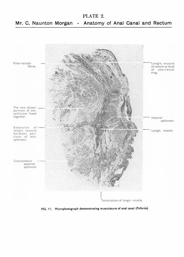
Mr. C. Naunton MorganPLATE 2.Anatomy of Anal.Canal and Rectum
Pubo-rectalis -
fibres.
The two deeperportions of ext.sphincter fusedtogether.
Extension oflongit. musclebetween por-tions of ext.sphincter.
Subcutaneousexternalsphincter
Longit. muscleof rectum at levelof ano-rectalring.
-- Internalsphincter.
-- Longit. muscle.
ITermination of longit. muscle.
FIG. 11. Microphotograph demonstrating musculature of anal canal (Pallaris)


