Embed Size (px)
Citation preview
the
IVIECHANISMS of CARBON DTOXIDE FIXATÏON
and IRON OXIDATION
by Ferrobacillus ferrooxidans
by
George A. Din
A thesis
submitted to
Faculty of Graduate Studies and Research
University of Manitoba
of the
In partial fulfilment
requirements for the degree
Doctor of Philosophv
Lg66
'ABSTRACT
carbon dioxide fixation was studied in intact cells
and cel-l-free extracts of Ferrobacil-lus fqrrooxidans. The
major pàthways of fixation were found to be the carboxy-
dismutase system and the phosphoenolpyruvate carboxylase
system. Phosphoenolpyruvate carboxylase activity was shown
to be under metabolic regulation, similar to the regulation
established in heterotrophic microorganj-sms.
Acetyl coenzyme A stimulated phosphoenolpyruvate
carboxylase activity, while the presence of aspartate was
inhibitory. The Ferrobacillus ferrooxidans enzyme appeared
to have a neutral- or acidic pH optimum, in contrast to the
same enzyme isolated from heterotrophs.
Althouqh intact cells were able to oxidize ferrous
iron with oxygen, crude sonicates and cell-free extracts
Iacked oxidase activity. The iron oxidation system involved
the cytochrome system, namely cyt. c and an g-type
cytochrome. The initial enzyme of the oxidation system was
found to be ferrous iron-cytochrome c reductase, which
l-inked the ferrous íron to the cell electron transport chain.
II.
iii.
The oxidase system was extremely labile although the
individual enzymes retained their activil-.2 aF]-ar J-ha system
was destroyed. The inability to isolate cell-free extracts
capable of oxidizing ferrous iron manometrically was found
to be due to the disruption of the cytochrome chain
alienating cytochrome c from the a-type cytochrome, thus
terminating electron transport at cyt. c rather than at
oxygen.
Ferrous iron-cytochrome c reductase lvas purified
6O-TO fold from cell-free extracts. The enzyme was judged
to be approximately )Oft pure by disc electrophoresis,
sedimentation, and DEAE-cellulose column chromatography.
RNA, whose presence !\ras obligatory for catalysis, was
cl-osely associated with the enzyme. The enzyme was
dissociated into two subuníts. one a r:rotein devoid of
catalytic activity and the other an RNA subunit. Molecular
weight analyses of the holoenzyme and its protein and RNA
subunits revealed weights of I00 110,O00, 2T-3Or00O, and
3f5 330,0O0 respectively.
Detailed kinetic analyses of the enzyme revealed the
reaction mechanism to be Ping Pong Bi Bi. The K* values for
the substrates were found to be KFe++ = O.59 mM and
av.
K^--* ^ = 0.085 mM. The presence of non-heme iron on theñ\zr I
enzyme was implicated as the responsible factor for the
oscillation of the enzvme between its two forms.
ACKNOIVLEDGEMENTS
The author is most qrateful to Dr. I" Suzuki for his
encouragement, advice, and patient guidance throughout the
course of this investigation and especially in the
preparat.ion of this manuscript. Gratitude is expressed to
Dr" H. Lees for introducing the author to Lhe fascinating
field of autotrophy and for his continued interest and advice
in this research. Thanks are also due to Dr" B, D" Sanwal,
Robert Cook, and Peter Maeba for their stimulating discussions
and advice concerning various aspects of this investi-gation,
v"
TABLE OF CONTENTS
îL ^+ -^ ^+¡tJ-rÞ Lr cr\- L .
Acknowl-edgements. . . .
Table of Contents.
LISE OI 'f'4I) Ies . . .
List of Figures. .
Abbreviations
INTRODUCTION
HISTORICAL..
MATERIALS AND METHODS
Organism and medium..
CO^ fixation by whole cells and crude extracts. . .¿
Paper chromatography
'l Dan* *per
2. Solvents
? CnrJ . -ì'- ays
4. Location of radioactive compounds. . .
5. Elution of isolated compounds. . . .
6. Identification of radioactive cornpounds. . . .
Carboxydismutase acLivity of cell--free extracts. .
V
vi
xi
xiii
xvi
I
)r-
PAGE
II
24
26
/'\ 17
28
28
2B
2Q
30
30
<l
vl_.
vl- l- .
PAGE
TABLE OF CONTENTS CONT'D
Isotopic assay of cell-free phosphoenolpyruvatecarboxylase activitY
Spectrophotometric assay of cell-free phospho-enolpyruvate carboxylase activíty
J.I
Preparation of cell-free Fe' '-cyt. ô reductase. . .
Estimation of protein. . .
Ferrous iron determination
Manometry . . .
Assay of ferrous iron-cytochrome c reductase^ ^! i --i !--dLJLIVILV. .
Iron oxidase activitY
Cytochrome oxidase activitY
Dual wavelength spectrophotometry. . .
SpectroscoPY
Spe ctrophot of luor ometrY
Determination of sulfide
RNA determination
DNA determinationJT
Iron content of Fe' '-cyt. c reductase. . .
Molecular weiqht determinations
Disc polyacrylamide gel electrophoresis. . .
?2
34
34
35
35
35
JO
3T
Jo
3B
39
40
40
4r
4z
4z
)taTJ
44
TABLE OF CONTENTS CONT'D
Preparation of cytochrome c monomer and dimer
Chemicals
viii.
PAGE
4B
/¡QTU
5o
rl
6l+
o5
OJ
66
v)tlT
T7
7T
Bz
B4
B4
)tr+2
46
RESULTS
I. Mechanisms of Carbon Dioxide
Carbon dioxide fixation by
Fixation
whole cells andcrude extracts
Separation and identification of radio-active compounds
The formation of ?-PGA bw r-e1l-freeJ
extracts
The formation
II. Iron Oxidation
of oxalacetate
Electron transport components
Oxidation of ferrous iron bv
Oxídation of ferrous iron by
intact cell-s. .
cell-freeextracts
ÏTÏ. Ferrous fron-Cytochrome c Reductase
Enzvme purification
Enzyme storage
--++pìtr 1 f \r ^ï
H'ô -¡rzt f rarlrr¡f_, _. _ase
Effect of pH on enzyme activity
l-x.
PAGE
TABLE OF CONTENTS CONT'D
Effect of substrate concentration onenzyme activity
Effect of inhibitors. .
Effect of some potential activators onenzyme activity
The formation of enzYme subuni-ts
Composition of Fe---cyt. c reductase and.: ! ^ ^,,-L-,"^ .i .t- ^IL.Þ ÞUJJU]IJLÞ. . ¡
Iron and labite sulfide content
Protein and nucleic acid content
Molecular weiqht studies
Enzyme fluorescence
Kinetic studies and reaction mechanism.....
Determination of Michaelis constantstK I\^-/'
Initial velocity studies using a
constant ratio of substrateconcentrations
BB
97
100
103
End-product
Inhibition b
inhibition studies. . . .. .. .
y sodj-um chl-oride. .
++c with Fe -cYt. c
IOB
1ôO
110
tt<
1J_O
L2T
12F,
i?o
t<l
1 ?1r
'l ?qReaction of cyt.reductase.......
TABLE OF CONTENTS CONT'D
DISCUSS ION
I. Carbon Dioxide Fixation
II. Iron Oxidation. . .
III. Ferrous Ïron-Cvtochrome c Reductase.
Reaction mechanism...
Composition of iron-cytochrome c reductase.
REFERENCES
X.
PAGE
I 1rq
1Ãô
142
r r/¡
1^7
L62
LIST OF TABLES
TABLE
I. COZ f ixation by whole cel-ls and crude^--$-r ^l -€.ã.LJ- eL, LÞ . c . . o . . . . r . . e . r . . o . . . . . . . . . . . t . . . . . . . 4g
II. Carboxydismutase activity of cell-freeextracts... o. ..... ô. c o... o o. ó r... o r. o t r. o.... 53
III. Phosphoenolpyruvate carboxylase activity ofcell-free extracts. o... ......c.. o oo.... oo,Õ. o 5T
IV. Effect of various compounds on cell-freephosphoenolpyruvate carboxylase activity 58
V. Effect of potential activators on phospho-enolpyruvate carboxylase acLivity o. o.... 59
VI. Effect of activators and potential inhibitorson phosphoenolpyruvate carboxylase activity.. 6O
VII. Effect of pH on phosphoenolpyruvatecarboxylase activity.. o...... c. o, o o..,.. o. o. o 6z
VIII. Absorption spectrum of whole cells afteranaeroloic incubation with ferrous iron andsodium dithionite.... o..... o..,.......c.... o . 65
IX. Effect of sonic oscillation on iron oxidase^--=--ity..ôo. o.co.oo... ..o.toe .c.c.c¿uL¿,ritv .oo'...o.'.e .c.c. 76
xo Purification of Fe++-cyt" c reductaseooc.o.oo 83
XI. Effect of sulfhydryl and flavin inhibitors onFe++-cyt, g reductase activity. o o, o. ó o. o . a o ô. 99
XII. Effect of proteolytic enzymes and ribo-nuclease on the activity of Fe---cyt' creductasêo.noooó.o.ooo.o,.c o,o.o 101
XIÏI, Effect of some potential activators on Fe**-cyt. c reductase activity..... o..o.â...o 104
PAGE
Xlo
xl_l_.
LTST OF TABLES CONTID
TABLE PAGE
XIV. Iron and tabile sulfide content of Fe**-cyt. c reductase. lll_
XV. Protein and nucleotide content of Fe**-cyt. c reductase. 114
JIXVT. Cytochrome c reduction by Fe' '-cyt. creductase. . . 143
LIST OF FTGURES
FIGURE PAGE
1. Effect of pH on the oxidation of ferrousiron by intact cells 6T
2. Effect of ferrous iron concentration on ironoxidation by whole cells 68
3. The oxidation of ferrous iron and ascorbateby whole cells
4. The oxidation of ferrous iron by whole cells¡nÄ ¡rrrÄa avf-ra¡J-c
5. The oxidation of reduced cyt. c by wholecells and crude extracts
6. Gradient elution of Fe**-cvt. c reductasefrom a f ast-f l-owing DeAE-cålluîose col-umn. . . BO
I.L
T . Gradient elution of Fe' '-cyt. c reductasefrom a slow-flowing DEAE-cellulose column. . . Bf
B. Relation of Fe++-cyt. c reductase activityto pH using 24 ¡rmoles Na acetate-acetic acid?rrrf far 9^UU
I¿
9 . Rel-ation of Fe ' '-cyt. c reductase activityto pH using 240 pLmoles Na acetate-aceticacid buffer BZ
10. Relation of Fe**-cyt. c reductase activityto pH using 24 pLmoles Na barbital-acetic¡^iA l-rrrffar Qn(JY
11. Relation of Fef*-cyt. c reductase activityto pH using 24 ¡rmoles Na barbital-aceticacid buffer under anaerobic conditions...... q0
L2. Effect of ferrous iron concentration on Fe+*-
To
72
73
xl-al-.
xiv.
LIST OF FIGURES CONT'D
FÏGURE PAGE
cyt. c reductase activity q3
13. Double reciprocal plot to find Fe** ^--rzdPP\n... gu
f4. Effect of oxidized cyt. c concentration onFer-f -cyt. c reductase activity 95
l-5. Double reciprocal plot to find oxidizedcyt.' c appK* 96
L6. Absorption spectra of Fe"*-cyt. c reductaseholoenzyme and its protein and nucleic acidsubunits 106
LT . Molecular weight of Fe++-cyt. c reductaseusing oxidized cyt. g as marker LLT
fB. Molecular weight of Fe+t--cyt. c reductaseusing hemoglobin as marker lfg
19 . Molecul-ar weight of Fe-F*-cyt. c reductasesubunits using oxidized cyt. c as marker. . . . 119
.LJ20. Molecular weight of Fe' '-cyt. c reductasesubunits using hemoglobin as marker... 1.ZO
21. Double reciprocal plot of 1,/v versus L/Fe++varying the substrate concentrations LZ6
22. Replot to find oxidized cyt. c Km LZT
23. Double reciprocal plot of l/v versus L/(oxidized cyt. c) varying the substrateconcentrations I2B
24. Replot to find ferrous iron K*. . IZ9
25. DoubIe reciprocal plot (t/v versus L/Fe++)
FÏGURE
26.
D7
? r)
LÏST OF FIGURES CONTID
maintaining a constant ratio of thesubstrate concentrati-ons (ge++:oxidized
-\õ\ff õ <.l l"J'.
=, J.L,/
XV
f.ê,Lr11
| \/
| <h\
t<rl
1?7
r ¡ôI lat
r JrnfTv
r J¡rIlI
Double reciprcr¡.|- ..)) main-¿ -. =_t /
substrate con
ocal plot (t/u versus L/(ox.taining a constant ratio of thecentrations ( ¡'e++ : oxidized
cyt. 9, 3,1)
Product inhibition c reductaseby ferric iron
28.
2A
Replot to find ferrj_c
Product inhibition ofby reduced cyt. c
Replot to find reduced
Inhibition of Fe++-cyt
Replot to find NaCl K1
f r^n K.--a' 'J, J-
Fta' '-nrz'f- I roÁrr¡l->ca--¿ " '
arzf- õ K ,vYe.I
. c reductase by NaC1.
ABBREVIATÏONS
aceto-acetyl CoA aceto-acetyl coenzyme A
acetyl CoA acetyl coenzyme A
ADP adenosine l'-diphosphate
AMP adenosine !'-monophosphate
3r-AMP adenosine l'-monophosphate
ATP adenosine l'-triphosphate
DEAE-cellulose diethvlaminoethyl-cellulose
C"1
c_J
uJrT
cyt. a
cyt" c
DNA
DNAse
EDTA
It nIr-+-r
FAD
one carbon
- three carbon
four carbon
cytochrome g
cytochrome c
deoxyribonucl-e ic acid
deoxyr ibonuc lease
- ethylenediamine tetracetic acid
erythrose -phosphate
flavin adenine dinucleotide
FDP fructose tr6-aipnosphate
FMN flavin mononucleotide
fructose b-phosphateT'-O-.H
XVI.
L\/'r 1
GA-3P
GSH
fDP
ITP
1,3-DPGA
RNA
NAD - nicotinamide adenine dinucleotide
NADP, NADP-- - nicotinamide adenine dinucleotide phosphate
NADPH - reduced nicotinamide adenine dinucleotídephosphate
PEP - phosphoenolpyruvic acid
PcA, 3-PcA J-phosphoglyceric acid
glyceraldehyde l-phosphate
glutathione (reduced)
inosine !' -diphosphate
inosine l' -triphosphate
1, 3-diphosphoglyceric acid
- ribonucl-eic acid
RNAse - ribonuclease
R-5-P
RUDP
Ru-l-P
SDP
S-7-P
TCA
'r'rl_s
x-Ã-Þ
- ribose l-phosphate
- ribulose 1r!-diphosphate
- ribulose l-phosphate
sedoheptulose 1, /-diphosphate
sedoheptulose /-phosphate
- Lrichloracetic acid
- tris (nyaroxymethyl ) aminoethane
- xylulose l-phosphate
INTRODUCTION
Although the metabolic mechanisms and general
physiology of autotrophic organisms are well established,
virtually nothing is known of the autotrophic iron-oxidizers.
This lack of knowledge is due partly to the belated isolation
of these organisms (temple and Colmer, L95L; Leathen, Kinsel,
and Braley, f956; Kinsel, L96O), and partly to the inability
of researchers to obtain sufficient quantities of the cells
for detailed enzyme studies. Research in this field was
Iimited to physiological studies with intact cells until the
recent development of Silverman and Lundgren (1959a;A),
who were able to obtain sufficiently large yields of cells
required for cell-free enzyme studies.
The major pathways of carlcon dioxide fixation in
autotrophic microorganisms have already been establ-ished,
but this aspect has not been extensively studied in the iron-
oxidizers. The recent work of Maciag and Lundgren (t964) ,
using intact cells of Ferrobacillus ferrooxidans, and Gale
and Beck (tg66), using cell-free extracts of Th¿qþe-cilIus
ferrooxidans, indicated the presence of carboxydismutase and
the possible existence of an enzyme catalysinS C, and C,
condensation (Wood-Werkman). The first problem arising from
these findings was to establish defínitely the major pathways
of Co2 fixation in Ferrobacillus ferrooxidans.
Both intact cells and cell-free extracts were used to
determine the major pathways of carbon dioxide fixation.
Amarsingham (L959) demonstrated that the major pathway of C4
synthesis feeding the tricarboxylic acid cycle in the
Enterobacteriaceae was phosphoenolpyruvate carboxylase. More
recent research (cínovas and Kornberg, L965; Sanwal and
Maeba, 1965¡ 1966) inOicated that this enzyme is under the
control of intermediary metabolites.
Autotrophs utilize carbon dioxide exclusively as
their sole carbon source. Since carboxydismutase is found
solely in autotrophic organisms, it was decided that a study
of the "heterotrophic" carbon dioxide fixing enzyme, phospho-
enolpyruvate carboxylase, would be carried out so that
comparisons coul-d be made with the findings established in
the Enterobacteriaceae.
The existing knowledge concerning the mechanism of
iron oxidation in the chemoautotrophs is scant. Vernon
et al. (WeO) proposed a scheme of electron transport during
iron oxidation by Thiobacillus ferrooxidans which included
the presence of both cytochrome c and cytochrome a. These
workers proposed that the electrons originating at ferrous
iron were transferred via cytochrome c and cvtochrome a to
oxygen. In L964, Blaylock and Nason suggested the same
scheme for Ferrobacillus ferrooxidanå and in addition
isolated a ne\^z enzyme, f errous iron-cytochrome c reductase .
A study of the electron transport chain was undertaken
to understand further the mechanism of iron oxidation and to
attempt to elucidate the enzymes involved. To aid in the
explanation of iron oxidation, ferrous iron-cytochrome c
reductase was isolated and purified. The results of Blaylock
and Nason (tg64) and the initial results of this study
indicated that a detailed kinetic analysis was required to
explain the unusual properties of the enzyme and the reaction
mechanism utilized in vivo.
Because the research encompassed a variety of
subjects, this thesis has been divided into three sections:
carbon dioxide fixation, iron oxidation, and a study of the
initial enzyme of the iron oxidation scheme (ferrous iron-
cytochrome c reductase).

HISTORICAL
HTSTORTCAL
In 1BBT Vfinogradsky established the fundamental
principles of autotrophy with his classic treatise on the
sulphur bacteria. His original concept was that autotrophic
bacteria adhered to two basic principles: (f) the only
carbon source used for cell synthesis was Co2î and (Z)
the sole source of energy was of an inorganic nature. In
later work he discovered other bacteria capable of oxidizing
ferrous iron (fBSB) and ammonia (f$gf). Although sulosequent
research revealed that his oríginal work was carried out
using the heterotrophic Beggiatoa, the original work can
be justly cal-Ied classic.
winogradsky's original concept of autotrophy led to
a belief that only the autotrophs were capable of fixing COz.
It was not until L935 (Wood and Werkman) that this concept
was challenged. Present knowledge reveals that all
organisms fix CO2 but that autotrophs are the only organisms
capable of using CO2 for all their carbon requirements. In
fg54 autotrophic bacteria were re-defined into two classes:
the chemolithotrophs and the photolithotrophs. The former
4
organisms gain their energy by the oxidation of inorganic
compounds and the latter use light as an energy source;
both classes of organisms use CO2 as their sole source of
carbon.
There is some controversy as to the rigidity of
definition of inorganic energy source (nlsden, 1962; Bhat
and Barker, f94B). Are carbon monoxide, methylamine,
methanol-, methane, and formate organic or can they be
classed as inorganic? All of these compounds can supply
certain bacteria with all of the energy required for growth,
with co2 as the sole carbon source (nlsden, L962). At the
present time this controversy has not been settled and the
definition of a chemolithotroph needs to be clarified.
The concept that growth of obligate autotrophs is
inhibited by organic compounds arose with Winogradsky's
original findings (fBBZ). More recent studies show that
although organic compounds alone cannot support the growth
of chemoautotrophs, organic compounds can be metabolized
into cell constituents. Waksman and Starky (tgZZ; L923)
found that glucose increased the rate of sulfur oxidation
in Thiobacillus thiooxidans and that the slucose slowly
disappeared from the medium. More recently, Suzuki (fgf8)
6
found that rhi_qþgçi1_Ius thiooxidans metabolized radioactive
glucose into amino acids, sugars, and polysaccharides. These
findingsshedsomelight.onthefundamentaldifferences
between heterotrophs and chemoautotrophs
Themetabo]ismofglucosebyautotrophsshowsthat
some organic compounds may enter the celt and be metabolizedo
but that the metabolism of organic compounds may not provide
sufficient energy f ot the growth of the autotroph ' The
growth of autotrophs may in some cases be stimulated by the
presence of a utilizable organic compound (Waksman and
Starky , L1ZZ¡ I923i Remsen and Lundgren, 1963; Butler and
umbreit , ]1965; London and Rittenberg, L965; Krulwich and
Funk , Lg65). Although some carbohydrates can enter obligate
autotrophs,theideathatautotrophshavecellwallsoT
membranes impermeable to organic compounds cannot be
completely discarded. The inability of obligate autotrophs
to grow on organic compounds could be due either to the slow
permeability of organic compounds or to the inability of the
organic compounds to generate sufficient energy for growth '
Thisdiscussionwilldea]-withthechemoautotrophs
that oxidize ferrous iron compounds for their energy' The
organisms are classified into two genera: Thiobacillus and

T
Ferrobacillus. For discussions on the thiobacilli and
iron-oxidizers the reviews of Van Niel- (ryf[), Lees (t955¡
i960) , Vishniac and Santer (ry>f ) , and Mulder (tg04) should
be consulted. since the mechanisms of co2 fixation in
bacteria have been ably reviewed by Elsden (t962), and Wood
and Stjernholm (t962), these details wil-t be only briefly
outlined.
Chemoautotrophic iron gxidizinq bacteria
Atthough tr{inogradsky first reported growth of a
chemoautotroph on ferrous iron in }BBB it was not until L949
that a pure culture of such a bacterium was isolated (Colmer,
Temple, and Hinkle , L/SO; Temple and Colmer, L95f). The
f irst iron-oxi ð,tzer isolated \^/as named Thiobacillus
ferrooxidans because of its close relationship to Tbiobacillus
thiooxidans in acid mine water (1.emple and Colmer, f951) .
Leathen, Braley, and Mclntyre (L953) also isolated an iron-
oxidizer from acid mine water and named the bacterium
Ferrobacillug ferroo:lidans (leathen, Kinsel, and Braley, L956)
These authors found that this bacterium oxidLzed ferrous iron
only and would not utilize either sulfur or thiosulfate as
oxidizable substrates .
a)o
The isolation of other iron-oxidizers from mine water
and sulfide ores contínued. In f95B Beck and Elsden isolated
another bacterium that oxidized ferrous iron and sulfur;
this bacterium was thought to be similar to the bacterium
isolared by Temple and Colmer (fgff). Kinsel (tg6O) also
isolated a similar bacterium which she named Thiobacillus
sulfooxidans. In L96L Vnz and Lundgren questioned the
taxonomic validity of the genus Fegrobacillus. They found
that all of the above bacteria vvere capable of utilizing Fel-+
and sulfur or thiosulfate as oxidizable substrates, although
the generation time of these bacteria varied with the substrate.
They suggested that the genus Ferrobacillug be abolished and
that Ferrobacillus ferrooxidans be identified as a strain of
Thiobacillus ferrooxidans. This controversy as to taxonomic
position still has not been settled.
Thiobacillus ferrooxidans (remple and Colmer, L)JL;
Beck. Iq6O ) . Ferrobacillus fef¡qq¡li-qen-s (Leathen, Kinsel, andt -t
Braley , 119,6), and Ferrobacillus sulfooxidans (Xinsel , L96O)
are capable of autotrophic arov,ith using ferrous iron as theír
sol-e energy source. Thiobacillus lerrooxidgns (Col-mer,
Temple, and Hinkle , l95O; Temple and Colmer, L95L; Beck, L)6O;
Colmer , Lg6L) can, âs well, utiLíze sulfur or thiosulfate as
9
an alternative energy source " Ferrobacill-us sulfooxidans
(xinset, L96O) exhibits the same ability to utilize an
alternative substrate, in this case sulfur. The
utilization of thiosulfate as an alternative substrate
has been questioned because in acid conditions thiosulfate
rapidly decomposes and the bacterium could then oxidize
the released sulfur, further lowering the pH and increasing
thiosulf ate decomposition "
Previously, it was believed that the chemosynthetic
iron oxidizers were obligate autotrophs, but in this
connection Schnaitman and Lundgren (t965a) and Remsen and
Lundgren (lg6Z) showed that protoplasts and. mildly sonicated
cells of Ferrobacillus ferrooxidans \^iere capable of hetero'-
trophic Arowth on glucose. Remsen and Lundgren (tg63) were
able to train the bacteria to grow on glucose and contend.ed
that Ferrobacillus ferrooxidans is a facultative bacterium
rather than an obligate autotroph. Schnaitman and Lundgren
(W6SA) found. that pyruvic acid was excreted into the spent
med.ium by autotrophically gro\À7n cells of Ferrobacillus
ferrooxidanq. This work has not been substantiated by other
workers and ihe significance of pyruvate excretion is not
understood.
10
All of the iron oxidizers use ammonium salts as
their sole source of nitrogen. Temple and Colmer (fgff)
found that nitrates can replace ammonium in the growth of
Thiobacillus ferrooxidans. Bryner and Jamieson (fgfB)
reported that (Ug¿+)eSO4, urea, and cysteine can all supply
the necessary nitrogen for growth of a bacterium similar
to Thiobacil-tus ferrooxidans. Remsen and Lundgren (fg6S)
found that alanine, glutamic acid, and lysine were capable
of substituting for (m+)ZSO4 during growth of Ferrobacillus
ferrooxidans.
Lazaroff (tg63) found that the presence of sulfate
is obligatory for iron oxidation in Thiobacillul ferrooxj-dans.
Dugan and Lundgren (L965) obtained a similar result in
Ferrobacill-us ferrooxidans. High concentrations of phosphate
appear to inhibit oxidation and growth in Thiobacillus
ferrooxidans (nazzell and Trussell , 1963) and Ferrgþaci11u,1
ferrooxidans (Silverman and Lundgren, L959a¡b). p.azzeL1- and
Trussell_ (tg63) also reported that iron oxidation in
Thiobacillus ferrooxidans is inhibited by chloride and
thiosulfate and they suggested that high concentrations of
anions are inhibitorY.
It
Mechanism of iron oxidation
Leathen, Braley, and Mclntyre (t953) ' working with
chemosynthetic autotrophs found in mine water, and Temple
and Delchamps (tgSZ), using Thioþacillus ferrooxidans also
isolated from mine water, showed that iron is oxidized
according to the following equation:
4l'eso4 + oz + 2Hrso4 ---> 2 Fer(so4 )3 2 HzO
Silverman and l,undgren
ferrooxidags, and Beck
isolate of Thiobacillus ferrooxidans. found the same
stoichiometrY.
Vernon, Mangum, Beck, and Shafia (1960) isolated
cytochrome c and cytochrome a from Thiobacillus ferrooxidans
and postulated that iron is oxidized through the cytochrome
system with oxygen as the final electron acceptor.
Fe++ cytochrome c -->
cytochrome g ---> 02
In L963, Blaylock and Nason proposed
Ferrobacillus ferrooxidans when they
(W59a) , using Ferrobacillgs
and Elsden (tgSB), using a different
the same mechanism in
found that cell-free
and a and an iron-extracts contained cytochromes Þ, s,
L2
cytochrome g reductase. These authors also reported an
iron oxidase system whose activity was measured by the
decrease of ferrous iron concentration (colorimetrically
not manometricalfv). Lundgren (tg6=) and Beck (W6f) have
not been able to find this iron oxidase activity, either
colorimetrically or manometrically, in similar extracts of
Ferrobacillus ferrooxidans or Thiobacillus ferrooxidans
and the original report of Btaylock and Nason (t963) tras
not been further substantiated.
Blaylock and Nason (tg63) advanced knowledge of the
iron oxidation mechanísm with their finding that the initial
electron carrier of the system is cytochrome g' Their
discovery of ferrous iron-cytochrome c reductase and
cytochrome a (cytochrome c oxidase) tras essentially
substantiated the original hypothesís of vernon, Mangum,
Beck, and Shafia (fg[O) ttrat ttre oxidation mechanism proceeds
through the cytochrome chain with oxygen as the terminal
electron acceptor.
schnaitman and Lundgren (1965a) prepared protoplasts
ofFerrobacíllusfeËl9.9><ider'åandshowedthatintact
protoplasts were capable of oxidizing ferrous iron (mano-
metrically) but lysed protoplasts lost almost alt of their
t?
oxidizing ability. This finding illustrates that the cell
wall plays no part in the oxidation mechanism, but that the
cytoplasmic membrane is the important structure in iron
oxidation. Anderson and Lundgren (tg65) manometrically
detected iron oxidase activity in ceIl-free extracts of
Ferrobacill-us ferrooxidans but this finding has not been
repeated (Lundgren, L965) .
In f965, Dugan and Lundgren constructed a model to
depict iron oxidation in intact cells of Fe¡qqþ4qi-llus
ferrooxidans. These authors envisaged ferrous iron
complexing with oxygen (and phosphate ?) in the medium, thus
oxidizing the ferrous iron to form a peroxide complex. They
postulated that this complex then binds the cell membrane
where iron oxygenase (ot oxidase) would release an electron
to a loound electron-deficient sulfate group (Lazaroff, L963) .
Once the electron is released by the enzyme the complex
splits and ferric iron is released from the membrane into
the medium; the sulfate group then releases the electron into
the electron transport chain ( ftavin or quinone ) .
Alternatively, these authors suggest that the iron complex
could also release its electron directly to the flavoprotein,
thus bypassing sulfate. The oxygenase transferring the
r4
electron from the complex to the cell membrane would split
off one oxygen atom, which could act as the terminal
el-ectron acceptor, and the other atom would remain with
the oxidized substrate (ferric iron). If the oxidase is
the functional errzyme, then neither oxygen atom remains
with the oxidized substrate and the ferric iron diffuses
to react with water and sulfate in the external medium.
This model mechanism attempts to explain both the
activation of the ferrous iron and the ability of the cell
to transport rapidly the oxidized substrate so that the
cetl does not become clogged with excessive substrate. The
electron transport chain involved does not differ
substantially from the models of vernon, Mangum, Beck, and
Shafia (fg6O) or Blaylock and Nason (tgæ). The essential
difference in this model is the requirement for oxygen in
order for electron transfer to take place and the requirement
fox sulfate in the oxidation of the substrate.
Transfgr of enerqy
Lees ( fg6O , 1962 ) ably reviewed energy transfer and
energetics in chemosynthetic systems. This author mentions
the difficulty in expl-aining the aloility of some
chemoautotrophs (most notably nitrite and ferrous iron
oxidizers ) to reduce pyridine nucleotides soleIy by the
oxidation of their inorganic substrates. Iron oxidizers
surmount a formidable energy gap (n'o ps++/psl-*+ + O.TT v,
E'o red. cyt. ilo*. cyt. c + 0.31 v, E'o NAD/NADH O.32
v) that must be overcome in at least a two-step process
(po=sibly five steps). The ferrous iron first reduces
cyt. c (Vernon, Mangum, Beck, and Shafia , L96O) whích lowers
the redox potential but still leaves a potential gap of O.6
v to overcome. The theoretical reversal of the electron
transport chain from cyt. c to NAD requires a minimum of
ZT kcaL (Lees, 1962), which necessitates an energy input of
3-4 afp molecules (afe to ADP). The reversal of the electron
transport chai-n in chemosynthetic autotrophs as yet has not
been completelY resolved.
Aleem, Lees, and Nichotas (tg63) found that extracts
of Nitrobacter, Nitrosomonas, and Eerågþ-acilluå were able to
reduce NAD in the presence of ATP and reduced cyt. c. The
ATp:NAD ratio was tO-20:I rather than 4:1. This system had
an absol_ute reguirement for ATP and reduced cyt. c and was
inhibited by atabrin, thus implicating the involvement of
f lavin(s). Kiesow (f9æ, 1964) showed similar resul-ts with
T6
Nitrobacter winoqradskyi. These workers showed that both
systems of energy transfer, production of ATP and reduction
of NAD, were present in Nitrobacter. The system for NAD
reduction in iron oxidizers, although present (Al-eem, Lees,
and Nicholas , 1963) , has not been studied further.
Oxidative or substrate phosphorylation has never
been demonstrated in the chemosynthetic iron oxídizers.
Beck and Shafia (WA+) reported an increased iron oxidation
by aged cells of Thiobacillus ferrooxidans in the presence of
phosphate. The same workers reported that 2,4-dinitrophenol
inhibited CO2 fixation but not iron oxidation. These results
with intact cel-l-s impty the existence of an oxidative
phosphorylation system, but the proof of such a system is
delayed because of the inability of researchers consistently
to find cell-free extracts capable of manometric iron
oxidation (Lundgren, L965; Beck, 1965).
Carbon dioxide fixation in chemoautotrophic bacteria
Although the ability of bacteria to fix carbon dioxide
was discovered in the late 1BOO's (winogradsky, fBBT), it was
not until f935 (Wood and i¡Ierkman, f935; 1938) tfrat research
on the processes involved received an impetus. With the
LT
availability of Ct4 the field of autotrophic CO2 fixation
surged forward. The California group of Calvin, using new
techniques of chromatography and radioautography, determined
the sequence of formation of labelled products and inter-
mediates after "14oo
incorporation by Chlorella.
Calvin for:nd that the first product of autotrophic
CoZ fixation was 3-PcA (catvin and Benson, 1948). Calvin
and Massini (tgSZ) postulated that RuDP was the acceptor of
the labelled COZ. Itlith the sequence of labelled products
known, enzymologists turned their efforts to finding the
enzymes responsibl-e for the cyclic mechanism. The primary
carboxylation enzyme carboxylating RuDP was discovered in
Chlorella (Quayte et aI. , 1954) .
The formation of RuDP was discovered to be an
enzymatic reaction catalyzed by phosphoribulokinase (tturwitz
et al., 1956). These two enzymes, the carboxylation enzyme
(carboxydismutase) and phosphoribulokinase, are unique in
autotrophic organisms. The discovery of the other enzymes
involved in the cycl-ic mechanism either preceded the initial
work of Calvin's group or was made later, but their occurrence
is ubiquitous in nature.
Hexose phosphates are synthesized by a reversal of the
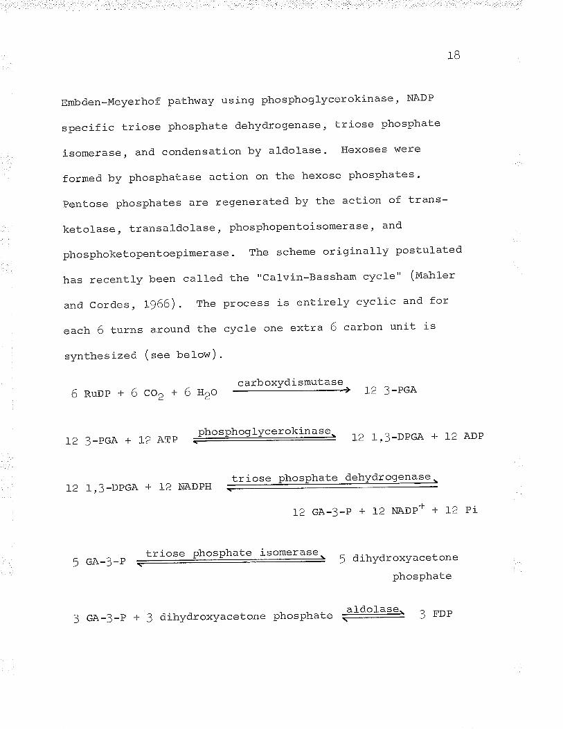
1B
Embden-Meyerhof pathway using phosphoglycerokinase, NADP
specific triose phosphate dehydrogenase, LTiose phosphate
isomerase, and condensation by aldolase. Hexoses were
formed by phosphatase action on the hexose phosphates.
pentose phosphates are regenerated by the action of trans-
ketolase, transaldolase, phosphopentoisomerase, and
phosphoketopentoepimerase. The scheme originally postulated
has recently been cal-Ied the "Calvin-Bassham cycle" (Mahler
and Cordes, L966). The process is entirely cyclic and for
each 6 turns around the cycle one extra 6 carbon unit is
synthes tzed ( see below) .
carboxvdismutase 12 3-PGA6Ruop+6cor+6n2o
phosphoglycerokinase\ 1ô 1 . nnñ^ ! r. ^rìD12 3-PGA + 12 ATP L2 l,3-DPGA + L¿ t\uy
triose Phosphate dehydrogenase.12 l,3-DPGA + 12 NADPH
-
L2 GA-3-P + 12 NADP- + L2 Pi
5 GA-3-Ptriose phosPhate i?"*St."=- 5 dihydroxyacetone
PhosPhate
3 GA-3-P + 3 dihydroxyacetone phosphate E13"1* 3 FDP

tq
3 FDp + 3 HrO phosphatase, 3 F 6-p + 3 pi
2¡'-6-p+2GA-3-P transkeLolasq z ¡ -u-P + 2 x-5-P
2 n-4-p + 2 dihydroxyacetone phosphate +doIas'g' 2 sDP
z sDp +2H2o phosphatase, 2 s-z-p +2 pi
2 s-7-P +2GA-3-P tsanskeioraset 2R-5-P +2x-5-P
D R_6_p phosphopentoisomerase. z Ru-5-p
rr-¡-ñ phosphoketopentoepimerase- 4nu-5-e+-ô.-)-Y
-
6 nu_5_e + 6 ATp phosphoribulokinase 6 nuOp + 6 eop
SUM
6 nupp + 6 co, + tB etp + l-2 NADPH
6 Ruop + F-6-P + tB aop + 18 Pi + 12 NADP+
In 1955, Santer and Vishniac were the first to report
a cell-free extract of a chemoautotroph (rhiobacillus tftigp"t*-)
capable of fixing CO2" Using alumina extracts of Thiobacill-us
thioparus these authors reported the incorporation of
20
radioactive Cl4or into the carboxyl group of 3-PGA
RuDP as the acceptor. In the same year Trudinger,
utilizing
working
with extracts of Thiobacillus denitrificans. also
demonstrated carboxydismutase and phosphoribulokinase
activity in a chemoautotroph.
Trudinger (tg=6), following the pattern laid down in
plant studies, was able further to demonstrate the following
enzyme activities: phosphoglycerokinase, NAD triose
phosphate dehydrogenase, triose phosphate isomerase, aldolase,
hexose diphosphatase, transketolase, and transaldolase in
extracts of Thiobacitlus denitrificans. From these results
Trudinger (tgS6) concluded that ce1l-free extracts were able
to slmthesize hexose phosphates from CO2 by a mechanism
similar to the mechanism established in Chlorella.
Working independently, the French group of Aubert
(aubert et aL., 1956¡ L95Ta,b) were able to show with time
course studies using radioactive CO, and RUDP that the
pattern of CO2 fixation in whole cells of Thiobacillus
denitrificans mimicked the results Calvin in his school
obtained with Chlorella. Both Aubert and Calvin showed a
high labelling content in aspartic acid. At that time the
enzyme system forming C4 compounds from C3 compounds was still
2!
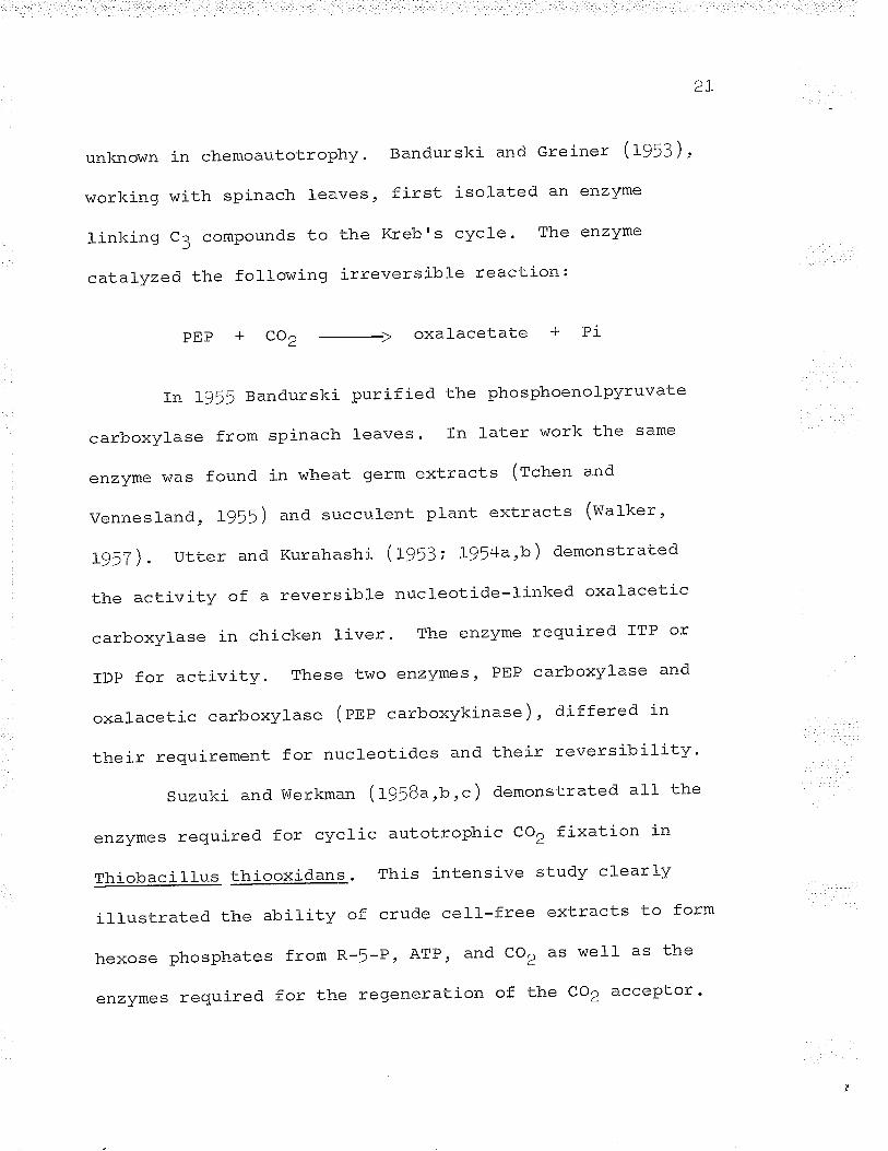
unkno\^in in chemoautotrophy. Bandurski and Greiner (lgSZ)
working with spinach leaves, first isolated an enzyme
linking C3 compounds to the Kreb's cycle ' The enzyme
catalyzed the following irreversible reaction:
PEP coz oxalacetate PI
In Lg55 Bandurski purified the phosphoenolpyruvate
carboxylase from spinach leaves. In later work the same
enzyme was found in wheat germ extracts (rchen and
vennesland, Lg55) and succulent ptant extracts (watker,
1957).UtterandKurahashí(1953;I)J4a,b)demonstrated
the activity of a reversible nucleotide-linked oxalacetic
carboxylase in chicken liver. The enzyme required ÏTP or
ïDp for activity. These two enzymes, PEP carboxylase and
oxalacetic carboxylase (ene carboxykinase), differed in
their requirement for nucleotides and their reversibility'
suzuki and werkman (r958a ,b ,c ) demonstrated all the
enzymes required for cyclic autotrophLc co, fixation in
Thiobacillus thiooxidana. This intensive study clearly
ill-ustrated the ability of crude cell-free extracts to form
hexosephosphatesfromR-5-PrATPrandCOraswellasthe
enzymes required for the regeneration of the co2 acceptor '
22
These workers also were first to demonstrate two enzymes
capable of C3 to C/¡ conversion in bactería, PEP carboxylase
and an ITP-linked oxalacetic carboxyl-ase.
carboxydismutase activity has also been reported in
Hydroqenomonas facilis (eergmann et al., I95B), Thiobacillus
novellus (aleem and Huang, L965; charles, 1966), and
Nitrobacter aqilis (Aleem, L965). In the facultative
thiobacilli, the synthesis of carboxydismutase is repressed
in the presence of exogenous carbohydrates. Vishniac and
Trudinger (t962), Aleem (tg6=), and charles (t966) reported
that the level of carboxydismutase activity during hetero-
trophic growth of Thiobacillus govs,!þg was 2/o or less of
autotrophically grown cells.
In the iron oxidizers only two reports of co2 fixation
have been published. Maciag and Lundgren (t964), using
intact cells of Fqqsobacllll¿_s f errooxidans, were able tor /r
show that after ! minutes in the presence of CfaO, and Fe++
the bacterium synthesized labelled phosphorylated carbo-
hydrates, hexoses, amino acids and peptides, Krebrs cycle
intermediates. and two and three carbon compounds. Gale and
Beck (tg66) showed that extracts of Thiobacillus ferrooxidansr /r
in the presence of R-!-P, ATP, and ct-oz were able to fix the
2?
radíoactive label-. These authors found that AMP is
competitive with ATP in the incorporation of radioactivity
and postulated that AMP is a competitive inhibitor of
phosphoribulokinase activity. This report is contradistinct
from that of Aleem and Huang (W6f) who reported that AMP
inhibited carboxydismutase activity in Thiobacil-lus novellus.
MATERTALS AND METHODS
MATERIALS AND METHODS
Orqagism and medium
The organism used in
ferrooxidans, was originallY
1-his investigation
isolated by it'i. W.
. Ferrobacillus
Leathen
(leathen, Kinsel, and Braley, L956) and was a gift of M.I.H.
Aleem. The organism was grown on the !K medium of Silverman
and Lundgren (lgSga) . The medium contained z 4.5 g (ivn+)2so4,
0.f5 g KCl, O.T5 g K2HPo¡, O.75 g MgSo4'7HZo, O.O2 g
Ca(mO3)2.4UrO and distil-ted water to a f inal volume of 1,O5O
m]. The energy source, ferrous sulfate, was made by dissolving
66.33 9 Feso4 .THZ> (t4 .f 4%) i" 45O ml distilted water
containing 1.5 mt 10 N H2SO4. After 45O ml of the ferronrs
sulfate solution was added to the mineral-sa1ts solution to
give a t.otal volume of lrl0O m1, tþO m1 aliquots were sierile-
filtered and placed in sterile 500 ml steel-capped Erlenmeyer
flasks.
The organism was rou-tinely maintained by twice a week
transfer wi'th a L/o inocu]um and grown at 2Bo C in a g¡zrotary
shaker. Large batches of cells were grown in 5 gallon
polyethylene carboys containing L5 I of unsteril-ized !K medíum
24
2^
After inoculation with 1lO ml freshly grolvn cells the
carboys were aerated by passing compressed air through two
sintered glass spargers (Z I/min) into the bottom of the
medium. The carbovs were incubated at e8o C and the ferrous
iron content of the medium was assayed daily. When T5% ot
the original amount of ferrous iron present was utilized,
45O g Feso4.THZ) díssolved in fOO ml distilled water and f0
ml 10 N H2So4 was added (elaylock and Nason, L963). Again
the ferrous iron content was assayed daily; when the ferrous
iron content was again down to 2J/o of Lhe original content
another similar addition was made.
When the second ferrous iron addition had been
utilized, the carboys were disconnected from the air lines
and were incubated at O-5o c for 12 hours. This storage in
the cold allowed some precipitation of the ferric salts, thus
.c^^i 1.:+^+..1-^LcrurrrLÕLrrrv subsequent recovery of the cells. The cells were
harvested in a steam-driven Sharples centrifuge and suspended
in cold H2o-H2So4 (ps 3.0).
After harvesting and prior to further use the cells
were cleaned by centrifugation at 1,000 x g fox I minutes to
remove any ferric iron precipitate and. centrifuged at 10rO0O
x g for t0 minutes to remove any sol-uble salts. The I0rO00 x g
¿O
precipitate was resuspended again in cotd H2O-H2SO4 (pU 3.0)
and this cleaning process was repeated J-! times. After
cleaning, the cells were stored in H2O-HZSO4 (pH 3.0) at
o-5o c.
If the "cleaned" cells were not used for any
experiment before 4 days of storage had elapsed, then the
cells were placed in lOO ml !K medium and incubated at 2Bo C
until T5% of the ferrous iron had disappeared ' The
revitalized cells were then harvested and cleaned in the
normal manner.
CO fixation by whole cells and crude extracts
carbon dioxide fixation by whole cells and crude
extracts was carried out in open test tubes at room
temperature. The reaction mixtures contained: O'62 ml O.O25
M citrate O.O5 M phosphate buf fer (pII 4.5), 20 mg (wet
weight) intact cells or an equal amount of crude extract
(prepared by sonicating a LO/" (r/u) suspension of intact
cells for 4! minutes in a 1O KC Raytheon) , 2.5 pmoles sodium
6 tl+bícarbonate , 2.5 x IOU cpm NaHCt*o3, and H2O-H2SO4 (pH 4.¡).
The reaction was started by the addition of 10 ¡rmoles
FeSo4 .[HZo immediately followed by
bicarbonate. Other additions were
cyt . c where indicated. The final
was 1.0 ml.
the radioactive
0.016 plmoles oxidized
vo]ume of the reaction
At various time intervals the reaction was stopped
by the addition of 0.5 ml 95% ei-lnanol. Suitable samples
were counted after acidification with 0.1 ml glacial acetic
acid and drying the planchets at 600 c in a vacuum hood.
The radioactive sampl-es were then extracted twice
with IO ml of BO/" ethanol by heating to boiling. The
residue was then discarded and the extract was dried and
redissolved in water. The samples were treated batchwise
with the H+ form of Dowex 5OW-XI2 to remove cations. After
drying, the samples were redissol-ved in water ( f mf ) and
used for chromatography and other experiments.
Paper Chromatographv
Ascending one and two dimensional chromatograms \^7ere
run in large round I gallon glass tanks at room temperature
2B
l. Paper
I,rihatman #t paper was used in this investigation.
The paper was washed for 12 hours in 2 N acetic acid, then
careful-]y rinsed with deionized water for 12 hours or untíl
the rinses were a neutral pH. The paper was then air dried.
and stored before use.
2. Solvents
The following sol-vents were used z BOfi phenol and
ethyl acetate-acetic acid-H2o (3tf:0.5). The phenol solvent
was kept frozen and the ethyl acetate-acetic acid-Hto was
freshly prepared prior to use.
3. Sprays
A variety of sprays was used to locate various
compounds on the chromatograms. Organic acids were detected
by a mixed acid-base indicator as described by Aronoff (lgS6).
Amino acids were found with ninhydrin reagent (Kornberg,
L95B) and sugars were located by the aniline phthalate spray
of Partridge (tg4g). Phosphorylated compounds were detected
2Q
by the procedure of
ammonium molybdate
Bandurski and Axelrod (lg>t) using an
spray and U.V. 1i9ht.
Location of radioactive compounds
The unknown radioactive compounds were located on
the chromatograms with an Amperex GM tube fitted with a
mica window and connected to a Phillips PW 4035 scaler.
Strips were then cut out and fed into a Nuclear Chicago
Model D47 gas fl_ow, lead shielded GM counter f itted with
Nuclear Chicago Model C-IOOB Actigraph II ratemeter.
After elution of the spots, the el-uted paper and
the concentrated eluent were counted in a Nuclear Chicago
D47O gas flow counter fitted with an end window. The
papers were counted by cementing the paper to aluminum
planchets and counting the samples directly with no
correction for self absorption. Eluents of chromatograms
(O.f mf ) \^/ere spread evenly over aluminum planchets with
the aid of 0.5 mt acetone and dried at BOo C. The films
\^iere sufficientlv thin to eliminate self absorption. All
counts vvere corrected for background radiation.
30
5. Elution of iso1ated compounds
Compounds isolated by chromatography were eluted
by cutting out radioactive spots and placing them in 100
ml- of water overnight. The paper \iüas then taken out, dried,
and counted" The el-uent was evaporated to a util-izable
volume.
In some cases the radioactive sirots were cut out as
a narrow strip. The end of the strip closest to the
isolated compound was cut into a wedge (v sfraped) and the
compound was eluted by a descending elution from a trough
of water. When 10 mI was collected the paper was dried and
counted, and the eluent was evaporated to a small volume.
6. Identification of radioactive compounds
The eluents were rechromatographed on a one dimension
chromatogram and again located with an end-window counter.
After a second elution the unknown radioactive compound was
spotted alone and mixed with a known; the known alone was
also spotted on the same chromatogram. The chromatogram was
then developed, dried, and sprayed with the appropriate
indicator. If the three spots coincided (nf) and the first
<l
two spots (unknown, and a mixture of the unknown and
known) contained all the radioactivity, the compound was
considered to be positivelV identified.
Carboxydismutase activity of cell-free extracts
Carboxydismutase activity of cell-free extracts was
measured after the method of Suzuki and Werkman (f958a;c).
The reaction mixture contained: 10 ¡rmoles R-!-P , L5 pmoles
ATP, 20 ¡rmoles MgC12, 4f ¡rmoles NaHCO3 , l-2O pmoles Tris-
H2So4 buffer (pn 7.5), 10 ¡rmoles GSH, I pmoles cysteine, 2
¡rmoles EDTA , 2.5 x 10⁶ cpm NaHCt4oa, cell-free extract (4.2
mg protein, prepared by sonicating a LOfr suspension (*/u) of
"washed" cells in O.1 M Tris-H2SO4 buffer (pH 7.5) for 45
minutes in a I0 KC Raytheon and using the 10,OOO x I
supernatant after 1! minutes centrifugation), and water in a
total volume of 2.0 ml-. The reaction was carried out in
double sidearm Warburg flasks stoppered with serum caps.
The reaction vessels were flushed out for 5 minutes with N2
gas and then the contents of both sidearms (n.ncl4oa and
cell-free extract) were tipped into the main compartment.
After incu.bation for one hour at ZBo C the reaction
?2
was stopped by the addition of 0.5 ml 50%TCA and the
vessels were flushed wíth N2 gas for an additional 5
minutes. The assay mixtures were centrifuged at 10,000 x g
for 1! minutes and suitable samples were placed in aluminum
planchets with 0.5 ml acetone and dried at 600 C. The
samples were then counted in a Nuclear chicago Model D470
gas flow counter fitted wíth an end window. AII counts were
corrected for background radiation and the samples were
sufficiently thin to exclude self absorption '
Theassavmixtureswerethentreatedbatchwisewith+the H' form of Dowex 5OW-XI2 and chromatographed for
identification of radioactive compounds '
Isotopic assaY of cell-free hosphoenol ruvate carboxYlase
activity
a. Phosphoenolpyruvate carboxylase activity of cell--
free extracts was assayed by a modification of the method of
Suzuki and Werkman (f95$a;¡). The complete reaction mixture
contaíned: lO pLmoles PEP, 30 ¡rmoles GSH, I ¡rmoles M9CI2, 45
¡rmoles NaHCO3: I3O pmoles Tris-H2SO2* buffer (pU 7.5), 2 x 10⁵
ceIl-free extract (4.e mg protein, see11icpm NaHC- '03,
33
carboxydismutase assay), and water in a total- volume of
2.0 ml. The reaction was carried out in double sidearm
Warburg vessels sealed with serum caps.
PEP and cell-free extract were each placed in
separate sidearms and the flask was gassed with N2 for 5
minutes. After tipping in the contents of the sidearms
the reaction mixture was incubated for l- hour at 2Bo C.
The reaction was stopped by addition of O.! ml 5O/" TICX and
the contents of the flask were gassed with "col-d" COD for
à mì n rrl-a q) rr.LLt
The reaction mixtures \Alere clarified and the radio-
activity was determined as described in the previous section.
b. The same assay constituents as the spectro-
photometric assay (see below) containing an addition of
2 x 10⁵ cpm NaHCtUoa were also used to assay the enzyme
activity isotopically. The reaction was run at 25o c for I
hour in N2 f ill-ed Warburg vessel-s and was stopped with O.5
ml- 50% TCA. After clarification (see carboxydismutase assay)
the samples were gassed with "col-d" CO, for 5 minutes; then
suitabl-e aliquots were counted and treated with the H-F form
of Dowex 5OVü-X12 prior to chromatography for the
identification of labelled compounds.
34
Spectrophotometric assay of cell:free phosphoenolpvruvate
carboxvlase activity
Phosphoenolpyruvate carboxylase activity was measured
by coupling the formation of oxalacetate to the oxidation of
NADH and the production of malic acid in the presence of
excess malic dehydrogenase. The reaction mixture contained:
JOO ¡rmoles Tris-H2So4 buffer (pH 9.0), O.3 ¡tmoles NADH, L5
¡:moles MgC12, 30 pmoles NaHCo3, 7.5 pmoles PEP, enzyme, ! Pg
malic dehydrogenase, and water in a total volume of 3.0 ml
(Sanwal and Maeba, L966). The oxidation of NADH was
followed at 340 mp. in a Gilford model 2000 spectrophotometer.
Preparation of cell--free Fe**-cyt. g reductase
Fe++-cyt. c reductase was prepared by sonicating a IO%
(*/u) suspension of "washed" cells in 0.1 M Tris-H2So4 buffer
(pu 8.0) for one hour in a 10 Kc Raytheon. After centri-
fugation at 25,OOO x g for 15 minutes the precipitate was
discarded and the supernatant was recentrifuged at 1!0,000 x
g for one hour. The resul-tant supernatant was stored at
O-5o C and the precipitate was suspended in hal-f the original
volume of 0.1 M Tris-H2So¡ (pH B.f). The particulate
35
Suspension was resonicated for one hour; after recentri-
fugation at 15Or000 x g for one hour the supernatant
sol_ution was mixed with the first 150,000 x I supernate
and used as the crude enzyme source. The extract was
stored at -2Oo C without any l-oss of activity.
Estimation of Protein
protein was determined by the colorimetric assay of
Lowry et aI . (wrt) using crystalline bovine serum albumen
as the standard.
Ferrous iron determination
Ferrous iron was determined by the acid-2,2ldipyridyl
method of sc]-ulek ana FtJderer (L939) .= outlined by snell
and snell (tg4g).
Manometr)a
All manometric experiments were carried out in a
Barcroft-Warburg apparatus with double sidearm flasks.
Standard techniques described by Umbreit et a1. (tg64 ) were
used. The flasks were equilibrated at lOo C for 10 minutes,
! minutes open to the atmosphere and ! minutes after the
JO
flasks were closed. prior to the start of the reaction.
Reactions were initiated by tipping in the contents of
the sidearm( s ) which contained either the substrate or
the cells (extracts) studied.
Assay of ferrous iron-cytochrome c reductase activity
Fe++-cyt. c reductase activity was measured by
followJ-ng the rate of reduction of horse heart cytochrome
c spectrophotometrically at 550 mp in a Unicam SP7O0
spectrophotometer (e}aylock and Nason, L963). The reaction
mixture contained: 24 ¡-r,moles Na barbital-acetic acid
buf fer (ps 7.0), 12 prmoles FeSo4'THZ} in 0.15 mI H2O-H2SO4
(pH 3.0), O.45 ¡-lmoles cyt. c (Sigma, type lrr) ' errzyme'
and water in a total volume of 3.0 mt"
The enzymatic and non-enzymatic reactions were
followed against a control cuvette containing O.45 ¡rmoles
cyt. c and water in a final volume of 3.0 ml. The reaction
mixture minus the ferrous iron was incubated for 6 L/2
minutes at 25o C príor to the start of the reaction. The
reaction was initiated by the introduction of ferrous iron
and the change in optical density at !!o m¡-L was followed
for two minutes. The reaction was linear only for a short
37
time, so only the linear initial velocities or rates
were recorded (elaylock and Nason , L963),
Iron oxidase activitY
Iron oxidase activity was assayed colorimetrically
foltowing the disappearance of ferrous iron by a
modification of the method described by Blaylock and Nason
(t9(3) . The reaction mixtures contained 6 times the volume
of the Blaylock and Nason method and the ferrous iron
remaining after the reaction was stopped was measured by
the method of Schulek and ¡'ldderer (f939) as described
elsewhere in Materials and Methods. The reaction mixture
contained: 0.6 ml enzyme, 1.8 ml O.OO25 M citrate-9.005
M phosphate buffer (pH 5.0), 0.6 ml IO-2 M Feso4'¡Hzo (pH
2.5 with tt2SO¿¡). Manometrically iron oxidase activity was
assayed using the same reactants. Spectrophotometrically
iron oxidase activity was measured by following the
reduction and subsequent oxidation of cellular cytochrome I
in an Aminco Dual l¡/avelength spectrophotometer' (see Figs'
t, \3 ano +/.
<-i
Cvtochrome oxidase activity
Cytochrome oxidase activity was assayed spectro-
photometrically by a modification of the method of Blaylock
and Nason ( f963 ) . The reactíon mixture contained 3 mt of
enzyme, O.O25 M citrate-O.05 M phosphate buffer (pH 4.5),
and 2 pmoles reduced cytochrome c. The activity was
assayed in I0 mm cuvettes at 55O mpL in a Unicam SP700
spectrophotometer. Activity was also assayed at pH 5.T in
0.01 M Na acetate-acetic acid buffer in an Aminco Dual
Wavelength spectrophotometer ( see Fig. 5) . Reduced
cytochrome c was prepared by reduction of oxidized cytochrome
c (Sigma, type III) dissolved in water with excess sodium
ascorbate. After the reduction was completed (measured
spectrophotometricatly) tfre solution was dialysed in two
changes of 4 t of distilled water prior to dilution to
achieve the required concentration.
Dual wavelength spectrophotometry
Dual wavelength spectrophotometry was carried out
in an Aminco-Chance Dual Wavelength spectrophotometer. All
reactions (aerobic or anaerobic) were performed in 10 mm
39
cuvettes fused. at the base of a Thunberg tube so that gas
evacuation of the tube was possible. In anaerobic
experiments the gas phase was evacuated by means of a
water as¡cirator and the tube was f illed with N, gas. This
process was repeated. three times to ensure anaerobiosis.
The substrate used was in the sidearm of the tube
and after the machine was adjusted the substrate was tipped
in and the reaction was followed on a transmission strir>
chart recorder and visually on the oscil-loscope screen.
The response of the machine was adjusted to the appropriate
| ^ -.¿ ^ - ^¿ \ .setting \O-5%, O-IO% etc. full scale deflection) and the
reaction was followed with extreme sensitivitv.
Spectroscolcy
The absorption spectrum of whole cells was observed
in a Hartridge Reversion Spectroscope manufactured by Beck
(r,ondon). The spectroscopy was carried out in evacuated
Thunberg tubes containing 50 mg (wet. weight) intact cells
in 3 .9 ml Hro-H2so4 (pH 3 .o ) . Ten umoles Fet* or sodium
dithionite (O.f mI H2o-Hçsot, (pu 3.O))was placed in the
sidearm prior to evacuation of the tube. The tubes were
4o
then evacuated and the absorption spectrum recorded before
and after the addition of Fe++ or dithionite.
S pe ctr oph of of luor ometry
Spectrophotofluorometric quenching studies were
carried out after the method of velick (fgf8). The
fluorescence of the enzyme was measured in an Aminco-Bowman
spectrophotofluorometer. The excitation wavelength was
varied from 200-BOO mU and fluorescence spectra v/ere
recorded from 200-B0O m¡r. The enzyme fluorescence had a
peak at 350 rntL when the enzyme was excited at 2TO-29O n+t'
(eBO m¡r peak). The fluorescence quenching was corrected
for dilution by using ordinary buffer instead of the
substrates or end-products of the enzyme ' Quenching by
non-specific reactions was checked for by using a
concentration of tryptophan exhibiting the same fluorescence
as the enzyme and adding the same quantities of substrates
and end-products used to quench the enzyme.
Determination of sulfide
Sulfide content of Fe**-cyt. c reductase was assayed
by the method of Fogo and Popowsì<y (1g49)¡ the colorimetric
?¡
4r
assay was performed
spectrophotometer .
RNA determinatíon
usinq IO mm cuvettes in a Unicam SPTOO
RNA content was estimated using the orcinol method
described by Schneider (tg5T). Yeast nucl-eic acid,
AMP, and ribose were used as standards.
RIriA was assayed spectrophotometrically after the
method described by Magasanik and Chargaf.f (fglf ). These
workers calculated that an extinction coefficient (e (p))
of IO,OOO at e60 ry. for intact RNA corresponded to a
quantitative phosphate analysis of the RNA assayed. Based
on this extinction coefficient for each nucleotide residue
and assuming each enzyme molecule contains one protein
molecule and one RNA molecule. the calculation of the number
of nucleotide residues per protein molecule is simply a
division of the number of nucleotide residues by the moles
of protein. Assuming an average nucleotide (p) weight of
340, the molecular weight of the RNA mol-ecule was calculated
by muttiplying the number of moles of nucleotid.e residue per
mole of protein by 340. The weight of RNA (g) was calculated
by multiplying the moles of RNA (moles of protein) try the
molecular weight of the RNA (calculated) '
moles of nucleotide (p) per protein =
+2
moles of protein
per protein x 340
of protein) x M.W. of RIIüA
moles of nucleotide found
M.W. RNA = moles of nucleotide
RNA (g) = moles of RNA (moles
DNA determination
The DNA content of Fe
by the diphenYlamine method
+*-"yt. c reductase was assaYed
as described. by Schneider (tg57)
Iron content of Fe-i-+-cyt. c reductase
The iron content of the enzyme was assayed by a
modification of the methods of Rajagopalan and Handler (f964)
and Massey (L957) r= described by Suzuki and Silver (t966).
Whenlargeconcentrationsoftheenzymewereused,l
ml of enzyme was mixed with l mI of saturated 2,2',dipyridyl
and all0wed to incubate for one hour in the dark. After o.2
ml saturated ammonium acetate and 0.3 ml H2O were added the
mixture was incubated in the dark for an additional hour '
The optical density of the solution was read at 520 n\u in a
unicam SPTOO spectrophotometer against an H2o blank in 10 mm
4:
cuvettes. This val-ue was the ferrous iron content.
The total iron content was determined by adding 0.1
ml 2% Narsro4 and incubating the mixture in the dark for one
hour. After dil-ution with Hro to a ro ml f inal- vol_ume the
opticar density was again read against a water blank at 5zo
mp in 40 mm cuvettes.
Mol-ecular weight determina.tions of the enzvme and
enzyme subunits were carried out by the sucrose densicy
gradient method described by Martin and Ames (196r). A L-zo%
sucrose gradient was prepared in 5 ml lusteroid spinco
centrifuge tubes using cold (o-¡" c ) sucrose solutions
dissolved in 0.01 M sodium barbital-acetate buffer (pu 7.0).The sample solutions (o.e mr) were then carefully lavered on
the top of the gradients and tle tubes were centrifuqed for
12 hours at JÇ,000 rpm in a swJÇL rotor (tuoael L spinco).
The decel-eration of the rotor was not braked. when
the tubes had come to rest the botLom of the tubes was
pierced and two drop fractions were coll-ected. The fractions
were either assayed for enzyme activity or difuted to 2 ml
with Hto and the optical density of the difuted fractions was
+4
recorded in a Gilford model 2000 spectrophotometer. The
enzyme (Fe++-cyt. c reductase) and its subunits were found
by recording the optical density of the diluted fractrons
at 260 and eBO mp. Both marker proteins, cyt. c and
hemoglobin, were found by recording the optical density of
the diluted fractions at 4O7 mp ("¡ absorption band).
AII tests \^iere run separately. There was no mixing
of marker and unknown to preclude the possibítity of an
interaction between them. In one case both enzyme subunits
were run together when reconst'i tution of the holoenzyme was
attempted.
Disc polyacrvlamide qel- electrophorgsis
Disc electrophoresis was used to study the purity of
the Fe++-cyt. c reductase preÞarations. The polyacrylamide
gel electrophoresis was performed after the method described
by orenstein (tg64) and Davis (fç64). The electrophoresis
was carried out in a trial kit purchased from Canalco
(eettresda) and was run at .ru B.J in Tris-glycine buffer.
Samples of enzyme (50-f5O pg) were used and the
electrophoresis was run for 30-60 minutes. The gels were
then stained with amido black to dye the protein and
)tr+)
destained using 2O/o aceLLc acid in the same current as
the electrophoresis was run. After destaining, the gels
were stored in water and were viewed in a backqround of
Iight to provide sharp contrast. Since no other electron
acceptor could. replace cyt. 9, no s;oecif ic enzyme stain
\^ras used to f ind the location of the active band or fraction.
Preparation of cytochrome c monomer and dimer
Monomer and polymers of cytochrome c were prepared
by a modification of the method of Margoliash and Lustgarten
(ryeZ) . Monomer was prepared by dissolving cytochrome ct. \ ,(type III, Sigma) in water and lowering the pH to 2.0 with
the addition of qlacial acetic acid. After 10 minutes
incubation the solution was dialvsed for 18 hours in O.O1 M
Na acetate-acetic acid buffer (pU 5.7). The monomer was
then diluted to the prolcer concentration with the same buffer
Cyt. c dimer was prepared by making a cyt. c solutíon
(typ" III, Sigma) 6O% with respect to ethanol concentration,
and allowing the solutj-on to incubate for one hour at room
temperature. The sol-ution was cooled to O-5o C and enough
concentrated NaCl solution was added to make the cytochrome
solution 0.015 M NaCl. After centrifugation at 2J,OOO x g
46
for I! minutes the precipitate was washed twice in a TO%
ethanol solution by repeated resuspension and centrifugation.
After the second washing the precipitate was dissolved in a
minimal volume of 0.5 N NH4OH. The solution was then
dialysed for two hours in deionized water and lyophilized.
The cyt. c was weighed, dissolved in water, and dialysed in
O.O1 M Na acetate-acetic acid buffer (pu 5.7) for 18 hours
prior to dil-ution with the same buffer to make up the
required concentration of cytochrome.
Chemicals
Standard reagent grade chemicals were used. Type fII
horse heart cytochrome 9, hemoglobin, protease type VII/ - - ,-, . \(subtilisin), t.tiose phosphate dehydrogenase, NADH,
N-ethylmaleimide, p-hydroxymercuribenzoate, 3'AMP, aceto-
acetyl CoA, acetyl CoA, phosphoenolpyruvate, ribose-l-
phosphate, ATP, 3-PcA, yeast RNA, DEAE-celluIose, and
ca3(Po4)2 gel were purchased from Sigma chemical company.
Ribonuclease, carboxypeptidase, and malic dehydrogenase were
obtained from Worthington Biochemicals; and trypsin,
o,-chymotrypsin, and malic dehydrogenase from Boehringer and
Soehne (eermany). Na2Cl4oa r.= obtained from the Radiochemical
lt-Tl
Centre, Amersham, England; quinacrine hydrochloride
(atabrir), Mann Research Laboratories; powdered bovine
albumen, Armour Laboratories, and benzyl viologen,
phenazine methosul-fate, and 212'dipyridyl, British Drug
Houses.

RESULTS
I.
RESULTS
Mechanisms of Cgrbon Dioxlde Fixation
Carbon dioxide fixation bY
was studied in intact cells, crude
extracts,
Ferrobací1lus ferrooxidans
extracts, and cell-free
Carbon dioxide fixation by whole cqlls and crude extracts
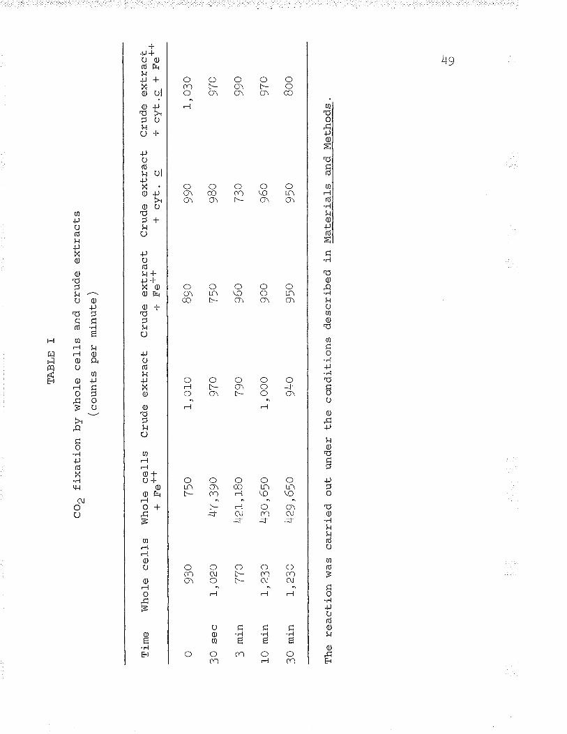
Intact cells and crude extracts of Ferrobacil-l-usrlr
ferrooxidans both fixed radioactive C-'OZ in the alosence of
an added energy source. The amount of C'[O, fixed was very
small and could be accounted for by the presence of endo-
genous metabolites (fante I). In the presence of an energy
source, ferrous iron, only intact ce1ls showed an appreciable
increase in the amount of Cf4Oo incorporated. The inability
of crude extracLs to incorporate additional ctuo, in the
presence of ferrous iron is another evidence of the failure
of crude extracts to metabolize ferrous iron which leads to
the exhaustion of intermediates for CO, fixation and ATP
(see Tables T, II, and III).
JrQ
Tim
e W
ho1e
cel
ls
ô
?ô
qaõ
Jv
<
mln
10 m
in
?O m
'i n
TA
BLE
I
CO
Z fi
xatio
n by
who
le c
ells
an
d cr
ude
extr
acts
(cou
nts
per
min
ute)
930
1,02
O
770
L,23
O
L,23
O
Who
le c
ells
+ F
e-F
*
The
rea
ctio
n w
as c
arrie
d ou
t un
der
the
cond
ition
s de
scrib
ed i
n M
ater
ials
an
d M
etho
ds.
750
4T ,3
9o
42t,t
8o
43o,
65o
Lça
Áno
Cru
de e
xtra
ct
1,01
0
9To
7go
1, O
0O
940
Cru
de e
xtra
ct.l-
J-
+ f
,'e' '
89o
,7tr
^I)
v
960
900
950
Cru
de e
xtra
ct*
cyt.
c
990
980
730
960
950
Cru
de e
xtra
ct-!
¡r
z{-
-l E
totf
1,03
o
a7Õ
ttv 99o
970
Boo
|-'_
\o
5o
The incorporation of Ct4op shows that metabolizLng
intact cells have the abil-ity to regenerate the necessary
intermediates of CO2 fixation and also illustrates the
ability of intact cells to generate ATP while metabolizing
ferrous iron. Tvpical results are found in Tab1e I.
Separation and identification of radioactive compounds
Ethanol extracts of the 30 second fixation by intact
cells metabolizíng ferrous iron (rafte I) were separated by
two dimension ascending chromatography. The following
1abelled compounds were found: J-PGA, aspartic acid,
glutamic acid, and phosphorylated sugars. The majority of
the radioactivity was found to be in the 3-PC=4, which
contained approximate tV T9% of the "t4oo
fixed. Aspartic
acid made vp 7/" of the activity fixed .lU ntuaamic acid
contained 2%.
The appearance of 3-PGA indicates an autotrophic COt
fixation mechanism similar to that first found ín plants
(Calvin and Benson, I94B; Bassham et â1., I95O; Benson et al.\-I95O) and in other chemolithotrophic bacteria (see Elsden,
L962). The high activity in aspartic and glutamic acids
indicates the operation of the heterotrophic l¡Vood-Werkman
5l
reaction (eee carboxylase) first shown in bacteria by
Suzuki and Werkman (f958a,¡).
The formation of 3-PGA bv ceIl-free extractå
Autotrophic CO, fixation mechani-sms, cyclic
mechanisms where the CO2 acceptor is regenerated, contain
two enzymes, phosphoribulokinase and carboxydismutase, not
found in any other metabolic system (eager , 1954; Quayle et
al., L954; Weissbach et êf., f954; Hurwitz et al., L956;
Weissbach et al., L956) . The autotrophic system is able to
regenerate RUDP from 3-PGA. The other enzymes found in the
regeneration mechanism are not unj-que to autotrophic systems
and appear ubiquitous.
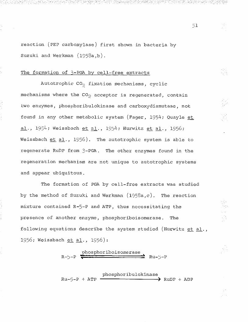
The formation of PGA bv cell--free extracts was studied
by the method of Suzuki and Werkman (f958a,c). The reaction
mixture contained R-5-P and ATP, thus necessitating the
presence of another enzyme, phosphoriboisomerase. The
fol-lowing equations describe the system studied (tturwitz e-t aI.,
- ^-- - - ^--\1956; Weissbach et aI., L956) z
phosphor iboisomeraseR-5-P Prr -Ã -Þ
phosphoribulokinaseRu-!-P + ATP RUDP + ADP
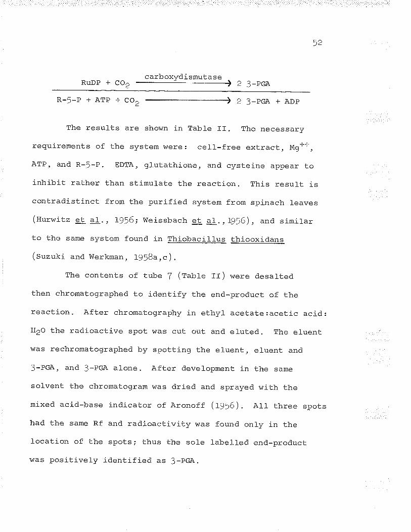
42
RuDp + co2 carboxydismutase , z 3-pGA
R-5-P+ATP+CO\ 2 3_PcA+ADP
The results are shown in Table II. The necessary
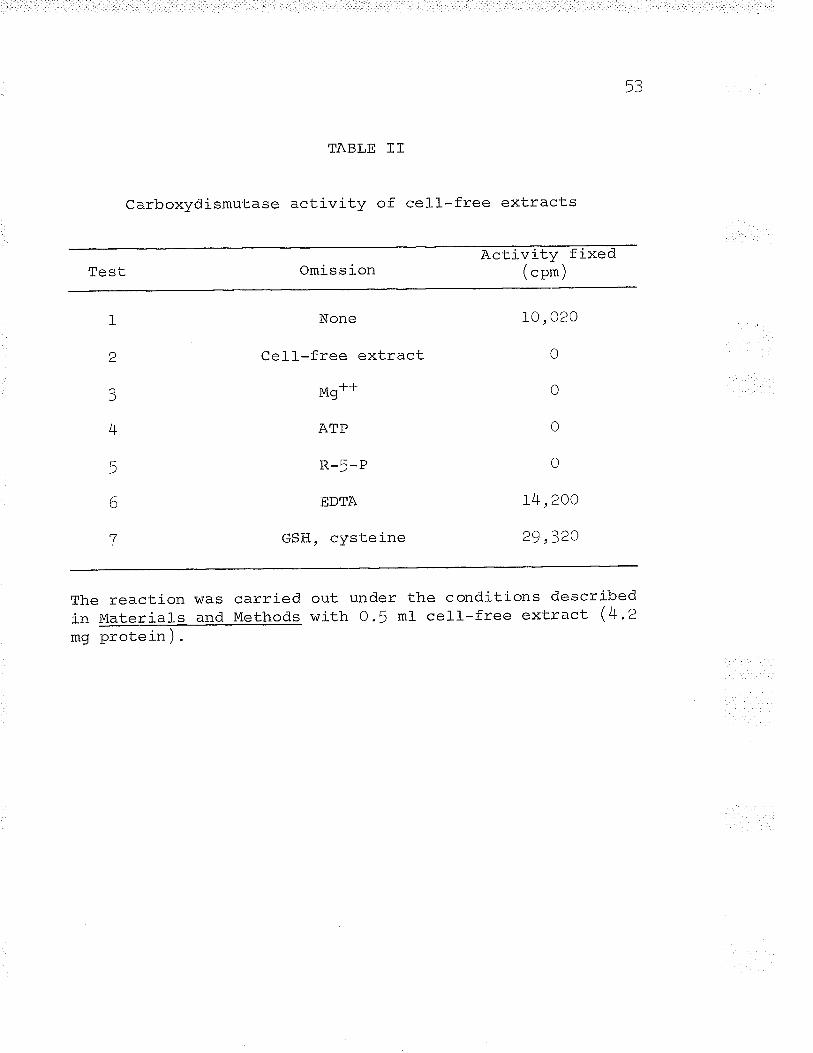
requirements of the system were: cell-free extract, Mgo*,
ATP, and R-!-P. EDTA, glutathione, and cysteine appear to
inhibit rather than stimulate the reaction. This resul_t is
contradistinct from the purified system from spinach leaves
(Hurwitz et aI. , 1956; Weissbach et al. , L9j6), and similar
to the same system found in Thiobacillus thiooxidans
(Suzuki and Werkman, 195Ba,c).
The contents of tube T (fatrte II) were desalted
then chromatographed to identify the end-product of the
reaction. After chromatography in ethyl acetate:acetic acid:
H2o the radioactive spot was cut out and eruted. The eluent
was rechromatographed by spotting the el-uent, eluent and
3-PGA, and 3-PC=A alone. After development in the same
solvent the chromatogram \^7as dried and sprayed with the
mixed acid-base indicator of Aronoff (lgf6). All three spots
had the same Rf and radioactivity was found onry in the
location of the spots; thus the sole labelred end-product
\^ias positively identífied as 3-pC=A.
53
TABLE II
Carboxydismutase activity of cell-free extracts
ActivityOmission ("p*)
t
L
3
4
5
6
7
None
Cel-l-free extract!¿Mg' '
ATP
R-5-P
EDTA
GSH, cysteine
10,020
0
0
0
0
L4,2OO
)Q ?20
The reaction was carriedin Materials and Methodsmg proteln/.
out under the conditions describedwith 0.5 ml- cell--free extract (4.2
Ã/l
The carboxyd.ismutase activity of cell-free extracts
was also assayed. spectrophotometrically by using "coId"
bicarbonate in place of HCt o, and adding 10 pmoles ATP,
O.! ¡rmoles NADH, and triose phosphate dehydrogenase (Sigma)
to the isotopic reaction mixture contents. The reaction
was carried out in 10 mm cuvettes at 340 mp in a Gilford
model- 2000 spectrophotometer. This assay provided evidence
for the presence of phosphoriboisomerase, phosphoribulokinase,
carboxydismutase, and phosphoglycerokinase in cell-free
extracts. Extracts also exhibited very weak glyceraldehyd.e-l-
phosphate dehydrogenase activity using 3-PGA, ATP, and NADH
as substrates.
Extracts of Ferrobacillus ferrooxidans contained. al-l-
the enzymes necessary for the autotrophic fixation of COt in
addition to the enzymes required to generate glyceraldehyde-J-
phosphate rìecessary for the isomerization and condensation
into a hexose in order to make the fixation process cyclic.
The formation of oxalacetate
The results of Ct4o^ fixation by whole cells indicateda
t C3 + CI reaction feeding the Krebs cycle, Ieading to the
formation of labelted aspartic and glutamic acids. This
55
"heterotrophic" COZ fixation is common to all organisms
and is required. for repleníshment of necessary intermediates
of the Krebs cycle. rn bacteria four major systems of. co,
fixation (ot exchange) feeding in necessary intermediares
have been foUnd: the malic ênz\zmê ÞTilÞ .-larbOxylase, PEp
carboxykinase, and pyruvate carboxylase.
Kornberg (tg6S) discussed the "anaplerotic" function
of PEP carboxylase and has reviewed evidence that the onlv
enzyme responsible for feeding c¿, compounds into the Krebs
cycle in many bacteria is PEP carboxylase. Single mutants
of E. coli (amarsingham, L959; Ashworth and Kornberg, L963)
and S. Fyphimurium (theodore and Engl.='b"rg, L964) tacking
PEP carboxylase were unable to grow on pyruvate or its
precursors unless Krebs cycle intermediates were added to
the growth medium.
Suzuki and Werkman (f958a,¡) tirst demonstrated the
existence of PEP carboxylase in cell--free extracts of bacteria
(fhiobacillus thiooxidans ) along with an ITP-Iinked oxalacetic
decarboxylase. since the Enterobacteriaceae contain the
malic enzyme, PEP carboxylase and an ATp-linked oxalacetic
decarboxylase and only one enzyme, pEp carboxylase, serves an
"anaplerotic" function, this same premise may holo true for
"-ln
chemolithotrophs ( chemoautotrophs ) .
The PEP carboxylase activity of cel-l-free extracts
was assayed by c'uo, incorporation in .the presence of pEp
and Mg++ (iur"**). The results are shown in Table frr. The
tests indicated a requirement for pEp, Mg** or Mn*+, and
cell-free extract. Test T ill-ustrated a lack of pvruvic
carboxylase in the cel_I-free extracts.
Other experiments to investigate the possibl_e
metabolic control-s exhibited on PEP carboxylase vvere carried
out linking the enzyme to malic dehydrogenase and NADH
oxid.ation" The reaction was measured. by following the rate
of NADH oxidation or measuring the amount of radioactivity
i-ncorporated. The pEp carboxylase of E" col! (cánovas and
Kornberg, L965; Nishikido et al., L96D) and s" tvphimurium
(Sanwal and Maeba, 1965¡ 1966) was activated by acetyl CoA,
fructose-lr6-Oiphosphate, and inhibited by aspartate.
Celf-free extracts of Ferrobaciltus ferrooxidans
contained PEP carboxylase activity and the sole end-product
of the linked reaction was maric acid (test L, Tabl_e rv).
The results shown in Tabres rv, v, and vr il-l-ustrate the
effects of various intermediates of the Krebs cvcle. amino
acids of the aspartic family, glutamic acid, aceto-acetyl coA,
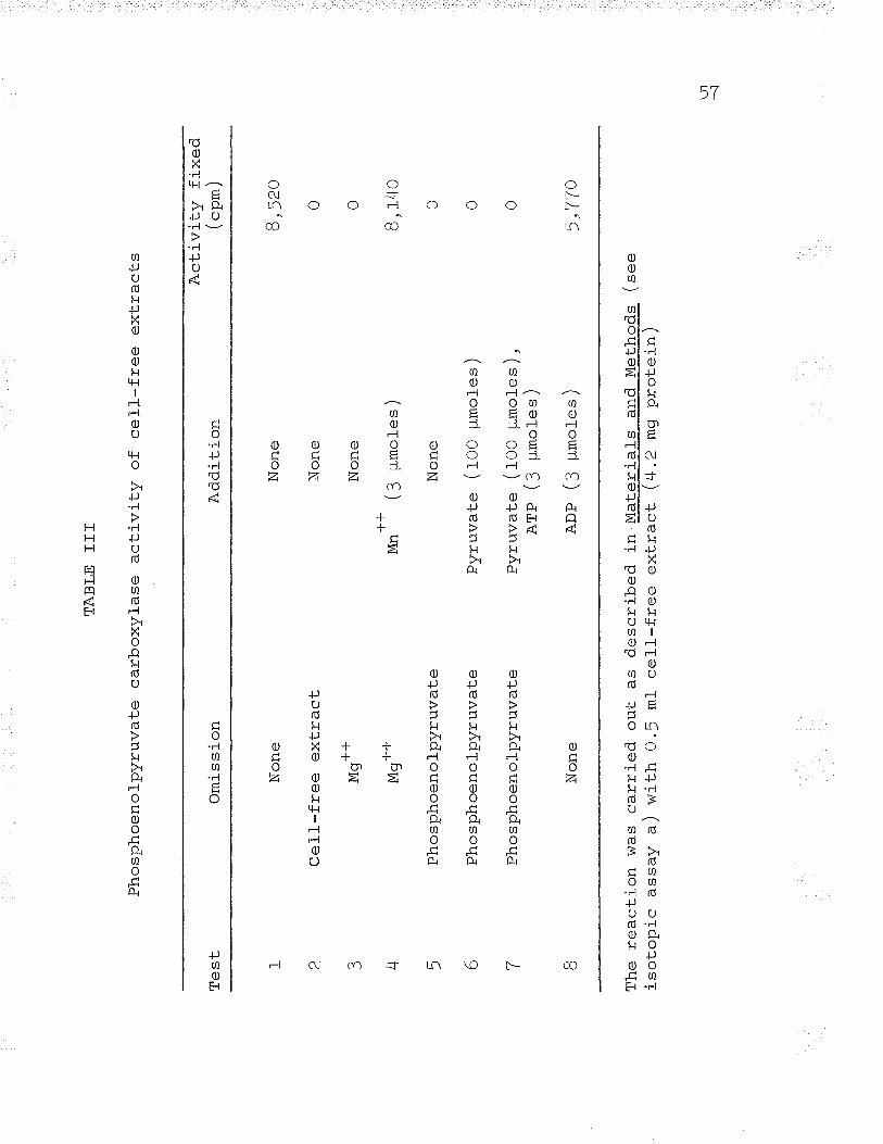
TA
BLE
IT
I
Pho
spho
enol
pyru
vate
ca
rbox
ylas
e ac
tivity
of
ce
ll-fr
ee
extr
acts
f,cb
L I a 3 4 5 6 7
Non
e
Ce1
1-fr
ee e
xtra
ct+
-l-M
g' -l-J
Mg'
'
Pho
spho
enol
pyru
vate
Pho
spho
enol
pyru
vate
Pho
spho
enol
pyru
vate
Non
e
Om
issi
on
B
The
rea
ctio
n w
asis
otop
ic
assa
y a)
Add
ition
Non
e
Non
e
Non
e
-t-+
/^
- \
lvlrl
t
<
llmÕ
lê
s )
\J
carr
ied
out
with
0 .5
ml-
Non
e
Pyr
uvat
e (f
OO
¡rm
oles
)
Pyr
uvat
e (f
OO
pr.
mol
es )
,A
TP
(S
pm
oles
)
AD
P (
: ¡-
imol
es )
Act
ivity
fix
ed("
p*)
as d
escr
ibed
in
Mat
eria
ls
and
Met
hods
( se
ece
ll-fr
ee
extr
act
(4.2
mg
prot
ein)
R q
2o
0 0
B, t4
o
0 0 0
5,T
TO
Lft
--l
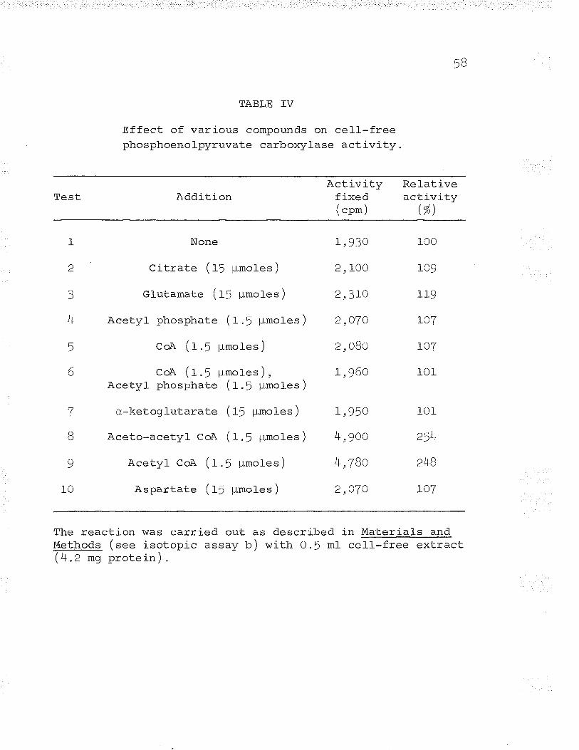
1a)
TABLE IV
Effect of various compounds on cell-freephosphoenolpyruvate carboxylase activity
Test AdditionActivity Relativefixed activity( "p* ) (%)
1
a
3
4
5
6
7
B
9
10
None
Citrate (ff ¡rmoles )
Glutamate (f ! ¡rmofes )
Acety1 phosphate ( f .f ¡rmoles )
CoA (t.f pmoles )
t- \uof\ \r.) pmores/,Acetyl ptrosphate (t.f pmoles )
cr,-ketoglutarate (ff ¡-LmoIes )
Aceto-acetyl CoA (f .¡ ¡rmoles)
Acetyl CoA ( f .f ¡-Lmoles )
Aspartate ( ff pmoles )
r,930
2, 100
2,3LO
2,O7O
2, o8o
L,960
L,950
4,9oo
4,78o
2,O7O
100
109
'l'tq
IOT
L07
101
101
248
r07
The reaction was carried out as describedMethods (see isotopic assay b) with O.5 ml(4.2 mg protei.,).
in Material-s andcell-free extract
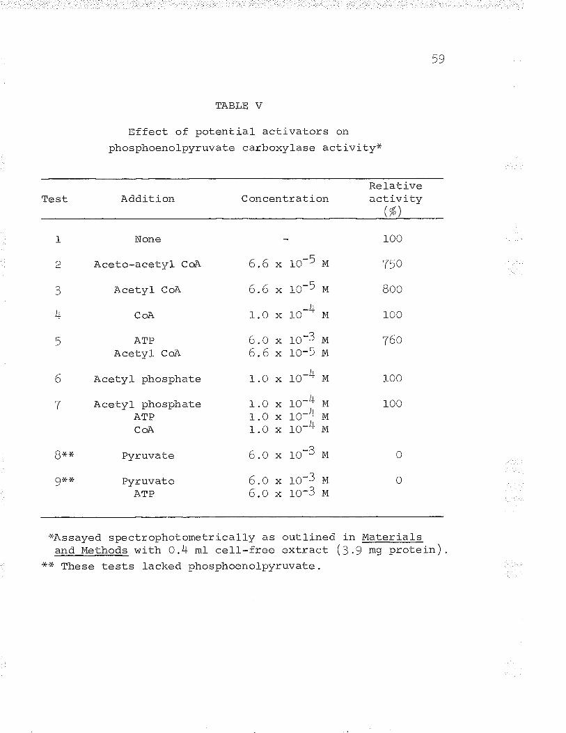
59
TABLE V
Effect of potential activators on
phosphoenolpyruvate carboxylase activity*
Test Addition ConcentrationRelativeactivity
(%)
-L
2
3
4
5
Bx+É
grçx
6
T
None
Aceto-acetyl CoA
Acety1 CoA
CoA
ATPAcetyl CoA
Acetyl phosphate
Acetyl phosphateATPCoA
ñ----'.--â + ^ryrLrvclLe
ñ---^"---^L ^ry l- Lrv c¿ LeATP
6.6
6.6
1.0
6.o6.6
1.0
1.01.01.0
6.o
6.o6.o
-Ãlo)Mro-5 ¡,i
_)l10M
ro-3 ¡,i
rO-5 vi
ro-4 lvl
ro-4 rvi
ro-4 vt
ro-4 irn
lOJM
ro-3 u1o-3 M
roo
750
Boo
100
760
100
100
x
Xx
x
Xxx
0
oL
x
*Assayed spectrophotometrically as outlinedand Methods with 0.4 mI cell-free extract
*x These tests lacked phosphoenolpvruvate.
in Materials/^ . \\ J .y m9 protrer-n /
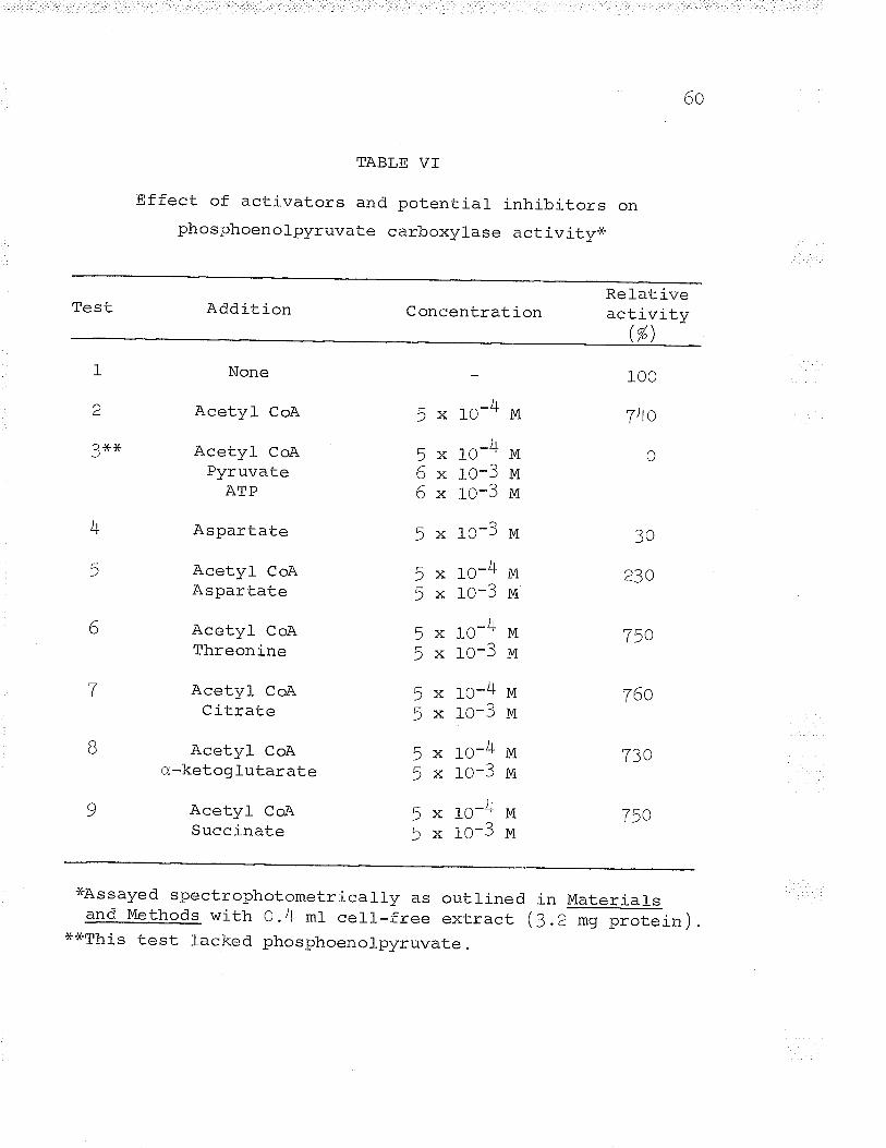
6o
TABLE VT
Effect of activators and potential inhibitors onphosphoenolpyruvate carboxylase activityx
Test Addition ConcentrationRel-ativeactivity
(%)
1
'¿
3rÊ*.
None
Acetyl CoA
Acetyl CoAPyruvate
ATP
Aspartate
Acetyl CoAAspartate
Acetyl CoAThreonine
Acetyl CoACitrate
Acetyl CoAo,-ketog l-utarate
Acetyl CoASuccinate
5 x 1O-4
5 x 1O-46 x rO-36 x rO-3
- .^-?)xJUJ
5 x 1o-45 x 1O-3
5 x 1O-45 x 1O-3
J x to-45 x 1O-3
5 x 1o-45 x 1O-3
5 x 1o-4- -^-?)xl-uJ
M
M
M
M
M
M
M
M
M
M
M
MM
M
M
100
740
0
30
230
.7tr.^l)v
760
7?atJv
750
)lT
5
6
7
B
q
xAssayed spectrophotometrically as outlinedand Methods with 0.4 ml cel_l-free extract
x*This test lacked phosphoenolpyruvate.
. in Material_s
ß.2 mg protein)
OI
and acetvl CoA.
The reaction was similarly activated by aceto-acetyl
CoA and acetyl CoA (tatrte IV), unlike the S. tvphimurium
enzyme where aceto-acetyl CoA could not completely substitute
for acetyl CoA (Vtaeba, 1966). The PEP carboxylase activity
was inhibited by aspartate in the presence or absence of
acetyl CoA (fatrte VI ) . This result is in agreement with the
results of sanwal and Maeba (1965)' Thus it appears that the
"heterotrophic " COZ fixation mechanism of a chemoautotroph
is subjected to the same control mechanisms as heterotrophic
bacteria. Once thís similarity was found, the question of
control was not further Pursued.
The pEP carboxylase of Ferrobacillus ferrooxídans Ïtas
a pH optimum close to pH /.0 or on the acid side of neutrality.
This is illustrated in Table VII . It was not determi-ned
whether the actual carbon moeity fixed was COZ or HCO3- but
the results indicate that the substrate fixed may be co2
rather than HCO3-. Keeping in mind that the bacterium grows
optimally at an acid pH this result would not be unexpected.
On the other hand it has never been demonstrated that PEP
carboxylase fixed free COp.
The cell--free extracts containj-ng PEP carboxylase
6z
TABLE VTI
Effect of pH on phosphoenol;oyruvate carboxylase activity*
LJLL Relative activity (%)
oo
A^
7n
100
111
167-v I
cF,6,
lrnn'taa
xThe reaction was assayed spectrophotometrically in lO mmcuvettes at 340 ¡qr in a Gil-ford model 2000 spectro-photometer. The reaction mixture contained: 600 pmolesTris-HCl buffer, L.5 plmoles acetyl CoA, L5 ¡-Lmoles tttgCl-'2,10 ¡-Lmoles PEP , 45 ¡rmoles NaHCOj , I.5 ¡rmoles NADH, 5 ug pigheart malic dehydrogenase (Worttrington), cell-free extractß.2 mg protein) and water in a final volume of 3.0 ml .
OJ
activity !üere tested for malic enzyme activity spectro-
photometrically, using both NAD and NADp coenzymes but no
activity was found. some heterotrophic bacteria have been
found to contain the malic enzyme. but rather than acr as
a pathway into C4 metabolism the enzyme replenished Ca
compounds when the bacteria were grown on Krebs cycle
intermediates (Wood and Stjernholm, 1962; Kornberg, L96Ð "
II. Iron Oxidation
Iron oxidation \^ias studied in both intact cells and
celr-free extracts in an attempt to elucidate the oxidation
mechanism.
El-ectron transport cgmponents
Vernon, Mangum, Beck, and Shafia (lg6O) first
demonstrated the existence of cytochromes in an iron oxidizer.
These workers, who found that cel_l-free extracts of
Thiobacill-uq ferrooxidans contained cytochromes c and a,
purified the cytochrome c. Blaylock and Nason (1g63) found
cytochromes b, s, and a in extracts of Ferrobacillus
ferrooxidans and postulated that iron oxidation involved only
cytochromes g and a. The cytochrome content of Ferrobacill-us
t-tl
ferrooxidans was investigated spectroscopically in intact
cells and spectrophotometrically in celf-free extracts
( sonic extracts ) .
When intact cell-s were treated with either ferrous
iron or sodium dithionite spectroscopy revealed absorption
bands at 409-4t0 m¡r, 5IO-525 m*, and 552-555 nV, which
coincide with the y, ?, and a bands of reduced cytochrome c;
and at 4ZO-425 r;lp" and 60Z-605 mp, which coincide with the y
and c¿ bands of a reduced a-type cytochrome (smitfr, L96I).
Contrary to the results of Blaylock and Nason (WAS) there
was no evidence of the presence of cytochrome b. Spectro-
photometrícally, sonic cell-free extracts exhíbited the same
type of absorption spectrum. The results of the spectroscopic
studies are shown in Table VIII.
Intact cells and sonic extracts were extracted with
4O/" Autanol. The butanol layer was dried and redissol-ved ín
butanol- prior to spectrophotometric analysis. The yellow
colour of the dissolved substance and the spectrophotometric
absorption spectrum (263, 2TL, and 2BO m¡r absorption peaks )
indicated the presence of a quinone-type compound (eishop,
Pandya, and King, 1962). Attempts to reduce the compound
with potassium borohydride failed and the spectrum remained
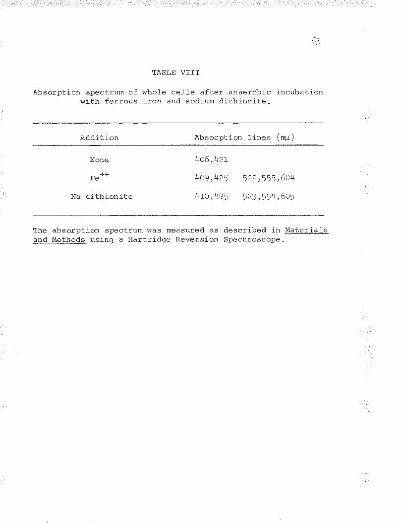
r)1
TABLE VÏTI
Absorption spectrum of whole ce1ls after anaerobic incubationwith ferrous iron and sodium dithionite.
Addition Absorption lines (rq-i)
None
J.-LUç
Na dithionite
4o6,4zt
4o9 ,425 522,555,604
[to,)+25 523,:r54,605
The absorption spectrum was measured as described in Materiqlsan{l ethods using a Hartridge Reversion Spectroscope.
66
unchanged unlike the coenzyme Q of Thiobacitlus thiooxidans
(cook and Umbreit, L963). The compound resembled the K
vitamins of E. coli (eisnop, Pandya, and King, L962). The
role of this guinone-type compound in the electron transport
chain was not pursued further.
Oxidation of ferrous iron by intact ce1ls
Reported optimum conditions for ferrous iron oxidation
by Thiobacillus ferrooxidans and Ferrobacillus ferrooxidans
vary considerably (Silverman and Lundgren , L)J)arb; Beck,
L96O; RazzeLI and Trussell, 1963; Landesman, Duncan, and
Walden, L966). Experiments designed to find the optimum pH
and iron concentra.tion for intact cell iron oxidation were
performed. Using 'washedr intact cell_s, the optimum pH
(initiat) i" a water-sulfuric acid mixture was pH 3.0 (rig.
1). This pH optimum coincides with the results of Silverman
and Lundgren (tgSgA) who used the same organism. At an
initial pH of 3.0 the optimum ferrous iron content was found
to be 2!O ¡,Lmoles FeSo¿1 ./HZo in a volume of 2.8 ml (eig. 2).
This result is at variance with the results of Landesman,
Duncan, and Walden (tg66) who reported an optimum iron
concentration of 4OO-BOO ¡-Lmoles for ThiçþaqiJlus ferrooxidans.
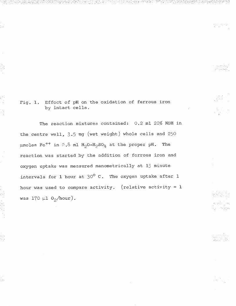
Fig. 1. Effect of pH on the oxidation of ferrous ironby intact cell-s.
The reaction mixtures contained: 0.2 mt ZO/" XOLS in
the centre well , 3.5 mg (wet weight ) whole cell-s and 25O
¡-Lmoles Fe++ in 2.8 mI H2O-H2SO4 at the proper pH. The
reaction was started by the addition of ferrous iron and
oxygen uptake !\ias measured manometrically at 15 minute
intervals for l- hour at 3Oo C. The oxygen uptake after I
hour was used to compare activity. (relative activity = I
was 170 pl or/nour) .
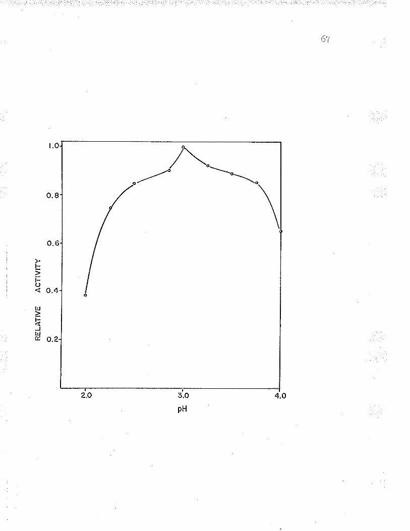
oo
AC
T¡V
ITY
I å
RE
LAT
IVE
I f\)
Or
--l
!u_ o
EO
J*o
.-Þ
_o
Fig. 2. Effect of ferrous iron concentration on ironoxidation by whol-e cel1s.
The reaction mixture contained: 0.2 ml- 20% IKOH
in the centre well, 5 mg (wet weight) whole cells in O.5
mt H2o-Heso4 (pH 3.0) in the sidearm, and Fe** in 2.3 mt
H2O-H'SO4 (pU 3.0) in the main compartment. The reaction
was initiated bv the addition of the cells from the side-
arm. oxygen uptake was manometricatly measured at 3Oo C.
A = IO plmoles Fe*-F
-LJB = 100 ¡-Lmoles Fe' '
.L-LC = 210 ¡-Lmoles Fe'
D = 5OO pLmoles Fe**.L-L
E = flO pLmoles Fe'
AJF = 1000 pmoles Fe' '
-L-Lc - O pmoles Fe'
6B
ãtrJlt<t
fLþON
I
,bc
-¿
B
,'il'r,
.i,(/'
,.ù
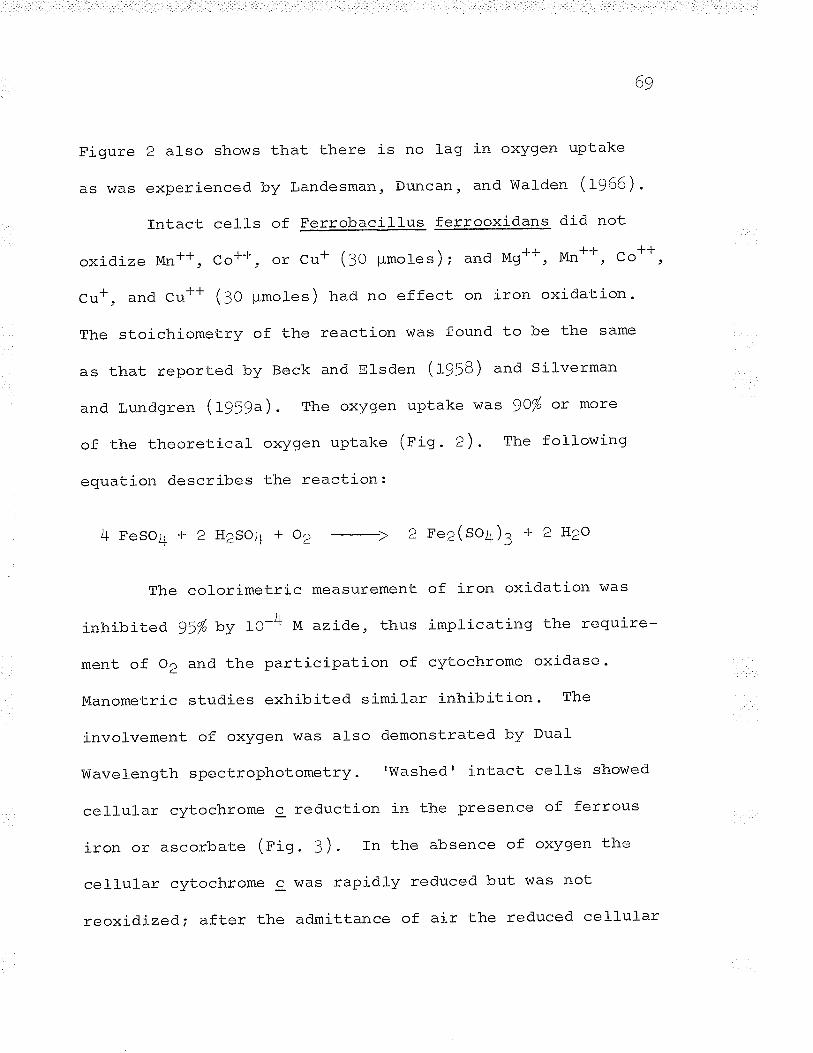
69
Figure
AS WAS
2 also shows that there is no lag in oxygen uptake
experienced by Landesman, Duncan, and Walden GgAe).
Intact cells of Ferrobacillus ferrooxidans did not
oxidize Mn++, co**, or cu+ (:o ¡rmotes); and M9++, Mn++, co**,
Cr*, and cu** (30 ¡rmotes) had no effect on iron oxidation.
The stoichiometry of the reaction was found to be the same
as that reported by Beck and Elsden (WSg) and Silverman
and Lundgren (tgSgÐ. The oxygen uptake was )Oy'" ox more
of the theoretical oxygen uptake (pig . 2). The following
equation describes the reaction:
4 l'eSO¿t + 2 H2SO4 + OZ -->
2Ee2(so4)3 +2H2o
The col0rimetric measurement of iron oxidation was
inhibited gi% by IO-4 M azide, thus implicating the require-
ment of 02 and the participation of cytochrome oxidase.
Manometric studies exhibited similar inhibition. The
involvement of oxygen was also demonstrated by Dual
Wavelength spectrophotometry.'Washed' intact cells showed
cellular cytochrome c reduction in the presence of ferrous
iron or ascorbate (eig. 3 ) . In the absence of oxygen the
cellular cytochrome c was rapidly reduced but was not
reoxidized; after the admittance of air the reduced cellular
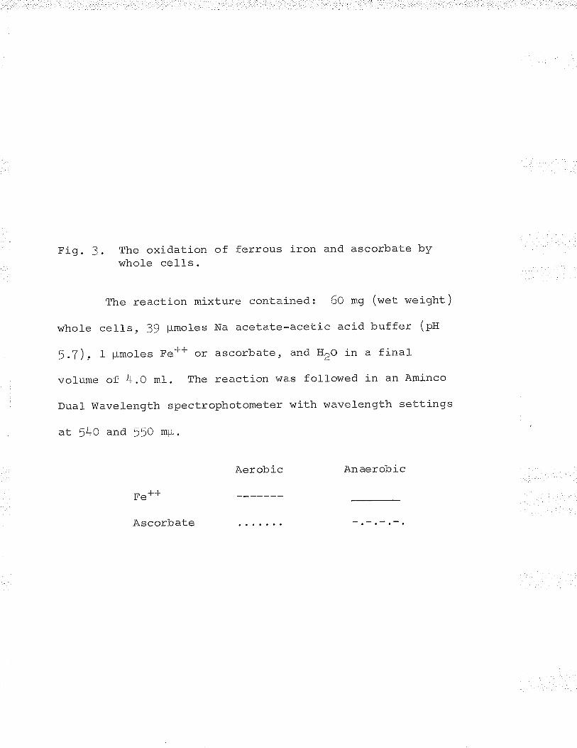
Fig. 3. The oxidation of ferrous iron and ascorbate bywhole cells.
The reaction mixture contained: 6O mg (wet weight)
whole cells , 39 ¡,Lmoles Na acetate-acetic acid buffer (pH
5.7), 1 pmoles Fe** or ascorbate, and H2o in a final
vol-ume of 4.0 mt. The reaction was followed in an Aminco
Dual Wavelength spectrophotometer with wavelength settings
at 54O and 550 mp.
Aerobic Anaerobic
Fe++
Ascorbate
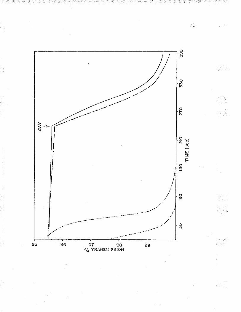
AIR ti t¡ -a\
--.1
390
??.a
I
I
(c (tl
(o g)
x -(o
-t{
7 U' = (!) =(o
=f.A
, (9 (Ð
270
150
2'o
TtM
E (
sec)
9030
7r
cytochrome c was rapidly oxidized (elg. 3). These resulLs
indicate a two-step process in the oxj-dation scheme, (1)
the reduction of cytochrome c by ferrous iron i and (Z) the
sulosequent reoxidation of the reduced cvtochrome c in the
presence of oxygen via cytochrome oxidase, âfl e-type
cytochrome.
Intact cells were stored for months at O-5o C and
stilI retained some oxidizing ability. The rate of iron
oxidation by intact cells decreases with increased duration
nF cl-nraaa syv . Landesman, Duncan, and Walden (ryeA ) f ound that
Thiobacil-lus ferrooxidans cells stored for lonqer than four
days l-ost a large percentage of their oxidizing ability, so
old cells were revitalized prior to use in all experiments
(see 'Mêterials and Methods'). Intact cell-s lose 9O-IOO% of
their oxidizing ability upon freezing at -2Oo C, even for
bríef periods (l-fO minutes) (rigs. 4 and !). The toss in
activity could not be recovered by incubating the cel_l_s with
ferrous iron. This freezing phenomenom is also exhibited by
Nitrosomonas (lees, L964) and is not unique in the iron
oxidizers.
Althouqh frozen cells lose their abilitv to oxidize
ferrous iron. crude sonicates of these same inactive cells
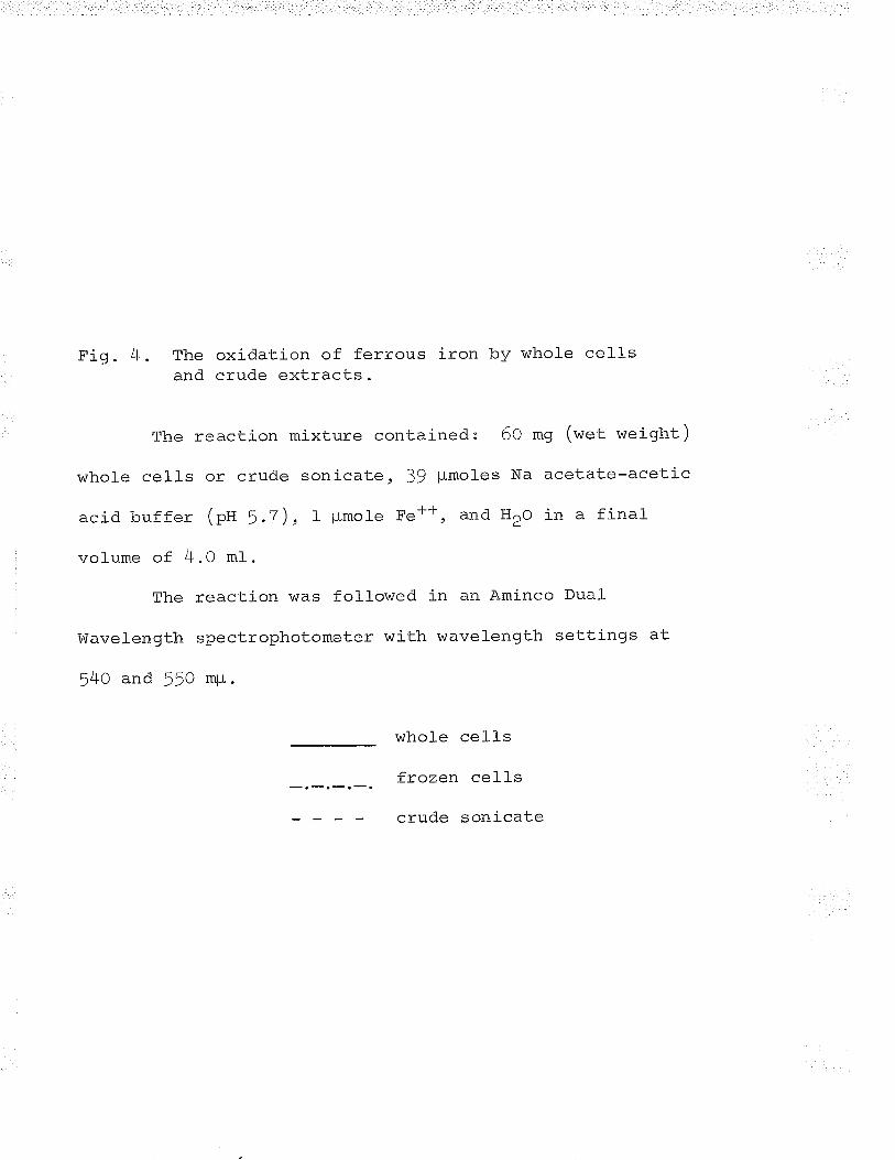
Fig. 4. The oxidation of ferrous iron by whole cellsand crude extracts.
The reaction mixture contained: 60 mg (wet weight )
whole cells or crude sonicate, 39 pmoles Na acetate-acetic
acid buffer (pa 5.7), I ¡rmole Fe*+, and H2o in a final-
volume of 4.0 mt.
The reaction was followed in an Aminco Dual
Wavelength spectrophotometer with wavelength settings at
540 and 550 rqu.
whole cell-s
frozen cells
crude sonicate
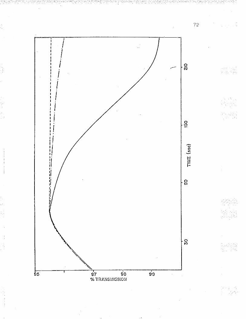
--1 h)
2ro
(or
(''l I I I I
'1
i I
*o-
-t -ú
-s't Þ -;¿ L9 :./ õ (n õ
so,
*¿@ (o
-C
D
t50
TIM
E (
sec)
9030
Fig. 5. The oxidation of reduced cyt. c by whole cellsand crude extracts.
The reaction mixture contained: 60 mg (wet weight)
whole cells or crude sonicate, 39 plmoles Na acetate-acetic
acid buffer (pn 5.7), reduced cyt. 9, and H2o in a final-
volume of 4.0 mt. The reaction was followed in an Aminco
Dual !üavelength spectrophotometer with vvavelength settings
.-t,^at 54O and 550 mp.
A = sonicated cells (6.f f m¡rmo]e reduced cyt. c )
Al = whole cells (6.15 m¡lmoles reduced cyt. c)
A' ' = sonicated cells, anaerobic (6.75 m¡rmoles reduced cyt. c )
A I ¡ t = frozen cells (6.f> mpmoles reduced cyt. c )
B = sonicated cells dituted I/LO (tO.tZ5 rqumoles reducedcyt. c )
c = sonicated cel-l-s (::.8¡ mpmoles reduced cyt. c)
IItttIt,,¡
.t,,,
..f/
t
i
IiI
C).
\-......-:J ..-.-.--.:
tk,gIIIII
CIo \(,
q
73
to
¡'-
ro
.zE
þJEþ
78 gO 84 88 92 96
% TRANS[\ll$Slof\|
7)lfT
exhibited both partial reactions of the oxidation mechanism
(rigs. 4 and 5) . These extracts contain both ferrous iron-
cytochrome c reductase and cytochrome oxidase activities.
The loss of iron oxidizing ability by fxozen intact cells
must mean that the electron transport chain is interrupted
or broken between cytochrome c and cytochrome a.
Oxidation of ferrous iron bv cell--free extracts
B1aylock and Nason (1g63) reported that they were
able to isolate ce11-free extracts of Ferrobacillus
ferrooxi.Qans capable of oxidizing ferrous iron (colorimetric
assay). These authors showed that oxidation by cell--free
extracts required oxygen and was inhilcited by cytochrome
oxidase inhibitors. Attempts by others to duplicate these
results faited (aleem, f964; Beck, L965; Lundgren, 1965¡
Silverman, f965). In this investigation, use of the same
methods as those described by Blaylock and Nason (tg63)
failed to uncover the activitv of an iron oxidase that
requires oxygen as its terminal- electron acceptor.
'I,riashed' intact cel-l-s were sonicated f or various
time intervals and iron oxidase activitv was measured at
pH 3.0, manometrically, and at pH 5.O, colorimetrically and
T5
manometrically. Oxygen uptake at pH 3.0 decreased with
increase of sonication time; after 10 minutes sonication
the extracts exhibited no oxygen uptake. There was no
retention or increase of oxygen uptake at pH 5.O¡ the
manometric oxidation at pH 1.0 paralleled the results
obtained at pH 3.0. The results are illustrated in Table
fx. similar results were obtained when ce11-free extracts
rather than crude extracts were util_ized (there was no
oxygen uptake at any time ) .
Colorimetrically, crude and cell--free extracts
oxidized iron at pH 5.0, but the oxidation of ferrous iron
was not oxygen dependent (see Fig. 4). Ferrous iron
oxidation by ce11 extracts was dependent on cellular
cytochrome g reduction. The oxidation of iron and the
reduction of cyt. c took place equalry as well anaerobically
as aerobically. Once the cel-lular cyt. c was reduced the
reaction stopped and there was no reoxidation of the reduced
cyt. c, although cytochrome oxidase was present in the
extracts (nigs. 4 and 5) .
Attempts to prepare cellular extracts capable of
manometric iron oxidation failed although al-l of the extracts
contained both ferrous iron-cyt. c reductase and cytochrome
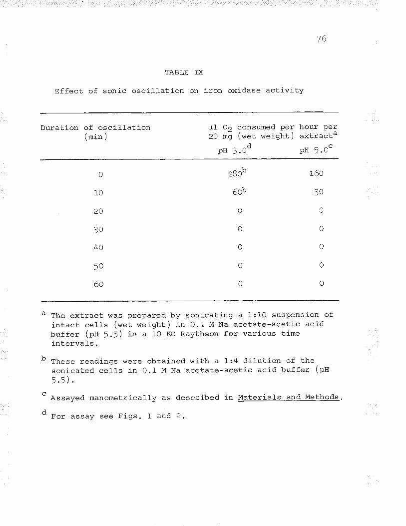
76
TABLE IX
Effect of sonic oscillation on iron oxidase activity
Duration of oscill-ation(min )
pf Oe consumed per hour Per20 m9 (wet weight ) extra-cta
pH 3.Od pH 5.Oc
0
10
20
3o
4o
50
60
't-2BO"
r ^l¡OU-
o
0
0
o
0
160
3O
o
0
0
o
0
The extract was prepared by sonicating a l:10 suspension ofintact cells (wet weight) i" O.t M Na acetate-acetic acidbuffer (pH 5.5) in a lO KC Raytheon for various timeintervals.
b Th"=" readings were obtained with a 1:4 dilution of thesonicated cel-l-s in O.l- M Na acetate-acetic acid buffer (pU- -\h1t).t I .
d
Assayed manometrically as described
For assay see Figs. 1 and 2.
in Materials and Methods.
TT
oxidase activities. The inability of cellular extracts
to oxidize ferrous iron manometricallv seems to be due to
a blockage of the electron transport chain between cyt. c
and cyt. a. FAD, FlvlN or the quinone isolated from intact
cells did not overcome thís blockaqe in the el-ectron
transport chain.
III. Ferrous Iron-Cytochrome c Reductase
Since studies with whole cel-ls and crude extracts
indicated that the primary enzyme in ferrous iron oxidation++r^7ãc Eaa -¡rz{- c reductase, this enzyme was purified and theeJ e.
mechanism of the reaction was studied. Ferrous iron-cvt. c
reductase was found to be tiqhtlv bound to the cell and the
enzyme was not easily solubilized, thus necessitating the
use of extreme conditions for extraction (a high pH extraction
buffer and an extended duration of sonication).
Enzyme purification
Alf manipulations during the enzyme purification
were carried out at O-5o C unless otherwise stated. Crude
ce11-free extracts of Ferrobacillus ferrooxidans (flOrOOO x g
supernatant, see Malerials and Methods) were adjusted to
7B
pH T.o with 10 N sul-furic acid. The extract was then
heated rapidly Lo jo-)Jo c in a boil-ing water bath and
maintained at this temperature for five minutes. The extract
was cooled quickly to !o c in an ice-water bath and was
centrifuged at 25,OOO x g for 1! minutes.
The resulta-nt 25,ooo x g supernatant was adjusted to
pH 5./ with I N acetic acid. Cal_cium phosphate gel
suspended in 0.01 M acetate-acetic acid buffer (pH 5.2) was
added srowly, with stirring, until the concentration of
gel:protein was 5:f (¿ry weight). The mixture vüas stirred
slowly for lo minutes, then centrifuged at 25,ooo x g for
1! minutes. The precipitate was discarded and col_d ethanol
(-zoo c) was added with constant stirring to the enzyme
incubated in an ice-salt bath. The enzyme temperature was
maintained befow oo c during the ethanol- addition.
There was no formation of precipitate untir- the
ethanol- concentration reached 60%. since the precipitate
was sl-ow in forming, the ethanol-enzyme sol-ution was
incubated at -2oo c for 12 hours to allow maximum
precipitation to occur. The ethanol precipitate was
centrifuged down in a pre-cooled centrifuge ( -zoo c ) at
25 'ooo x g for 1l minutes. The precipitated enzyme was
79
sol-ubilized with 5 ml of 0.0I M acetate-acetic acid buffer
(pH 5.7). The enzyme \^/as then dialysed for 12 hours against
2 changes of 4 I 0.01 M acetate-acetic acid buffer (pg 5.7).
Further purification was attempted usÍng (ivtt+)2so4
precipitation but the enzyme was labile at high salt
concentrations and no further purification was achieved.
DEAE-cellulose adsorbed the enzyme very strongly.
A I x l-0 cm DEAE-cell-ufose column prepared by the method of
Sober et al. (DfA) was equilibrated with O.Ol M acetate-
acetic acid buffer (pU 5.7). After the enzyme was adsorbed
(l-Z mg protein) a linear gradient of O-2 M NaCl in O.OI M
acetate-acetic acid buffer (pU 5./) was applied to the
column in order to el-ute the enzyme. The DEAE-cellulose
chromatography resul-ted in two patterns of elution. When
the column was tightly packed and slow-flowing two peaks
were eluted, while only one peak was eluted from a ]6n<aìrz
packed, fast-flowing col-umn (nigs. 6 and T). The peaks
were found by measuring the optical density at 260 and
280 mpL of 5 ml eluent fractions.
The fractions constituting the peaks were pooled and
concentrated by dial-ysis in a saturated solution of sucrose
(O.Of M acetate-acetic acid buffer (pu 5.7)). The sucrose
Fig. 6. Gradient efution of Fe*l--cyt. c reductase from afast-f r-owing (z.o mlrlmin. ) DeAE-cellu]ose cor_umn.
260 mp"
--2BO m¡r .'o.'...o..
Bo
æI
æeia
60 too trÈo
TUBE l{UldËER ( Srnl/îr¡bo)

Fig. T . Gradient efution of Fe**-cyt. c reductase from as low-f lowing ( O . ¡ ml/min . )- onan-ce Ilul_ose col_umn .
)6Õ ^tr r{*
2RÔ -' ¡ r.Q'-rgor
Br
ö('zLrJ()-¿4ûlr-oCNcr¡
t.o ûr
Joä
60 tooFR/ÀO'TION NUI\"4BER
20 t40 r80
Bz
was then dialysed out against 2 changes of 4 I 0.01 M
acetate-acetic acid buffer (ptt 5.7). Only the single peak
el-uted from the fast-fl-owing column exhibited enzyme
activity. Neither peak eluted from the slow-flowing column
exhibited any enzyme activity and attempts to mj-x the two
peaks to reactivate the enzyme failed.
Because the single peak eluted from the fast-flowing
column showed no increase in specific activity, DEAE-
cellulose chromatography was not included in the purification
scheme. The ethanol-precipitated enzyme was purified 6O-7O
times with a 2O/o recovery and the specific activity was
greater than that achieved by Blaylock and Nason (1g63).
The purif ication scheme is outl-ined in Table X.
Enzyme sLorage
Fe++-cyt. c reductase could be stored at O-5o c for
one week without any loss in activity. At O-5o C the enzyme
was dialysed against O.01 M acetate buffer (pH 5.7) for
periods up to 24 hours without subsequent loss in activity.
At room temperature the enzyme retained complete activity
after six hours incubation. Routinely, the enzyme was
stored either undiluted or diluted at -2Oo C without anv
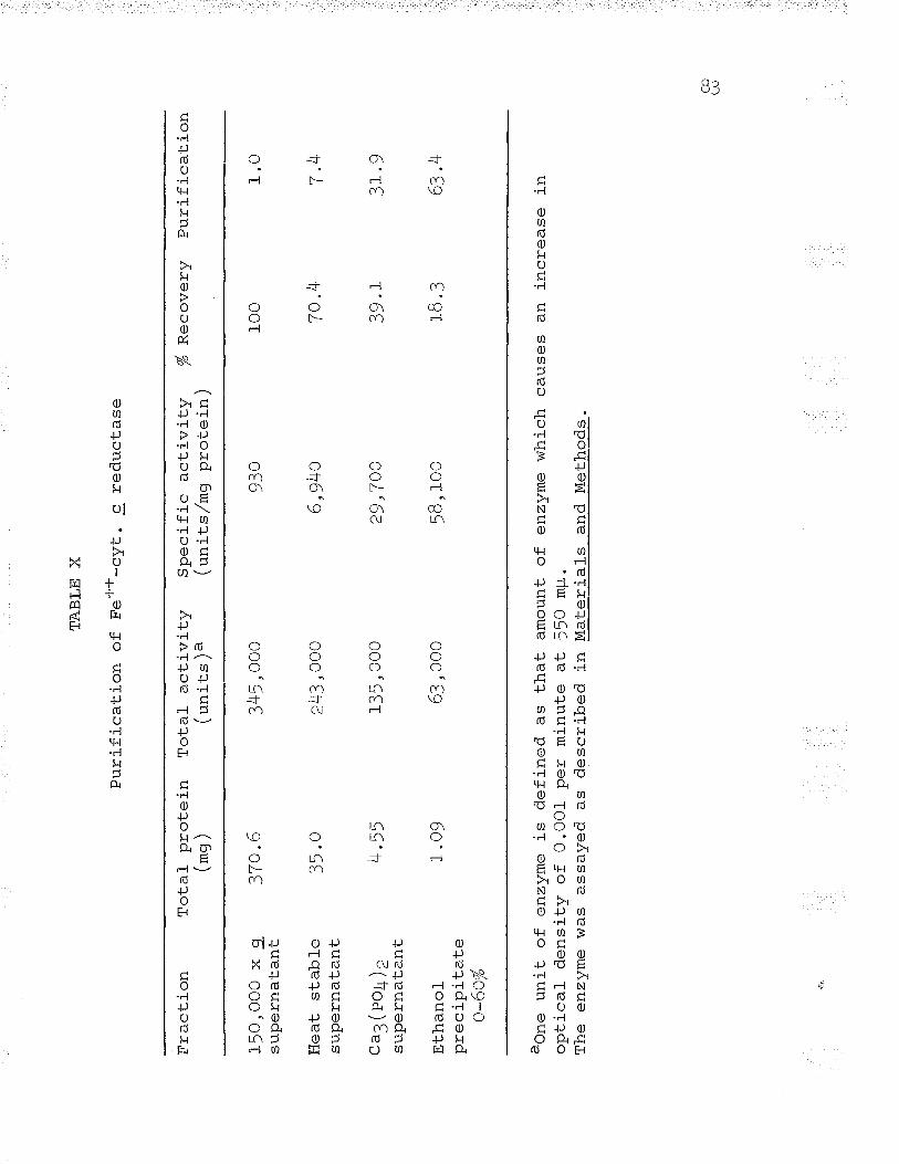
Fra
ctio
n
I5O
,OO
O x
gsu
pern
atan
t
Hea
t st
able
supe
rnat
ant
".3(
po4)
esu
pern
atan
t
Eth
anol
TT
^õ
ì ni
#a
f-a
o-60
%
Tot
al
prot
ein
(*g
)
TA
BLE
X
Pur
ifica
Lion
of
F
e*+
-cvt
. c
redu
ctas
e
?7 î
t6
35.o
lr r¡
'.2t
I .09
Tot
al
activ
ity/
', \ã
t un
r_cs
/ *
aone
uni
t of
enz
yme
is d
efin
ed a
s th
atop
tical
de
nsity
of
O.O
O1
per
min
ute
atT
he e
nzvm
e w
as a
ssav
ed a
s de
scrib
ed in
345,
000
243,
OO
o
135,
000
63,
ooo
Spe
cific
ac
tivity
(uni
ts/m
g pr
otei
n)
930
6,94
o
29,7
OO
cB r
oo
ft R
ecov
ery
Pur
ifica
tion
amou
nt o
f en
zym
e w
hich
cau
ses
an in
crea
se i
n55
o m
p.M
ater
ials
an
d M
etho
ds.
r00 70
.4
?4 1
18 .
3
1.0
7L ?1 0
/'¡
lrrl<
4
Q/¡
appreciable
Purity of Fe
loss in activity for periods up to 6 months
++' '-cyt. c reductase
A11 preparations of purified Fe++-cyt. c reductase
contained nucleic acid similar to the enzyme purified by
Blaylock and Nason (fg6]). The presence of extraneous
proteins in the purified enzyme was determined by disc
electrophoresis. Samples (lO-f5O pLg protein) were run in
Tris-glycine buffer (pH 8.3), then stained with Amido black.
The resultant gels revealed one dark, broad band and two
f aint, narrow bands . These resul-ts indicated that the
enzyme preparation was approximately )O-LOOFI pure. The
results of the ge1 electrophoresis were supported both by
DEAE-cellulose chromatography (fast-flowing column), where
only one band was el-uted (eig . 6) , and by molecular weight
studies, where all the protein was found in only one band
(rigs . LT and tB). No attempt was made to stain specifically
the active enzyme band in the ge1 since no artificial
electron acceptor could be found f.or the enzyme.
Effect of pH on enzyme activitv
The effect of pH on enzyme activity was tested using
Õ'l
various buffers; only one buffer proved to be satisf¿arnrrz
Blaylock and Nason (tg63) routinely assayed Fe++-cyt. c
reductase in B mn acetate buffer (pH 5.7) and showed that
the enzyme activity has no pH optimum in B or BO myl acetate
buffers. These authors reported that a plateau was evident
at pH 5.7-6.0 at a concentration of B m¡,i. In this
investigation similar results were obtained using acetate
buffer at the same concentrations (B and BO mM). The results
obtained with acetate buffer are illustrated in Fiqs. B and
9. The higher concentration acetate buffer (BO m¿)
increases the non-enzymatic reaction and decreases the
enzymatic reaction (ion effect).
When cytochrome 9, an extremely basic protein, was
added to the B mu acetate buffered reaction mixture, the pH
immediately rose to pH 7.5-7.8, depending on the initial plf
of the acetate buffer (pU 5,25 6.5). In the presence of
such a high concentration of cyt. c the B m¡ti acetate buf fer
proved to be too weak to maintain a constant pH. The pH
values reported in Fig. B represent the initial pH of the
acetate buffer rather than the actual- r¡H of the reaction
mixture. Althouqh the BO m¿ acetate buffer was shown to
have better buffering action, its use was l-imited because of
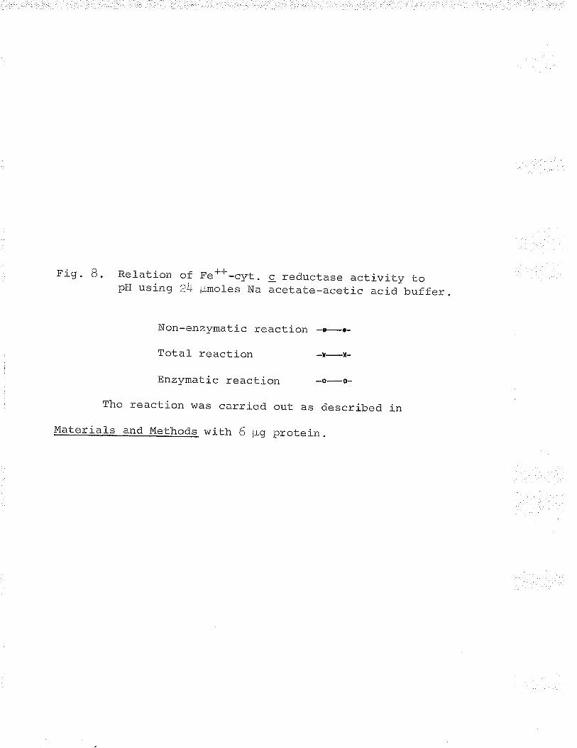
Fig. B. Relation of Fe*-F-cyt. c reductase activity topH using 24 ¡-Lmoles Na acetate-acetic acid buf fer.
Non-enzymatic reaction -r-.-
Total reaction -I-X-
EnzYmatic reaction -o-o-
The reaction was carried out as described in
Materials and Methods with 6 $g protein.
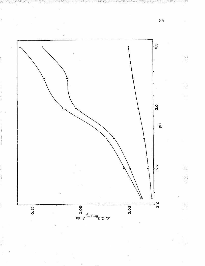
CO o'\
6.5
I I
o 85
l I I
6.O
='E o.
oe-
o t4) Ð o o
p!'l
5.5
o.03
- 5.U
Fi o q Re] ation of Fe**-elrt r. redrrcf a-^ -^+'i -'; +r' 'r-- *¡3. ) . ..*r*tl-On Ot !'e -L, - esÐc cluLrvrLy uO pHusing 240 pmoles Na acetate-acetic acid buffer.
Non-enzymatic reaction -o-o-
Total reaction -f-I-
Enzlnnatic reaction -e-v-
The reaction was carried out as described in
Uaterials and Methods with 6 pg protein.
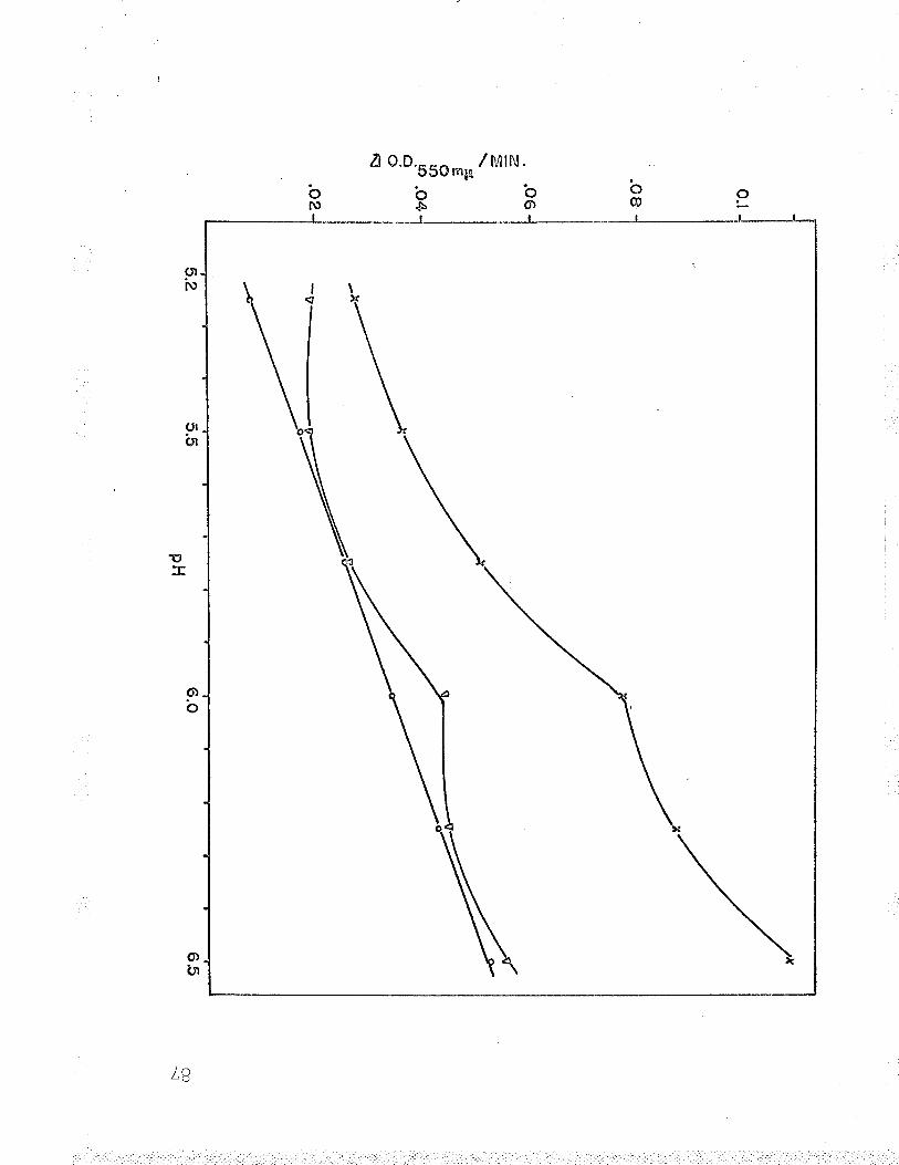
bfu
¿ o.D.-_^ /r/trN.cÞu m¡¡
b'o"ts O
LB
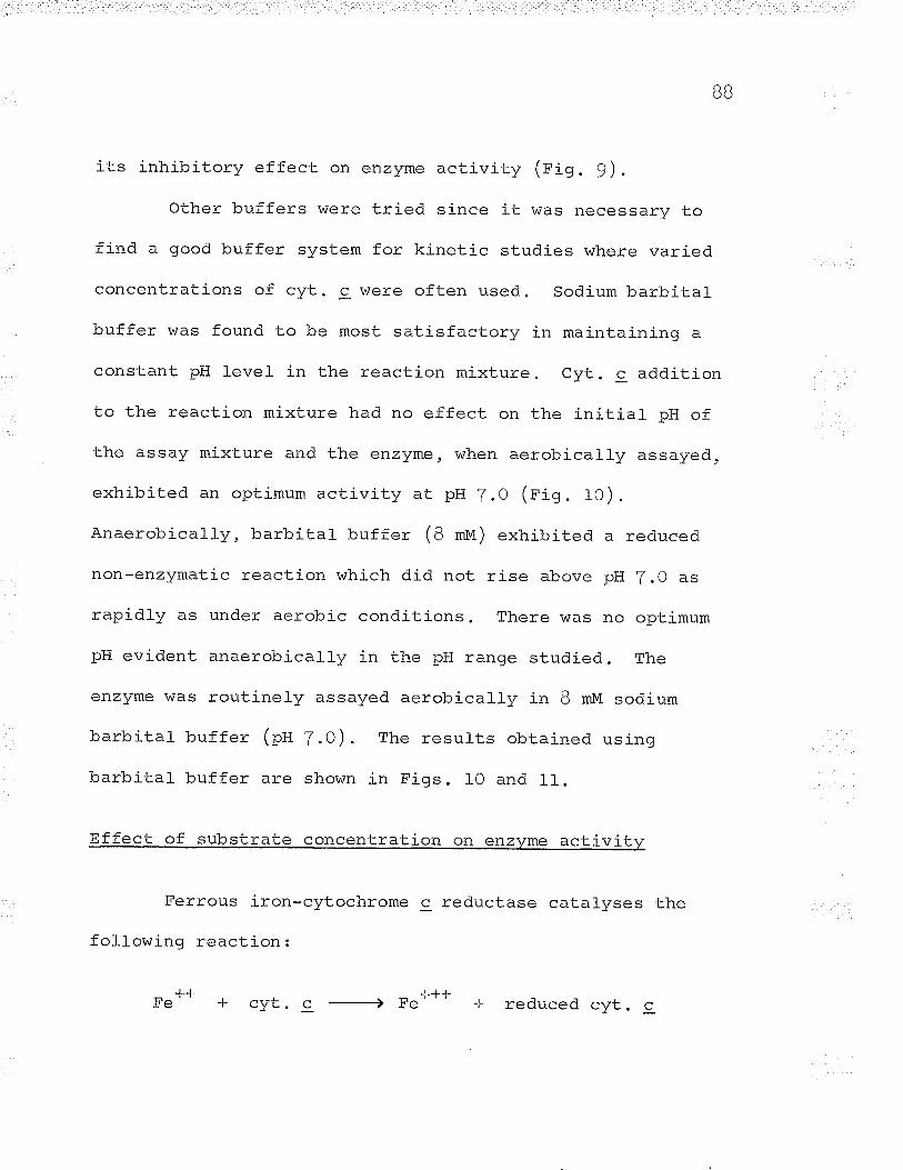
BB
its inhibitory effect on enzyme activity (eig . 9).
Other buffers were tried since it was necessarv to
find a good buffer system for kinetic studies where varied
concentrations of cyt. c were often used. sodium barbital
buffer was found to be most satisfactory in maintaining a
constant pH level in the reaction mixture. Cvt. c addition
to the reaction mixture had no effect on the initial pH of
the assay mixture and the enzyme, when aerobically assayed,
exhibited an optimum activity at pH /.0 (rig. 1O).
Anaerobically, barbital buffer (B *u) exhibited a reduced
non-enzymatic reaction which did not rise above pH 2.0 as
rapidly as under aerobÍc conditions. There was no optimum
pH evident anaerobically in the pH range studied. The
enzyme was routinely assayed aerobically in B mM sodium
barbital buffer (pH 7.O). The results obtained using
barbital- buf fer are shown in Figs. l0 and 11.
Effect of substrate concentration on enzvme activitv
Ferrous iron-cytochrome c reductase catalyses the
following reaction:
-L.LIcyt. c Fe"'J--t-
reduced cyt. c
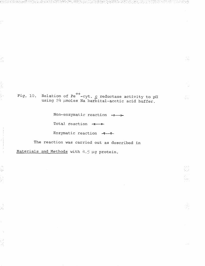
Fig. 10. Relation of Fe**-cyt. c reductase activity to pHusíng 24 ¡-Lmoles Na barbital-acetic acid buffer.
Non-enzymatic reaction -o-o-
Total reaction -x-x-
Enzymatic reaction -V-V-
The reaction was carried out as described in
Materials and Methods with 4.5 þg protein.
¡ôn
/¿
JV .
U.S
SO
nn* /
t*'ti
itt.
o
CO \o
9P ¡\
) p (¡
E
_,1 b ì{ N
x
\ I I f
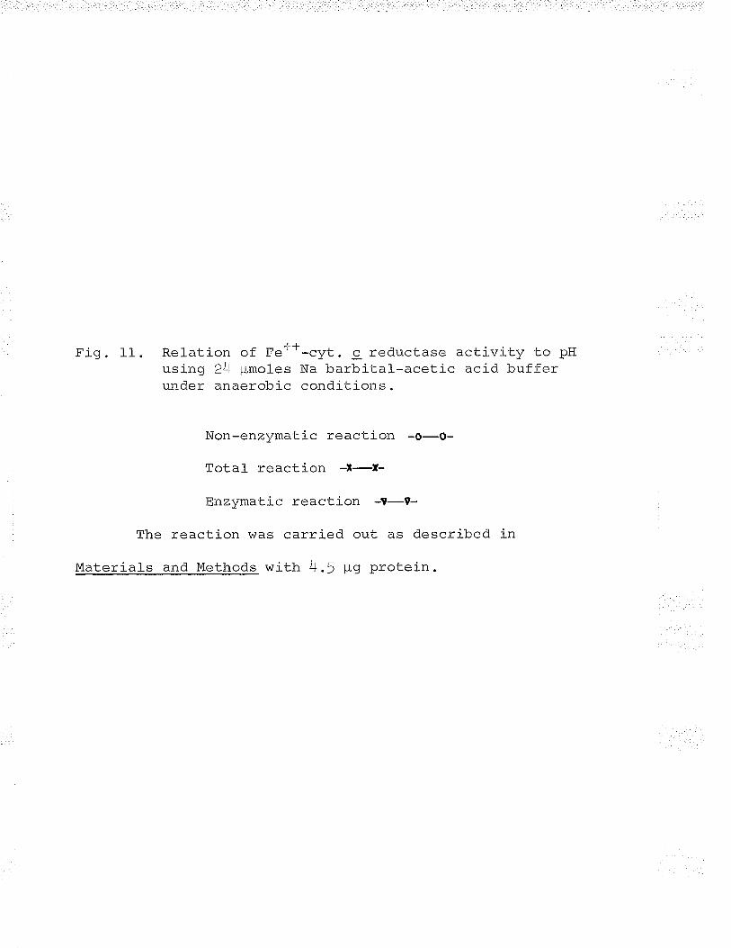
Fig. 11. Relation of Fer-+-cyt. c reductase activity to pHusing 24 pimoles Na barbital--acetic acid bufferunder anaerobic conditions.
Non-enzvmatic reaction -o-o-
Tota1 reaction -t-r-
EnzYmatic reaction -T-9-
The reaction was carried out as described in
Materials and Methods with 4.5 ¡-ig protein.
o.B
o.6
o.4
o.2
z. =E o tf,)
a o ña
o
* v
6.2
,/r
.5
-<
PH
7.O
7.2
\o o
QI
The effect of both substrates, ferrous iron and cytochromê 9,
on enzyme activity was measured by assaying the initial
reaction velocity while varying the concentration of one
substrate. At the same time, the concentration of the
second substrate was maintained at a constant level. Under
these conditions the concentration of the first substrate
(S) is rel-ated to the initial velocity (") according to the
equation:
VI.trw-r
1+
This is easily transposed to the double reciprocal form of
Lineweaver and Burk (fg]4)
1-K¿ I +1t^\
Vr \Þ/ vff
where K¿ is the apparent Michaelis constant of the varied
substrate (s) (either ferrous iron or cytochrome c) and Vf
is the apparent maximum velocity. When data is plotted in
the double reciprocal form, K' values may be calculated from
intercept on the horizontal axís, where I - 0 and I t
Tal^ Þ̂
GT
/¡ \ lultù, .NJ
92
| - --\(see t'r-gs. 15 ano r) ) .
The results of the effect of ferrous iron on enzyme
activity are shown in Figs. 12 and 13. At low ferrous iron
concentrations enzyme activity increased in a normal manner,
but at higher concentrations the curve deviated from the
normal hyperbolic function and exhibited substrate
inhibition. The enzyme was saturated by 4 mm ferrous iron.
Similar resul-ts were obtained with cyt. 9, which indicated
that the enzyme was saturated at a 0.15 mM concentration
(see Figs. 14 and L5).
The double reciprocal replots of Figs. 12 and 14
revealed apparent Michaelis constants of 1.0 mM and 0.095 mM
for ferrous iron and cyt. c respectively. Blaylock and
Nason (t963) reported apparent K*'= of 0.f5 mM and 0.OB mM
for ferrous íron and cvt. c. Their results of substrate
saturation are at variance with the resul-ts of this
investigation. The concentrations of substrates that
saturated the enzyme (rigs. 12 and 14) !üere used in alt
routine assays (4 t¡n ferrous iron and 0.f5 mM cytochrome c,
see Materialq and Methods ) .
Other substances were tried in attempts to substitute
f.or ferrous iron but no other el-ectron donor could replace
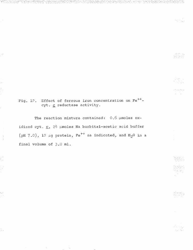
Fig. l-2. Effect of ferrous iron concentration on Fe'--cyt. c reductase activity.
The reaction mixture
idized cyt. 9, 24 pmoles Na
| -- - ^\(pH 7.0), L2 ¡rg protein, Fe
final volume of 3.0 ml.
contained: O .6
barbital--aceticL-L as indicated,
pmoles ox-
acid buffer
and HrO in a
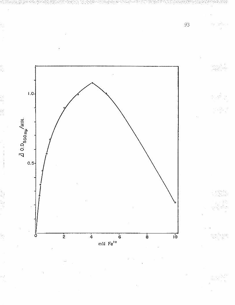
93
t.
z=
a,Eolf)roâ
o\
o.
oä46örbrnfl,l FerI
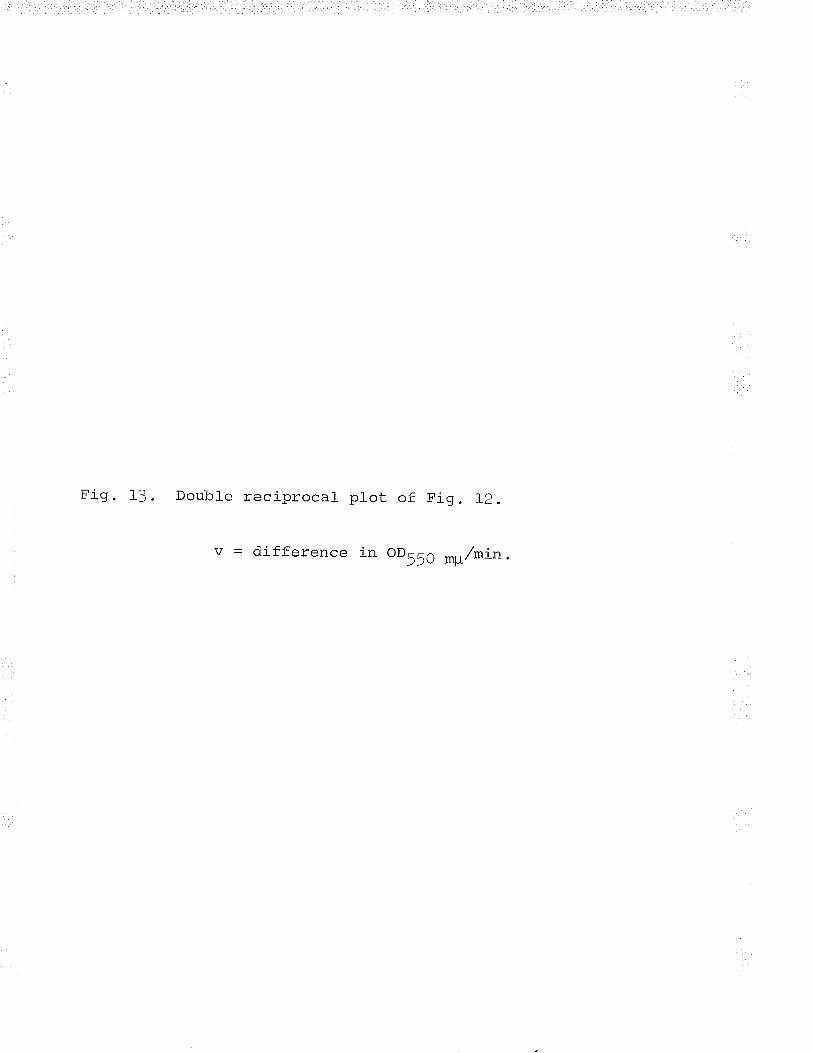
Fig. 13. Double reciprocal plot of Fig . Ip.
v = difference in oD55O mg/min.
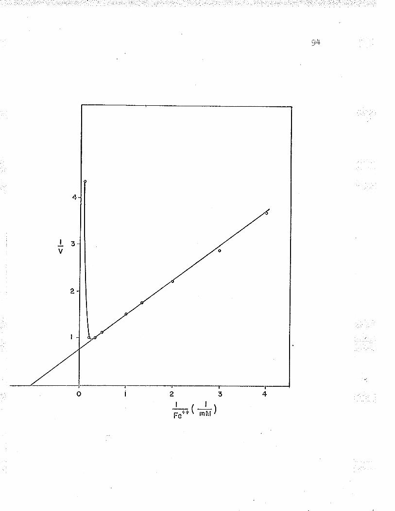
94
l3V
| ( l-rFe'Þù
\ rnft"l /
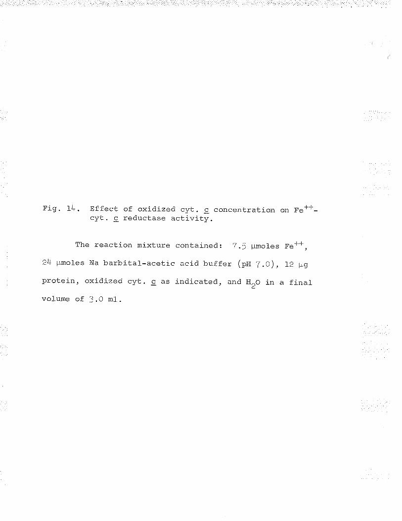
Fig. 14. Effect of oxidized cyt. c concentration on Fe*-F-cyt. c reductase activity.
The reaction mixture contained: 7.5 ¡rmoles F.**,
24 ¡lmoles Na barbital-acetic acid buffer (pII Z.O), IZ þg
protein, oxidized cyt. c as indicated, and HrO in a final
volume of 3.0 ml.
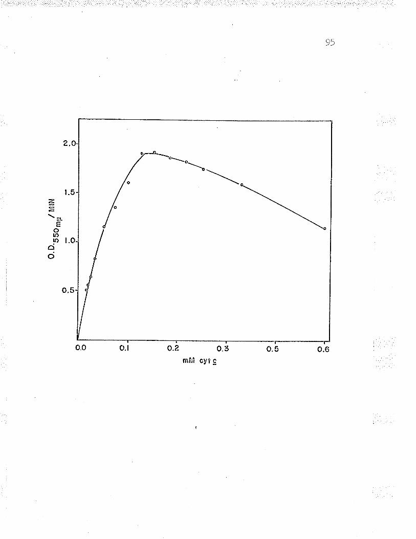
95
t.5
2.O
o.5
æ,E\*
E()ro!o l.Oâ
o
0.3o.?-o.lo.o
mM cyt g
\
o.6o.5
Fig. L5 . Doubl-e reciprocal plot
= difference in
-t,or ¡'ag. 1+.
OD--^ ^,,/min.) )v ttLFL
vn
20 40
ahn('*')
97
ferrous iron. The following substances were tried: NADH,
NADPH, co**, Mn++, cr*, and Hg+. Artificial acceptors -
dichlorophenolindophenol, phenazine methosulfate, methylene
blue, teLrazolium chl-oride, and benzyl viologen - woul-d
not substitute for cytochrome c; consequently it was
impossible to use an easily permeable electron acceptor for
identification of the active enzyme in gel electrophoresis.
Effect of inhibitors
Ferrous iron cyt. c reducta.se was weakly inhibited
by sulfhydryl inhibitors. This inhibition appeared to be
ionic rather than specific binding to sulfhydryl groups, âs
demonstrated by results with mercuric chloride (rafte XI).
I,rIhen mercuric chloride was added to the assay mixture the
reaction was substantiatly inhibited, but upon dialysis in
O.Ol M acetate-acetic acid buffer (pU 5.7) the enzyme
activity returned to normal levels. If the mercury was
bound to the sulfhydryl group(s) of the enzyme, the inhibition
should have remained after dialysis. The ability of the
enzyme to overcome the inhibition of mercury salts after
dialysis indicates that sulfhydryl groups are not required
for catalysis.
9B
Blaylock and Nason (WAZ) reported that Fe++-cyt.
reductase was inhibited by atabrin, thus indicating the
possible invol-vement of f lavin(s ) in catalysis. However
these authors were unabre to demonstrate the presence of
flavin(s) in the enzyme. Although atabrin does inhibit
enzyme, this inhibition also appears to be ionic rather
specific binding of the enzyme at the flavin site(s).
inabil-ity of ultra violet light, a potent inhibitor of
the
than
The
flavin enzymes, to inhibit Fe++-cyt. c reductase indicates
that flavins are not involved in the reaction. When Fe**-
cyt. c reductase was excited at 350-375 m¡r the enzyme did
not fluoresce. This fluorescence, ât 350-375 m¡r excitation,
is characteristic of some f lavin enzymes (tuiassey , 1963) .
These results lead to the conclusion that Fe++-cyt. c is not
a flavin enzyme. As a further proof, the absorption
spectrum (rig. L6) of Fe++-cyt. c reductase failed to
exhibit any of the spectral properties of fl_avin enzymes.
The results of inhibition by flavin and sulfhydryl inhibitors
are shown in Table XI.
The effect of proteolytic enzymes and ribonuclease
(nivase ) o. Fe++-cyt. c reductase was tested by incubating
1OO pg of the various enzymes with Fe-F*-...2t .r rarlrr..|-¿ss for
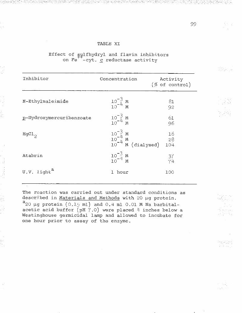
99
TABLE XI
Effect of çplfhydryl and flavin inhibitorsTTon Fe -cyt. c reductase activitv
Tnhibitor Concentration 1^!.: --.: !--.¿-1'(-j LrV r Ly
/.J -\\7o ox control,/
N-Ethylmaleimide
p-Hydroxlzme rcur iben zo ate
HgCL,
Atabrin
u . v. Il_ginc
BT
92
6t96
I62B
f -, \ - -(dialysed) 104
100
_?r_0íMlo-- M
-?10;Ml-o-- M
-?10 i,1O-;lo--
1O-;10--
t hour
MMM
3Tz)tlT
M
M
The reaction was carried out under standard conditions asdescribed in Materials and Methods with 20 ¡cr nroto'in-PYoc(\ t a nrnJ-ai / a ' \ l¡ aLv r.!y ¡,,v--,fl. (O.15 ml) and 0.4 mt 0.O1 M Na barbital-acetic acid buffer (pH 7.0) v/ere placed 4 inches below alVestinghouse germicidal lamp and al-lowed to incubate forone hour prior to assay of the enzlzme.
100
one hour prior to enzyme assay. None of the proteases -
trypsin, o-chymotrypsin, carboxypeptidase, and subtilisin
affected enzyme activity. Ribonuclease, oh the other hand,
exhibited a 25-30% inhibition of enzyme activity. This
inhibition was immediate, requiring no preincubation, thus
Ieading to the concl-usion that it was not due to the
hydrolysis of the RNA-portion of the enzyme. Blaylock and
Nason (tg63) also reported that Fe++-cyt. c reductase showed
remarkable stability in the presence of various proteases,
DNAse, and RNAse. Table XII illustrates the effect of
proteolytic enzymes and RNAse on Fe++-cyt. c reductase
activity. Fel-*-cyt. c reductase activity was also inhibited
by ionic inhibitors (w. acetate, NaCl, etc.). This ionic
inhibition was evident when enzyme activity was compared in
B and BO mM acetate buffer (see Figs. B and Ç). The
inhibition by sodium chloride was studied in more detail and
the results are shown in Figs. 3I and l2 (see Kinetic
Analysis ) .
Effect of some potential activators on enzvme activitv
Mora, Creskoff, and Person (t06=) reported that
certain carbohydrates were abl-e to reduce cytochrome c.
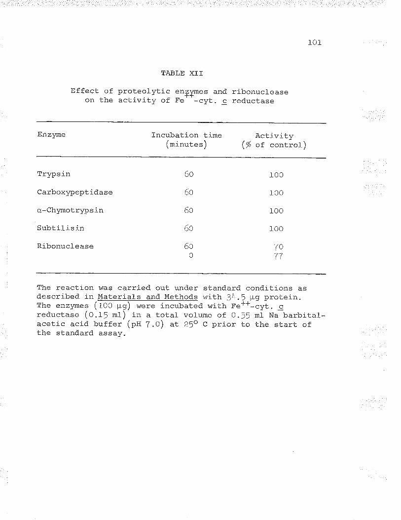
101
TABLE XTT
Effect of proteolytic enlymes andon the activity of Fe' '-cyt.
cribonucleasereductase
Enzyne Incubation timelmi nrr.|- a.'ì\ -"'--^-' --- /
A^f 117r +rtt.r/ -\\þ or. concrol/
'l'r\ñê t h
Carboxlzpeptidase
o-ChymotrYPsin
Subtil is in
Ribonuclease
6o
6o
6o
6o
r00
100
r00
r00
7o77
6oo
The reaction was carried out under standard conditions asdescribed in Materials and Methods v¡ith 34.5 ¡rg protein.The enzymes (f oo ¡rg) v/ere incubated with Fe++-cyt. creductase (0.f5 ml) in a total volume of O.!! ml Na barbital-acetic acid buffer (pII 7.0) at 25o C prior to the start ofthe standard assay.
ro2
This reduction of cytochrome c was a time-dependent reaction
in contrast to the instantaneous oxidation or reduction
caused by oxidizing or reducing agents (ascorbate and ferri-
cyanide). More recently, Person, Fine, Mora, and Z:-pper
(ry6f) reported on the reversible polyion reduction and
oxidation of cvtochrome c. These workers found that
polycations reduced cytochrome g, while polyanions oxidized
reduced cytochrome c. Since these results \^/ere unexpected
and not completely explained, the effect of some of the
substances causing reduction of cytochrome c was tested with
Fe++-cyt. c reductase in a hope that these compounds may
convert cyt. c to a more readily reducible conformation.
None of the sugars used - ribose, glucose, or
arabinose - had any effect on Fe**-cyt. c reductase activity.
Only RNAse had any effect, and this effect was inhibitory
rather than stimulatory. Possibly, the reason for the
absence of stimulation of enzyme activity was the short
duration of assay (t minute), whereas reduction of cyt. c
with these compounds was observed by the above workers only
after a long duration (Z-24 hours )
The effect of cytochrome c
(Margoliash and Lustgarten, L962)
monomer and polymers
on Fe**-cyt. c reductase
103
was also assayed to determine which form the enzyme preferred
as its substrate. Although the dimer form (potymer) was not
purified, the enzyme showed no preference for either form
(monomer or polymer ) . The results of the effects of these
substances on Fe++-cyt. c reductase activity are shown in
Table XIII.
The formation of enzvme subunits
Fe**-cvt. c reductase is associated with nucleic acid
when isolated (elaylock and Nason, L963). The enzyme was
dissociated into subunits bv adsorption on a DEAE-ce1l-ul_ose
column and by slow elution with 0-2 M NaCl in 0.01 M acetate-
acetic acid buffer (pg 5.7) (see Fig. T). A similar resul-t
was obtained when the enzyme \^ras treated with DEAE-cel1u1ose
in a batch process . Fet*-cyt. c reductase (f -Z mg ) \^zas
incubated with DEAE-cellulose (5 *f bed volume in a total-
volume of 10 ml 0.5 M NaCl, O.O1 M acetate-acetic acid
buffer (pH 5.7)) in the cold (O-5" c) and stirred stowly for
12 hours.
The enzyme-DEAE-cellulose mixture was then poured
into a I x I0 cm column and was slowlv eluted with 10 ml-
O.5 M NaCl, 0.01 M acetate-acetic acid buffer (ptt 5.7).
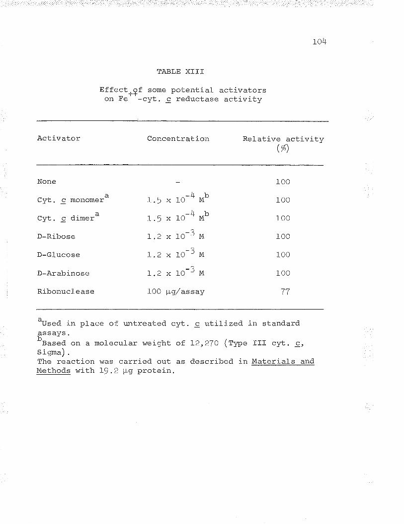
't a\/r
Effecton Fe
TABLE X]TT
, af some potential1--l-
^a,1- -^,¡.. ^+ ^ ^ ^-uJu. U rEuL¿(-Lc¿ÞEactivators
¡r'J-i r¡i'l-r¡
Activator Concentration Relative activi tw( oÁ\\/"1
None
Cyt. c monomer
(1t¡4- ¡ À'i mar-"J ".
D-Ribose
D-Glucose
D-Arabinose
Ribonuclease
Jr r^I.' X IU IVI
JrxI.) x ru lvr
-?1.2 x l0 'M_?1.2 x 10 'M-?1.2 x l0 'M
Lvv FtY/ o.ÞÞay
r00
r00
r00
100
100
r00
T7
a__-Used in place of untreated cyt
b.-Based on a molecular weiqht ofS igrna) .
c utilLzed
L2,27o (ryp.
described in
in standard
IIf cyt. c,
Materials andThe reaction was carried out asMethods with L9.2 ¡-Lg protein.
105
When the liquid level reached the top of the DEAE-cellulose
the elution was stopped. This first elution at 0.5 M NaCl
contained the protein portion of the enzyme and corresponded
to the first peak eluted from the slow-flowíng DEAE-
cel-l-ul-ose col-umn during chromatography (eig . T).
The column was next eluted with 1l mt 1.0 M NaCl,
O.Of M acetate-acetic acid buffer (pH 5.7). This second
fraction, consisting of the RNA portion of the enzyme,
corresponded to the second peak eluted from a slow-flowing
DEAE col-umn (see Fig. 7). These results were quite similar
to those of Symons (tg6S), who used DEAE-cellulose
chromatography to separate the DNA component from yeast
cytochrome br.
The two fractions were then concentrated by dialysis
in a saturated sucrose solution (O.Of M acetate-acetic acid
buffer (pti 5 .T))¡ the sugar and NaCl were dialysed out
against 2 changes of 4 I 0.Ol M acetate-acetic acid buffer
(pH 5.7).
Absorption spectra of the enzyme and its subunits
i^/ere recorded in a Unicam SP70O spectrophotometer which
revealed differences in the compositions of the subunits
(eig. 16) . Both methods of forming enzyme subunits were
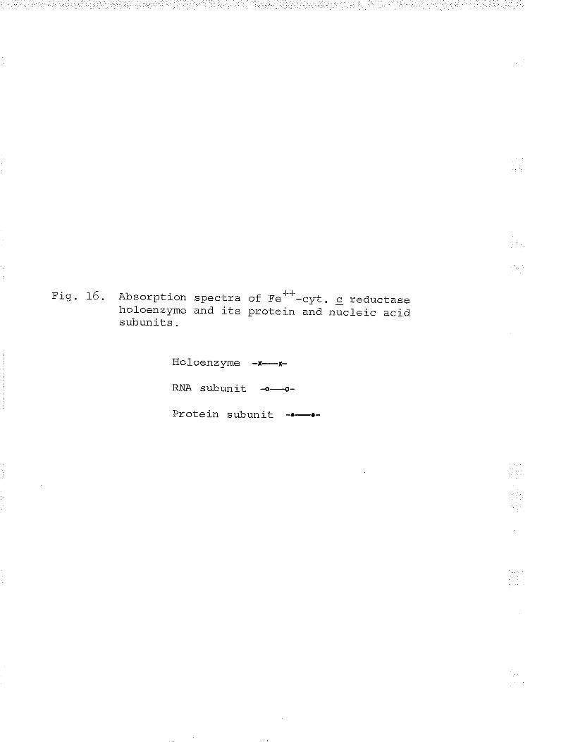
Fig. t6. Absorption spectra of Fe**-cyt. c reductaseholoenzyme and its protein ,.rA ,rl"t"i. acidsubunits.
Holoenzyme -t-x-
RNA subunit -o-----€-
Protein subunit -.-.-
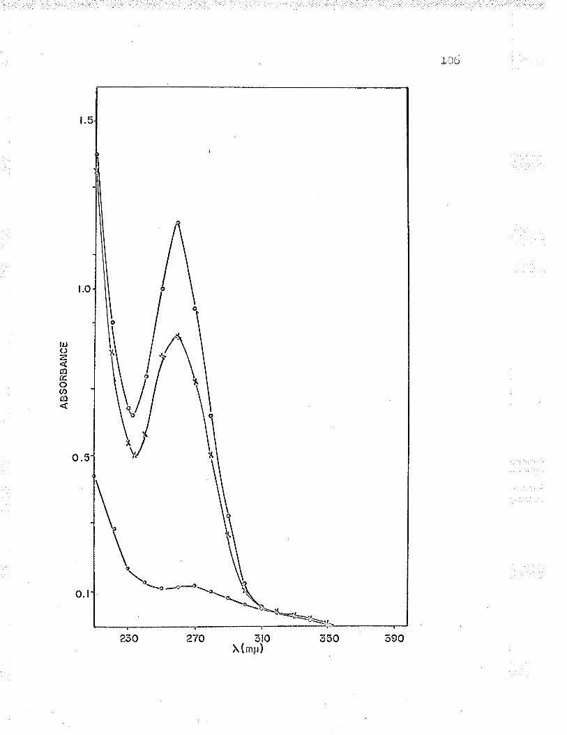
n : n!a,...*L 1..â!.J
¡.o
t.
o.5
-ëo:-o\ Yr
-\.o ,\
\*\ =\\
t¿¡(J-¿,
@Eo(n
cÐ
o.l
230 270 3tO 350 390X ( rnp)
r07
equally effective; however, the batch method was much
simpler and the results were always consistent.
The first fraction el-uted from the DEAE-cellulose
contained all of the protein adsorbed (see Tabl-e XIV), but
rl ., Fe**-cvtochrome c reductase activity was found in this
subunit. Since the second subunit contained RNA ( see Table
xV), attempts \^7ere made to anneal the protein and RNA
subunits by the method of Hall and Spiegelman (fg6f).
Mixtures of the two subunits were boited and slowly cooled,
or heated to 7Oo C and slowly cooled, in attempts to
reconstitute the oxLginal holoenzyme.
Restoration of the original catalytic activity of the
holoenzyme, by mixing equal quantities of the subunits or by
annealing, was not successful. Incubation of the two
subunits in dioxane 5-25%, ethanol LO-6O%, and p-mercapto-)r
ethanol 1O-- M also failed to reactivate the original
catalytic activity. The subunits were mixed in 0.5 M NaCl,
and 1.0 M NaCl. O.O1 M acetate-acetic acid buffer (pH 5-T)
and allowed to incubate for 6 hours prior to dialysis
-^-.in-+ . ^h=-aoo nf rt .ì'l M aCetate-acetic acid bufferd.9c¿lrrÞL ¿ \-rlorl9sÞ v! v.v! ¡
(pn 5.7). These attempts also faited to reactivate the
enzyme. Molecular weight determinations indicated that
108
there was no change in the originar subunits and that no
new species was formed.
Yeast RNA and ribose did not restore the oriqinal
catalytic activity to the protein subunit. Later studies
showed that the RNA subunit formed a higher mol_ecular weight
species than the original holoenzyme. All of the methods
attempted failed to split the RNA subunit into the small_er
molecurar weight species necessary, perhaps, for restoration
of the original enzyme structure and catalysis.
JJComposítion of Fe ' '-cyt. g reductase and its subunits
Fe++-cyt. c reductase must be preincubated with
cyt. c prior to assay or the reaction rate is not l-inear
(etaylock and Nason , L963). During the preincubation period
the optical density at !10 m¡-L was recorded and it was
noticed that there was an ever decreasing oscillation in the
optical density, which was finally damped after 4-5 minutes.
The optical density showed an increase above the oriqinal
optical density of the cytochrome c solution. An absorption
spectrum was recorded before and after the addition of
enzyme; it was observed that after preincubation there was
some reductíon of the cytochrome c.
r09
The amount of cyt. c reduction varied with the enzyme
preparation. This reduction of cyt. c by the enzyme l-ed to
the belief that some subsLrate might be bound to the enzyme
or that the enzyme existed in two stable forms, âh oxidized
and a reduced form. Assays were performed to determine the
amount of iron bound to the enzyme. Since the presence of
iron on an enzyme is usually accompanied by sulfide (wtitter
and Massey, L965; Brumby, Miller, and Massey, 1965), the
sulf ide content of the enzyme lras al-so assayed.
Iron and l-abile sulfide content
The total iron content of the holoenzvme was constant
although the ferrous:ferric ratio varied considerably.
Despite the fact that the iron bound to the holoenzyme was
non-dialysable, the protein subunit retained only IO/o of the
bound iron. This indicated that onl-y I mole of the iron was
extremely tightly bound to the enzyme. Assuming a molecular
weight of 301000 for the protein portion of the enzyme, the
iron:protein ratios of the hol-oenzyme and the protein subunit
were 10:1 and 1:1 respectively.
The presence of tightly-bound iron is characteristic
of non-heme iron proteins (Mason, 1965), the majority of
r10
which contain sulfide closery rinked to the iron on the
protein structure. sulfide analysis failed to detect any
sulfide in either the hol-oenzyme or protein subunit. rf the
ratio of protein:iron:sulfide was 1:1:1. then the amounts of
sulfide that should have been present were well within the
limits of the assay and should have been detected (Xing,
1964). The results of the iron and sul-fide assays are shown
l_n '1'aþIe xl.v .
An attempt was made to determine if the iron bound to
the enzyme was replaced slowly or if the binding was
instantaneous. To remove the bound iron. the enzvme was
incubated with saturated 2 12' dipyridyl and the mixture was
dialysed against 2 changes of 4 I 0.o1 M acetate-acetic acid
hrrffar /^T{ Ã z\ T¡lhan +lanvu!rç! \y!L ). t J . yvrrerr. Lrlc iron-free enzyme was assayed
there was no detectable lag in cyt. c reduction compared
with the normal holoenzyme. This resul-t indicated that the
tightly bound iron was rapidly replaced and the iron-
protein interaction was not time-dependent, which is true
for some enzyme-cofactor systems.
Protein and nucleic acid content
Since the presence of nucl-eotide was obligatory for
Fra
ctio
n
TA
BLE
XT
V
Iron
an
d. la
bile
su
lfide
co
nten
t of
F
e**
Hol
oenz
yme
l-
Hol
oenz
yme
2
Hol
oenz
yme
3'l-
,IV
.rlo
l-oen
zym
e +
l-\H
rot'e
ansu
buni
t
Pro
tein
ll^
mn'
l ^^
-l'!
ì, ¡t
tvrç
Þ
20 4B
O.O
X I
U
ia I.O
X I
U
aBas
ed on
a m
o.l-e
cula
r w
eigh
t of
1^r -T
he p
rote
in s
ubun
it w
as d
eriv
ed
300
I .0
?16
r ô6
J-
/ +
.
" t
300
I .0
Fer
rous
iron
/-\
\mor
es/
-l_0
-o ax1
0
,-q
1.4
x IO
',-
a4.
3 x
IO' a
t.u
x IU
aJ.
' x
t-u
_-o
ô,.0
x L
O '
Tot
aliro
n( m
oles
)
10 10
-cyt
. c
redu
ctas
e
^-a
5.U
x IO
- al.U
x 1
0
-T-L
-L
-L-L
Fe"
:Fe"
'
1.0
1.1
1.0
30,0
00 f
or t
he p
rote
in m
oeity
(se
e F
igs.
from
hol
oenz
yme
4 w
ith a
95.
Zrt
pro
Le:-
n
-aLO
' 17
10-
f a10
- "
Iron
:pro
tein
La
bile
sul f
ide
( m
oles
)
't I 1
?l ?2 1 1 I
f
2.I
B.B
:1
10.
6:1
10: I
10:
1
l.t1Ã
0 0 0 0
L9 a
nd 2
0).
yiel
d.
H ts F
Tl-2
enzyme activity, the protein and nucleic acid contents of
the enzyme \^/ere assayed. The constant 260:280 m¡r ratio of-.t--t^"-'i€'i^'i o^' '-cyt. c reductase meant that different batchesyq! r!rçu ¡ 9
of enzyme had the same protein-nucfeic acid composition.
When the nature of nucl-eic acid was studied, only RNA was
found; there was no DNA present (rante XV). Since the
enzyme was found to be rather resistant to RNAse hydrolysist^ . - ----\(Table XII), this resistance was tested in the holoenzyme
and RNA subunit. RNA hydrolysis was measured by dialysing
the hydrolysed nucleotides from the reaction mixture and
assaying these nucleotides by the orcinol method (ta-trte XV).
Alkali completely hydrolysed the enzyme-RNA a.s well
as the RNA subunit. RNAse, on the other hand, hydrolysed
the complete RNA subunit but only 50% ot the RNA of the
holoenzyme. This resistance to RNAse hydrolysis by the
holoenzyme must mean that the RNA is an integral part of the
enzyme and the complete structure is not accessible to RNAse
action. Assumíng a mol-ecular weight of 30TOOO (see Figs. 1!
and 20) for the protein portion of the holoenzyme, the
mol-ecul-ar weight of the RNA-portion of the enzyme was
spectrophotometrically calculated to be approximately 65rOOO.
The combined molecular weights of the protein subunit and
1l?
RNA-portion of the enzyme closely approximated the molecular
weight of the holoenzyme (see Figs . LT and 18). The resul-ts
of the protein and nucleotide assays are shown in Table XV.
Molecul-ar weiqht studies
Schachman (tgSg) calculated the molecular weights of
macromolecules by means of sedimentation equilibrium.
Schachman, in his ultracentrifugation study, showed that the
sedimentation constants of two kinds of macromolecules were
related to their respective molecul-ar weights according to
the following equation :
(r) . -?/3s'r / wr ì._' '
-:=l-ls2 \we t
where S = sedimentation constant
MW = molecular weight
Martin and Ames (196t) demonstrated that molecul-ar
weights of protein mixtures coul-d be determined by a
measurement of sedimentation velocity after centrifugation
in a sucrose gradient. The distance (o) a protein travelled
from the meniscus was a direct measure of its sedimentation
velocity and, s1/52 equalled DL/D2. Substituting in (r),
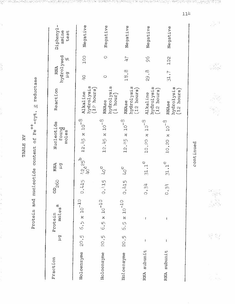
Fra
ctio
n
Hol
oenz
yme
20.5
6.5
* 1
0-10
O.I;
LD
Pro
tein
and
nuc
leot
ide
Pro
tein
pg
mol
esa
Hol
oenz
yme
Zo
.D
6.5
* IO
-10
0.41
5
Hol
oenz
yme
20.5
6.j
* 10
-10
0.4f
5 40
"
TA
BLE
XV
cont
ent
of
F.*
*-cy
t. c
redu
ctas
e
OD
^.^
¿O
U
RN
A s
ubun
it
RN
A
t-Lg
RN
A s
ubun
it 0.
34
31.1
"
1-)+
ç 2^
"'*
å*/
40-
4o'
Nuc
leot
ide
farr
nrl
.o.
mol
_es
ôL2
.+5
x 10
Rea
ctio
n
^ ¡1
, 'c
-B
u. J
4 31
.1
10 .2
0 x
t0
ôL2
.+5
x 10
Alk
alin
ehy
drol
ysis
l- ^
- 1
lrz h
ours
]
RN
Ase
hydr
olys
is/-
,
\\ t_
nou
r/
RN
Ase
hydr
olys
ist-
^
. \
\Jl
nour
s/
Al-k
alin
ehy
drol
ysis
I l,
h^rr
-o\
RN
Ase
hydr
olys
ist1
^ .
\\a
z ]'ì
.our
s/
_,q
L2.4
5 x
10
RN
A
Dip
heny
l_hy
drol
ysed
am
ine
p9
%
rest
40
lOO
-R10
.20
x tO
cont
inue
d
Neg
ativ
e
18. B
47
N
egat
ive
Neg
ativ
e
29 .B
96
N
egar
íve
3L.7
IO
2 N
eqat
ive
H H l-
a n B
ased
on
a m
olec
ular
wei
ght
"Bas
ed o
n a
mol
ecul
ar w
eigh
tin
g f!2
m
gles
P p
er m
ole
of^l
'¡ /
s\<
¿tt
I aê
ê |
-^-^
.i ^^
lr{
.Jrc
l-nor
ass
ayV
- -
¡^/*
\B
ased
on
an E
(P)
of
1Or0
00
of 3
0r00
0of
65,
ooo
prot
ein
and
TA
BLE
XV
(co
ntin
ued)
for
the
prot
ein
subu
nit (
see
Fig
s. 1
!as
cal
-cul
ated
fro
m s
pect
roph
otom
etric
usin
g an
ave
rage
nuc
leot
ide
mol
ecul
ar
and
20).
data
sho
w-
wei
ght
of
ts ts LFI
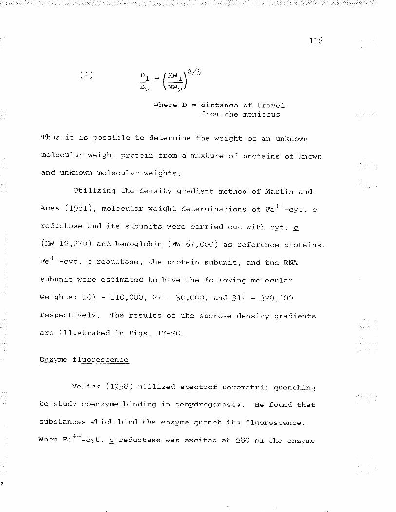
r_ J_o
(z),> /ADl : /mnl.,\"'.-¡--l
D2 \MWZ /
where D = distance of travelfrom the meniscus
Thus it is possibte to determine the weiqht of an unknown
molecular weight protein from a mixture of proteins of known
and unknown molecular lveights.
Utilizing the density gradient method of Martin and
Ames (f96f ), mol-ecular weight determinations of Fel-*-cvt. c
reductase and its subunits were carried out with cvt. c
(mvu L2,2To ) and hemogrobin (m,il 67 ,ooo) u= reference proteins .
-L-LFe"-cyt. c reductase, the protein subunit, and the RNA
subunit were estimated to have the followinq molecul-ar
weights: 103 I10,000, 27 3O,0OO, and 114 3Z9,OOO
respectively. The resurts of the sucrose density gradients
are illustrated in Figs . LT-7O.
Enzvme fl-uorescence
Velick (fg¡g) utilized spectrofluorometric quenching
to study coenzyme binding in dehydrogenases. He found that
substances which bind the enzyme quench its fluorescence.
When Fe++-cyt. c reductase was excited at 28O mr r J-lre êr1 z\zmê
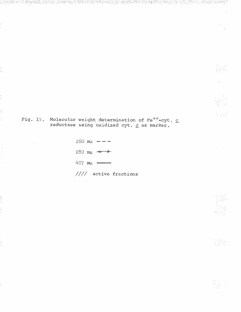
Fig. LT . Molecular weight determina-tion of Fe*+-cyt .
reductase using oxidized cyt. c as marker.
260 m¡r
,Ro -, r ....H-
40f mp
//// active fractions
tr*
.-l
FI
oi o.
¿o
o TO
P
15
20F
RA
CT
IO6\
¡ hJ
L,fv
gBE
R
Fig. fB. Molecular weight determination of Fe4*-cyt. cusing hemoglobin as marker.
260 m¡-L
280 m¡-L --É
+u( mþ
//// active fractions
-l .-t
FR
AC
T¡O
S3
h¡L'
MgE
R
Fig. 19. Mor-ecurar weight determination of Fe+*-cyt. creductase subunits using oxidized cyt. g- asmarker.
2n{l mrl
280 m¡-L -o+
407 mp
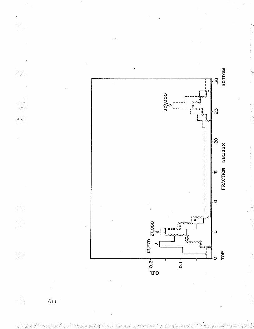
o'\
rl r-l
i o.
2o
t2.^
27v. 'ü z
lyoo
c
o TO
P
t5
?o
FR
AC
TIO
n\¡
Nit,
Mts
ER
30B
OT
TO
M
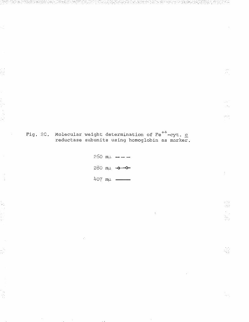
F"i o 20 Mol ar.rr I ar tn¡c i ohJ- determination of Fe---cvt - c¡ç _çJç. _reductase subunits using hemoglobin as marker.
260 mp
280 mp ..o+
+nl mp
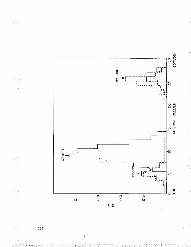
oO p----qig -ÞI-- - -'¡*o\r -L----- 1.-*1r) ------ì
oc)a--*Fs(0
IIII
t--
IL -- --l
Il- -r
ILr
o¿ï
ctbJfrl:-:ä"¿
a,Oþc)4trcu-
Í\1+nJ
¡It-
L2L
exhibited a f luorescence peak at 350 mpL. Enzyme f l-uorescence
was quenched by both substrates and end-products, Fe++,
+++Fe' ' , cyt. 9, and reduced cyt. c.
The quenching of enzyme fluorescence was specific and
was not evident when tryptophan was titrated with equal
quantities of the substrates or end-products used to quench
the enzyme. The decrease in fl-uorescence of tryptophan by
substrates or end-products was equal to the dilution effect
evident when enzyme or tryptophan was titrated with equal
volumes of buffer (same vol-ume as substrates or end-products).
The quenching of fl-uorescence by both substrates and both
end-products suggested a mechanism in which the enzyme is
bound in a random order, ot the existence of two enzyme
forms. Kinetic studies were reguired to differentiate which
of these mechanisms was Trresent.
Kinetic studies and reaction mechanism
The presence of non-heme iron in Fe*+-cyt. c reductase
suggested its possible involvement in the reduction of
cytochrome c by ferrous iron. Detailed kinetic studies were
carried out in order to elucidate the exact role of non-heme
iron and the reaction mechanism.
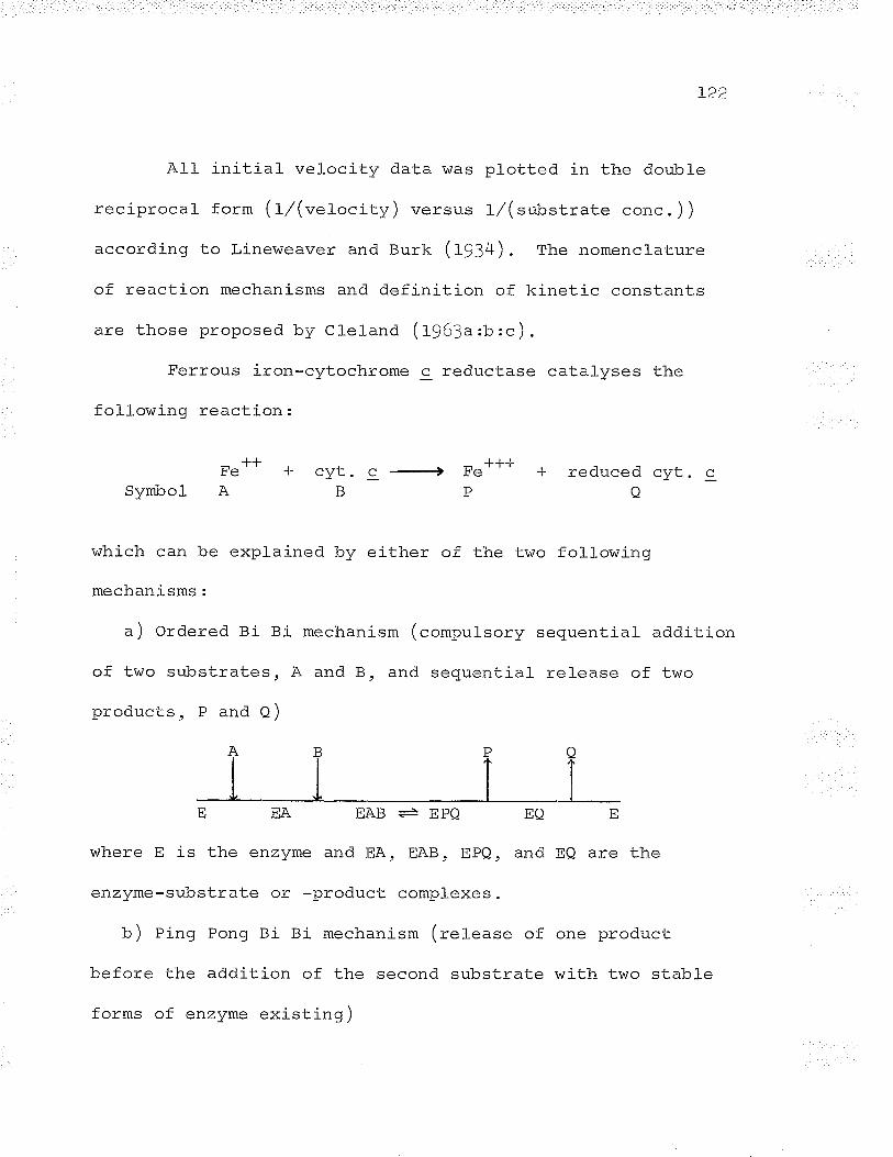
L22
All initial velocity data was plotted in the doubl-e
reciprocal form (1,/(vefocity) versus 1/(substrate conc. ))
according to Lineweaver and Burk (fg:+). The nomenclature
of reaction mechanisms and definition of kinetic constants
are those proposed by Cleland (t963a:b:c).
Ferrous iron-cytochrome c reductase catalyses the
fol-lowinq reaction:
Symbol
-J--l-
n̂
¡rrl- õ-J-'B
-Ll--L+ Fe'P
reduced cyt.o
c
which can be explained by either of the two following
mechanisms:
a) ordered Bi Bi mechanism (compulsory sequential addition
of two substrates, A and B, and sequential release of two
products, P and Q)
EA EQ
and EQ are thewhere E is the enzyme and EA, EAB, EPQ,
enzyme-substrate or -product complexes.
b) Ping Pong Bi Bi mechanism (release of one product
before the addition of the second substrate with two stable
forms of enzyme existirrg)
EAB + EPQ
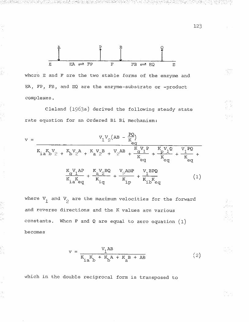
EEA
whereEandFare
EA, FP, FB, and EQ
compl-exes.
+FP
the two stable forms of the enzvme and
are the enzyme-substrate or -product
Cl-eland (t963a) derived the following steady state
rate equation for an Ordered Bi Bi mechanism:
v, v^c* _ #)r'¿- êñ
FB .- EQ
K. -K-V^ , K-V^A , K-V^B V^AB ^Otrnraþ'¿ + þ¿ + a'¿ + '¿ *-Ë-eq
K V.Q V-PQ. pr t-----r.KKeq eq
K V.APgr
--|
K.Kra eq^.t2"Q
K.i-q
V2ABP+._
K,r_p
VI BPQ+*K.. K
r_.o eqfrl
where V, and \/ =ro 1-lra -=ximum velocities for the forward- -1 '2
and reverse directi ons and the K values are various
constants. When P and Q are equal to zero equation (t)
becomes
tt*(z)*i"\*I1o+KaB+AB
which in the double reciprocal form is transposed. to
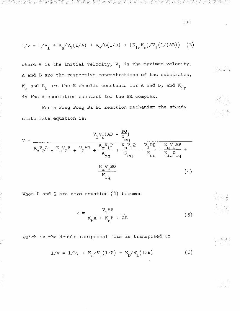
V
B
r'¿+
is the initial velocity, V. is the maximum velocity,
are the respective concentrations of the substrates,
and K.'ìã
steady
vrv2(A3 - #leq
L/v = t/vt + K*/VLG/A) + Kb/B(rlø) + (o,_r\) /vJL/(Æ) ) (:)
where
A and
l( ano
is the
^l^.1-¡
K are the Michael-is constants for A and B,þ
dissociation constant for the EA complex.
For a Ping Pong Bi Bi reaction mechanism the
rate equation is:
K V^A K V^Bb2 + a2 +K V-Pv^ArJ g I
/T--l--K
fa \7 Þ.ñI\ V ^uY
ctL
u1*
K V,Qpr-K
çL¿
K,
lVhen P and Q are zero equation (4) becomes
I1A+KaB+AB
which in the double reciprocal form is transposed to
V,PQ K V"AP. l- CIr- K - IL-K- =
eq ra eq
(4)
(¡)
(6)L/v = t/vt + Kd/vLGle) + WvLQ/B)
12tr.'
Determination of Michaelis constant= (f*)
The K values are defined as the concentration ofm
-"he{-r={-a ^ir¡in^ nna-h='r f 1¿¡imum VelOCitV (V ) iat initialÞuvÐ u!auç yrv r¡rv v¡rç-rtQrr r[o^llllulrt vçIUUf max.
velocity studies when the second substrate is in saturating
concentrations. O* values v/ere det.ermined by measuring
initial velocities when one substrate was varied and the
second substrate v/as fixed at several concentrations. The
data was plotted in the double reciprocal form (t,/v versus
L/(varted. substrat") ) . A replot of the reciprocal of rppV*"*
( intercept at the vertical axis) for each concentration of
fixed substrate against the reciprocal of the fixed substrate
concentration was made (Florini and Vestling, L957). Since
appv esuals (r + R /A\/v- or (t + K/B\/v- in equationsMAXAIþI/^\ I t a\(3) and (6), the replot intercept on the horizontal axis
corresponds to -t/Rm,
The results of initial velocity
reploLs are shown in Figs. 2L-24. The
from the replot analyses (fígs. 22 and
0.59 mM and K , = 0.085 mM. ThesecyE. c
studies and their
K values cal-culatedm
^ t, \'¿+) v/ere K'^** =
resul-ts compared
favourably with the apparent K* values (Ki.++ = 1.0 mM and
Kårrt. c = 0.095 ÍìM) previously calculated. (see Figs. 13 and.
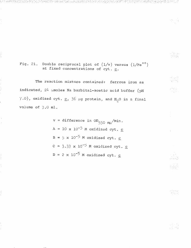
Fig. 2L. Doubfe reciprocal plot of (l/v) versus (lrlFe++)at fixed concentrations of cyt. c.
The reaction mixture contained: ferrous iron as
indicaLed, 24 ¡rmoles Na barbitar--acetic acid buffer (pn
7.0), oxidized cyt. 9, 36 $g protein, and HoO in a final_
volume of 3 .0 ml .
v = difference ir oo55O mg/mín.A - 10 x IO-5 M oxidized cyt. c
^-ÃB = 5 x IO-) M oxidized cyt. c
C = 3.33 x fO-5 M oxidized r--wt- c
D = 2 x 1O-5 M oxidized cyt. c
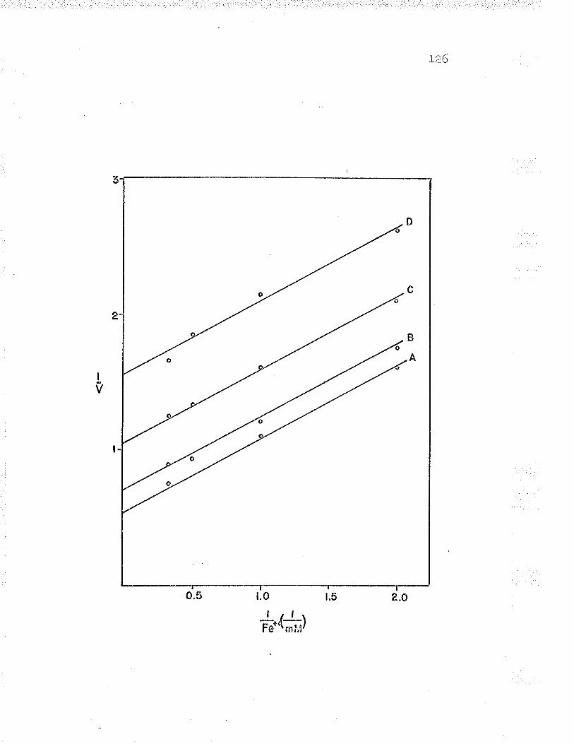
]-26
a
0
c
B
A
I
v
o.5 t.0 t.5 2.0| .r-l-r
Fett\nrlJ/
Fig. 22. Replot of Fig. pI, intercept (I/(appv*r*)) versus(t/cyt. c).
H N)
-\]
+-*
(**ù
lÐ tË-t
> l¿ I c¡
,rg
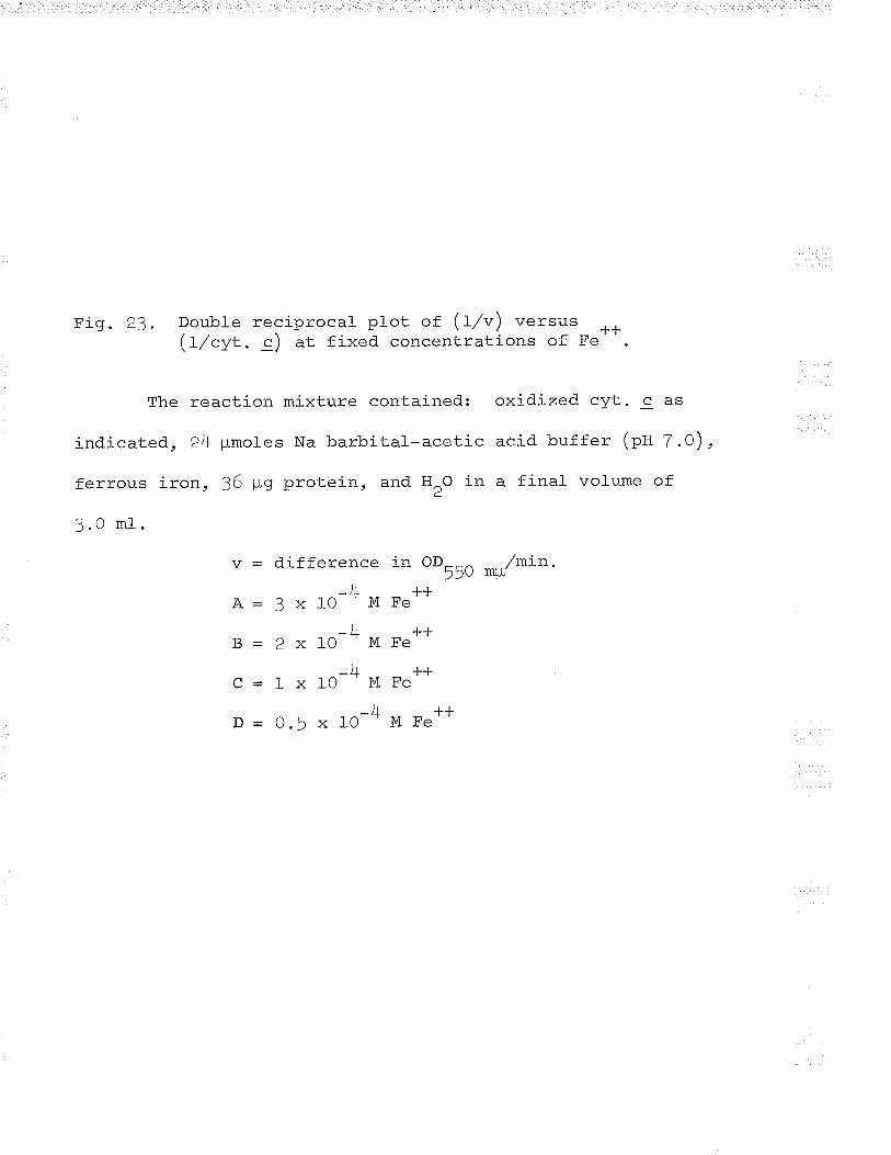
Fig. 23. Double reciprocal plot of (L/u) versus -L-L(t/cyt. c) at fixed concentrations of Fe".
The reaction mixture contained: oxidized cyt. c as
indicated, 24 ¡-Lmoles Na barbital-acetic acid. buffer (pH 7.0),
ferrous iron, 36 pg protein, and HrO in a final volume of
J.O ml .
v = difference in OD--^ /min.??u mpJ r -¡--¡-
A-3x10 MFeJt ¿r
þ = 2 x 1O - M Fe"lr r¡-
c - I x 1O-* M Fe"J r -¡--¡-
þ=O.5xlO--MFe"
128
4
õ
IVz
o o.r o.2 0.3 0.4 0.sJ- IJ-\Cltc\¡¡li{r
A
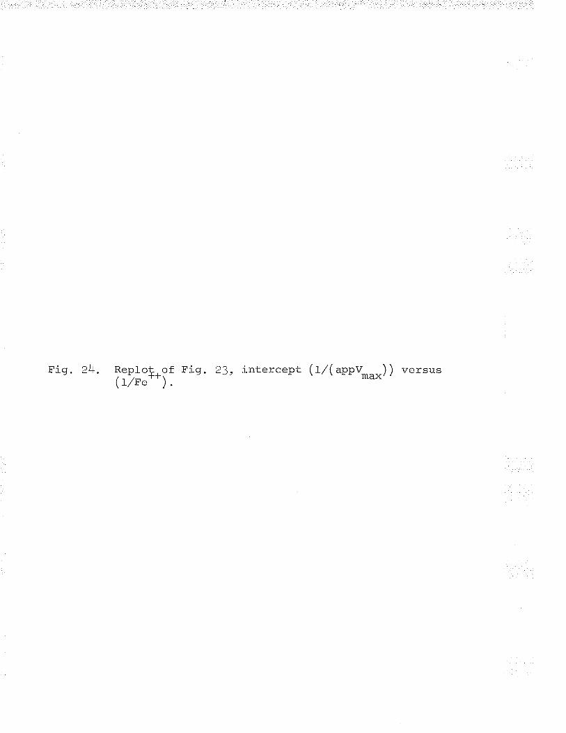
^t, , f - /l -- \\Fig. 2+. ReploÇ, of Fig . 23, intercept ( trz( appv___ ) ) versus" ( LTF'++) max''
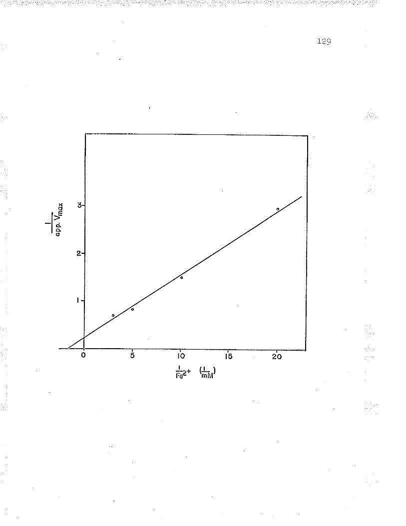
oPP
.Vm
ox
ts n) \o
. -\f5) . The paral_lel lines
mechanism is Ping Pong Bi
130
obtained indicate that the reaction
Bi rather than Ordered Bi Bi.
l.nrt'r ocit studies using a constant rati of substrate
cteland (rg6s") described a method to differentiate a
Ping Pong Bi Bi mechanism from Ordered. and Rapid Ecruil-ibrium
Random Bi Bi mechanisms. The latter mechanism assumes the
random addition of substrates A and B, and. the random release
of products P and Q; and that all steps are very rapid
except the interconversion of the central ternary complexes,
EAB and EPQ. The rate equation is identical to that of an
Ordered Bi Bi mechanism.
When initial velocities are measured at various
concentrations of one substrate, keeping a constant ratio of
the two substrate concentrations, a double reciprocal plot of
a Ping Pong Bi Bi reaction is the only mechanism that results
in a straight line relationship. When ArlB is constant,/ ^\equaEron ( Jl þecomes
I/v = t/vt * consrarrt(L/A) * consrant(t7e)2
and. equation ( 6) becomes
(r)
concentrations
t<l
L/v = t/vt + consrant(t/e) (B)
Maintaining a constant ratio of Fe**: cyt. c
-LJconcentrations (3tf), double reciprocal plots of Fe"-"yt. S
reductase activity resulted in a linear relationship as
shown in Figs . 25 and 26, Thus the mechanism of Fe**-cyt'. c
reductase is Ping Pong Bi Bi.
End-product inhibition studies
Kinetic studies on the inhibition of enzyme activity
by end-prod.uct( s) can supply valuable information required to
establ-ish a particular reaction mechanism (cleland, 1963btc) .
/.\ - /r,\Equations (1) and (4) indicate that when Q = 0 and P is used
as the inhibitor, l/v versus L/B ploLs should result in a
family of l-inear l-ines intersecting at the same point on the
vertical axis (competitive inhibition) for Ping Pong Bi Bi
mechanisms, but not for Ordered Bi Bi mechanisms. When P = 0
and Q is used as the inhibitor, l/v versus L/A p1ots for
both mechanisms should resul-t in comtretitive inhibition
plots. Inhibition constants are obtained by replotting the
slopes (appK /appV ) aqainst the concentrations ofm max'
inhibitor. The intercerrt at the horizontal axis crives the
J¿Fig. 25. Double reciprocal plot ((tl") versus (r7re"))maintainíng a conslqnt ratio of the substrateconcentrations ( f"' ' : cyt. c, 3t l) .
The reaction mixture contained: 24 umoles Na
barbital-acetic acid. buffer (pH 7.0), ferrous iron and ox-
'iÄ'i-aÀ ¡ttr as indicated, 9 ¡rg protein, and H^O in a final"J '.
volume of 3.0 ml.
v : dif f erence tr oo55. */min.

1?2
o.5
*.-t*l
^a f f - t \Fig. 26. Double reciprocal plot \lt/v)maintaining a consÇçnt ratioconcentrations (Fe--rcyt. g,
were the same as
\7êrqrr< (1/rtrl- -\\=ttof the substrate
a.rl
those outl-ined inThe conditions
H'I d 24- -Y. 1).
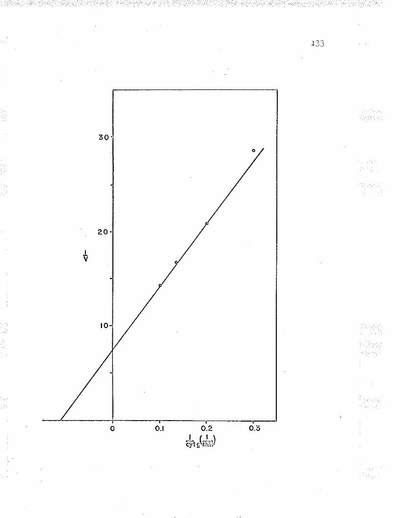
l??
o.2
'.$'*(**)
t<r+
value of the inhibitor constant.
End-product inhibitor studies showed that ferric iron
competitively inhibited the binding of oxidized cytochrome c
(fig. 27) and reduced cytochrome c competitively inhibited
the binding of ferrous iron (eig. 29) to Fe++-cyt. c
reductase. These results are compatible with a Ping Pong Bi
Bi mechanism,
plno n l'l.o
The inhibition constants for F"*** and reduced cyt. c
are O.L37 mM and 0.0135 mM respectively (eigs . 28 and lO).
The competitive inhibition by the Fe**-red. cyt. c and
J--LAFe- - --cyt. c pairs shows that there are probably two enzyme
, +++ -L-L.forms, E(Fe"') and E(re" ), and the reaction mechanism is
Ping Pong Bi Bi.
Inhibitiog bv sodium chloride
Inhibition studies \^/ere carried out usinq sodium
chloride. Initial velocities were measured when ferrous
iron concentra-tion was varied and cytochrome c concentration
IJ-
e(re' ' )cvt. c1l'-It
JJf_t_ | ¡ | \E(Fe Jred. cyt. c
, +++.E(Fe )
(g)
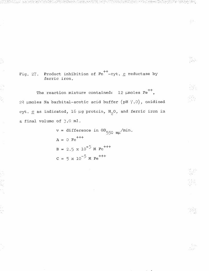
fJ
Fig. 27. Product inhibition of Fe' -cyt. c reductase byferric íron.
++The reaction mixture contained; 12 ¡,Lmoles Fe ,
24 pmoles Na barbital-acetic acíd buffer (pH 7.0), oxidized
^-,+- -i-,{.i^-r-^^ -tá ,'^ protein. H^O. and ferric iron inUyL. : clÞ rf,ru¿ud.Lçur f u FrY 2
a final volume of 3.0 ml.
v = difference in oD--^ /mín.??u mp+++
Õ - v ¿ç
- Ã ]-++B - 2.5 x LO'M Fe'
-5 +++C-5x10-MFe
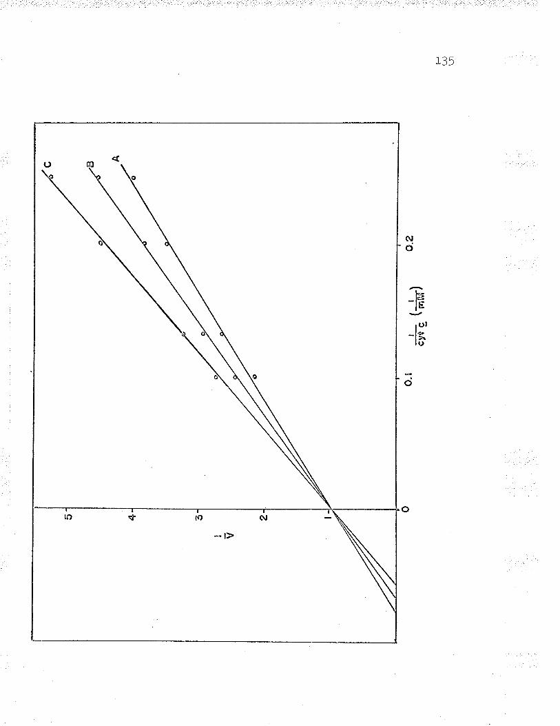
c ,tEt A
H UJ \n
s .t 2 I
,r
I ll
clf
g \r
¿¡M
/
v
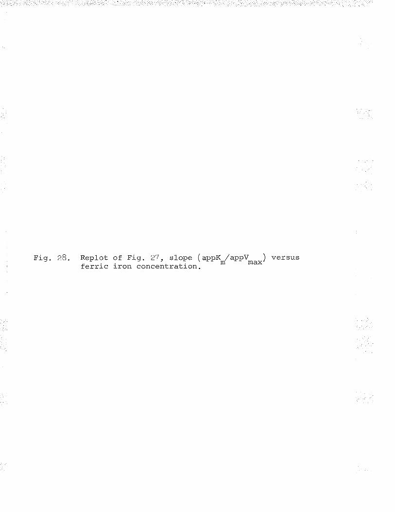
Replot of Fig. 27 , slope ( -ppferric iron concentration. ^,/m
appv ) versusmax'
¡.o
a) o õ o.
zs
o.50
o.
oo.
o25
0.o5
Fe+
+(¡
"nM
)
H o-\
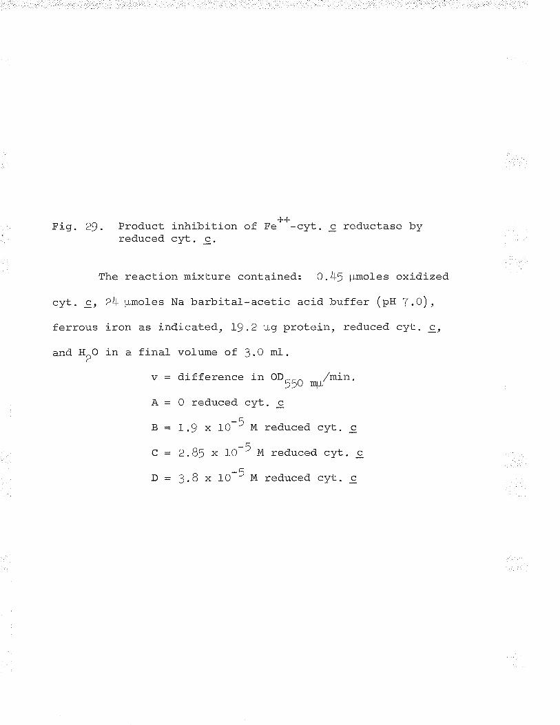
Fig. 29. Product inhibition of F.*+-cyt. c reductase byreduced cvt. c.
The reaction mixture conLained: O.45 ¡rmoles oxidizedr, . / -- - ^\cyt. c, 24 ¡rmoles Na barbital-acetic acid buffer (pH 7.0),
ferrous iron as indicated, 19 .2 plg protein, reduced cyt. c,
and HrO in a final volume of 3.0 ml.
v = difference in oD--^ ^,,/min.??U Í\U
ft: 0 reduced cyt. c
-ÃB - I.9 x l0 - M reduced cyt. c
Ç = 2.85 x to-5 M t.d..rced cyt. c
þ = 3.B x IO-5 M reduced cyt. c
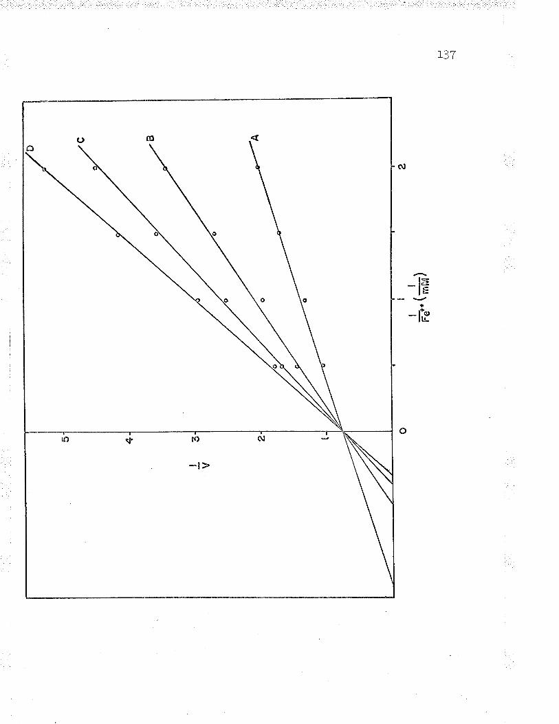
1?7
o mô
uD sf sÐ $J
. -t>
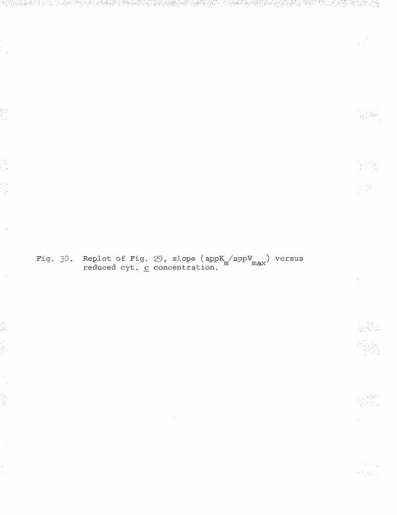
Fig. 30. Replot of Fig. 29, slope (app{*,Zapp!*^--) versusreduced cyt. c concentration. m max'
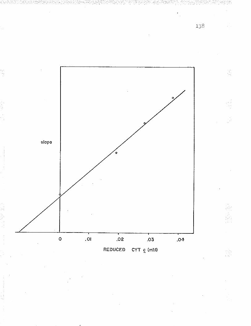
I aa)
.o2
REDUCED
.oõ
CYT c (mFdl
'ì ?q
was kept constant at three concentrations of NaCl. An
uncompetitive pattern of inhibition (eig. 31) \^/as evident
from the 1/v versus l.Tve++ plots. Replots of this data,
when l,/appV*rx was plotted against inhibitor concentration,
resulted in an inhibition constant of 5.8 mM (rig. 32). The
uncompetitive inhibition by the Fe"*-Nacl pair indicated
that the sodium chloride acted as a dead-end inhibitor.
binding the ferrous form of the enzyme, E(Fe++), but not the
ferric rorm, n(re+++) (see (g) ) (cretand , t963c).
Reaction of cyt. c with Fe++-cyt. c reductase
Spectrophotofluorometric and kinetic analyses indicated
the reaction mechanism of Fe++-cyt. c reductase is possibly
a Ping Pong Bi Bi (cfeland, L963a). This mechanism is
accepted for transaminases where both forms of the enzyme
are stable and easily interconvertibl-e; both enzyme f orms
are capable of carrying out their respective half reactions.
The ability of Fe++-cyt. c reductase to carry out a half
reaction was examined. Fe**-cyt. c reductase and cyt. c
were allowed to interact in the absence of ferrous iron.
The amount of enzvme non-heme iron oxidized and the amount
of cyt. c reduced were then measured.
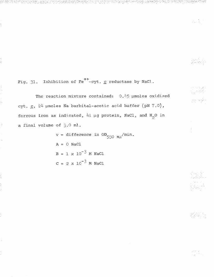
Fig. 31. Inhibition of F.**-cyt. c reductase by NaCl.
The reaction mixture contained: O.4J pmoles oxidized
cyt. 9, 24 pmoles Na barbital-acetic acid buffer (pH 7.0),
ferrous iron as indicated, 4f ¡rg protein, NaCl, and HrO in
a final volume of J.0 ml.
v = difference tr oo55o *u/min.A-0NaCl
B-lxlO-3MNacl
Q=2xlO-3MNacl-
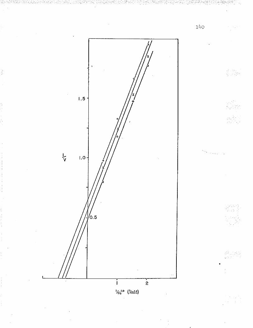
r /r^ITV
H'1 d <)¡ ¡ìr. J4. Replot of Fig. 3f, interceptNaCl concentration.
( L/appv ) versusmax'
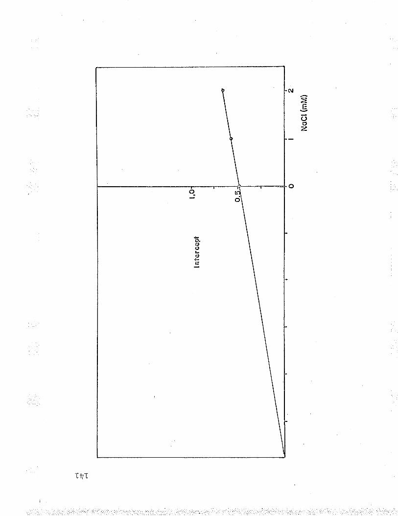
r-'l
--+ .-l
I nt
erce
pt
NoC
t(nn
M)
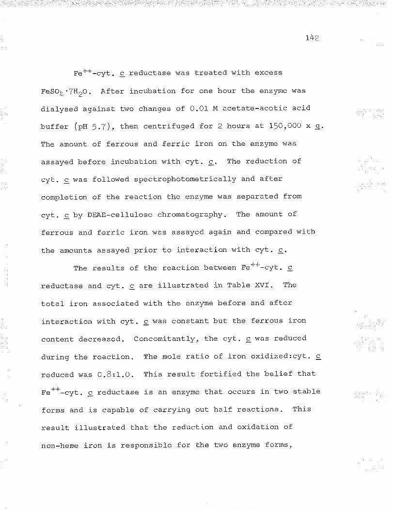
f Jr^Laa
Fe++-cyt. c reductase was treated with excess
FeSO4'THZO. After incubation for one hour the enzyme was
dialysed against two changes of 0.01 M acetate-acetic acid
buffer (ptt 5.7), then centrifuged for 2 hours at 150,OOO x 9.
The amount of ferrous and ferric iron on the enzyme was
assayed before incubation with cyt. c. The reduction of
cyt. c \^las followed spectrophotometrically and after
completion of the reaction the enzyme was separated from
cyt. c by DEAE-cellulose chromatography. The amount of
ferrous and ferric iron was assayed again and compared with
the amounts assayed prior to interaction with cyt. c.
The results of the reaction between Fe++-cyt. c
reductase and cvt. c are illustrated in Tabfe XVI. The
total iron associated with the enzyme before and after
interaction with cyt. c was constant but the ferrous iron
content decreased. Concomitantly, the cyt. c was reduced
during the reaction. The mole ratio of iron oxidized:cyt. c
reduced was O.B:1.0. This result fortified the belief that
Fe++-cyt. c reductase is an errzyme that occurs in two stable
forms and is capable of carrying out Ïralf reactions. This
result illustrated that the reduction and oxidation of
non-heme iron is responsible for the two enzyme forms,
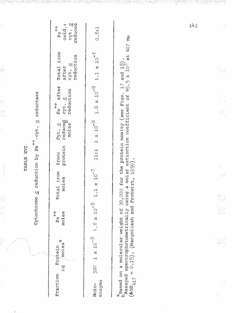
Fra
ctio
n P
rote
inug
m
oles
Hol
-o-
300
enzy
me
TA
BLE
X\Æ
Cyt
ochr
ome
c re
duct
ion
by F
e++
-cyt
. c
red.
ucta
se
.)^5
aseq
on
v-l\s
saye
o.(n
oo,'
17 =
T!l
-l--¡
.
Fe'
'm
oles
I x
to-B
4.6
x r
o-B
1.r
x to
-7
a m
olec
ular
wei
ght
ofsp
e ct
rop
hoto
me
tr ic
al 1
y, -\
/-
,O
.15)
, (M
argo
liash
and
Tot
al i
ron
mol
esfr
on:
Cyt
. c
prot
ein
redu
cefl n
mol
es"
3010
00 f
or t
he p
rote
in m
oeity
(se
e F
igs.
1/
and
usin
g a
mol
ar e
xtin
ctio
n co
effic
ient
of
45.
5 x
Fro
hwirt
, 19
59)
.11:1
++
Fe
afte
r T
otal
iro
ncy
t. c
afte
rre
duct
ion
cyt.
cre
duct
ion
1x t
o-B
3.
8 x
to-B
1.1
++
Fe
oxid
.:rr
rzf
õ-J
v.
_=
redu
ced
-7X
TU
1g)
.
lOJ
at
0. B
:1
4rZ
mp
H L!
r44
similar to the
interconversion
pyridoxal phosphate-pyridoxamine phosphate
found with transaminases.

DÏSCUSSfON
DTSCUSSTON
Io Carbon Dioxide Fixation
Ce1l-free extracts, in the presence of certain
accanli¡1 i-+ermediates, and metabolizing intact cells of
Ferrobacilrus ferrooxidans are capable of fixing carbon
dioxide. studies with intact cells revealed two classes of
compounds as the primary intermediates, phosphorylated
carbohyd.rates and amino acids formed from Krebs cycre
intermediates. Maciag and Lundgren (t964) using intact
cells of Ferrobacillus ferrooxidans showed that after 5
minutes in the presence of Fe** and ctUo, the same two
classes of labelled. compounds appeared (sugars and.
phosphorylated carbohyd.rates; Krebs cycle intermediates and
amino acids ) " This overall picture is consistent with
results previously obtained with prants by calvin and hÍs
co-workers, and. with other chemoautotrophic bacteria (El-sden,
1962). The primary compounds indicate that the autotrophic
"Calvin-Bassham cycle" (Mahler and Cord.es, L966) and the
heterotrophic wood-I,verkman (cq + ct condensation) reaction
t¿+'l
L46
are the two major pathways of COt fixation.
The presence of a high percentage of labelled 3-PGA
in whole cell fixation studies suggested the presence of
phosphoribulokinase and carboxydismutase. These enzymes
have been investigated in the CO2 fixation of other chemo-
autotrophs: the thiobacilli (Trudinger, f955; L956; Aubert
et al., L957aì L957b; Santer and Vishniac , 1955; Suzuki and
Werkman , L95Bc; Aleem and Huang, L965; Gale and Beck, L966),
the hydrogen bacteria (eergman et al., l95B) and the
nitrifiers (aleem, 1965). The results indicate that the
primary co2 fixation reaction in Ferrobacillus is the
carboxylation of RuDP which in turn feeds intermediates into
pathways leading to hexose synthesis, the tricarboxylic acid
cycIe, and regeneration of R-!-P essential for the cyclic
autotrophic mechanism to continue (Suzuki and Werkman, }!!Barc)
cell-free extracts exhibited the following enzyme
activities : phosphoriboisomerase, phosphoribulokinase,
carboxydismutase, phosphoglycerokinase, and triose phosphate
dehydrogenase necessary to form c'A-3P from R-5-P. The other
enzymes necessary for the conversion of GA-]P to R-l-P are
ubiquitous and their activity was not assayed (elsden , 1962) '
The triose phosphate dehydrogenase activity, similar to the
148
enzyme was present in crude av#r:¡'l-c ^ialysed extracts
were lacking this activity. The enzyme could be similar '¿o
the E. coli enzyme (Kornberg, f965) favouring the conversion
of C4 compounds to Ca compounds when there is an excess of
Krebs cycle intermedi.t"= and a paucitV of C, compounds.
Although the presence of other carboxylation schemes
is not excluded, the results implicate only the initial
carboxylation of RUDP by carboxydismutase and the
carboxylation by PEP carboxylase. The presence of the malic
enzyme activiiy could not be demonstrated, similar to the
f inding of Suzuki (f gf B ) in Thiobacil-lug thiooxidans .
Kornberg (L965) postulated that only the PEP carboxylase of
Enterobacteriaceae serves the "anaþlerotic" function and is
the only major enzyme responsible for C4 synthesis from C3
compounds leading to tricarboxylic acid cycle intermediates.
The results in Ferrobacillus ferrooxidans lead to the same
conclusion.
The met.abolic control of PEP carboxvlase in
Ferrobacillus mimics the control of the "heterotrophic" enzyme
in the Enterobacteriaceae. Acetyl CoA and. aceLo-acetyl CoA
activate the enzyme's activity, similar to results found
f ^a - - ^aFin E. col-i (Cänovas and Kornberg, 1965; Nishikido et al.,
r49
L965) and S. tvphimurium (sanwal and Maeba, L965)- The
enzyme activity is ínhibited by aspartate (misfritido et al.,
1965; Sanwal and Maeba, 1965), thus implicating "allosteric"
control of its "anaplerotic" function. The similarity in
results between the heterotro;ohic Enterobacteriaceae and the
chemoautotroph Ferrobaclllus supports the contention that
"heterotrophic" CO2 fixation is subjected to the same control
mechanisms.
coz fixation studies in cell-free extracts provide
evidence to support the findings in ce}]-free iron oxidation.
The inability of cell-free extracts to oxidize Fe** is reflected
in the inability of these extracts to fix COt without the
addition of necessary intermediates (ene, R-5-P) and an energy
source (afe). Also, the requirement of cell-free extracts for
exogenous NADH in order to reduce ]r3-DPGA exhibits the same
inability. On the other hand, intact cells must be able to
synthesLze ATP, \ADH, and necessary intermediates fox co2
fixation by the oxidation of Fe** alone, thus substantiating
the chemoautotrophic nature of this bacterium.
150
ïI. Iron Oxidation
Optimal
Ferrobaciflus
iron oxidation bv intact
ferrooxidans was found to
cell-s of
occur at pH 1.0
(Hzo-Hrso4) irr the presence of 2!o pmoles Feso4.THpo (i.t
2.8 ml). The optimal pH for iron oxidation coincided wíth
the results of Silverman and Lundgren (tgSgA) using
Ferrobacillus ferrooxidans but d.iffered from those of Beck
and Elsden (rg¡g) and Landesman, Duncan and walden Ggea)
using Thiobacil-luq ferrooxidanå. rhro¡gcirlus- ferrooxidans
seems to prefer a lower pH, while Ferrobacillus ferrooxidans
prefers a fower Fe+* concentration. rn other resþec'ts the
oxidatíon mechanism in these two bacteria is the same.
The inhibition of iron oxidation in intact celfs bv
azide points to the involvement of a cytochrome oxidase in
the oxidation mechanism. vernon, Mangum, Beck, and shafia
Ggao) and Blaylock and Nason Qgas) posrurated rhar iron
oxidation is accomplished by means of cyt. c and cyt. a.
The resurts of studies with intact cer-fs and cell-free
extracts lead to the same concl-usion. cerlular cyi. c under
anaerobic conditions in the nrosênôê nf F"**- asr:ortla.l-a (\r'" ,
sodium dithionite is rapidly reduced" under aerobic
'l 5'l
conditions this same reduction takes place but there r-s
a rapid reoxidation of the cellular cytochrome c. The
presence of both cyt. c and cyt. a in Ferrobacillus
ferrooxidans indicates the involvement of both these enzymes
in the oxidation mechanism.
The inability to isolate cell-free extracts capable
of oxidizing iron and the loss of iron oxidase activity in
frozen cells indicate that the electron transport chain is
disrupted between the initial reduction of cellular cyt. c
and the subsequent reoxidation of cyt. c. The presence of
both enzymes, Fe++-cyt. c reductase and cytochrome oxidase,
in extracts of inactive frozen cells shows that there is no
loss of activity in the enzymes involved, oflly an inability
of the electrons to pass from one electron carrier to the
other. This disruption of the cytochrome chain between cyt.
c and cyt. a is borne out by experiments with frozen intact
cells and cell-free extracts. Although the reason for the
disruption of the electron transport chain is known, the
cause of the dísruption is not understood.
FAD, FMN, and quinone were unable rn ran:ir r-l''e
disruption in the electron transport chain. The breakage
in the electron transport chain may be due to: (f) a
1^2
physical disruption of the erectron transport svstem
separating cyt. c and cyt. a in the membrane so that
electron f low terminates at ¡r¡{- | Z) a loss of
necessary cofactors bridging the cyt. c - cyt. a gap.
The former seems the most probable. The loss of oxidizinq
ability by such a mild treatment as freezing points to the
extreme labilit,y of the system. rn order to isolate cell-
free extracts capable of oxidizing iron, milder techniques
(..g. protoplast lysis) might be employed rather than the
harsh techniques of sonic oscillation, grinding, oî other
gross physícal methods.
ïïï. Ferrous Iron-Cvtochrome c Reductase
The results presented here indicate that the properties
of ferrous iron-cyt. c reductase are similar to those
reported by Blaylock and Nason (tg63) ror the same enzyme.
Ferrous iron-cyt. c reductase appears to be the initiar
enzyme in the iron-oxidizing system of Ferrobacilluq
ferrooxidans. The compactness and close association of the
system to the celr (membrane) necessitated the use of harsh
treatment to solubilize the enzyme. once the enzyme was
solubilized, it displayed remarkabre stability for a protein;
'1 6?
this stabirity appeared to be attributabfe to its cl_ose
association with nucleic acid (elaylock and Nason , 1963).
In acetate buffer (figs. B and !) there was no pH
optimum of catalytic activity evident (etaylock and Nason,
19æ) . rn B ml sodium barbitaf-acetic acid buffer the
enzyme displayed an activity optimum at pH f.O (I'ig. IO).
However, when the enzyme was assayed anaerobically in B m¡n
barbital no pH optimum was evident and under these conditions
the actual pH optimum may be hígher than T .5. since the
oxygen tension in the environment of the enzyme is unknown,
no definite statement can be made as to the actual pH
optimum of the enzyme in vivo. Aerobically, in vitro, a pH
optimum at T .o in barbital buffer was reproducibte. but the
non-enzymatic reaction above pH 2.0 increased rapidlv to
more than Jofi of the total reaction (enzymatic plus non-
enzymatic) and measurement of the reaction became extremely
difficult.
Barbital buffer was found to be much better than
acetate buffer in maintaining a constant pH of the reaction
mixture. This increased buffering action was especiarly
required when varying concentrations of cvt. c were used to
find the effect of cyt. c concentration on enzyme acrivity.
151].
The use of different buffers with varied bufferinq
capabilities courd account for the discrepancies in the
concentrations of substrates saturating the enzyme and the
appKm values with those previously reported by Blaylock and
Nason Qgez). rn barbital buffer, the concentrations of
substrate saturating the enzyme and the applin values were:
4 mtvt and t mM Fe*+ , o .r5 mM and o .o95 mM cyt . c respectively.
Using acetate buffer (pH 5.2), Blaylock and Nason Gge=)
reported saturating substrate concentrations of 3 mM Fe*+
and 0.18 mM cyt. c, and apparent 5*,= of O.l5 mM Fe** and
O.OB mM cyt. c.
Reaction mechanisE
The kinetic analyses of initial verocity studies with+-{-Fe' '-cyt. c reductase indicated that the enzyme adhered to a
Ping Pong Bi Bi mechanism. The existence of this mechanism
was substantiated by three criteria. when one substrate was
varied at several fixed concentrations of the second
substrate, double reciprocal plots of 1,/v versus I/(varj-.eð,
substrate concentration) resulted in a family of l-inear
parallel fines (figs . 2L and 23 ) . It was evident that
equation (6) conforms with the following equation,
't 55
y=mx+b
lvhere y = L/v, m = ou/ul, x = L/A, and b = I/VL(L + n/ø)when A was the varied substrate. Thus at anv fixed
concentration of B the line has the same slope Ku/Y.; at
fixed values of B, when A is varied, parallel lines should
arise in dor-rble reciprocal plots (eulos and Handler, f965;
Ray and Roscel-l-i, L964). In contrast, neither an Ordered
or Rapid Equilibrium Random Bi Bi mechanism produces
paralle1 l-ines under the same conditions (equation (:))t
both the slopes and the intercepts of the lines would change
with a change in the concentration of substrate B.
When a constant ratio of substrates (Fe++:cvt- c :\* -
3rf) was used in initial velocity studies, a linear relation
was evident in double reciprocal plots of l/v versus
1,/(substrate conc. ) (n'igs. 2! and 26). ordered and Rapid
Equilibrium Random Bi Bi mechanisms would produce a parabola
in the same círcumstances (see equations (7) and (B))r thus
it is evident that equations (2) and (:) do not describe the
results of initial- velocity studies of Fe+l--cyt. c reductase .
Cleland (f963¡;c) demonstrated that a reaction
mechanism could be identified in some cases by end-product
inhibition studies. F.++-reduced cyt. c and Fei-++-cyt. c
I?O
pairs exhibited competitive inhibítion, which is consistent
with a Ping Pong Bi Bi or a Random Bi Bi mechanism. However.
a Random Bi Bi mechanism can be ruled. out because of the
linear double reciprocal plots obtained when I/v is plotted
against 1/S where S is either substrate. A Random Bi Bi
mechanism woul-d yield a non-linear relationship (cleland,
r963a). The Random Bi Bi mechanism can arso be discounted
because of the linear double reciprocal plots obtained when
both su]:strates were varied but the ratio of one substrate
to the other was constant.
The results are consistent with a ping pong Bi Bi
mechanism (-= depicted in (g)), where there are two stable
forms of the enzyme, E(Fe+++) and E(F.++). rn this type of
mechanism each enzyme form binds only one substrate (ne++ or
cyt. c). When the end-product (Fe++-l- or reduced cyt. c) is
v^'l ^-^^¡ +l^^r,ËrecrÞcL¡, Lrre enzyme changes its form (E(Fe+++) to E(Fe++), -LJ.L. .to E(Fe"')). The enzyme oscillates between the two forms,
acting as a shuttfe mechanism carrying some reactant from
the first to the second substrat-e lin th.is case an electron) -
The ability of the enzyme to carry out a half reaction
was measured and the results fortified the belief that the
factor responsible for the existence of two enzyme forms was
1\7
the non-heme iron tightly bound to the enzyme (onfy one mole
of non-heme iron is tightly bound to the enzyme, âs
demonstrated in Table XIV). This ability of the enzyme to
reduce cyt. c in the absence of exLernally added ferrous iron
is consistent with a Ping Pong Bi Bi mechanism.
Composition of iron-cytochrome c reductase
All active preparations of the enzyme contained RNA,
a fact consistent with the report of Blaylock and Nason
(t063). Although the enzyme could be dissociated into
protein and RNA subunits, the resultant protein subunít was
devoid of catalytic activity. This unique phenomenon has
not previously been reported, although several enzymes have
been isolated associated with either DNA or RNA (Barth and
Hele , 1964; Hel-e , 1964; Symons , 1965; Hofer and Pette
L)6Ja,b; Hel-e and Barth, L966).
The inabitity of the RNA-free enzyme to catalyse the
reaction can be explained only by a change in conformation
of the protein (Koshland, L964). The RNA of the enzyme must
ptay some role in binding the enzyme to the cell- (membrane)
in the proper position and in maintaining the enzyme in the
proper conformation for catalysis. Once the enzyme is
released from the RNA. the effect of the release on the
protein conformation may be substantial indeed.
In both yeast cytochro*" Þ2 (lactate-cyt. c oxido-
reductase) and Fe*4-cyt. c reductase the DNA- and RNA-
components appear to be specifi_c and are found in al_l_
fractions containing catalytic activity (see Fig. l.6) , but
the yeast cytochro*. Þ2 catalytic activity is unaffected
when the DNA-component is removed (iviahter and pereira, L)62;
Armstrong et åf_., f963; symons , 1965) . Hofer and pette
(W6SA) reported that rabbit muscle phosphofructokinase
activity is modulated by the nucleic acid associated with
the enzyme. The presence of nucleic acid on phosphofructo-
kinase al-ters the ability of the enzyme to catalyze the
reverse reaction and results in a fowered forward:backward
reaction ratio. Other cases of modulation of catalvtic
activity by nucleic acids have also been reported by Hele
and Barth (Wee) and Allende et al- . OgAA) with aminoacyl
ribonucleic ¿nìrl crznf-lra{-ãceS. AlthOugh the Catalytic
activity of these enzymes is modified by RNA, the presence
of RNA is not obligatory, in contrast to Fe-F*-cyt. c
reductase.
Associated with the inability of the RNA-free protein
164
to catalyze the reaction is the failure of attempts to
reconstitute the holoenzyme from the protein and RNA
subunits. This f ail-ure to reconstitute the catal-ytic
activity can be explained by the change in the RNA portion
of the enzyme (Armstrong et âf., L963; Jackson et a1., 1965) .
Yeast cyt. Þp (vtatrter and Pereira, 1962) contains about lJ
nucleotide residues in each molecule of protein. which
indicates a molecular weight of approximately 10,000 for the
DNA portion of the enzyme. When the DNA portion of the enzyme
..i ^ -^J-^,f 'r¿Ð Ðçyqlquçu oy chromatography (symons , 1965), the DNA
molecular weight increases to lOO l2OrOOO (Armstrong et al.,
-^-^-^/-\1963; Jackson et al., L965). This increase ín molecular
weight seems to be d.ue to the DNA portion of the enzyme
aggregating to form a ne\^7 structure of higher molecular
weight. The same type of problem exists with Fel-+-cyt. c
reductase. As shown in Figs. 1/ and 18, the holoenzyme
exhibits the sedimentation velocity of a particle with
molecular \^Teight of 100 IlOr000. Upon dissociation, the
enzyme subunits have molecular weights of 30r000 for the
protein portion and 3OO 32O,OOO for the RNA portion (rigs.
. ^^ \19 and 2O). If the weight of the RNA portion of the enzyme
is 65,OOO (rafte XV), then the protein portion:RNA portion
r60
ratio in the holoenzyme is 1:l since the protein subunit/ . \ f _,(portion) has a molecular weight of 2T-3O,000 (Figs. 17 and
-ô\lU). The appearance of an RNA molecule weighing 300
320r000 after dissociation can be explained by assuming that
there is an aggregation of 5-6 ruUe molecules to form the
RNA subunit. The failure to reconstitute the holoenzvme
from its subunits al-so hinqed upon the failure of the RNA
subunit to segregate into the 5-6 RNA molecules required
to reunite with the protein subunÍ'b..
The presence of iron on the enzyme is consistent
with the belief that the reaction mechanism is Ping Pong
Bi Bi. Since the enzyme adheres to a Ping Pong Bi Bi
mechanism, the two forms of enzyme must differ from one
another in that one form carries an electron from the first
substrate (r"-t*) to tfre second substrate ("yt. c). since
iron is capable of oxidation-reduction, a non-heme iron atom
incorporated in the enzyme structure woul-d be consistent
with this premise. Non-heme íron analysis revealed 9-f0
iron atoms attached to the holoenzyme, but only one non-heme
iron atom was tightly bound and found with the protein
- f _ - - -----\subunit (fabte XIV). The presence of the other iron atoms
on the enzyme can not be explained unless one assumes that
LOI
the other atoms are bound non-specifical-ly to the RNA
portion of the holoenzyme. Attempts to show that the
bínding of iron to an iron-free enzyme is time-dependent
fail-ed and the results indicated that the incoricoration of
the non-heme iron was instantaneous. Analvses carried out
to detect the presence of l-abile sulfide and flavin(s) were
also negative. Thus the sole factor necessary for catalysis
in addition to the enzyme, was non-heme iron.

REFERENCES
REFERENCES
Aleem, M. I. H. GgeU) . personal communication.
Aleem, M. I. H. (tg6=). path of carbon and assimifatory
power in chemosynthetic bacteria. I. Nitrobacter
aqil-is . Biochim. Biophys . Acta . IOT: f4-28 .
Aleem, M. r. H. and Huang, E. (lg6s). carbon dioxide fixation
and carboxyd.ismutase in Thiobacitlus nove1lus.
Biochim. Biophys. Res. Commun . ZO:jlj-jzo.
Aleem, M. I. H., Lees, H. and Nj-cholas, D. J. D. GgeS).
Adenosine triphosphate-dependent reduction of
nicotinamide adenine dinucleotide by ferro-cvtochrome
c in chemoautotrophic bacteria. Nature. 2OO:Tj9-76f.
Allende, C. C., A1lende, J" E., Gatica, M., Ce1is, J.,
Mora, G. and Matama1a, M. (1g66). The aminoacyl
ribonucleic acid synthetases. I. properties of the
threonyladenylate-enzyme complex. J. Biol. Chem.
24r:224j-zzjL.
Amarsingham, C. R. (tgSg) " The relative roles of mal_ic
L62
IOJ
enzyme and oxalacetic carboxylase in acetate
metabolism. Fed . proc . IB :18f.
Andersen, K. J. and Lundgren, D. G. GgAf). Enzyme analyses
of the iron-oxidizlng bacterium, Ferrobacillul
ferrooxidans . Bacteriol. proc. p. 83 .
Armstrong, J. McD., Coates, J. H. and Morton, R. K. (tg6Z).
Physicochemical studies on cytochro*" Þ2. Some
properties of modified forms of the enzyme and of the
deoxyribonucl-eic acid component. Biochem. J.
BB:266-226.
Aronoff, S. (1956). Techniques of radiochemistry. Iowa
State College Press, Ames, Iowa.
Ashworth, J. M. and Kornberg, H. L. (fg63). The fine control_
of the glyoxylate cycle by allosteric inhibition of
isocitrate lyase. Biochim. Bíophys. Acta. T3:519-522.
Aubert, J. P., Milhaud, G. and Mitlet, J. (WnA). yiJtabolisme
du carbone dans la chimioautotrophie. Mode
d' incorporation de 1'anhydride carbonique. Compt.
rend . z4z:2OJ)-2O62.
t64
Aubert, J. P. , Milhaud, G. and Millet, J. (ry,f ") .
L'assimilation de 1'anhydride carbonique par les
bactíries chimioautotrophes. I-V. Ann. Inst.
Pasteur . 92:5f5-524.
Aubert, J. P., Milhaud, G. and Millet, ,J. (WffA).
L'assimilation de 1'anhydride carbonique par les
bacte'ríes chimioautotrophes . VI-VIII. Ann. Inst.
Pasteur . 92:679-683.
Bandurski, R. S" (ryf=). Further studies on the enzymatic
synthesis of oxalacetate from phosphoenolpyruvate and
carbon dioxide. J. Biol. Chem. 2I7:IJf-IJO.
Bandurski, R. S. and Axelrod, B. (fgff). The chromatographic
identification of some biologically important
phosphate esters. J. Biol. Chem. f93:405-4f0.
Bandurski, R. S. and Greiner, C. M. (fgf]). The enzymatic
synthesis of oxalacetate from phosphoenolpyruvate and
carbon dio>cide . J. Biol_ . Chem . ZO4:f BVf 86 .
Barth, P. T. and He1e, P. (1g64). possible a1l-osteric
effects controllinq the kinetic behaviour of amino
tfìî
acid-dependent pyrophosphate exchange reactions.
Biochem. J. 93r4p.
Bassham, J. 4., Benson, A. A. and Calvin, M. (fgfO). The
path of carbon in photosynthesis. VIII. The role of
malic acid. J. Biot. Chem. IB5 zTBI-TBT .
f - ^a^ \ -Beck, 'J. V. (19ô0) . A ferrous-ion-oxidjzlng bacterium. I.
fsolation and some general physiological
characteristics. J. Bacteriol. T9z5O2-5O9.
| - ^.- \ -Beck, J. V. (I9ô5). Personal- communication.
Beck, J. V. and Elsden, S. R. (fg¡B). Isolation and some
characteristics of an iron-oxidizinq bacterium.
J. Gen. Microbiol. 19:i.
Beck, J. V. and Shafia, F. M. ¡gA+). Effect of phosphate
ion and 2rA-dinitrophenol on the activity of intact
cells of Thíobacillus ferrooxidans. J. Bacteriol.
BB : B5o-857 .
Benson, A.4., Bassham, J.4., Calvin, M., Goodale, T. C.,
Haas, V. A. and Stepka, W. (fg¡O). The path of carbon
in photosynthesis. V. Paper chromatography and
IOO
radioautography of the products. J. Am. Chem. Soc.
72:17LO-I718.
Bergmann, F. H., Towne, J. C. and Burris, R. H. (fgf8).
Assimilation of carbon dioxide by hydrogen bacteria.
J. Biol. Chem. 23OzL3-24.
Bhat, J. v. and Barker, H. A. (fg48). studies on a new
oxalate-decomposing bacterium, Vibrio oxaliticgs .
J. Bacteriol. 55.359-368.
Bishop, D. H. L., Pandya, K. P. and King, H. K. (lg6Z).
llbiquinone and vitamin K in bacteria. Biochem. J.
83.6o6-6L4.
Blaylock, B. A. and Nason, A. (fg63). Electron transport
systems of the chemoautotroph Ferrobacillus ferro-
oxidans. I. Cytochrome c-containing iron oxidase.
J. Biol . chem . 48 ß4534462 .
Brumby, P. E., Miller, R. w. and Massey, v. (tg65). The
content and possible significance of labile sulfide
in some metalloflavoproteins. J. Biol. Chem.
24o zzzz2-2228 .
L67
Bryner, L. c. and Jamieson, A. K. (fgf8). Microorganisms in
leaching sulfide minerals. App. Microbiol. 6zZB|-ZBT.
Bulos, B . and Handler, P. QgA¡) . Kinetics of beef heart
glutamic-alanine transaminase. J. Biol. Chem.
z4o:32834294 .
Butler, R. G. and Umbreit, W. i/v. (tg65). The utilization of
organic substances by the strictly autotrophic
bacterium. Thiobacillus thiooxidans. Bacteriol. Proc.
p. 84.
Calvin, M. and Benson, A. A. (fg48). The path of carbon in
photosynthesis. Science. 10/:4f6-48O.
Calvin, M. and Massini, P. (tgSZ). The path of carbon in
photosynthesis. )C(. The steady state. Experentia.
F,. )-t)-ttr,-[,tr2v. I tJ ,)1.
cJnovas, J. L. and Kornberg: H. L. (t065). Fine control- of
phosphopyruvate carboxylase activity in Escherichia
coli. Biochim. Biophys. Acta. 96tL69-IT2.
Charles, A. M. GgAe ) . Ph.D. thesis . university of Manitoba,
Winnipeg, Manitoba.
L6B
Cleland, Irü. W. (fg63a). The kinetics of enzyme-catalysed
reactions with two or more substrates or products.
I. Nomenclature and rate equations. Biochim. Biophys.
Acra . 6T 1104-137.
Cleland, W. w. (fg6S¡). The kinetics of enzyme-catalysed
reactions with two or more substrates or products.
II. Inhibition: Nomenclature and theory. Biochim.
Biophys. Acta " 6f ztT3-LBT .
Cleland, W. W. (fg6gc). The kinetics of enzyme-catalysed
reactions with two or more substrates or products.
III. Prediction of initial velocity and inhibition by
inspection. Biochim. Biophys. Acta . 6T tf88-f96.
cslmer, A. R. (fç0f). Relatíon of the iron oxidizer,
Thiobagillus ferrooxidans, to thiosulfate.
J. Bacteriol. 63 :T6t-765.
Colmer, A. R., Temple, K. L. and Hinkle, M. E. (fg¡O). An
iron-oxidizing bacterium from the acid drainage of
some bituminous coal mines . J . Bacteriol . 59 :3LT 428
Cook, T. M. and Umbreit, W. I^i. (fg63). The occurrence of
16A
cytochrome and coenzyme Q in Thiobacillus thiooxidans.
Biochem. 2zL)4-L96.
Davis, B. J. GgA+). Disc electrophoresis- II. Method and
application to human serum proteins. Annal-s New York
Acad. Sci . L2Lz4O4-427.
Dugan, P. and Lundgren, D. c. (L964). Microdetection of
menadione, coenzyme Q5,, and certain related quinoid
compounds in biological- materials using elec'Lron
capture-gas chromatography. Anal-. Biochem. B:312-318.
Dugan, P. R. and Lundgren, D. c. (W6f). Energy supply for
the chemoautotroph Ferrobacillus ferrooxidans.
J. Bacteriol. 89:Bz5-834.
Elsden, S. R. (tgíZ). Photosynthesis and lithotrophic
carbon dioxide fixation. Irr, The Bacteria, vol. III,
- l¡ tpp 1-40, I. C. Gunsalus and R. Y. Stanier, eds.,
Academic Press Inc., New York, New York.
Fager, E. W. (tgS4). Phosphoglyceric acid formation by
carbon dioxide fixation in 'plant extracts. Biochem. J.
57.264-272.
TTO
Florini, J. R. and Vestling, C. S. (tg5T). Graphical
determination of the dissociation constants for two-
substrate enzyme systems. Biochim. Biophys. Acta.
ctr,.tr,2tr,_trzR¿).)|))Iv.
Fogo, J. K. and Popowsky, M. (tg4g). Spectrophotometric
determination of hydrogen sulfide, methylene bl-ue
method. Anal-. Chem . 2L:732-734 .
Gale, N. L. and Beck, J. V. (1966). Competitive inhibition
of phosphoribulokinase by AMP. Biochim. Biophys.
Res. Commun . 24:792-796.
Hall, B. D. and Spiegelman, S. (fg6f). Sequence
complementarity of T, - DNA and T, specific RNA.
Proc. Natl. Acad. Sci . (u.s . ) 47 zt3T-t46.
Hele, P. GgAU). On the role of s-RNA in regulating the
rates of amino acid-dependent pyrophosphate exchange
reactions : A possibl-e "al-Iosteric control" phenomenon.
Biochim. Biophys. Acta . BT r44g-459.
Hele, P. and Barth, P. T . (tg66) . Possibl-e " allosteric"
effects controllinq the kinetic behaviour of amino
T7T
acid-dependent pyrophosphate exchange reactions .
Biochim. Biophys . Acta. 114 :L49-L5T .
Hofer, H. w. and Pette, D. (ry6na) . The complex nature of
phosphofructokinase a nucleic acid containing
enzyme. Life Sciences . 4:L59L-L596.
Hofer, H. W. and Pette, D. (tg6SA). on the functional-
significance of the nucleic acid-protein complex of
phosphofructokinase. Life Sciences. 421597-16OZ.
Hurwitz , J., Weissl:ach, A., Horecker, B. L. and Smyrniotis,
P. z. (lgS6) . spinach phosphoribulokinase.
J. Biol. Chem. 218 2769-T83.
Jackson, J. F., Kornberg, R. D., Berg, P., RajbhandatY, U. L.,
Stuart , A., I{:orana, H. G. and. Kornberg, A. (tg65).
On the heterogeneity of the deoxyribonucleic acid
associated with crystalline yeast cytochrome þ2.
Biochim. Biophys. Acta. lOB :243-248.
.)À*Kiesow, L. (tg6r. Ûb"r die reduktion von diphosphopyridin-
nucleoti-d bei der chemosvthese. Biochem. Z.
33B,4oo-406.
172
Kiesow, L. (tg64). on the assimilation of energy from
inorganic sources in autotrophic forms of life. Proc.
Narl . Acad . sci . (u .s . ) 5z zgBO-9BB .
King, T. E. (L964). Stoichiometry of labile sulfide, non-
heme iron and flavin in reconstitutivel-v active
succinic dehydrogenase from heart mitochondria.
Biochim. Biophys. Res. Commun . L6z5II-5L5.
Kinsel, N. A. (fg6O). New sul-fur oxidizing iron bacterium,
Ferrobacill-us sulfooìlidans sp. n.. J. Bacteriol.
Bo t62B-632.
Kornberg, H. L. (fgf8). The metabolism of C, compounds in. -t )tmicroorganisms. f. The incorporation of (et*C)
acetate by Pseudomonas fluorescens and by a
Corvnebacterium grown on ammonium acetate. Biochem.
r. 682535-542.
Kornberg, H. L. (tg6>). Anaplerotic sequences in microbial
metabolism. I. Angew. Chem. 42558-565.
Koshland, D. E. Gge+). Conformation changes at the active
site during enzyme action. Fed. Proc. 23zTL9-726.
17?
Krulwich, T. and Funk, H. B. (tg6S). Stimulation of
N. aqilis by biotin. J. Bacteriol. 90¿729-733.
Landesman, J., Duncan, D. I4T. and Wald.en, C. C. GgAA). Iron
oxidation by washed ce11 suspension of the chemoauto-
troph, Thiobacillus ferrooxidans. Can. J. Microbiol.
L2 I2J-JJ .
Lazaroff, N. (fg6:). suffate requirement for iron oxidation
by Thiobacillqq lerlooxidans. J. Bacteriol. 85;78-83.
Leathen, W. w., Braley, Sr., S. A. and Mcfntyre, L. D. (fg¡S).
The role of loacteria in the formation of acid from
certain sulfuritic constituents associated with
bituminous coal. II. Ferrous iron oxidizing bacteria.
App. Microbiol- . L:65-68 .
Leathen, W. W., Kinsel, N. A. and Braley, Sr., S. A. (Wn6).
Ferrobacillus ferrooxidans : a- chemosynthetic
autotrophic bacterium. J. Bacteriol-. T2z7OO-TO4.
Lees, H. (Wnn). Biochemistry of autotrophic bacteria.
Butterworths Scientific Publications, London, England..
Lees, H. (fgeO). Energy metabolism in chemolithotrophÍc
17 )J,¿t I
bacteria. Ann. Rev. Microbiol. f4:83-98.
Lees, H. (tgAZ). Symposium in autotrophy. IV. some thoughts
on the energetics of chemosynthesis. Bacteriol. Revs.
'¿o'. to)- ro ( .
T,ees, H. GgA+). Personal communication.
Lineweaver, H. and Burk, D. (fg:4). The determination of
enzyme dissociation constants. J. Am. Chem. Soc.
-a --^ aaatO:OtÕ-ooo.
London, J. and Rittenberg, S. C. (tg6=). Effects of organic
matter on the growth of Thiobacillus fnËerrne-qiÆ-.
J. Bacteriol. 9I11062-1069.
Lowry, O. H., Rosebrough, N. J., Farr, A. L. and Randall,
R. J. (fgff). Protein measurement with the Folin
phenol reagent. J. Biol. Chem. L93.265-275.
Lundgren, D. G. (tg6>). Personal communi-cation.
Maciag, W. J. and Lundgren, D. G. (fg64). Carbon dioxide
fíxation in the chemoautotroph, Ferrobacillus ferro-
oxidans. Biochim. Biophys. Res. Commun . 17 1603-60T .
17^
Maeba, P. (t066) . Personal communica'tion.
Magasanik, B. and Chargaff, E. (fgff). Studies on the
structure of ribonucleic acids. Biochim. Biophys.
Acta . T r396-4t2.
Mahler, H. R. and cordes, E. H. Ggee). Biological
Chemistry. Harper and Row, New York, New York.
Ma-hler, H. R. and Pereira, A. da S. OgeZ). Properties of
the DNA associated with crystalline yeast l-actic
dehydrogenase . J. Mot. Biol . 5 =325-347 .
Margoliash, E. and Frohwirt, N. (lgjg). Appendix-Spectrum
of horse heart cyt. c. Biochem. J. TL:57O-572.
Margoliash, E. and Lustgarten, J. (tg6Z). Intraconversion
of horse heart cytochrome c monomer and polymers.
J. Biot. chem . 237 8397-3405 .
Martin, R. G. and Ames, B. N. (tg6t). A method for
determininq the sedimentation behaviour of enzymes:
Applications to protein mixtures. J. BioI. Chem.
236 zL3T2-L379 .
LT6
Mason, H. S. (tg65). oxidases. Ann. Rev. of Biochem.
34:595-634 .
Massey, V. (WSf ). Studies on succinic dehydrogenase. VII.
Valency state of the iron in beef heart succinic
dehydrogenase. J. Biol. Chem . 229 2763-770.
Massey, V. (t963). Lípoy1 dehydrogenases. In, The Enzymes,
vol. 7, pp 275-306, P. D. Boyer, H. Lardy and
K. Myrbäck, eds., Academic Press Inc., New York,
New York.
Miller, R. W. and Masseyr V. (tg65). Dihydroorotic dehydro-
genase. I. Some properties of the enzyme. J. Biol.
chem. 24o2t453-L465.
Mora, P. T., Creskoff, E. and Person, P. Gge=). Reduction-
l-ike effect of carbohydrates on cytochrome c.
science. r49:642-644.
Mulder, E. c. (t964). Iron bacteria, particularly those sr
the Sphaerotilus-Leptothrix Group, and industrial
problems. J. App. Bacteriol. 2T:L5L-L73.
Nishikido, T., Í.zttí, R., Iwatani, A., Katsuki, H. and
L77
Tanaka, S. (tg65). Inhibition of carbon dioxide
fixation in E. coli by the compounds related to TCA
cycle. Biochim. Biophys. Res. Commun . 2I:94-99.
orenstein, L. GgA+). Disc electrophoresis- I. Background
and theory. Annals New York Acad. Sci. I2L:.32L-349.
Partridge, S. M. (tg[g). Aniline hydrogen phthalate as a
spraying reagent for chromatography of sugars.
Nature . L642443.
Person, P., Fine, A. S., Mora, P. T. and Zipper, H. (tg65).
Macroion interactions involving components of the
cytochrome system. IV. Polyion-induced reversible
oxidations and reductions of cytochrome c. J. Biol-.
chem. 24Or3L59-3I64.
Quayle, J. R., Fuller, R. C., Benson, A. A. and Calvin, M.
(Wf4) . Enzymatic carboxylation of ribulose diphosphate.
J. Am. chem. soc . T6:36fO-36ff .
Rajagopalan, K. V. and Handler, P. (t964). The absorption
spectra of iron-flavoproteins. J. Biol. Chem.
239 zL5o9-L5r4.
LTB
Ray, I^i. J. and Roscelli, G. A. QgA+). A kinetic study of
the phosphoglucomutase pathway. J. Biol. Chem.
239:L228-L236.
F.azzeLl, W. E. and Trussell, P. c. (f963). Isolation and
properties of an iron-oxidizing Thiobacil-Ius.
J. Bacteriol. 85 2595-6O3.
Remsen, C. C. and Lundgren, D. c. (f963). The heterotrophic
qrowth of the chemoautotroph, Ferrobacillus ferro-
oxidans . Bacteriol. Proc . p. 33 .
Santer, M. and Vishniac, W. (tgSZ). CoZ incorporation by
extracts of Thiobaci-Llus thioparus. Biochim. Biophys.
Acta . 18 : L57 -L5B .
Sanwal, B. D. and Maeba, P. (tg65). Feedback inhibition of
phosphoenolpyruvic carboxylase of Salmonella. Biochim.
Biophys. Res. Commun . 2L2503-508.
Sanwal, B. D. and Maeba, P. (t966). Regulation of the
activity of phosphoenolpyruvate carboxylase by
fructose diphosphate. Biochim. Biophys. Res. Commun.
22.L94-L99 .
174
Schachman, H. K. (tg>g). Ultracentrifugation in Biochemistry,
Academic Press Inc., New York, New York.
Schnaitman, c. and Lundgren, D. G. (tO6=a). Production and
properties of spheroplasts of Ferrobacillus ferro-
oxidans. Bacteriol. Proc. p. 83.
Schnaitman, c. and Lundgren, D. G. Qge>A). organic
compounds in the spent medium of FerrqþeqiIlilq ferro-
oxidans. Can. J. Microbiol. LLz23-27 .
Schneider, w. c. (Wff ). Determination of nucleic acids in
tissues by pentose analysis. fn, Methods in
Enzymology, Vol. IIf, pp 680-684, S. P. Colewick and
N. O. Kaplan, eds., Academic Press Inc., New York,
New York.
*s"hrt-"k, E. and Flóderer, r . (rg:g ) . colorimetric
determination of ferrous and ferric iron in the
presence of aluminum, manganese, mercury, copper,
phosphoric acid, etc., or organic substances with
particular reference to pharmaceutical products.
Z. Anal. Chem . IIT TIT6-L95.
lBO
Silverman, M. P. (tg6|) . personal_ communication.
Silverman, M. P. and Lundgren, D. c. (tgfg"). Studies on the
chemoautotrophic iron bacterium Ferrobacilrus ferro-
oxidans. I. An improved medium and a harvesting
procedure for securing high cell- yields. J. Bacteriol.
TT z64z-647 .
Silverman, M. P. and Lundgren, D. c. (tg>gA). Studies on
the chemoautotrophic iron bacterium Ferrobacifrus
ferrooxidans. rr. Manometric studies. J. Bacteriol.
TB ß26-33r .
Smith, L. (fg6f). Cytochrome systems in aerobic elecrron
transport. fn, The Bacteria, vol. II, pp 360-396,
I. C. Gunsalus and R. y. Stanier, eds., Academic
Press Inc., New York, New York.
Snell, E. D. and Snel], C. T. (f949). Colorimetric methods
of analysis, vol. II, D. Van Nostrand Co. Inc.,
Princeton, New Jersey.
Sober, H. 4., Gutter, F. J. and tr{ychoff , M. M. (D=A).
Chromatography of proteins. fI. Fractionation of
rBl
serum protein on anion-exchange cellulose. J. Am.
chem. soc . TBrT56-763.
Suzuki, I. (fgf8). Ph.D. thesis. Iowa State College, Ames,
Iowa.
suzuki, I. and silver, M. (tg66). The initial product and
properties of the sulfur-oxidizing enzyme of
thiobacilti. Biochim. Biophys. Acta . L22222-33.
Suzuki, I. and Werkman, C. H. (fgl8a). Chemoautotrophic
fixation of carbon dioxide bv Thiobacil-lus
thiooxidans. Iowa State Coll. J. Sci. 3Zz4T5-483.
Suzuki, I. and Werkman, c. H. (fgfgU). Chemoautotrophic
carbon dioxide fixation by extracts of Thiobacillus
thiooxidans. I. Formation of oxalacetic acid. Arch.
Biochem. Biophys. /6:1Ol-lfl.
Suzuki, I. and I¡üerkman, c. H. (fg¡Bc). Chemoautotrophic
carbon dioxide fixation bv exLracts of Thiobacillus
thiooxidans. II. Formation of phosphoglyceric acid.
Arch. Biochem. Biophys . T7 ILL2-L23.
Symons, R. H. (t065). The DNA component of cytochrome þ2.
IB2
I. Isolation of þ'-DNA and. the behaviour of cytochrome
þ2 during chromatography on DEAE-cellulose and
sedimentation in a sucrose gradient. Biochim.
Biophys . Acta. 103 :298-310.
Tchen, T. T. and Vennesland, B. (1955). Enzymatic carbon
dioxide fixation into oxalacetate in wheat serm.
J. BioI. chem. 2L32533-546.
Ternple, K. L. and Colmer, A. R. (fgff ). The autotrophic
oxidation of iron by a new bacterium. Thiobacil-lus
ferqooxidans. J. Bacteriol. 6Z:6O5-6tt.
Temple, K. L. and Delchamps, E. vü. (tg53). Autotrophic
bacteria and. the formation of acid in bituminous coal
mines. App. Microbiol. L2255-258.
¡FJraaÄnra rF q ¡nrl É-nn] oql- r ' ^ 't' \ Mrr{-=nl- nf'rneouo.fe, r'. L. ---Jerg, !j . |\l-yo4l. Ivlutant oI
S. tvphimurium deficient in the carbon dioxide-
fixing enzyme phosphoenolpyruvic carboxylase.
J. Bacteriol. BB :946-955.
Trudinger, P. A . (tg=->). Phosphoglycerate formation from
pentose ;ohosphate by extracts of Thiobacillus
lB3
den=Ltrificans. Biochim. Biophys. Acta. 18 :5BL-582.
Trudinger, P. A. (lgS6) . Fixation of carbon dioxide by
extracts of the strict autotroph Thiobacillus
denitrificans. Biochem. J. 64zZT4-286.
Umbreit, W. V,I., Burris, R. H. and Stauffer, F. (tg64).
Manometric techniques, 4th edition, Burgess pubrishing
Co. , Minneapolis, Minn.
lJnz , R. F. and Lundgren, D. c. (f g6f ) . A comparative
nutritional- study of three chemoautotrophic bacteria:
Ferrobacilluq ferrooxidanå, and Thiobacill_us thio-
oxidans. Soil. Sc. 92:3O2-3L3.
Utter, M. F. and Kurahashi, K. (tg=S). Mechanism of action
of oxalacetic carboxylase from liver. J. Am. Chem.
Soc. T5 r75B .
Utter, M. F. and Kurahashi, K. (fgl4a). purification of
oxal-acetic carboxylase from chicken liver. J. BioI.
chem . 2OT ITBT-BOp.
Utter, M. F. and Kurahashi, K. (fgl4¡). Mechanism of action
of oxalacetic carboxylase. J. Biol_. Chem. ZOT;BZf-84f.
IB4
Van Niel, C. B. (tgSU) . The chemoautotrophic and photo-
synthetic bacteria. Ann. Rev. Microbiot. B:105-132.
velick, S. P. (fg¡B). Fluorescence spectra and polarization
of glyceraldehyde-l-phosphate and lactic dehydrogenase
coenzyme complexes. J. Biol. Chem . 233 :1455-1467 .
Vernon, L. P., Mangum, H. H., Beck, J. V. and Shafia, F. M.
(fg6O). studies on a ferrous-ion*oxid:-zLng bacterium.
II. Cytochrome composition. Arch. Biochem. Biophys.
BB z22T-23L.
vishniac, w. and santer, M. (Wfl ) . The thiobacilli .
Bacteriol. Revs . 2IzL95-2I3.
Vishniac, W. and Trudinger, P. A. (tg6Z). Symposium on
autotrophy. V. Carbon dioxide fixation and substrate
oxidation in the chemosynthetic sulfur bacteria.
Bacteriol. Revs . 26:L6B-I75.
lVaksman, S. A. and Starkey, R. L. (tgZZ). Carbon
assimilation and respiration of autotrophic bacteria.
Proc. Soc. Exper. Biot. and Med. 20 z9-L4 .
Waksman, S. A. and Starkey, R. L. çt923). on the growth and
IO?
respiration of autotrophic bacteria. J. Gen. Physiol.
5 3285-3LO .
walker, D. A. (ry=f ). Physiological studies on acid
metabolism. IV. Phosphoenolpyruvic carboxylase
activity in extracts of Crassulacean plants. Biochem.
r. 6T ,73-Tg .
Weissbach, A., Horecker, B. L. and HurwiLz, J. (tg)6). The
enzymatic formation of phosphoglyceric acid from
ribulose diphosphate and carbon dioxide. J. Biol.
chem. zLB.T75-BLO.
Weissbach, 4., Smyrniotis, P. Z. and Horecker, B. L. (tg=4).
Pentose phosphate and CO, fixation with spinach
extracts. J. Am. Chem. Soc. 76I36LI-36I2.
*wirrogr.dsky, s . (fBBT ) . Über schwef elbakterien. Botan.
zts.\5:489-526.
*!üirrogr-dsky, s. (fBBB). Üeber Bisenbakterien. Botan. ztg.
46 226I.
ì+^winogradsky, S. (f8gf). Ann. Inst. Pasteur. 5:92 and JZf.
rB6
wood, H. c. and stjernhorm, R. L. (ryaz). Assimiration ofcarbon dioxide by heterotrophic organisms. rn. The
Bacteria, vol. III, pp 4t_tt7, f. C. Gunsalus and
R. Y. Stanier, eds., Academic press Inc., New york,
New york.
wood, H. G. and werkman, c. H. (t%5). The utilization of
CO, by the propionic acid bacteria in the
dissimiration of glycerol. J. Bacterior. 30 z33z .
Irlood, H. c. and werkman, c. H. (rg:g). The utirization ofCO, by the propionic acid bacterj_a. Biochem. J.
J2 2r262-L27L.
Not read in the original


