Embed Size (px)
Citation preview
Answers to Questions
Chapter 11-1. Answer: c
Osteoporosis results from net bone resorption. Therefore, there is net loss of bone. There is no change in the standard mineral to matrix ratio. Corti-cal diameter increases and bone mineral density (BMD) by dual-energy X-ray absorptiometry (DEXA) decreases.
1-2. Answer: e
Paget’s is a disease of typically older males and results from increased bone turnover. Bone formation and bone resorption both increase.
1-3. Answer: d
The mechanical properties of bone depend largely on its unique integrated lamellar structure. Apatite is the primary mineral and the collagen is type I. (Remember, the word ONE is found in BONE.)
1-4. Answer: a
Without doubt, the common denominator in these and other similar dis-eases is muscle imbalance. This imbalance results in abnormal agonist–antagonist relationships, leading to joint contractures, fixed deformities, subluxation, and dislocation. In cerebral palsy (CP), the imbalance is spastic muscle versus more spastic muscle, in spina bifida the imbalance is largely weak muscle versus absent muscle, and in polio the imbalance is absent and weak muscle versus normal muscle. Nevertheless, the net result is the same: muscle imbalance.
1-5. Answer: c
Vitamin C deprivation causes the condition known as scurvy. Dilantin is toxic to liver microsomes, hence blocking normal pathways of vitamin D metabolism.
505
506 Answers to Questions
1-6. Answer: e
All are typical characteristics of hyaline cartilage.
1-7. Answer: b
Achondroplasia is an abnormality of the proliferating zone of the physis resulting primarily in short stature. Typically, these individuals have bow-legs, kyphotic spines, and are of normal intelligence.
1-8. Answer: e
Rheumatoid arthritis is a synovial disease characterized by hyperemia and hyperplasia of the synovium. There is NO repair attempt made; hence, all the changes around the joint are atrophic. Vascular hyperemia is the mech-anism of resorption on both sides of the joint.
1-9. Answer: a
Gout produces typically focal changes around the joints as a result of the deposition of urate.
1-10. Answer: b
Polio does NOT affect the sensory nerves. It is purely a motor neuropathy; therefore, neuropathic joints are not seen.
1-11. Answer: e
All are typical of avascular necrosis (AVN). The talus, proximal femur, and scaphoid all have a retrograde blood fl ow, making them vulnerable to AVN.
1-12. Answer: c
Collagen is a linear protein molecule that is highly cross-linked at multiple sites in the triple helix called tropocollagen. Both cell populations can synthesize the molecule despite the fact that the amino acid sequence is different. Type I collagen is found in bone and type II collagen in cartilage. The primary mechanical role of collagen is to provide tensile strength to the tissue.
Chapter 22-1. Answer: e
Twisting-type forces, which cause torsional loading to bone, produce spiral fractures. These fractures appear as an oblique fracture in both anteropos-terior and lateral radiographs.
Answers to Questions 507
2-2. Answer: b
Axonotmesis is an anatomic disruption of the axon in its intact sheath. In neuropraxia, the nerve is anatomically intact and physiologically nonfunc-tional. Neurotmesis is an anatomic disruption of the nerve itself (axons and sheath).
2-3. Answer: b
In the metabolic phase of fracture healing, the soft callous is reworked by a number of specific cellular elements to produce a firm, hard callous sat-isfactory for meeting the mechanical demands placed upon the fracture in the early phase. This process usually begins 4 to 6 weeks after the injury.
2-4. Answer: h
All the above answers are documented complications of fractures. See the chapter for more detail.
2-5. Answer: c
Midshaft radius and ulna fractures, or “both bone forearm fractures,” require anatomic reduction and rigid fixation to allow early range of motion and less stiffness. The remaining fractures can all be treated conservatively and nonsurgically, with reasonable expectation for regaining excellent function of the extremity.
2-6. Answer: b
The middle column includes the posterior half of the vertebral body and the posterior longitudinal ligament. The anterior column includes the ante-rior half of the vertebral body and the anterior longitudinal ligament. The posterior column includes the pedicles and the lamina (see Figure 2-15).
2-7. Answer: c
An open fracture of the pelvis with injury to the bowel and the urogenital system still carries with it a mortality rate of 50%. Great care in evaluating the patient is essential. A rectal and vaginal examination is required to assure that the fracture is not open through those soft tissue structures.
2-8. Answer: c
These fractures occur through an area below the lesser trochanter and do not heal quite as rapidly as the intertrochanteric injuries. In the younger population, subtrochanteric fractures usually follow the severe trauma of motor vehicle accidents. In the elderly, they are caused by severe osteopo-rosis or a pathologic process in the subtrochanteric area.
508 Answers to Questions
2-9. Answer: c
This injury is the result of very severe trauma. True dislocation of the knee is a very serious injury notable for producing arterial damage to the popli-teal vessels. The popliteal artery is fixed anatomically at the level of the proximal tibia by the interosseous membrane and, therefore, is placed at great risk when the knee dislocates.
2-10. Answer: d
All displaced ankle fractures should be treated surgically.
Chapter 33-1. Answer: c
The involucrum refers to an area of new or reactive bone growth around a sequestrum or focus of necrotic bone.
3-2. Answer: d
Staphylococcus aureus is by far and away the most common organism found in pediatric osteomyelitis.
3-3. Answer: b
Plain radiographs rarely demonstrate evidence of changes within the bone until 10 days to 2 weeks.
3-4. Answer: b
Staphylococcus aureus is the most common isolate from an adult septic joint.
3-5. Answer: c
It is the phenotypic alterations in the bacteria encased in the slime layer that most directly renders the antibiotic resistance.
3-6. Answer: a
Hematogenous inoculation is the most common cause of osteomyelitis in the pediatric population.
3-7. Answer: d
All the above are known risk factors for the development of a septic arthritis.
Answers to Questions 509
3-8. Answer: b
Soft tissue swelling is the earliest radiographic change that may be evident on plain films; this is particularly important in the distal aspect of the extremities where these soft tissue changes may be readily detectable.
3-9. Answer: d
In general, wounds associated with open fractures should be carefully inspected and then dressed in preparation for an emergent operative debridement. There is little or no reason to obtain predebridement cultures.
3-10. Answer: c
This would be considered a grade 1 open fracture. It is recommended that a first-generation cephalosporin alone be used in this setting.
Chapter 44-1. Answer: d
Both answers a and b describe benign or benign aggressive bone tumors, and answer c describes sarcomas of the soft tissues. Osteosarcoma and Ewing’s sarcoma are the two most commonly diagnosed primary bone sarcomas in patients less than 30 years of age.
4-2. Answer: a
Sarcomas tend to grow in a centripetal manner and push and compress the surrounding tissues. This tendency is important in that limb-sparing sur-geries can typically take place due to this “buffer zone” with few or any malignant cells noted. It is very rare for sarcomas to be found in adjacent lymph nodes.
4-3. Answer: d
A large open biopsy can cause contamination of the surrounding soft tissues, making a limb-sparing procedure impossible. Although staging studies are crucial in diagnosing a sarcoma, one should always perform a core biopsy under CT guidance if there is a question of malignancy. Mul-tiple cores are usually obtained and provide an accurate diagnosis in more than 95% of all patients.
4-4. Answer: d
Any and all of these characteristics can be displayed by an osteosarcoma.
510 Answers to Questions
4-5. Answer: a
The distal femur, proximal tibia, and proximal humerus are the most common anatomic sites, in descending order. Tumors arising in the proxi-mal femur and pelvis are less common. The spine is the most common site of metastatic carcinomas, usually after the age of 40 years.
4-6. Answer: d
Each of these diagnoses is seen in older adults; they are commonly found in the flat bone of the pelvis and may result in a pathologic fracture.
4-7. Answer: b
Batson’s plexus is a venous system (plexus) surrounding the spine (vertebral bodies) and arises from the abdominal and thoracic cavity structures. There are no valves within this system, thus permitting blood carrying tumor cells to travel ‘backward’ away from the heart during valsalva type of breathing and seed the spine. This is thought to be the primary physical mechanism for carcinoma metastases to the spine. The brachial plexus is the correct name for the answer given in a.
4-8. Answer: d
Benign bone tumors do not metastasize but often require treatment for all the reasons in answers a–c; for example, benign UCBS often present with a fracture, osteoid osteomas are always painful and require removal, and cartilage tumors as well as GCTS may become malignant.
4-9. Answer: d
Surgery, chemotherapy, and radiation therapy are all utilized for high-grade soft tissue sarcomas. Surgery and radiation therapy given postopera-tively are almost always required. Chemotherapy, either preoperative (called induction chemotherapy) or postoperative, is recommended for high-grade soft tissue sarcomas that are more than 5 cm in size and are deep, that is, occurring below the deep fascia.
4-10. Answer: b
A lipoma characteristically appears homogeneous and bright on T1
sequence on an MRI. The signal looks identical to the surrounding subcutaneous fat, whereas most sarcomas are dark on T1 and bright on T2.
Answers to Questions 511
Chapter 55-1. Answer: b
Most are hereditary and are characterized by generalized skeletal abnor-malities. The face is commonly involved, and typically there is genu varum or genu valgum.
5-2. Answer: d
It is multifactorial with environment and genetics playing a role. The rea-son that the left hip is much more frequently involved is the subject of speculation.
5-3. Answer: a
The child’s periosteum has an inner cambial or osteogenic layer. The struc-ture is of great mechanical significance as a dense fibrous membrane usually assisting in fracture reduction and maintenance.
5-4. Answer: e
Achondroplasia is a physeal dysplasia with the proliferating zone being most affected.
5-5. Answer: b
Slipped capital femoral epiphysis (SCFE) is common in obese adolescents, presenting as a painful limp. It is classified based on the ability of the child to bear weight on the limb: stable or unstable (cannot bear weight).
5-6. Answer: d
Avascular necrosis (AVN) of the femoral head is the definition of Perthes’ and a complication of the other two.
5-7. Answer: d
Because of the ligamentous laxity, typical in these children, the arch col-lapses and they have flatfeet.
5-8. Answer: a
Fracture of the parietal bone is the most common skull fracture and rarely indicates battery. Rather, suspicion should be raised if the fracture is not typical: frontal, temporal, etc.
5-9. Answer: e
Osteolysis of the metaphysis is a radiographic hallmark of leukemia.
512 Answers to Questions
5-10. Answer: c
A Trendelenburg gait results from putting the Trendelenburg sign into motion. The Trendelenburg sign is caused by either weakness of the abduc-tor muscles or shortening of the lever arm over which they act. When the patient is asked to stand on the affected side, because of inability to stabilize the pelvis the iliac crest on the contralateral side is seen to drop. Put into motion, the child throws the upper body over the affected hip to prevent falling.
5-11. Answer: 3
Displacement alone is the most forgiving, especially in the younger age groups.
5-12. Answer: e
The Pavlik harness has become the worldwide standard for the manage-ment of developmental dysplasia of the hip (DDH) in infants. If, however, the position is extreme (especially in abduction), avascular necrosis can occur. Overall, the device is capable of normalizing the hip in about 90% of affected.
Chapter 66-1. Answer: c
Type I collagen is the major constituent of tendon, comprising 86% of its dry weight. It is the high concentration of collagen in combination with its parallel orientation that gives it high tensile strength.
6-2. Answer: d
Dancing is most commonly associated with an overuse of the flexor hallicus longus tendon. This is a classic finding specific to this activity. Other ten-donopathies are sport specific and can be found in Table 6-1.
6-3. Answer: d
A grade III ligamentous injury is best characterized by complete ligamen-tous rupture without any structural integrity remaining. A common example is that of a complete rupture of the anterior cruciate ligament that results in increased translation of the tibia to anteriorly directed force.
6-4. Answer: b
Muscular strains most commonly occur with passive stretching and length-ening that occur during eccentric muscular contractions.
Answers to Questions 513
6-5. Answer: a
The proteoglycan component of articular cartilage makes it most effective in resisting compression across a joint.
6-6. Answer: e
The meniscus has a poor blood supply, limited largely to the peripheral one-third of its structure. The potential for meniscal tears to heal is based primarily on the location of the tear as well as the morphology. Tears best associated with potential to heal with repair are red–red longitudinal tears of the meniscus.
6-7. Answer: c
Injury patterns in sports medicine are best described as either microtrau-matic or macrotraumatic. Microtraumatic injuries include overuse injuries. Of those injuries listed, the only injury not caused by a single episode of trauma is a stress fracture of the hip.
6-8. Answer: b
Achilles tendonitis represents an overuse injury. Initial management of all overuse injury involves activity modification; this is an essential component to management of all entities that involve an overuse component. Often, this requires cessation of the sport in question.
6-9. Answer: d
At their attachments to bone, the transition from ligament to bone occurs gradually in a series of distinct phases. These phase range from ligament to fibrocartilage, from fibrocartilage to mineralized fi brocartilage, and from mineralized fibrocartilage to bone. The size of each zone varies from ligament to ligament and is related to its structural properties. Col-lagen fibers, known as Sharpey’s fibers, run in continuity throughout this zone of transition and have an important role in securing the ligament to bone.
6-10. Answer: a
Collagen chains are linked together to form fibrils that in turn are bound together by a proteoglycan matrix to form a fascicle, the primary unit in tendon structure. Fascicles in turn are bound by the endotenon, a layer of elastin-containing loose connective tissue that supports the blood, lym-phatic, and neural supply to the tendon unit. It is the endotenon that is contiguous with both the muscle fibers and the periosteum at the muscu-lotendinous and tendo-osseous junctions, respectively.
514 Answers to Questions
Chapter 77-1. Answer: e
Any or all of the above may be seen in a patient with cervical myelopathy. Abnormality of gait, particularly a broad-based and shuffling gait, is the hallmark abnormality of cervical myelopathy, but any of the above can be seen.
7-2. Answer: c
The triceps reflex is innovated by C7, and a diminished triceps reflex would be seen in a C7 radiculopathy.
7-3. Answer: a
Cervical myelopathy results in upper motor neuron findings for spasticity including hyperreflexia of the lower extremities, up-going toes, and, depending on the level of the spinal cord compression, hyperactivity in the upper extremities.
7-4. Answer: a
Spondylosis of the cervical (or lumbar) spine includes disk degeneration. The first and most striking finding in disk degeneration is a decrease in water content of the nucleus pulposus. All the other abnormalities are indeed seen in cervical and lumbar spondylosis.
7-5. Answer: d
In a relatively healthy, middle-aged patient, the presence of cervical myelopathy represents a fairly clear-cut indication for surgical treatment. The presence of chronic severe axial neck pain is usually treated nonop-eratively. Patients with a herniated disk, even with evidence of radiculopa-thy, are usually treated nonoperatively, and that is certainly the first lineof treatment in most cases.
7-6. Answer: a
All the above patterns of instability, including mixed patterns involving C1–C2, the occipitocervical junction, and the subaxial spine can be seen. Instability at C1–C2, however, is the most common pattern of instability seen.
7-7. Answer: d
It has been estimated that between 60% and 80% of adults in the United States will have at least one episode of significant low back pain in their lifetime.
Answers to Questions 515
7-8. Answer: d
The extensor hallucis longus is innervated by L5; weakness of this muscle would be evidence of an L5 radiculopathy.
7-9. Answer: c
Spondylolysis is believed to be a stress or fatigue fracture of the pars inter-articularis occurring because of repetitive shear stresses from repetitive hyperextension in individuals with a hereditary predisposition. It occurs most commonly at L5, is more common in boys than in girls and in athletes, particularly gymnasts.
7-10. Answer: d
Urinary retention results from lower motor neuron bladder dysfunction seen in cauda equina compression (CEC) syndrome. Patients with CEC syndrome may also present with severe back pain, saddle anesthesia, pain down the back of lower extremities, or even foot drop, but the most typical and most important manifestation is bladder dysfunction.
Chapter 88-1. Answer: c
The scapula and posterior thorax articulate through a number of bursae, but there is no articular surface.
8-2. Answer: b
The anterior band of the inferior glenohumeral ligament complex is the main stabilizer of the humeral head when it is abducted and externally rotated.
8-3. Answer: a
Central erosion of the glenoid is more common in inflammatory arthro pathy. In glenohumeral osteoarthritis, posterior glenoid wear is more common.
8-4. Answer: e
The spinal accessory nerve is the 11th cranial nerve and innervates the trapezius muscle.
8-5. Answer: c
The deltoid muscle is critical for elevation and abduction of the shoulder girdle, but it is not part of the rotator cuff.
8-6. Answer: a
Although all the above regions can produce referred pain into the shoulder, the cervical spine is the most common origin of shoulder pain that does
516 Answers to Questions
not emanate from the shoulder girdle. Most patients with cervical spine pathology presenting as shoulder pain localize the pain to the trapezial and posterior scapular regions.
8-7. Answer: d
Total shoulder arthroplasty and humeral head replacement are the most common surgical procedures used to treat end-stage osteoarthritis of the glenohumeral joint. Arthroscopic debridement of the glenohumeral joint is also a reasonable alternative in some patients with osteoarthritis of the glenohumeral joint. Glenohumeral fusion is an option for the treatment of osteoarthritis but is not generally recommended because of the severe restriction in motion that occurs.
8-8. Answer: e
The MRI scan is currently the gold standard for noninvasive evaluation of the rotator cuff tendons.
8-9. Answer: a
Adhesive capsulitis is initially treated with physical therapy for capsular stretching and NSAIDs. Corticosteroid injections into the glenohumeral joint are also helpful for pain relief. Arthroscopic adhesiolysis and manipu-lation under anesthesia are both options for patients who fail nonoperative treatment.
8-10. Answer: e
Most rotator cuff tears are degenerative in nature. The presence of rotator cuff tears has been documented by MRI scans in normal patients, and, in this asymptomatic population, the incidence of rotator cuff tears increases with the age of the patient.
Chapter 99-1. Answer: c
The thenar musculature is supplied by the recurrent motor branch of the median nerve, so it is never involved in isolated cubital tunnel syndrome. It is a late finding in carpal tunnel syndrome. The other signs are all common in cubital tunnel syndrome.
9-2. Answer: e
Treatment of lateral epicondylitis should focus on conservative manage-ment with rest, activity modification, modalities, bracing, physical therapy, and injections. When all else fails, arthroscopic and open surgical options are available.
Answers to Questions 517
9-3. Answer: d
The musculocutaneous nerve innervates the coracobrachialis, the biceps brachii, and the brachialis.
9-4. Answer: b
This term has been coined to describe the pathologic tissue in lateral epi-condylitis based on its microscopic appearance. It has little, if any, inflam-matory component.
9-5. Answer: d
For fractures around the elbow, it is critical to obtain good alignment and begin early motion. The only way to achieve this for the fracture described is through rigid internal fixation.
9-6. Answer: c
The brachioradialis is a powerful elbow flexor when the forearm is pronated.
9-7. Answer: a
The arcade of Frohse is a ligamentous band between the two heads of the supinator. It can compress the posterior interosseous branch of the radial nerve but not the ulnar nerve.
9-8. Answer: e
The above-mentioned muscles encompass the motor innervation of the anterior interosseous branch of the median nerve.
9-9. Answer: At this time, arthroscopic or endoscopic techniques are not indicated for median nerve release at the elbow. All the other procedures have been described by authors with reasonably good results when done arthroscopically.
9-10. Answer: e
MRI can be helpful for all the conditions listed.
Chapter 1010-1. Answer: d
Aside from skin cancer, such as squamous cell carcinoma, malignancies in the hand are extremely rare. The most common ones are epithelioid sarcoma, synovial cell sarcoma, and malignant fibrous histiocytoma.
518 Answers to Questions
Isolated metastatic disease to the bones of the hand are particularly uncom-mon conditions.
10-2. Answer: e
Although most patients who develop carpal tunnel syndrome have no known underlying causative factor, several medical conditions can contrib-ute to the onset of this disease. All of the diagnoses mentioned as well as other conditions such as amyloidosis or antiinflammatory arthropathies can contribute to compression of the median nerve at the wrist.
10-3. Answer: b
When a patient presents with a radial aplasia such as a radial clubhand or thumb deficiency, evaluation by the appropriate pediatric subspecialist should be performed for visceral anomalies: these includes vertebral anomalies, imperforate anus, tracheoesophageal problems, thrombocyto-penia, and other potentially life-threatening problems. It is uncommon for the other anomalies mentioned to have significant visceral involvement.
10-4. Answer: a
Osteoarthritis of the hand and wrist is a very common condition that mark-edly diminishes a patient’s hand function. Commonly involved joints include the thumb CMC joint, the scaphotrapeziotrapezoid joint, the PIP joints, and the DIP joints. The MP joints of the index through small finger are most commonly spared until very late in the disease process.
10-5. Answer: d
Forced ulnar deviation of the thumb and wrist causing severe pain over the fi rst dorsal compartment is a positive Finkelstein’s test. It is indicative of de Quervain’s tenosynovitis. One of the chief conditions in the differential diagnosis is thumb CMC arthritis, which is further differentiated by a CMC grind test.
10-6. Answer: c
The small finger receives nearly all its sensory function from the ulnar nerve and, as such, carpal tunnel syndrome rarely causes isolated small fi nger numbness. Patients often have a sensation of global numbness and until specifically tested do not realize their small finger is spared.
10-7. Answer: d
Open hand wounds should almost never be explored on initial evaluation. They should be covered with a sterile dressing, and a careful documenta-
Answers to Questions 519
tion of nerve, tendon, and vascular function distal to the injury should be obtained before applying an anesthetic block. After this evaluation, defini-tive management can be performed either in the emergency room, if appro-priate, or in the operating room, if necessary.
10-8. Answer: e
Human bite wounds are fairly common injuries usually caused by the patient punching another person in the mouth. Many different bacteria can be involved. Staphylococus aureus is the most common infectious agent in this situation, but one must cover Eikenella corrodens as well, because it is a common bacterium in the human mouth.
10-9. Answer: e
Scaphoid fractures, although the most common fractures of the carpal bones, are often difficult to diagnose and treat because of the poor blood supply. Nonunion and osteonecrosis occur at relatively high rates. One should maintain a high index of suspicion when patients present with radial-sided wrist pain and anatomic snuff box tenderness, even if initial X-rays are negative.
10-10. Answer: c
Bone destruction is a very uncommon finding in suppurative flexor teno-synovitis. The remaining four options constitute Kanavel’s four signs, which are pathognomonic for the disease process.
10-11. Answer: d
Gamekeeper’s thumb is an ulnar collateral ligament rupture of the thumb MP joint. It can be associated with a Stener lesion, in which the ruptured ligament button-holes into the adductor aponeurosis and becomes incar-cerated there.
Chapter 1111-1. Answer: d
The artery of the ligamentum teres, a branch of the obturator artery, only supplies approximately 10% to 20% of the blood supply to the femoral head. The majority of the blood supply comes from the small retinacular vessels that run in the synovial space. They are supplied by the medial and lateral femoral circumflex vessels from the profunda femoris artery. The internal iliac artery and the superior gluteal artery do not contribute to the femoral head.
520 Answers to Questions
11-2. Answer: c
The primary internal rotator of the hip is the gluteus medius muscle. The anterior one-third of that muscle runs from the iliac wing to the anterior greater trochanter. The iliopsoas and piriformis are external rotators. The rectus femoris and iliopsoas are hip flexors. The gluteus maximus is a hip extensor.
11-3. Answer: b
Rheumatoid arthritis is an inflammatory arthritis. Inflammatory arthritis results from an autoimmune attack of the articular cartilage, which results in involvement in the entire joint. Therefore, there is no preserved cartilage to rotate into a weight-bearing area. Inflammatory arthritis is a contrain-dication to osteotomy. The other conditions listed are indications for osteotomy.
11-4. Answer: d
The rate of deep venous thrombosis (DVT) is between 10% and 20%. DVT is the most common complication after hip replacement. The rate of loosening is approximately 0.5% to 1% per year, of perioperative fracture is approximately 1%, and of infection is approximately 0.1% to 0.4%. The rate of dislocation after total hip replacement is between 1% and 5%.
11-5 Answer: e
All the listed factors alone or in combination can be responsible for the dislocation of a total hip replacement.
11-6. Answer: d
Patients at high risk for the formation of heterotopic ossification after total hip replacement can be treated with 700 to 800 cGy radiation therapy to reduce the risk of bone formation; this is usually administered as a single dose of therapy. The other treatment that has been shown to be successful is the use of NSAI medications.
11-7. Answer: b
A fracture of the femoral neck can result in disruption of the small reti-nacular vessels that lie in the synovial space; this will destroy the blood supply of the femoral head and result in avascular necrosis even if the fracture is repaired.
11-8. Answer: a
The anterior approach to the hip detaches a portion of the gluteus medius from the greater trochanter; this can result in a limp postoperatively if it
Answers to Questions 521
is not properly repaired. The tensor fascia lata is displaced anteriorly and does not attach to the trochanter. The quadratus femoris, piriformis, and gluteus maximus all are posterior.
11-9. Answer: c
Deep periprosthetic infection is a devastating complication that requires at least two surgeries and 6 to 8 weeks of intravenous antibiotics to treat. It will occur more frequently in patients taking oral corticosteroids. Anti-biotics alone either orally or intravenously cannot be used to treat peri-prosthetic sepsis.
11-10. Answer: d
A coxalgic gait pattern results from a decreased stance phase and an abductor lurch. An antalgic or painful gait pattern is any gait with a reduced stance phase. The stance phase is reduced to decrease the time standing on a painful lower extremity. An abductor lurch results from weakened hip abductors. To compensate for the weakened abductors, the patient shifts the upper body over the affected hip to reduce to stress on the hip abductors, resulting in a lurch. Combining the two patterns results in a coxalgic gait pattern.
Chapter 1212-1. Answer: a
Posterior translation of the tibia relative to the femur is primarily restricted by the posterior cruciate ligament. The quadriceps and the extensor mech-anism are secondary restraints to posterior translation. The anterior trans-lation of the knee is resisted by the anterior cruciate ligament. Varus and valgus opening are resisted by the lateral and medial collateral ligaments, respectively.
12-2. Answer: a
In all modern knee replacements, the anterior cruciate ligament is removed. The function of the ligament is replaced by the design of the implants for the arthroplasty. The posterior cruciate ligament can either be retained or taken for the arthroplasty. The implant design varies depending upon what is done with the ligament. The medial, lateral, and patellar ligaments are necessary for the proper functioning of a total knee replacement.
12-3. Answer: d
The most common complication after total knee replacement is infection. The rate of deep venous thrombosis is approximately 10%. The second
522 Answers to Questions
most frequent complication after total knee replacement is stiffness, occur-ring in approximately 2% of cases. The other listed complications occur at a rate of less than 0.5%.
12-4. Answer: b
Osteotomy of the knee is indicated for patients with osteoarthritis of the knee isolated to one part of the knee, that is, either the medial, lateral, or patellofemoral compartment. Valgus osteoarthritis is best treated with a varus osteotomy to correct the valgus deformity; this can be done on either the tibia or femur. Osteotomy is contraindicated in rheu-matoid arthritis and in tricompartmental osteoarthritis. Patellofe-moral osteoarthritis cannot be treated with either a varus or valgus osteotomy.
12-5. Answer: b
This patient is developing an early wound complication after total knee replacement. This is an urgent situation. If it is not rapidly addressed, the patient will quickly develop a deep wound infection, which can require multiple surgeries to correct and long courses of antibiotic. Infection is the most feared complication after a joint replacement arthroplasty.
12-6. Answer: e
If a knee replacement becomes loose before 5 years after implantation, it should be considered infected until proven otherwise. Stiffness and neurovascular injury will occur early after the surgery. Fracture can occur at any time after the surgery and is associated with osteoporosis. Osteoporosis is not associated with loosening after total knee replacement.
12-7. Answer: d
Resection is a salvage procedure used to treat a multiply operated knee that has failed. This procedure is also best reserved for minimally active sedentary patients. Patients with chronic instability and arthritis are best treated by reconstruction or replacement.
12-8. Answer: c
The patient with osteoarthritis may experience more pain after the proce-dure than they had before. The results of arthroscopy for osteoarthritis are not highly successful and do not have a good long-term success. The ability of arthroscopy to delay total knee arthroplasty is unproven, and it is not necessary to do it before total knee replacement.
Answers to Questions 523
12-9. Answer: e
All the measures are important in the nonoperative management of knee osteoarthritis. There is never a rush to surgery. All patients should be tried in a good conscientious course of conservative management before total knee replacement.
12-10. Answer: e
All the listed symptoms are commonly seen in patients with chondroma-lacia of the patella. Pain with prolonged sitting is also referred to as movie sign.
Chapter 1313-1. Answer: b
The distal fibula lies laterally and slightly posterior to the tibia and is held there by the inferior tibiofibular ligaments. The lateral surface of the distal tibia has a sulcus to accommodate the adjacent fibula, forming the distal tibiofibular joint.
13-2. Answer: d
The talar dome is the superior portion of the talar body that articulates with the mortise of the tibia and fibula. The dome is wider anteriorly, which allows for stability in the mortise during dorsification.
13-3. Answer: e
The foot is composed of 7 tarsals, 5 metatarsals, and 14 phalanges. Three anatomic groupings are defined for descriptive purposes: the hindfoot, the midfoot, and the forefoot (see Figure 13-3). In the hindfoot lies the largest bone in the foot, the calcaneus.
13-4. Answer: c
Ligaments of the ankle syndesmosis include the anterior tibiofibular, pos-terior tibiofibular, and inerosseous ligaments.
13-5. Answer: e
The tibial and common peroneal nerves are terminal branches of the sciatic nerve, which arises from the lumbosacral plexus. The common peroneal nerve from L5 branches into the superficial peroneal nerve and deep peroneal nerve, which terminally supply sensation to the dorsal foot and first web space, respectively. The tibial nerve, a branch of S1, travels through the popliteal fossa into the deep posterior compartment. The sural
524 Answers to Questions
nerve is a sensory branch of the tibial nerve and provides sensation to the posterolateral hindfoot and lateral border of the foot.
13-6. Answer: e
Radiographic studies of the foot and ankle require weight-bearing X-rays when possible. Important views involve the anteroposterior (AP), lateral, and oblique views of the foot, and AP, lateral, and mortise views of the ankle.
13-7. Answer: c
Pilon fractures involve the intraarticular fractures of the tibial metaphysis, which extend to the weight-bearing portion of the tibia; a is a Lisfranc fracture-dislocation, and e is also known as a Jones fracture.
13-8. Answer: e
All the above answers define a hallux valgus, or bunion, deformity. Answers a, c, and d may be observed clinically. Answers a, b, and c may be seen radiographically.
13-9. Answer: b
Because the insole of a sneaker can be colonized with the Pseudomonasorganism, care should be taken to treat the patient with an infection from a puncture wound for this organism.
13-10. Answer: c
Overuse of the posterior tibial tendon causes conditions that range from mild tendonitis to complete rupture and asymmetrical flatfoot deformity. As the tendon continues to deteriorate and becomes incompetent, a pro-gressive asymmetrical flatfoot deformity develops with lateral hindfoot impingement.

Case Studies
Degenerative Spondylolisthesis
William C. Lauerman
History
E.D. is a 72-year-old woman with a 2-year history of progressively worsen-ing back pain, with radiation of pain into the buttocks, posterior thighs, and more recently into her legs bilaterally. The pain is worse with walking and is relieved when she stops to sit down. She denies any bowel or bladder changes. She notes that it is significantly easier walking in a grocery store pushing a cart than it is to walk in a mall. Her past medical history is posi-tive for adult-onset diabetes mellitus. Past medical history, family history, and social history are otherwise unremarkable. Review of systems is noncontributory.
On physical examination, she has a normal gait. She tends to stand in a forward flexed position, and she has limited extension of the lumbar spine, with pain on extension. Pulses are palpable in both feet. Neurologic exami-nation demonstrates normal motor and sensory function. Her deep tendon reflexes are symmetrically diminished at the knee and ankle. Her toes are down-going. Straight leg raising is negative. (See Figs. 1, 2.)
Treatment
The patient had previously been treated with physical therapy, including Williams flexion exercises, which improved her symptoms for several months but did not give her long-lasting or adequate relief. She underwent a trial of lumbar epidural steroids. Each injection led to good relief of her symptoms but that relief lasted only 2 to 3 weeks. Having failed nonopera-tive treatment, she elected surgery and underwent a decompressive lami-nectomy at L4–L5 with posterolateral fusion, utilizing pedicle screw instrumentation and iliac crest autograft. (See Fig. 3.)
527
528 W.C. Lauerman
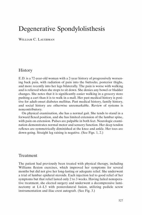
Figure 1. Lateral radiograph demonstrates a grade 1 degenerative spondylolisthe-sis at L4–L5.
Discussion
Degenerative spondylolisthesis is common and increases in prevalence with increasing age. It is more common in women than in men, in blacks than in whites, and is found with increased frequency in patients with dia-betes. Degeneration of the disk and facet joints, most commonly at L4–L5, leads to anterior slippage of the cephalad vertebrae (L4 in this case) on the level below (L5). It is common to see spinal stenosis in association with degenerative spondylolisthesis, and this patient’s symptoms, including back pain and stiffness with aching pain into the buttocks, thighs, and legs, are common. A common complaint is pain with walking that is often relieved by stopping and sitting down. Patients frequently note that they can walk farther in a grocery store pushing a cart than they can in a mall.
In spinal stenosis, positions of fl exion, such as pushing a grocery cart or sitting down, are more comfortable than extension, and the most common
Degenerative Spondylolisthesis 529
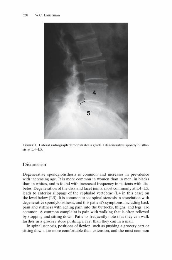
Figure 2. Sagittal (A) and axial (B) magnetic resonance (MR) images. The sagit-tal view demonstrates the spondylolisthesis, as well as the significant thecal sac narrowing at L4–L5. The axial view demonstrates enlargement and arthritic change in the facet joints bilaterally, thickening of the ligament flavum, and severe central and lateral recess stenosis.
B
A
530 W.C. Lauerman
physical finding is limited extension of the lumbar spine with pain on extension. Treatment options include nonoperative measures such as a daily back exercise regimen, flexion exercises, physical therapy modalities, nonsteroidal antiinflammatory medications, or epidural steroid injections. When nonoperative measures fail, surgery is often indicated, usually with good results. The primary goal of the surgical treatment of spinal stenosis is decompression of the affected nerve roots and/or thecal sac. In cases of degenerative spondylolisthesis, because of the risks of further slippage, concomitant fusion is routinely employed.
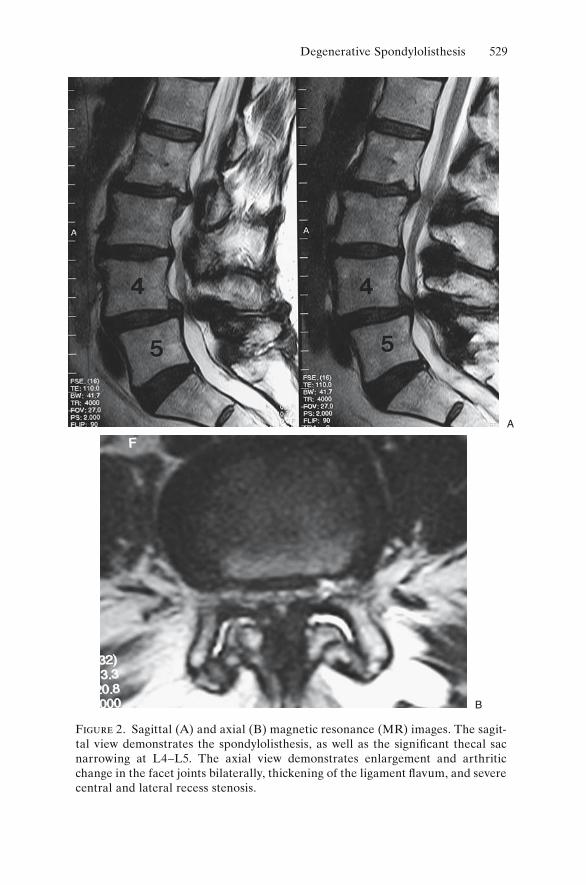
Figure 3. Decompressive laminectomy at L4–L5 with posterolateral fusion, utiliz-ing pedicle screw instrumentation and iliac crest autograft.
Hallux Valgus
Scott T. Sauer
History
A 52-year-old woman presents with worsening bilateral forefoot pain over the last three years. She describes pain in the left foot that is greater than the right foot. It is an achy, sometimes sharp intermittent pain over the medial aspect of both first metatarsophalangeal (MTP) joints. She rates the pain as 4 of 10 with 10 being the worst pain she has ever felt. The pain she feels is worse with shoe wear and walking and is relieved by wearing sandals. She also reports having to wear multiple layers of socks to keep her shoes from rubbing on the medial aspects of her forefeet. She describes no pain with arising, no swelling, no loss of sensation, no joint stiffness, and no flatfoot. Her past medical history is significant for breast cancer, for which she has had a mastectomy. Currently, the only medication she is taking is tamoxifen. She has no known drug allergies. She is a nonsmoker, and her review of systems is noncontributory.
Physical Examination
This is a well-developed, well-nourished female in no apparent distress. She is 5 feet 3 inches in height and weighs 118 pounds. She walks with a normal-appearing gait. Her station shows bilateral hallux valgus with normal-appearing arches and heels in slight valgus positioning. She is able to double-toe heel rise with minimal tenderness in her bilateral first MTP joints, plantar and medial. Examination of her right foot and ankle shows a moderate hallux valgus formation with a medial protuberance noted. No skin lesions or edema. Dorsalis pedis and posterior tibial artery pulses are palpable. There is tenderness to palpation over the medial and plantar medial aspect of the first MTP joint. There is no pain plantarly over the medial or lateral sesamoid, no lesser toe pain, and no medial midfoot or hindfoot pain. No ankle pain noted. Ankle range of motion is 5 degrees of dorsiflexion, 40 degrees of plantar flexion, 10 degrees of inversion, and
531
532 S.T. Sauer
5 degrees of eversion. First MTP joint range of motion is approximately 30 degrees of extension and 20 degrees of flexion with no ligamentous instabil-ity. Ankle dorsiflexion, plantarflexion, inversion, eversion, and toe exten-sor and flexion strength are 5/5. Sensation is intact on the dorsal, medial, lateral, and plantar aspects of the foot.
Examination of the left foot and ankle shows a moderate hallux valgus with protuberance medially noted. No skin lesions or edema noted. Dor-salis pedis and posterior tibial artery pulses are palpable. There is tender-ness to palpation over the medial first MTP joint, as well as plantar medially in the same region. There is no sesamoid pain noted. No medial midfoot or hindfoot pain is noted. No ankle pain noted. Ankle range of motion is 5 degrees of dorsiflexion, 40 degrees of plantar flexion, 10 degrees of inver-sion, and 5 degrees eversion. First MTP joint range of motion is 30 to 40 degrees of extension and 20 degrees of flexion with no ligamentous instabil-ity noted. Ankle dorsiflexion, plantarflexion, inversion, eversion, and toe extension and flexion strength are 5/5. Sensation is intact in the dorsal, medial, lateral, and plantar aspects of the foot.
X-Rays
Weightbearing anteroposterior (AP), lateral, and mortise views of her bilateral feet show bilateral mild hallux valgus deformities with slight sub-luxation laterally of the sesamoids with congruent first MTP joints. Medial exostosis present. First MTP joint appears without degenerative change. First and second intermetatarsal (IM) angle on the left foot is 12 degrees with a hallux valgus angle of 20 to 25 degrees. No other bony abnormalities noted.
Treatment
Surgical correction of left hallux valgus using distal chevron bunionec-tomy, which includes distal metatarsal osteotomy with exostectomy and medial first MTP joint capsule tightening.
Discussion
This patient has a symptomatic hallux valgus deformity; the left foot is clinically worse than the right foot. Clinically, hallux valgus can be prob-lematic in that it makes shoe wear difficult. With the progressive valgus deformity of the toe, extra strain is placed on the medial structures of the fi rst MTP joint, and this area becomes more prominent. With shoe wear, the inner aspect of the shoe pushes on the medial aspect of the first MTP
Hallux Valgus 533
joint, which can cause friction and pain. With this repetitive stress, as well as progression of the bunion deformity, bony buildup can occur on the medial aspect of the first metatarsal head; this perpetuates continued discomfort as the exostosis enlarges. As the bunion deformity progresses, it can affect the lesser toes, creating hammer toes, which have hyperexten-sion at the MTP and DIP joints and hyperflexion of the PIP joints.
Treatment is aimed at relieving pain, which can be done conservatively and nonsurgically in early stages of hallux valgus by shoe wear modifica-tion utilizing a wider toe box to prevent rubbing or friction. Toe spacers, which keep the first toe in a straight orientation in relation to the other toes, can also be used, as well as physically altering the shoe with shoe stretching or cutting out the medial side of the forefoot toe box to accom-modate the bunion deformity. When these modifications are unsuccessful, surgical options can be helpful. Surgical treatment goals include removing the exostosis, correcting the hallux valgus deformity, and straightening the toe, as well as relocating the sesamoid complex to be congruent with the underside of the first metatarsal head.
Many surgical options are available for bunion correction and are dependent on the degree of severity of the bunion deformity. The severity is typically measured radiographically with weight-bearing X-rays. The fi rst and second IM angle, and the hallux valgus angle, the angle between the proximal phalanx and the first metatarsal, are measured. Treatment parameters for hallux valgus generally include whether the first MTP joint is congruent. An incongruent joint is one in which the proximal phalanx articular surface does not line up with the metatarsal head articular surface because of extreme hallux valgus. A normal hallux valgus angle is less than 20 degrees. Generally, when it approaches 30 degrees and is symptomatic, it requires correction. The IM angle is generally about 9 degrees. When the IM angle is less than 12 to 13 degrees, a distal correc-tive osteotomy may be done in the first metatarsal. With an IM angle greater than 13 degrees, one should consider a proximal first metatarsal osteotomy with distal soft tissue balancing. One final consideration is the sesamoids. Typically, they are subluxed laterally, and surgery should be tailored to make these bones congruent in their articulations with the base of the first metatarsal.
In this case, because of the IM angle, hallux valgus angle, and slight incongruence, a distal first metatarsal osteotomy was performed, and the distal aspect of the metatarsal was moved laterally to reduce the sesa-moids. The medial capsule was imbricated and tightened, allowing for adequate stability of the first toe, as well as holding a corrected position. Postoperative management included weight-bearing as tolerated in a postoperative shoe for 6 weeks, as well as a first–second toe spacer for 3 months. See the preoperative, immediate postoperative, and late follow-up pictures (Figs. 1, 2, 3, respectively) for details and radiographic results.
A
B
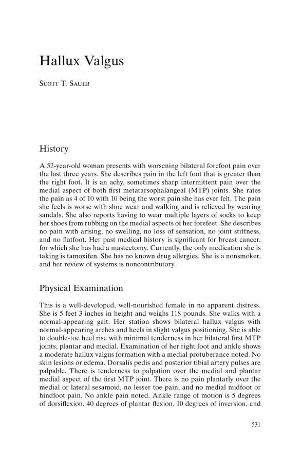
Figure 1. Preoperative radiographs with radiographic evidence of hallux valgus.
B
A
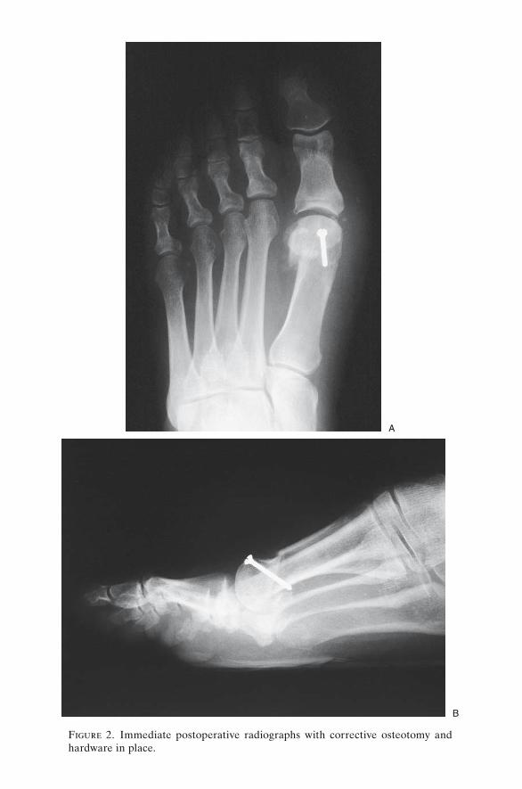
Figure 2. Immediate postoperative radiographs with corrective osteotomy and hardware in place.
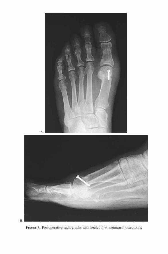
Figure 3. Postoperative radiographs with healed first metatarsal osteotomy.
A
B
Trauma: Open Tibia Fracture
Scott T. Sauer
History
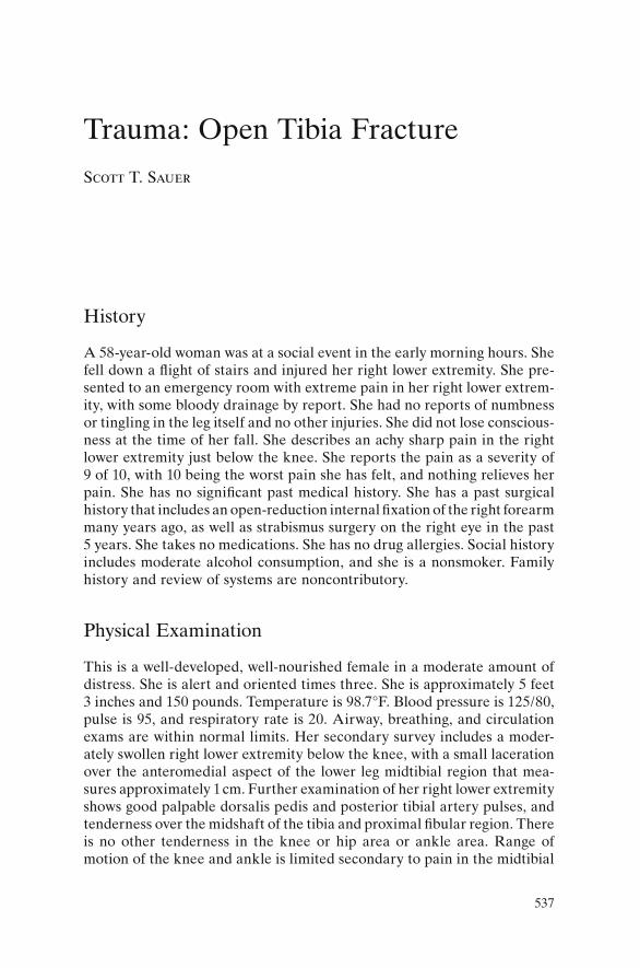
A 58-year-old woman was at a social event in the early morning hours. She fell down a flight of stairs and injured her right lower extremity. She pre-sented to an emergency room with extreme pain in her right lower extrem-ity, with some bloody drainage by report. She had no reports of numbness or tingling in the leg itself and no other injuries. She did not lose conscious-ness at the time of her fall. She describes an achy sharp pain in the right lower extremity just below the knee. She reports the pain as a severity of 9 of 10, with 10 being the worst pain she has felt, and nothing relieves her pain. She has no significant past medical history. She has a past surgical history that includes an open-reduction internal fixation of the right forearm many years ago, as well as strabismus surgery on the right eye in the past 5 years. She takes no medications. She has no drug allergies. Social history includes moderate alcohol consumption, and she is a nonsmoker. Family history and review of systems are noncontributory.
Physical Examination
This is a well-developed, well-nourished female in a moderate amount of distress. She is alert and oriented times three. She is approximately 5 feet 3 inches and 150 pounds. Temperature is 98.7°F. Blood pressure is 125/80, pulse is 95, and respiratory rate is 20. Airway, breathing, and circulation exams are within normal limits. Her secondary survey includes a moder-ately swollen right lower extremity below the knee, with a small laceration over the anteromedial aspect of the lower leg midtibial region that mea-sures approximately 1 cm. Further examination of her right lower extremity shows good palpable dorsalis pedis and posterior tibial artery pulses, and tenderness over the midshaft of the tibia and proximal fibular region. There is no other tenderness in the knee or hip area or ankle area. Range of motion of the knee and ankle is limited secondary to pain in the midtibial
537
538 S.T. Sauer
region. Hip range of motion internal–external rotation is 45 to 50 degrees without pain. No obvious evidence of ligamentous instability. Ankle dor-siflexion, plantarflexion, inversion, and eversion strength is 4+/5. Sensation is intact on the dorsal, medial, lateral, and plantar aspects of the foot. No pain with passive range of motion of the foot and toes.
X-Rays
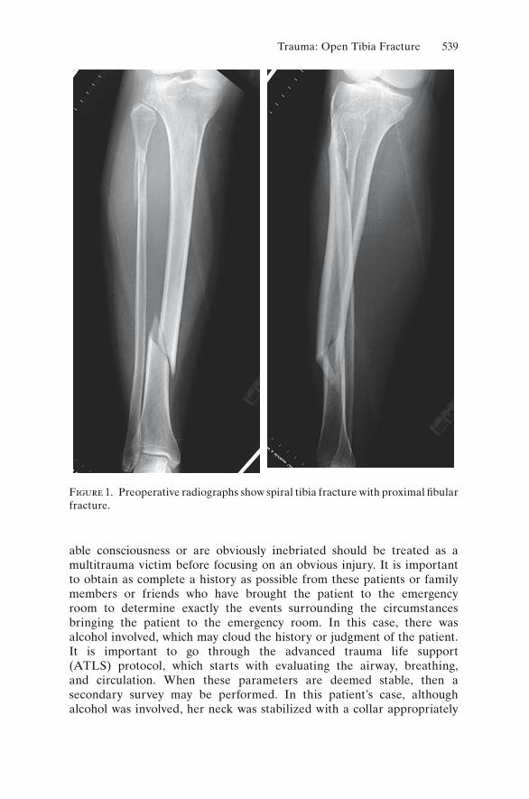
Anteroposterior (AP) and lateral radiographs of her right tibia and fibula, AP, lateral, and mortise view radiographs of her right ankle, and AP and lateral radiographs of her right knee show a spiral fracture of the middle shaft to distal third of the tibia with a proximal fibular fracture with mod-erate displacement. No obvious deformity or fracture is seen in the ankle mortise or around the distal femur.
Laboratory Values
Laboratory values, EKG, and chest X-ray are within normal limits.
Assessment
Right open tibia and fibular fracture with spiral fracture of the midshaft distal third tibia, proximal fibular fracture; no signs of neurologic injury or compartment syndrome.
Treatment
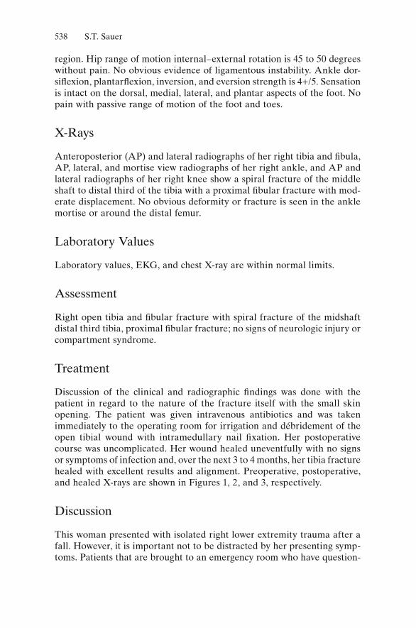
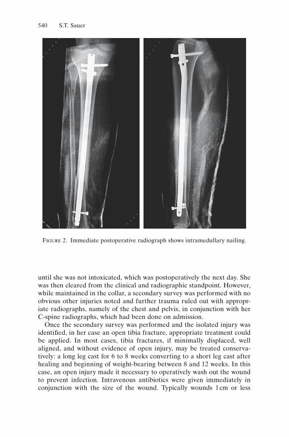
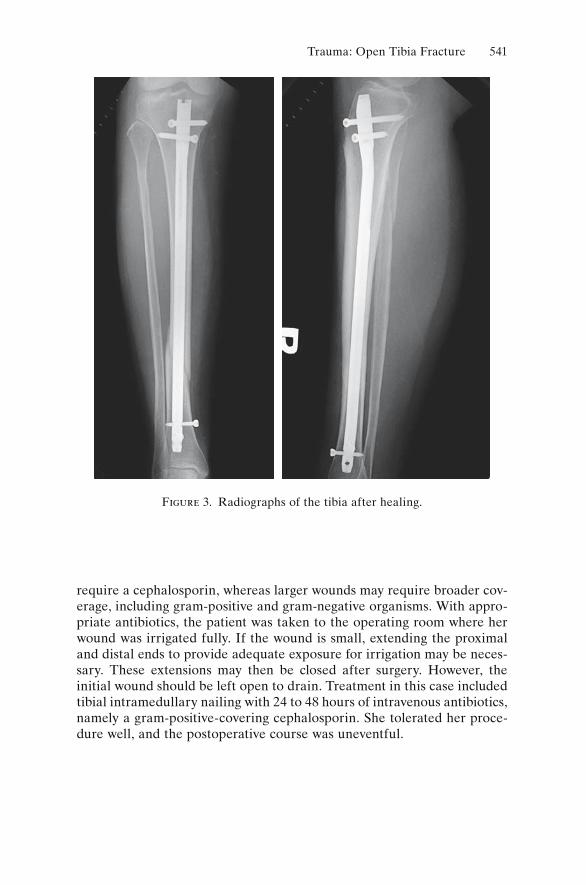
Discussion of the clinical and radiographic findings was done with the patient in regard to the nature of the fracture itself with the small skin opening. The patient was given intravenous antibiotics and was taken immediately to the operating room for irrigation and débridement of the open tibial wound with intramedullary nail fixation. Her postoperative course was uncomplicated. Her wound healed uneventfully with no signs or symptoms of infection and, over the next 3 to 4 months, her tibia fracture healed with excellent results and alignment. Preoperative, postoperative, and healed X-rays are shown in Figures 1, 2, and 3, respectively.
Discussion
This woman presented with isolated right lower extremity trauma after a fall. However, it is important not to be distracted by her presenting symp-toms. Patients that are brought to an emergency room who have question-
Trauma: Open Tibia Fracture 539
Figure 1. Preoperative radiographs show spiral tibia fracture with proximal fibular fracture.
able consciousness or are obviously inebriated should be treated as a multitrauma victim before focusing on an obvious injury. It is important to obtain as complete a history as possible from these patients or family members or friends who have brought the patient to the emergency room to determine exactly the events surrounding the circumstances bringing the patient to the emergency room. In this case, there was alcohol involved, which may cloud the history or judgment of the patient. It is important to go through the advanced trauma life support (ATLS) protocol, which starts with evaluating the airway, breathing, and circulation. When these parameters are deemed stable, then a secondary survey may be performed. In this patient’s case, although alcohol was involved, her neck was stabilized with a collar appropriately
540 S.T. Sauer
Figure 2. Immediate postoperative radiograph shows intramedullary nailing.
until she was not intoxicated, which was postoperatively the next day. She was then cleared from the clinical and radiographic standpoint. However, while maintained in the collar, a secondary survey was performed with no obvious other injuries noted and further trauma ruled out with appropr-iate radiographs, namely of the chest and pelvis, in conjunction with her C-spine radiographs, which had been done on admission.
Once the secondary survey was performed and the isolated injury was identified, in her case an open tibia fracture, appropriate treatment could be applied. In most cases, tibia fractures, if minimally displaced, well aligned, and without evidence of open injury, may be treated conserva-tively: a long leg cast for 6 to 8 weeks converting to a short leg cast after healing and beginning of weight-bearing between 8 and 12 weeks. In this case, an open injury made it necessary to operatively wash out the wound to prevent infection. Intravenous antibiotics were given immediately in conjunction with the size of the wound. Typically wounds 1 cm or less
Trauma: Open Tibia Fracture 541
require a cephalosporin, whereas larger wounds may require broader cov-erage, including gram-positive and gram-negative organisms. With appro-priate antibiotics, the patient was taken to the operating room where her wound was irrigated fully. If the wound is small, extending the proximal and distal ends to provide adequate exposure for irrigation may be neces-sary. These extensions may then be closed after surgery. However, the initial wound should be left open to drain. Treatment in this case included tibial intramedullary nailing with 24 to 48 hours of intravenous antibiotics, namely a gram-positive-covering cephalosporin. She tolerated her proce-dure well, and the postoperative course was uneventful.
Figure 3. Radiographs of the tibia after healing.
Herniated Nucleus Pulposus
Sam W. Wiesel
History
A 34-year-old lawyer arose with the hope of taking an early-morning run. As he began, he felt sudden back pain to the point where he had to stop running. Walking, the back pain became less intense, but he noticed pain down the leg all the way into the foot with paresthesia (numbness and tin-gling) into the foot. He had trouble walking and came into the office for an evaluation.
Physical Examination
The patient is a well-nourished young man in moderate distress. He had decreased range of motion of the lumbosacral spine with no palpable spasm. His straight leg raising test was positive in that it produced pain all the way down the leg. He had a weak extensor hallioxic longus (EHL). His hip examination was normal.
Laboratory Studies
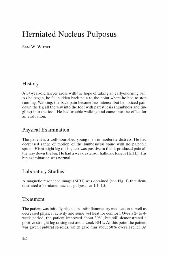
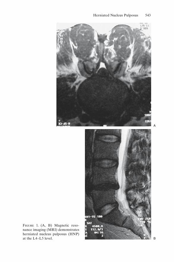
A magnetic resonance image (MRI) was obtained (see Fig. 1) that dem-onstrated a herniated nucleus pulposus at L4–L5.
Treatment
The patient was initially placed on antiinflammatory medication as well as decreased physical activity and some wet heat for comfort. Over a 2- to 4-week period, the patient improved about 30%, but still demonstrated a positive straight leg raising test and a weak EHL. At this point the patient was given epidural steroids, which gave him about 50% overall relief. At
542
Herniated Nucleus Pulposus 543
B
A
Figure 1. (A, B) Magnetic reso-nance imaging (MRI) demonstrates herniated nucleus pulposus (HNP) at the L4–L5 level.
the 10-week mark, because the patient was really unable to function, it was recommended that he have a laminectomy with removal of the herniated disk at L4–L5. Patient underwent the procedure, and postoperatively his pain and paresthesias disappeared. He was placed in a rehabilitation program and 1 year later was doing well.
Discussion
This is a classical history for a herniated nucleus pulposus. It generally occurs in the second and third decades of life. This patient had a positive straight leg raising and a positive neurologic in the form of an EHL that coincided with his herniated disk between L4 and L5 involving the L5 nerve root. Conservative treatment was instituted because the simple fact of a neurologic deficit such as a weak EHL does not mandate surgical intervention. If the patient can get good subjective pain relief using non-operative measures, it is preferred. The patient was treated nonoperatively for a total of 10 weeks. The recommendation is that if one cannot get sat-isfactory pain relief within 3 months and there is a correlation between the physical examination and an MRI, surgery is indicated. This patient did well with the laminectomy between L4 and L5 and was able to resume all his activities.
544 S.W. Wiesel
Osteomyelitis
Steven C. Scherping, Jr.
History
The patient is an 18-year-old man who is referred to the office for evalua-tion of a painful and swollen left knee. Two weeks before presentation he suffered a minor twisting injury to the knee while playing soccer. This injury was not associated with any significant pain at the time, and he was able to continue his routine activities, including playing soccer. Over the ensuing 4 to 5 days, the knee and the distal thigh became progressively more painful with some swelling noted in the knee. He denies any giving-way or locking of the knee. The pain is present at all times, only slightly worsened with weight-bearing activities. He denies any prior history of injury to the extremity. He denies pain at any other site. He has no history of recent illness other than a laceration to his foot that occurred 3 weeks ago and which was complicated by a local wound infection requiring treat-ment with oral antibiotics. The laceration subsequently healed without event, and he has no residual swelling or pain in the foot. His past medical history is otherwise benign. On review of symptoms, he has noted a mild sense of fatigue, in association with a sense of a low-grade fever, although he has not taken his temperature and denies any frank rigors or chills.
Physical Examination
This generally healthy-appearing young adult male is afebrile. Gait is antalgic with restricted motion through the left knee. His left knee is swollen in appearance with an effusion. Tenderness is appreciable over the distal femur, more so of the medial condyle. Range of motion of the knee is from 0 to 120 degrees in association with some discomfort. No mechani-cal block or instability is detected. His neurovascular examination is normal in the extremity. No adenopathy is appreciated in the groin or popliteal fossa. Range of motion of his ankle and hip is painless and sym-metrical with the uninvolved extremity.
545
546 S.C. Scherping, Jr.
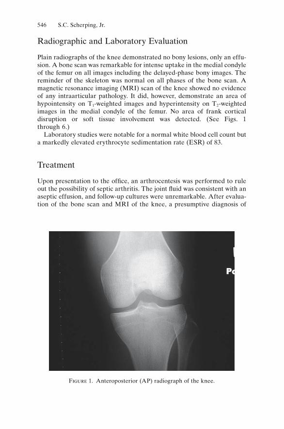
Radiographic and Laboratory Evaluation
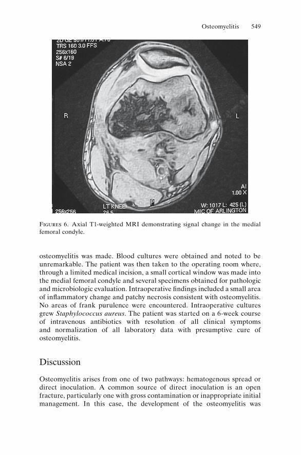
Plain radiographs of the knee demonstrated no bony lesions, only an effu-sion. A bone scan was remarkable for intense uptake in the medial condyle of the femur on all images including the delayed-phase bony images. The reminder of the skeleton was normal on all phases of the bone scan. A magnetic resonance imaging (MRI) scan of the knee showed no evidence of any intraarticular pathology. It did, however, demonstrate an area of hypointensity on T1-weighted images and hyperintensity on T2-weighted images in the medial condyle of the femur. No area of frank cortical disruption or soft tissue involvement was detected. (See Figs. 1 through 6.)
Laboratory studies were notable for a normal white blood cell count but a markedly elevated erythrocyte sedimentation rate (ESR) of 83.
Treatment
Upon presentation to the office, an arthrocentesis was performed to rule out the possibility of septic arthritis. The joint fl uid was consistent with an aseptic effusion, and follow-up cultures were unremarkable. After evalua-tion of the bone scan and MRI of the knee, a presumptive diagnosis of
Figure 1. Anteroposterior (AP) radiograph of the knee.
Osteomyelitis 547
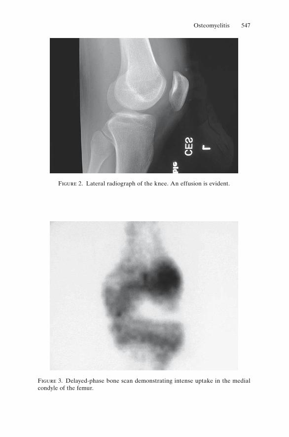
Figure 2. Lateral radiograph of the knee. An effusion is evident.
Figure 3. Delayed-phase bone scan demonstrating intense uptake in the medial condyle of the femur.
548 S.C. Scherping, Jr.
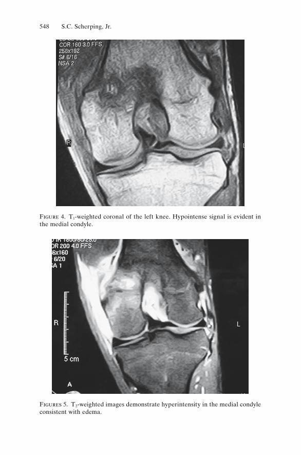
Figure 4. T1-weighted coronal of the left knee. Hypointense signal is evident in the medial condyle.
Figures 5. T2-weighted images demonstrate hyperintensity in the medial condyle consistent with edema.
Osteomyelitis 549
osteomyelitis was made. Blood cultures were obtained and noted to be unremarkable. The patient was then taken to the operating room where, through a limited medical incision, a small cortical window was made into the medial femoral condyle and several specimens obtained for pathologic and microbiologic evaluation. Intraoperative findings included a small area of inflammatory change and patchy necrosis consistent with osteomyelitis. No areas of frank purulence were encountered. Intraoperative cultures grew Staphylococcus aureus. The patient was started on a 6-week course of intravenous antibiotics with resolution of all clinical symptoms and normalization of all laboratory data with presumptive cure of osteomyelitis.
Discussion
Osteomyelitis arises from one of two pathways: hematogenous spread or direct inoculation. A common source of direct inoculation is an open fracture, particularly one with gross contamination or inappropriate initial management. In this case, the development of the osteomyelitis was
Figures 6. Axial T1-weighted MRI demonstrating signal change in the medial femoral condyle.
550 S.C. Scherping, Jr.
undoubtedly from transient bacteria related to the superficial foot infec-tion. If found early, before the development of an abscess or a bony seques-trum, osteomyelitis is generally be successfully treated with appropriate antibiotic therapy alone, which is why the timely diagnosis of a bone infec-tion plays a critical role in minimizing the morbidity of treatment. In some instances, however, surgical treatment, or at a minimum a biopsy, will be necessary to precisely define the bacteriologic origin and thereby allow a specific course of antibiotic therapy. In this case, as the presumed osteo-myelitis was of uncertain origin, the biopsy and limited debridement were performed to refine the selection and course of antibiotic therapy. In most instances of a confirmed osteomyelitis, a minimum of 6 to 8 weeks of treatment is necessary.
Hip Osteoarthritis
Brian G. Evans
History
This patient is a 65-year-old woman who has had a long-standing history of pain in the right groin. The pain has been gradually getting more severe, resulting in the patient limping when she walks for more than 10 to 15 minutes. The pain began approximately 6 to 7 years ago. Initially, she thought the pain was caused by a groin pull. She tried treating it on her own with nonsteroidal antiinflammatory medications such as ibuprofen, which provided good symptomatic relief. Over the past 1 to 2 years, however, the pain has become much more pronounced. She has pain at night when she turns over and significant pain and stiffness in the morning when she fi rst gets up out of bed. Also, with prolonged sitting, such as during a car ride or if she goes out to dinner, after approximately 30 to 40 minutes, she has significant pain in the hip that requires her to get up and move about to alleviate the pain. If she has been sitting for more than 10 to 15 minutes, when she arises from the sitting position she also has a significant degree of stiffness when she first starts walking. Initially, this pain was well treated with ibuprofen; however, currently she is having a significant degree of pain and discomfort, even when taking 600 to 800 mg ibuprofen three times a day. She has also had increasing difficulty putting on her socks and tying shoes. She has been unable to clip her toenails for several years, particularly on the right side. She has a history of some mild hypertension, but other-wise she is in good health and feels quite limited by her right-sided groin pain. She denies any numbness, tingling, or weakness in the leg. She has not had any buckling. The pain does not worsen with coughing or sneezing, and she has no pain in the buttock or posterior thigh.
Physical Examination
The patient walks with a significant coxalgic gait consisting of a reduced stance phase and a Trendelenburg lurch. When she stands, she stands with the right knee slightly flexed with her weight on the left side. She does have
551
552 B.G. Evans
stiffness and a limp when she moves from a chair in the examination room to sit on the examination table.
When examining the lower extremities, she has 5/5 motor strength in the extensor hallucis longus, tibialis anterior, gastrocnemius-soleus, and quad-riceps muscles. She has no decrease in sensation to light touch over both lower extremities. With the patient supine, both legs have equal limb lengths. On assessing range of motion, on the right side she has flexion toapproximately 85 degrees, extension to the table, internal rotation to 0 degrees, and external rotation of approximately 45 degrees, and adduction of 5 degrees and abduction of approximately 30 degrees. All range of motion is limited to pain. On assessing for flexion contracture with the Thomas test, she has a 15 degree flexion contracture on the right hip and none on the left. On the left side she has flexion to 105 degrees, full exten-sion, internal rotation of 15, external rotation of 70, and adduction of 20 and abduction of 45 degrees. She has no pain with range of motion of her knees or ankles. She has no exacerbation of her pain with extension or fl exion or lateral bending of the lumbar spine.
Radiographs
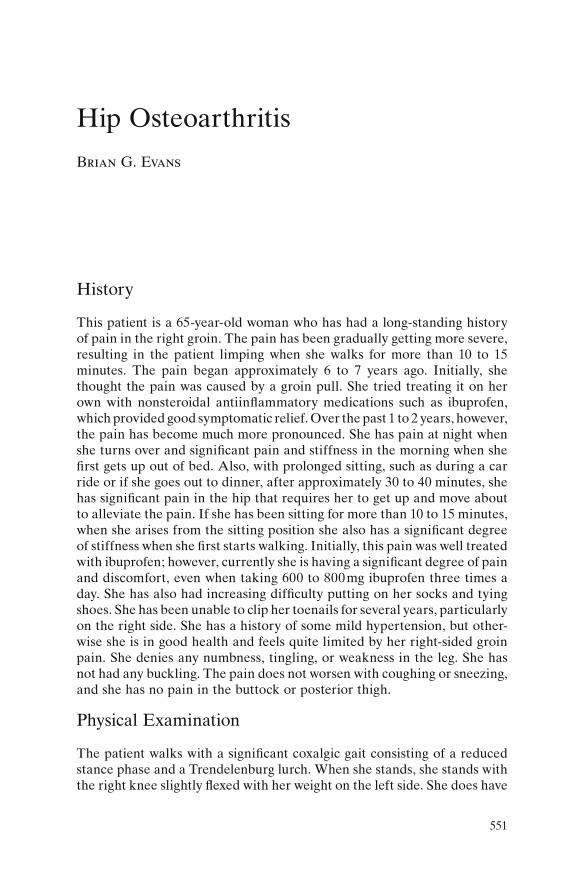
Figure 1 is an anteroposterior (AP) radiograph of the pelvis showing both hips that demonstrates a normal left hip with a well-maintained articular space and no osteophyte formation. The right hip, however, has extensive osteoarthritic changes. There is no articular space, extensive osteophyte
Figure 1. Anteroposterior (AP) radiograph of the pelvis showing both hips.
Hip Osteoarthritis 553
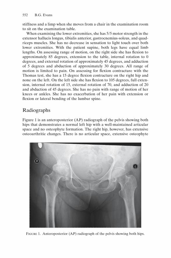
Figure 2. Lateral radiograph of the right hip demonstrates extensive osteoarthri-tis of the hip, with no articular space, subchondral sclerosis, and osteophyte formation.
formation is noted, and sclerosis or increased density in the bone is noted both in the femoral head as well as on the acetabular side. Figure 2, a lateral radiograph of the right hip, again demonstrates extensive osteoarthritis of the hip, with no articular space, subchondral sclerosis, and osteophyte formation.
Diagnosis
A patient with substantial osteoarthritis of the right hip, which is signifi -cant clinically in terms of pain and reduction in function and radiographi-cally in terms of marked changes on the plain radiographs.
Treatment
The treatment alternatives for this patient are either to continue with current medications, with stronger antiinflammatories, and with reduction in activities or use of a cane. The other treatment alternative would be a total hip arthroplasty. The benefit of total hip arthroplasty would be relief
554 B.G. Evans
of pain and restoration of ambulatory function and ability. The risks of the surgery, as indicated in Chapter 11, are the risks of bleeding, thromboem-bolic disease, dislocation, and loosening of the components. These possi-bilities were reviewed with the patient in detail, and the patient elected to proceed with total hip arthroplasty.
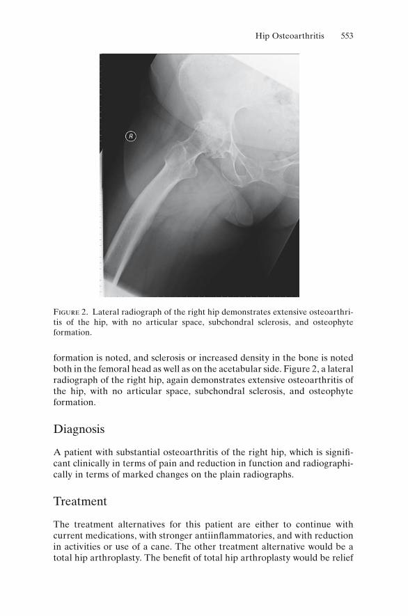
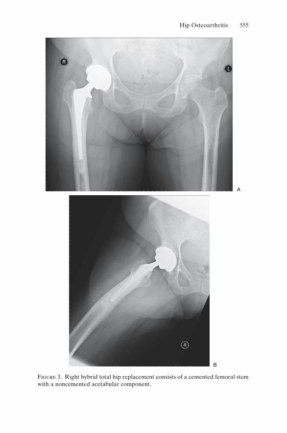
Figure 3 shows the patient after having had a right hybrid total hip replacement. This replacement consists of a cemented femoral stem with a noncemented acetabular component. The components appear to be well fi xed and well aligned. There is good interdigitation of the bone cement into the femoral cortex, and the overall limb alignment and anatomy have been restored.
The patient did well subsequent to the surgery. She returned to the office at 2 weeks postoperatively, able to ambulate with only the use of a cane with no pain, taking no pain medications. She returned again 6 weeks after the surgery and was ambulating freely without the use of a cane, crutch, or walker. She was able to ambulate for 40 to 60 minutes with no difficulty whatsoever and had no limp in her gait and no pain with routine activities. She was also able to put a sock on her right foot, clip her right toenails, and tie a shoe on her right foot without difficulty.
This patient has done well after a total hip arthroplasty. She should enjoy continued success with the arthroplasty for approximately 15 to 20 years.
Discussion
The treatment of osteoarthritis of the hip is based upon exploring nonop-erative management as long as possible. Nonoperative management includes NSAIDs, physical therapy, the use of a cane, and, if appropriate, weight loss. In the end, however, the arthritis becomes increasingly symptomatic. As the pain and limitation of mobility become more severe, the next step in treatment is total hip replacement, done after nonoperative manage-ment fails. The timing for total hip replacement is elective; it is based solely upon the patient’s pain and the impact of the pain upon their life and activities.
Total hip replacement is an excellent surgical option. It has an extremely high success rate, and after recovery the patient moves and ambulates completely normally, which allows a near-complete return to normal activi-ties. However, the patients should not participate in running or cutting activity such as jogging and racquet sports after total hip replacement as these activities may result in early loosening and wear of the implanted devices. They are encouraged to keep aerobically fit with nonimpact aerobic activities such as cycling and swimming.
Total hip replacement commonly results in excellent functional return. Some patients, years after surgery, may have to stop and think which hip they had replaced. That is the definition of a good result.
Hip Osteoarthritis 555
Figure 3. Right hybrid total hip replacement consists of a cemented femoral stem with a noncemented acetabular component.
A
B
Slipped Capital Femoral Epiphysis
John N. Delahay
History
V.C. is a slightly overweight 11-year-old Caucasian girl who presented with the chief complaint of leg pain and limp for a period of 3 months. The pain began somewhat insidiously and without antecedent trauma. The pain was dull and aching in character and without significant radiation. The pain was relieved by rest and accentuated by standing and walking. She had actually attempted to play soccer during this period but had to stop because of the pain.
Her past medical history was entirely negative. However, of note, her brother had previously been diagnosed with slipped capital epiphysis and had been operated upon sequentially for bilateral disease.
Physical Examination
Pertinent findings were confined to the musculoskeletal system. Observa-tion of gait demonstrated an antalgic limp on the right side. She was, however, able to walk without an assistive device and in fact was able to stand only on the affected side with the opposite leg off the ground.
In the resting position (hip extended), the leg tended to lie in the exter-nally rotated position. When evaluating range of motion (ROM), the hip was noted to flex up in the externally rotated position. Attempts to internally rotate the hip in flexion or extension demonstrated the inability to do so as well as guarding because of pain. The flexion–extension range was normal. Leg lengths were equal, and the neurovascular status of the limb intact.
Laboratory and Radiographic Evaluation
Complete blood count (CBC), sedimentation rate, C-reactive protein (CRP), thyroid hormone assays, and renal function studies (BUN, creati-nine, UA) were all within normal limits.
556
Slipped Capital Femoral Epiphysis 557
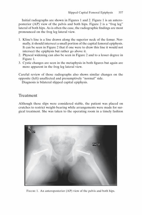
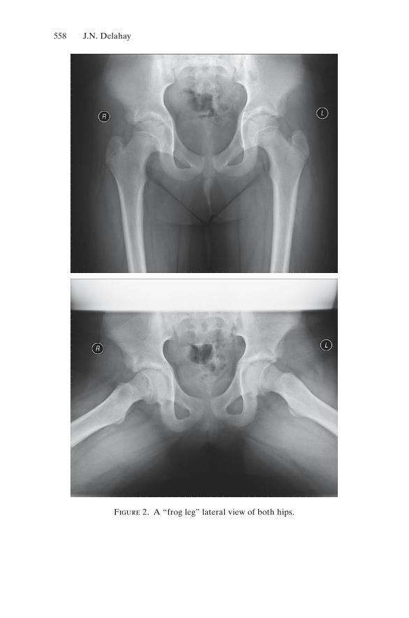
Initial radiographs are shown in Figures 1 and 2. Figure 1 is an antero-posterior (AP) view of the pelvis and both hips. Figure 2 is a “frog leg” lateral of both hips. As is often the case, the radiographic findings are most pronounced on the frog leg lateral view.
1. Kline’s line is a line drawn along the superior neck of the femur. Nor-mally, it should intersect a small portion of the capital femoral epiphysis. It can be seen in Figure 2 that if one were to draw this line it would not intersect the epiphysis but rather go above it.
2. Physeal widening can also be seen in Figure 2 and to a lesser degree in Figure 1.
3. Cystic changes are seen in the metaphysis in both figures but again are more apparent in the frog leg lateral view.
Careful review of those radiographs also shows similar changes on the opposite (left) unaffected and presumptively “normal” side.
Diagnosis is bilateral slipped capital epiphysis.
Treatment
Although these slips were considered stable, the patient was placed on crutches to restrict weight-bearing while arrangements were made for sur-gical treatment. She was taken to the operating room in a timely fashion
Figure 1. An anteroposterior (AP) view of the pelvis and both hips.
558 J.N. Delahay
Figure 2. A “frog leg” lateral view of both hips.
Slipped Capital Femoral Epiphysis 559
(2 days after presentation) and underwent bilateral percutaneous pinning in situ using 7.3-mm cannulated screws. One screw was used on each side. No attempt was made to reduce the slips. Postoperatively, she was dis-charged home on crutches. Her weight-bearing status was slowly advanced, and the crutches were discontinued when the adductor spasm had resolved. No plans exist to remove hardware. Follow-up X-rays are planed at 6-month intervals until growth completion.
Discussion
Slipped capital femoral epiphysis (SCFE) is a relatively common cause of hip pain and limp in the preadolescent population. Typically, however, it affects Afro-Americans more commonly than Caucasians and males more commonly than females. Typically the patient is somewhat overweight and has had the pain and limp for several months at the time of presentation. The incidence of bilaterality is somewhat debated, but most texts report it to be about 25%. There is rarely a family history (as is the case here), and that of course raises the question of genetic association (which has never been demonstrated).
Because the patient was a female and Caucasian with a strong family history, the other issue to be considered is an endocrine etiology, and this was the rationale for obtaining thyroid and renal function studies. One other area to be considered would be growth hormone abnormalities.
Her presenting complaints were rather typical. The pain was more local-ized to the thigh; this phenomenon is usually explained as referral along the course of the obturator nerve. The pain was mechanical and relieved by rest. The limp was antalgic with a shortened stance time on the affected limb.
The X-rays were diagnostic, showing the most typical findings: positive Kline’s line on the right and arguably the left, and physeal widening and metaphyseal cysts on both sides. Additional imaging studies are not needed in this patient. Occasionally, when the diagnosis is in question, a magnetic resonance imaging (MRI) scan or a Tc-99 bone scan may provide confir-mation of the diagnosis, although some authors question the efficacy of either. There are many who believe that if a child presents with the classic history and physical findings, they should be considered as having “pre-slips” and treated as such.
This girl’s treatment was the norm: restrict weight-bearing and pin the SCFE in situ in a timely manner. No attempt to reduce the slip should be made when the slip is stable as doing so increases the risk of vascular necrosis. A child with a stable slip can still bear weight on the limb and walk. The child with an unstable slip cannot bear weight on the extremity. Pinning in situ is the standard treatment for this condition; the goal of the
560 J.N. Delahay
treatment is primarily to stop any further slipping and initiate or facilitate plate closure.
In this case, both hips were pinned because there was radiographic evi-dence of bilateral disease. This author and others routinely pin both sides whether or not there is evidence of bilaterality, the rationale being the rela-tively high incidence of bilaterality (this patient’s brother had to have his pinned sequentially) and resultant leg length discrepancy that results from unilateral pinning. In the case of an 11-year-old girl, who would have about 3 years of growth left, one could expect a 1.8-cm discrepancy (6 mm × 3).
This girl’s postoperative course was benign. The two major complica-tions of SCFE are avascular necrosis (AVN) and chondrolysis. AVN is usually considered a complication of the treatment of the disease, resulting from aggressive manipulation to reduce the slip or osteotomies of the femoral neck. Chondrolysis is considered by most to be a complication of the disease itself. This loss of the articular cartilage is much more likely to be seen in African-Americans and can actually be seen at the time of presentation before treatment has been initiated.
Overall, the prognosis for SCFE is good. With early diagnosis and appro-priate surgical treatment, most children can be restored to normal function rather quickly.
Cubital Tunnel Syndrome
Mustafa A. Haque
History
This patient is a healthy, 34-year-old woman who started to develop pain, numbness, and tingling in her dominant right hand 3 months ago. She points to the medial elbow as the source of her symptoms but notes radiation all the way to the tips of her ring and small finger. She is a laboratory medical researcher and has worsening of her symptoms while pipetting under a sterile hood. She is starting to have weakness with opening jars or grasping objects. She has no history of injury or trauma. Her remaining medical and surgical history are negative; in particular, she has no history of diabetes, hypothy-roidism, or renal failure. She is not on any medications and has no allergies.
Physical Examination
The patient is a healthy, athletic-appearing female in no distress. Her upper extremities and left hand have no deformity and full active range of motion. She is tender to palpation over the ulnar nerve just posterior to the medial epicondyle, and tapping over the nerve at that site creates tingling that radi-ates all the way to the small fingertip. Full elbow flexion for more than 30 seconds reproduces her symptoms. Elbow flexion greater than 90 degrees causes subluxation of the ulnar nerve on to the medial epicondyle. The patient is nontender over the medial and lateral epicondyle or over the wrist. Carpal tunnel compression and Phalen’s test are negative, as is Tinel’s testing at the wrist. Sensation to light touch is normal over the thumb through radial border of the ring finger and subjectively decreased over the ulnar border of the ring fi nger and all of the small finger. She has slight finger abduction weakness.
Radiographs and Laboratory Values
This patient’s X-rays were completely normal. Her laboratory studies were also unremarkable; in particular, serum glucose and thyroid function tests were within normal limits.
561
562 M.A. Haque
Diagnosis and Treatment
This patient has compression of the ulnar nerve at the elbow, more com-monly known as cubital tunnel syndrome. She was treated with nonsteroi-dal antiinflammatory drugs (NSAIDs), a custom-molded night splint to avoid elbow flexion greater than 30 degrees, ergonomic workstation adjust-ments, and therapy. Her symptoms unfortunately worsened over the next 3 months despite these interventions, and she developed significant finger abduction weakness. She was then surgically treated with an anterior sub-cutaneous transposition of the ulnar nerve. Within a few weeks her symp-toms resolved, and by 3 months after surgery, she had regained completely normal hand and elbow function.
Discussion
Cubital tunnel syndrome results from compression of the ulnar nerve around the elbow. Patients classically present with medial elbow pain and paresthesias in an ulnar nerve sensory distribution (the ulnar half of the ring finger and all the small finger). As the condition progresses, patients can develop weakness to finger abduction. Tinel’s sign, or tapping on the nerve dorsal to the medial epicondyle, causes paresthesias radiating to the small finger. A positive elbow flexion test reproduces the patient’s symp-toms after 60 seconds of maximal elbow flexion. Another classic late finding is the Froment’s sign, in which attempts at key pinch result in flexion of the thumb interphalangeal (IP) joint as the extensor pollicis longus (EPL) and fl exor pollicis longus (FPL) compensate for adductor weakness.
The differential diagnosis includes medial epicondylitis, cervical radicu-lopathy, thoracic outlet syndrome, carpal tunnel syndrome, and ulnar tunnel syndrome, and several of these conditions can occur concurrently. History and physical examination are the keys to making the correct diagnosis, but EMG and nerve conduction studies are helpful. The electri-cal criteria for confirming cubital tunnel syndrome includes a drop in conduction velocity of 11 m/s or greater across the elbow compared to below the elbow. Lesser velocity decreases, however, do not rule out the disease.
Treatment is usually conservative with nighttime extension splinting, NSAIDs, and rest. Occasionally, hand therapy with nerve and tendon gliding exercises can be helpful. If a patient’s symptoms do not resolve after 3 months of this management, one can consider surgical intervention. The options include in situ decompression, medial epicondylectomy, and anterior transposition of the ulnar nerve. The anterior transpositions include subcutaneous, intramuscular, and submuscular techniques. It is important to completely release the arcade of Struthers (see Fig. 1), the medial intermuscular septum (see Fig. 2), the fascia over the groove dorsal
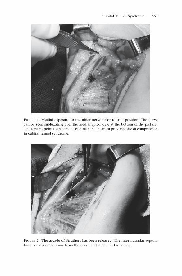
Cubital Tunnel Syndrome 563
Figure 1. Medial exposure to the ulnar nerve prior to transposition. The nerve can be seen subluxating over the medial epicondyle at the bottom of the picture. The forceps point to the arcade of Struthers, the most proximal site of compression in cubital tunnel syndrome.
Figure 2. The arcade of Struthers has been released. The intermuscular septum has been dissected away from the nerve and is held in the forcep.
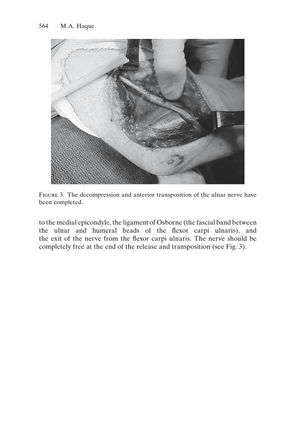
564 M.A. Haque
Figure 3. The decompression and anterior transposition of the ulnar nerve have been completed.
to the medial epicondyle, the ligament of Osborne (the fascial band between the ulnar and humeral heads of the flexor carpi ulnaris), and the exit of the nerve from the flexor carpi ulnaris. The nerve should be completely free at the end of the release and transposition (see Fig. 3).
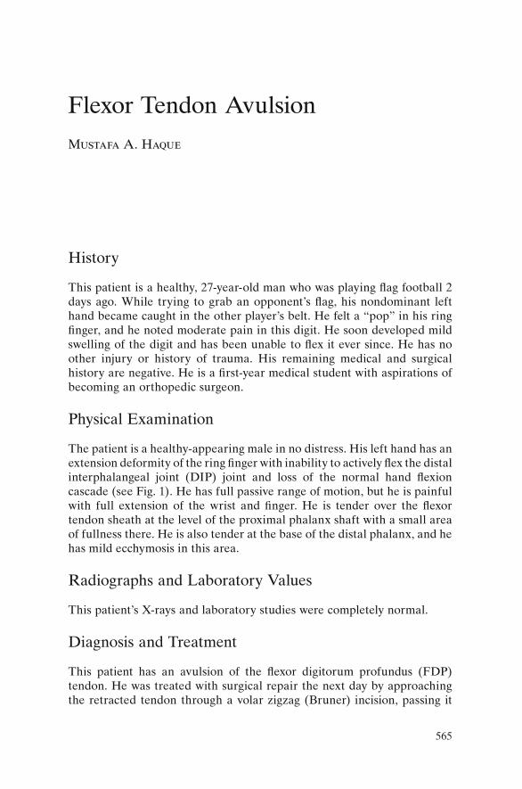
Flexor Tendon Avulsion
Mustafa A. Haque
History
This patient is a healthy, 27-year-old man who was playing flag football 2 days ago. While trying to grab an opponent’s flag, his nondominant left hand became caught in the other player’s belt. He felt a “pop” in his ring fi nger, and he noted moderate pain in this digit. He soon developed mild swelling of the digit and has been unable to flex it ever since. He has no other injury or history of trauma. His remaining medical and surgical history are negative. He is a first-year medical student with aspirations of becoming an orthopedic surgeon.
Physical Examination
The patient is a healthy-appearing male in no distress. His left hand has an extension deformity of the ring finger with inability to actively flex the distal interphalangeal joint (DIP) joint and loss of the normal hand flexion cascade (see Fig. 1). He has full passive range of motion, but he is painful with full extension of the wrist and finger. He is tender over the flexor tendon sheath at the level of the proximal phalanx shaft with a small area of fullness there. He is also tender at the base of the distal phalanx, and he has mild ecchymosis in this area.
Radiographs and Laboratory Values
This patient’s X-rays and laboratory studies were completely normal.
Diagnosis and Treatment
This patient has an avulsion of the flexor digitorum profundus (FDP) tendon. He was treated with surgical repair the next day by approaching the retracted tendon through a volar zigzag (Bruner) incision, passing it
565
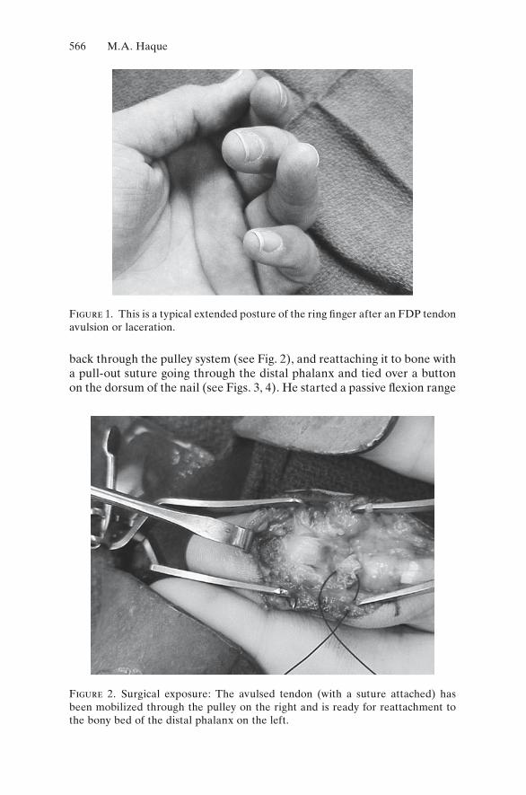
566 M.A. Haque
back through the pulley system (see Fig. 2), and reattaching it to bone with a pull-out suture going through the distal phalanx and tied over a button on the dorsum of the nail (see Figs. 3, 4). He started a passive flexion range
Figure 1. This is a typical extended posture of the ring finger after an FDP tendon avulsion or laceration.
Figure 2. Surgical exposure: The avulsed tendon (with a suture attached) has been mobilized through the pulley on the right and is ready for reattachment to the bony bed of the distal phalanx on the left.
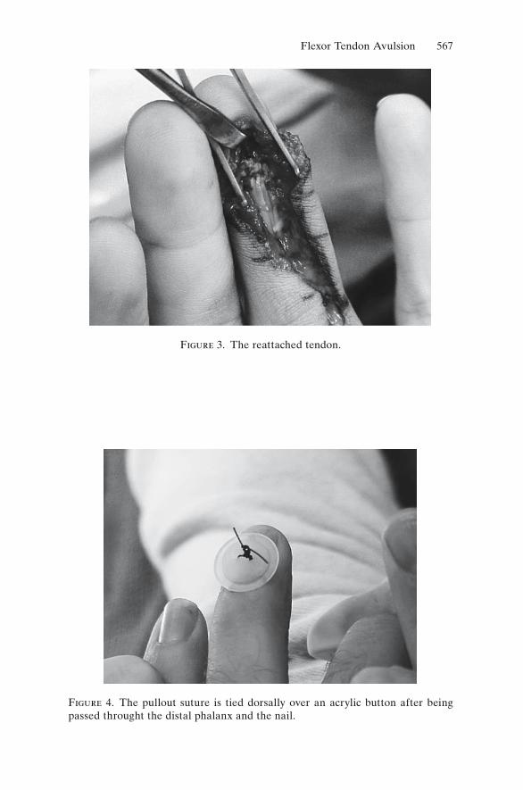
Flexor Tendon Avulsion 567
Figure 3. The reattached tendon.
Figure 4. The pullout suture is tied dorsally over an acrylic button after being passed throught the distal phalanx and the nail.
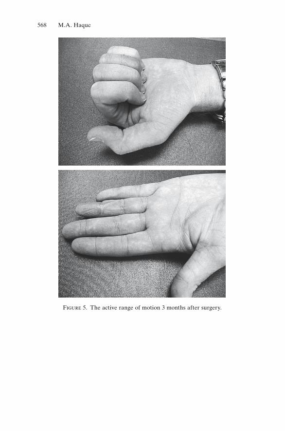
568 M.A. Haque
Figure 5. The active range of motion 3 months after surgery.
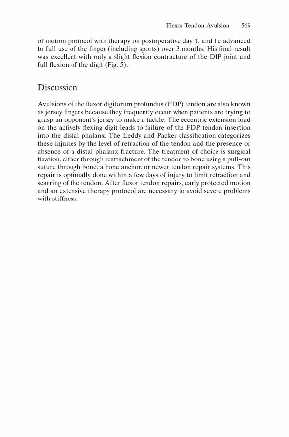
Flexor Tendon Avulsion 569
of motion protocol with therapy on postoperative day 1, and he advanced to full use of the finger (including sports) over 3 months. His final result was excellent with only a slight flexion contracture of the DIP joint and full flexion of the digit (Fig. 5).
Discussion
Avulsions of the flexor digitorum profundus (FDP) tendon are also known as jersey fingers because they frequently occur when patients are trying to grasp an opponent’s jersey to make a tackle. The eccentric extension load on the actively flexing digit leads to failure of the FDP tendon insertion into the distal phalanx. The Leddy and Packer classification categorizes these injuries by the level of retraction of the tendon and the presence or absence of a distal phalanx fracture. The treatment of choice is surgical fi xation, either through reattachment of the tendon to bone using a pull-out suture through bone, a bone anchor, or newer tendon repair systems. This repair is optimally done within a few days of injury to limit retraction and scarring of the tendon. After flexor tendon repairs, early protected motion and an extensive therapy protocol are necessary to avoid severe problems with stiffness.
Acute Anterior Cruciate Ligament Rupture with Strain of the Medial Collateral Ligament
John J. Klimkiewicz
History
This patient is a 20-year-old college football player who sustained an injury to his left knee. He is a lineman and stated another player had fallen on the lateral aspect of his left knee. He stated that his knee was extended at the time of the injury and described a “pop” within his knee. He was unable to continue playing, and required assistance off the field. He stated that his knee subsequently swelled over the next few hours and is currently unable to bear weight without significant discomfort. He denies any prior injury to his knee.
Physical Examination
His height is 6 feet 3 inches and his weight 245 lbs. He is unable to ambulate without crutches. His knee shows no signs of bruising or ecchymoses. His range of motion is 5 degrees to 90 degrees of flexion. He has a significant effusion (see Fig. 1). There is no patellar tenderness and no crepitation with range of motion, and no patellar apprehension. Patient has tenderness over the medial epicondyle of the distal femur and over the lateral joint line. There is no medial joint line tenderness. McMurray sign is positive for pain at 90 degrees of flexion over the lateral joint line. He is stable to varus stress maneuvers at both full extension and 30 degrees of flexion. Although stable to valgus stress maneuvers in full extension, at 30 degrees the patient has 1+ laxity with pain during this maneuver. He has a grade II Lachman with no endpoint, and also has grade II laxity to anterior drawer maneuvers at 90 degrees of flexion. There is a positive pivot shift. There is no increased laxity to posterior drawer maneuver at 90 degrees of flexion. His neurologic examination and vascular examination are unremarkable.
570
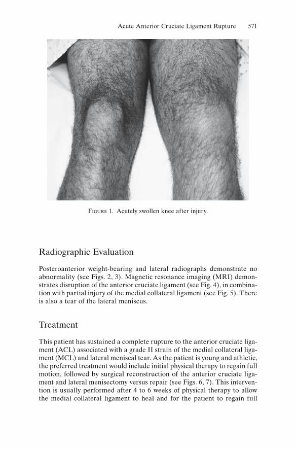
Acute Anterior Cruciate Ligament Rupture 571
Radiographic Evaluation
Posteroanterior weight-bearing and lateral radiographs demonstrate no abnormality (see Figs. 2, 3). Magnetic resonance imaging (MRI) demon-strates disruption of the anterior cruciate ligament (see Fig. 4), in combina-tion with partial injury of the medial collateral ligament (see Fig. 5). There is also a tear of the lateral meniscus.
Treatment
This patient has sustained a complete rupture to the anterior cruciate liga-ment (ACL) associated with a grade II strain of the medial collateral liga-ment (MCL) and lateral meniscal tear. As the patient is young and athletic, the preferred treatment would include initial physical therapy to regain full motion, followed by surgical reconstruction of the anterior cruciate liga-ment and lateral menisectomy versus repair (see Figs. 6, 7). This interven-tion is usually performed after 4 to 6 weeks of physical therapy to allow the medial collateral ligament to heal and for the patient to regain full
Figure 1. Acutely swollen knee after injury.
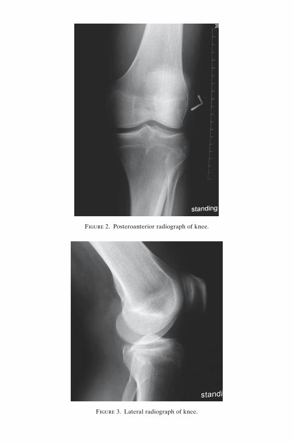
Figure 2. Posteroanterior radiograph of knee.
Figure 3. Lateral radiograph of knee.
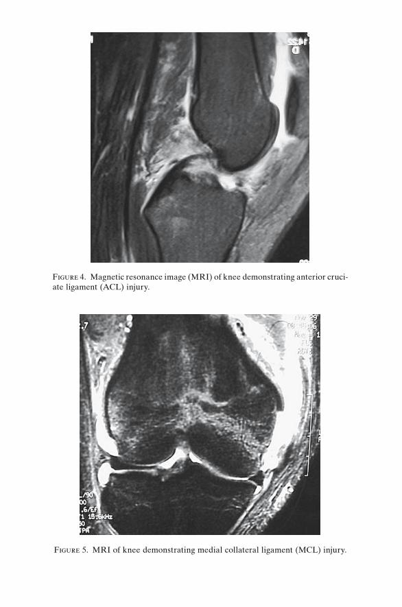
Figure 4. Magnetic resonance image (MRI) of knee demonstrating anterior cruci-ate ligament (ACL) injury.
Figure 5. MRI of knee demonstrating medial collateral ligament (MCL) injury.
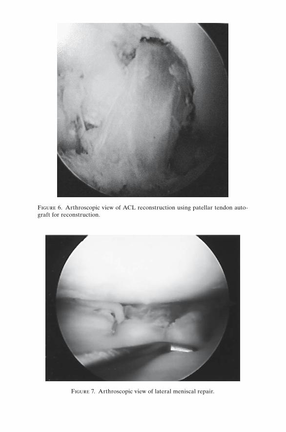
Figure 6. Arthroscopic view of ACL reconstruction using patellar tendon auto-graft for reconstruction.
Figure 7. Arthroscopic view of lateral meniscal repair.
Acute Anterior Cruciate Ligament Rupture 575
range of motion. Although different graft options exist to reconstruct the anterior cruciate ligament, a central third patellar tendon autograft was chosen. Intense physical therapy is necessary postoperatively to allow a return to sport at 6 months after the procedure.
Discussion
This patient suffered an injury to the ACL, MCL, and lateral meniscus. The mechanism for this injury involved a valgus load to the extended knee. The patient often recalls hearing a pop that is shortly followed by significant swelling. There is often an immediate inability to ambulate or a significant antalgic (painful) gait. These injuries can also be associated with injuries to the collateral ligaments and injuries to the menisci or articular cartilage. This patient suffered the triad of injuries that include: an ACL rupture, strain or rupture of the MCL, and a lateral meniscal tear.
This patient’s examination is significant for increased laxity to anterior stress tests including the Lachman exam at 30 degrees and anterior drawer at 90 degrees. These tests are categorized by grades based on the degree of translation. Grade I injuries are those with laxity 0 to 5 mm greater than the contralateral side, grade II, 5 to 10 mm, and grade III, more than 10 mm. There is also a positive pivot shift that results when a valgus load is applied to the knee through a range of motion, and there is a palpable shift at 30 degrees to full extension when the tibia translates from an unreduced position secondary to ACL disruption to a reduced position in full extension. This is pathognomonic for an ACL rupture. This patient also demonstrates a partial injury to his MCL with pain at its origin (medial epicondyle). Grade I injuries involve a strain with no increased laxity on exam, grade II injuries involve increased laxity with an endpoint of this structure, and grade III injuries involve increased laxity of this structure with no endpoint present, thus representing a complete tear. This was the case for this patient. The presence of lateral joint line ten-derness with a positive McMurray test is consistent with the lateral menis-cal tear.
Current treatment for anterior cruciate ligament injuries involves an initial period of physical therapy to emphasize range of motion and modali-ties to decrease swelling. After this initial period of rest, a reconstruction is the recommended form of treatment to allow the patient to return to cutting sports. Conservative modification of activity and bracing can be considered for less active individuals. Reconstruction can be performed with the patient’s own tissue (autograft) or through the use of cadaveric tissue (allograft). At the time of surgery, associated injuries to the articular cartilage or meniscus are also addressed. This patient sustained a tear of the lateral meniscus in the vascular zone of this structure, making meniscal
576 J.J. Klimkiewicz
repair the preferred surgical technique, as compared to menisectomy. Return to sport is possible with this technique at 6 to 9 months. Long-term results are dictated by not only providing the patient with a stable recon-struction but also through preserving the patient’s meniscal structures as well as articular cartilage.
Shoulder Osteoarthritis
Raymond M. Carroll
History
A 57-year-old man presents to your office with a chief complaint of right shoulder pain. On further questioning, the patient reports that the pain began a few years ago with sharp pain in the shoulder with various activities such as reaching overhead and out to the side. He denies trauma or injury. He notes that the pain has become constant and dull at rest. With activity, the pain is sharp and is becoming unbea-rable. He reports that he is unable to use the extremity because of the pain and loss of motion. Nonsteroidal antiinflammatory medications alleviated the pain in the past but have minimal effect at this time. He denies any systemic symptoms. He has no significant past medical history other than hypertension, which is controlled with diet and exercise.
Physical Examination
In general, the patient is mildly overweight. His cervical spine motion is within normal range and is pain free. His right shoulder girdle reveals a loss of the normal contour of the anterior aspect relative to the left shoulder girdle. He has no scars, wounds, or localized atrophy. He has tenderness diffusely on the anterior and posterior joint lines. There is no fluctuance or induration noted on palpation. His active range of motion is as follows: forward elevation to 100 degrees with sharp pain at the end range of motion, external rotation is limited to 15 degrees, also painful, and internal rotation is limited to the buttock region. His passive range of motion is similarly affected. He has no focal motor or sensory deficits in the upper extremity. He has a strong and symmetrical radial pulse.
577
Differential Diagnosis
There are a number of important aspects to consider in this patient when formulating a differential diagnosis. The age of the patient is critical. Common diagnoses in this age group include rotator cuff disease and osteoarthritis. Also, it is important to note that there is no history of trauma or prior surgery. The documentation of a loss of active and passive motion in the shoulder suggests arthropathy or capsular adhesion as is seen with adhesive capsulitis. The most likely diagnoses are osteoarthritis or adhesive capsulitis (idiopathic). Although the patient may have a degenera-tive rotator cuff condition, the loss of passive motion indicates that the rotator cuff is not likely to be the culprit. Patients can develop secondary adhesive capsulitis following shoulder injuries such as a traumatic rotator cuff tear, but this patient does not have a history that is consistent with such an event.
Laboratory Results and Studies
Because of the lack of systemic or additional joint involvement, there is no indication to obtain any laboratory studies. A radiographic shoulder series including anteroposterior (AP), lateral, and axillary views is the next diagnostic step.
Imaging
The first image is an AP radiograph of the shoulder (see Fig. 1). The radio-graph shows a loss of the glenohumeral joint space, subchondral sclerosis, and osteophyte formation at the inferior aspects of the humeral head and glenoid, and subchondral cysts are easily identified. The second image is an axillary view of the shoulder (see Fig. 2). The findings are similar to those noted on the AP radiograph. There is mild wear on the posterior aspect of the glenoid, which is commonly seen in osteoarthritis of the glenohumeral joint.
Treatment
Treatment for osteoarthritis of the glenohumeral joint includes activity modification, rest, and antiinflammatory medication. Corticosteroid injec-tions may be used sparingly but are often not helpful in alleviating pain. Patients with osteoarthritis who have pain that is not responsive to these modalities may require narcotic pain medicine for relief of their symptoms.
578 R.M. Carroll
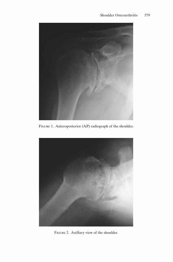
Shoulder Osteoarthritis 579
Figure 1. Anteroposterior (AP) radiograph of the shoulder.
Figure 2. Axillary view of the shoulder.
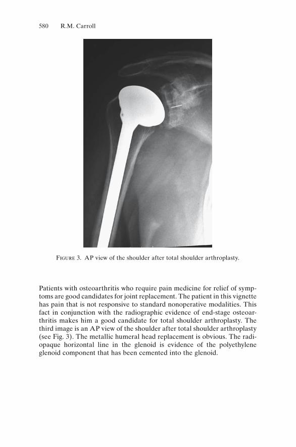
Patients with osteoarthritis who require pain medicine for relief of symp-toms are good candidates for joint replacement. The patient in this vignette has pain that is not responsive to standard nonoperative modalities. This fact in conjunction with the radiographic evidence of end-stage osteoar-thritis makes him a good candidate for total shoulder arthroplasty. The third image is an AP view of the shoulder after total shoulder arthroplasty (see Fig. 3). The metallic humeral head replacement is obvious. The radi-opaque horizontal line in the glenoid is evidence of the polyethylene glenoid component that has been cemented into the glenoid.
Figure 3. AP view of the shoulder after total shoulder arthroplasty.
580 R.M. Carroll
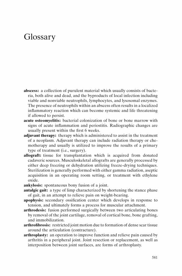
Glossary
abscess: a collection of purulent material which usually consists of bacte-ria, both alive and dead, and the byproducts of local infection including viable and nonviable neutrophils, lymphocytes, and lysosomal enzymes. The presence of neutrophils within an abscess often results in a localized inflammatory reaction which can become systemic and life threatening if allowed to persist.
acute osteomyelitis: bacterial colonization of bone or bone marrow with signs of acute inflammation and periostitis. Radiographic changes are usually present within the first 6 weeks.
adjuvant therapy: therapy which is administered to assist in the treatment of a neoplasm. Adjuvant therapy can include radiation therapy or che-motherapy and usually is utilized to improve the results of a primary type of treatment (i.e., surgery).
allograft: tissue for transplantation which is acquired from donated cadaveric sources. Musculoskeletal allografts are generally processed by either deep freezing or dehydration utilizing freeze-drying techniques. Sterilization is generally performed with either gamma radiation, aseptic acquisition in an operating room setting, or treatment with ethylene oxide.
ankylosis: spontaneous bony fusion of a joint.antalgic gait: a type of limp characterized by shortening the stance phase
of gait, in an attempt to relieve pain on weight-bearing.apophysis: secondary ossification center which develops in response to
tension, and ultimately forms a process for muscular attachment.arthrodesis: fusion performed surgically between two articulating bones
by removal of the joint cartilage, removal of cortical bone, bone grafting, and immobilization.
arthrofibrosis: restricted joint motion due to formation of dense scar tissue around the articulation (contracture).
arthroplasty: an operation to improve function and relieve pain caused by arthritis in a peripheral joint. Joint resection or replacement, as well as interposition between joint surfaces, are forms of arthroplasty.
581
582 Glossary
arthroscopy: a procedure to inspect, and operate on, the contents of a joint through a small portal utilizing a fiber-optic light source and specialized viewing and operative instruments.
benign: a neoplasm which has local capability for growth, with well-differentiated cells that are not capable of vascular or lymphatic invasion. Benign neoplasms can be either latent, with limited local growth, or aggressive, with growth proceeding in a destructive manner.
biopsy: the acquisition of material from a lesion, whether it be neoplastic or infectious, for diagnostic review. An adequate biopsy requires obtain-ing enough material for complete pathological review to arrive at a definitive diagnosis.
bone cement: polymethylmethacrylate (PMMA), used as a filler to enhance the fixation of total joint components.
bone graft: bone used to promote fracture healing, reconstruct a defect in bone, or enhance fusion by providing an organic matrix, osteoblasts, and hormonal factors that contribute to osteogenesis.
calcification: the deposition of calcium within a cartilaginous matrix. This occurs secondary to mineralization of an existing lobule of cartilage which may appear punctate, comma shaped, or popcorn-like on radiographs.
callus: reparative tissue at the site of a fracture that evolves and matures, leading to fracture healing.
cancellous bone: mature bone found in the epiphysis and metaphysis of long bones, and in flat bones, comprised of a three-dimensional lattice of trabecular bone that is less densely packed than cortical bone.
chondrocyte: cartilage matrix-producing cells which rely on nutrition from synovial fl uid and not blood vessels. These cells are often arranged in lacunae which are arranged in rather distinct layers.
chronic osteomyelitis: a chronic infection of bone, usually involving the presence of an involucrum or sequestrum, in addition to radiographic changes within the bone. Sclerosis surrounding chronic sites of radiolu-cency on radiographs and a sinus tract may be present. Chronic osteo-myelitis generally requires at least 6 weeks in order to demonstrate radiographic changes.
Codman’s triangle: a region at the periphery of a bone tumor which is formed secondary to the deposition of reactive bone underneath the periosteum. As the periosteum lays down new bone in response to stress, the bony trabecular patterns in this region are usually at a right angle to the underlying cortical bone. A Codman’s triangle usually represents a rapidly growing tumor or osteomyelitis, with elevation of the perios-teum off of the bone secondary to neoplastic tissue, bone edema, or purulent material.
comminution: disruption of a fractured bone into more than two fragments.
Glossary 583
compartment syndrome: an increase in the resting pressure in a contained fi bro-osseous compartment, such as the forearm or leg, resulting sequen-tially in decreased lymphatic drainage, decreased venous drainage, loss of arterial inflow, and finally death to the muscle contained in the affected compartment. Sequelae include contracture, pain, and severe functional disability.
computerized axial tomography/computerized tomography (CAT/CT scan): an imaging modality which utilizes computer-generated analysis and imaging resulting from multi-planar exposure through either an extremity or the spine. These scans provide clinicians with excellent axial representation of body segments that were previously not available. Tissue density is graded based on Hounsfield units with dense structures being represented by a bright or white image.
contracture: fixed loss of motion in one direction caused by hypertrophy and shortening of periarticular soft-tissue structures such as tendon, ligament or capsule.
cortical bone: mature, organized, densely packed bone, making up the periphery of flat bones and the diaphysis of long bones.
crepitus: audible or palpable grinding, usually located in a peripheral joint, with motion.
curettage: the mechanical removal of neoplastic or infectious tissue from a primary site. This generally involves entering a lesion and scraping its contents from within its lesional cavity, and has the potential for leaving residual disease, at the microscopic level, in the periphery. This type of “intralesional resection” is generally utilized for benign neoplasms.
cyst: a fl uid-filled cavity which results from the production of fl uid from a surrounding glandular membrane. The majority of cysts in orthopaedic terminology, such as a simple bone cyst or aneurysmal bone cyst are not true cysts, since the fl uid does not directly result from the surrounding mesenchymal tissue present in the wall lining, but from the passive accumulation of fl uid within the marrow cavity of bone.
debridement: the removal of infected or devitalized bone, muscle, and skin. The purpose of debridement is to remove any material that can serve as a substrate which harbors bacteria and to enable antibiotics, via parental or local routes, to reach colonies of bacteria.
delayed union: failure of a fracture to heal within the desired and expected time frame, with the potential still present for eventual union.
developmental: pertaining to growth and differentiation.diarthrodial joint: a joint which consists of connections between two rigid
parts of the musculoskeletal system which is lined by synovial tissue, lubricated by synovial fl uid, and demonstrates appreciable ranges of motion. The ends of bones in diathrodial joints are usually covered with hyaline cartilage.
diaphysis: the tubular midportion of a long bone, consisting primarily of cortical bone.
584 Glossary
dislocation: loss of normal articular congruity of a joint, with no contact between opposing articular surfaces.
dysostosis: an isolated disruption of normal bone growth with no identifi -able etiology.
dysplasia: intrinsic defect in normal bone growth, which may be localized or generalized.
dystrophy: alteration in bone growth due to extrinsic defect, typically an endocrine abnormality.
electromyography (EMG): recording of the variations of electric poten-tial or voltage from skeletal muscle. The EMG/nerve conduction velocity test is useful in determining the site of injury of a peripheral nerve or nerve root, and in identifying peripheral neuropathy caused by meta-bolic abnormalities.
enchondral ossification: bone formation following the template of a car-tilaginous matrix.
enchondroma: a benign tumor of bone, with a cartilaginous matrix, com-monly occurring in the hand.
epiphysis: a secondary ossification center, adjacent to the physis, which develops in response to compression and is covered by articular cartilage.
external fixation: the use of an extracorporeal device to stabilize a part of the skeleton, usually following an open fracture.
fracture: a cortical disruption, ranging from incomplete and non-displaced to completely displaced.
fracture healing: the process of biologic repair of a fracture in response to hormonal, biochemical, and mechanical factors. Fracture healing encompasses the phases of inflammation, soft callus, hard callus, and remodeling.
free tissue transfer: one-stage transplantation of distant autogenous com-posite tissue from a donor site to a recipient site. Free tissue transfer can involve transplantation of muscle, fasciocutaneous tissue, or bone with or without attached soft tissue. This type of transfer requires immediate revascularization, utilizing microsurgical anastomosis of graft and recip-ient site arteries and veins.
frozen section: the preparation of pathological sections from fresh tissue, used primarily in the operating room for rapid diagnosis which may impact on surgical decision making.
ganglion: a soft, mucin-filled cyst arising from a tendon, tendon-sheath, or joint capsule. Most common about the hand and wrist, and more common in women.
gigantism (overgrowth): hypertrophy of a single digit or entire limb, pri-marily involving soft tissues. Causes include neurofibromatosis, tumor, or vascular anomaly.
haversian bone: cortical bone composed of vascular channels surrounded by mature (lamellar) bone.
Glossary 585
herniated nucleus pulposus (HNP): Extrusion of gelatinous nucleus pulposus through the anulus fibrosus, into the spinal canal or neural foramen. When an HNP results in nerve root compression, radicular pain, numbness, or weakness may be seen. Herniated disc, ruptured disc.
hydroxyapatite: the calcium mineral crystal component of bone.internal fixation: the use of an implant to stabilize the skeleton, usually
after a fracture.involucrum: newly formed reactive bone, usually occurring at the inter-
face between diseased bone and healthy tissue. An involucrum consists of viable bone which is the opposite of a sequestrum, which is composed of dead bone.
joint reaction force: the force across a joint that results from a combina-tion of weight bearing and muscular contraction.
kyphosis: forward bending of the spine, when viewed from the side, which is normal in the thoracic spine.
laminectomy: removal of a lamina from its superior to its inferior margin, performed as surgical treatment for spinal stenosis or HNP. Laminotomy or hemilaminectomy refer to partial removal of the lamina.
lordosis: backward bending, or “sway,” of the spine when viewed from the side. Lordosis is normal in the neck and low back.
low back strain: nonspecific term referring to acute onset of pain in the low back, occasionally radiating into the buttocks, with associated muscle spasm. Low back sprain, lumbago.
magnetic resonance imaging (MRI): an imaging modality utilizing reso-nance phenomenon resulting in the absorption and/or emission of elec-tromagnetic energy by nuclei or electrons in a static magnetic field. Magnetic resonance imaging requires unpaired electrons that are excited by exposure to a magnetic field with a particular signal being emitted once that field is removed. Differences in density of tissues are then represented on images as varying shades of gray, black, or white, depend-ing on their concentration of hydrogen. The modality is extremely useful in the evaluation of musculoskeletal tumors, as well as disorders of the spine, knee, shoulder, and foot.
malignancy: a neoplasm consisting of undifferentiated or dedifferentiated cells which have the active capability of vessel invasion, transport, and establishment of a secondary site of neoplastic growth in a distant organ.
malunion: healing of a fracture in a nonanatomic position.membranous bone formation: bone formation occurring directly from a
fi brous, mesenchymal, connective tissue template.metaphysis: the transition segment of a long bone from the enlarged end
(epiphysis) to the tubular shaft (diaphysis). The funnel-shaped metaphy-sis is usually made up of abundant cancellous bone, and during growth, woven bone.
586 Glossary
metastasis: the deposition to secondary sites of neoplastic cells from a primary neoplasm.
myelopathy: noninflammatory dysfunction of the spinal cord resulting in long-tract signs and symptoms, most commonly caused by mechanical compression in the cervical spine.
neoadjuvant therapy: the application of adjuvant treatment, usually che-motherapy or radiation, prior to a primary procedure in an attempt to facilitate surgical removal of the primary tumor.
neuroma: a nodule, frequently painful, developing in a nerve that has been partially or completely lacerated or traumatized.
nonunion: failure of a fracture to heal within the upper range of time expected, with sequential radiographic documentation of a lack of any progression towards healing.
nonsteroidal anti-inflammatory drugs (NSAIDs): these agents are in widespread use to treat arthritis as well as a variety of soft-tissue injuries and for pain relief. Gastrointestinal side effects are common.
open fracture: a fracture which has come into contact with the outside, extracorporeal environment. Implications primarily relate to an increased risk of infection and impaired fracture healing secondary to soft-tissue injury.
ossification: the deposition of mineral along osteoid matrix, produced by either normal (fracture healing) or malignant osteoblasts (osteosarcoma).
osteoarthritis: noninflammatory degeneration of a diarthrodial joint char-acterized by loss of articular cartilage, effusion, crepitus, deformity, and pain. Radiographic changes include loss of joint space, subchondral scle-rosis, cyst formation, and periarticular osteophytes.
osteoblast: a type of differentiated mesenchymal cell that is essential for the process of osteogenesis or ossification. Osteoblasts alone can produce the organic intercellular substance or matrix which makes up bone tissue, called osteoid.
osteoclasis: to manually fracture a bone, usually to correct malposition.osteoclast: bone resorbing cell.osteocyte: histologically and possibly biochemically inert osteoblast,
usually present in bone which is not undergoing any active remodeling or repair.
osteoid: organic component of bone.osteomalacia: metabolic disorder of bone characterized by inadequate
mineralization of normal osteoid. Less common than osteoporosis, causes include Ca++ deficiency and renal disease.
osteomyelitis: infection involving bone. (See acute and chronic osteomyelitis)
osteoporosis: pathologic condition of bone characterized by a decrease in bone matrix with normal mineralization of the matrix that is present. Causes include postmenopausal estrogen deficiency, steroid usage,
Glossary 587
immobilization, and bed rest. Fractures of the spine, hip, wrist, and shoulder are common.
osteotomy: surgical cutting and realignment of bone to change the mechanical environment of adjacent joints, posture, or appearance.
pannus: hyperplastic, hypertrophic synovium seen in the inflammatory arthritides. Source of degradative enzymes.
paraesthesia: a “pins and needles” or “tingling” sensation in an extremity, typically along the distribution of a peripheral nerve or nerve root.
pathologic fracture: a fracture occurring in bone that is of abnormal quality, either from metabolic changes (e.g., osteoporosis), tumor, or infection.
periosteum: thick fibrous tissue covering of a bone.physis (epiphyseal plate, growth plate): a highly specialized cartilaginous
structure, at each end of long bones, through which longitudinal growth of the bone occurs. The physis regresses and closes at the end of active skeletal growth.
polydactyly: the presence of an extra digit, either partial or complete. Most common in the hand, polydactyly may occur on either the ulnar or radial side.
prosthesis: a mechanical replacement for a removed portion of either bone or soft tissue. Prostheses include endoprostheses, which are internal replacements (joints, valves, ect.), and exoprostheses as would be used following an amputation.
radial club hand: congenital absence of all or part of the radius, result-ing in a near-normal hand attached to, but radially deviated on, the forearm.
radicular pain: pain traveling down the extremity in a dermatomal distri-bution, often associated with paraesthesia. Pain should typically extend distal to the elbow or knee to be considered radicular.
radiculopathy: noninflammatory dysfunction of a spinal nerve with abnor-mal neurologic findings, most commonly secondary to a herniated nucleus pulposus or spinal stenosis.
referred pain: sclerotomal pain in a region that shares a common embryo-logic origin with the diseased region, such as the trapezius and shoulders for cervical disc pathology or the buttocks and posterior thighs for the lumbar spine.
remodeling: continuous process whereby older bone is removed and replaced with new bone, usually in response to mechanical stresses.
rheumatoid arthritis: inflammatory arthritis characterized by morning stiffness, swelling of peripheral joints, subcutaneous nodules, pain, and deformity.
rheumatoid factor: a blood marker that is usually, but not always, present in patients with rheumatoid arthritis. Some patients who do not have rheumatoid arthritis may have a positive rheumatoid factor.
588 Glossary
rotation tissue transfer: soft tissue, such as muscle, or bone which is trans-planted for wound coverage from one local site to another without tran-secting the blood supply to the transplanted tissue.
scintigraphy: a sensitive but nonspecific imaging modality for the diagno-sis and staging of various orthopaedic problems. The standard “bone scan” utilizes technetium 99 m, a gamma-emitting radioisotope with a half-life of 6 hours, coupled with methylene diphosphonate, a bone-seeking mineral material. Positive bone scans usually demonstrate increased uptake in areas of bony repair or destruction caused by neo-plasm, fracture, infection, inflammation, or arthritis.
sclerosis: a rim of host bone reaction around the periphery of a lesion, or in an area of degeneration, which appears white (radiopaque) on radiographs.
scoliosis: coronal plane curvature, typically associated with rotation, leading to asymmetric rib or flank prominence. Common etiologies are idiopathic, neuromuscular, and congenital.
segmental instability: abnormal motion pattern between adjacent vertebrae resulting in excessive motion under physiologic loads and causing, in some individuals, characteristic mechanical back or neck pain.
septic arthritis: the presence of bacteria or their by-products within a diarthrodial joint.
sequestrum: a microscopic or macroscopic island of necrotic bone found at the nidus of an infection within viable bone. Often, sequestered frag-ments are surrounded by purulent material and infected granulation tissue and occur secondary to devitalization of cortical bone.
spinal fusion: arthrodesis of two or more adjacent vertebrae for painful or unstable conditions of the spine including spinal deformity, trauma, or degenerative segmental instability. Bone graft or bone-graft substitute is utilized and internal fixation may be used. Fusion may be performed anteriorly (interbody) or posterolaterally (intertransverse).
spinal stenosis: narrowing of the spinal canal or neural foramen resulting in compression of the cord or nerve roots. Causes include congenital narrowing, degenerative changes, and spondylolisthesis.
spondylolysis: defect of the pars interarticularis, most commonly a stress fracture that occurs during childhood. May be associated with back pain and lead to spondylolisthesis.
spondylolisthesis: the forward slippage of a vertebrae on the one below it (most commonly L5 on S1). The most common types are isthmic (secondary to spondylolysis), degenerative (caused by degenerative instability of the disc and facet joints) or postsurgical.
sprain: partial or complete injury to ligament resulting from excessive stretching.
straight-leg raising (SLR) test: with the patient supine or seated, simul-taneous hip flexion and knee extension places the sciatic nerve on stretch.
Glossary 589
Reproduction of ipsilateral sciatica (not back pain) constitutes a positive SLR sign.
subluxation: partial dislocation of a joint, with some retained contact of the articular surfaces.
syndactyly: webbing of the fingers, usually resulting from congenital failure of formation, and commonly seen with other congenital abnormalities.
synostosis: congenital fusion, either partial or complete, of adjacent bones. Synostosis is far more common in the upper than in the lower extremity, and may involve the elbow, carpus, metatcarpals, or phalanges.
synovectomy: removal of the synovium (lining surface of a diarthrodial joint), for inflammatory conditions such as rheumatoid arthritis or infection.
synovial membrane: a soft glandular tissue which lines diarthrodial joints and produces synovial fl uid. The synovial membrane has a rich capillary network and serves both in phagocytosis of foreign debris and the pro-duction of synovial fl uid.
tension sign: a physical finding produced by placing the involved nerve root on stretch to assess for mechanical compression. A positive tension sign requires reproduction of pain, in a dermatomal distribution, down the extremity.
Trendelenburg gait: a type of limp, caused by ineffective hip abduction, marked by swaying the torso over the effected hip on weight-bearing.
Trendelenburg sign: an abnormal drooping of the pelvis, on single-leg stance, away from the effected side due to ineffective hip abduction sec-ondary to neurologic or mechanical factors, or pain.
ulnar club hand: congenital absence of all or part of the ulna, resulting in a near-normal hand attached to, but ulnarly deviated on, the forearm.
valgus: malalignment of an extremity, in the coronal plane, with the apex of the deformity pointing to the midline (“knock-knees”).
varus: malalignment of an extremity, in the coronal plane, with the apex of the deformity pointing away from the midline (“bow-legs”).
volar: the palmar surface of the hand and forearm.Wolff’s Law: physiologic phenomenon whereby new bone forms in
response to mechanical stress and resorbs in the absence of it (“form follows function”).
woven bone: immature bone found in the embryo and newborns, fracture callus, tumors, and osteogenesis imperfecta or Paget’s disease.
zone of transition: the boundary between what is perceived as normal bone and lesional tissue on radiographs. The zone of transition can be very sharply defined, indicative of a slow-growing process (benign tumor), or very poorly defined, indicative of an aggressive process (neoplasm or infection).
Index
AAbscess
infection pathway of, 89pediatric, 87–90sequestrum and, 90
AC. See Acromioclavicular jointAchilles tendon, 258
rupture, 79–80Achondroplasia, pediatric, 199–201ACL. See Anterior cruciate ligamentAcromioclavicular joint (AC),
337–38deformity at, 269of shoulder, 334
Acute hematogenous osteomyelitis, pediatric, 87–93
Adhesive capsulitis, of shoulderdifferential diagnosis, 357examination, 357history, 357radiographs, 358treatment, 358
Adriamycin. See Doxorubicin hydrochloride
Aggressive fibromatosis, 165Aggressive synovitis, 151–53Aneurysmal bone cysts, 148–49,
408Angiography, radiographic evaluation,
115, 157Angiomatosis, 165Ankle. See Foot and ankleAnkylosis, 287Anterior cruciate ligament (ACL)
arthroscopy, 574
591
with MCL straindiscussion, 575–76history, 570MRI of, 571, 573physical exam, 570–71radiograph of, 571–72treatment of, 571, 575
repair of, 77Antibiotics
bacteria and, 87, 103infection, 92
ArmBrachialgia in, 297herniated disk and, 285nerve root compression and, 297pain in neck and, 277
Arthritis, 383–84. See alsoOsteoarthritis; Osteomyelitis; Rheumatoid arthritis; Septic arthritis
crystalline arthropathy in, 28elbow, 383–84foot and ankle, 494–96hand, 394–97hip, 433hip replacement for, 69knee, 465–69NSAIDs for, 465pediatric, 93–96, 189–91rheumatoid, 26–28, 289–90, 331,
384, 395–97, 494–96Arthropathy
arthritis, 28Charcot, 357inflammatory, 356
592 Index
Arthroplasty. See also Joint arthroplasty
infection with, 102–3shoulder, 355, 580
Arthroscopic adhesiolysis, of shoulder, 358
Arthroscopy, 271of ACL, 574elbow, 372hand, 390hip and femur, 436injury evaluation with, 271shoulder, 358, 361in sports medicine, 273
Articular cartilage, 262–64. See alsoCartilage
lesions, 264morphology of, 262–63musculoskeletal tissues, 262–64outerbridge classification of, 264sports medicine and, 262–64
Aspirin, DVT and, 447Avascular necrosis (AVN), 30
etiologies ofidiopathic, 32occlusive phenomena, 31–32trauma, 31
of femoral head, 431fracture complication of, 53in hip dislocation, 73of humeral head, 356–57lesions, 432with SCFE, 560trauma and, 31
AVN. See Avascular NecrosisAxonotmesis, 46
BBabinski’s signs, in cervical spine
disorders, 279Back pain. See Cervical spine
disorders/injuries; Low back pain; Lumbar spine disorders/injuries
Bacteriaantibiotics and, 87, 103molecular sequence of, 86
Bankart lesion, 360
Barlow test, for pediatric hip dislocation, 208
Batson’s plexus, metastatic bone disease and, 139
Battered child syndrome, 235–37Biceps tendon
long head of, 336–37O’Brien’s test of, 345
Biopsycore-needle, 115–16for Ewing’s sarcoma, 136–37for metastatic bone disease, 140open incisional, 116of sarcomas, 115–16of tumors, in musculoskeletal
system, 115–16Blood supply
to femoral head, 431to long bone, 88of meniscus, 265
BMD. See Bone mineral densityBone disease, metabolic
arthritis, 26–28crystalline arthropathy in, 28
eucalcemic states of, osteoporosis, 11–16
formation/resorption and, 10–11hypercalcemic states of
hyperparathyroidism, 16–17rickets and osteomalacia, 16–17
miscellaneous types of, renal osteodystrophy, 20–21
neurodevelopment disorders indevelopmental/congenital defects,
34–37neurologic diseases, 34
ochronosis, 29Paget’s disease, 23–25
therapeutic approaches for, 23–24
trabecular thickening in, 24–25viral particles in, 25–25
sick cell syndromes inosteogenesis imperfecta, 22osteopetrosis, 23–24
vascular circulatory disease inavascular necrosis, 30hematologic syndromes, 33sickle-cell anemia, 33
Index 593
Bone disease, metastaticdiagnosis of
Batson’s plexus and, 139biopsy for, 140bone scans for, 139–40clinical characteristics/physical
examination, 139computed tomography for, 140–41magnetic resonance imaging, 140microscopic characteristics in,
140, 142radiographic findings in, 139–40,
143staging studies for, 139treatment of, 142
in hip, 141Bone growth, abnormal
disease categories of, 10metabolism and, 9–10
Bone growth, normalbone organization in, 6cartilage
histologic types of, 9hyaline, 9
coupling, 8formation/resorption in, 6–8growth plate events, 5immature, 6–8mature, 6–7mechanical stress and, 8ossification
endochondral model of, 2–3mesenchymal model of, 2–3
osteoblasts vs. osteoclasts in, 6–7piezoelectric effect in, 8–9postnatal development, 6primary ossification center in, 3–4secondary ossification center in, 4turnover and remodeling in, 6–9voltage gradient in, 8–9Wolff’s law, 8
Bone(s), joints and. See also Pelvic bones
aneurysmal cysts, 148–49aspiration, Cefazolin after, 92Batson’s plexus, 139distraction, replacement vs., 98fi brosarcoma of, radiographic
features, 134
infection, 550infection in, exogenous vs.
hematogenous pathways to, 84–85
loss, 12–15pediatric, 170
biological plasticity of, 231remodeling of, 233–35
pelvic, chondrosarcomas of, 128sarcomas of. See Sarcomas
Bone mineral density (BMD), DEXA scanning for, 15
Bone replacement, bone distraction vs., 98
Bone scans, 139. See alsoElectromyography; Magnetic resonance imaging; Radiographic evaluation; X-rays
osteomyelitis, 91–92of STS, 157with Technetium-99, 372of tibia, 270
Bone scintigraphy, 270Borrelia burgdorferi. See Lyme
diseaseBowlegs. See Genu varumBraces and corsets, for lumbar spine
disorders, 313, 329Brachial artery, elbow, 368–69Brachialgia, 297Brachial plexus, 339Brooker grading, of HO, 449Bursitis, in shoulder, 359
CCalcific tendonitis, 358
differential diagnosis, 358examination, 358history, 358radiographs, 358treatment, 358
Calcitonin, osteoporosis treatment with, 16
Calcium pyrophosphate dihydrate deposition, of hand, 407
Cancer, in United States, 106Carisoprodol (Soma)
for herniated disk pain, 311
594 Index
Carisoprodol (Soma) (cont.)for low back pain, 311
Carpal tunnel syndrome, 398Cartilage
articular, 262–64lesions, 264morphology of, 262–63musculoskeletal tissues, 262–64outerbridge classification of, 264
histologic types of, 9hyaline, 9pediatric, 171X-ray, 171
Cauda equina compression (CEC), 301–2, 317
CEC. See Cauda equina compressionCefazolin, following bone aspiration,
92Cerebral palsy, pediatric, 201–2Cervical hypertension injuries, 290–91Cervical radiculopathy, symptoms and
fi ndings with, 278Cervical spine disorders/injuries. See
also Lumbar spine disorders/injuries
algorithm for, 291–94Babinski’s signs in, 279clinical conditions
acute herniated disk, 283–86cervical hypertension injuries,
290–91cervical spondylosis, 286–88cervical spondylosis with
myelopathy, 231, 287–88neck sprain/ache, 283rheumatoid arthritis, 289–90
clonus and, 279conservative treatment for, 295
with arm pain predominant, 297drug therapy, 298–99exercises, 300immobilization, 298manipulation, 300with neck pain predominant,
296–97traction, 299trigger-point injection, 300
degeneration, 296diagnostic studies for, 279–83
CT, 282EMG, 282MRI, 280–81myelography, 281–82radiographs, 279–80
history in, 276–77Hoffman’s sign in, 279hyperreflexia and, 279lidocaine for, 295muscle spasm and, 299physical examination of
myelopathic findings in, 279neurological deficit in, 278range of motion in, 277sensory deficits and, 279upper extremities, 279
Cervical spondylosis, 286–88with myelopathy, 231, 279, 287–88treatment of, 287–88
Charcot arthropathy, 357Chemotherapy
Adriamycin for, 158cisplatin for, 158with Dacarazine, 158for Ewing’s sarcoma, 137ifosfamide for, 158with methotrexate, 158for osteosarcoma, 119–21for tumors, 119–21
Children’s orthopedicsacute hematogenous osteomyelitis,
87–93anatomic structures and
bone, 170cartilage, 171femur, 205–6growth plate, 171–73ligament, 170periosteum, 170–71peripheral plate structures, 171
arthritis, 93–96, 189–91biologic differences
growth, 169remodeling, 170
bone, 170, 231, 233–35bone fracture, classifications of,
231–32cartilage, 171cerebral palsy, 201–2
Index 595
congenital scoliosis, 245–46femur, Perthes’ disease in, 213–16fl atfoot
congenital hypermobile, 224rigid, 224
foot and ankle, 223congenital clubfoot, 224–26fl atfoot, 223–24metatarsus adductus in, 226–27
fractures, special types of, 47–48growth plate, 171–73hematogenous inoculation, 87hip
Barlow test, 208dysplasia of, 230septic arthritis of, 186–87
infection, 181–89abscess development in, 87–90complications of, 187–88specific organisms in, 90
infection in, 181–89knee
discoid meniscus, 221–22Osgood-Schlatter’s disease of, 220osteochondritis dissecans of, 221popliteal cysts in, 222–23
ligaments, 170limp, 237–40long bone
hematogenous inoculation in, 87radial dysplasia of, 230
neuromuscular deformity, 246osteomyelitis, 87–93osteomyelitis in
clinical feature, 181diagnosis, 182–83trauma and, 87–88treatment, 184–85
peripheral plate structures, 171Perthes’ disease, 213–16septic arthritis, 93–96skeletal affectations in
battered child syndrome, 235–37childhood arthritis, 189–91congenital/neurodevelopmental,
195–201foot, 223–27infection, 181–89knee, 219–23
limp evaluation, 237–40metabolic disease, 191–93neuromuscular disease, 201–5pediatric trauma, 231–35pediatric upper extremity/neck,
228–31regional orthopedic problems,
205–19spine, 240–54
skeletal growth factorscoexistent disease, 174endocrine, 174environmental, 174genetic impact, 174nutrition, 174
skeletal growth variations inangular, 177–79differential diagnosis of, 179–80torsional, 175–76
slipped capital femoral epiphysis, 216–19
spinekyphosis of, 247scoliosis of, 240–45skeletal affectations in, 240–54
supracondylar fracture in, 60trauma, 231–35upper extremity and neck
congenital muscular torticollis, 229–30
congenital trigger thumb, 231radial anomalies, 230Sprengel’s deformity, 228–29
vascular/hematologic disease, 193–95
Chondroblastoma bone tumors, 146Chondrogenic bone tumors, 110Chondrolysis, with SCFE, 560Chondromyxoid fibroma, 500Chondrosarcoma
central/peripheral, 127clear cell, 131clinical characteristics/physical
examination, 127–29computed tomography of, 128differentiated, 131endosteal scalloping in, 127, 129grading/prognosis, 130mesenchymal, 131
596 Index
Chondrosarcoma (cont.)microscopic characteristics of, 130of pelvic bones, 128radiographic findings of, 128–29treatment, 130variations of, 130–31
Cierny classification, of osteomyelitis, 97
Cisplatin, for chemotherapy, 158Clonus, in cervical spine disorders,
279Cobb technique, for Scheuermann’s
kyphosis, 249Codeine, for neck pain, 299Colles fracture, of wrist, 65Complex Regional Pain Syndrome
(CPRS), 500Computed tomography (CT)
for cervical spine disorders, 282of chondrosarcoma, 128for elbow, 372of hip, 429for lumbar spine disorders, 306, 308lumbar spine disorders/injuries, 306,
308for metastatic bone disease, 140–41with myelography, 282of pelvis, 429radiographic evaluation, 114of shoulder, 348for STS, MRI vs., 155
Congenital and neurodevelopmentalconstriction band syndrome, 394hand differences, 390, 392–94
duplication, 391–92failure, 391part formation, 391
pediatricachondroplasia, 199–201clubfoot, 224–26Down syndrome, 196–98hip, 207–13muscular torticollis, 229–30scoliosis, 245–46skeletal dysplasias, 198–99Sprengel’s deformity, 228–29trigger thumb, 231
Contusions, muscular, 261–62Core-needle biopsy technique, 115–16
Corticosteroid injections, elbow rehabilitation and, 373, 376
Coumadin, with THR, 446–47CPRS. See Complex Regional Pain
SyndromeCryosurgery, for giant cell tumor, 132Crystalline arthropathy, arthritis and,
28CT. See Computed tomographyCubital tunnel syndrome, 398
case study ofdiagnosis/treatment, 562history, 561physical examination, 561radiographs/laboratory values,
561treatment, 562–64
of elbow, 381NSAIDs for, 562ulnar nerve in, 562
Cyclobenzaprine (Flexeril), for low back pain, 327
DDacarazine (DTIC), for
chemotherapy, 158DDH. See Developmental dislocation
of hipDedifferentiated chondrosarcoma,
131Deep vein thrombosis (DVT)
aspirin and, 447hip replacement and, 447oral estrogen complication of, 15
Demerol. See MeperidineDevelopmental dislocation of hip
(DDH)pathology of, 207–9treatment of, 210–13
DEXA scanning. See Dual-energy X-ray absoptiometry scanning
Diazepam (Valium)for herniated disk pain, 312for low back pain, 321
Dilantin therapy, rickets and, 192Diplegia, 202Discoid meniscus, in pediatric knee,
221–22Disk. See Herniated disk
Index 597
Distal femoral, anatomic compartment of, 113
Distal timoral, fracture, 74–75Dowager’s hump, 12Down, Langdon, 196Down’s syndrome, 196–98Doxorubicin hydrochloride
(Adriamycin), for chemotherapy, 158
DTIC. See DacarazineDual-energy X-ray absoptiometry
scanning (DEXA), for bone mineral density, 15
Dupuytren’s Contracture, of hand, 400DVT. See Deep vein thrombosisDysplasias. See Skeletal dysplasias
EEarly osteoarthritis. See OAElbow
anterior/posterior views of, 365atraumatic/overuse injuries of
arthritis, 383–84lateral/medial epicondylitis,
379–80Little Leaguer’s Elbow, 381–82Osteochondritis Dissecans,
382–83ulnar nerve compression, 381
brachial artery, 368–69cubital tunnel syndrome, 381dislocation of, 61evaluation
with arthroscopy, 372CT, 372with EMG, 372history, 369–70MRI, 372physical examination, 370–72stress X-rays, 372Technetium-99 bone scan, 372
functional anatomy ofneurovascular, 367–69skeletal, 364–66
heterotopic ossification of, 376HO of, 376ligament complexes of, 365–66ligament injuries to, 378–79ligaments of, 365–66, 378–79
median nerve, 368neurovascular
brachial artery, 368–69median nerve, 368musculocutaneous nerve, 368radial nerve, 368ulnar nerve, 368
nonoperative treatment forcorticosteroid injections, 373, 376rehabilitation, 373, 376
radial nerve, 368sagittal view of, 366–67tendon, ruptures of, 379trauma
dislocations, 377–78fractures, 377ligamentous injuries, 378–79tendon ruptures, 379
treatment foralgorithmic approach to, 373–75operative, 376–77
ulnar nerve, 368Electromyography (EMG)
for cervical spine disorders, 282of elbow, 372of shoulder, 348
Embryology, bone development inchondrification process, 1–2interzones, 1–2ossification, 1–5
Enchondromas bone tumors, 145–46Endocrine system, skeletal
development and, 174Endosteal scalloping, in
chondrosarcomas, 129Enneking, Dr. William F., 110Eosinophilic granuloma, 151Epidural steroid injection, for lumbar
spine disorders, 323, 327–28Epiphysis. See Slipped capital femoral
epiphysisEpithelioid sarcoma, 161Ewing’s sarcoma, 119
biopsy consideration for, 136–37chemotherapy for, 137clinical characteristics/physical
examination of, 135–36combined multimodality treatment
for, 137
598 Index
Ewing’s sarcoma (cont.)microscopic characteristics of, 137natural history of, 136radiation therapy for, 137radiographic evaluation/staging of,
136surgical treatment for, 138
Exercisefor cervical spine disorders, 300for lumbar spine disorders, 329–30
FFDP. See Flexor digitorum profundusFemoral shaft fractures, 73–74Femur
distal, sarcomas in, 117, 129fractures of, 69–73
nondisplaced vs. displaced, 70head
AVN of, 431blood supply to, 431vascular anatomy of, 420–21
intertrochanteric fracture of, 71–72Perthes’ disease in, 213–16proximal, metastatic carcinoma in,
117, 129slipped capital femoral epiphysis in,
216–19supracondylar fracture of, 60
Fetus, ossification in, 1–5, 172Fibrogenic bone tumors, 110Fibrosarcoma, 134, 161
clinical characteristics, 134radiographic features, 134
Flatfoot, pediatriccongenital hypermobile, 224rigid, 224
Flexeril. See CyclobenzaprineFlexor digitorum profundus (FDP)
avulsion of, 565–69Leddy/Packer classification of, 569
Flexor tendon avulsion, case study ofdiagnosis/treatment, 565–69discussion, 569history, 565physical examination, 565radiographs/laboratory values, 565
Foot and ankleacquired deformities of
hallux rigidus, 490hallux valgus, 489–90, 531–36hallux varus, 490
arthritic conditions of, 494–95rheumatoid, 495–96
clinical evaluation ofhistory/physical exam, 482radiology of, 482–83
diagnosis/treatment of, algorithms for, 501–2
Foot and ankle, injuriesforefoot, 487
bones/joints of, 472–77diseases of, 483–88gait cycle of, 481inversion stress testing of, 488ligaments of, 477–79nerves/vessels of, 480–81
fractures, soft tissueAchilles tendon rupture, 79–80,
258ankle sprains, 80distal, 79Lisfranc dislocation, 81Os Calcis, 79talar neck, 80
heel pain, 493–94infections of
diabetic, 498paronychia, 497–98
inversion stress testing of, 488Lauge-Hansen classification or,
77–79midfoot, 487pediatric, 223–27
congenital clubfoot, 224–26fl atfoot, 223–24metatarsus adductus in, 226–27skeletal affectations in, 223–27
sprains of, 80, 487–88tendon
Achilles, 491–93anterior tibial, 491peroneal, 491posterior, 492
trauma tocalcaneus fractures, 486pilon fractures, 484syndesmosis injuries, 484
Index 599
talus fractures, 484–86tumors
bone, 499–500soft tissue lesions, 499
Fracture(s), dislocationsof ankle
Lauge-Hansen classification of, 77–79
pilon, 77complications of
AVN, 53chronic regional pain syndrome,
53implant failure, 53infection, 52myositis ossificans, 53stiffness/loss of motion in, 52union problems in, 50
deformity inangulation, 41–42displacement, 41–42rotation, 41–42
descriptors ofcomplete vs. incomplete, 40open vs. closed, 40simple vs. comminuted, 40
distal timoral, 74–75of elbow and forearm, 377
distal humerus, adults, 61at head of radius (elbow), 63multiple bones, 62olecranon, 62–64supracondylar fracture, 60
epiphyseal plate, Salter classification of, 44
of femur, 60, 69–73intertrochanteric, 69–70neck, 69shaft, 73–74
of foot, Os Calcis (Heel Bone), 79gamekeeper’s thumb, 65of hand, 65–66
scaphoid, 66healing phases of, 50–51, 233–35
mechanical, 48metabolic, 48vascular, 48
of hip, 73avascular necrosis, 73
femoral neck, 71Jefferson, 68of knee, 75
distal femoral, 74–75patella, 75–76tibial plateau, 75tibial shaft, 75–77
loading mode in, 41–42, 231of lower extremity, 69–81of metatarsals/toes, 81orthopedic emergencies of, 49patterns of, 41–45pediatric
classifications of, 231–32complications of, 233greenstick, 170multiple, 236pathologic, 189special types of, 47–48supracondylar, 60torus, 170
of pelvis, 69phalangeal, 65of proximal humerus, Neer
classification, 56of shoulder
AC separation, 58glenohumeral dislocation, 53humerus shaft, 58of proximal humerus, 53, 56–57recurrent, 58
in shoulder region, of clavicle, 53soft tissue damage in
arterial injury, 44–48ligament, 44–48muscle and tendon, 44–48neural injury, 44–48vascular injury, 44–48
special types ofincomplete, 47intraarticular, 48pathological, 48pediatric, 48physeal, 48stress, 48
of spine, 67–68Hangman’s fracture, 68Jefferson fracture, 68odontoid fractures, 68
600 Index
Fracture(s), dislocations (cont.)treatment of, 68
treatment principles for, 49in upper extremity
elbow and forearm, 60–65shoulder, 53–60
of wrists/handsdistal radius, 65gamekeeper’s thumb, 65phalangeal, 65scaphoid, 66
Fracture(s), open, 99–102antibiotic treatment for, 99–100Gustilo-Anderson classification of,
99–100management algorithm for, 101operative emergency of, 99–100osteomyelitis and, 102, 549–50soft tissue coverage of, 99–101tibia, 537–39
assessment, 538discussion, 538–41history, 537laboratory values, 538pain with, 537–38physical examination, 537–38radiographs of, 539–41treatment, 538X-rays, 538
wound healing of, 102Franconi syndrome, 230Frozen shoulder. See Adhesive
capsulitisFunctional restoration approach, to
low back strain, 309
GGaleazzi fracture-dislocation, 62–63,
209Gamekeeper’s thumb fracture, 65Ganglia, soft tissue lesion of, 153GCT. See Giant cell tumorGenu valgum, 175, 177, 179–80Genu varum, 175, 177, 179–80GH. See Glenohumeral jointGiant cell tumor
clinical characteristics/physical examination, 131
cryosurgery for, 132
microscopic characteristics, 132natural history/malignancy
potential, 131–32radiographic/clinical evaluation,
132–33treatment of, 132
Glenohumeral joint (GH), 347, 356anterior perspective, 347Charcot arthropathy in, 357functional anatomy of, 333–34ligaments of, 334–35osteoarthritis in
differential diagnosis, 354examination, 353history, 353radiographs, 354treatment, 355
X-rays of, 347, 354, 356Glomerular disease, rickets and, 192Gout, in hand, 407Growth plate, pediatric, zones of,
171–73Gustilo-Anderson classification, of
open fractures, 99–100
HHallux rigidus, foot and ankle, 490Hallux valgus
of foot, 489–90discussion, 532–36history, 531metatarsophalangeal joints with,
531–32pain with, 531–33physical examination, 531–32radiographs of, 534–36surgical options for, 533treatment, 532X-rays, 532
foot and ankle, 489–90, 531–36Hallux varus, foot and ankle, 490Hand
arthroscopy, 390calcium pyrophosphate dihydrate
deposition of, 407cold injury to, 406congenital differences, 390–94developmental/acquired diseases of
arthritis, 394–97
Index 601
Dupuytren’s Contracture, 400Kienböck’s Disease, 400nerve compression syndromes,
397–99tendon compression, 399
fractures, dislocations of, 65–66distal radius, 65gamekeeper’s thumb, 65phalangeal, 65scaphoid (navicular), 65–66
gout in, 407history, 387–88imaging, 389–90inflammation/infection of, 400–402Kienböck’s Disease of, 400management protocols for,
algorithm, 410–12metabolic disease and, 406–7neoplasms of
skin cancer, 408–9soft tissue masses, 408tumors, 409–10
nerve compression, 397–99open wounds of, 389pathophysiology, 390phalangeal fracture of, 65physical examination of, 388–89rheumatoid arthritis of, 395–97skin cancer, 408trauma
fractures/dislocations, 403–5lacerations, 402mallet finger, 406other, 406severe/complex, 405–6
vascular disease, 406–8wrist, aneurysms of, 408
Hangman’s fracture, 68Hawkins’s impingement, of shoulder,
344–45HEA. See Hypotensive epidural
anesthesiaHemangiomas, in benign tumors, 165Hematogenous inoculation, pediatric,
87Hematopoietic bone tumors, 110Hemiplegia, 202Hemophilia, juvenile rheumatoid
disease and, 190
Heredityskeletal growth, pediatric, 174spondylolisthesis and, 313
Herniated diskarm and, 285in cervical spine
acute, 283–86treatment for, 286
clinical conditions of, 304–7, 309–12diazepam (Valium) for, 312long-term prognosis for, 312in lumbar spine
clinical features of, 304–7, 309–12diagnosis of, 304, 306
pain, 310–11carisoprodol for, 311methocarbamol (Robaxin), 311Valium for, 312
pain in neck and, 285treatment of, psychosocial
evaluation in, 321Herniated nucleus pulposus
case study ofdiscussion of, 544history, 542laboratory studies, 542physical examination, 542treatment of, 542–44
MRI of, 543Heterotopic ossification
around THR, 448Brooker grading of, 449of elbow, 376
Hill-Sachs lesion, 360Hindfoot, fractures to
calcaneus, 486talus, 484–86
Hipanterior thigh pain and, 323arthritis, 433avascular necrosis of, 73CT of, 429dislocation, AVN with, 73dislocation of, 73fl exion contracture of, 423–24immature, 93joint embryology of, 205metastatic bone disease in, 141MRI of, 428–30
602 Index
Hip (cont.)pediatric
congenital/developmental dislocation of, 206–13
dysplasia of, 230septic arthritis of, 186–87suppurative, 188transient synovitis in, 219
replacementfor arthritis, 69DVT and, 447for femoral neck fractures, 71for osteoarthritis, 69
rheumatoid arthritis of, 190Hip and femur
anatomybiomechanics of, 421development of, 415–16gait, 421osteology/musculature of, 416–20vascular, 416–20
patient evaluationhip arthritis, 433–36hip pathology, 430–33history, 422physical examination, 423–25radiographic, 425–30
surgical managementarthrodesis, 438–39arthroscopy, 436arthrotomy, 437complications, 446–50osteotomy, 437–38replacement, 439–46
Histiocytosis X. See Eosinophilic granuloma
HNP. See Herniated nucleus pulposusHO. See Heterotopic ossificationHoffman’s sign, 288
in cervical spine disorders, 279Horner’s syndrome, 290Humeral head
avascular necrosis of, 356–57replacement of, 355
Hyperextension injuries, cervical, 290–91
Hyperparathyroidism, 16–17Hyperreflexia, cervical spine disorders
and, 279
Hypotensive epidural anesthesia, in THR, 446
IIfosfamide, for chemotherapy, 158Imaging, of hand, 389–90Immobilization, for cervical spine
injuries, 295, 298Infection, 550
abscess, pathway of, 89acute, orthoscopic debridement in,
96, 100antibiotics for, 92aspiration for, 92of bones/joints, exogenous vs.
hematogenous pathways to, 84–85
in children’s orthopedics, 181–89
foot and ankle, diabetic, 498of hand, 400–402with joint arthroplasty, 102–3localized abscess from, 89medical management of, 90from microcirculation of metaphysis,
88orthopedic, with joint arthroplasty,
102–3orthoscopic debridement and, 96,
100pediatric, 181–89
abscess development in, 87–90acute hematogenous osteomyelitis,
87–93chronic, 189osteomyelitis, 181–85pathologic fracture, 189septic arthritis, 93–96, 185–89specific organisms in, 90
Injury evaluation, 257history, 266–67physical examination, 267–68special tests, 268–71
arthroscopy, 271bone scintigraphy, 270MRI, 269–70X-rays, 268
strength assessment in, 267Internal femoral torsion, 175–76
Index 603
Intertrochanteric fracture, of femur, 71–72
Inversion stress testing, of foot and ankle, 488
Involucrumradiographs of, 90sequestrum and, 90
Irritable hip syndrome. See Transient synovitis
JJefferson fracture, 68Joint arthroplasty
infections associated with, 102treatment of, 103
Joint tumors, specific diagnoses, pigmented villonodular synovitis, 151–53
JRA. See juvenile rheumatoid diseaseJuvenile rheumatoid disease (JRA)
hemophilia and, 190Lyme disease and, 190–91ophthalmologist care and, 190synovium histology of, 189–90
KKanavel’s signs, 402Kienböck’s Disease, in hand, 400Kline’s line, 559Knee
anatomy ofbiomechanics, 458lateral collateral ligament, 454–57medial collateral ligament, 454posterior cruciate ligament,
454–57arthritis of, 465–69dislocation of, 75fractures of
distal femoral, 74–75patella, 75–76tibial shaft, 75–77tibia plateau, 75
LCL of, 454–57ligamentous injuries to, 77meniscal tear of, 463–64meniscus of, 264–65MRI of, 463osteoarthritis of, 461
pathologyligament injuries, 464meniscal tears, 463–64patellofemoral, 464
PCL of, 454–57pediatric
discoid meniscus, 221–22Osgood-Schlatter’s disease of, 220osteochondritis dissecans of, 221popliteal cysts in, 222–23skeletal affectations in, 219–23
TKR, 465–69Knee, pain, evaluation of
history, 458–60imaging, 461–63physical examination, 460–61
Knock-knees. See Genu valgumKyphosis
pediatric, 245, 247–50Scheuermann’s, 249
LLabrum, 336Lateral collateral ligament (LCL), of
knee, 454–57Lateral/medial epicondylitis, of elbow,
379–80Lauge-Hansen classification, of ankle
fractures, 77–79LCL. See Lateral collateral ligamentLeddy/Packer classification of, FDP,
569Legg-Calve Perthes’ disease. See
Perthes’ diseaseLesions
articular cartilage, 264of AVN, 432Bankart, 360ganglia, 153Hill-Sachs, 360in shoulder, 360
Leukemiabone manifestations of, 195pediatric, 194–95
LHBT. See Biceps tendon, long head of
Lidocaine (Xylocaine)for cervical spine disorder
treatment, 295, 300
604 Index
Lidocaine (Xylocaine) (cont.)for low back pain, 320for posterior thigh pain, 324
Ligaments, 259–61of elbow, 365–66, 378–79of forefoot, 477–79glenohumeral, 334–36macrotraumatic injuries to, 259–61musculoskeletal tissues, 259–61pediatric, 170shoulder, 334–35trauma to, 260
Ligaments, injuriesto anterior cruciate, 77classification of, 259–60elbow, 378–79to knee, 77, 464sports-related, 77
Limppediatric, 237–40
antalgic, 237evaluation of, 237–40short leg, 237Trendelenburg, 237
trauma and, 237, 240Lipogenic bone tumors, 110Lipomas
intra/intermuscular, 163pleomorphic, 162simple, 162spindle cell, 162
Liposarcoma, 160–61Lisfranc dislocation, foot and ankle,
81Little Leaguer’s Elbow, 381–82Long bone
blood supply to, 88pediatric
hematogenous inoculation in, 87radial dysplasia of, 230
Low back education, 320–21Low back pain. See also Lumbar spine
disorders/injuriescyclobenzaprine (Flexeril) for, 327diazepam (Valium) for, 321lidocaine for, 320treatment for, 319–21, 326–30
Lower limbs, muscles, neurosegmental innervation of, 204
Lumbar spinal disorders/injuries, neural compression in, 310
Lumbar spine disorders/injuries. See also Cervical spine disorders/injuries
additional treatment modalities for, 330
algorithm for, 315–18braces and corsets for, 313, 329clinical conditions of
back strain-lumbago, 308–9herniated disk, 304–7, 309–12spinal stenosis, 287, 312, 528spondylolisthesis, 313
conservative treatment for, 319with anterior thigh pain, 323–24bed rest, 325–26braces and corsets, 329drug therapy, 321, 326–27epidural steroid injection, 323,
327–28exercises, 329–30with low back pain, 320–21manipulation, 329physical therapy, 330with posterior thigh pain, 324–25with sciatica, 321–23traction, 328trigger-point injection, 327
diagnostic studies for, 302–4CT, 306, 308electrodiagnostic testing in, 208false positives, 304MRI, 304–5myelography, 305–6radiographs, 304
history, 301lifetime incidence of, 300, 332low back education and, 320–21physical examination, 301–2psychiatric problems and, 321SLRT for, 301–3, 322
Lyme disease, juvenile rheumatoid disease and, 190–91
MMacrotraumatic injuries, 257
to ligaments, 259–61treatment of, 271–72
Index 605
Magnetic resonance imaging (MRI), 257, 269–70
of ACL, 571, 573for cervical spine disorders,
280–81of degenerative spondylolisthesis,
529of elbow, 372herniated nucleus pulposus, 543of hip, 428–30injury evaluation with, 269–70of knee, 463for lumbar spine disorders, 304–5for metastatic bone disease, 140osteomyelitis, 548–49radiographic evaluation of, 114of rotator cuff, 351–52of shoulder, 348for STS, CT vs., 155
Malignant fibrous histiocytoma (MFH), 160
clinical characteristics of, 134radiographic characteristics of,
134treatment for, 134
Mallet finger, 406Manipulation
for cervical spine disorders/injuries, 300
for lumbar spine disorders, 329Marfan’s syndrome. See ScoliosisMCL. See Medial collateral ligamentMDI. See Multidirectional instabilityMedial collateral ligament (MCL), of
knee, 454–57Median nerve, 368
elbow, 368Meniscus, 221–22, 264–66
blood supply to, 265of knee, 264–65musculoskeletal tissues, 264–65tears of, 265, 463–64
Meperidine (Demerol), for low back pain, 326
Mesenchymal chondrosarcoma, 131Metabolic arthritides, crystalline
arthropathy in, 28Metabolic disease
hand and, 406–7
pediatric, skeletal affectations and, 191–93
Metastatic carcinoma, in femur, 117, 129
Metatarsophalangeal joints (MTP), with hallux valgus, 531–32
Metatarsus adductus, in pediatric foot, 226–27
Methocarbamol (Robaxin)for herniated disk pain, 311for low back pain, 327
Methotrexate, for STS chemotherapy, 158
MFH. See Malignant fibrous histiocytoma
Microtraumatic injuries, 257, 266NSAIDs for, 272treatment of, 272–73
Monteggia fracture-dislocation, 62MRI. See Magnetic resonance imagingMTP. See Metatarsophalangeal jointsMultidirectional instability (MDI), of
shoulder, 359differential diagnosis, 360examination, 360history, 360radiographs, 360treatment, 360–61
Multiple myeloma/plasmocytoma, 138Muscle, 261–62. See also
Neuromuscular diseasecontusions, myositis ossificans from,
262lower limb, neurosegmental
innervation of, 204soft tissue damage, 44–48
Muscular torticollis, 229–30hip dysplasia and, 230
Musculocutaneous nerve, 368Musculoskeletal infection. See also
Infection; Orthopedic infections
diagnosis of, 103Musculoskeletal tissues
articular cartilage, 262–64ligaments, 259–61meniscus, 264–65muscle, 261–62tendons, 258
606 Index
Musculoskeletal Tumor Society, surgical staging system of, 110
Myelodysplasia. See Spinal bifidaMyelography
for cervical spine disorder, 281–82CT with, 282for lumbar spine disorders, 305–6
Myelopathy, cervical spondylosis with, 231, 287–88
Myositis ossificans, 53fracture complication of, 53from muscular contusions, 262
NNCV. See Nerve conduction velocity
testingNeck. See also Upper extremity and
neckpain in
with arm involvement, 277codeine for, 299differential diagnosis of, 294spondylosis and, 287
sprain/ache in, 283Neer classification, of proximal
humeral fractures, 56Neer impingement, of shoulder, 344Neoplasms
of handskin cancer, 408soft tissue masses, 408tumors of, 409–10
of musculoskeletal system, 107Nerve compression syndromes, hand,
397Nerve conduction velocity testing
(NCV), of shoulder, 348Neural compression, in lumbar spinal
injury, 310myelography for, 306
Neural injury inaxonotmesis, 46neuropraxia, 46neurotmesis, 46
Neurilemmoma (schwannoma), 163–64
Neurofibroma, 164Neurofibromatosis
aggressive, 165
rickets and, 192Neurogenic bone tumors, 110Neuromuscular deformity, pediatric,
246Neuromuscular disease, pediatric
cerebral palsy, 201–2polio, 203spinal bifida, 203–5
Neuropraxia, 46Neurosegmental innervation, of lower
limb muscles, 204Neurotmesis, 46Neurovascular. See also Vascular
diseaseelbow
brachial artery, 368–69median nerve, 368musculocutaneous nerve, 368radial nerve, 368ulnar nerve, 368
Nonsteroidal antiinflammatory drugs (NSAIDs)
for arthritis, 465for cubital tunnel syndrome, 562for hip osteoarthritis, 554for microtraumatic injuries, 272for transient synovitis of hip, 219
Notochordal bone tumors, 110NSAIDs. See Nonsteroidal
antiinflammatory drugsNutrition
skeletal development and, 174skeletal growth and, 174soft tissue healing and, 102
OOA. See Osteoarthritis, earlyO’Brien’s test, of biceps tendon, 345Ochronosis, 29Odontoid fractures, 68Ollier’s disease, 145Open fractures. See Fracture(s), openOpen incisional, biopsy technique,
116Open reduction and internal fixation
(ORIF), for femur neck fractures, 70
ORIF. See Open reduction and internal fixation

Index 607
Orthopedic infectionswith joint arthroplasty, 102–3osteomyelitis
acute vs. chronic, 84inflammatory/immunological
pathways in, 85pathogenesis of, 84surgical treatment of, 97–98
pediatricacute hematogenous osteomyelitis,
87–93septic arthritis, 93–96
Orthoscopic debridement, in acute infections, 96, 100
Ortolani test, for pediatric hip dislocation, 208
OS. See OsteosarcomaOsgood-Schlatter’s disease, of
pediatric knee, 220Ossification
embryology, 1–5endochondral model of, 2–3in fetal bone growth, 1–5, 172heterotopic
around THR, 448Brooker grading of, 449of elbow, 376
mesenchymal model of, 2–3primary vs. secondary, 3–4
Osteoarthritis, 26in glenohumeral joint
differential diagnosis, 354examination, 353history, 353radiographs, 354treatment, 355
of hip, 433–36diagnosis, 553discussion, 554history, 551NSAIDs for, 219, 554physical examination,
551–52radiographs of, 553–55treatment, 553–54
hip replacement for, 69joint replacement for, 580of knees, 461pain from, 580
primary interventions, nonoperative, 434
primary vs. secondary, 26of shoulder
arthroplasty, 580differential diagnosis, 578history, 577imaging, 578–80laboratory results/studies, 578treatment, 578
spinal canal and, 312THR for, 554–55X-ray of, 355
Osteoarthritis, early (OA), 353Osteoblastoma bone tumors, 146Osteochondritis Dissecans (Panner’s
Disease)elbow, 382–83of elbow, 382–83
of pediatric knee, 221Osteochondromas bone tumors,
143–45Osteodystrophy, renal, 20–21Osteogenesis imperfecta,
196–97deformity in, 22radiograph of, 23scoliosis and, 196sick cell syndrome of, Silence
classification in, 22–23Osteogenic bone tumors, 110Osteoid osteoma
clinical characteristics/physical examination of, 146–47
common anatomic sites of, 146physical characteristics of, 146radiographic appearance/evaluation
of, 147treatment of, 148
Osteomalaciadiseases associated with, 18
mineralization defects and, 19miscellaneous, 19phosphate depletion and, 19Vitamin D and, 19
Looser’s lines in, 17, 19mineral to matrix ration in, 11radiograph of, 19rickets and, 19
608 Index
Osteomyelitisacute vs. chronic, 84adult, septic arthritis in, 96–99blood count and, 91bone cortices, devascularization of,
92bone scanning for, 91–92case study
discussion, 549–50history, 545MRI, 548–49physical examination, 545radiographic/laboratory
evaluation, 546–47treatment, 546, 549
in children’s orthopedicsclinical feature, 181diagnosis, 182–83trauma and, 87–88treatment, 184–85
Cierny classification of, 97diagnosis and treatment of, 84–86,
91–93, 181–85hematogenous spread of, 84inflammatory/immunological
pathways in, 85localization of, 182metaphyseal, 186pathogenesis of, parameter
classification in, 84pathophysiology of, 84–87pediatric, 87–93, 181–85with prosthesis/fracture implant, 87radiography of, 91Salmonella in, 194treatment, 87
surgical, 97–98Osteopenia
DEXA scanning and, 15in osteogenesis imperfecta, 196–97
Osteopetrosisradiological appearance of, 24sick cell syndromes in, 23–24
OsteoporosisCalcitonin treatment of, 16etiologies of, 11–13eucalcemic states of, 11–16involutional, 14mineral to matrix ration in, 11
postmenopausal, 11in rheumatoid arthritis, 28spine radiograph of, 14treatment regimens for
bisphosphonates, 16Calcitonin, 16prophylactic, 15therapeutic, 15–16
Osteosarcoma. See also Sarcomaschemotherapy for, 119–21classical, 119–26clinical characteristics/physical
examination, 119–20history/prognosis, 119–20limb-sparing resection for, 121microscopic characteristic of,
119–20parosteal, 127radiography of, 119–20skeletal reconstruction and, 121variants, parosteal, 127
Outerbridge classification, of articular cartilage, 264
Oxycodone (Percodan, Tylox), for low back pain, 321, 326
PPain
in anterior thigh, 323–24in degenerative spondylolisthesis,
528foot and ankle, 493–94with hallux valgus, 531–33herniated disk, 310–11
carisoprodol for, 311diazepam (Valium) for, 312methocarbamol for, 311Valium for, 312
location of, 276, 301low back
carisoprodol for, 311cyclobenzaprine (Flexeril) for, 327diazepam (Valium) for, 321lidocaine for, 320meperidine (Demerol) for, 326methocarbamol (Robaxin) for,
327treatment for, 319–21, 326–30
neck, 283, 296–97

Index 609
differential diagnosis of, 294psychosocial evaluation of, 297
from osteoarthritis, 580in posterior thigh, 324–25posterior thigh, lidocaine
(Xylocaine), 324sources of, 278tenderness with, types of, 277–88thigh, 323–25Xylocaine for, 324
Parosteal osteosarcoma, 127Pars defect. See SpondylolisthesisPatella fracture, 75–76Pathologic fracture, pediatric, 189Pavlic harness, 212PCL. See Posterior cruciate ligamentPediatric orthopedics. See Children’s
orthopedicsPelvic bones
chondrosarcoma of, 128CT of, 429fractures
stable, 69traumatic, 69treatment of, 69
sarcomas, 128trauma to, 69, 429
Periosteum, pediatric, 170–71Peripheral nerve tumors
aggressive fibromatosis, 165benign fibrous, 164neurilemmoma, 163–64neurofibroma, 164
Peripheral plate structures, pediatric, 171
Perthes’ disease, 206, 213femoral head deformity in, 214–15pediatric, 213–16prognosis for, 216
Pes valgus, 202Phalangeal fracture, of hand, 65Physical therapy, for lumbar spine
disorders, 330Physis. See Growth platePigmented villonodular synovitis,
151–53Pilon fractures, of ankle, 77Pleomorphic lipomas, 162Polio, pediatric, 203
Ponseti method, for congenital clubfoot, 225
Popliteal cysts, in pediatric knee, 222–23
POS. See Parosteal osteosarcomaPosterior cruciate ligament (PCL), of
knee, 454–57Psychosocial evaluation
for herniated disk, 321for neck pain, 297
PVNS. See Pigmented villonodular synovitis
QQuadriplegia, 202
RRadial dysplasia, long bone, 230Radial nerve, 368
elbow, 368Radiation therapy
for Ewing’s sarcoma, 137for soft tissue sarcomas, 159
Radiculopathy, 287Radiographic evaluation
ACL, 571–72angiography, 115, 157calcific tendonitis, 358of cervical spine disorders, 279–81of chondrosarcoma, 128–29of cubital tunnel syndrome, 561of Ewing’s sarcoma, 136of fibrosarcoma, 134of flexor tendon avulsion, 565of foot and ankle, 482–83of hallux valgus, 534–36of involucrum, 90of lumbar spine disorders, 304of osteomyelitis, 91, 546–47of osteosarcoma, 119–20for parosteal osteosarcoma, 127of renal osteodystrophy, 20–21of rheumatoid arthritis, 28of rickets, 19of rotator cuff, 351of SCFE, 556–58of shoulder, 346of spondylolisthesis, 315
Rang, Dr. Mercer, 175
610 Index
RC. See Rotator cuffRenal osteodystrophy, 20–21
metabolic bone disease of, 20radiograph of, 20–21unmineralized osteoid presence in,
20Rheumatoid arthritis
in cervical spine, 289–90, 331of elbow, 384foot and ankle, 495–96hand, 395–97hands, 395–97osteoporosis in, 28radiograph of, 28synovial membrane impact by,
26–27Rheumatoid disease, juvenile,
189–91Rickets
Dilantin therapy and, 192etiology of, 192Glomerular disease and, 192histologic appearance of, 192neurofibromatosis and, 192osteomalacia and, 16–17radiograph of, 19renal, 192Vitamin D deficiency, 192–93
Robaxin. See MethocarbamolRotator cuff, 337
calcific tendonitis of, 358–59disease of, 349
algorithmic diagnosis of, 350differential diagnosis, 351examination, 351history, 351initial treatment for, 352MRI scan for, 351–52radiographic evaluation, 351surgical treatment for, 353
impingement of, 344–45Rotator interval, 336
SSalk vaccine, 203Salmonella, in osteomyelitis, 194Salter classification, of epiphyseal
plate fractures, 44
Sarcomas, 106. See alsoChondrosarcoma; Ewing’s sarcoma; Osteosarcoma; Soft tissue sarcomas
behavior patterns, 109–10biological behavior of, 107biopsy of, 115–16chemotherapy for, 158distal femur and, 117Ewing’s, 106in femur, 117, 129ganglion, 153local anatomy impact on, 107of pelvic bones, 128skip metastases and, 110spindle cell, 107surgical staging system of, 110–11tumor staging of, 110–11
Scaphoid fracture, of wrists/hands, 66Scapula, Sprengel’s deformity and,
228–29SCFE. See Slipped capital femoral
epiphysisScheuermann’s kyphosis, 247–48
Cobb technique for, 249Schwannoma. See NeurilemmomaSciatica
lumbar spine and, 321–23refractory patients with, 321
Scoliosisadult, 242congenital, 245–46neuromuscular causes of, 246osteogenesis imperfecta and, 196pediatric, 240
etiology of, 241kyphosis and, 245management of, 242–45
of spine, 240–45Scottish-Rite orthosis, 216Selective estrogen receptor modulators
(SERMs), therapeutic regimen of, 15–16
Sepsis, after THR, 448Septic arthritis, 93–99, 186–87
in adult osteomyelitis, 96–99adult, synovial fl uid, 99aspiration in, 95common pathogens in, 90, 98
Index 611
pathogens and treatment for, 94pediatric, 93–96, 185–89
diagnosis and treatment, 94–96, 185–87
from osteomyelitis, 93susceptible joints, 94–96
Septic joint destruction, pediatric, 187Sequestrum
abscess formation and, 90involucrum and, 90
SERMs. See Selective estrogen receptor modulators
Shoulder. See also Glenohumeral jointAC joint of, 334
deformity at, 269anterior view of, 334apprehension test and, 268, 345arthroplasty, 355arthroscopic adhesiolysis of, 358arthroscopy, 358, 361bursitis in, 359clinical examination of, 339–40
functional assessment, 341history, 340–41imaging studies and, 346–48inspection, 341–42neurological examination, 343palpitation, 342range of motion, 342–43special tests, signs, 344strength assessment, 342–43
CT of, 348EMG of, 348evaluation and treatment
adhesive capsulitis, 357–58calcific tendonitis, 358–59miscellaneous arthropathy,
356–57multidirectional instability,
357–61osteoarthritis, 353–55rotator cuff disease, 349–53
fractures-dislocations of, 53–60functional anatomy of
AC, 337–38biceps tendon, long head, 336–37brachial plexus, 339glenohumeral joint, 333–34glenohumeral ligaments, 334–35
labrum, 336rotator cuff, 337rotator interval, 336scapulothoracic articulation, 339sternoclavicular joint, 338subacromial space, 337
Hawkins’s impingement of, 344–45hemophilic arthropathy of, 191instability tests for
apprehension, 345load-and-shift, 346
lesions in, 360MRI of, 348NCV of, 348Neer impingement of, 344osteoarthritis of
arthroplasty, 580differential diagnosis, 578history, 577imaging, 578–80laboratory results/studies, 578treatment, 578
radiographs of, 346separated, 269
Sickle-cell disease, 33pediatric, 194
Simple lipoma, 162Skeletal disease, diagnostic groups of,
10Skeletal dysplasias
achondroplasia, 199–201pediatric, 198–99
Skeletal growth, pediatricfactors in
coexistent disease, 174endocrine, 174environmental, 174genetic impact, 174nutrition, 174
variations inangular, 177–79differential diagnosis of, 179–80torsional, 175–76
Skin cancer, hand, 408Skip metastases, sarcomas and, 110Slipped capital femoral epiphysis
(SCFE), 206, 216–19, 268among Afro-Americans vs.
Caucasians, 559
612 Index
Slipped capital femoral epiphysis (SCFE) (cont.)
AVN wt., 560case study of
discussion, 559–60Kline’s line, 559MRI, 559pain, 559physical examination, 556treatment, 557, 559
complications of, 560SLRT. See Straight leg-raising testSofi eld-Millar intramedullary rod
fi xation, Williams modification of, 197
Soft disk protrusion, 284Soft tissue, healing of, protein/caloric
malnutrition and, 102Soft tissue sarcomas (STS), 153
biological behavior, 154chemotherapy for
with cisplatin, 158with Dacarazine, 158with doxorubicin hydrochloride
(Adriamycin), 158with ifosfamide, 158with methotrexate, 158
clinical findings/physical examination, 153–54
decision-making process in, 155–56epithelioid sarcoma, 161fi brosarcoma, 161liposarcoma, 160–61malignant fibrous histiocytoma, 160natural history, 154–55pathology/staging of, 155radiographic evaluation of
angiography, 157bone scans, 157MRI vs. CT, 155
synovial sarcoma, 161treatment of
with chemotherapy, 157–58with radiation therapy, 159with surgery, 159–60
Soma. See CarisoprodolSpecific organisms, in pediatric bone
infection, 90Spinal bifida (myelodysplasia),
pediatric, 203–5
Spinal canal, osteoarthritis and, 312Spinal column, anatomy of, 67Spinal stenosis, 287, 312, 528Spindle cell lipoma, 162Spine. See also Cervical spine
disorders/injuries; Lumbar spine disorders/injuries
cervicaldrug therapy, 298–99EMG for, 282exercise for, 300
herniated disk of, clinical features of, 304–7, 309–12
lumbarbraces and corsets for, 313drug therapy, 321, 326–27exercise for, 329–30
pediatric, 240–54kyphosis of, 245, 247–50scoliosis of, 240–45skeletal affectations in, 240–54
Spondylolisthesis, 322degenerative
decompressive laminectomy for, 527, 530
discussion, 528–30history, 527lateral radiograph of, 528MRI of, 529pain in, 528treatment, 527–28
grading system for, 314heredity and, 313pediatric, 250–53radiographic evaluation of, 315
Sports medicinearthroscopy in, 273injury evaluation
history, 266–67physical examination, 267–68special tests, 268–71
injury patterns, 257microtraumatic injuries, treatment
of, 272–73musculoskeletal tissues
articular cartilage, 262–64ligaments, 259–61meniscus, 264–65muscle, 261–62tendons, 258
Index 613
prevention and, 273treatment, of macrotraumatic
injuries, 271–72Sprengel’s deformity, in upper
extremity and neck, 228–29Sternoclavicular joint, 338Steroids. See Epidural steroid
injection; Trigger-point injection
Still’s disease. See Juvenile rheumatoid disease
Straight leg-raising test (SLRT), for lumbar spine disorders, 301–2, 322
Strains, muscular, 261Subacromial space, 337Supracondylar fracture
of elbow and forearm, 60of femur, 60pediatric, 60
Surgical proceduresamputation, 118reconstruction, 118–19, 123, 125resection, 117segmental prostheses, 125
Surgical staging system (SSS), of sarcoma tumors, 110–11
Synovial inflammation, corticosteroid injection for, 358
Synovial sarcoma, 161Synovitis
aggressive, 151–53pigmented villonodular synovitis,
151–53in rheumatoid arthritis, 26–27
TTechnetium-99, 372Tendonosis, 258Tendons, 258
elbow, 379foot and ankle
Achilles, 491–93anterior tibial, 491peroneal, 491posterior, 492
hand, compression syndromes, 399–400
soft tissue damage to, 44–48sports injuries sights in, 259
Thigh, pain inanterior, 323–24lidocaine for, 324posterior, 324–25
Thoracic outlet syndrome, 278THR. See Total hip replacementThromboembolic disease, after THR,
446Tibia
bone scan of, 270open fracture of
assessment, 538discussion, 538–41history, 537laboratory values, 538pain with, 537–38physical examination, 537–38radiographs of, 539–41treatment, 538X-rays, 538
Tibial torsion, pediatric, 175–76Tibia plateau fracture, 76Tibia shaft fracture, 76–77TKR. See Total knee replacementTorsional variations, measuring of, 176Total hip replacement (THR), 439,
442–46Coumadin with, 446–47MIS approach, 440–41for osteoarthritis, 554–55sepsis after, 448
Total knee replacement (TKR), for knee arthritis, 465–69
Tractionfor cervical spine disorders, 299for lumbar spine disorders, 328
Transient synovitis, pediatric, hip, 219Trauma, 388
arthritis and, 395avascular necrosis and, 31AVN, 31elbow
dislocations, 377–78fractures, 377ligamentous injuries, 378–79tendon ruptures, 379
foot and ankle, 79–81calcaneus fractures, 486pilon fractures, 484syndesmosis injuries, 484
614 Index
Trauma (cont.)talus fractures, 484–86
fractures (See also Galeazzi fracture-dislocation; Monteggia fracture-dislocation)
around the ankle, 77–78both forearm bones, 62complications of, 49–53deformity in, 41–42descriptors of, 2, 40of femur, 69–73healing phases of, 41–42knee, 74–77loading mode in, 41–42patterns of, 41–45pediatric, 231–36of pelvis, 69soft tissue damage in, 44–48special types, 47–48of spine, 67–68treatment principles for, 49of wrists and hands, 65–66
fractures, soft tissue injuries, of foot, 79–81
hand, 65–66fractures/dislocations, 403–5lacerations, 402mallet finger, 406other, 406severe/complex, 405–6
to ligaments, 260muscular contusions from, 261–62neural injury in
axonotmesis, 46neuropraxia, 46neurotmesis, 46
open tibia fractureassessment, 538discussion, 538–41history, 537laboratory values, 538pain from, 537–38physical examination, 537–38radiograph, 539–41treatment, 538X-rays, 538
orthopedic emergencies of, 49patient evaluation and, 49pediatric, 87–88, 231–35
Battered child syndrome, 235to pelvis, 69, 429to shoulder, 349
Treatmentfor cervical spine injuries, 298–300of chondrosarcoma, 130for lumbar spine injuries, 325–30of macrotraumatic injuries, 271of microtraumatic injuries, 272–73
Trendelenburg gait, 423Trendelenburg limp, 237Trigger-point injection
for cervical spine disorders, 300for lumbar spine disorders, 327
Tumors. See also Sarcomasanatomic compartment
determination of, 108aneurysmal bone cysts, 148–49behavioral classification of, 109benign, 107, 118, 142, 162, 165
curettage/marginal excision for, 118
fi brous, 164growth mechanisms of, 107microscopic characteristics of, 162pleomorphic, 162simple lipoma, 162soft tissue, 162spindle cell, 162vascular, 165
biology/growth of, 107–8biopsy considerations/importance,
115–16bone/soft tissue, 107–11chemotherapy for, 119–21chondroblastoma, 146chondrogenic, 110chondrosarcomas, 127–31decision-making process in,
111–12dissemination of, 108–11early detection of, 106enchondromas, 145–46eosinophilic granuloma, 151Ewing’s sarcoma, 119, 135–38fi brogenic, 110fi brosarcoma of bone, 134of foot and ankle, 499–500general classification of, 110
Index 615
giant cell, 131, 132hematopoietic, 110of joint, 151–53lipogenic, 110lipomas, 163mesenchymal neoplasms, 107metastatic bone disease, 138–42multiple myeloma/plasmocytoma,
138neurogenic, 110notochordal, 110osteoblastoma, 146osteochondromas, 143–45osteogenic, 110osteoid osteoma, 146pathologic fracture, 138–42in peripheral nerves, 163–65radiographic imaging/evaluation of,
106, 113–15, 122–24sarcoma behavior patterns, 108–9soft tissue
behavioral classification of, 109biology/growth of, 107–8dissemination of, 108–11general classification of, 110sarcoma behavior patterns, 108–9
soft tissue sarcomas, 153–61surgical procedures for, 117–19, 123,
125–26tissue types and, 106treatment strategy for, 126typical locations for, 108unicameral bone cysts, 148, 150–51unknown origin, 110vascular, 110
Tumors, malignantchondrosarcoma, 127–31fi brous histiocytoma, 134, 160osteosarcoma
chemotherapy for, 119–21classical, 119–26clinical characteristics/physical
examination, 119–20history/prognosis, 119–20microscopic characteristic of,
119–20radiography of, 119–20
variants, 126–27osteosarcoma, variants, 126–27
UUBC. See Unicameral bone cystsUlnar nerve, 368
in cubital tunnel syndrome, 562decompression/anterior
transposition of, 381elbow, 368, 381
Unicameral bone cysts, 148, 150–51United States, cancer in, 106Upper extremity and neck
cervical spine disorders and, 279pediatric, 228–31
skeletal affectations in, 228–31Sprengel’s deformity, 228–29
VValium. See DiazepamVascular bone tumors, 110Vascular disease
hand and, 406–8pediatric, 193–95
leukemia, 194–95sickle cell disease, 194skeletal affectations in, 193–95
Vater syndrome, 230Vitamin D deficiency, rickets and, 193Volkmann’s contracture, 60
WWhiplash. See Cervical hypertension
injuriesWolff’s law, 8Wrist
aneurysms of, 408Colles fracture of, 65scaphoid fracture, 66
XX-rays, 268. See also Radiographic
evaluationcartilage, 171of glenohumeral joint, 347, 354, 356radiographic evaluation, 113–14
Xylocaine. see Lidocaine


