Embed Size (px)
Citation preview

This article appeared in a journal published by Elsevier. The attachedcopy is furnished to the author for internal non-commercial researchand education use, including for instruction at the authors institution
and sharing with colleagues.
Other uses, including reproduction and distribution, or selling orlicensing copies, or posting to personal, institutional or third party
websites are prohibited.
In most cases authors are permitted to post their version of thearticle (e.g. in Word or Tex form) to their personal website orinstitutional repository. Authors requiring further information
regarding Elsevier’s archiving and manuscript policies areencouraged to visit:
http://www.elsevier.com/copyright

Author's personal copy
Anterior insular cortex anticipates impending stimulus significance
Kathryn L. Lovero a,b, Alan N. Simmons a,b, Jennifer L. Aron a, Martin P. Paulus a,b,⁎a Department of Psychiatry, University of California San Diego, 8950 Villa La Jolla Drive, Suite B218, La Jolla, CA 92037, USAb VA San Diego Healthcare System, Psychiatry Service, 3350 La Jolla Village Drive, San Diego, CA 92161, USA
a b s t r a c ta r t i c l e i n f o
Article history:Received 24 April 2008Revised 7 December 2008Accepted 31 December 2008Available online 20 January 2009
Touch is a fundamental, but complex, element of everyday interaction that impacts one's sensory andaffective experience via interoceptive processing. The insular cortex is an integral component of the neuralprocesses involved in interoception, i.e. the generation of an “emotional moment in time” through thesensing of the internal body state (Craig, A.D., 2002. How do you feel? Interoception: the sense of thephysiological condition of the body. Nat. Rev. Neurosci 3, 655–666.). Here, we examine the contribution ofdifferent parts of the insular cortex in the representation of both affective and sensory aspects of touch. Tothat end, subjects were administered a cued application of touch during functional MRI. We find thatstimulus-related activation occurs in the mid-to-posterior insula, whereas anticipatory related activation isseen mostly in anterior insula. Moreover, the degree of activation in anterior insula during anticipation iscorrelated with the degree of activation in the posterior insula and caudate during stimulus processing.Finally, the degree of activation in the anterior insula during anticipation is also correlated with experiencedintensity of the touch. Taken together, these results are consistent with the hypothesis that the anteriorinsula is preparing for the sensory and affective impact of touch. This preparatory function has importantimplications for the understanding of both anxiety and addictive disorders because dysfunctions inanticipatory processing are a fundamental part of the psychopathology.
© 2009 Elsevier Inc. All rights reserved.
Introduction
Touch impacts both sensory and affective processes and, assuch, acts as an important component of daily human interactions.In particular, human touch can attenuate stress-related activity inboth the brain and body (Coan et al., 2006; Devries et al., 2003)and provides for supportive social behavior and. Moreover, touchby a human hand can produce pleasure and is often associatedwith significant reward value (Rolls, 2000). Finally, touch intensifiesemotional displays from other modalities (Knapp and Hall, 1997)and is even able to convey distinct types of emotion (Hertensteinet al., 2006).
Touch by an external object, including another human being,affects a number of subcutaneous receptors, which signal a change inthe internal body state. Therefore, touch is a quintessential example ofan exteroceptive stimulus that affects interoceptive processing.Interoception, i.e. the sensing of the internal body state, is mediatedvia afferent unmyelinated C-fibers that converge within the posteriorinsular cortex (Craig, 2002). The C-fiber ascending pathway has beenlinked traditionally to pain processing. However, the function of thesefibers has beenwidely expanded to include a range of sensations such
as temperature (Craig and Bushnell, 1994), itch (Schmelz et al., 1997),tickle (Lahuerta et al., 1990), and sensual touch (Olausson et al., 2002;Robinson et al., 2005; Vallbo et al., 1995). One important function ofthe interoceptive system is to evaluate the emotional intensity of anexperience and to modulate subsequent assessment of subjectivevalue of the stimulus, providing a link between the internal body stateand cognitive-affective processes.
The insular cortex is a critical neural substrate for interoceptiveprocessing and the production of a general feeling state based on thephysiological condition of the body (Craig, 2003). It has also beensuggested to play a key role in evaluating the impact that upcomingenvironmental stimuli may have on the interoceptive body state(Craig, 2002). Thus, the insular cortex is implicated in bothanticipatory and stimulus related processing. The anterior insulareceives afferent projections from structures involved in emotionprocessing and motivated behaviors, such as the entorhinal cortex,striatum, and periamygdaloid areas. In comparison, posterior insulareceives afferent projections from the primary somatosensory cortex,superior temporal sulcus, and the thalamus (Dupont et al., 2003),providing it with somatosensory and visceral input. Recent literaturehas suggested a parallel functional organization of insular cortex,implicating the anterior insula in interoceptive awareness andmotivation, and the posterior insula in the processing of sensoryinput (Craig, 2002, 2005; Critchley et al., 2004; Knapp and Hall, 1997).
Here, we use a cued application of different types of touch (real andlatex hand) (Fig. 1) during functional magnetic resonance imaging
NeuroImage 45 (2009) 976–983
⁎ Corresponding author. UCSD Department of Psychiatry, Laboratory of BiologicalDynamics and Theoretical Medicine, University of California San Diego, 8950 Villa LaJolla Drive, Suite C213, La Jolla, CA 92037-0985, USA. Fax: +1 858 534 9450.
E-mail address: [email protected] (M.P. Paulus).
1053-8119/$ – see front matter © 2009 Elsevier Inc. All rights reserved.doi:10.1016/j.neuroimage.2008.12.070
Contents lists available at ScienceDirect
NeuroImage
j ourna l homepage: www.e lsev ie r.com/ locate /yn img

Author's personal copy
(fMRI) to investigate the role of the insular cortex in anticipation ofand stimulation by an emotionally relevant stimulus. By employing acued application of touch, we are able to focus our analysis on areasactive in expectation of touch—a purely affective process—versus areasactive during stimulation by touch—a combination of affective andsensory processes.
We have previously hypothesized that the anterior insula not onlyreceives interoceptive information but is also able to generate apredictive model (Paulus and Stein, 2006), which provides theindividual with a signal of how the body will feel, similar to the “asif” loop in the Damasio somatic marker model (Damasio, 1994). In thisformulation, Damasio's theory extends the James Lang theory ofemotion because the insula can instantiate body sensation withoutnecessarily receiving peripheral inputs. Therefore, in understandingthe anticipatory processing of an emotional stimulus, like touch, wemay gain further insight into the abnormal processing in mood andaddiction disorders that have expectancy modulation as one of themain psychopathologies.
Materials and measures
Subjects
Twenty-one non-smoking, right-handed healthy volunteers wereinterviewed to participate in the study. Nineteen were chosen to
complete the fMRI session (11 females; mean age 23.1 years, s.d.±2.6 years; average education level 15.9 years, s.d.±0.03 years). Allparticipants denied a history of drug or alcohol dependence or regularuse of prescription medications other than oral contraceptives.Additionally, subjects gave written informed consent, as approvedby the University of California, San Diego School of Medicine HumanResearch Protection Program.
Tactile stimuli
For each subject an experimenter of opposite sex, trained prior toscanning, administered the tactile stimuli. During fMRI, the experi-menter received a voice instruction via headphones about thestimulus type and stimulus administration timing. The real (human)hand stimulus consisted of the application of light touch heldstationary for two seconds to the subject's left palm by theexperimenter's own right hand, while the latex hand stimulusconsisted of a 170 g latex right hand–held by the experimenter–applied to the left palm for two seconds. The administrators weretrained to apply both real and latex stimuli with equal pressure.
Measures
Thirteen of the nineteen participants completed a questionnaireaimed at quantifying various aspects of the touch sensations using avisual analog scale (VAS)—six subjects were run on the fMRI taskbefore the addition of VAS scales to the protocol. Both prior to andafter the fMRI session, each individual received a touch by a real and alatex hand and was instructed to rate his/her experience from “not atall” (rating of 0) to “extremely” (rating of 10) on the followingdimensions: pleasant, unpleasant, intensity, tickle, warm, cold, andsoft.
Experimental task design
During the fMRI session, individuals performed a behavioral taskthat combined a continuous performance task and a cued stimuluspresentation, to examine the effects of anticipation and of thestimulus administration (Fig. 1). The continuous performance taskwas administered to assure that the individual was attending to thevisual stimuli, as well as to provide for a baseline in further analysis.Accordingly, a screenwas used to present a left or right pointing arrowevery three seconds, and subjects were instructed to identify thedirection of the arrow by pressing the left or right button on a buttonbox. The cued stimulus presentation had two phases: an anticipatoryphase, when the characteristics of the background behind the arrowchanged and signaled the impending presentation of a tactile stimulus(detailed below) and a stimulation phase, when subjects received thereal or latex touch stimulus. Additionally, the paradigm had two trialtypes, one in which a touch was administered (stimulus trial) and onein which the subjects expected to receive a touch but none waspresented (no-stimulus trial). Throughout the baseline of the task(continuous performance task only), a black arrow on a graybackground was presented on a screen. For the anticipation phase,subjects were informed that a blue background on the screen predictsthe subsequent touching by a real hand, whereas a yellow backgroundpredicts touching by a latex hand. The anticipation phase consisted ofa 6 seconds period of arrows surrounded by either a blue or yellowbackground, and was followed by a real or latex 2 second stimulus,respectively, or no stimulus at all. The arrow and background from theanticipation period continued through the stimulus. As the colors usedpresent minimal bias, the background colors were not counter-balanced across subjects. Although the real cue for the human handwas presented thirty times, the human touch occurred during onlytwenty of these trials, while the remaining ten cues resulted in the no-stimulus trial. Similarly, the latex cues and stimuli were administered
Fig. 1. fMRI paradigm combining a continuous performance task–responding to left orright pointing arrows–and a cued application of touch. The arrows were presented inthree second intervals. A background color change to blue indicated an upcoming realhand touch (a); a background color change to yellow indicated an upcoming latex handtouch (b). The anticipatory cue lasted six seconds, and the following touch stimuluslasted two seconds.
977K.L. Lovero et al. / NeuroImage 45 (2009) 976–983

Author's personal copy
with the same frequency. Thus, in total, sixty cues were presentedthroughout the task (thirty real, thirty latex), followed by acorresponding stimulus type two-thirds of the time (twenty real,twenty latex), or no stimulus one-third of the time (ten real no-stimulus, ten latex no-stimulus).
fMRI protocol analysis pathway
The blood oxygen level dependent (BOLD) fMRI data werecollected during the task using a Signa EXCITE 3.0 Tesla-GE scanner(T2⁎-weighted echo planar imaging (EPI) scans, TR=2000 ms,TE=32 ms, FOV=230×230 mm3, 64×64 matrix, thirty 2.6 mm axialslices with a 1.6 mm gap, 315 whole-brain acquisitions). Theexperimental task was performed over two 10 min and 30 s EPI runs.For anatomical reference, a high-resolution T1-weighted image (spoiledgradient recalled (SPGR), TI=450, TR=8 ms, TE=3 ms, FOV=250 mm,flip angle=12°, 176 sagitally acquired slices 1×0.97×0.97 mm3 voxels)was obtained during the same session.
All image processing and analysis was done with the Analysis ofFunctional Neuroimages Software (AFNI) package (Cox, 1996). Forpreprocessing, EPI images were interpolated to correct for three-dimensional motion, time-corrected for non-simultaneous sliceacquisition, and normalized to Talairach coordinates (Talairach andTournoux, 1988). Time series data for each individual were thenanalyzed using a multiple regression model. Six regressors of interestwere created to measure the neural substrates contributing to eachelement of the task: (1) real hand anticipation condition (AntR), the6 second anticipatory phase marked by the background color changeto blue prior to real hand stimulus, (2) latex hand anticipationcondition (AntL), the 6 second anticipatory phase following back-ground color change to yellow prior to the touch by the latex hand, (3)the real touch stimulus condition (StimR), which consisted of a2 second stimulation phase, (4) the latex touch stimulus condition(StimL), also a 2 second phase (5) the real no-stimulus condition, the2 second phase when the individual expected a real touch but did notreceive any stimulation (StimNR), and (6) the latex no-stimuluscondition when the individual expected a touch by the latex hand butdid not receive any stimulation (StimNL). Additionally, three move-ment-related nuisance regressors were used to account for residualmotion (in the roll, pitch, and yaw directions), a white matter maskwas used to control for physiological noise (Strigo et al., 2006), andbaseline and linear trend nuisance regressors were used to eliminateslow signal drifts.
The regressors of interest were convolved with a modified gammavariate function to account for the delay and dispersion relatingpresumed neural activation to hemodynamic changes measured bythe BOLD response (Boynton et al., 1996). The AFNI program3dDeconvolve calculated the estimated voxel-wise response ampli-tude for each regressor of interest, in addition voxel-wise linearcontrast were created for specific planned comparison: (1) anticipa-tion phase (AntR+AntL), (2) stimulation phase (StimR+StimL), (3) no-stimulus phase (StimNR+StimNL), (4) difference between the stimu-lation phase in the real stimulus and the real no-stimulus trials (StimR−StimNR), and (5) difference between the stimulation phase in thelatex stimulus and the latex no-stimulus trials (StimL−StimNL).Subsequently, a Gaussian filter with FWHM 6 mm was applied tovoxel-wise percent signal change data to account for individualanatomical variations. Finally, statistical significance for the plannedcomparisons specified above was determined through a series of one-sample t-tests, which included: (1) AntR+AntL, (2) StimR+StimL, (3)StimNR+StimNL, (4) StimR−StimNR, and (5) StimL−StimNL.
Voxel-wise percent signal change data for the whole brain wereentered into a t-test to examine effects of real and latex touchanticipation (AntR+AntL) and stimulation (StimR+StimL). Due to theunconstrained nature of whole brain investigation, we aimed toidentify only peak areas of activation; thus, we chose to only consider
regions of activation significant at the pb0.001 level in order toremain conservative in our results. A threshold adjustment methodbased on Monte Carlo simulations was used to guard againstidentifying areas of false positive activation (Forman et al., 1995).Based on these calculations, a prior voxel-wise probability of pb0.001in a 448 μL cluster resulted in cluster-wise false positive probability ofpb0.001. Thus, only activations which satisfied both the volume andvoxel connection constraints were used for further analysis.
Additionally, a priori regions of interest (defined by the Talairachdemon atlas (Lancaster et al., 2000)) in bilateral amygdala, cingulate,and insular cortex were used as anatomical masks in a region ofinterest (ROI) based analysis. Within these constrained search regions,a voxel-wise a priori probability of pb0.01 resulted in a correctedcluster-wise activation probability of pb0.01 when using a minimumvolume of 128 μL for an amygdala cluster, or 256 μL for an insular orcingulate cortex cluster.
A secondary analysis was conducted to examine the overlap ofactivation during the anticipation phase (AntR+AntL) and thestimulation phase (StimR+StimL) in the insular cortex, amygdala,anterior cingulated cortex (ACC). Clusters of significant activationfound in the ROI analysis for the anticipation condition andstimulation condition were submitted to a conjunction analysis, andregions active during “anticipation only,” “stimulation only,” and theintersection of the two (“anticipation∩stimulation”) were identified.The signal from within these regions during the anticipation andstimulation phases were extracted and entered into three pairedt-tests.
Finally, voxel-wise correlation analyses were carried out to findregions of activation during the stimulation phase that correlated toanterior insular activation during the anticipation phase—found byextracting the anterior regions (yN0) of the “anticipation only” ROI.Using the statistical package R (Ihaka and Gentleman, 1996), clustersof significant correlation (VN256 μL, pb0.01) in the stimulation phasewere found using an anatomically defined ROI based analysis,including insula, amygdala, cingulate, and striatal regions. The percentsignal change found in these areas was extracted and used forsubsequent scatter plots. This same voxel-wise correlation techniquewas used to correlate VAS scale ratings and fMRI data for theanticipation phase as well as the stimulation phase.
Statistical analysis
All analyses were carried out using Statistical Package for the SocialSciences (SPSS) 11.0 (Norusis, 2002). The seven dimensions of the VASfrom before and after fMRI sessionwere entered into awithin-subjectsrepeated measures analysis of variance (ANOVA). Additionally,behavioral data regarding task accuracy and reaction time duringfMRI were collected and entered into within-subjects repeatedmeasures ANOVA to compare data across task condition (Baseline,AntR, AntL, StimR, StimL, StimNR, or StimNL) and touch type. Onesubject was removed from behavioral analysis due to complicationwith the button box, resulting in inaccurate recording of thecontinuous performance task. To account for multiple contrasts, aBonferroni correction was used for between condition comparisons.
Results
Subjective assessment of touch
Subjects were asked to rate the real and latex touch on sevendimensions–pleasant, unpleasant, intensity, tickle, warm, cold, andsoft–using VAS outside of the fMRI session. Results from the VAS,completed by subjects before and after the task, revealed significantdifferences in the affective qualities of real and latex touch. There wasno significant difference in ratings taken before or after fMRI (F=0.556,p=0.470), thus ratings were averaged for further analysis (Fig. 2).
978 K.L. Lovero et al. / NeuroImage 45 (2009) 976–983

Author's personal copy
Across all subjective dimensions, subjects rated touch by a real handsignificantly different from that of the latex hand (F(1, 12)=15.27,pb0.01, partial eta-squared=0.56). Post-hoc analyses revealed thatindividuals rated the real hand touch more pleasant (t=3.999,pb0.01), less unpleasant (t=−2.711, p=0.019), warmer (t=4.639,pb0.01), and softer (t=2.853, p=0.015) than the touch by the latexhand. There were no differences between the real hand touch and thelatex hand touch on intensity (t=0.021, p=0.98), tickle (t=0.00,p=1.00), and cold (t=−1.00, p=0.34) ratings. These results indicatethat the different touch types induced significantly different experi-ences in subjects, and the real hand touch induced significantly morepositive ratings than that of the latex hand.
Task performance
Subjects maintained a high level of accuracy on the continuousperformance task across the real and latex anticipation, stimulation,and no-stimulus conditions (Fig. 3). Although selecting the correctchoice was lowest during the real hand and latex hand touch no-stimulus conditions (mean=93%, s.d.=0.06), and highest during theanticipation conditions (mean=94%, s.d.=0.06), there were nosignificant differences across phases (F(2,16)=0.284, p=0.754) oracross stimulus type (F(1,17)=0.099, p=0.756). Alternatively, subjectsresponded with significantly different latencies during the differenttask phases (F(2,16)=4.553, p=0.018), although type of stimulus didnot significantly alter latency (F(1,17)=1.286, p=0.273). Post-hocanalyses revealed that individuals responded significantly fasterduring the no-stimulus phase relative to the anticipation (t(18)=4.222, pb0.01) and stimulation phases (t(18)=2.632, p=0.017).
fMRI results
Stimulus effectWhole brain analysis revealed robust areas of significant activation
and deactivation during the real and latex stimulation conditions (seeSupplementary Table 1 online). Relative to the baseline, both the realand latex stimulation produced significant activation (pvoxelb0.001,VN448 μL) in primary sensory areas, both cortical (bilateral postcentralgyrus) and subcortical (thalamus), as well as areas associated withreward (caudate and putamen). There were no areas of significantly
Fig. 2. Subjects' reported ratings of the characteristics of real and latex hand touch (n=13). The real touch was rated significantly higher in the pleasant, warm, and soft dimensions,and significantly lower in the unpleasant dimension.
Fig. 3. Percent correct choices made (a) and reaction time (b) in arrow discriminationtask during fMRI (n=18). There were no significant differences in accuracy acrossstimulus type or phase of the task. The “no-stimulus” conditions for both the real andlatex hands produced a significantly faster reaction time (⁎) compared to theanticipation and stimulation conditions.
979K.L. Lovero et al. / NeuroImage 45 (2009) 976–983

Author's personal copy
different activation between the real and latex stimulation conditions inthe whole brain analysis.
A combined threshold-volume method (pvoxelb0.01, VN256 μL)was used for the ROI based analysis to compare activation across taskphase and stimulus type. Bilateral insula and dorsal anterior cingulatecortex (dACC) significantly activated during the stimulation conditions,irrespective of real or latex touch. Touch by the latex hand wasassociated with significant bilateral amygdala activation. In compar-ison, touch by the real hand produced significant activation only in theright amygdala. Furthermore, latex, but not real hand touch resulted insignificant activation within ventral anterior cingulate cortex (vACC)and subgenual cingulate. There were no areas of significant differenceacross stimulus type.
Anticipatory effectDuring the real and latex stimulus anticipation conditions, whole
brain analysis showed significant activation (pvoxelb0.001, VN448 μL)in bilateral insula, cingulate cortex, postcentral gyrus, thalamus,posterior aspects of the rostral medial frontal gyrus, and lateralnucleus. Furthermore, significant activation was seen in the rightinferior frontal gyrus and claustrum. In contrast, areas of significantdeactivation were found in bilateral superior frontal gyrus, anterioraspects of the rostral medial frontal gyrus, and postcentral gyrus (seeSupplementary Table 2 online).
Additionally, the ROI based analysis (pvoxelb0.01, VN256 μL)revealed that, relative to the baseline, there was also significantactivation in the dACC and bilateral amygdala (pvoxelb0.01, VN128 μL)during anticipation of both the real and latex stimulus.
“No-stimulus” effectTo examine which neural substrates were specific to expected, but
not received, stimulation, and also to verify our stimulus conditionoutcome as resultant of touch and not some other factor, we analyzedthe no-stimulus trials relative to the real and/or latex touch trials.Consistent with the stimulation effect findings, both the real and latexstimulation conditions had significantly greater activation in posteriorinsula, cingulate cortex, right amygdala, and somatosensory cortexthan their respective no-stimulus condition. Additionally, the latextouch produced significantly greater activation in subgenual cingulateand vACC than the latex no-stimulus condition; whereas the realtouch did not. Thus, areas found to have significantly greateractivation when comparing stimulation condition to baseline werealso found to have significantly greater activationwhen comparing thestimulation trials to the no stimulation trials. The consistency of theseresults supports that the regions active in the stimulus condition can
be attributed to tactile stimulation and not interference of thecontinuous performance task.
Conjunction analysis
A conjunction analysis was carried out to clarify which portions ofthe insula were active during the anticipation phase, the stimulationphase, and the intersection of the two. While themiddle and posteriorof the insula was active during both anticipation and stimulus, it isinteresting to note that the anticipation phase displayed significantactivation in the anterior portions of the insula while the stimulusphase did not. Post-hoc t-tests were conducted to directly compareanticipation and stimulation processing in each of the three areas(Fig. 4). Activation during the anticipatory phase was found to begreater than activation during the stimulation phase in the“anticipation only” region of the insula (right insula: t=2.354,p=0.03; left insula: t=1.360, p=0.191), whereas activation during thestimulation phase was found to have significantly greater activationin the “stimulation only” (right insula: t=−4.733, pb0.0001; leftinsula: t=−4.223, p=0.001) and “anticipation∩stimulation” (rightinsula: t=−3.765, p=0.013; left insula: t=−2.386, p=0.028) regions.To further verify this activity, the time course of activity for the“anticipation only”, “stimulation only”, and “both” was extracted andplotted (Supplementary Fig. 1). Data from the conjunction analysisfor bilateral amygdala and ACC can be found in Supplementary Fig. 2.
Correlation analyses
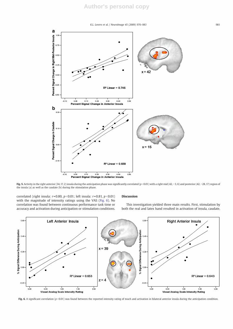
A voxel-wise correlation analysis between activation in theanterior insular portion of the “anticipation only” ROI during theanticipation phase (AntR+AntL) of the task and activation in otherregions of interest–insula, striatum, and cingulate cortex–during thestimulation phase (StimR+StimL) of the task was carried out toexamine any modulatory functions the anterior insula might becontributing to during anticipation (Fig. 5). A significant positivecorrelation was found in regions of the right mid and posterior insula(r=0.86, pb0.01) as well as the caudate (r=0.92, pb0.01).
Brain behavior relationships
A voxel-wise regression analysis was carried out to determinewhether the degree of activation in the insular cortex was related tosubjective assessment of the touch experience recorded in the VASscales. We found that the degree of activation during the anticipationphase (AntR+AntL) in bilateral anterior insula was significantly
Fig. 4. The distinct regions of insular activation during anticipation only and stimulation only (yellow and blue, respectively), and the intersection of these areas of activation (green).The signal within these regions during the anticipation and stimulation phases of the task (AntR+AntL and StimR+StimL) was extracted and entered into a paired t-test for each ROI.Activation during stimulation was significantly (pb0.017) greater than anticipation in the stimulation only and anticipation∩stimulation regions.
980 K.L. Lovero et al. / NeuroImage 45 (2009) 976–983
Author's personal copy
correlated (right insula: r=0.80, pb0.01; left insula: r=0.81, pb0.01)with the magnitude of intensity ratings using the VAS (Fig. 6). Nocorrelation was found between continuous performance task time oraccuracy and activation during anticipation or stimulation conditions.
Discussion
This investigation yielded three main results. First, stimulation byboth the real and latex hand resulted in activation of insula, caudate,
Fig. 6. A significant correlation (pb0.01) was found between the reported intensity rating of touch and activation in bilateral anterior insula during the anticipation condition.
Fig. 5. Activity in the right anterior (34,17, 2) insula during the anticipation phasewas significantly correlated (pb0.01) with a rightmid (42, −3, 6) and posterior (42, −28,17) region ofthe insula (a) as well as the caudate (b) during the stimulation phase.
981K.L. Lovero et al. / NeuroImage 45 (2009) 976–983

Author's personal copy
cingulate, and amygdala–areas commonly related to emotion andreward–as well as thalamus and somatosensory cortices. Thus, thetouch paradigm engaged systems involved in both affective andsensory processing. Second, the anterior portion of the insula wasnotably active during anticipation of touch, whereas the mid-to-posterior insular cortex was active for both anticipation and stimula-tion. Third, the degree of activation during anticipation in the bilateralanterior insular cortex was associated with stimulus related proces-sing in the mid and posterior insula and caudate, as well as the ratingsof intensity acquired in the VAS scales. Taken together, the datasupport the hypothesis that the anterior insula generates ananticipatory signal of expected stimulus intensity and thus mayhave a preparatory function to integrate the affective and sensoryexperiences associated with interoceptive processing.
During both the real and latex hand stimulation, areas implicatedin affective processing, along with areas responsible for sensoryprocessingwere significantly activated. In previous studies, the insularcortex has been implicated in interoceptive (Critchley et al., 2004) andemotion-related (Phan et al., 2002) processes. Cingulate cortex andmedial frontal gyrus have previously been linked to self-focus andmonitoring emotional state (Amodio and Frith, 2006; Beauregard et al.,2001; Gusnard et al., 2001; Mars et al., 2005; Northoff and Bermpohl,2004;Wicker et al., 2003), as well as in stimulation by pleasant touch(Francis et al., 1999). The amygdala has also been shown to activateduring pleasant sensory stimuli (O'Doherty et al., 2001), and duringpleasant mood induction (Schneider et al., 1997a). Moreover, caudateand putamen have continually shown relations to reward processing(Knutson and Cooper, 2005b; O'Doherty, 2004). Finally, primarysensory areas, including thalamus and post-central gyrus, were activeduring the stimulation phase. The activation of all these areas duringthe stimulation phase support that the touch paradigm utilized hereprovided for a stimulus that acts on both affective and sensorysystems.
Whereas activation was seen in areas related to affective andsensory processing during the stimulation phase, there was a muchgreater recruitment of areas implicated in affective processing duringthe anticipation phase. Similar to the stimulus condition, anteriorcingulate cortex, medial frontal gyrus, and amygdala were signifi-cantly activated during anticipation. Additionally, a region of theinsular cortex was found to be significantly active during anticipation;however, this region appeared to extend more into anterior regionsthan those found active during stimulation.
In order to better segregate these topographical differences, weperformed a conjunction analysis to identify regions of the insulaactive during anticipation only, stimulation only, and the intersectionof the two. Our results show that the anterior insula was active onlyduring the anticipation phase of the task. Moreover, post-hoc t-testsrevealed that there was greater activation in this “anticipation only”region during anticipation than stimulation, as opposed to themid-to-posterior insular “stimulation only” region in which stimulationresulted in significantly greater activation than anticipation. Takeninto consideration that the anterior insular cortex has been implicatedin modulation of the affective response (Craig, 2005, 2008) and giventhe current finding of anticipatory signaling in this structure, wesuggest that this anticipatory periodmay be critical for various aspectsof affective encoding of the stimulus.
Additionally, the anterior insula activation during anticipation wascorrelated with the stimulus related activation in a mid and posteriorregion of the insula, as well as with a region in the caudate. Theposterior insula is part of SII and therefore is directly involved insensory and interoceptive processing (Craig, 2002, 2003; Critchley etal., 2004). Therefore, this correlation is consistent with the notion thatthe anterior insula may have a preparatory function to set thesensitivity of the posterior insula when processing the sensorycomponent of the upcoming stimulus. Moreover, the correlationwith the caudate, which is important for processing rewarding stimuli
(Knutson and Cooper, 2005a), may indicate that the level ofanticipatory activity in the anterior insula could function to alter thedegree to the rewarding properties of the upcoming stimulus. Finally,the correlation between reported intensity ratings of touch andactivation in bilateral anterior insula during anticipation relates thesubjective experience to the modulatory role of the anterior insula. Insum, these results are in support of the notion that the anterior insulaanticipates the affective value of the stimulus, whereas a more mid-posterior region is primarily involved in processing sensory compo-nents of a stimulus.
Contrary to our expectation, we did not find strong differencesbetween the real and latex stimuli. This lack of differential processingmay be due to the design of stimulus delivery. Specifically, the latexhand touch was administered by a person holding the hand, whichadds an interpersonal element to the stimulus and renders the latexand real hand condition less different than initially conceptualized.Therefore, future studies will need to parse out this element in orderto gain further understanding of different dimensions of affectiveprocessing during anticipation of and stimulation by a touchstimulus.
This study examines the affective nature of human touch, and therole the insular cortex plays in anticipating and processing thisemotionally significant stimuli. The insular cortex is well suited, bothanatomically and functionally, to integrate the sensory and psycho-logical self (Craig, 2002; Paulus and Stein, 2006). Specifically, theanterior inferior insula has an agranular columnar organization,whereas posterior insular is characterized by a granular corticalarchitecture. This type of transition is found elsewhere in the brainwhere cortical re-representations are based on modulatory orselective feedback circuits (Shipp, 2005). Furthermore, previousstudies have outlined the role of anterior insula in interoceptiveprocessing, emotional awareness (Critchley et al., 2004; Schneideret al., 1997b), and meditation of urges (Evans et al., 2002). Togetherwith our results, it appears that the insular cortex is an importantneural substrate for the integration of the affective and sensoryaspects of touch. The evaluation of stimulus valence occurs in anteriorinsula prior to stimulation, and this activity is correlated to that of amore mid-to-posterior region that responds during stimulation,suggesting some type of modulatory relationship between theregions.
The functional division of the insula seen in this study not onlyprovides interesting information on the topography of this region ofthe brain, but also provides a greater understanding of the temporalprocessing of an exteroceptive/interoceptive stimulus, as well as theassociation between anticipatory and affective processes. The role ofanterior insula in setting a tone for an emotionally significantexperience has strong implications for various social phobias, anxietydisorders, and drug abuse. Altered anticipatory processing, frequentlyrelated to greater insular activation, has been found in individualswith social phobia and other anxiety disorders (Chua et al., 1999;Lorberbaum et al., 2004; Nitschke et al., 2006b; Simmons et al., 2006).Also, insular activation has been associated with imagery-induceddrug craving (Kilts et al., 2001), and has been implicated inmaintaining urge related use of drugs (Naqvi et al., 2007). Recently,Nitschke et al. showed that expectancy modulates insular response tounpleasant taste (Nitschke et al., 2006a). Thus, understanding theneural substrates of human touch, beyond providing insight into abasic sensory system, may additionally provide a highly useful probefor different stages of emotionally relevant stimulation to examinewhether individuals with mood, anxiety, or addiction disorders showaltered anticipation or processing of expectancy modulated stimuli.
Acknowledgments
This research was supported by grants from NIDA (R01DA016663,R01DA018307) and by a VA Merit Grant.
982 K.L. Lovero et al. / NeuroImage 45 (2009) 976–983

Author's personal copy
Appendix A. Supplementary data
Supplementary data associated with this article can be found, inthe online version, at doi:10.1016/j.neuroimage.2008.12.070.
References
Amodio, D.M., Frith, C.D., 2006. Meeting of minds: the medial frontal cortex and socialcognition. Nat. Rev. Neurosci. 7, 268–277.
Beauregard, M., Levesque, J., Bourgouin, P., 2001. Neural correlates of conscious self-regulation of emotion. J. Neurosci. 21, RC165.
Boynton, G.M., Engel, S.A., Glover, G.H., Heeger, D.J., 1996. Linear systems analysis offunctional magnetic resonance imaging in human V1. J. Neurosci. 16, 4207–4221.
Chua, P., Krams, M., Toni, I., Passingham, R., Dolan, R., 1999. A functional anatomy ofanticipatory anxiety. NeuroImage 9, 563–571.
Coan, J.A., Schaefer, H.S., Davidson, R.J., 2006. Lending a hand: social regulation of theneural response to threat. Psychol. Sci. 17, 1032–1039.
Cox, R.W., 1996. AFNI: software for analysis and visualization of functional magneticresonance neuroimages. Comput. Biomed. Res. 29, 162–173.
Craig, A.D., 2002. How do you feel? Interoception: the sense of the physiologicalcondition of the body. Nat. Rev. Neurosci 3, 655–666.
Craig, A.D., 2003. Interoception: the sense of the physiological condition of the body.Curr. Opin. Neurobiol. 13, 500–505.
Craig, A.D., 2005. Forebrain emotional asymmetry: a neuroanatomical basis? TrendsCogn. Sci. 9, 566–571.
Craig, A.D., 2008. Interoception and Emotion: a Neuroanatomical Perspective. Hand-book of Emotion, 3rd ed. The Guilford Press.
Craig, A.D., Bushnell, M.C., 1994. The thermal grill illusion: unmasking the burn of coldpain. Science 265, 252–255.
Critchley, H.D., Wiens, S., Rotshtein, P., Ohman, A., Dolan, R.J., 2004. Neural systemssupporting interoceptive awareness. Nat. Neurosci. 7, 189–195.
Damasio, A.R., 1994. Descartes' error and the future of human life. Sci. Am. 271, 144.Devries, A.C., Glasper, E.R., Detillion, C.E., 2003. Social modulation of stress responses.
Physiol. Behav. 79, 399–407.Dupont, S., Bouilleret, V., Hasboun, D., Semah, F., Baulac, M., 2003. Functional anatomy
of the insula: new insights from imaging. Surg. Radiol. Anat. 25, 113–119.Evans, K.C., Banzett, R.B., Adams, L., McKay, L., Frackowiak, R.S., Corfield, D.R., 2002.
BOLD fMRI identifies limbic, paralimbic, and cerebellar activation during air hunger.J. Neurophysiol. 88, 1500–1511.
Forman, S.D., Cohen, J.D., Fitzgerald, M., Eddy, W.F., Mintun, M.A., Noll, D.C., 1995.Improved assessment of significant activation in functional magnetic resonanceimaging (fMRI): use of a cluster-size threshold. Magn. Reson. Med. 33, 636–647.
Francis, S., Rolls, E.T., Bowtell, R., McGlone, F., O'Doherty, J., Browning, A., Clare, S., Smith,E., 1999. The representation of pleasant touch in the brain and its relationship withtaste and olfactory areas. NeuroReport 10, 453–459.
Gusnard, D.A., Akbudak, E., Shulman, G.L., Raichle, M.E., 2001. Medial prefrontal cortexand self-referential mental activity: relation to a default mode of brain function.Proc. Natl. Acad. Sci. U. S. A. 98, 4259–4264.
Hertenstein, M.J., Keltner, D., App, B., Bulleit, B.A., Jaskolka, A.R., 2006. Touchcommunicates distinct emotions. Emotion 6, 528–533.
Ihaka, R., Gentleman, R., 1996. R: a language for data analysis and graphics. J. Comput.Graph. Stat. 5, 299–314.
Kilts, C.D., Schweitzer, J.B., Quinn, C.K., Gross, R.E., Faber, T.L., Muhammad, F., Ely, T.D.,Hoffman, J.M., Drexler, K.P., 2001. Neural activity related to drug craving in cocaineaddiction. Arch. Gen. Psychiatry 58, 334–341.
Knapp, M.L., Hall, J.A., 1997. Nonverbal Communication in Verbal Interaction. HarcourtBrace College, Fort Worth, TX.
Knutson, B., Cooper, J.C., 2005b. Functional magnetic resonance imaging of rewardprediction. Curr. Opin. Neurol. 18, 411–417.
Knutson, B., Cooper, J.C., 2005a. Functional magnetic resonance imaging of rewardprediction. Curr. Opin. Neurol. 18, 411–417.
Lahuerta, J., Bowsher, D., Campbell, J., Lipton, S., 1990. Clinical and instrumentalevaluation of sensory function before and after percutaneous anterolateralcordotomy at cervical level in man. Pain 42, 23–30.
Lancaster, J.L., Woldorff, M.G., Parsons, L.M., Liotti, M., Freitas, C.S., Rainey, L., Kochunov,P.V., Nickerson, D., Mikiten, S.A., Fox, P.T., 2000. Automated Talairach atlas labels forfunctional brain mapping. Hum. Brain Mapp. 10, 120–131.
Lorberbaum, J.P., Kose, S., Johnson, M.R., Arana, G.W., Sullivan, L.K., Hamner, M.B.,Ballenger, J.C., Lydiard, R.B., Brodrick, P.S., Bohning, D.E., George, M.S., 2004. Neuralcorrelates of speech anticipatory anxiety in generalized social phobia. NeuroReport15, 2701–2705.
Mars, R.B., Coles, M.G., Grol, M.J., Holroyd, C.B., Nieuwenhuis, S., Hulstijn, W., Toni, I.,2005. Neural dynamics of error processing in medial frontal cortex. NeuroImage 28,1007–1013.
Naqvi, N.H., Rudrauf, D., Damasio, H., Bechara, A., 2007. Damage to the insula disruptsaddiction to cigarette smoking. Science 315, 531–534.
Nitschke, J.B., Dixon, G.E., Sarinopoulos, I., Short, S.J., Cohen, J.D., Smith, E.E., Kosslyn,S.M., Rose, R.M., Davidson, R.J., 2006a. Altering expectancy dampens neuralresponse to aversive taste in primary taste cortex. Nat. Neurosci. 9, 435–442.
Nitschke, J.B., Sarinopoulos, I., Mackiewicz, K.L., Schaefer, H.S., Davidson, R.J., 2006b.Functional neuroanatomy of aversion and its anticipation. NeuroImage 29, 106–116.
Northoff, G., Bermpohl, F., 2004. Cortical midline structures and the self. Trends Cogn.Sci. 8, 102–107.
Norusis, M.J., 2002. Statistical Package for the Social Sciences: SPSS 11. SPSS. Inc, Chicago.O'Doherty, J.P., 2004. Reward representations and reward-related learning in the
human brain: insights from neuroimaging. Curr. Opin. Neurobiol. 14, 769–776.O'Doherty, J., Rolls, E.T., Francis, S., Bowtell, R., McGlone, F., 2001. Representation of
pleasant and aversive taste in the human brain. J. Neurophysiol. 85, 1315–1321.Olausson, H., Lamarre, Y., Backlund, H., Morin, C., Wallin, B.G., Starck, G., Ekholm, S.,
Strigo, I., Worsley, K., Vallbo, A.B., Bushnell, M.C., 2002. Unmyelinated tactileafferents signal touch and project to insular cortex. Nat. Neurosci. 5, 900–904.
Paulus, M.P., Stein, M.B., 2006. An insular view of anxiety. Biol. Psychiatry 60, 383–387.Phan, K.L., Wager, T., Taylor, S.F., Liberzon, I., 2002. Functional neuroanatomy of emotion: a
meta-analysis of emotion activation studies in PETand fMRI. NeuroImage 16, 331–348.Robinson, S.K., Viirre, E.S., Bailey, K.A., Gerke, M.A., Harris, J.P., Stein, M.B., 2005.
Randomized placebo-controlled trial of a selective serotonin reuptake inhibitor inthe treatment of nondepressed tinnitus subjects. Psychosom. Med. 67, 981–988.
Rolls, E.T., 2000. The orbitofrontal cortex and reward. Cereb. Cortex 10, 284–294.Schmelz, M., Schmidt, R., Bickel, A., Handwerker, H.O., Torebjork, H.E., 1997. Specific
C-receptors for itch in human skin. J. Neurosci. 17, 8003–8008.Schneider, F., Grodd, W., Weiss, U., Klose, U., Mayer, K.R., Nagele, T., Gur, R.C., 1997a.
Functional MRI reveals left amygdala activation during emotion. Psychiatry Res. 76,75–82.
Schneider, F., Grodd, W., Weiss, U., Klose, U., Mayer, K.R., Nagele, T., Gur, R.C., 1997b.Functional MRI reveals left amygdala activation during emotion. Psychiatry Res. 76,75–82.
Shipp, S., 2005. The importance of being agranular: a comparative account of visual andmotor cortex. Philos. Trans. R. Soc. Lond. B Biol. Sci. 360, 797–814.
Simmons, A., Strigo, I., Matthews, S.C., Paulus, M.P., Stein, M.B., 2006. Anticipation ofaversive visual stimuli is associated with increased insula activation in anxiety-prone subjects. Biol. Psychiatry 60, 402–409.
Strigo, I., Simmons, A.N., Craig, A.D., Paulus, M.P., 2006. Breathing and BOLD fMRI:watch out.
Talairach, J., Tournoux, P., 1988. Co-planar Stereotaxic Atlas of the Human Brain: a 3-Dimensional Proportional System, an Approach to Cerebral Imaging. G. Thieme;Thieme Medical Publishers, Stuttgart; New York New York.
Vallbo, A.B., Olausson, H., Wessberg, J., Kakuda, N., 1995. Receptive field characteristicsof tactile units with myelinated afferents in hairy skin of human subjects. J. Physiol.483, 783–795.
Wicker, B., Ruby, P., Royet, J.P., Fonlupt, P., 2003. A relation between rest and the self inthe brain? Brain Res. Brain Res. Rev. 43, 224–230.
983K.L. Lovero et al. / NeuroImage 45 (2009) 976–983


