Embed Size (px)
Citation preview

REVIEWpublished: 28 March 2017
doi: 10.3389/fmicb.2017.00495
Frontiers in Microbiology | www.frontiersin.org 1 March 2017 | Volume 8 | Article 495
Edited by:
Kuldeep Dhama,
Indian Veterinary Research Institute,
India
Reviewed by:
Maryam Dadar,
Razi Vaccine and Serum Research
Institute, Iran
Hafiz M. N. Iqbal,
Monterrey Institute of Technology and
Higher Education, Mexico
Ruchi Tiwari,
DUVASU Mathura UP, India
*Correspondence:
Shihua Wang
Specialty section:
This article was submitted to
Microbial Immunology,
a section of the journal
Frontiers in Microbiology
Received: 26 January 2017
Accepted: 09 March 2017
Published: 28 March 2017
Citation:
Saeed AFUH, Wang R, Ling S and
Wang S (2017) Antibody Engineering
for Pursuing a Healthier Future.
Front. Microbiol. 8:495.
doi: 10.3389/fmicb.2017.00495
Antibody Engineering for Pursuing aHealthier FutureAbdullah F. U. H. Saeed, Rongzhi Wang, Sumei Ling and Shihua Wang*
Key Laboratory of Pathogenic Fungi and Mycotoxins of Fujian Province, Key Laboratory of Biopesticide and Chemical
Biology of Education Ministry, and School of Life Sciences, Fujian Agriculture and Forestry University, Fuzhou, China
Since the development of antibody-production techniques, a number of
immunoglobulins have been developed on a large scale using conventional methods.
Hybridoma technology opened a new horizon in the production of antibodies against
target antigens of infectious pathogens, malignant diseases including autoimmune
disorders, and numerous potent toxins. However, these clinical humanized or chimeric
murine antibodies have several limitations and complexities. Therefore, to overcome
these difficulties, recent advances in genetic engineering techniques and phage display
technique have allowed the production of highly specific recombinant antibodies.
These engineered antibodies have been constructed in the hunt for novel therapeutic
drugs equipped with enhanced immunoprotective abilities, such as engaging immune
effector functions, effective development of fusion proteins, efficient tumor and
tissue penetration, and high-affinity antibodies directed against conserved targets.
Advanced antibody engineering techniques have extensive applications in the fields
of immunology, biotechnology, diagnostics, and therapeutic medicines. However,
there is limited knowledge regarding dynamic antibody development approaches.
Therefore, this review extends beyond our understanding of conventional polyclonal
and monoclonal antibodies. Furthermore, recent advances in antibody engineering
techniques together with antibody fragments, display technologies, immunomodulation,
and broad applications of antibodies are discussed to enhance innovative antibody
production in pursuit of a healthier future for humans.
Keywords: antibody engineering, hybridoma technology, antibody fragments, scFv, phage display technology,
immunomodulation, immunology
INTRODUCTION
In recent years, the development of polyclonal and monoclonal antibody by means of laboratoryanimals has become a vital approach to protect against a number of pathogenic contagions(Marasco and Sui, 2007). These immunoprotective molecules provide defense against transmissiblediseases and can eliminate the infection. Their prophylactic and therapeutic protection abilitywas first discovered in the late nineteenth century by the passive transmission of antibodies froma diseased animal that provided immunity against diphtheria. Subsequently, immune sera fromvarious herbivores and humans were obtained, pooled, and used as therapeutics. Since then,the management of infectious diseases such as diphtheria, tetanus, pneumococcal pneumonia,meningococcal meningitis, and toxin-mediated diseases has considerably improved patient survival(Casadevall, 1999).
Saeed et al. Antibody Engineering and Recent Advances in Immunology
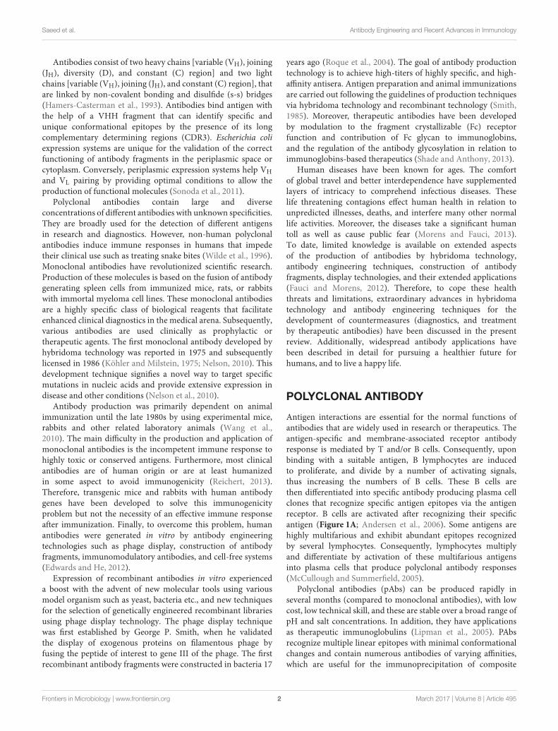
Antibodies consist of two heavy chains [variable (VH), joining(JH), diversity (D), and constant (C) region] and two lightchains [variable (VH), joining (JH), and constant (C) region], thatare linked by non-covalent bonding and disulfide (s-s) bridges(Hamers-Casterman et al., 1993). Antibodies bind antigen withthe help of a VHH fragment that can identify specific andunique conformational epitopes by the presence of its longcomplementary determining regions (CDR3). Escherichia coliexpression systems are unique for the validation of the correctfunctioning of antibody fragments in the periplasmic space orcytoplasm. Conversely, periplasmic expression systems help VH
and VL pairing by providing optimal conditions to allow theproduction of functional molecules (Sonoda et al., 2011).
Polyclonal antibodies contain large and diverseconcentrations of different antibodies with unknown specificities.They are broadly used for the detection of different antigensin research and diagnostics. However, non-human polyclonalantibodies induce immune responses in humans that impedetheir clinical use such as treating snake bites (Wilde et al., 1996).Monoclonal antibodies have revolutionized scientific research.Production of these molecules is based on the fusion of antibodygenerating spleen cells from immunized mice, rats, or rabbitswith immortal myeloma cell lines. These monoclonal antibodiesare a highly specific class of biological reagents that facilitateenhanced clinical diagnostics in the medical arena. Subsequently,various antibodies are used clinically as prophylactic ortherapeutic agents. The first monoclonal antibody developed byhybridoma technology was reported in 1975 and subsequentlylicensed in 1986 (Köhler and Milstein, 1975; Nelson, 2010). Thisdevelopment technique signifies a novel way to target specificmutations in nucleic acids and provide extensive expression indisease and other conditions (Nelson et al., 2010).
Antibody production was primarily dependent on animalimmunization until the late 1980s by using experimental mice,rabbits and other related laboratory animals (Wang et al.,2010). The main difficulty in the production and application ofmonoclonal antibodies is the incompetent immune response tohighly toxic or conserved antigens. Furthermore, most clinicalantibodies are of human origin or are at least humanizedin some aspect to avoid immunogenicity (Reichert, 2013).Therefore, transgenic mice and rabbits with human antibodygenes have been developed to solve this immunogenicityproblem but not the necessity of an effective immune responseafter immunization. Finally, to overcome this problem, humanantibodies were generated in vitro by antibody engineeringtechnologies such as phage display, construction of antibodyfragments, immunomodulatory antibodies, and cell-free systems(Edwards and He, 2012).
Expression of recombinant antibodies in vitro experienceda boost with the advent of new molecular tools using variousmodel organism such as yeast, bacteria etc., and new techniquesfor the selection of genetically engineered recombinant librariesusing phage display technology. The phage display techniquewas first established by George P. Smith, when he validatedthe display of exogenous proteins on filamentous phage byfusing the peptide of interest to gene III of the phage. The firstrecombinant antibody fragments were constructed in bacteria 17
years ago (Roque et al., 2004). The goal of antibody productiontechnology is to achieve high-titers of highly specific, and high-affinity antisera. Antigen preparation and animal immunizationsare carried out following the guidelines of production techniquesvia hybridoma technology and recombinant technology (Smith,1985). Moreover, therapeutic antibodies have been developedby modulation to the fragment crystallizable (Fc) receptorfunction and contribution of Fc glycan to immunoglobins,and the regulation of the antibody glycosylation in relation toimmunoglobins-based therapeutics (Shade and Anthony, 2013).
Human diseases have been known for ages. The comfortof global travel and better interdependence have supplementedlayers of intricacy to comprehend infectious diseases. Theselife threatening contagions effect human health in relation tounpredicted illnesses, deaths, and interfere many other normallife activities. Moreover, the diseases take a significant humantoll as well as cause public fear (Morens and Fauci, 2013).To date, limited knowledge is available on extended aspectsof the production of antibodies by hybridoma technology,antibody engineering techniques, construction of antibodyfragments, display technologies, and their extended applications(Fauci and Morens, 2012). Therefore, to cope these healththreats and limitations, extraordinary advances in hybridomatechnology and antibody engineering techniques for thedevelopment of countermeasures (diagnostics, and treatmentby therapeutic antibodies) have been discussed in the presentreview. Additionally, widespread antibody applications havebeen described in detail for pursuing a healthier future forhumans, and to live a happy life.
POLYCLONAL ANTIBODY
Antigen interactions are essential for the normal functions ofantibodies that are widely used in research or therapeutics. Theantigen-specific and membrane-associated receptor antibodyresponse is mediated by T and/or B cells. Consequently, uponbinding with a suitable antigen, B lymphocytes are inducedto proliferate, and divide by a number of activating signals,thus increasing the numbers of B cells. These B cells arethen differentiated into specific antibody producing plasma cellclones that recognize specific antigen epitopes via the antigenreceptor. B cells are activated after recognizing their specificantigen (Figure 1A; Andersen et al., 2006). Some antigens arehighly multifarious and exhibit abundant epitopes recognizedby several lymphocytes. Consequently, lymphocytes multiplyand differentiate by activation of these multifarious antigensinto plasma cells that produce polyclonal antibody responses(McCullough and Summerfield, 2005).
Polyclonal antibodies (pAbs) can be produced rapidly inseveral months (compared to monoclonal antibodies), with lowcost, low technical skill, and these are stable over a broad range ofpH and salt concentrations. In addition, they have applicationsas therapeutic immunoglobulins (Lipman et al., 2005). PAbsrecognize multiple linear epitopes with minimal conformationalchanges and contain numerous antibodies of varying affinities,which are useful for the immunoprecipitation of composite
Frontiers in Microbiology | www.frontiersin.org 2 March 2017 | Volume 8 | Article 495

Saeed et al. Antibody Engineering and Recent Advances in Immunology
FIGURE 1 | Interaction of antibodies with numerous antigens present on the surface of target cell. (A) Interaction of polyclonal antibodies with specific
surface antigen activates B lymphocytes to divide and differentiate into plasma cell clones producing more antibodies that recognize antigens. (B) Interaction of
monoclonal antibodies with specific surface antigen activates B lymphocytes to divide and differentiate into plasma cell clones that further recruit homogeneous and
mono-specific antibodies.
antigens to form a large precipitating lattice. Mice are usedoften to produce pAbs because of their small size and bloodvolume. As an alternative, pAbs are produced as ascites in mice.Moreover, these antibodies have superior specificity comparedwith monoclonal antibodies because they are generated by a largenumber of B-cell clones each producing antibodies to a specificepitope (Hudson et al., 2012; Zhuang et al., 2014).
MONOCLONAL ANTIBODY
Monoclonal antibodies (mAbs) are clinically significanthomogeneous and mono-specific scientific biomoleculesproduced from hybridoma cells by hybridoma technology(Zhang, 2012). mAbs arise from single cell clone compared tomultiple cell clones for pAbs (Figure 1B; Andersen et al., 2006).Since their discovery, these molecules have been used as researchtools and have revolutionized the fields of biotechnology,immunology, diagnostics, and medicine. The technology wasdescribed for the first time by Köhler and Milstein (1975) in themid-1970s in the journal Nature, and they were later awardedthe Nobel Prize (Saeed and Awan, 2016).
Currently, mAb products approved by the US Food and DrugAdministration (FDA) are increasing worldwide i.e., about fournew products per year. Currently, 47 mAb products in the US,Europe and global markets have been approved for the treatmentof a variety of diseases (Table 1; Ecker et al., 2015). At the currentrate, about 70 mAb products will be on the market by 2020,and collective global trade will be approximately $125 billion(Ecker et al., 2015). Improvements in hybridoma technology arebased on research demand, cost effectiveness, human labor, andreduced development time. Similarly, the production of mAbsrequires multiple phases, long duration, and high cost. Currently,mAbs have been produced against a number of mycotoxins suchas fumonisin B1 (Yuan et al., 2012; Ling et al., 2014, 2015b),
citreoviridin (Jin et al., 2014), marine toxins (Saeed and Wang,2016), and other exo- and endo-antigens. Similarly, mAbs againsttransmembrane enzymes have been produced (Yuan et al., 2012).
Method of Antibody ProductionHybridoma technology has been a significant and essentialplatform for producing high-quality mAbs (Zhang, 2012). Itpermits generation of therapeutic antibodies in a native form.However, technical difficulties in hybridoma production haveupdated the mainstream antibody production into new ways likedisplay and transgenic mice techniques. Nevertheless, hybridomatechnology is a classical and established route of generatingspecific antibodies all around the globe (Glukhova et al., 2016).The technology begins with immunization of test animals withan antigen of interest and serum antibody titer is determinedby enzyme linked immunosorbent assay (ELISA). Subsequently,the spleen is aseptically removed and splenocytes are fusedwith myeloma cells to produce hybridoma cells. Hybridomacells are then cultured in 96-well plates in the presence ofhypoxanthine-aminopterin-thymidine (HAT) selection mediumfor high throughput screening. Later, hybridoma cells producingdesired antibodies are screened by conventional ELISA andnovel nanoparticle-probed immunoassay (colloidal gold or silvernanoparticles; Figure 2). Cell culture systems in vitro withspecific mAb cell lines were then subjected to mass generation bymedia selection, shaker flasks, and bench-scale bioreactors (Linget al., 2014).
The optimal conditions such as temperature, percent carbondioxide, and humidity for cell cultures should be determined(Sen and Roychoudhury, 2013), and then transferred to apilot scale for scalability and toxicology studies. In addition,clinical materials should be produced on a large scale underthe existing good manufacturing practice (cGMP) regulations.After production on a small scale, products that are already
Frontiers in Microbiology | www.frontiersin.org 3 March 2017 | Volume 8 | Article 495

Saeed et al. Antibody Engineering and Recent Advances in Immunology
TABLE 1 | Monoclonal antibody products in the US, Europe, and global markets approved for diseases.
Brand name Company reporting US
sales
Company reporting EU
sales
Year of
approval
Treatment
AlprolIX (Factor IX Fc fusion
protein)
Biogen Idec Sobi and Biogen Idec 2014 Hemophilia B
Cyramza (ramucirumab) Eli Lilly and Co. Eli Lilly and Co. 2014 Gastric cancer and non-small cell lung cancer
Eloctate (Factor VIII Fc
fusion protein)
Biogen Idec Sobi and Biogen Idec 2014 Anti-hemophilic Factor
Entyvio (vedolizumab) Takeda Pharmaceutical
Co.
Takeda Pharmaceutical
Co.
2014 Ulcerative colitis (UC)/Crohn’s disease (CD)
Keytruda (pembrolizumab) Merck & Co. Merck & Co. 2014 Melanoma
Sylvant (siltuximab) Johnson & Johnson Johnson & Johnson 2014 Multicentric Castleman’s Disease (MCD)
Inflectra (infliximab
[biosimilar])
N/A Hospira 2013 Tumor necrosis
Kadcyla (ado-trastuzumab
emtansine)
Roche Roche 2013 Metastatic breast cancer
Lemtrada (alemtuzumab) N/A Sanofi 2013 Relapsing form of multiple sclerosis (MS)
Gazyva (obinutuzumab) Roche Roche 2013 Chronic lymphocytic leukemia (CLL)
Remsima (infliximab
[biosimilar])
N/A Celltrion 2013 Rheumatoid arthritis, psoriatic arthritis, ulcerative colitis, Crohn’s
disease, ankylosing spondylitis, and plaque psoriasis
Perjeta (pertuzumab) Roche Roche 2012 Human epidermal growth factor receptor 2 (HER2)/neu-positive)
metastatic breast cancer
Abthrax (raxibacumab) GlaxoSmithKline GlaxoSmithKline 2012 Inhalational anthrax
Prolia (denosumab) Amgen GlaxoSmithKline 2011 Osteoporosis
Adcetris (brentuximab
vedotin)
Seattle Genetics Takeda Pharmaceutical
Co.
2011 Hodgkin lymphoma
Benlysta (belimumab) GlaxoSmithKline GlaxoSmithKline 2011 Systemic lupus erythematosus (SLE/lupus)
Eylea (aflibercept) Regeneron
Pharmaceuticals
Bayer Healthcare
Pharmaceuticals
2011 Macular degeneration
Nulojix (belatacept) Bristol-Myers Squibb Bristol-Myers Squibb 2011 Prevention of transplant rejection
Xgeva (denosumab) Amgen Amgen 2010 Prevention of bone fractures and other skeletal bone tumor
conditions
Arzerra (ofatumumab) GlaxoSmithKline GlaxoSmithKline 2009 Chronic lymphocytic leukemia (CLL)
Ilaris (canakinumab) Novartis Pharmaceuticals Novartis Pharmaceuticals 2009 Systemic Juvenile Idiopathic Arthritis (SJIA)
Actemra (tocilizumab) Roche Roche 2009 Moderate to severe rheumatoid arthritis
Simponi Aria (golimumab) Johnson & Johnson Merck & Co. 2009 Rheumatoid arthritis
Stelara (ustekinumab) Johnson & Johnson Johnson & Johnson 2009 Plaque psoriasis, psoriatic arthritis and Crohn’s disease
Removab (catumaxomab) N/A NeoPharm Group 2009 Malignant ascites
Arcalyst (rilonacept) Regeneron
Pharmaceuticals
Regeneron
Pharmaceuticals
2008 Familial Cold Auto-inflammatory Syndrome/Muckle-Wells
Syndrome
Cimzia (certolizumab pegol) UCB UCB 2008 Rheumatoid Arthritis
Nplate (romiplostim) Amgen Amgen 2008 Low blood platelet counts
Soliris (eculizumab) Alexion Pharmaceuticals Alexion Pharmaceuticals 2007 Paroxysmal nocturnal hemoglobinuria (PNH), and Atypical
hemolytic uremic syndrome (aHUS)
Lucentis (ranibizumab) Roche Novartis Pharmaceuticals 2006 Macular degeneration
Vectibix (panitumumab) Amgen Amgen 2006 Metastatic colorectal cancer
Orencia (abatacept) Bristol-Myers Squibb Bristol-Myers Squibb 2005 Rheumatoid arthritis
Avastin (bevacizumab) Roche Roche 2004 Various cancers and eye disease
Tysabri (natalizumab) Biogen Idec Biogen Idec 2004 MS and Crohn’s disease
Erbitux (cetuximab) Bristol-Myers Squibb Merck KGaA 2004 Metastatic colorectal cancer, metastatic non-small cell lung
cancer, head and neck cancer
Humira (adalimumab) AbbVie AbbVie 2002 Rheumatoid arthritis, juvenile idiopathic arthritis, psoriatic arthritis,
ankylosing spondylitis, plaque psoriasis, and hidradenitis
suppurativa
Enbrel (etanercept) Amgen Pfizer 1998 Rheumatoid arthritis
(Continued)
Frontiers in Microbiology | www.frontiersin.org 4 March 2017 | Volume 8 | Article 495

Saeed et al. Antibody Engineering and Recent Advances in Immunology
TABLE 1 | Continued
Brand name Company reporting US
sales
Company reporting EU
sales
Year of
approval
Treatment
Herceptin (trastuzumab) Roche Roche 1998 Breast cancer/stomach cancer
Remicade (infliximab) Johnson & Johnson Merck & Co. 1998 Crohn’s Disease
Simulect (basiliximab) Novartis Pharmaceuticals Novartis Pharmaceuticals 1998 Prevention of organ rejection
Synagis (palivizumab) AstraZeneca Abbvie 1998 Lung disease caused by respiratory syncytial virus (RSV)
Rituxan (rituximab) Roche Roche 1997 Non-Hodgkin’s lymphoma or chronic lymphocytic leukemia
ReoPro (abciximab) Lilly Lilly 1994 Prevention of blood clots
FIGURE 2 | Illustration showing the production route of hybridoma technology. Monoclonal antibodies are generated by immunizing laboratory animals with a
target antigen. B cells and myeloma cells are fused and then selected in HAT medium. Finally, hybridoma cells producing the desired antibodies are screened.
cultured in a laboratory are then transferred to pilot scalecommercialization. The process then performs characterization,scaling, technology transfer, and validation. Commercial cellculture for the production of a biological product is completedby pilot scale laboratory methods (Li et al., 2010). Recently,commercialization is initiated by process characterization, scale-up, technology transfer, and validation of the manufacturingprocess (Li et al., 2006).
ELISA is enzyme-based colorimetric assay, requireslarge sample volumes, several incubation steps and has lowdetection sensitivity (Tang and Hewlett, 2010). Conversely,nanotechnology and nanoparticles (NPs) use nanomaterials withlength scale of 1–100 nanometers (nm). Nanomaterials haveunique biological properties such as small size, large surface-to-volume ratio, sharp melting temperature, magnetic properties,unusual target binding properties, and size based multi-coloring(Qi and Wang, 2004). NPs such as gold particles have been usedto improve assay sensitivity and specificity of antibodies, lowlimit of detection (LOD), dose response over 10,000-fold and thedetection sensitivity by 1,000-fold compared to ELISA (Tang andHewlett, 2010).
Similarly, an essential factor of NPs selection is thatnanometer-sized (colloidal gold or silver) particles can beconjugated with targeting ligands, including mAbs, peptides,or small molecules with functional and structural propertiesthat are not available from either discrete molecules or bulkmaterials. Nanoparticles are probed with mAbs that can beused to target any antigen of interest (Ling et al., 2015a).The use of colloidal gold is a rapid, less time consuming, andcost effective technique. This technology has been used in thedevelopment of immunochromatic strip assays based on mAb.The technique has widespread applications for the detection of anumber of antigens (exogenous antigens, endogenous antigens,autoantigens, neoantigens, viral antigens, and tumor antigens)with high specificity and binding affinity (Schumacher andSchreiber, 2015).
Antigen PreparationAntigen preparation including quality and quantity is essentialfor the production of antibodies. The purification of antigensis difficult and time consuming but antigen purity is vital foradequate immune responses (Leenaars and Hendriksen, 2005).
Frontiers in Microbiology | www.frontiersin.org 5 March 2017 | Volume 8 | Article 495

Saeed et al. Antibody Engineering and Recent Advances in Immunology
Insignificant impurities (<1%) can be immunodominant inthe case of various microbial antigens. They show non-specificactivity against the antigens of interest and specific activityagainst impurities. Higher concentrations of specific antibodiesare obtained after purification and the exclusion of impuritiesby extensive absorption procedures such as antibody affinitychromatography (Leenaars et al., 1997; Mazzer et al., 2015).
Before scheduling immunization, antigen contaminationshould be considered by the researcher. The antigen and diluentsshould be free of endotoxins such as lipopolysaccharide, orchemical residues that have been utilized to neutralize themicroorganism. Additionally, the pH must be adjusted toprevent undesirable effects in the animal to be immunized(Hendriksen and Hau, 2003). Moreover, sterile workingconditions, antigen concentration, animal preparation, andinjection quality must be confirmed. These conditions arenecessary to avoid suppression, sensitization, tolerance, or othersuperfluous immunomodulation and to induce effective immuneresponses. The required antigen concentration (µg to mg) isbased on the intrinsic properties of the antigen. Usual doses ofantigen conjugated with Freund’s adjuvant for rabbits are inthe range of 50–1,000µg; for mice, 10–200µg; and for goatsand sheep, 250–5,000µg (Leenaars et al., 1997). The inherentproperties of antigen quantity include purification, animals to beimmunized, type, and quality of the adjuvant (to elicit high-titerserum responses), route and the immunization (injection)frequency (Hanly et al., 1995).
ImmunizationAntibody production involves the immunization or injectionof laboratory animals with an immunogenic protein and testsampling of antiserum. The immunization is performed inspecific animal species and the adjuvant is processed to form animmunogenic emulsion that is insoluble in water (Leenaars andHendriksen, 2005). Conjugation of smaller or less immunogenicantigens i.e., haptens (numerous myco- and marine toxins)to carrier proteins is essential before animal injection. Afterimmunization, the animal is monitored daily or three times aweek for side effects (clinical and pathological examination).Severe pathological changes such as tissue reactivity, infectionand anaphylactic reactions in case of booster injections have beenreported in the absence of visual clinical or behavioral changes(Leenaars et al., 1997).
The immunization route is based on the choice of animalspecies, adjuvant, concentration and quality parameters of theantigen (Apostolico Jde et al., 2016). Suitable immunizationroutes include subcutaneous (s.c.), intradermal (i.d.),intramuscular (i.m.), intraperitoneal (i.p.), and intravenous(i.v.). Blood sample handling should not interfere with theimmunization site to avoid pain and distress to the animal whiletaking blood sample (Leenaars et al., 1997). The i.v. route forwater-in-oil emulsions such as Freund’s complete and incompleteadjuvants (FCA and FIA) may be fatal as embolisms causedby large particulates or viscous gel adjuvants (e.g., aluminumsalts) can occur (Hanly et al., 1995). The s.c. administrationof FCA is immunologically effective (0.1 mL in mice and 0.25mL in rabbits) with some reports of pathological changes.
The i.p. administration of FCA is not suggested for antibodydevelopment because it induces infection, peritonitis, and socialvariations. There is high risk of anaphylactic shock for boosterinjections by the i.v. or i.p. route (Leenaars et al., 1997).
The volume of the immunogenic mixture depends on thequality of the antigen and the degree of the lesions formed.Therefore, smaller volumes of mixtures are injected to induceantibody responses except when increased concentrations ofantigen are required (Leenaars and Hendriksen, 2005). Theimmunization volume also depends on the animal species, route,and chemistry of the injection mixture. The volume of FCAhas lethal effects on animal physiology and the extent of lesionsproduced is associated with high volumes of FCA (Ramos-Varaand Miller, 2014).
Booster injections have a significant effect on the outcomes ofimmunization and induction of B memory cells. Class switchingof B cells depends on the time interval between two consecutiveinjections. A small volume of antigen can be used for a boosterinjection without adjuvant (Leenaars et al., 1997). A boosterinjection is used when the antibody titer has reached a plateauor begins to decline. Antibody titers typically peak at 2–3 weeksafter immunization in the absence of the depot-forming adjuvantused in the first immunization. A booster injection is used after 4weeks in the presence of depot-forming adjuvant. FCA should beused in the first s.c. injection to avoid subsequent (Mycobacteriaproteins) severe tissue reactions (Hendriksen and Hau, 2003).
Hybridoma ScreeningHybridoma technology is essential for the production of high-quality mAbs as well as research and diagnostic reagents, andit is currently the most rapidly growing class of therapeutics(Hnasko and Stanker, 2015). MAbs can be effectively screenedby a number of techniques such as ELISA, phage display,and other related technologies. Screening identifies and picksonly specific antibody producing hybridomas. In hybridomatechnology, laboratory animals are immunized with an antigenof interest and specific antibody-producing B cells are isolatedfrom the spleen and then fused with immortal myeloma cells(such as sp2/0; Bradbury et al., 2011). Subsequently, expansionsof clonal populations are produced from serially diluted sub-cloned individual hybridoma cells in a microtiter plate. Next,the hybridoma supernatant is checked by ELISA or other relatedimmunoassays for desired antibody activity (El Debs et al., 2012).
After screening, hybridoma cells are expanded in bigger wellsor in culture flasks to maintain the hybridoma and providesufficient cells for cryopreservation and supernatants for laterinvestigations (Nelson et al., 2010; El Debs et al., 2012). However,this technique restricts the number of clones that can be screenedto no more than a few thousand. Consequently, insufficientnumbers of cells remain available for clonal expansion andcell immortalization. Therefore, improved techniques such asantigen-based microarrays have been used for the screening of>105 clones in <12 h, or lithographically fabricated microwellsfor individual cell compartmentalization. Nevertheless, thesetechniques only screen for binding activity and do not allowfunctional assays (El Debs et al., 2012).
Frontiers in Microbiology | www.frontiersin.org 6 March 2017 | Volume 8 | Article 495

Saeed et al. Antibody Engineering and Recent Advances in Immunology
Characterization of Monoclonal AntibodiesMAb characterization is based on a determination of itsphysicochemical and immunochemical character, heterogeneity,purity, impurities, biological activity, potency, and concentration(Berkowitz et al., 2012). For physicochemical properties, theisotype (class, subclass, light chain composition) and primarystructure of the mAb are determined. Immunological propertiesinclude binding assay, affinity, avidity, immunoreactivity (cross-reactivity with other structurally homologous proteins), analysisof CDR, epitope characterization, and determination of effectorfunctions (CHMP, 2008).
Heterogeneity (including chromatographic andelectrophoretic methods) and quantity are determined forthe characterization of mAb purity, impurity, contaminants, andconcentration. Specificity of mAbs is also checked. Peptide map,anti-idiotype immunoassay, potency, and other appropriate
methods are used to determine its identity and biological activity(CHMP, 2008).
Applications of Monoclonal AntibodiesMAb-based products exhibit superior specificity for a particularantigen. This characteristic feature of the immunoglobins makesthem an ideal tool for many applications including diseasediagnosis and therapy (Table 2; Modjtahedi et al., 2012; Redmanet al., 2015; Steplewski et al., 2015). Diagnostic applicationsinclude biochemical analysis and imaging. It involves a numberof immunoassays for the detection of hormonal, tissue, andcell products. Imaging is carried out using radiolabeled mAbsfor diagnostics of infectious diseases. Therapeutic mAbs have awide range of applications. They are used in the treatment ofcancer, transplantation of bonemarrow and organs, autoimmunediseases, cardiovascular diseases, and various infectious diseases.
TABLE 2 | Summary of various applications of monoclonal antibodies.
Application Features Diagnosis, treatment, and gains
DIAGNOSIS
Biochemical analysis Diagnostic tests are regularly used in radioimmunoassay (RIA)
and ELISA in the laboratory to quantify the circulating
concentrations of hormones and several other tissue and cell
products.
Pregnancy: human chorionic gonadotropin; cancers: colorectal cancer,
prostate cancer, tumor markers; hormonal disorders: thyroxine,
triiodothyronine, thyroid; infectious diseases, sexually transmitted
diseases (STDs) include Neisseria gonorrhoeae, herpes simplex virus.
Diagnostic imaging The technique is also called immunoscintigraphy.
Radiolabeled—mAbs are used in the diagnostic imaging of
the diseases. The radioisotopes commonly used for labeling
mAbs are iodine—131 and technetium—99. Imaging tool
include single photon emission computed tomography
(SPECT) cameras.
Myocardial infarction: myocardial necrosis; deep vein thrombosis:
thrombus in thigh, pelvis, calf, knee; atherosclerosis: coronary and
peripheral arteries; immunohistopathology of cancers: colon, stomach,
pancreas, liver, germ cells of testes, choriocarcinoma, prostate cancer,
melanoma; hematopoietic malignancies: hematopoietic stem cells
malignancy; bacterial infections.
THERAPEUTIC APPLICATIONS
Direct therapeutic agents Monoclonal antibodies can be directly used for augmenting
the immune function of the host triggering minimal toxicity to
the target tissues or the host.
Opsonization and phagocytosis of pathogenic organisms: hepatitis
B-virus, E. coli, haemophilus influenza, streptococcus sp.
pseudomonas sp; cancer treatment: ADCC, CDC, phagocytosis of
cancer cells, colorectal cancer, lymphoma, melanoma;
immunosuppression of organ transplantation; treatment of AIDS;
treatment of autoimmune diseases: rheumatoid arthritis, MS.
Targeting agents in therapy Toxins, drugs, radioisotopes etc., can be attached or
conjugated to the tissue-specific mAbs and carried to target
tissues for efficient action.
Immunotoxins: diphtheria toxin, pseudomonas exotoxin, toxins used
for cancer treatment; drug delivery: antibody-directed enzyme pro-drug
therapy (ADEPT), liposomes coupled mAb drug delivery; dissolution of
blood clots: thrombus in coronary or cerebral artery; immunotherapy
(RAIT): yttrium-90, indium-111.
Protein Purification MAbs are produced against protein of interest and
conveniently used for the purification of that particular protein.
In a single step, it is likely to reach more than 5,000-fold purification of
interferon-α2.
MISCELLANEOUS APPLICATIONS
Catalytic mAbs (ABZYMES) The antibody enzymes, appropriately regarded as abzymes,
are the catalytic antibodies. Hapten-carrier complex is formed
that resembles the transition state of an ester undergoing
hydrolysis. This hapten conjugate is used to produce
anti-hapten mAbs.
Widespread applications include splicing of peptides and
deoxyribonucleic acids, dissolution of blood clots, and killing of viruses.
Autoantibody fingerprinting Recently, a new class of individual specific (IS) autoantibodies
have been documented in recent years. These
IS-autoantibodies are developed after birth and extend
maximum in number by 2 years, and then stay persistent
afterwards.
Autoantibodies collected from blood, saliva, semen and tears are used
for detection, and identification of individuals especially for forensic
sciences.
Frontiers in Microbiology | www.frontiersin.org 7 March 2017 | Volume 8 | Article 495

Saeed et al. Antibody Engineering and Recent Advances in Immunology
Therapy can be carried out by direct use of mAbs as therapeuticagents and as targeting agents respectively (Modjtahedi et al.,2012).
Computational and Bioinformatics Toolsfor Antibody SelectionComputational and bioinformatics approaches play an essentialrole for antibody selection and epitope prediction. It isan interdisciplinary science and the term can be definedas “the application of computer tools to handle biologicalinformation.” Several computational and bioinformatics tools forprediction of antibody binders include RANKPEP, nHLAPred,NetMHC, kernel-based inter-allele peptide binding predictionsystem (KISS) with support vector machine (SVM; Bhasin andRaghava, 2007; Lundegaard et al., 2008). Moreover, these toolsuse databases that contain known epitopes from SYFPEITHI,MHCBN, LANL, and IEDB for protein epitope prediction (Soria-Guerra et al., 2015).
Furthermore, other tools include position specific scoringmatrices (PSSM) used for sequence alignment, IEDB analysisresource database uses NetMHCpan for peptide affinity,quantitative matrices (QM), whole antigen processing pathway(WAPP), Matthews correlation coefficient (MCC), and spatialepitope prediction of protein antigens (SEPPA) (Soria-Guerraet al., 2015). Recently, a simulation tool C-ImmSim wasdeveloped for the study of a number of different immunologicalprocesses. The processes include simulations of immuneresponse by representing pathogens, as well as lymphocytesreceptors, amino acid sequences and T and B cell epitopeprediction (Rapin et al., 2010).
ANTIBODY ENGINEERING
The first fully human mAb was developed over 25 years agoby phage display and a selection of antigen-specific bindersfrom blood lymphocyte libraries (Gavilondo and Larrick, 2000).This technique employed transgenic animals such as miceand rabbits with integrated human immunoglobulin (Ig) loci.Germline-configured chimeric constructs confirmed that human,mouse, and all mammalian Ig loci function in very similar ways(Neuberger and Bruggemann, 1997). Antibody developmenthas progressed from hybridoma technology to a recombinantdeoxyribonucleic acid (DNA) approach. In the last few years, anumber of engineered antibody drugs have been approved orinvestigated in phase II or III clinical trials (Table 3; Nelson et al.,2010; Dantas-Barbosa et al., 2012; Nixon et al., 2014).
MAb immunoglobulins (IgG) are the starting material forthe generation of smaller antibody fragments in lymphoid ornon-lymphoid cells (Klimka et al., 2000). Traditional hybridomatechnology has several limitations such as being exclusivelymurine based, time consuming, and exhibiting low-affinity inconventional assays. Therefore, antibody engineering, displaysystem, and immunomodulation methods are now used toproduce efficient therapeutic antibodies (Klimka et al., 2000).
The first study of recombinant antibodies in bacteria wasdifficult because of interference from disoriented proteins in
the bacterial cytoplasm. A new antibody expression techniquewas developed to produce smaller antibody molecules (Fab orFv fragments; Okamoto et al., 2004) where numerous typesof vectors (phagemid) are used with competent E. coli. Thistechnique involves the expression of antibody fragments forrecombinant antibody construction. In this way, a number ofgenetically engineered antibodies have been constructed, suchas Fab fragments, Fv fragments, single-chain variable fragments,bivalent antibodies, and bispecific antibodies (Little et al., 2000).
Antibody Fragment DisplayDifferent antibody fragments are used in phage displaytechnology: scFv (single chain fragment variable), Fv (fragmentvariable), Fab (antigen binding fragment) and their derivatives,V-gene domain, bispecific or bivalent antibodies, and otheroligomers (Figure 3; Nelson, 2010). Fab fragments are the linkageof VH–CH and VL–CL by disulfide bridges, and radiolabeledFabs are used in tumor imaging. Fv is used for the constructionof VL and VH domains or their modifications such as scFv,which is the most popular fragment. A (Gly4Ser)3 linker isused for the stabilization of VL–VH and proper antigen bindingsite formation without the loss of antibody affinity. Chelatingrecombinant antibodies (CRAbs) are scFv segments with a highbinding affinity. These constructs consist of two scFv fragmentsspecific to the identical antigen and adjacent epitopes. Thesefragments are connected by a short linker (up to 10 amino acids)for the dimerization and formation of diabodies (Wright andDeonarain, 2007; Nelson, 2010).
Single-Chain Variable Fragment (scFv)ScFv has high affinity, highly solubility, multi-domains, andhigh binding specificity with their target antigen and theyare used for antibody engineering, biotechnology, cancerresearch, and biomedical applications. ScFv have been engineeredto improve the effector functions of full-length antibodies,carrying toxins to kill cells, or cytokines to activate theimmune system. Furthermore, bispecific antibodies have beenconstructed to target multiple receptors (AlDeghaither et al.,2015). Recombinant antibody engineering and recombinantDNA technology has facilitated successful expression and cloningof widespread antibody fragments in bacteria (E. coli), as wellas mammalian (Chinese hamster ovary (CHO), or myelomacell lines e.g., Sp2/0), yeast (Pichia pastoris), plant (Arabidopsis),and insect cells (Drosophila melanogaster) (Frenzel et al., 2013).These smaller fragments have several advantages over full-length antibodies such as tumor and tissue penetration, bloodclearance, short retention times, and reduced immunogenicity.Likewise, they have better fusion in bacteria, and display on afilamentous phage. These fragments permit the production ofhomogenous proteins for diagnostic and therapeutic purposesas well as structural studies (Frenzel et al., 2013; Wu et al.,2016).
ScFv fusion proteins are constructed by the association ofheavy (VH) and light chains (VL) of immunoglobulins viaa short peptide linker. Antigen specific scFv can be easilygenerated by phage display. These fusion proteins have extensiveapplications in cancer therapeutics such as in lymphatic invasion
Frontiers in Microbiology | www.frontiersin.org 8 March 2017 | Volume 8 | Article 495

Saeed et al. Antibody Engineering and Recent Advances in Immunology
TABLE 3 | Engineered antibody drugs in phase II, phase III clinical trials or approved.
International
drug code
Company/sponsor Type/source Target antigen Progress status Disorder(s) Administration
route
Nivolumab
(OPDIVO)
Bristol-Myers
Squibb
Human IgG4 monoclonal
antibody
Programmed cell death
protein 1 (PD-1)
Approved (2017) Locally advanced or
metastatic urothelial
carcinoma
IV
Necitumumab
(Portrazza)
Eli Lilly and
Company
Humanized IgG1
monoclonal antibody
Epidermal Growth Factor
Receptor (EGFR)
Approved (2015) Cancer (NSCLC) IV
Ramucirumab
(Cyramza)
Eli Lilly and
Company
Human IgG1 monoclonal
antibody
Vascular endothelial growth
factor receptor 2 (VEGFR2)
Approved (2015) Cancer (NSCLC, breast,
metastatic gastric
adenocarcinoma)
IV
Raxibacumab
(ABthrax)
CAT technology and
Human Genome
Sciences-
GlaxoSmithKline
Recombinant Human IgG1λ
Monoclonal Antibody
Protective antigen (PA)
component of anthrax
(Bacillus anthracis)
Approved (2012) Prophylaxis, anthrax Oral or IV
Ipilimumab
(Yervoy)
Bristol-Myers
Squibb
Human IgG1κ monoclonal
antibody
Human cytotoxic
T-lymphocyte antigen 4
(CTLA-4)
Approved (2011) Melanoma, Metastatic IV
Belimumab
(Benlysta)
GlaxoSmithKline Recombinant human IgG1λ
monoclonal antibody
B-lymphocyte stimulator
(BLyS)
Approved (2011) Autoantibody-positive,
systemic lupus
IV
Ecallantide
(Kalbitor)
Dyax Corp. human IgG1 monoclonal
antibody
Plasma kallikrein Approved (2009) Hereditary angioedema SI
Romiplostim
(Nplate)
Amgen Inc. Peptide-Fc fusions or
peptibody
Thrombopoietin receptor
(TPOR)
Approved (2008) Immune thrombocytopenic
purpura
SI
Ranibizumab
(Lucentis)
Genentech Recombinant humanized
IgG1κ monoclonal antibody
fragment
Vascular endothelial growth
factor A (VEGF-A)
Approved (2006) Neovascular (wet) age-related
macular degeneration
Intravitreal
injection
Adalimumab
(Humira)
Abbott Laboratories Recombinant human IgG1
monoclonal antibody
Tumor necrosis factor-α
(TNF)
Approved (2002) Rheumatoid arthritis, juvenile
idiopathic arthritis, psoriatic
arthritis, ankylosing
spondylitis, crohn’s disease,
ulcerative colitis, plaque
psoriasis
SI
Solofuse
(Xyntha)
Wyeth
Pharmaceuticals/
Pfizer
Recombinant factor VIII
product
Factor VIII Approved (2008) Hemophilia A IV
Guselkumab
(CNTO 1959)
MorphoSys/Janssen
Biotech
Human IgG1 monoclonal
antibody
Interleukin 23 (IL-23p19) Phase III Psoriasis IV or SI
Gantenerumab Roche Human IgG1 monoclonal
antibody
Beta-amyloid Phase III Alzheimer IV or SI
Trebananib
(AMG 386)
Amgen Inc. Angiopoietin
1/2-neutralizing peptibody
Angiopoietin 1 and 2
neutralizing peptibody
(Ang1/2)
Phase III Cancer (ovarian, peritoneal,
fallopian tube)
IV
Foravirumab
(CL-184)
Sanofi/Crucell Human IgG1κ monoclonal
antibody
Rabies virus glycoprotein Phase III Prophylaxis, inhaled anthrax IV
Bimagrumab
(BYM338)
Novartis
Pharmaceuticals
Human IgG1λ monoclonal
antibody
Activin receptor IIB (ActRIIB) Phase IIb/III Pathological muscle loss and
weakness
IV
ZMapp Mapp
Biopharmaceutical,
Inc.
Chimeric monoclonal
antibody cocktail of
MB-003, ZMab, c2G4 and
c4G7
Ebola virus protein sGP Phase II Ebola virus disease (EVD) IV
GRN1005 Angiochem Inc. Peptide-drug conjugate Lipoprotein receptor
protein-1
Phase II Non-small Cell Lung Cancer
(NSCLC) With Brain
Metastases
IV
Ganitumab
(AMG 479)
Amgen Inc. Human IgG1 monoclonal
antibody
Insulin-like growth factor
receptor (IGF-1R)
Phase II Cancer (pancreatic, colorectal
breast, NSCLC)
IV
Cixutumumab
(IMC-A12)
Eli Lilly and Co. Human IgG1 monoclonal
antibody
Insulin-like growth factor-1
receptor (IGF-1R)
Phase II Cancer (NSCLC, metastatic
melanoma of the eye, liver)
IV
(Continued)
Frontiers in Microbiology | www.frontiersin.org 9 March 2017 | Volume 8 | Article 495

Saeed et al. Antibody Engineering and Recent Advances in Immunology
TABLE 3 | Continued
International
drug code
Company/sponsor Type/source Target antigen Progress status Disorder(s) Administration
route
MM-121 Merrimack
Pharmaceutical
partner with Sanofi
Human IgG2 monoclonal
antibody
ErbB3 gene Phase II Cancer (advanced ovarian,
hormone sensitive breast
cancer, NSCLC, and HER2
negative neoadjuvant breast
cancer)
IV
BIIB 033 Biogen Idec Human aglycosyl IgG1
monoclonal antibody
Leucine-rich repeat and
immunoglobulin-like domain
containing, Nogo
receptor-interacting protein
(LINGO 1)
Phase II Acute optic neuritis, MS IV
Mapatumumab GSK company Human IgG1 monoclonal
antibody
TNF-related
apoptosis-inducing ligand
receptor 4 (TRAIL-4)
Phase II Cancer (NSCLC,
non-Hodgkin lymphoma, liver,
cervical)
IV
Tralokinumab
(CAT-354)
AstraZeneca Human IgG4 monoclonal
antibody
Interleukin-13 Phase IIb Ulcerative colitis, pulmonary
fibrosis, asthma
SI
Mavrilimumab
(CAM-3001)
MedImmune Human IgG4 monoclonal
antibody
Granulocyte
macrophage-colony
stimulating factor receptor
(GM-CSF)
Phase IIb Rheumatoid arthritis IV
Bertilimumab Immune
Pharmaceuticals
Human IgG4 monoclonal
antibody
Eotaxin-1 (CCL-11 gene) Phase II Ulcerative colitis IV
GSK3196165
(MOR103)
MorphoSys/ GSK
company
Recombinant Human IgG1
monoclonal antibody
Granulocyte macrophage
colony-stimulating factor
(GM-CSF)
Phase II Inflammatory diseases
(rheumatoid arthritis)
IV
BHQ880 Novartis
Pharmaceuticals
Human IgG1 monoclonal
antibody
Dickkopf-1 (DKK1 gene) Phase II Multiple myeloma IV
Carlumab
(CNTO 888)
Centocor Research
& Development, Inc.
Human IgG1κ monoclonal
antibody
Monocyte Chemoattractant
Protein-1 (MCP-1), CCL-2
gene
Phase II Prostate cancer IV
MLDL1278A
(BI-204)
Genentech/
Bioinvent
Recombinant Human IgG1
antibody
Oxidized low-density
lipoprotein (LDL)
Phase II Stable atherosclerotic
vascular disease
IV or SC
Adecatumumab
(MT201)
Amgen Inc. Recombinant Human IgG1
monoclonal antibody
Epithelial cell adhesion
molecule (EpCAM)
Phase II Colorectal cancer (CRC), liver
and breast metastases
IV
Fresolimumab
(GC-1008)
Sanofi/Genzyme Human IgG4 monoclonal
antibody/immunomodulator
Transforming growth
factor-beta (TGF-β) 1, 2,
and 3
Phase II Primary brain tumors, primary
focal segmental
glomerulosclerosis, diopathic
pulmonary fibrosis, cancer
IV
BI-505 BioInvent Human IgG1 monoclonal
antibody
Intercellular Adhesion
Molecule 1 (ICAM-1) or
Cluster of Differentiation 54
(CD54)
Phase II Cancer (multiple myeloma) IV
NSCLC, non-small cell lung cancer; IV, intravenous; SI, subcutaneous injection; IM-intramuscular injection.
vessels, colon cancer, hepatocarcinoma, and diagnostics ofhuman disease (Tonelli et al., 2012). Moreover, scFv have beenwidely used with phage display panning i.e., an affinity selectiontechnique, to construct ligands to detect toxins produced byvarious pathogenic entities in vitro or in vivo. Similarly, co-expression vector systems have been used to prevent and curediseases by scFv. Currently, various scFv fragments have beenconstructed against toxins and virulence factors of pathogens(Tonelli et al., 2012; Wang et al., 2013, 2016). Additionally,a method for rapid and effective high-affinity GFP-basedantibody production corresponding to scFv was developed. Itwas demonstrated by inserting CDR3 into green fluorescenceprotein (GFP) loops for improved disease diagnosis and therapy
(Wang et al., 2014b). Skp co-expressing scFv has high solubilityand binding activity to antigen thermolabile hemolysin (TLH)(a pathogenic factor of Vibrio parahaemolyticus) and wasdeveloped by using pACYC-Duet-skp co-expression vector. ThisscFv was constructed to detect TLH directly in real samples.Furthermore, an antibody was developed against TLH thatshowed strong neutralizing effects on TLH-induced cell toxicity(Wang et al., 2006, 2007, 2012, 2013, 2014a; Chen et al.,2015).
Library Construction of scFvPhage-displayed antibody libraries have been widely usedfor the construction of high-affinity target-specific antibodies
Frontiers in Microbiology | www.frontiersin.org 10 March 2017 | Volume 8 | Article 495

Saeed et al. Antibody Engineering and Recent Advances in Immunology
FIGURE 3 | Complete antibody and various types of antibody fragments. These fragments are constructed by antibody engineering techniques for enhanced
therapeutic applications.
(Chen et al., 2008). ScFv is a non-covalent heterodimer thatconsists of VH and VL domains. Moreover, antibody repertoiresfrom phage-displayed libraries are constructed by harvestingmessenger ribonucleic acids (mRNAs) from peripheral bloodlymphocytes, hybridoma, spleen, bone marrow, tonsil, andsimilar other sources (Chen et al., 2008). Large libraries witha diverse range of antibodies and genes are created usingreverse transcribed (RT) process into cDNA to function as atemplate for antibody gene amplification (PCR) (Lim et al.,2010).
Libraries are also created by PCR assembly, phagemid,and sequential cloning or combinatorial infection. The VH
and VL chains are combined (linker orientation dependent)and cloned to construct a combinatorial scFv library forantigen selection (Ahmad et al., 2012). Another techniquefor recombinant antibody production is the utilization ofphage recombinants displaying antibodies at their tips, andwhich undergo biopanning for the in vitro selection of scFvfrom large libraries of variable domains circumventing thetraditional hybridoma method. Numerous scFv fragments havebeen constructed against haptens (Wang et al., 2006, 2014a),proteins, carbohydrates, receptors, and tumor antigens formedical therapies and diagnostic applications (Wang et al., 2012,2013).
Screening by Phage DisplayPhage display is a powerful biological technique for screeningspecific peptides or proteins. Screening of antibody libraries byphage display permits the rapid selection of scFvs to isolateVH and VL chains for mAb transformation. Thus, therapeuticantibodies against noxious or highly conserved antigens, plasmamembrane proteins, and receptors can be obtained in their nativeconformation while avoiding animal immunization (Rader andBarbas, 1997). The peptide libraries are incubated on a platecoated with the antigen of interest. Next, unbound phages arewashed away. Then, the phages are amplified using host bacteriafor high-affinity elution and binding/amplification cycles toexpand the pool in favor of binding sequences. Finally, individualclones are characterized after 3–5 rounds by DNA sequencingand ELISA to achieve the resultant structural and functionaldetails (Catanese et al., 2007).
Expression of scFvNumerous expression systems such as E. coli, yeast, mammalian,insect cell, wheat germ cell-free expression system, and plant-based expression system have been used for the successfulisolation of scFv and display as fragments (Wang et al.,2013). The expression and activation of scFv is performed byappropriate folding and in vitro refolding for aggregation. The
Frontiers in Microbiology | www.frontiersin.org 11 March 2017 | Volume 8 | Article 495

Saeed et al. Antibody Engineering and Recent Advances in Immunology
expression systems for the production of active scFv antibody areselected, designed, and constructed based on hosts. The bacterialexpression system is the most suitable and widely used methodfor the production of scFv antibody fragments compared to otheravailable expression strategies (Frenzel et al., 2013).
E. coli is a valuable tool for expression systems in the fields ofgenetics and biochemistry. The system has numerous advantagesincluding enhanced folding, low cost, high throughput, well-studied physiology and genetics, rapid growth, high yieldsup to 10–30% of total cellular protein, and simple handling.Additionally, it can be used for multi-plexed cloning, expression,and purification of proteins for structural genomics (Rosano andCeccarelli, 2014).
Characterization of scFvAntibody characterization involves peptide mapping, glycancharacterization analysis, purification, fragmentation of antibodypharmacokinetics, and quality assurance for many applicationsin basic research (Roth et al., 2012). The scFv antibody isusually characterized by affinity, isotype, cross binding, phage-ELISA, and immunoblot. Biochemical characterization includesthe expression of scFv antibody in a soluble form in infectedE. coli cells. In addition, further characterization is carried out byimmunofluorescence antibody test (IFAT), mass spectrometry,sequencing and indirect immunofluorescence (IFI) assays,cytotoxicity analysis, surface plasmon resonance, and NMRspectroscopy (Wang et al., 2012; Yuasa et al., 2014; Levenhagenet al., 2015).
Other Antibody FragmentsAntibody engineering has become an aesthete discipline coveringa wide range of production technologies. Moreover, techniquesinclude the modification of clinically significant therapeuticdrugs by antibody fragments especially for clinical imaging andto target multiple disease associated antigens (Spiess et al., 2015).
FabFab antibody fragments are smaller with better tissue and tumorpenetration than intact mAbs. Fab lack a constant region andtherefore, antibody effector functions (Nelson, 2010). Moreover,Fab bind to specific antigens and are used in non-clinical studies(e.g., staining experiments) and clinical therapeutics such as anti-TNFα PEGylated fab fragment. This fragment has a 14 day serumhalf-life and is used to improve anti-tumor activity and to reduceimmunogenicity (Chames et al., 2009).
Bispecific AntibodyThe tumor necrosis factor (TNF) and interleukins 1 and 6(IL-1 and IL-6) proinflammatory cytokines cause multifactorialdisease such as cancer and systemic inflammations. Moreover,these factors are involved in redundancy of disease-mediationand crosstalk between signal cascades (Arango Duque andDescoteaux, 2014). Similarly, upregulation of alternativereceptors and pathway switching is often related to resistance totherapy (Dong et al., 2011). The obstruction of several targetsor multiple sites on one target is associated with improvedtherapeutic efficacy. Over the past decade, dual targeting with
bispecific antibodies has gradually switched to combinatorialor cocktail therapy. This targeting technique is based on thetargeting of multiple disease-modifying reagents with onedrug. Several bispecific molecules such as diabodies, IgG-liketetravalent Di-diabodies, IgG-scFv fusion proteins, and bispecificAdnectinsTM have been developed to target tumor mediatedreceptors such as members of the epidermal growth factor (EGF)receptor family, i.e., EGFR, HER2, HER3, and HER4,45 and theinsulin-like growth factor 1 receptor (IGF-1R; Tao et al., 2007;Kontermann and Brinkmann, 2015). The application of a single,bispecific molecule is advantageous because it is less complicatedto administer to patients, requires reduced preclinical and clinicaltesting, and has cost effective manufacturing (Kontermann andBrinkmann, 2015).
Other Gene Engineered AntibodyFragmentsSmaller antibody fragments permit in depth tissue penetrationassociated with the affinity of the antibody fragments. Moreover,a high concentration of complete antibody restricts its abilityto infiltrate tumors (Weiner and Adams, 2000). Additionalengineered antibody fragments include CDRs, Fab, F(ab’)2,monospecific Fab2, bispecific Fab2, trispecific Fab3, monovalentIgG, bispecific diabody, trispecific triabody, scFv-Fc, minibody,new antigen receptor (IgNAR), variable new antigen receptor (V-NAR) domains in sharks, camelid heavy chain IgG (hcIgG), andVhH (Nelson, 2010; Rodrigo et al., 2015).
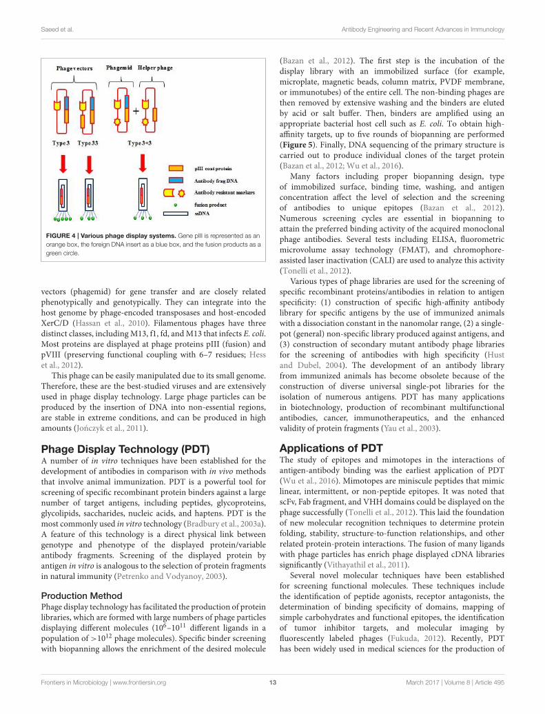
PHAGE DISPLAY SYSTEMS
Phage display is a selection technique for fusion proteins andphage coat proteins that are expressed on the phage surface. Thelibrary is developed by careful genetic manipulation (Figure 4;Chan et al., 2014). Peptide or protein coding genes are insertedinto a vector fused to the 5′-terminal of pIII or pVIII thatare phage coat proteins. The bacteria are transformed withphagemid libraries, and then infected with a helper phage toassemble phage particles that express fusion proteins on theirsurface. Subsequently, the displayed proteins/antibody fragmentsare rooted to the surface of the coat protein, and permit affinitypurification with its analogous genes (Barbas et al., 1991; Chanet al., 2014).
Principles of Phage DisplayFilamentous bacteriophages used in phage display techniques areviruses that belong to the Inoviridae family. There are fewerof these filamentous phages in this genus compared with tailedphages. Inovirus virions are 7 mm in diameter, contain circularDNA enclosed in a protein capsid, and infect both Gram negativeand positive bacteria. They do not lyse host cells, instead, they arepacked and extrude at the surface (Marvin et al., 2014).
The genomes of these viruses consist of double strand DNA(dsDNA), single stranded DNA (ssDNA), double strand RNA(dsRNA), and single strand RNA (ssRNA). The viruses enterhost cells via pilli and are involved in genome replication,and virion structure, assembly and regulation (Stassen et al.,1994). These viruses undergo extensive recombination, act as
Frontiers in Microbiology | www.frontiersin.org 12 March 2017 | Volume 8 | Article 495
Saeed et al. Antibody Engineering and Recent Advances in Immunology
FIGURE 4 | Various phage display systems. Gene pIII is represented as an
orange box, the foreign DNA insert as a blue box, and the fusion products as a
green circle.
vectors (phagemid) for gene transfer and are closely relatedphenotypically and genotypically. They can integrate into thehost genome by phage-encoded transposases and host-encodedXerC/D (Hassan et al., 2010). Filamentous phages have threedistinct classes, includingM13, f1, fd, andM13 that infects E. coli.Most proteins are displayed at phage proteins pIII (fusion) andpVIII (preserving functional coupling with 6–7 residues; Hesset al., 2012).
This phage can be easily manipulated due to its small genome.Therefore, these are the best-studied viruses and are extensivelyused in phage display technology. Large phage particles can beproduced by the insertion of DNA into non-essential regions,are stable in extreme conditions, and can be produced in highamounts (Jonczyk et al., 2011).
Phage Display Technology (PDT)A number of in vitro techniques have been established for thedevelopment of antibodies in comparison with in vivo methodsthat involve animal immunization. PDT is a powerful tool forscreening of specific recombinant protein binders against a largenumber of target antigens, including peptides, glycoproteins,glycolipids, saccharides, nucleic acids, and haptens. PDT is themost commonly used in vitro technology (Bradbury et al., 2003a).A feature of this technology is a direct physical link betweengenotype and phenotype of the displayed protein/variableantibody fragments. Screening of the displayed protein byantigen in vitro is analogous to the selection of protein fragmentsin natural immunity (Petrenko and Vodyanoy, 2003).
Production MethodPhage display technology has facilitated the production of proteinlibraries, which are formed with large numbers of phage particlesdisplaying different molecules (106–1011 different ligands in apopulation of >1012 phage molecules). Specific binder screeningwith biopanning allows the enrichment of the desired molecule
(Bazan et al., 2012). The first step is the incubation of thedisplay library with an immobilized surface (for example,microplate, magnetic beads, column matrix, PVDF membrane,or immunotubes) of the entire cell. The non-binding phages arethen removed by extensive washing and the binders are elutedby acid or salt buffer. Then, binders are amplified using anappropriate bacterial host cell such as E. coli. To obtain high-affinity targets, up to five rounds of biopanning are performed(Figure 5). Finally, DNA sequencing of the primary structure iscarried out to produce individual clones of the target protein(Bazan et al., 2012; Wu et al., 2016).
Many factors including proper biopanning design, typeof immobilized surface, binding time, washing, and antigenconcentration affect the level of selection and the screeningof antibodies to unique epitopes (Bazan et al., 2012).Numerous screening cycles are essential in biopanning toattain the preferred binding activity of the acquired monoclonalphage antibodies. Several tests including ELISA, fluorometricmicrovolume assay technology (FMAT), and chromophore-assisted laser inactivation (CALI) are used to analyze this activity(Tonelli et al., 2012).
Various types of phage libraries are used for the screening ofspecific recombinant proteins/antibodies in relation to antigenspecificity: (1) construction of specific high-affinity antibodylibrary for specific antigens by the use of immunized animalswith a dissociation constant in the nanomolar range, (2) a single-pot (general) non-specific library produced against antigens, and(3) construction of secondary mutant antibody phage librariesfor the screening of antibodies with high specificity (Hustand Dubel, 2004). The development of an antibody libraryfrom immunized animals has become obsolete because of theconstruction of diverse universal single-pot libraries for theisolation of numerous antigens. PDT has many applicationsin biotechnology, production of recombinant multifunctionalantibodies, cancer, immunotherapeutics, and the enhancedvalidity of protein fragments (Yau et al., 2003).
Applications of PDTThe study of epitopes and mimotopes in the interactions ofantigen-antibody binding was the earliest application of PDT(Wu et al., 2016). Mimotopes are miniscule peptides that mimiclinear, intermittent, or non-peptide epitopes. It was noted thatscFv, Fab fragment, and VHH domains could be displayed on thephage successfully (Tonelli et al., 2012). This laid the foundationof new molecular recognition techniques to determine proteinfolding, stability, structure-to-function relationships, and otherrelated protein-protein interactions. The fusion of many ligandswith phage particles has enrich phage displayed cDNA librariessignificantly (Vithayathil et al., 2011).
Several novel molecular techniques have been establishedfor screening functional molecules. These techniques includethe identification of peptide agonists, receptor antagonists, thedetermination of binding specificity of domains, mapping ofsimple carbohydrates and functional epitopes, the identificationof tumor inhibitor targets, and molecular imaging byfluorescently labeled phages (Fukuda, 2012). Recently, PDThas been widely used in medical sciences for the production of
Frontiers in Microbiology | www.frontiersin.org 13 March 2017 | Volume 8 | Article 495

Saeed et al. Antibody Engineering and Recent Advances in Immunology
FIGURE 5 | Schematic illustration of the biopanning technique. The target is attached to a phage library that is immobilized on a solid surface. Unbound phages
are washed out, and specific phages are eluted and amplified. After several rounds of biopanning, the phages are analyzed to obtain diagnostic and therapeutic
agents.
a large number of humanized antibodies and the production ofnew therapeutics. These antibodies have preclinical and clinicalapplications (Rothe et al., 2006).
Transfusion MedicineA large number of antibody reagents are being developedfor hematological applications such as cell subpopulationidentification, directed therapeutics, and in vivo imaging. Anti-ABO, anti-Rh, and anti-Kell hemagglutination antibodies havebeen developed against red blood antigens (Marks et al., 1993).Anti-Rh (D) and anti-HPA-Ia bispecific diabodies developed byPDT that are useful for hemagglutination assays. These diabodiesare being used for the treatment of neonatal alloimmunethrombocytopenia (Watkins et al., 1999).
Moreover, various antibody reagents have been raised againstfetal red blood cells (Huie et al., 2001). Additionally, thistechnique has helped the production of antibodies againstdendritic cells, white blood cells (WBC) (Fitting et al., 2011),hairy cell leukemia (Kreitman et al., 2012), myeloma protein(paraproteins) (O’Nuallain et al., 2007), B and T cells (Maedaet al., 2009), clotting factors, AITP, GPIa, and GPIIIa antigens,CD antigens (Chu et al., 2006), and 11-dehydro-thromboxane B2(11D-TX) antigens (Siegel et al., 2003).
Autoimmune Diseases and Neurological TherapeuticsHuman immune libraries developed by PDT facilitate thestudy of autoimmune and neurological disorder physiology,clinical diagnostics, and the treatment of AITP (plateletdisorder caused by anti-platelet autoantibodies), MS, myasthenia
gravis (MG) [antibodies against nicotinic acetylcholine receptor(AcChoR)], thrombotic thrombocytopenic purpura (TTP),Cogan’s syndrome (CS) caused by systemic vasculitis, acuteanterior uveitis (AAU), ocular inflammation, insulin dependentdiabetes mellitus (IDDM) caused by the destruction of pancreaticbeta cells, Wegener’s granulomatosis (Finnern et al., 1997),autoimmune thyroid disease (Latrofa et al., 2003), primary biliarycirrhosis (PBC), and Sjögren’s syndrome (SS). Additionally,the technique has therapeutic uses in blistering skin diseases,pemphigus vulgaris (PV) (Payne et al., 2005), pemphigusfoliaceus (PF) (Ishii et al., 2008), autoimmune hepatitis(AIH), primary biliary cirrhosis (PBC), mixed cryoglobulinemia(CryoII), systemic sclerosis (SSc), autoimmune cholangitis(AIC), antiphospholipid syndrome (APS), vitiligo rheumatoidarthritis, Crohn’s disease, Graves’ disease (GD), and celiac disease(genetic inflammatory disorder) (Zohreh and Hossein, 2008).
Neurological disorders are treated by intracellular antibodyfragments (intrabodies), which are potentially therapeutic.Intrabodies select abnormal intracellular proteins. However,there are several limitations in the extracellular binding andinternalization of DNA transfected by viral based vectors,lipofection or electroporation (Jazi et al., 2012). These arenot efficient in vivo techniques and can alter cell viability.This problem can be overcome by fusing protein transductiondomains (PTD) to antibodies (Langedijk et al., 2004). Phagedisplay libraries have been utilized for novel immunotherapeuticstrategies for the treatment of neurotoxins, Creutzfeldt–Jakobdisease (CJD), and Gerstmann-Sträussler-Scheinker syndrome(GSS). They are also used for kuru disease, familial fatal
Frontiers in Microbiology | www.frontiersin.org 14 March 2017 | Volume 8 | Article 495

Saeed et al. Antibody Engineering and Recent Advances in Immunology
insomnia by the accumulation of abnormal prion protein (PrPSc)(Thanongsaksrikul and Chaicumpa, 2011), Huntington’s disease,and Parkinson’s disease. Moreover, they have been employed ininhibitory studies of β-amyloid formation, and enzyme therapyof the brain vasculature and brain parenchyma (Chen et al.,2009).
Peptide Homing in Organs and Molecular ImagingBiopanning in vivo with phage display libraries has facilitatedthe isolation of peptides homing to all types of organs in thehuman body. Phage display is applied to stem cells for cellbased regenerative medicine (Gothard et al., 2011). Moreover,this technique has assisted in the guided delivery of variouspeptides/drugs such as proapoptotic peptides, cytotoxic drugs,metalloprotease inhibitors, and cytokines to specific targets(Nixon et al., 2014). The binding of peptides with the extracellulardomain of the LOX-1 receptor is associated with hypertensionand atherogenesis (Nixon et al., 2014). Other studies havereported that the homing of an RGD-motif-containing peptideto angiogenic vasculature was linked to a proapoptotic peptideand was successfully used for the treatment of collagen-inducedarthritis in mice. Phage libraries have also been used for anti-obesity, microparticle (MP), avb3 integrin angiogenesis therapy,and in targeting vascular endothelial growth factor (VEGF)(Cooke et al., 2001).
Similarly, phage displays are used for tumor targeting agentse.g., the scFv (MFE-23)molecule is specific for carcino embryonicantigen (CEA) (Edwards et al., 2008). This technique hasreplaced radiolabeled antibodies that havemultiple disadvantagesincluding reduced natural immunity (Adachi et al., 2011).Furthermore, PDT has been used to isolate a number of peptidesfor molecular imaging. Its advantages are small size, rapidblood clearance, lack of immunogenicity, tissue penetration, andincreased diffusion. Numerous peptides for tumor targeting wereisolated using human B cell lymphoma (McGuire et al., 2006),cervical (Robinson et al., 2005), colon (Rasmussen et al., 2002),gastric (Liang et al., 2006), breast (Askoxylakis et al., 2005), lung(Chang et al., 2009), glioblastoma (Wu et al., 2008), hepatic (Duet al., 2006), prostate (Zitzmann et al., 2005), neuroblastoma(Askoxylakis et al., 2006), and thyroid (Zitzmann et al., 2007)carcinoma cell cultures. However, about 80% of these peptideshave not been reported to function in vivo. This inactivity wasobserved in peptides that recognized mouse double minute 2homolog-p53 protein (MDM2/p53) (Pazgier et al., 2009), IL-11 receptor (Zurita et al., 2004), prostate specific antigen (PSA)(Pakkala et al., 2004), heat shock protein 90 (Kim et al., 2006),and growth factors (Hetian et al., 2002).
HYBRIDOMA TECHNOLOGY VS. PDT
Hybridoma technology is a well-established method for thegeneration of murine mAb cell lines by the fusion of splenocytes(harvested from immunized mice) with myeloma cells. Thetechnology remains a feasible method for laboratories thatimplement basic cell biological research. Hybridoma technologyis a comparatively simple procedure with minimal cost forthe steady production of native whole immunoglobulins
(Tomita and Tsumoto, 2011). Nevertheless, this technologyhas various limitations such as antibodies produced by thehybridoma technique are strictly murine proteins that limits theirtherapeutic use in humans. In addition, they also trigger humananti-mouse antibody (HAMA) responses (Tjandra et al., 1990).Moreover, indefinite production costs, low fusion efficiency,limited number of mAbs, difficulty in developing mAbs againststrictly conserved and toxin antigens and time consumption areother disadvantages (Hnasko and Stanker, 2015).
The production and amplification of antibodies in vitro usingbacteria by PDT has a low turnaround time compared withother methods. Additionally, the library comprises of diversevariants up to 1013, which can be selected against a variedrange of biological and inorganic targets (Sblattero and Bradbury,2000). The experimental conditions can be controlled, and therequired equipment and libraries are available commercially.Disadvantages include a difficult procedure and lack of antibodiesdisplayed on the surface of each bacteriophage yielding a smallnumber of mAbs (Willats, 2002).
OTHER ANTIBODY ENGINEERINGTECHNIQUES
Antibody engineering is a remarkable modification techniquefor production of highly specific and efficient antibody products.However, antibodies are bulky macromolecules that encompasschallenges in construction, optimal pharmacokinetics,manufacturing, stability, and process development. Nonetheless,progress in antibody engineering technologies such as phagedisplay, yeast display, bacterial display, and ribosomal or cell-freedisplay continue to advance our capacity to rapidly screenand refine stable binding immunoglobins. These engineeringtechniques further improve biological properties significantly inthe effector domains of the mAbs (Filpula, 2007).
Engineered Immunomodulatory AntibodiesImmunomodulatory techniques are persistently progressing toexpand the clinical efficacy of therapeutic antibodies. Cell surfaceantigens exhibit a wide array of targets that are overexpressed,mutated or selectively expressed, and selected for modulatedantibody-based therapeutics. The technology functions throughengineering alterations in antigen or receptor function, theimmune system i.e., altering Fc function and T cell activationand antibody conjugated drug delivery system (DDS) targetinga specific antigen. Immunomodulatory antibodies have gainedsignificant clinical success (Scott et al., 2012).
The Fc region is modulated by engineering the effectorfunction, for example to increase or lessen binding to Fc gammareceptors (FcγRs) or complement factors and the half-life of IgG.The half-life can be extended by improving affinity of Fc forFc neonatal receptor (FcRn). Moreover, it can be prolonged byengineering pH-dependent antigen binding to enhance recyclingof IgG via FcRn, and effective binding to the target molecule.Engineering the Fc region permits the development of moleculesthat are better suited to the pharmacology activity required ofthem (Vincent and Zurini, 2012; Rath et al., 2015). Recently,
Frontiers in Microbiology | www.frontiersin.org 15 March 2017 | Volume 8 | Article 495

Saeed et al. Antibody Engineering and Recent Advances in Immunology
a study investigates engineering the pH-dependent interactionbetween IgG and FcRn. It involves modulation of constant Fcpart of monoclonal human IgG1 (hIgG1) antibodies to improveeffector functions and clinical efficacy of next-generation IgG1-based therapeutics (Grevys et al., 2015).
Similarly, new opportunities have been created by thedevelopment of antibody-drug conjugates (ADCs) to treat theinfectious diseases or target cancer cells. ADCs are beingdeveloped by progressing in antibody generation, selection ofexceedingly cytotoxic molecules, and construction of stablelinkers that can be investigated in clinical trials (Vincentand Zurini, 2012). Cytotoxic therapeutic mAbs often helptarget cell-killing by eliciting immune effector functions.These include antibody-dependent cell-mediated cytotoxicity(ADCC), antibody-dependent cellular phagocytosis (ADCP)mediated by innate immune effector cells, and complement-dependent cytotoxicity (CDC)mediated by humoral components(Figure 6). in vitro studies, Fc engineering methods have beenspecifically designed to modulate ADCC, ADCP, and CDCenvisioned for therapeutic mediation (Kinder et al., 2015).
Natural killer (NK) cells exhibit essential role in immunityin the context of mAb treatment by exerting direct cytotoxicitytoward infected or tumor cells and contributing in modeling theadaptive response (Cheng et al., 2013). Several T- or NK-cellmodulators such as ipilimumab and nivolumab were approvedfor the treatment of metastatic melanoma (Berman et al., 2015).
Fc-engineered antibodies improve the ADCC/ADCPpotential and target CD19, CD20, CD40, and Her2.Consequently, they enhance the therapeutic potential ofmAbs. NK cells are exclusive in exhibiting low-affinity activatingFcγRIIIa (CD16), and no inhibitory antibody receptors, featuringa substantial role in ADCC. Several studies have established alink between activating Fc receptors and the efficacy of mAbtherapy using mouse tumor models (Romain et al., 2014).
Recently, glyco-engineering technique has been used toproduce recombinant therapeutic proteins with optimizedefficacy, half-life, specificity, and antigenicity. Glyco-engineering
FIGURE 6 | The Fc region of an antibody mediates effector functions
such as CDC and ADCC.
of expression platforms is progressively documented as anessential approach to advance biopharmaceuticals (Ferrer-Miralles et al., 2009; Dicker and Strasser, 2015). The techniquehas been applied to in vivo expression systems that includemammalian cells, insect cells, yeast, and plants for the productionof recombinant proteins. The underlined approaches aim atdeveloping glycoproteins with homogeneous N- and O-linkedglycans of defined composition (Dicker and Strasser, 2015).Moreover, multi-level glyco-engineering techniques havebeen investigated to generate IgG with defined Fc-glycans ineukaryotic cells (Dekkers et al., 2016). Additionally, E. coliexpression have been successfully employed to producerecombinant human interleukin-2 (IL-2) (Kamionka, 2011).
Yeast, Bacterial, and Ribosomal DisplayTechniquesAntibodies are engineered with superior properties such asbinding affinity, stability, and catalytic activity by several otherdisplay tools (for example, yeast and bacterial display) forbroad spectrum of biotechnology, medicine, and biomedicalapplications. Yeast surface display exhibit development ofrecombinant antibodies by displaying on the surface ofSaccharomyces cerevisiae via genetic fusion to an abundant cellwall protein (Cherf and Cochran, 2015).
Yeast display technique has been used for engineeringprotein affinity, stability, and enzymatic activity. Moreover, it isextensively applied in protein epitope mapping, identificationof protein-protein interactions, and uses of displayed proteinsin industry and medicine (Cherf and Cochran, 2015). Severalrecombinant antibodies have been generated by yeast display forlethal infections such as highly pathogenic H5N1 avian influenzavirus (Lei et al., 2016), cell tumor (Li et al., 2017) and humantumor endothelial marker 1 (TEM1) (Yuan et al., 2017).
Similarly, several bacterial display systems have beenestablished for Gram-negative bacteria and Gram-positivebacteria (Lee et al., 2003). The display systems comprised of acarrier protein as an anchoring motif, a target protein, and a hoststrain. Proteins developed for use as anchoring motifs includeouter membrane proteins, lipoproteins, autotransporters,subunits of surface appendages, and S-layer proteins (Hanand Lee, 2015). Bacterial display has widespread applicationsincluding live vaccine development, screening-displayed peptidelibraries, biosorbents, whole-cell biocatalysts, and biosensordevelopment. Moreover, the promising technology is helpingin the remediation of pollutants, biofuel production, andproduction of enantiomerically pure compounds (Han and Lee,2015; Ramanan et al., 2016).
Ribosome display is a cell-free display system, and a techniqueto perform entirely in vitro selection of proteins or peptidesto bind desired ligand. Ribosome display consists of bothprokaryotic and eukaryotic display systems (Zahnd et al., 2007).It forms stable protein-ribosome-mRNA (PRM) complexes andlinks individual nascent proteins (phenotypes) to their analogousmessenger RNA (mRNA) (genotypes). Ribosome display allowssynchronized isolation of a functional nascent protein, throughaffinity for a ligand together with the encoding mRNA. The
Frontiers in Microbiology | www.frontiersin.org 16 March 2017 | Volume 8 | Article 495

Saeed et al. Antibody Engineering and Recent Advances in Immunology
encoding mRNA is then transformed and amplified as DNAfor further manipulation, including repeated cycles or proteinexpression. The advantages of ribosome display over othercell based methods include displaying very large libraries,generating toxic, proteolytically sensitive and unstable proteins,and incorporation of modified amino acids or mutations atdistinct positions (He and Taussig, 2002; Zahnd et al., 2007).Ribosome display systems have been investigated to identifypotential antigens of Clonorchis sinensis (Kasi et al., 2017), andhuman tumor necrosis factor α (hTNFα) for diagnosis andtreatment (Zhao et al., 2016).
Advantages and DisadvantagesThe large size of mAbs limits tumor penetration, andtheir long serum half-life is not suitable for therapy andimaging applications. Therefore, antibody fragments have beenconstructed in various formats as they are small, monovalent,penetrate tumor tissues efficiently, and are rapidly eliminated byrenal clearance (Chames et al., 2009).
Similarly, recombinant antibodies have several advantages:(i) bacteria, yeast, plants, or animals can be used to produceantibodies, (ii) no need for immunization, and (iii) intrinsicproperties (immunogenicity, binding affinity, pharmacokinetics,specificity, and stability of antibodies) can be modified easilyusing mutagenesis techniques. Genetically engineered antibodieshave integral characteristics that suit various downstreamapplications or can be converted into functional wholeimmunoglobulins (Bradbury et al., 2003b). Antibodies exhibitstrong immunity to defend against foreign antigens and non-self-agents. However, a variety of recombinant antibodies is neededto interact these hostile antigens. Over the last decade, the useof antibody engineering or recombinant antibody technology hasshaped the genetic manipulation of a diverse range of antibodyfragments for research, diagnosis, and therapy (Kontermann andMuller, 1999). This technology has resulted in better affinity andspecificity of manipulated antibody fragments and has facilitatedthe replacement of hybridoma technology with various displaysystems for unlimited antibody production against any knownantigen (Gram et al., 1992).
Conversely, engineered antibodies have various disadvantagessuch as they exhibit greater expense and complexity inmanufacturing compared to antibodies developed by hybridomatechnology (Spiess et al., 2015). Due to their foreign nature,engineered therapeutic antibodies lead to allotypic immuneresponses that results in rapid clearance from body by kidney,elicit on T-cell help, and have reduced antibody avidity.Moreover, engineered antibodies exhibit reduced half-life due tolack of an Fc domain and prevention of FcRn-mediated recycling.Likewise, antibody based therapies have more limitations basedon the fact that many targets (sometimes in low level) have notyet been determined for various diseases (Chames et al., 2009;Attarwala, 2010).
APPLICATIONS OF ANTIBODIES
In the 1890s, von Behring and Kitasato worked on tetanusantitoxin that lead to the development of a new discipline,
immunology. They described antibodies for the first time anddiscovered that inactive toxins can elicit a protective immuneresponse against active toxins in animals (Kantha, 1991). Thetransfusion of serum from these protected animals elicitedan immune response in other animals. Therefore, antibodieswere originally called “antitoxins.” Since then, antibodies havebeen shown to have a wider repertoire of antigen recognition.Antibodies are widely used in diagnostic tests referred as“immunoassays.” These are used to confirm diagnoses and forrapidly growing antibody based technologies (Mukherjee et al.,2012).
Similarly, antibodies have remarkable applications in the fieldof diagnostics, therapeutics and targeted DDS. They have beenused to study various diseases such as cancer, metabolic andhormonal disorders, and infections caused by bacteria, viruses,fungi, algae, protozoa and other agents (Sundar and Prajapati,2012). Moreover, these biomolecules have numerous applicationin the diagnosis of lymphoid and myeloid malignancies.Likewise, immunoglobins are used in tissue typing, ELISA, radioimmunoassay, serotyping of microorganisms, immunologicalintervention with passive antibody, analyzing a patient’s antibodyprofile, and antiidiotype inhibition (Siddiqui, 2010).
Detection and Immunodiagnostic Test KitsThe detection of antibodies against infectious agents andthe construction of rapid and sensitive antibody-basedimmunodiagnostic test kits is an important part of basicand clinical research. Assays have been developed againstpathogens, toxins, and infectious proteins/peptides (Ahmadet al., 2012). The production of antibodies depends on specificprotein titers, extent of immunity, and identification of Bcell responses (Burbelo et al., 2010). Antibodies are routinelydetected by ELISA and other immunoassays. Accessibility offull length DNA sequences is useful for the rapid and robustsystematic identification of antigens by recombinant proteinsusing protein arrays for complete proteome analysis (Kiernyet al., 2012). Furthermore, numerous novel high-throughputdominant immunoassays are used to detect antibody responsesagainst antigens (Burbelo et al., 2010).
Enzyme-Linked Immunosorbent Assay (ELISA)ELISA is the most common method for the quantitation ofpathological antigens. In some cases, modified ELISA have beenused in combination with various specific proteins/peptides. Itis rapid, consistent, relatively easy to analyze, and adaptable tohigh-throughput screening (Alonso et al., 2015).
Principally, specific antibody is immobilized on high bindingELISA plates by incubation overnight at 4◦C or for 1–2 h at 37◦C,and then followed by 3–5 washes with PBST (137 mM NaCl,2.7 mM KCl, 8.1 mM Na2HPO4, 1.5 mM KH2PO4, and 0.5%Tween 20; Murayama et al., 1996). Then, plates are blocked withirrelevant protein e.g., albumin (for example, PBS containing 4–5% skimmed milk), and incubate overnight at 4◦C or for 1–2 hat 37◦C. After washes, samples and standard dilutions are addedto the wells to be captured by bound protein, incubate for 1–2 hat 37◦C and wash properly. Next, specific peroxidase-conjugatedenzyme labeled detection antibody is added to the wells to
Frontiers in Microbiology | www.frontiersin.org 17 March 2017 | Volume 8 | Article 495

Saeed et al. Antibody Engineering and Recent Advances in Immunology
enable detection of the captured protein and incubate for 1 hat 37◦C. After appropriate wash, colorimetric substrate is addedto the wells and incubated for 15–20 min at 37◦C for the colordevelopment as catalyzed by the enzyme. For instance, additionof 3,3′,5,5′-tetramethylbenzidine (TMB) substrate develops bluecolor that can be read by microplate plate reader at thewavelength of 562 nm. Additionally, addition of 0.16–2 Msulfuric acid (H2SO4) as a stop reaction solution develops yellowcolor that can be read at 405–450 nm correspondingly. Inaddition, the incubation conditions and reagent formulationsshould be preferably optimized based on the type of ELISA(Grange et al., 2014; Thiha and Ibrahim, 2015).
There are four ELISA methods. Direct ELISA (dELISA) usescompetition between unwanted proteins for plastic bindingspots. Antigen is attached to the solid phase followed by anenzyme-labeled antibody. This assay is used to detect variouspathogenic antigens (Brasino et al., 2015).
Indirect ELISA (iELISA) uses an antigen attached to a solidphase followed by the addition of unlabeled primary antibody.Instead, a peroxidase enzyme-conjugated secondary antibody isadded onto the first antibody. The iELISA is used to detectspecific antibodies in sera (Peng et al., 2016).
Competitive ELISA (cELISA) is used for the detection ofsmall molecules lacking multiple epitopes. Specific antibodies tothe analyte of interest are immobilized on a micro-titer plate.Then, enzyme-conjugated antigen is incubated with captureantibody and the same antigen in its unconjugated form. Thisstep yields a color following the addition of substrate. The signalsproduced are directly proportional to the quantity of conjugatedenzyme bound and inversely proportional to the quantity ofunconjugated antigen present (Dupont-Deshorgue et al., 2015).
Sandwich ELISA (sELISA) is used for larger proteins withmultiple epitopes and two antibodies can be used consecutively.The capture antibody is immobilized on a microtiter plate.Then, unknown or known samples are added into the matrixto minimize attachment to the solid phase. Peroxidase-enzymelabeled antibody is then added for coloration, which is directlyproportional to the amount of antigen present (Qu et al., 2016).
ELISA has been extensively used in the detection of variouspathological antigens from viral, bacterial, fungal, protozoa, algal,and numerous other sources (Thavaselvam and Vijayaraghavan,2010). An improved ELISA has been used for detecting anti-melanoma differentiation-associated gene 5 (MDA5) antibodiesthat are expressed in patients with dermatomyositis. Clinicalstudy of this newly developed ELISA exhibited efficient detectionof anti-MDA5 antibodies and showed promising potential toassist the routine clinical check of anti-MDA5 antibodies inpatients who supposed to have DM (Sato et al., 2016).
Western BlotWestern blot assay (WBA) is also called immunoblotting.This technique is used for the determination of molecularweight and amount of relevant proteins present in a sample.Proteins in a sample are first separated by electrophoresis andthen transferred to a nitrocellulose or polyvinylidene difluoride(PVDF) membrane for the detection of bound primary proteins
with antibodies specific to the protein of interest (Ness et al.,2015).
Nonspecific sites in the membrane are then blocked withBSA, non-fat milk powder, or casein. Finally, a labeled secondaryantibody is added for detection by chemiluminescence orfluorescence (Lewis et al., 2016). WBA has been used forthe confirmation of presence of purified proteins producedagainst various pathological antigens such as the lethal toxin ofClostridium sordellii (Arya et al., 2013), shiga toxin Stx2f (Skinneret al., 2013), Staphylococcus aureus alpha-hemolysin (alpha-toxin) (Ladhani et al., 2001), Selenocosmia huwena huwentoxin-IV (HWTX-IV) (Yu et al., 2014), and V. parahaemolyticusthermolabile hemolysin (TLH) (Wang et al., 2015).
Dot BlotDot Blot (DB) assays are used to measure protein concentrationssemi-quantitatively. It is slightly different from the WBA.Proteins in the sample are not separated by electrophoresisbut are spotted directly on a membrane and hybridizedwith an antibody probe (Emmerich and Cohen, 2015). Thistechnique is cost effective and uses avidin-biotin technologywith diaminobenzidine as a chromogen. It is used for theanalysis and quantitation of 14-3-3 protein in cerebrospinal fluid(CSF) samples from cases of Creutzfeldt-Jakob disease (CJD),and for disease control of other neurodegenerative diseasessuch as Alzheimer’s disease (AD) and Parkinson’s disease (PD)(Subramanian et al., 2016).
ImmunohistochemistryImmunohistochemistry (IHC) is used for the detection andidentification of proteins and their localization in tissues. Itis essential to retain the morphology of tissues, cells and theavailability of antigen sites. Fresh, rapidly frozen tissue sectionsare preferably used for IHC by chemically fixing tissues informalin and embedding in wax (Zhu et al., 2015). Additionally,fixing crosslinks amino acids in the tissue that block accessto the epitope sites and prevent the action of any proteinspecific antibodies. The exposure of hidden epitope regions isperformed by digestion with an enzyme or by heat treatment,which removes endogenous peroxidase activity and non-specificsites are blocked. A labeled antibody or an unlabeled primaryantibody specific to the protein of interest is used, followed by theaddition of a secondary labeled antibody specific to the primaryantibody (Diorio et al., 2016).
IHC has recently been used for determination andidentification of expressed proteins such as lysosome-associatedprotein transmembrane-4 beta (LAPTM4B) associated with theprognosis of several human malignancies (Xiao et al., 2017),glucagon-like peptide-1 (GLP-1) against renal tubular injury(Guo et al., 2017), and CD147 trans-membrane protein thatinduces expression and activity of matrix metalloproteinases(MMP) (Dana et al., 2016).
ImmunoprecipitationImmunoprecipitation (IP) is used for the study of protein-protein interactions, specific enzyme activity, posttranslationalmodifications, protein quantification, and determination of
Frontiers in Microbiology | www.frontiersin.org 18 March 2017 | Volume 8 | Article 495

Saeed et al. Antibody Engineering and Recent Advances in Immunology
molecular weight of protein antigens. This technique includesantibody/antigen purification complexes at conditions thatspecifically bind antibodies. Rare proteins can be accumulatedup to 10,000-fold by IP (Dwane and Kiely, 2011). Luciferaseimmunoprecipitation systems (LIPS) have been developed for therapid detection of antibodies against peste des petits ruminantsvirus (PPRV) (Berguido et al., 2016), varicella-zoster virus (VZV)(Cohen et al., 2014), zinc transporter (ZnT8) autoantibodies(Ustinova et al., 2014), and pancreatic and duodenal homeobox1 autoantibodies (PAA) (Donelan et al., 2013).
Flow CytometryFlow cytometry (FC) is used to study antibodies on thecell surface and their related physiochemical properties. Thistechnique was developed over 40 years ago (Robinson et al.,2012), and allows the rapid detection of various proteins on cells.Proteins produce fluorescence or scattered light when passedthrough the machine sensing point to generate quantitative dataon a large number of cells. Labeled antibodies are used for thedetection of target protein molecules or antigens on the surfaceof cells (Álvarez-Barrientos et al., 2000). FC has been used forthe visualization of pulmonary clearance (Zhou et al., 2015),fluorescence decay lifetimes, control sorting (Houston et al.,2010), quantification of DNA-end resection in mammalian cells(Forment et al., 2012), microsphere-based protease assays, high-throughput screening (Saunders et al., 2010a), and botulinumneurotoxin type A light chain protease inhibitors (Saunders et al.,2010b).
Fluorescence Activated Cell SortingFluorescence Activated Cell Sorting (FACS) is used for thedetection of specific cells from a mixed population of cells basedon their distinctive fluorescence or light scattering characteristics(Yilmaz et al., 2010). The technique allows the isolation of cellsby flow cytometry. FACS has been used to isolate high-lipidstrains of Tetraselmis suecica (Montero et al., 2011), high-lipidmutants of Nannochloropsis (Doan and Obbard, 2011), E. coliO157, (Ozawa et al., 2016), high-lipid Chlamydomonas mutants(Terashima et al., 2015), and Chlorella (Manandhar-Shrestha andHildebrand, 2013). In addition, the technology has been used forquantification of the cellular uptake of cell-penetrating peptidesand mRNA (Date et al., 2014).
Enzyme Linked ImmunospotEnzyme linked immunospot (Elispot) assay is used formonitoring cellular immune responses in humans and otheranimals (Whiteside et al., 2003). It involves a polyvinylidenedifluoride (PVDF) assisted microtiter plate pre-coated withantibodies specific to the antigen of interest. A capture antibodybinds to the analyte of interest under precise conditions. Then, abiotinylated antibody specific to the analyte of interest is addedto detect the original antibody after a wash to remove cell debris.Finally, an enzyme labeled conjugate is added after a second washto remove unbound antibody and to visualize a colored product.The product is typically a black spot representing a single cellthat produces the antigen of interest (Janetzki et al., 2005). Thistechnique was used in the development of a coxsackievirus A16
neutralization test (Hou et al., 2015) and the determination ofrotavirus infectivity (Li et al., 2014). Diagnostic applicationsinclude diagnosing sensitization to house dust mites (Changet al., 2016), pulmonary tuberculosis (Pang et al., 2016), pleuraltuberculosis (Adilistya et al., 2016), smear-negative tuberculosis(Li et al., 2015), spinal tuberculosis (Yuan et al., 2015), andcytomegalovirus infection (Nesher et al., 2016).
Lateral Flow TestThe lateral flow immunochromatographic test (LFT) is a simpleand cost effective device used to detect the presence or absenceof a target antigen. It is widely used for medical diagnostics athome (pregnancy test) or in a laboratory testing. The technologyinvolves the transportation of fluids (e.g., urine) through capillarybeds, fragments of porous paper, microstructured polymers, orsintered polymers (Hansson et al., 2016).
The technique comprises various components and steps. Asample pad acts as a sponge and absorbs the fluids. Then,fluids migrate to a conjugate pad containing protein conjugatesimmobilized on the surface of bio-active particles in a salt-sugarmatrix that reacts with the antigen. Next, sample fluids dissolvethe conjugate salt-sugar matrix and antibody-particles. The fluidmix flows through the porous structure causing the analyte tobind with particles while migrating further through the thirdcapillary bed. Finally, there is a third capture molecule in stripedareas that binds to the fluid mix containing analytes and theparticle consequently changes color (Yu et al., 2012). LFTs canbe used as a competitive or sandwich assay. Latex (blue color),nanometer-sized particles of gold (red color), or fluorescent ormagnetic labeled particles are also used (Seydack, 2005). Thetechnique is qualitative, nevertheless, the quantity of analyte ina sample can be measured by the intensity of the test line color.It is usually carried out by optical and non-optical lateral flowreaders or biosensors (LFBs) such as complementary metal-oxidesemiconductor (CMOS) or a charge-coupled device (CCD) andmagnetic immunoassay (MIA) (Gui et al., 2014).
Diagnosis and CureProgress in hybridoma technology and the production ofhighly specific mAbs has revolutionized the therapeutic useof antibodies for the diagnosis and cure of infections, thedevelopment of vaccines, antigenic characterization, and geneticmanipulation. Antibodies have widespread applications indiagnostics, therapeutics and targeted DDS against potentpathogens, cancer, and physiological disorders (Tiwari et al.,2012).
They are used for the diagnosis of lymphoid and myeloidmalignancies, tissue characterization, ELISA, radiolabeledimmunoassay, and serotyping of microbes. In addition, theyare used in the diagnosis of immunological interpolation withpassive antibodies, anti-idiotype suppression, or magic bullettreatments with cytotoxic agents coupled to anti-mouse specificantibodies (Keller and Stiehm, 2000). Similarly, recombinantDNA technology (rDNA) has revolutionized the reconstructionof mAbs by genetic engineering using chimeric antibodies,humanized antibodies, CDR grafted antibodies for therapeutic
Frontiers in Microbiology | www.frontiersin.org 19 March 2017 | Volume 8 | Article 495

Saeed et al. Antibody Engineering and Recent Advances in Immunology
use (Dimitrov and Marks, 2009; Shen et al., 2014; Fitting et al.,2015).
ImagingMolecular imaging provides a sensitive, non-invasivemethod for the molecular characterization of a cell surfacephenotype for disease diagnosis and treatment. It is a rapidlygrowing multidiscipline that involves molecular biology,immunology, and medicine (Massoud and Gambhir, 2003). Thetherapeutic applications of antibodies include clinical diagnosisand treatment in hematology, cardiology, immunology,autoimmunity, infectious diseases, and oncology (Neves andKwok, 2015).
MAbs can be used as molecular imaging probes forinvestigating cell surfaces in vivo. Coupling of cell surfacetargets with advances in antibody technology have facilitated theproduction of antibodies optimized for non-invasive imaging(Manning et al., 2008). They have numerous applications suchas radioimmunoscintigraphy, antibody-based positron emissiontomography (immuno-PET), and single-photon emissioncomputed tomography (SPECT or SPET; Knowles and Wu,2012).
Epitope MappingPhage libraries are useful for mapping antibody epitopes,and display millions of peptides/proteins with unique randomsequences. Antibodies select peptides according to the affinity ofparatopes (their combining sites) from the libraries (Forsstromet al., 2014).
Immuno-Protective Epitope MappingAntibodies protect a host against invading microbes. Thesenatural vaccines neutralize toxins and induce microbicidaleffector functions. Antibody-mediated identification ofpathogens during infection is essential for revealingimmunoprotective responses in the host (Sharon et al.,2014). B cell epitope (variable region of protective antibodies incontact with infectious antigens) mapping is important for thedevelopment of effective vaccines in support of sero-diagnostics.This technique identifies protective epitopes for vaccinedevelopment and the estimation of conventional vaccines (killedor attenuated pathogens; Malito et al., 2015).
B and T cell epitope conformation is determined by pepscantechnique, which has led to the development of antibodytherapeutics, vaccine design, and recognition of protectiveantibody responses (Ahmad et al., 2016). The technique alsotargets pathogens with novel antimicrobials. Examples includethe V-shaped Ab52 glycan epitope in the O-antigen of Francisellatularensis, the CR6261 peptidic epitope in influenza virus H1N1,and the PG16 glycopeptidic epitope in the gp120 V1/V2 loopof HIV type 1 (Sharon et al., 2014). Peptides are screened bybiopanning with antibodies from the sera of various humandiseases, including severe acute respiratory syndrome (SARS),human papillomavirus (HPV), and avian influenza viruses (AIV).Moreover, peptide-based antigens are also used for serologicaldiagnosis and development of vaccines (Wu et al., 2016).
Surface Plasmon ResonanceThe surface plasmon resonance (SPR) technique was first usedin the year 2000, for the epitope mapping of polysaccharidesin L. pneumophila and the epitope mapping of various lowaffinity peptide/protein interactions. SPR is analogous to theworking principles of the ELISA technique, but it is label free(Säfsten, 2009). This technique screens molecular interactionsin real-time with complex physical principles. Binding ofrecombinant peptide/protein can be analyzed against naturalligands and mAbs (Säfsten, 2009). SPR was used for the epitopemapping of vitamin B12 (holo-transcobalamin), hemophiliadisease, ricin toxin and the manganese transport protein C(MntC) of Staphylococcus sp. It also identified Goodpastureauto-immune disease epitopes in combination with PDT(Nguyen et al., 2014).
Next Generation Phage DisplayPhage display panning methods have failed to yield specificresults because of the presence of parasitic phage clonesthat are often not removed by washing. To overcome theselimitations, next generation sequencing (NGS) technology isused to sequence sub-libraries in biopanning experiments (Naqidet al., 2016). Next generation phage display (NGPD) can sequence3,000 up to a million reads per panning round. This methodis more rapid compared with traditional ELISA screening.NGPD has been used to identify target specific ligands by asingle panning round using a library or ligand motif (Ravnet al., 2013). This technique has widespread applications for theidentification of thousands of ligands and mapping antibodiesin response to infectious particles. Clinical targets and antigen-related ligands have been discovered with the help of geneticallyencoded peptide libraries. Correspondingly, ligands are selectedusing NGPD by the detection of low abundant copy cloneswithout numerous rounds of selection. Subsequently, NGShas facilitated the quantification of gene expression, genomeassembly, and metagenome analysis. Additionally, it is used forPDT with the ion torrent technique (Matochko and Derda,2015).
Use of Antibody in Treatment of InfectiousDiseases for Healthier FutureAntibodies are critical for immunity against infectious diseases,and extensively used for prevention and treatment of infectioncaused by bacteria, virus, and other infectious agents to improvepublic health. Emil von Behring was awarded the first Nobel Prizein Physiology or Medicine in 1901 for his discovery of serumtherapy for diphtheria. This led to the term antitoxin and laternamed as “antikörper” translated “antibody” by Paul Erlich ina 1891 paper (Graham and Ambrosino, 2015). The emergenceof recombinant technologies has revolutionized the selection,humanization, and development of therapeutic antibodies andpermitted the strategic design of antibody-based elements forspecificity and diversity. A number of genetically engineeredmAbs have been used for the treatment of numerous infectiousdiseases (Hudson and Souriau, 2003).
Ebola virus disease (EVD) is a severe, often fatal, zoonoticinfection documented with most recent widespread outbreak in
Frontiers in Microbiology | www.frontiersin.org 20 March 2017 | Volume 8 | Article 495

Saeed et al. Antibody Engineering and Recent Advances in Immunology
west Africa. It is caused by a virus of the Filoviridae family(genus Ebolavirus). The disease is transmitted from humanto human via contact with body fluids of the patients, theincubation period is 1–21 days and case fatality rates range from30 to 90% (Beeching et al., 2014). A mAb named ZMapp ismanufactured in the tobacco plant Nicotiana benthamiana totreat EVD patients and to improve public health. It containsa cocktail of different mAbs (MB-003, ZMab, c2G4, and c4G7)that work to avert the spread of the disease within the body(Qiu et al., 2014). A recent study investigated the treatment ofEVD patient with ZMapp, a buffy coat transfusion from an Ebolasurvivor, and the broad-spectrum antiviral GS-5734. The patientis a first neonate documented to have successfully survived EVD(Dörnemann et al., 2017).
Therapeutic antibodies have been generated to fight offseveral infectious cancers. Cancer is a group of diseasesthat involve abnormal cell growth and proliferation causedby alterations, or mutations, in the genetic material ofthe cells. The cell outgrowths or lumps may be benign ormetastatic (Marusyk and Polyak, 2010). Cancer outpacesthe immune system, avoids detection, or blocks immunesystem activity. Thus, numerous therapeutic antibodiessuch as necitumumab, ramucirumab, GRN1005, ganitumab,cixutumumab MM-121, BIIB 033, mapatumumab, trebananib,BHQ880, and carlumab have been investigated for thesuccessful treatment of human cancers. The malignanciesinclude NSCLC, metastatic gastric adenocarcinoma, brainmetastasis, breast, ovarian, peritoneal, fallopian tube,prostate, pancreatic, and colorectal cancers, melanoma ofthe eye and liver, non-Hodgkin lymphoma, and multiplemyeloma (Dantas-Barbosa et al., 2012; Nixon et al.,2014).
Similarly, engineered antibodies have been used for thetreatment of various types of arthritis. Arthritis (from Greekword “joint”) is a chronic multifactorial disease in whichimmune system attacks and starts degrading the body’s joints(Persidis, 1999). Therapeutic antibodies such as adalimumab,mavrilimumab, and GSK3196165 have been developed for thetreatment of rheumatoid arthritis, juvenile idiopathic arthritis,psoriatic arthritis, and ankylosing spondylitis (Dantas-Barbosaet al., 2012; Nixon et al., 2014).
Therapeutic antibodies developed for the treatment of otherinfectious diseases include prophylaxis, anthrax, autoantibody-positive, lupus, angioedema, immune thrombocytopenicpurpura, macular degeneration, hemophilia A, psoriasis,alzheimer, muscle loss and weakness, optic neuritis, ulcerativecolitis, pulmonary fibrosis, and asthma (Dantas-Barbosa et al.,2012; Nixon et al., 2014).
Prognosis of a disease is a complex process that involvededuction from several observable symptoms and the choiceof treatment based on therapeutic effectiveness. Antibodies,because of their exquisite specificity, and mAbs in general exhibitgreater specificity. Therefore, they are used widely in a variety
of assay formats, in the diagnosis and treatment of infectiousdiseases (Zola and Thomson, 2005). Moreover, they show rapididentification of new or rare infectious agents that is an importantpublic health measure. Shortly, development of novel mAbs arehelping to monitor and lessen the likelihoods of epidemics andother disease threats imposed on human health by prevalentinfectious agents (Zola et al., 2013).
CONCLUSIONS
In the past few decades, antibodies have been developed by usingconventional techniques such as hybridomas with significantapplications in therapeutics. However, more recently, therehave been advancements in recombinant technologies thathave enhanced the generation of efficient immunoglobulinsand their fragments. Antibody engineering offers rapid, cost-effective, and efficient biomedical tools for the discovery ofhigh-affinity peptides/proteins, investigation of protein-proteininteractions, receptor binding, and epitope identification.This powerful technology is used in a variety of systemsto approach different questions from a background of cellbiology and biotechnology. Moreover, it is used for theproduction of different types of engineered antibodies againstany target molecule or highly unique conserved antigens.Additionally, it is facilitating in diagnosis and treatment ofinfections to improve human health. Antibody engineeringpossesses broad spectrum of biological, biotechnological,medical, and antibody applications for the development ofnovel therapeutics in various disease fields. Numerous noveltherapeutic drugs have been developed by in vitro screening andselection techniques based on several high throughput immuneeffector functions, engineered antibodies, and high-affinityantibody fragments. This review has comprehensively describedhybridoma technology, advances in antibody engineeringtechniques, engineered antibodies, antibody fragments, displaytechnologies, and applications of antibodies. In conclusion,the study will help in understanding the significance ofantibody fabrication approaches with extensive uses inmolecular, immunological, diagnostic, imaging, biomedical,and biotechnological fields, pursuing a healthier future forhumans.
AUTHOR CONTRIBUTIONS
AS, RW, SL, and SW designed the study. AS and SW wrote thepaper and all the authors approved its submission.
ACKNOWLEDGMENTS
This work was supported by the Program from Key Scientificand Technology Project of Fujian Province of China (No.2014YZ0001-1, 2014Y4001), and Fujian Provincial Departmentof Ocean and Fisheries (Fujian Ocean Hi-tech [2015] 26).
Frontiers in Microbiology | www.frontiersin.org 21 March 2017 | Volume 8 | Article 495

Saeed et al. Antibody Engineering and Recent Advances in Immunology
REFERENCES
Adachi, S., Yoshimura, T., Matsuoka, T., Okada, K., Yasuda, T., and Kamei, K.(2011). Appearance of skin and nail toxicity in patients with breast cancer whounderwent trastuzumab-containing chemotherapy. Gan To Kagaku Ryoho 38,1453–1456.
Adilistya, T., Astrawinata, D. A., and Nasir, U. Z. (2016). Use of pleural fluidinterferon-gamma enzyme-linked immunospot assay in the diagnosis of pleuraltuberculosis. Acta Med. Indones. 48, 41–47.
Ahmad, T. A., Eweida, A. E., and Sheweita, S. A. (2016). B-cell epitope mappingfor the design of vaccines and effective diagnostics. Trials Vaccinol. 5, 71–83.doi: 10.1016/j.trivac.2016.04.003
Ahmad, Z. A., Yeap, S. K., Ali, A. M., Ho, W. Y., Alitheen, N. B., and Hamid, M.(2012). scFv antibody: principles and clinical application. Clin. Dev. Immunol.
2012:980250. doi: 10.1155/2012/980250AlDeghaither, D., Smaglo, B. G., and Weiner, L. M. (2015). Beyond peptides and
mAbs–current status and future perspectives for biotherapeutics with novelconstructs. J. Clin. Pharmacol. 55(Suppl. 3), S4–S20. doi: 10.1002/jcph.407
Alonso, N., Guillen, R., Chambers, J. W., and Leng, F. (2015). A rapid and sensitivehigh-throughput screening method to identify compounds targeting protein-nucleic acids interactions. Nucleic Acids Res. 43:e52. doi: 10.1093/nar/gkv069
Álvarez-Barrientos, A., Arroyo, J., Cantón, R., Nombela, C., and Sánchez-Pérez, M.(2000). Applications of flow cytometry to clinical microbiology.Clin. Microbiol.
Rev. 13, 167–195. doi: 10.1128/CMR.13.2.167-195.2000Andersen, M. H., Schrama, D., Thor Straten, P., and Becker, J. C. (2006). Cytotoxic
T cells. J. Invest. Dermatol. 126, 32–41. doi: 10.1038/sj.jid.5700001Apostolico Jde, S., Lunardelli, V. A., Coirada, F. C., Boscardin, S. B., and Rosa, D.
S. (2016). Adjuvants: classification, modus operandi, and licensing. J. Immunol.
Res. 2016:1459394. doi: 10.1155/2016/1459394Arango Duque, G., and Descoteaux, A. (2014). Macrophage cytokines:
involvement in immunity and infectious diseases. Front. Immunol. 5:491.doi: 10.3389/fimmu.2014.00491
Arya, P., Ponmariappan, S., Singh, L., and Kumar, O. (2013). Development ofELISA based detection system for lethal toxin of Clostridium sordellii. IndianJ. Med. Res. 137, 1180–1187.
Askoxylakis, V., Mier, W., Zitzmann, S., Ehemann, V., Zhang, J., Kramer, S., et al.(2006). Characterization and development of a peptide (p160) with affinity forneuroblastoma cells. J. Nucl. Med. 47, 981–988.
Askoxylakis, V., Zitzmann, S., Mier, W., Graham, K., Kramer, S., von Wegner, F.,et al. (2005). Preclinical evaluation of the breast cancer cell-binding peptide,p160. Clin. Cancer Res. 11, 6705–6712. doi: 10.1158/1078-0432.CCR-05-0432
Attarwala, H. (2010). Role of antibodies in cancer targeting. J. Nat. Sci. Biol. Med.
1, 53–56. doi: 10.4103/0976-9668.71675Barbas, C. F. III., Kang, A. S., Lerner, R. A., and Benkovic, S. J. (1991). Assembly of
combinatorial antibody libraries on phage surfaces: the gene III site. Proc. Natl.Acad. Sci. U.S.A. 88, 7978–7982. doi: 10.1073/pnas.88.18.7978
Bazan, J., Calkosinski, I., and Gamian, A. (2012). Phage display–a powerfultechnique for immunotherapy: 1. Introduction and potential of therapeuticapplications. Hum. Vaccin. Immunother. 8, 1817–1828. doi: 10.4161/hv.21703
Beeching, N. J., Fenech, M., and Houlihan, C. F. (2014). Ebola virus disease. BMJ
349:g7348. doi: 10.1136/bmj.g7348Berguido, F. J., Bodjo, S. C., Loitsch, A., and Diallo, A. (2016). Specific detection
of peste des petits ruminants virus antibodies in sheep and goat sera bythe luciferase immunoprecipitation system. J. Virol. Methods 227, 40–46.doi: 10.1016/j.jviromet.2015.10.008
Berkowitz, S. A., Engen, J. R., Mazzeo, J. R., and Jones, G. B. (2012). Analytical toolsfor characterizing biopharmaceuticals and the implications for biosimilars.Nat.Rev. Drug Discov. 11, 527–540. doi: 10.1038/nrd3746
Berman, D., Korman, A., Peck, R., Feltquate, D., Lonberg, N., and Canetta, R.(2015). The development of immunomodulatory monoclonal antibodies asa new therapeutic modality for cancer: the Bristol-Myers Squibb experience.Pharmacol. Ther. 148, 132–153. doi: 10.1016/j.pharmthera.2014.11.017
Bhasin, M., and Raghava, G. P. (2007). A hybrid approach for predictingpromiscuous MHC class I restricted T cell epitopes. J. Biosci. 32, 31–42.doi: 10.1007/s12038-007-0004-5
Bradbury, A., Velappan, N., Verzillo, V., Ovecka, M., Chasteen, L., Sblattero,D., et al. (2003a). Antibody in proteomics I. Trends Biotechnol. 21, 275–281.doi: 10.1016/S0167-7799(03)00112-4
Bradbury, A., Velappan, N., Verzillo, V., Ovecka, M., Chasteen, L., Sblattero,D., et al. (2003b). Antibodies in proteomics II: screening, high-throughputcharacterization and downstream applications. Trends Biotechnol. 21, 312–317.doi: 10.1016/S0167-7799(03)00117-3
Bradbury, A. R., Sidhu, S., Dubel, S., and McCafferty, J. (2011). Beyond naturalantibodies: the power of in vitro display technologies. Nat. Biotechnol. 29,245–254. doi: 10.1038/nbt.1791
Brasino, M., Lee, J. H., and Cha, J. N. (2015). Creating highly amplifiedenzyme-linked immunosorbent assay signals from genetically engineeredbacteriophage. Anal. Biochem. 470, 7–13. doi: 10.1016/j.ab.2014.10.006
Burbelo, P. D., Ching, K. H., Bush, E. R., Han, B. L., and Iadarola, M. J. (2010).Antibody-profiling technologies for studying humoral responses to infectiousagents. Expert Rev. Vaccines 9, 567–578. doi: 10.1586/erv.10.50
Casadevall, A. (1999). Passive antibody therapies: progress and continuingchallenges. Clin. Immunol. 93, 5–15. doi: 10.1006/clim.1999.4768
Catanese, M. T., Graziani, R., von Hahn, T., Moreau, M., Huby, T., Paonessa, G.,et al. (2007). High-avidity monoclonal antibodies against the human scavengerclass B type I receptor efficiently block hepatitis C virus infection in thepresence of high-density lipoprotein. J. Virol. 81, 8063–8071. doi: 10.1128/JVI.00193-07
Chames, P., Van Regenmortel, M., Weiss, E., and Baty, D. (2009). Therapeuticantibodies: successes, limitations and hopes for the future. Br. J. Pharmacol.
157, 220–233. doi: 10.1111/j.1476-5381.2009.00190.xChan, C. E., Lim, A. P., MacAry, P. A., and Hanson, B. J. (2014). The role of
phage display in therapeutic antibody discovery. Int. Immunol. 26, 649–657.doi: 10.1093/intimm/dxu082
Chang, D. K., Lin, C. T., Wu, C. H., and Wu, H. C. (2009). A novel peptideenhances therapeutic efficacy of liposomal anti-cancer drugs in mice modelsof human lung cancer. PLoS ONE 4:e4171. doi: 10.1371/journal.pone.0004171
Chang, D. Y., Lee, J., Choi, S. W., Lee, H. J., Kang, H., Yeo, S. C., et al. (2016).Interleukin-4 enzyme-linked immunospot assay may be useful for diagnosingsensitization to house dust mite. Int. Forum Allergy Rhinol. 6, 1007–1012.doi: 10.1002/alr.21786
Chen, W., Zhu, Z., Feng, Y., Xiao, X., and Dimitrov, D. S. (2008). Construction ofa large phage-displayed human antibody domain library with a scaffold basedon a newly identified highly soluble, stable heavy chain variable domain. J. Mol.
Biol. 382, 779–789. doi: 10.1016/j.jmb.2008.07.054Chen, Y., Huang, X., Wang, R., Wang, S., and Shi, N. (2015). The structure
of a GFP-based antibody (fluorobody) to TLH, a toxin from Vibrio
parahaemolyticus. Acta Crystallogr. F Struct. Biol. Commun. 71(Pt 7), 913–918.doi: 10.1107/S2053230X15008845
Chen, Y. H., Chang,M., andDavidson, B. L. (2009).Molecular signatures of diseasebrain endothelia provide new sites for CNS-directed enzyme therapy.Nat. Med.
15, 1215–1218. doi: 10.1038/nm.2025Cheng, M., Chen, Y., Xiao, W., Sun, R., and Tian, Z. (2013). NK cell-based
immunotherapy for malignant diseases. Cell. Mol. Immunol. 10, 230–252.doi: 10.1038/cmi.2013.10
Cherf, G. M., and Cochran, J. R. (2015). Applications of yeast surface display forprotein engineering. Methods Mol. Biol. 1319, 155–175. doi: 10.1007/978-1-4939-2748-7_8
Committee for Medicinal Products for Human Use (CHMP) (2008). Guidelineon Development, Production, Characterisation and Specification for Monoclonal
Antibodies and Related Products. EMA/CHMP/BWP/532517.Chu, X. X., Hou, M., Peng, J., Zhu, Y. Y., Ji, X. B., and Wang, L. (2006).
Effects of IgG and its F(ab’)2 fragments of some patients with idiopathicthrombocytopenic purpura on platelet aggregation. Eur. J. Haematol. 76,153–159. doi: 10.1111/j.1600-0609.2005.00557.x
Cohen, J. I., Ali, M. A., Bayat, A., Steinberg, S. P., Park, H., Gershon, A. A., et al.(2014). Detection of antibodies to varicella-zoster virus in recipients of thevaricella vaccine by using a luciferase immunoprecipitation system assay. Clin.Vaccine Immunol. 21, 1288–1291. doi: 10.1128/CVI.00250-14
Cooke, S. P., Boxer, G. M., Lawrence, L., Pedley, R. B., Spencer, D. I., Begent,R. H., et al. (2001). A strategy for antitumor vascular therapy by targetingthe vascular endothelial growth factor: receptor complex. Cancer Res. 61,3653–3659.
Dana, P., Kariya, R., Vaeteewoottacharn, K., Sawanyawisuth, K., Seubwai, W.,Matsuda, K., et al. (2016). Upregulation of CD147 promotes metastasis ofcholangiocarcinoma by modulating the epithelial-to-mesenchymal transitional
Frontiers in Microbiology | www.frontiersin.org 22 March 2017 | Volume 8 | Article 495
Saeed et al. Antibody Engineering and Recent Advances in Immunology
process. Oncol. Res. doi: 10.3727/096504016X14813899000565. [Epub ahead ofprint].
Dantas-Barbosa, C., de Macedo Brigido, M., and Maranhao, A. Q. (2012).Antibody phage display libraries: contributions to oncology. Int. J. Mol. Sci. 13,5420–5440. doi: 10.3390/ijms13055420
Date, A., Maeda, T., Watanabe, M., Hidaka, Y., Iwatani, Y., and Takano, T. (2014).An improved protocol for mRNA quantification after fluorescence-activatedcell sorting with an increased signal to noise ratio in flow cytometry. Mol.
Biotechnol. 56, 591–598. doi: 10.1007/s12033-014-9733-5Dekkers, G., Plomp, R., Koeleman, C. A., Visser, R., von Horsten, H. H., Sandig,
V., et al. (2016). Multi-level glyco-engineering techniques to generate IgG withdefined Fc-glycans. Sci. Rep. 6:36964. doi: 10.1038/srep36964
Dicker, M., and Strasser, R. (2015). Using glyco-engineering to produce therapeuticproteins. Expert Opin. Biol. Ther. 15, 1501–1516. doi: 10.1517/14712598.2015.1069271
Dimitrov, D. S., and Marks, J. D. (2009). Therapeutic antibodies: current state andfuture trends–is a paradigm change coming soon?MethodsMol. Biol. 525, 1–27.doi: 10.1007/978-1-59745-554-1_1
Diorio, C., Furrer, D., Michaud, A., Laberge, S., Popa, I., Jacob, S., et al. (2016).Validation of EP1 antibody clone for estrogen receptor immunohistochemistryfor breast cancer. Anticancer Res. 36, 435–437.
Doan, T. T. Y., and Obbard, J. P. (2011). Enhanced lipid production inNannochloropsis sp. using fluorescence-activated cell sorting. GCB Bioenergy 3,264–270. doi: 10.1111/j.1757-1707.2010.01076.x
Donelan, W., Wang, H., Li, S. W., Pittman, D., Li, Y., Han, S., et al. (2013). Noveldetection of pancreatic and duodenal homeobox 1 autoantibodies (PAA) inhuman sera using luciferase immunoprecipitation systems (LIPS) assay. Int. J.Clin. Exp. Pathol. 6, 1202–1210.
Dong, J., Sereno, A., Aivazian, D., Langley, E., Miller, B. R., Snyder, W. B., et al.(2011). A stable IgG-like bispecific antibody targeting the epidermal growthfactor receptor and the type I insulin-like growth factor receptor demonstratessuperior anti-tumor activity.MAbs 3, 273–288. doi: 10.4161/mabs.3.3.15188
Dörnemann, J., Burzio, C., Ronsse, A., Sprecher, A., De Clerck, H., Van Herp, M.,et al. (2017). First newborn baby to receive experimental therapies survivesebola virus disease. J. Infect. Dis. 215, 171–174. doi: 10.1093/infdis/jiw493
Du, B., Qian, M., Zhou, Z., Wang, P., Wang, L., Zhang, X., et al. (2006).in vitro panning of a targeting peptide to hepatocarcinoma from a phagedisplay peptide library. Biochem. Biophys. Res. Commun. 342, 956–962.doi: 10.1016/j.bbrc.2006.02.050
Dupont-Deshorgue, A., Oudart, J. B., Brassart, B., Deslee, G., Perotin, J. M.,Diebold, M. D., et al. (2015). A competitive enzyme-linked immunosorbentassay for quantification of tetrastatin in body fluids and tumor extracts. Anal.Biochem. 482, 16–21. doi: 10.1016/j.ab.2015.04.023
Dwane, S., and Kiely, P. A. (2011). Tools used to study how proteincomplexes are assembled in signaling cascades. Bioeng. Bugs 2, 247–259.doi: 10.4161/bbug.2.5.17844
Ecker, D. M., Jones, S. D., and Levine, H. L. (2015). The therapeutic monoclonalantibody market.MAbs 7, 9–14. doi: 10.4161/19420862.2015.989042
Edwards, B. M., and He, M. (2012). Evolution of antibodies in vitro byribosome display. Methods Mol. Biol. 907, 281–292. doi: 10.1007/978-1-61779-974-7_16
Edwards, W. B., Xu, B., Akers, W., Cheney, P. P., Liang, K., Rogers, B. E.,et al. (2008). Agonist-antagonist dilemma in molecular imaging: evaluationof a monomolecular multimodal imaging agent for the somatostatin receptor.Bioconjug. Chem. 19, 192–200. doi: 10.1021/bc700291m
El Debs, B., Utharala, R., Balyasnikova, I. V., Griffiths, A. D., and Merten, C.A. (2012). Functional single-cell hybridoma screening using droplet-basedmicrofluidics. Proc. Natl. Acad. Sci. U.S.A. 109, 11570–11575. doi: 10.1073/pnas.1204514109
Emmerich, C. H., and Cohen, P. (2015). Optimising methods for thepreservation, capture and identification of ubiquitin chains and ubiquitylatedproteins by immunoblotting. Biochem. Biophys. Res. Commun. 466, 1–14.doi: 10.1016/j.bbrc.2015.08.109
Fauci, A. S., and Morens, D. M. (2012). The perpetual challenge of infectiousdiseases. N. Engl. J. Med. 366, 454–461. doi: 10.1056/NEJMra1108296
Ferrer-Miralles, N., Domingo-Espin, J., Corchero, J. L., Vazquez, E., and Villaverde,A. (2009). Microbial factories for recombinant pharmaceuticals. Microb. Cell
Fact. 8:17. doi: 10.1186/1475-2859-8-17
Filpula, D. (2007). Antibody engineering and modification technologies. Biomol.
Eng. 24, 201–215. doi: 10.1016/j.bioeng.2007.03.004Finnern, R., Pedrollo, E., Fisch, I., Wieslander, J., Marks, J. D., Lockwood, C.
M., et al. (1997). Human autoimmune anti-proteinase 3 scFv from a phagedisplay library. Clin. Exp. Immunol. 107, 269–281. doi: 10.1111/j.1365-2249.1997.254-ce1127.x
Fitting, J., Blume, T., Ten Haaf, A., Blau, W., Gattenlohner, S., Tur, M. K.,et al. (2015). Phage display-based generation of novel internalizing antibodyfragments for immunotoxin-based treatment of acute myeloid leukemia.MAbs
7, 390–402. doi: 10.1080/19420862.2015.1007818Fitting, J., Killian, D., Junghanss, C., Willenbrock, S., Murua Escobar, H., and
Lange, S. (2011). Generation of recombinant antibody fragments that targetcanine dendritic cells by phage display technology. Vet. Comp. Oncol. 9,183–195. doi: 10.1111/j.1476-5829.2010.00246.x
Forment, J. V., Walker, R. V., and Jackson, S. P. (2012). A high-throughput, flowcytometry-based method to quantify DNA-end resection in mammalian cells.Cytometry A 81, 922–928. doi: 10.1002/cyto.a.22155
Forsstrom, B., Axnas, B. B., Stengele, K. P., Buhler, J., Albert, T. J.,Richmond, T. A., et al. (2014). Proteome-wide epitope mapping of antibodiesusing ultra-dense peptide arrays. Mol. Cell. Proteomics 13, 1585–1597.doi: 10.1074/mcp.M113.033308
Frenzel, A., Hust, M., and Schirrmann, T. (2013). Expression of recombinantantibodies. Front. Immunol. 4:217. doi: 10.3389/fimmu.2013.00217
Fukuda, M. N. (2012). Peptide displaying phage technology in glycobiology.Glycobiology 22, 318–325. doi: 10.1093/glycob/cwr140
Gavilondo, J. V., and Larrick, J. W. (2000). Antibody engineering at themillennium. Biotechniques 29, 128–132.
Glukhova, X. A., Prusakova, O. V., Trizna, J. A., Zaripov, M. M., Afanas’eva, G.V., Glukhov, A. S., et al. (2016). Updates on the production of therapeuticantibodies using human hybridoma technique. Curr. Pharm. Des. 22, 870–878.doi: 10.2174/1381612822666151223102845
Gothard, D., Tare, R. S., Mitchell, P. D., Dawson, J. I., and Oreffo, R. O.(2011). In search of the skeletal stem cell: isolation and separation strategiesat the macro/micro scale for skeletal regeneration. Lab Chip 11, 1206–1220.doi: 10.1039/c0lc00575d
Graham, B. S., and Ambrosino, D. M. (2015). History of passive antibodyadministration for prevention and treatment of infectious diseases. Curr. Opin.HIV AIDS 10, 129–134. doi: 10.1097/COH.0000000000000154
Gram, H., Marconi, L. A., Barbas, C. F. III., Collet, T. A., Lerner, R. A., and Kang,A. S. (1992). In vitro selection and affinity maturation of antibodies from anaive combinatorial immunoglobulin library. Proc. Natl. Acad. Sci. U.S.A. 89,3576–3580. doi: 10.1073/pnas.89.8.3576
Grange, R. D., Thompson, J. P., and Lambert, D. G. (2014). Radioimmunoassay,enzyme and non-enzyme-based immunoassays. Br. J. Anaesth. 112, 213–216.doi: 10.1093/bja/aet293
Grevys, A., Bern, M., Foss, S., Bratlie, D. B., Moen, A., Gunnarsen, K. S.,et al. (2015). Fc engineering of human IgG1 for altered binding to theneonatal Fc receptor affects Fc effector functions. J. Immunol. 194, 5497–5508.doi: 10.4049/jimmunol.1401218
Gui, C., Wang, K., Li, C., Dai, X., and Cui, D. (2014). A CCD-basedreader combined with CdS quantum dot-labeled lateral flow strips forultrasensitive quantitative detection of CagA. Nanoscale Res. Lett. 9:57.doi: 10.1186/1556-276X-9-57
Guo, H., Li, H., Wang, B., Ding, W., Ling, L., Yang, M., et al. (2017). Protectiveeffects of glucagon-like peptide-1 analog on renal tubular injury in mice onhigh-fat diet. Cell. Physiol. Biochem. 41, 1113–1124. doi: 10.1159/000464118
Hamers-Casterman, C., Atarhouch, T., Muyldermans, S., Robinson, G., Hamers,C., Songa, E. B., et al. (1993). Naturally occurring antibodies devoid of lightchains. Nature 363, 446–448. doi: 10.1038/363446a0
Han, M. J., and Lee, S. H. (2015). An efficient bacterial surface display system basedon a novel outermembrane anchoring element from the Escherichia coli proteinYiaT. FEMS Microbiol. Lett. 362, 1–7. doi: 10.1093/femsle/fnu002
Hanly, W. C., Artwohl, J. E., and Bennett, B. T. (1995). Review of polyclonalantibody production procedures in mammals and poultry. ILAR J. 37, 93–118.doi: 10.1093/ilar.37.3.93
Hansson, J., Yasuga, H., Haraldsson, T., and van der Wijngaart, W. (2016).Synthetic microfluidic paper: high surface area and high porosity polymermicropillar arrays. Lab Chip 16, 298–304. doi: 10.1039/C5LC01318F
Frontiers in Microbiology | www.frontiersin.org 23 March 2017 | Volume 8 | Article 495

Saeed et al. Antibody Engineering and Recent Advances in Immunology
Hassan, F., Kamruzzaman, M., Mekalanos, J. J., and Faruque, S. M. (2010). Satellitephage TLCϕ enables toxigenic conversion by CTX phage through dif sitealteration. Nature 467, 982–985. doi: 10.1038/nature09469
He, M., and Taussig, M. J. (2002). Ribosome display: cell-free protein displaytechnology. Brief. Funct. Genomic. Proteomic. 1, 204–212. doi: 10.1093/bfgp/1.2.204
Hendriksen, C., and Hau, J. (2003). “Production of polyclonal and monoclonalantibodies,” in Handbook of Laboratory Animal Science, 2nd Edn., eds J. Hauand S. J. Schapiro (Boca Raton, FL: CRC Press LLC), 391–411.
Hess, G. T., Cragnolini, J. J., Popp, M. W., Allen, M. A., Dougan, S. K., andSpooner, E. (2012). M13 bacteriophage display framework that allows sortase-mediated modification of surface-accessible phage proteins. Bioconjug. Chem.
23, 1478–1487. doi: 10.1021/bc300130zHetian, L., Ping, A., Shumei, S., Xiaoying, L., Luowen, H., Jian, W., et al.
(2002). A novel peptide isolated from a phage display library inhibits tumorgrowth and metastasis by blocking the binding of vascular endothelial growthfactor to its kinase domain receptor. J. Biol. Chem. 277, 43137–43142.doi: 10.1074/jbc.M203103200
Hnasko, R. M., and Stanker, L. H. (2015). Hybridoma technology. Methods Mol.
Biol. 1318, 15–28. doi: 10.1007/978-1-4939-2742-5_2Hou, W., Yang, L., He, D., Zheng, J., Xu, L., Liu, J., et al. (2015). Development
of a coxsackievirus A16 neutralization test based on the enzyme-linkedimmunospot assay. J. Virol. Methods 215–216, 56–60. doi: 10.1016/j.jviromet.2015.02.010
Houston, J. P., Naivar, M. A., and Freyer, J. P. (2010). Digital analysis andsorting of fluorescence lifetime by flow cytometry. Cytometry A 77, 861–872.doi: 10.1002/cyto.a.20930
Hudson, E. P., Uhlen, M., and Rockberg, J. (2012). Multiplex epitope mappingusing bacterial surface display reveals both linear and conformational epitopes.Sci. Rep. 2:706. doi: 10.1038/srep00706
Hudson, P. J., and Souriau, C. (2003). Engineered antibodies.Nat.Med. 9, 129–134.doi: 10.1038/nm0103-129
Huie, M. A., Cheung, M. C., Muench, M. O., Becerril, B., Kan, Y. W., andMarks, J. D. (2001). Antibodies to human fetal erythroid cells from anonimmune phage antibody library. Proc. Natl. Acad. Sci. U.S.A. 98, 2682–2687.doi: 10.1073/pnas.051631798
Hust, M., and Dubel, S. (2004). Mating antibody phage display with proteomics.Trends Biotechnol. 22, 8–14. doi: 10.1016/j.tibtech.2003.10.011
Ishii, K., Lin, C., Siegel, D. L., and Stanley, J. R. (2008). Isolation ofpathogenic monoclonal anti-desmoglein 1 human antibodies by phage displayof pemphigus foliaceus autoantibodies. J. Invest. Dermatol. 128, 939–948.doi: 10.1038/sj.jid.5701132
Janetzki, S., Cox, J. H., Oden, N., and Ferrari, G. (2005). Standardization andvalidation issues of the ELISPOT assay. Methods Mol. Biol. 302, 51–86.doi: 10.1385/1-59259-903-6:051
Jazi, M. H., Dabaghian, M., Tebianian, M., Gharagozlou, M. J., and Ebrahimi, S.M. (2012). In vivo electroporation enhances immunogenicity and protectionagainst influenza A virus challenge of an M2e-HSP70c DNA vaccine. Virus Res.167, 219–225. doi: 10.1016/j.virusres.2012.05.002
Jin, N., Ling, S., Yang, C., and Wang, S. (2014). Preparation and identification ofmonoclonal antibody against Citreoviridin and development of detection byIc-ELISA. Toxicon 90, 226–236. doi: 10.1016/j.toxicon.2014.08.057
Jonczyk, E., Kłak, M., Miedzybrodzki, R., and Górski, A. (2011). The influenceof external factors on bacteriophages-review. Folia Microbiol. (Praha). 56,191–200. doi: 10.1007/s12223-011-0039-8
Kamionka, M. (2011). Engineering of therapeutic proteins production inEscherichia coli. Curr. Pharm. Biotechnol. 12, 268–274. doi: 10.2174/138920111794295693
Kantha, S. S. (1991). A centennial review; the 1890 tetanus antitoxin paper of vonBehring and Kitasato and the related developments. Keio J. Med. 40, 35–39.doi: 10.2302/kjm.40.35
Kasi, D., Catherine, C., Lee, S.-W., Lee, K.-H., Kim, Y. J., Ro Lee, M.,et al. (2017). Cell-free translational screening of an expression sequence taglibrary of Clonorchis sinensis for novel antigen discovery. Biotechnol. Prog.doi: 10.1002/btpr.2440. [Epub ahead of print].
Keller, M. A., and Stiehm, E. R. (2000). Passive immunity in preventionand treatment of infectious diseases. Clin. Microbiol. Rev. 13, 602–614.doi: 10.1128/CMR.13.4.602-614.2000
Kierny, M. R., Cunningham, T. D., and Kay, B. K. (2012). Detection of biomarkersusing recombinant antibodies coupled to nanostructured platforms. Nano Rev.3:17240. doi: 10.3402/nano.v3i0.17240
Kim, Y., Lillo, A.M., Steiniger, S. C., Liu, Y., Ballatore, C., Anichini, A., et al. (2006).Targeting heat shock proteins on cancer cells: selection, characterization,and cell-penetrating properties of a peptidic GRP78 ligand. Biochemistry 45,9434–9444. doi: 10.1021/bi060264j
Kinder, M., Greenplate, A. R., Strohl, W. R., Jordan, R. E., and Brezski, R.J. (2015). An Fc engineering approach that modulates antibody-dependentcytokine release without altering cell-killing functions. MAbs 7, 494–504.doi: 10.1080/19420862.2015.1022692
Klimka, A., Matthey, B., Roovers, R. C., Barth, S., Arends, J. W., Engert, A., et al.(2000). Human anti-CD30 recombinant antibodies by guided phage antibodyselection using cell panning. Br. J. Cancer 83:252. doi: 10.1054/bjoc.2000.1226
Knowles, S. M., and Wu, A. M. (2012). Advances in immuno-positron emissiontomography: antibodies for molecular imaging in oncology. J. Clin. Oncol. 30,3884–3892. doi: 10.1200/JCO.2012.42.4887
Köhler, G., and Milstein, C. (1975). Continuous cultures of fused cells secretingantibody of predefined specificity. Nature 256, 495–497. doi: 10.1038/256495a0
Kontermann, R. E., and Brinkmann, U. (2015). Bispecific antibodies. Drug Discov.Today 20, 838–847. doi: 10.1016/j.drudis.2015.02.008
Kontermann, R. E., and Muller, R. (1999). Intracellular and cell surfacedisplayed single-chain diabodies. J. Immunol. Methods 226, 179–188.doi: 10.1016/S0022-1759(99)00062-9
Kreitman, R. J., Tallman, M. S., Robak, T., Coutre, S., Wilson, W.H., Stetler-Stevenson, M., et al. (2012). Phase I trial of anti-CD22recombinant immunotoxin moxetumomab pasudotox (CAT-8015 orHA22) in patients with hairy cell leukemia. J. Clin. Oncol. 30, 1822–1828.doi: 10.1200/JCO.2011.38.1756
Ladhani, S., Robbie, S., Garratt, R. C., Chapple, D. S., Joannou, C. L., and Evans, R.W. (2001). Development and evaluation of detection systems for staphylococcalexfoliative toxin A responsible for scalded-skin syndrome. J. Clin. Microbiol. 39,2050–2054. doi: 10.1128/JCM.39.6.2050-2054.2001
Langedijk, J. P., Olijhoek, T., Schut, D., Autar, R., and Meloen, R. H.(2004). New transport peptides broaden the horizon of applications forpeptidic pharmaceuticals.Mol. Divers. 8, 101–111. doi: 10.1023/B:MODI.0000025653.26130.ce
Latrofa, F., Pichurin, P., Guo, J., Rapoport, B., and McLachlan, S. M. (2003).Thyroglobulin-thyroperoxidase autoantibodies are polyreactive, not bispecific:analysis using human monoclonal autoantibodies. J. Clin. Endocrinol. Metab.
88, 371–378. doi: 10.1210/jc.2002-021073Lee, S. Y., Choi, J. H., and Xu, Z. (2003). Microbial cell-surface display. Trends
Biotechnol. 21, 45–52. doi: 10.1016/S0167-7799(02)00006-9Leenaars, M., and Hendriksen, C. F. (2005). Critical steps in the production of
polyclonal andmonoclonal antibodies: evaluation and recommendations. ILARJ. 46, 269–279. doi: 10.1093/ilar.46.3.269
Leenaars, P. P. A. M., Claassen, E., and Boersma, W. J. A. (1997). Antigens andAntigen Presentation. Immunology Methods Manual. London: Academic PressLtd.
Lei, H., Jin, S., Karlsson, E., Schultz-Cherry, S., and Ye, K. (2016). Yeast surface-displayed H5N1 avian influenza vaccines. J. Immunol. Res. 2016:4131324.doi: 10.1155/2016/4131324
Levenhagen, M. A., de Almeida Araujo Santos, F., Fujimura, P. T., Carneiro, A.P., Costa-Cruz, J. M., and Goulart, L. R. (2015). Structural and functionalcharacterization of a novel scFv anti-HSP60 of Strongyloides sp. Sci. Rep.5:10447. doi: 10.1038/srep10447
Lewis, C. W., Taylor, R. G., and Golsteyn, R. M. (2016). Measurementof Cdk1/Cyclin B kinase activity by specific antibodies and westernblotting. Methods Mol. Biol. 1342, 337–348. doi: 10.1007/978-1-4939-2957-3_21
Li, D., Gong, R., Zheng, J., Chen, X., Dimitrov, D. S., and Zhao, Q.(2017). Engineered antibody CH2 domains binding to nucleolin: isolation,characterization and improvement of aggregation. Biochem. Biophys. Res.
Commun. 485, 446–453. doi: 10.1016/j.bbrc.2017.02.058Li, F., Hashimura, Y., Pendleton, R., Harms, J., Collins, E., and Lee, B.
(2006). A systematic approach for scale-down model development andcharacterization of commercial cell culture processes. Biotechnol. Prog. 22,696–703. doi: 10.1021/bp0504041
Frontiers in Microbiology | www.frontiersin.org 24 March 2017 | Volume 8 | Article 495

Saeed et al. Antibody Engineering and Recent Advances in Immunology
Li, F., Vijayasankaran, N., Shen, A. Y., Kiss, R., and Amanullah, A. (2010). Cellculture processes for monoclonal antibody production. MAbs 2, 466–479.doi: 10.4161/mabs.2.5.12720
Li, T., Lin, H., Yu, L., Xue, M., Ge, S., Zhao, Q., et al. (2014). Development ofan enzyme-linked immunospot assay for determination of rotavirus infectivity.J. Virol. Methods 209, 7–14. doi: 10.1016/j.jviromet.2014.08.012
Li, Z., Qin, W., Li, L., Wu, Q., and Chen, X. (2015). Accuracy ofbronchoalveolar lavage enzyme-linked immunospot assay to diagnose smear-negative tuberculosis: a meta-analysis. Int. J. Clin. Exp. Med. 8, 12637–12643.
Liang, S., Lin, T., Ding, J., Pan, Y., Dang, D., Guo, C., et al. (2006). Screening andidentification of vascular-endothelial-cell-specific binding peptide in gastriccancer. J. Mol. Med. 84, 764–773. doi: 10.1007/s00109-006-0064-2
Lim, T. S., Mollova, S., Rubelt, F., Sievert, V., Dubel, S., Lehrach, H., et al. (2010).V-gene amplification revisited - an optimised procedure for amplification ofrearranged human antibody genes of different isotypes. N. Biotechnol. 27,108–117. doi: 10.1016/j.nbt.2010.01.001
Ling, S., Chen, Q. A., Zhang, Y., Wang, R., Jin, N., Pang, J., et al. (2015a).Development of ELISA and colloidal gold immunoassay for tetrodotoxindetetcion based on monoclonal antibody. Biosens. Bioelectron. 71, 256–260.doi: 10.1016/j.bios.2015.04.049
Ling, S., Pang, J., Yu, J., Wang, R., Liu, L., Ma, Y., et al. (2014). Preparation andidentification of monoclonal antibody against fumonisin B(1) and developmentof detection by Ic-ELISA. Toxicon 80, 64–72. doi: 10.1016/j.toxicon.2013.12.008
Ling, S., Wang, R., Gu, X., Wen, C., Chen, L., Chen, Z., et al. (2015b). Rapiddetection of fumonisin B1 using a colloidal gold immunoassay strip test in cornsamples. Toxicon 108, 210–215. doi: 10.1016/j.toxicon.2015.10.014
Lipman, N. S., Jackson, L. R., Trudel, L. J., andWeis-Garcia, F. (2005). Monoclonalversus polyclonal antibodies: distinguishing characteristics, applications, andinformation resources. ILAR J. 46, 258–268. doi: 10.1093/ilar.46.3.258
Little, M., Kipriyanov, S. M., Le Gall, F., and Moldenhauer, G. (2000). Of mice andmen: hybridoma and recombinant antibodies. Immunol. Today 21, 364–370.doi: 10.1016/S0167-5699(00)01668-6
Lundegaard, C., Lund, O., and Nielsen, M. (2008). Accurate approximationmethod for prediction of class I MHC affinities for peptides of length 8, 10and 11 using prediction tools trained on 9mers. Bioinformatics 24, 1397–1398.doi: 10.1093/bioinformatics/btn128
Maeda, M., Ito, Y., Hatanaka, T., Hashiguchi, S., Torikai, M., Nakashima, T., et al.(2009). Regulation of T cell response by blocking the ICOS signal with theB7RP-1-specific small antibody fragment isolated from human antibody phagelibrary.MAbs 1, 453–461. doi: 10.4161/mabs.1.5.9633
Malito, E., Carfi, A., and Bottomley, M. J. (2015). Protein crystallographyin vaccine research and development. Int. J. Mol. Sci. 16, 13106–13140.doi: 10.3390/ijms160613106
Manandhar-Shrestha, K., and Hildebrand, M. (2013). Development of flowcytometric procedures for the efficient isolation of improved lipid accumulationmutants in a Chlorella sp. microalga. J. Appl. Phycol. 25, 1643–1651.doi: 10.1007/s10811-013-0021-8
Manning, H. C., Lander, A., McKinley, E., and Mutic, N. J. (2008). Acceleratingthe development of novel molecular imaging probes: a role for high-throughput screening. J. Nucl. Med. 49, 1401–1404. doi: 10.2967/jnumed.108.053009
Marasco, W. A., and Sui, J. (2007). The growth and potential of humanantiviral monoclonal antibody therapeutics. Nat. Biotechnol. 25, 1421–1434.doi: 10.1038/nbt1363
Marks, J. D., Ouwehand, W. H., Bye, J. M., Finnern, R., Gorick, B. D., Voak,D., et al. (1993). Human antibody fragments specific for human bloodgroup antigens from a phage display library. Biotechnology 11, 1145–1149.doi: 10.1038/nbt1093-1145
Marusyk, A., and Polyak, K. (2010). Tumor heterogeneity: causes andconsequences. Biochim. Biophys. Acta 1805, 105–117. doi: 10.1016/j.bbcan.2009.11.002
Marvin, D. A., Symmons, M. F., and Straus, S. K. (2014). Structure andassembly of filamentous bacteriophages. Prog. Biophys. Mol. Biol. 114, 80–122.doi: 10.1016/j.pbiomolbio.2014.02.003
Massoud, T. F., and Gambhir, S. S. (2003). Molecular imaging in living subjects:seeing fundamental biological processes in a new light. Genes Dev. 17, 545–580.doi: 10.1101/gad.1047403
Matochko, W. L., and Derda, R. (2015). Next-generation sequencing ofphage-displayed peptide libraries. Methods Mol. Biol. 1248, 249–266.doi: 10.1007/978-1-4939-2020-4_17
Mazzer, A. R., Perraud, X., Halley, J., O’Hara, J., and Bracewell, D. G. (2015).Protein A chromatography increases monoclonal antibody aggregation rateduring subsequent low pH virus inactivation hold. J. Chromatogr. A 1415,83–90. doi: 10.1016/j.chroma.2015.08.068
McCullough, K. C., and Summerfield, A. (2005). Basic concepts of immuneresponse and defense development. ILAR J. 46, 230–240. doi: 10.1093/ilar.46.3.230
McGuire, M. J., Samli, K. N., Chang, Y. C., and Brown, K. C. (2006).Novel ligands for cancer diagnosis: selection of peptide ligands foridentification and isolation of B-cell lymphomas. Exp. Hematol. 34, 443–452.doi: 10.1016/j.exphem.2005.12.013
Modjtahedi, H., Ali, S., and Essapen, S. (2012). Therapeutic application ofmonoclonal antibodies in cancer: advances and challenges. Br. Med. Bull. 104,41–59. doi: 10.1093/bmb/lds032
Montero, M., Aristizabal, M., and Reina, G. (2011). Isolation of high-lipid contentstrains of the marine microalga Tetraselmis suecica for biodiesel productionby flow cytometry and sing-cell sorting. J. Appl. Phycol. 23, 1053–1057.doi: 10.1007/s10811-010-9623-6
Morens, D. M., and Fauci, A. S. (2013). Emerging infectious diseases:threats to human health and global stability. PLoS Pathog. 9:e1003467.doi: 10.1371/journal.ppat.1003467
Mukherjee, J., Tremblay, J. M., Leysath, C. E., Ofori, K., Baldwin, K., Feng,X., et al. (2012). A novel strategy for development of recombinant antitoxintherapeutics tested in a mouse botulism model. PLoS ONE 7:e29941.doi: 10.1371/journal.pone.0029941
Murayama, O., Nishida, H., and Sekiguchi, K. (1996). Novel peptide ligands forintegrin α6β1 selected from a phage display library. J. Biochem. 120, 445–451.doi: 10.1093/oxfordjournals.jbchem.a021431
Naqid, I. A., Owen, J. P., Maddison, B. C., Spiliotopoulos, A., Emes, R. D., Warry,A., et al. (2016). Mapping polyclonal antibody responses to bacterial infectionusing next generation phage display. Sci. Rep. 6:24232. doi: 10.1038/srep24232
Nelson, A. L. (2010). Antibody fragments: hope and hype. MAbs 2, 77–83.doi: 10.4161/mabs.2.1.10786
Nelson, A. L., Dhimolea, E., and Reichert, J. M. (2010). Development trends forhuman monoclonal antibody therapeutics. Nat. Rev. Drug Discov. 9, 767–774.doi: 10.1038/nrd3229
Nesher, L., Shah, D. P., Ariza-Heredia, E. J., Azzi, J. M., Siddiqui, H. K.,Ghantoji, S. S., et al. (2016). Utility of the enzyme-linked immunospotinterferon-gamma-release assay to predict the risk of cytomegalovirus infectionin hematopoietic cell transplant recipients. J. Infect. Dis. 213, 1701–1707.doi: 10.1093/infdis/jiw064
Ness, T. L., Robinson, R. L., Mojadedi, W., Peavy, L., and Weiland, M.H. (2015). A streamlined Western blot exercise: an efficient and greenerapproach in the laboratory classroom. Biochem. Mol. Biol. Educ. 43, 358–365.doi: 10.1002/bmb.20876
Neuberger, M., and Bruggemann, M. (1997). Monoclonal antibodies. Miceperform a human repertoire. Nature 386, 25–26. doi: 10.1038/386025a0
Neves, H., and Kwok, H. F. (2015). Recent advances in the field of anti-cancerimmunotherapy. BBA Clin. 3, 280–288. doi: 10.1016/j.bbacli.2015.04.001
Nguyen, P. C., Lewis, K. B., Ettinger, R. A., Schuman, J. T., Lin, J. C., and Healey, J.F. (2014). High-resolutionmapping of epitopes on the C2 domain of factor VIIIby analysis of point mutants using surface plasmon resonance. Blood Coagul.
Fibrinol. 123, 2732–2739. doi: 10.1182/blood-2013-09-527275Nixon, A. E., Sexton, D. J., and Ladner, R. C. (2014). Drugs derived from phage
display: from candidate identification to clinical practice. MAbs 6, 73–85.doi: 10.4161/mabs.27240
Okamoto, T., Mukai, Y., Yoshioka, Y., Shibata, H., Kawamura, M., Yamamoto,Y., et al. (2004). Optimal construction of non-immune scFv phage displaylibraries from mouse bone marrow and spleen established to select specificscFvs efficiently binding to antigen. Biochem. Biophys. Res. Commun. 323,583–591. doi: 10.1016/j.bbrc.2004.08.131
O’Nuallain, B., Allen, A., Ataman, D., Weiss, D. T., Solomon, A., and Wall, J.S. (2007). Phage display and peptide mapping of an immunoglobulin lightchain fibril-related conformational epitope. Biochem. Mol. Biol. Educ. 46,13049–13058. doi: 10.1021/bi701255m
Frontiers in Microbiology | www.frontiersin.org 25 March 2017 | Volume 8 | Article 495

Saeed et al. Antibody Engineering and Recent Advances in Immunology
Ozawa, S., Okabe, S., and Ishii, S. (2016). Specific single-cell isolation of Escherichiacoli O157 from environmental water samples by using flow cytometryand fluorescence-activated cell sorting. Foodborne Pathog. Dis. 13, 456–461.doi: 10.1089/fpd.2016.2125
Pakkala, M., Jylhäsalmi, A., Wu, P., Leinonen, J., Stenman, U. H., and Santa,H. (2004). Conformational and biochemical analysis of the cyclic peptideswhich modulate serine protease activity. J. Pept. Sci. 10, 439–447. doi: 10.1002/psc.557
Pang, C., Wu, Y., Wan, C., Shen, K., Hu, Y., Yang, T., et al. (2016). Accuracy ofthe bronchoalveolar lavage enzyme-linked immunospot assay for the diagnosisof pulmonary tuberculosis: a meta-analysis. Medicine (Baltimore). 95:e3183.doi: 10.1097/MD.0000000000003183
Payne, A. S., Ishii, K., Kacir, S., Lin, C., Li, H., Hanakawa, Y., et al. (2005). Geneticand functional characterization of human pemphigus vulgaris monoclonalautoantibodies isolated by phage display. J. Clin. Invest. 115, 888–899.doi: 10.1172/JCI24185
Pazgier, M., Liu, M., Zou, G., Yuan, W., Li, C., Li, C., et al. (2009).Structural basis for high-affinity peptide inhibition of p53 interactionswith MDM2 and MDMX. Proc. Natl. Acad. Sci. U.S.A. 106, 4665–4670.doi: 10.1073/pnas.0900947106
Peng, D., Yang, B., Pan, Y., Wang, Y., Chen, D., Liu, Z., et al. (2016). Developmentand validation of a sensitive monoclonal antibody-based indirect competitiveenzyme-linked immunosorbent assay for the determination of the aflatoxinM1levels in milk. Toxicon 113, 18–24. doi: 10.1016/j.toxicon.2016.02.006
Persidis, A. (1999). Arthritis drug discovery. Nat. Biotechnol. 17, 726–728.doi: 10.1038/10954
Petrenko, V. A., and Vodyanoy, V. J. (2003). Phage display for detectionof biological threat agents. J. Microbiol. Methods 53, 253–262.doi: 10.1016/S0167-7012(03)00029-0
Qi, W. H., and Wang, M. P. (2004). Size and shape dependent meltingtemperature of metallic nanoparticles. Mater. Chem. Phys. 88, 280–284.doi: 10.1016/j.matchemphys.2004.04.026
Qiu, X., Wong, G., Audet, J., Bello, A., Fernando, L., Alimonti, J. B., et al. (2014).Reversion of advanced Ebola virus disease in nonhuman primates with ZMapp.Nature 514, 47–53. doi: 10.1038/nature13777
Qu, H., Wang, X., Qu, B., Kong, H., Zhang, Y., Shan, W., et al. (2016). Sandwichenzyme-linked immunosorbent assay for naringin. Anal. Chim. Acta 903,149–155. doi: 10.1016/j.aca.2015.09.058
Rader, C., and Barbas, C. F. III. (1997). Phage display of combinatorialantibody libraries. Curr. Opin. Biotechnol. 8, 503–508. doi: 10.1016/S0958-1669(97)80075-4
Ramanan, R., Kim, B. H., Cho, D. H., Oh, H. M., and Kim, H. S. (2016). Algae-bacteria interactions: evolution, ecology and emerging applications. Biotechnol.Adv. 34, 14–29. doi: 10.1016/j.biotechadv.2015.12.003
Ramos-Vara, J. A., and Miller, M. A. (2014). When tissue antigens and antibodiesget along: revisiting the technical aspects of immunohistochemistry–the red,brown, and blue technique. Vet. Pathol. 51, 42–87. doi: 10.1177/0300985813505879
Rapin, N., Lund, O., Bernaschi, M., and Castiglione, F. (2010). Computationalimmunology meets bioinformatics: the use of prediction tools for molecularbinding in the simulation of the immune system. PLoS ONE 5:e9862.doi: 10.1371/journal.pone.0009862
Rasmussen, U. B., Schreiber, V., Schultz, H., Mischler, F., and Schughart, K.(2002). Tumor cell-targeting by phage-displayed peptides. Cancer Gene Ther.9, 606–612. doi: 10.1038/sj.cgt.7700476
Rath, T., Baker, K., Dumont, J. A., Peters, R. T., Jiang, H., Qiao, S. W.,et al. (2015). Fc-fusion proteins and FcRn: structural insights for longer-lasting and more effective therapeutics. Crit. Rev. Biotechnol. 35, 235–254.doi: 10.3109/07388551.2013.834293
Ravn, U., Didelot, G., Venet, S., Ng, K. T., Gueneau, F., Rousseau, F., et al. (2013).Deep sequencing of phage display libraries to support antibody discovery.Methods 60, 99–110. doi: 10.1016/j.ymeth.2013.03.001
Redman, J. M., Hill, E. M., AlDeghaither, D., and Weiner, L. M. (2015).Mechanisms of action of therapeutic antibodies for cancer.Mol. Immunol. 67(2Pt A), 28–45. doi: 10.1016/j.molimm.2015.04.002
Reichert, J. M. (2013). Which are the antibodies to watch in 2013? MAbs 5, 1–4.doi: 10.4161/mabs.22976
Robinson, J. P., Rajwa, B., Patsekin, V., and Davisson, V. J. (2012). Computationalanalysis of high-throughput flow cytometry data. Expert Opin. Drug Discov. 7,679–693. doi: 10.1517/17460441.2012.693475
Robinson, P., Stuber, D., Deryckère, F., Tedbury, P., Lagrange, M., andOrfanoudakis, G. (2005). Identification using phage display of peptidespromoting targeting and internalization into HPV-transformed cell lines. J.Mol. Recogn. 18, 175–182. doi: 10.1002/jmr.723
Rodrigo, G., Gruvegård,M., and Van Alstine, J. M. (2015). Antibody fragments andtheir purification by protein l affinity chromatography. Antibodies 4, 259–277.doi: 10.3390/antib4030259
Romain, G., Senyukov, V., Rey-Villamizar, N., Merouane, A., Kelton, W., Liadi,I., et al. (2014). Antibody Fc engineering improves frequency and promoteskinetic boosting of serial killing mediated by NK cells. Blood 124, 3241–3249.doi: 10.1182/blood-2014-04-569061
Roque, A. C., Lowe, C. R., and Taipa, M. A. (2004). Antibodies and geneticallyengineered relatedmolecules: production and purification. Biotechnol. Prog. 20,639–654. doi: 10.1021/bp030070k
Rosano, G. L., and Ceccarelli, E. A. (2014). Recombinant protein expressionin Escherichia coli: advances and challenges. Front. Microbiol. 5:172.doi: 10.3389/fmicb.2014.00172
Roth, Z., Yehezkel, G., and Khalaila, I. (2012). Identification and quantification ofprotein glycosylation. Int. J. Carbohydr. Chem. 2012, 1–10. doi: 10.1155/2012/640923
Rothe, A., Hosse, R. J., and Power, B. E. (2006). in vitro display technologies revealnovel biopharmaceutics. FASEB J. 20, 1599–1610. doi: 10.1096/fj.05-5650rev
Saeed, A. F., and Wang, S. (2016). In vitro improved production of monoclonalantibody against domoic acid in supplemented cell culture media. Sci. Int. 28,1197–1203.
Saeed, A. F. U. H., and Awan, S. A. (2016). Advances in monoclonal antibodiesproduction and cancer therapy.MOJ Immunol. 3:00099.
Säfsten, P. (2009). Epitope mapping by surface plasmon resonance. Methods Mol.
Biol. 524, 67–76. doi: 10.1007/978-1-59745-450-6_5Sato, S., Murakami, A., Kuwajima, A., Takehara, K., Mimori, T., Kawakami,
A., et al. (2016). Clinical utility of an enzyme-linked immunosorbent assayfor detecting anti-melanoma differentiation-associated gene 5 autoantibodies.PLoS ONE 11:e0154285. doi: 10.1371/journal.pone.0154285
Saunders, M. J., Edwards, B. S., Zhu, J., Sklar, L. A., and Graves, S. W. (2010a).Microsphere-based flow cytometry protease assays for use in protease activitydetection and high-throughput screening. Curr. Protoc. Cytom. Chapter 13,Unit 13.12. 1–17. doi: 10.1002/0471142956.cy1312s54
Saunders, M. J., Graves, S. W., Sklar, L. A., Oprea, T. I., and Edwards, B. S.(2010b). High-throughput multiplex flow cytometry screening for botulinumneurotoxin type a light chain protease inhibitors. Assay Drug Dev. Technol. 8,37–46. doi: 10.1089/adt.2009.0219
Sblattero, D., and Bradbury, A. (2000). Exploiting recombination in singlebacteria to make large phage antibody libraries. Nat. Biotechnol. 18, 75–80.doi: 10.1038/71958
Schumacher, T. N., and Schreiber, R. D. (2015). Neoantigens in cancerimmunotherapy. Science 348, 69–74. doi: 10.1126/science.aaa4971
Scott, A. M., Wolchok, J. D., and Old, L. J. (2012). Antibody therapy of cancer.Nat.Rev. Cancer 12, 278–287. doi: 10.1038/nrc3236
Sen, S., and Roychoudhury, P. K. (2013). Development of optimal mediumfor production of commercially important monoclonal antibody 520C9 byhybridoma cell. Cytotechnology 65, 233–252. doi: 10.1007/s10616-012-9480-z
Seydack, M. (2005). Nanoparticle labels in immunosensing using optical detectionmethods. Biosens. Bioelectron. 20, 2454–2469. doi: 10.1016/j.bios.2004.11.003
Shade, K., and Anthony, R. (2013). Antibody glycosylation and inflammation.Antibodies 2:392. doi: 10.3390/antib2030392
Sharon, J., Rynkiewicz, M. J., Lu, Z., and Yang, C. Y. (2014). Discovery ofprotective B-cell epitopes for development of antimicrobial vaccines andantibody therapeutics. Immunology 142, 1–23. doi: 10.1111/imm.12213
Shen, D., Tang, Y., Li, S., Xu, W., and Zhang, L. (2014). Successful constructionand massive expression of a novel Anti-CD19 human-mouse chimericantibody Hm2E8b.Monoclon. Antib. Immunodiagn. Immunother. 33, 215–220.doi: 10.1089/mab.2013.0079
Siddiqui, M. Z. (2010). Monoclonal antibodies as diagnostics; an appraisal. IndianJ. Pharm. Sci. 72, 12–17. doi: 10.4103/0250-474X.62229
Frontiers in Microbiology | www.frontiersin.org 26 March 2017 | Volume 8 | Article 495

Saeed et al. Antibody Engineering and Recent Advances in Immunology
Siegel, D., Loftu, J. C., and McMillan, R. (2003). Characterization of anITP patient-derived anti-alpha(2b)beta(3) monoclonal antibody that inhibitsplatelet aggregation. Blood 102, 87A-A.
Skinner, C., Patfield, S., Stanker, L., and He, X. (2013). Development of monoclonalantibodies and immunoassays for sensitive and specific detection of Shiga toxinStx2f. PLoS ONE 8:e76563. doi: 10.1371/journal.pone.0076563
Smith, G. P. (1985). Filamentous fusion phage: novel expression vectors thatdisplay cloned antigens on the virion surface. Science 228, 1315–1317.doi: 10.1126/science.4001944
Sonoda, H., Kumada, Y., Katsuda, T., and Yamaji, H. (2011). Effects ofcytoplasmic and periplasmic chaperones on secretory production of single-chain Fv antibody in Escherichia coli. J. Biosci. Bioeng. 111, 465–470.doi: 10.1016/j.jbiosc.2010.12.015
Soria-Guerra, R. E., Nieto-Gomez, R., Govea-Alonso, D. O., and Rosales-Mendoza, S. (2015). An overview of bioinformatics tools for epitope prediction:implications on vaccine development. J. Biomed. Inform. 53, 405–414.doi: 10.1016/j.jbi.2014.11.003
Spiess, C., Zhai, Q., and Carter, P. J. (2015). Alternative molecular formats andtherapeutic applications for bispecific antibodies. Mol. Immunol. 67(2 Pt A),95–106. doi: 10.1016/j.molimm.2015.01.003
Stassen, A. P., Folmer, R. H., Hilbers, C. W., and Konings, R. N. (1994). Single-stranded DNA binding protein encoded by the filamentous bacteriophageM13: structural and functional characteristics. Mol. Biol. Rep. 20, 109–127.doi: 10.1007/BF00990543
Steplewski, Z., Thurin, M., and Kieber-Emmons, T. (2015). Antibodies: at thenexus of antigens and cancer vaccines. J. Infect. Dis. 212(Suppl. 1), S59–S66.doi: 10.1093/infdis/jiu638
Subramanian, S., Mahadevan, A., Satishchandra, P., and Shankar, S. K. (2016).Development of a dot blot assay with antibodies to recombinant “core” 14-3-3 protein: evaluation of its usefulness in diagnosis of Creutzfeldt-Jakob disease.Ann. Indian Acad. Neurol. 19, 205–210. doi: 10.4103/0972-2327.176867
Sundar, S., and Prajapati, V. K. (2012). Drug targeting to infectious diseases bynanoparticles surface functionalized with special biomolecules. Curr. Med.
Chem. 19, 3196–3202. doi: 10.2174/092986712800784630Tang, S., and Hewlett, I. (2010). Nanoparticle-based immunoassays for sensitive
and early detection of HIV-1 capsid (p24) antigen. J. Infect. Dis. 201(Suppl. 1),S59–S64. doi: 10.1086/650386
Tao, Y., Pinzi, V., Bourhis, J., and Deutsch, E. (2007). Mechanisms ofdisease: signaling of the insulin-like growth factor 1 receptor pathway–therapeutic perspectives in cancer. Nat. Clin. Pract. Oncol. 4, 591–602.doi: 10.1038/ncponc0934
Terashima, M., Freeman, E. S., Jinkerson, R. E., and Jonikas, M. C. (2015). Afluorescence-activated cell sorting-based strategy for rapid isolation of high-lipid Chlamydomonas mutants. Plant J. 81, 147–159. doi: 10.1111/tpj.12682
Thanongsaksrikul, J., and Chaicumpa, W. (2011). Botulinum neurotoxinsand botulism: a novel therapeutic approach. Toxins (Basel). 3, 469–488.doi: 10.3390/toxins3050469
Thavaselvam, D., and Vijayaraghavan, R. (2010). Biological warfare agents. J.Pharm. Bioallied Sci. 2, 179–188. doi: 10.4103/0975-7406.68499
Thiha, A., and Ibrahim, F. (2015). A colorimetric enzyme-linked immunosorbentassay (ELISA) detection platform for a point-of-care dengue detectionsystem on a lab-on-compact-disc. Sensors (Basel). 15, 11431–11441.doi: 10.3390/s150511431
Tiwari, G., Tiwari, R., Sriwastawa, B., Bhati, L., Pandey, S., Pandey, P., et al. (2012).Drug delivery systems: an updated review. Int. J. Pharm. Investig. 2, 2–11.doi: 10.4103/2230-973X.96920
Tjandra, J. J., Ramadi, L., and McKenzie, I. F. (1990). Development of human anti-murine antibody (HAMA) response in patients. Immunol. Cell Biol. 68(Pt 6),367–376. doi: 10.1038/icb.1990.50
Tomita, M., and Tsumoto, K. (2011). Hybridoma technologies for antibodyproduction. Immunotherapy 3, 371–380. doi: 10.2217/imt.11.4
Tonelli, R. R., Colli, W., and Alves, M. J. (2012). Selection of binding targets inparasites using phage-display and aptamer libraries in vivo and in vitro. Front.Immunol. 3:419. doi: 10.3389/fimmu.2012.00419
Ustinova, J., Zusinaite, E., Utt, M., Metskula, K., Reimand, K., Huchaiah, V., et al.(2014). Development of a luciferase-based system for the detection of ZnT8autoantibodies. J. Immunol. Methods 405, 67–73. doi: 10.1016/j.jim.2014.01.009
Vincent, K. J., and Zurini, M. (2012). Current strategies in antibodyengineering: Fc engineering and pH-dependent antigen binding, bispecificantibodies and antibody drug conjugates. Biotechnol. J. 7, 1444–1450.doi: 10.1002/biot.201200250
Vithayathil, R., Hooy, R. M., Cocco, M. J., and Weiss, G. A. (2011). Thescope of phage display for membrane proteins. J. Mol. Biol. 414, 499–510.doi: 10.1016/j.jmb.2011.10.021
Wang, R., Fang, S., Wu, D., Lian, J., Fan, J., Zhang, Y., et al. (2012). Screeningfor a single-chain variable-fragment antibody that can effectively neutralizethe cytotoxicity of the Vibrio parahaemolyticus thermolabile hemolysin. Appl.Environ. Microbiol. 78, 4967–4975. doi: 10.1128/AEM.00435-12
Wang, R., Gu, X., Zhuang, Z., Zhong, Y., Yang, H., and Wang, S. (2016).Screening and molecular evolution of a single chain variable fragmentantibody (scFv) against citreoviridin toxin. J. Agric. Food Chem. 64, 7640–7648.doi: 10.1021/acs.jafc.6b02637
Wang, R., Huang, A., Liu, L., Xiang, S., Li, X., Ling, S., et al. (2014a).Construction of a single chain variable fragment antibody (scFv) againsttetrodotoxin (TTX) and its interaction with TTX. Toxicon 83, 22–34.doi: 10.1016/j.toxicon.2014.02.021
Wang, R., Xiang, S., Feng, Y., Srinivas, S., Zhang, Y., Lin, M., et al. (2013).Engineering production of functional scFv antibody in E. coli by co-expressing the molecule chaperone Skp. Front. Cell. Infect. Microbiol. 3:72.doi: 10.3389/fcimb.2013.00072
Wang, R., Xiang, S., Zhang, Y., Chen, Q., Zhong, Y., and Wang, S. (2014b).Development of a functional antibody by using a green fluorescentprotein frame as the template. Appl. Environ. Microbiol. 80, 4126–4137.doi: 10.1128/AEM.00936-14
Wang, R., Zhong, Y., Gu, X., Yuan, J., Saeed, A. F., and Wang, S. (2015). Thepathogenesis, detection, and prevention of Vibrio parahaemolyticus. Front.Microbiol. 6:144. doi: 10.3389/fmicb.2015.00144
Wang, S. H., Du, X. Y., Huang, Y. M., Lin, D. S., Hart, P. L., and Wang, Z.H. (2007). Detection of deoxynivalenol based on a single chain fragmentvariable(scFv) of the antideoxynivalenol antibody. FEMS Microbiol. Lett. 272,214–219. doi: 10.1111/j.1574-6968.2007.00765.x
Wang, S. H., Zhang, J. B., Zhang, Z. P., Zhou, Y. F., Yang, R. F., Chen, J., et al.(2006). Construction of single chain variable fragment (ScFv) and BiscFv-alkaline phosphatase fusion protein for detection of Bacillus anthracis. Anal.Chem. 78, 997–1004. doi: 10.1021/ac0512352
Wang, W., Xu, R., and Li, J. (2010). Production of native bispecific antibodies inrabbits. PLoS ONE 5:e10879. doi: 10.1371/journal.pone.0010879
Watkins, N. A., Armour, K. L., Smethurst, P. A., Metcalfe, P., Scott, M. L., andHughes, D. L. (1999). Rapid phenotyping of HPA-1a using either diabody-basedhemagglutination or recombinant IgG1-based assays. Transfusion 39, 781–789.doi: 10.1046/j.1537-2995.1999.39070781.x
Weiner, L. M., and Adams, G. P. (2000). New approaches to antibody therapy.Oncogene 19, 6144–6151. doi: 10.1038/sj.onc.1204000
Whiteside, T. L., Zhao, Y., Tsukishiro, T., Elder, E. M., Gooding, W., and Baar, J.(2003). Enzyme-linked immunospot, cytokine flow cytometry, and tetramers inthe detection of T-cell responses to a dendritic cell-based multipeptide vaccinein patients with melanoma. Clin. Cancer Res. 9, 641–649.
Wilde, H., Thipkong, P., Sitprija, V., and Chaiyabutr, N. (1996). Heterologousantisera and antivenins are essential biologicals: perspectives on a worldwidecrisis. Ann. Intern. Med. 125, 233–236. doi: 10.7326/0003-4819-125-3-199608010-00012
Willats, W. G. (2002). Phage display: practicalities and prospects. Plant Mol. Biol.
50, 837–854. doi: 10.1023/A:1021215516430Wright,M. J., andDeonarain,M. P. (2007). Phage display of chelating recombinant
antibody libraries.Mol. Immunol. 44, 2860–2869. doi: 10.1016/j.molimm.2007.01.026
Wu, C., Lo, S. L., Boulaire, J., Hong, M. L., Beh, H. M., and Leung, D. S. (2008). Apeptide-based carrier for intracellular delivery of proteins into malignant glialcells in vitro. J. Control. Release 130, 140–145. doi: 10.1016/j.jconrel.2008.05.015
Wu, C. H., Liu, I. J., Lu, R. M., and Wu, H. C. (2016). Advancement andapplications of peptide phage display technology in biomedical science. J.Biomed. Sci. 23:8. doi: 10.1186/s12929-016-0223-x
Xiao, M., Yang, S., Meng, F., Qin, Y., Yang, Y., Jia, S., et al. (2017). LAPTM4Bpredicts axillary lymph node metastasis in breast cancer and promotes breast
Frontiers in Microbiology | www.frontiersin.org 27 March 2017 | Volume 8 | Article 495

Saeed et al. Antibody Engineering and Recent Advances in Immunology
cancer cell aggressiveness in vitro. Cell. Physiol. Biochem. 41, 1072–1082.doi: 10.1159/000464115
Yau, K. Y., Lee, H., and Hall, J. C. (2003). Emerging trends in the synthesis andimprovement of hapten-specific recombinant antibodies. Biotechnol. Adv. 21,599–637. doi: 10.1016/S0734-9750(03)00104-6
Yilmaz, S., Haroon, M. F., Rabkin, B. A., Tyson, G. W., and Hugenholtz,P. (2010). Fixation-free fluorescence in situ hybridization for targetedenrichment of microbial populations. ISME J. 4, 1352–1356. doi: 10.1038/ismej.2010.73
Yu, A., Shang, J., Cheng, F., Paik, B. A., Kaplan, J. M., Andrade, R. B., et al.(2012). Biofunctional paper via covalent modification of cellulose. Langmuir
28, 11265–11273. doi: 10.1021/la301661xYu, H., Liu, H., Yan, Y., Duan, Z., and Wang, X. (2014). Detection and
identification of huwentoxin-IV interacting proteins by biotin-avidin chemistrycombined with mass spectrometry. J. Venom. Anim. Toxins Incl. Trop. Dis.
20:18. doi: 10.1186/1678-9199-20-18Yuan, J., Zhuang, C., Fang, S., Zhuang, Z., Ling, S., and Wang, S. (2012). A
monoclonal antibody against F1-F0 ATP synthase beta subunit.Hybridoma 31,352–357. doi: 10.1089/hyb.2012.0033
Yuan, K., Liang, D., Wu, X. Q., Yao, Z. S., Jin, D. X., Yang, Z. D., et al. (2015).Diagnostic value of enzyme-linked immunospot assay using CFP10/ESAT6fusion protein as antigen in spinal tuberculosis. Zhongguo Yi Xue Ke Xue YuanXue Bao 37, 44–49. doi: 10.3881/j.issn.1000-503X.2015.01.008
Yuan, X., Yang, M., Chen, X., Zhang, X., Sukhadia, S., Musolino, N., et al.(2017). Characterization of the first fully human anti-TEM1 scFv in modelsof solid tumor imaging and immunotoxin-based therapy. Cancer Immunol.
Immunother. 66, 367–378. doi: 10.1007/s00262-016-1937-zYuasa, N., Koyama, T., and Fujita-Yamaguchi, Y. (2014). Purification and refolding
of anti-T-antigen single chain antibodies (scFvs) expressed in Escherichia coli asinclusion bodies. Biosci. Trends 8, 24–31. doi: 10.5582/bst.8.24
Zahnd, C., Amstutz, P., and Pluckthun, A. (2007). Ribosome display: selectingand evolving proteins in vitro that specifically bind to a target. Nat. Methods
4, 269–279. doi: 10.1038/nmeth1003Zhang, C. (2012). Hybridoma technology for the generation of monoclonal
antibodies. Methods Mol. Biol. 901, 117–135. doi: 10.1007/978-1-61779-931-0_7
Zhao, X. L., Tian, L. F., Zhang, S. J., Li, J. M., Feng, H., Wang, L. M., et al.(2016). Novel human three-domain antibody fragments against sTNFalphaas well as tmTNFalpha with high affinity generated by the combination ofribosome display and E. coli expression system. Scand. J. Immunol. 83, 267–278.doi: 10.1111/sji.12417
Zhou, H., Gunsten, S. P., Zhegalova, N. G., Bloch, S., Achilefu, S., ChristopherHolley, J., et al. (2015). Visualization of pulmonary clearance mechanismsvia noninvasive optical imaging validated by near-infrared flow cytometry.Cytometry A 87, 419–427. doi: 10.1002/cyto.a.22658
Zhu, S., Schuerch, C., and Hunt, J. (2015). Review and updates ofimmunohistochemistry in selected salivary gland and head and neck tumors.Arch. Pathol. Lab. Med. 139, 55–66. doi: 10.5858/arpa.2014-0167-RA
Zhuang, Z. H., Que, S. J., Gao, Y. M., Yuan, J., Ye, Z., Du, M., et al. (2014).Artificial antigen synthesis and the development of polyclonal antibody-basedimmunoassay for citreoviridin determination. World J. Microbiol. Biotechnol.
30, 343–349. doi: 10.1007/s11274-013-1431-0Zitzmann, S., Kramer, S., Mier, W., Hebling, U., Altmann, A., Rother, A., et al.
(2007). Identification and evaluation of a new tumor cell-binding peptide,FROP-1. J. Nucl. Med. 48, 965–972. doi: 10.2967/jnumed.106.036699
Zitzmann, S., Mier, W., Schad, A., Kinscherf, R., Askoxylakis, V., Kramer, S., et al.(2005). A new prostate carcinoma binding peptide (DUP-1) for tumor imagingand therapy. Clin. Cancer Res. 11, 139–146.
Zohreh, J., and Hossein, S. (2008). The autoimmune diseases manifested byproduction of autoantibodies: the autoantigens identified by random peptidelibrary. Iran. J. Allergy Asthma Immunol. 7, 115–131. doi: 10.07.03/ijaai.115131
Zola, H., and Thomson, P. R. (2005). “Monoclonal antibodies: diagnostic uses,” ineLS (John Wiley & Sons, Ltd.). doi: 10.1038/npg.els.0004019
Zola, H., Mohandas, A. P., and Krumbiegel, D. (2013). “Monoclonalantibodies: diagnostic uses,” in eLS (Chichester: John Wiley & SonsLtd.). doi: 10.1002/9780470015902.a0002177.pub3. Available online at:http://www.els.net
Zurita, A. J., Troncoso, P., Cardó-Vila, M., Logothetis, C. J., Pasqualini, R., andArap, W. (2004). Combinatorial screenings in patients: the interleukin-11receptor alpha as a candidate target in the progression of human prostatecancer. Cancer Res. 64, 435–439. doi: 10.1158/0008-5472.CAN-03-2675
Conflict of Interest Statement: The authors declare that the research wasconducted in the absence of any commercial or financial relationships that couldbe construed as a potential conflict of interest.
Copyright © 2017 Saeed, Wang, Ling and Wang. This is an open-access article
distributed under the terms of the Creative Commons Attribution License (CC BY).
The use, distribution or reproduction in other forums is permitted, provided the
original author(s) or licensor are credited and that the original publication in this
journal is cited, in accordance with accepted academic practice. No use, distribution
or reproduction is permitted which does not comply with these terms.
Frontiers in Microbiology | www.frontiersin.org 28 March 2017 | Volume 8 | Article 495