Embed Size (px)
Citation preview

PDFlib PLOP: PDF Linearization, Optimization, Protection
Page inserted by evaluation versionwww.pdflib.com – [email protected]

Neuropsychiatric Aspects of Tuberous Sclerosis
P. CURATOLO, R. CUSMAI, AND F. CORTESI Institute of Child Neuropsychiatry
Rome, Italy
C. CHIRON, I. JAMBAQUE, AND 0. DULAC Hbpital St . Vincent de Paul
Paris, France
In 1880 Bournevillle first detailed the neurologic and pathologic findings of tuber- ous sclerosis (TSC), reporting on a 15-year-old girl with seizures starting in early infancy, right hemiplegia, and mental retardation. He concluded that the seizures had a focal origin and later progressed to generalized attacks. He attributed their partial onset to large sclerotic lesions involving the ascending frontal and parietal circumvolutions of the left cerebral hemisphere. I Since this first description, in- terest in neurologic and psychiatric manifestations of TSC, including seizures, mental retardation, motor deficits, and behavioral problems, has grown dramati- cally. These symptoms are now considered among the most common features in children with TSC and among the most pressing problems for their parents.*
In recent years, significant progress in the classification of seizures associated with TSC has been due to neurophysiologic techniques such as video-electroen- cephalographic (EEG) monitoring and computerized EEG t o p ~ g r a p h y . ~ . ~ Ana- tomic correlates have been identified by means of imaging techniques such as computerized tomography and magnetic resonance imaging (MRI), enabling in uiuo detection of cerebral ha rna r toma~ .~ .~ MRI, by combining multiplanar views, is now the most sensitive method of providing accurate localization of cortical tubers;7** this allows new correlative studies between clinical, EEG, and neuroimaging findings. However, despite this progress, many questions regarding the complex relations between cerebral lesions, seizures, and mental aspects remain unresolved. In particular, the pathogenesis of mental retardation, which occurs in about half the patients with TSC, still remains a p u ~ z l e . ~ A possible relation between number and location of cortical tubers and subsequent neuro- logic and psychiatric manifestations should be taken into account in children with TSC.
We carried out a retrospective study on 34 children with TSC in order to evaluate the possible relation between the natural history of epilepsy, the neuro- psychologic features, and the localization of EEG foci and MRI lesions. Detailed reports on specific issues in this field are in presslo or in preparation." This report summarizes the main results of our study.
MATERIAL AND METHOD
Thirty-four consecutive children, 17 boys and 17 girls, with a diagnosis of TSC confirmed by clinical and CT findings, were observed at the Institute of Child Neuropsychiatry in Rome and the Hdpital Saint Vincent de Paul in Paris during
8

CURATOLO ef al.: NEUROPSYCHIATRIC ASPECTS OF TSC 9
the period 1986-1987. Ages ranged from 5 months to 18 years (mean 5.5 years). All patients had epilepsy and were receiving anticonvulsant treatment. Age at onset of first seizure, type of seizure, and evolution of epilepsy were studied.
All patients underwent EEG recording and MRI at the same time. In six of the youngest patients MRI was repeated twice at an interval of 5-23 months.
MRI was performed with an 0.5 Tesla Magnet (Philips Gyroscan) in eight patients and a 1.5 Tesla Magnet (General Electric) in the others. All previous MRI studies in TS have shown that long spin-echo sequences (T2) are more efficient than are short ones (TI) in visualizing cortical lesions. I2-l4 Pathologic studies have proved these lesions to be cortical tubers.’ Therefore, we report only on the T2 data. Long spin-echo T2 sequences were studied using a repetitive time (TR) ranging from 1,500 to 2,000 ms, and an echo time (TE) of 100 ms. Each axial slice was 10 mm thick and was separated from the next one by 5 mm; the coronal slices were joint 7.5 mm thick. The acquisition and reconstruction matrix size was 256 x 256. We analyzed the total number, size, and localization of cortical lesions. Lesion measurement was performed on the axial slices, similarly for each exami- nation. A lesion was defined as large (over 10 mm in the axial plane and over 30 mm high), intermediate (over 5 mm in the axial plane and over 20 mm high), and small in the other instances. The topography of the lesions was determined ac- cording to the axial and coronal views, and lesions were classified as frontal anterior, rolandic, parietal, occipital, or temporal. Anterior lesions localized in front of a plane running through the anterior limit of the frontal horns of the lateral ventricles were considered as frontal. Serial EEG recordings were performed in all patients since the onset of epilepsy, and the tracings were retrospectively studied. EEG registrations included standard awake and sleep tracings in all patients and 24-hour recordings with Mediloga in I5 patients.
With regard to the different ability of MRI and EEG in localizing lesions, we studied only large and intermediate tubers, and we considered that one EEG focus and one tuber were topologically correlated when they were located in the same lobe. Twenty-three of 34 children with a minimum age of 5 years (range 5-16 years) were tested with a neuropsychologic battery including Wechsler scales (WPPSI, WISC-R), Stanford-Binet, and Brunet-Lezine. Autism was diagnosed on the basis of the family interview and repeated observations using the behavioral TSC questionnaire. I s
RESULTS
In 88% (30 of 34) of the patients, seizures appeared in the first year of life and consisted of partial seizures (10 cases), infantile spasms ( 1 1 cases), or both (9 cases). In the other four patients the seizures were partial and occurred between 14 months and 9 years of life. The vast majority of patients who had infantile spasms at onset (9 of 11 cases) later manifested either partial motor or complex partial seizures.
EEG Findings
EEG performed at the same time as MRI showed focal spikes and slow waves in all but three patients (91%). Focal ictal discharges were recorded in 38% (13 of 34) of the patients and apparently generalized discharges in 9% (3 of 34). The topography of the focal ictal discharge was similar to that of the interictal focus in

10 ANNALS NEW YORK ACADEMY OF SCIENCES
all cases. Nine patients had one EEG focus; the remaining 22 exhibited multiple EEG foci. The foci were located in all regions of the brain: frontal (19 cases), rolandic (15 cases), temporal (29 cases), and occipital (26 cases). These foci persisted during followup in all patients except two in whom a posterior focus disappeared after the age of 2 years. Additional foci became progressively evident in 50% (17 of 34) of the patients; these foci had a frontal localization in 14 of them. A secondary bilateral synchrony (SBS) occurred in 12 patients, all but one having multiple foci. SBS appeared only after patients were 2 years of age, during drows- iness and sleep.
A hypsarrhythmic EEG pattern was observed in only 2 of the 20 children with infantile spasms; they also exhibited EEG foci.
MRI Findings
MRI disclosed localized circumscribed cortical areas of high intensity signal (tubers) in all but two patients (94%). Total number of tubers ranged from 1 (4 cases) to more than 10 disseminated in the whole brain (15 cases). Of the 32 patients with tubers, 29 had at least one large or intermediate tuber; three patients exhibited exclusively small isolated tubers. The number of large and intermediate tubers ranged from 1 to 7 and involved all the regions of the cortex: frontal anterior (31 cases), rolandic (9 cases), parietal (30 cases), temporal (25 cases), and occipital (37 cases). These tubers were mainly located in the same region in six patients and were disseminated in the whole brain in 11.
In the six patients in whom MRI investigation was repeated, the number of tubers remained unchanged.
Relation between MRI and EEG Findings
Of the 34 patients, 26 (76%) exhibited both MRI cortical large or intermediate tubers and EEG foci. The remaining patients had either no tubers (2 cases), small tubers only (3 cases), or no EEG foci (3 cases).
We found an EEG/MRI topographic relation between at least one large or intermediate tuber and one EEG focus in patients who exhibited such abnormali- ties (Fig. 1). In 11 of 26 (42%) patients, the topography of each tuber corresponded to an EEG focus, and each EEG focus to a tuber. This correspondence was observed for anterior (frontal and rolandic) as well as for posterior tubers (tempo- ral, parietal, and occipital). In the 15 remaining patients, some tubers had no corresponding EEG focus and vice versa (TABLE 1). Cortical tubers without corresponding EEG foci were observed in 11 of these patients, all of whom had at
TABLE I. Topographic Relationships between MRI Tubers and EEG Foci in 26 Patients with Tuberous Sclerosis"
MRI - EEG +
1 1 4 (42%) (16%)
+
"Concordance (+ +); discordance (+ -; - +).

CURATOLO e l al.: NEUROPSYCHIATRIC ASPECTS OF TSC 11
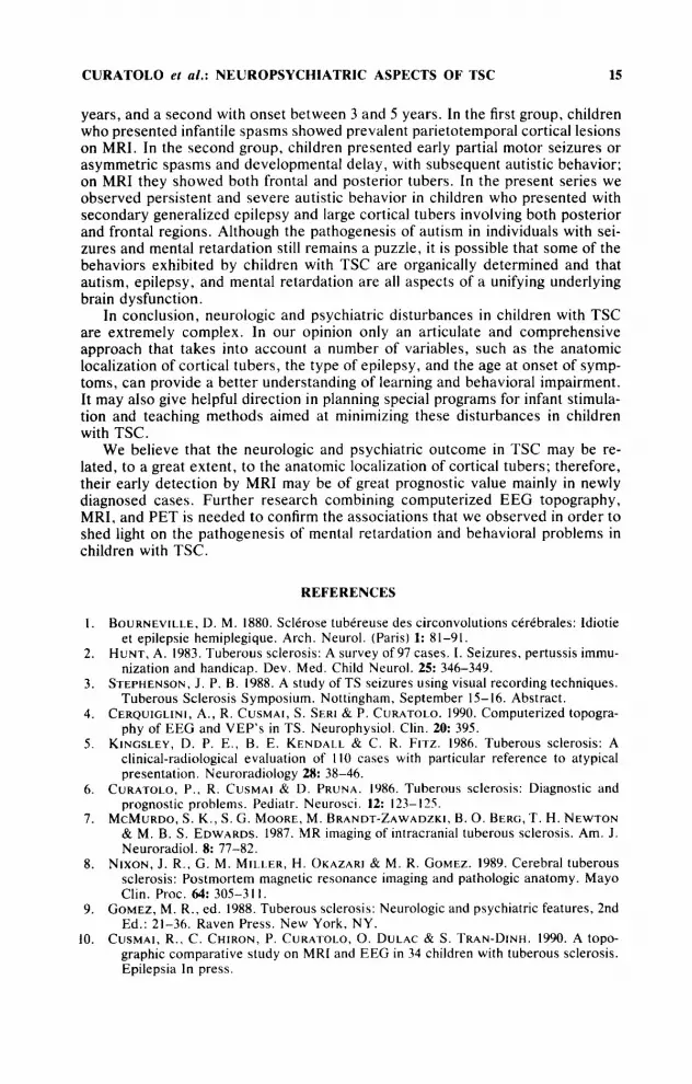
F ~ L ‘ A?. ‘ i ‘ ~ ; . . ~ i . - ~ ~ - ~ ~ ~ , ~ ~ ,
CJ A L ‘ - - \ ~ nr----y*.’*.i
TJ A Z L ~ ,ry J-.
0 2 A 2 2 -’
Fpl A I -,-\ , , - - ~ ~ , c . . _ r ~ ~ ~ ~ ~ ~ - ’ ~ ~ . , - , C3 A1 - v u ~ w + ,-
O J A I -. T3 A1 1 Y
- ss m t h s A M 1 <F 5’3077n1
sou4 1 sec
FIGURE 1. A 6-year-old child with multiple cortical lesions localized in the left frontal, right temporal, and biparietal zones on MR examination. Awake electroencephalography at this time showed a left frontal focus.
least two large or intermediate tubers. EEG foci without corresponding cortical tubers were observed in only four patients; they exhibited one to two tubers.
Although EEG was a sensitive tool in detecting focal abnormalities (91%), it was not specific enough to delineate their topographic localization (58%). In the entire series, 41% (14 of 34) of the patients exhibited cortical tubers without corresponding EEG foci. In six of them, tubers had a frontal location (mean age at MRI 1.4 years). Among the 12 patients with SBS, eight (67%) exhibited frontal tubers (bilateral in five and unilateral in three). The others had bilateral parietal (one case) or occipital tubers (three cases).
Neuropsychologic Findings
In 7 of the 23 children (30%) aged over 5 years and tested with a complete neuropsychologic battery, IQ was within normal limits (mean IQ IOO), and in 16

12 ANNALS NEW YORK ACADEMY OF SCIENCES
an IQ lower than 69 was found. Of the latter, 6 of 23 (26%) fulfilled the diagnostic criteria for infantile autism according to the behavioral TSC questionnaire; they all scored more than 10, and their behavior was regarded as severely autistic. Therefore, the series was classified as follows: normal intelligence (7 cases), mental retardation (10 cases), and mental retardation with persistent autistic be- havior (6 cases). Of the seven nonretarded children, specific neuropsycho- logic deficits such as speech delay, visuospatial disturbances, dyspraxia, and memory impairment were found.
Behavioral problems were present in almost all of the 10 retarded children; in particular, five had marked hyperactivity and two had aggressive and obsessive behavior and bulimia. Hyperkinetic behavior was also observed in three of the six autistic children.
Relation between Epilepsy, Neuropsychologic Findings, and MRI Findings
In our series, late-onset partial seizures (five cases) or transient infantile spasms (two cases) were the only types of seizure reported in the nonretarded individuals. All patients with favorable evolution of their epilepsy had normal psychomotor development before the onset of their first seizure. Children with normal intelligence showed small, isolated cortical tubers mainly localized in parietal and rolandic regions.
In contrast, all of our patients with mental retardation had had frequent partial seizures and later developed multifocal or secondary generalized epilepsy; they all showed multiple bilateral cortical tubers.
Patients with progressive intellectual deterioration and behavioral regression presented more frequent and intractable complex partial seizures. On EEG they showed frontal foci, with marked SBS and parasagittal frontal tubers on MR examination. Children with autism presented infantile spasms or partial seizures followed by secondary generalized epilepsy and showed both frontal and poste- rior tubers. Finally, most children who did not speak had large or intermediate left temporal tubers.
DISCUSSION
Epilepsy
Seizures are the most common neurologic symptom of TSC, occurring in 92% of a recently published series9 Although epilepsy may occur at any age, it most often begins in the first year of life with partial motor seizures and infantile spasms. In the same child, partial seizures may precede, coexist, or evolve into infantile spasms.16 Sometimes partial seizures are not recognized until the third or fourth month of life, when infantile spasms occur. Awake EEG at the onset shows multifocal or focal abnormalities; during sleep these abnormalities generalize to resemble hypsarrhythmia.
At the onset, older children exhibit complex partial or generalized tonic sei- zures. In these individuals EEG foci prevail in temporal or frontal regions with marked generalization during sleep, assuming the appearance of a Lennox-like pattern.” At this moment, the classification of epilepsy according to the predomi- nant type of seizure may be misleading, and the recognition of focal origin on standard EEG is more difficult. However, video-EEG recording and computer-

CURATOLO ef al.: NEUROPSYCHIATRIC ASPECTS OF TSC 13
ized EEG mapping may show that even in cases of apparently synchronous EEG abnormalities, seizures have a focal rigi in.^ Our data confirm that in addition to generalized seizures and diffuse EEG abnormalities, seizures in TSC mainly ex- hibit the characteristics of partial epilepsy with single or multiple spikes and slow wave foci on EEG.
Tubers as Epileptogenic Foci
In children with TSC, cortical lesions are thought to account for seizure^.^^^^ Cortical tubers are often radiologically isodense to normal brain tissue; therefore, most are overlooked by CT, but disclosed by MRI.6 Pathologically, they consist of large glial cells and atypical giant neurons with a low cellular density.lSz1 It has also been suggested that cortical tubers revealed by MRI represent the epilepto- genic foci in TSC.10.22
Our series showed a clear topographic correspondence between the EEG foci and the largest MRI high signal lesions, demonstrating the preponderant role of these tubers in epilepsy. The age of occurrence of EEG foci and the age of seizure onset depended on the localization of cortical tubers, with an earlier expression for parietooccipital lesions than for frontal ones. In patients with early-onset epilepsy, posterior foci were present and persisted during followup, whereas additional frontal EEG foci seemed to occur only after the age of 2 years. This finding is in accord with the posteroanterior migration of epileptic foci reported in childrenz3 and may result from maturational phenomena.24 We also found a rela- tion between the presence of frontal tubers and SBS. SBS is more likely to occur in frontal foci than in focal abnormalities located in other areas, because the degree of synchronization of EEG activity is higher in frontal than in occipital regions.25 However, the presence of more than one focus in half our patients suggests that SBS may also result from a complex interaction of multiple epilepto- genic regions. l 7 These findings have important implications not only in the clinical management of these patients, but also probably in the therapeutic approach. Selective surgical removal of involved tissue or anterior callosotomy could im- prove patients with SBS due to frontal tubers.26
In our series some patients have epilepsy with no detectable tuber on MRI. Several hypotheses can be proposed. It is known that MRI may fail to visualize some pathologically demonstrated cortical tubersx Furthermore, positron ernis- sion tomographic (PET) scans reveal hypometabolic regions not predicted by MRI in patients with TSC, suggesting that the disturbance of cerebral function is more extensive than that indicated by the morphologic finding^.^' Tubers may be overlooked on MRI because of physiologic hypomyelination in the first year of life;28 some tubers may also be undetected because of the particular relative proportion of astrocytes and neurones. l 4 Heterotopic clusters of giant cells in white matter have also been reported in TSC;i3.14 their radial localization, forming a continuous line between the ependyma and the tuber, recalls the embryologic migration of the spongioblasts.I9 Therefore, lack of some normal neuronal popula- tion in the corresponding cortical area could explain epilepsy in cases without a corresponding MRI high signal area.
Mental Retardation
The mental ability of patients affected by TSC varied from normal to severely retarded. Educational placement ranged from normal school to a special-care
14 ANNALS NEW YORK ACADEMY OF SCIENCES
school in severely affected cases.” In the Gomez series, mental retardation vary- ing from severe to mild occurred in 47% of case^.^ All the reported patients with severe mental retardation had seizure^.^^^* The mental outcome of TSC children with epilepsy is considered poor29 mainly in those with early-onset epilepsy, such as West syndrome.Is Patients who present with infantile spasms and hypsar- rhythmia are reported as more severely affected than are those with any form of epilepsy.”
There is sufficient evidence to support the concept that cumulative damage occurs from repeated and frequent seizures.’” Therefore, early diagnosis and early seizure control may be crucial in reducing the poor prognosis in such cases.31 The question arises as to whether seizures cause mental retardation or if mental retardation and epilepsy are two aspects of some unifying unakriying brain dysfunction. We found a consistent association between seizures and mental retardation in our series; however, this association did not present definite evi- dence for a cause-effect relation. In the study by Roach et a1.,12 there was consid- erable variation in the mental function of patients with five or fewer cortical lesions, but the developmental function of all patients with 10 or more cortical lesions was severely impaired. Our data suggest that both the number and the anatomic localization of cortical tubers play an important role in mental outcome, and they support the idea that epilepsy and mental retardation probably reflect the underlying brain dysfunction caused by cortical tubers. According to our data it is also possible that in patients with average intelligence some particular neuropsy- chologic functions can be affected independently of seizures, caused by strategi- cally located cortical tubers. Mental deterioration observed in children with per- sistent and frequent seizures may also be due to a particular epileptogenicity of parasagittal frontal tubers.
Behavioral Problems and Autism
Varied and multiple behavioral problems were reported in children with TSC: hyperactivity, screaming, destructiveness, temper tantrums, obsessions, aggres- siveness, sleeplessness, and self-mutilation. Children’s difficult behavior, includ- ing the lack of communication and speech, is reported as one of the most pressing parental problems, eventually leading to special-care classes. l5 However, at the moment there are no standardized neuropsychologic investigations related to the clinical and neuroradiologic findings.
A very high incidence of autism has been found in children affected by TSC. We believe that this association, which is not rare, might be more than just a coincidence. In Hunt’s series, over 50% of the 97 children showed psychotic behavior, 59% were hyperkinetic, and 13% were severely aggressive.
The majority of reported cases of children with TSC and autism had experi- enced infantile spasms. However, according to Hunt and Dennis,ls 57% of the children with TSC who had infantile spasms were autistic, compared to 13% in a Finnish series of children with infantile spasms from other causes.32 They sug- gested that autism should be related more to TSC than to infantile spasms. They also noted the possible relation between autistic behavior and limbic calcification as revealed by CT. In the study by Hunt and DennisI5 all the children with autistic behavior were severely mentally retarded, raising the question of cognitive de- fects as a primary cause of autism.Is
In a previous report33 we hypothesized that two groups of patients with autism and TSC can be outlined, one with onset of autistic behavior before the age of 2
CURATOLO et al.: NEUROPSYCHIATRIC ASPECTS OF TSC 15
years, and a second with onset between 3 and 5 years. In the first group, children who presented infantile spasms showed prevalent parietotemporal cortical lesions on MRI. In the second group, children presented early partial motor seizures or asymmetric spasms and developmental delay, with subsequent autistic behavior; on MRI they showed both frontal and posterior tubers. In the present series we observed persistent and severe autistic behavior in children who presented with secondary generalized epilepsy and large cortical tubers involving both posterior and frontal regions. Although the pathogenesis of autism in individuals with sei- zures and mental retardation still remains a puzzle, it is possible that some of the behaviors exhibited by children with TSC are organically determined and that autism, epilepsy, and mental retardation are all aspects of a unifying underlying brain dysfunction.
In conclusion, neurologic and psychiatric disturbances in children with TSC are extremely complex. In our opinion only an articulate and comprehensive approach that takes into account a number of variables, such as the anatomic localization of cortical tubers, the type of epilepsy, and the age at onset of symp- toms, can provide a better understanding of learning and behavioral impairment. It may also give helpful direction in planning special programs for infant stimula- tion and teaching methods aimed at minimizing these disturbances in children with TSC.
We believe that the neurologic and psychiatric outcome in TSC may be re- lated, to a great extent, to the anatomic localization of cortical tubers; therefore, their early detection by MRI may be of great prognostic value mainly in newly diagnosed cases. Further research combining computerized EEG topography, MRI, and PET is needed to confirm the associations that we observed in order to shed light on the pathogenesis of mental retardation and behavioral problems in children with TSC.
REFERENCES
I .
2.
3.
4.
5 .
6 .
7.
8.
9.
10.
BOURNEVIL1.E. D. M. 1880. Sclerose tubereuse des circonvolutions cerebrales: Idiotie et epilepsie hemiplegique. Arch. Neurol. (Paris) 1: 81-91.
HUNT, A. 1983. Tuberous sclerosis: A survey of 97 cases. I . Seizures, pertussis immu- nization and handicap. Dev. Med. Child Neurol. 25: 346-349.
STEPHENSON, J . P. B. 1988. A study of TS seizures using visual recording techniques. Tuberous Sclerosis Symposium. Nottingham, September 15-16. Abstract.
CERQUIGLINI, A, , R. CUSMAI, S. SERI & P. CURATOLO. 1990. Computerized topogra- phy of EEG and VEP's in TS. Neurophysiol. Clin. 20: 395.
KINGSLEY, D. P. E . , B. E. KENDALI. & C. R. FITZ. 1986. Tuberous sclerosis: A clinical-radiological evaluation of I10 cases with particular reference to atypical presentation. Neuroradiology 28: 38-46.
CURATOLO, P.. R. CUSMAI & D. PRUNA. 1986. Tuberous sclerosis: Diagnostic and prognostic problems. Pediatr. Neurosci. 1 2 123-125.
MCMURDO, S. K., S. G. MOORE, M. BRANDT-ZAWADZKI, B . 0. BERG, T. H . NEWTON & M. B. S. EDWARDS. 1987. MR imaging of intracranial tuberous sclerosis. Am. J . Neuroradiol. 8: 77-82.
NIXON, J . R . . G. M. MILLER, H. OKAZARI & M . R. GOMEZ. 1989. Cerebral tuberous sclerosis: Postmortem magnetic resonance imaging and pathologic anatomy. Mayo Clin. Proc. 64: 305-3 I 1 .
GOMEZ, M. R., ed. 1988. Tuberous sclerosis: Neurologic and psychiatric features, 2nd Ed.: 21-36. Raven Press. New York, NY.
CUSMAI, R., C . CHIRON, P. CURATOLO, 0. DULAC & S. TRAN-DINH. 1990. A topo- graphic comparative study on MRI and EEG in 34 children with tuberous sclerosis. Epilepsia In press.

16 ANNALS NEW YORK ACADEMY OF SCIENCES
11.
12.
13.
14.
15.
16.
17.
IS.
19.
20.
21.
2 2 .
23.
24.
25.
26.
27.
28.
29.
30.
31.
32.
33.
JAMBAQUE I. , R. CUSMAI. F. CORTESI. c . CHIRON, P. CURATOLO & 0. DULAC. Neuropsychological aspects in tuberous sclerosis: Relationships with epilepsy and MRI findings. In preparation.
ROACH, E . , D. P. WIi.LiAMs & D. W. LASTER. 1987. Magnetic resonance imaging in tuberous sclerosis. Arch. Neurol. 44: 301-303.
TERWEY. B. & H . DOOSE. 1987. Tuberous sclerosis: MRI of the brain. Neuropediatrics 18: 67-69.
MARTIN, N.. T. DE BROUCKER. J . CAMBIER, C. MARSAULT & H. NAHUM. 1987. MRI evaluation of tuberous sclerosis. Neuroradiology 29: 437-443.
HUNT, A. & J . DENNIS. 1987. Psychiatric disorder among children with tuberous sclerosis. Dev. Med. Child Neurol. 29: 190-198.
DULAC. 0.. A. LEMAITRE & P. PLOUIN. 1984. Maladie de Bourneville: Aspects cli- niques et electroencephalographiques de I'epilepsie dans la premiere annee. Boll. Lega It. Epil. 45-46: 39-42.
GASTAUT. H.. B. Zi txiN. A. MACAUDDA & E. MARIANI. 1987. Symptomatic partial epilepsies with secondary bilateral synchrony: Differentiation from symptomatic generalized epilepsy of Lennox-Gastaut type. I n Presurgical Evaluation of Epilep- tics, H . G. Wieser & C. E. Elger. eds.: 308-316. Springer-Verlag. Berlin.
ROGER, J . , C. DRAVET, C . BONIVIER, A. MAGAUDDA, M. BUREAU, E. FERNANDEZ- ALVAREZ, F. X. SANMARTI. I . FABREGUES, B. CENRAUD & J . L. LARRIEU. 1984. L'epilepsie dans la sclerose tubereuse de Bourneville. Boll. Lega It. Epil. 45: 33-38.
DONEGANI. G , , F. R. GRATTAROI.A & E. W I L D I . 1972. Tuberou\ sclerosis: Bourneville disease. I n Handbook of Clinical Neurology. Vol. 14. The Phakomatoses. P. J . Vinken & G . W. Hruyn. eds.: 340-389. North Holland Puhlishing Co. Amsterdam.
TROMBLEY. I . K. & S. S. MIRRA. 1981. Ultrastructure of tuberous sclerosis: Cortical tuber and subependymal tumor. Ann. Neurol. 9: 174-181.
HUTTENLOCHER, P. R. & P. T. HEYDLMANN. 1984. Fine structure of cortical tubers in tuberous sclerosis: A Golgi study. Ann. Neurol. 16: 59s-602.
CURATOLO, P. & R. CUSMAI. 1988. lmagerie par resonance magnetique nuclkaire dans la maladie de Bourneville: Relations avec les donnees electroencephalographiques. Neurophy4ol. Clin. 18: 149-167.
GIBBS. E. L. & F. A. GIRLS. 19.54. Disappearance and migration of epileptic foci in childhood. Am. .I. Dis. Child. 596-603.
CHUGANI H. T. , M. E. PHELPS & J . C . MAZZIOTTA. 1987. Positron Emission To- mography study of human brain functional development. Ann. Neurol. 22: 487-497.
BLUME. W. T. & N. PIi.i.AY. 1985. Electrographic and clinical correlates of secondary bilateral synchrony. Epilepsia 26: 636-641.
BYE, A. M., J . M. MATHESON, V. H . TOBIAS & R. A. MACKENZIE. 1989. Selective epilepsy surgery in tuberous sclerosis. Aust. Paediatr. J . 25: 243-245.
SZEILES, B.. K. HERHOLZ, W. D. HEISS. A. RACKI.. G . PAWI.IK. R. WAGNER, H. W. I L S E N & K . WIENHARD. 1983. Hypometabolic cortical lesions in tuberous sclerosis with epilepsy: Demonstration by Positron Emission Tomography. J . Comput. As- sist. Tomogr. 7: 946-9.53.
BARKOVICH. A . J . . 0. K . BENT, D. E. JACKSON & D. NORMAN. 1988. Normal matura- tion of the neonatal and infant brain: MR imaging at 1 .S TI. Radiology 166 173-180.
LAGOS, J. C. & M. R. GOMEZ. 1967. Tuberous sclerosis: Reappraisal of a clinical entity. Proc. Mayo Clin. 42: 26-49.
GOMEZ, M. R. , N. L. KUNTZ & B. F. WESTMORELAND. 1982. Tuberous sclerosis, early onset of seizures. and mental subnormality: Study of discordant monozygous twins. Neurology 32(h): 604-61 1.
LANE. V . W. & J . M. SAMPLES. 1984. Tuberous sclerosis: Case study of early seizure control and subsequent normal development. J . Aut. Dev. Disord. 14: 423-427.
RIJKONEN. R. & G . ANNELL. 1981. Psychiatric disorder in children with earlier infan- tile spasms. Dev. Med. Child Neurol. 23: 747-760.
CURATOLO P. & R. CusMAi. 1987. Autism and infantile spasms in children with tuber- ous sclerosis. Dev. Med. Child Neurol. 29: 550-S.51.


