Embed Size (px)
Citation preview
pathogens
Article
Antioxidant and Leishmanicidal Evaluation ofPulicaria Inuloides Root Extracts:A Bioguided Fractionation
Hamza Fadel 1, Ines Sifaoui 2,3, Atteneri López-Arencibia 2, María Reyes-Batlle 2,Ignacio A. Jiménez 4, Jacob Lorenzo-Morales 2, Nabil Ghedadba 5, Samir Benayache 1 ,José E. Piñero 2,* and Isabel L. Bazzocchi 4,*
1 Unité de recherche Valorisation des Ressources Naturelles, Molécules Bioactives et AnalysesPhysicochimique et Biologiques, Université Constantine-1, Route d’Ain El Bey, 25 000 Constantine, Algerie;[email protected] (H.F.); [email protected] (S.B.)
2 Instituto Universitario de Enfermedades Tropicales y Salud Pública de Canarias, Departamento deObstetricia y Ginecología, Pediatría, Medicina Preventiva y Salud Pública, Toxicología, Medicina Legal yForense y Parasitología, Universidad de La Laguna, Avda. Astrofısico Fco. Sanchez, S/N, 38203 La Laguna,Tenerife, Canary Islands, Spain; [email protected] (I.S.); [email protected] (A.L.-A.);[email protected] (M.R.-B.); [email protected] (J.L.-M.)
3 Laboratoire Materiaux-Molecules et Applications, IPEST, University of Carthage, 2070 La Marsa, Tunisia4 Instituto Universitario de Bio-Orgánica Antonio González, Departamento de Química Orgánica,
Universidad de La Laguna, Avenida Astrofísico Francisco Sánchez 2, 38206 La Laguna, Spain;[email protected]
5 Laboratory of Biotechnology of the Bioactive Molecules and Cellular Physiopathology, Department ofBiology, University of Batna, 05000 Batna, Algeria; [email protected]
* Correspondence: [email protected] (J.E.P.); [email protected] (I.L.B.)
Received: 31 July 2019; Accepted: 19 October 2019; Published: 23 October 2019�����������������
Abstract: Leishmaniasis remains a major world health problem, and in particular, Algeria rankssecond for the incidence of cutaneous leishmaniasis. Pulicaria inuloides is a well-known ArabianPeninsula medicinal plant. In the present study, the chloroform, ethyl acetate and n-butanol extractsfrom the roots of Pulicaria inuloides were analyzed for antioxidant activity and its correlation withthe total phenolic and flavonoid contents. The highest antioxidant activity using a DPPH assay wasshowed by the ethyl acetate extract (IC50 4.08 µg/mL), which also had the highest total phenoliccontent (307.12 µgAGE). Furthermore, P. inuloides root extracts were evaluated against Leishmaniaamazonensis and Leishmania donovani. The results highlighted the chloroform extract as the most activeone against both tested Leishmania strains. A bioguided fractionation of the chloroform extract led tothe isolation of (8R:8S)-(75:25 er)-10-isobutyryloxy-8,9-epoxy-thymol isobutyrate as the main bioactivecomponent, showing a potent leishmanicidal activity on L. amazonensis promatigote and amastigotestages (IC50 5.03 and 2.87 µM, respectively) and a good selectivity index on murine macrophages(CC50 19.37 µM). This study provides the first report of the antioxidant and leishmanicidal activitiesof P. inuloides root extracts and the results point to this species as a source of potential bioactive agents.
Keywords: Leishmania; antioxidant; Pulicaria; bioguided; therapy
1. Introduction
Leishmaniasis, a neglected tropical disease caused by protozoan parasites belonging to the genusLeishmania, remains a major world health problem [1]. In particular, Algeria is endemic for cutaneousleishmaniasis (CL) and visceral leishmaniasis (VL) [2]. CL, also called “Biskra boil” in the locallanguage, is a serious public health problem because the country has the second largest focus in the
Pathogens 2019, 8, 201; doi:10.3390/pathogens8040201 www.mdpi.com/journal/pathogens
Pathogens 2019, 8, 201 2 of 11
world after Afghanistan. It is reported to be endemic in 328 third sub-national administrative levelswith 10 million populations at risk, rising 13106 numbers of cases reported in 2017 [3]. Regarding VL,the cases have been detected mainly from the central and eastern parts of the Tell region, and newfoci have appeared and the number of cases has resurged, with 34 new cases reported in 2017 [4].The Leishmaniasis National Control Program (LNCP) in Algeria was established in 2006, with avector control program based on pyrethroids as insecticides, but without reservoir host controlprogram [5]. The treatment of the population is provided free of charge in the public sector, with thefollowing antileishmanial medicines included in the national List of Essential Medicines: amphotericinB deoxycholate, liposomal amphotericin B and meglumine antimoniate [6,7]. In spite of the highprevalence, current chemotherapy for leishmaniasis is compromised by high cost, toxicity associatedwith long-term treatments, route of administration, drug resistance, and different strain sensitivityto the available drugs [8]. These drawbacks lead to an urgent need to develop new treatments withacceptable efficacy and safety profile. In this regard, plants have demonstrated to be an importantsource of leishmanicidal drugs owing to their accessibility, structural diversity, low cost and possiblerapid biodegradation [9].
The genus Pulicaria belonging to the Asteraceae family (tribe Inuleae) consists of about 100 speciesdistributed in Europe, North Africa and Asia, particularly around the Mediterranean region [10,11].Some Pulicaria species are used as traditional medicines for treatment of a variety of illnesses due to theiranticancer, antioxidant, antimicrobial, antifungal and antimalarial properties, among others [11–13].Pulicaria inuloides (Poir.) DC is a well-known Yemeni medicinal plant. The leaves are claimed tohave antibacterial and antioxidant properties, and are also used to flavor foods and to make anherbal tea [14]. The chemical composition [14–16] and medicinal uses as anticancer, antibacterial andantioxidant [14,17] of the P. inuloides essential oil obtained by hydro-destillation of the leaves andflowers have been reported. Moreover, the antioxidant properties and total phenol and flavonoidcontents of different extracts from the leaves of P. inuloides were analyzed by Al-Hajj and co-workers [15].In addition, the isolation of ent-kaurane diterpenoids and flavonoids from the aerial part of P. inuloideswas reported, along with their antimicrobial, cytotoxic and antioxidant evaluation [18].
Our previous works reported the in vitro antiprotozoal evaluation of extracts from the aerial part ofP. inuloides, and the characterization of quercetagetin-3,5,7,3’-tetramethyl ether as the main componentof the active CHCl3 extract [19]. In the present work, and as a continuation of the P. inuloides medicinalplant research, the antioxidant activity, total phenolic and flavonoid contents, and leishmanicidalproperties of P. inuloides roots are reported. The bioassay-guided fractionation of the active CHCl3extract led to the isolation and characterization of 10-isobutyryloxy-8,9-epoxythymol isobutyrate as themain leishmanicidal component, highlighting this plant as a promising source of antiparasitic agents.
2. Results and Discussion
2.1. DPPH Radical Scavenging Activity
Oxidative stress has been associated with the development and progression of severaldiseases, including cancer, inflammation, atherosclerosis, cardiovascular diseases or diabetes. Thus,administration of exogenous antioxidants is a promising way of combating the undesirable effects ofreactive oxygen species (ROS) induced oxidative damage. In this regard, plants have demonstrated tobe an important source of antioxidants such as polyphenols or flavonoids [20]. Moreover, taking intoconsideration that some Pulicaria species, and in particular P. inuloides, are claimed to have antioxidantproperties [11], the antioxidant activity of P. inuloides roots was analyzed.
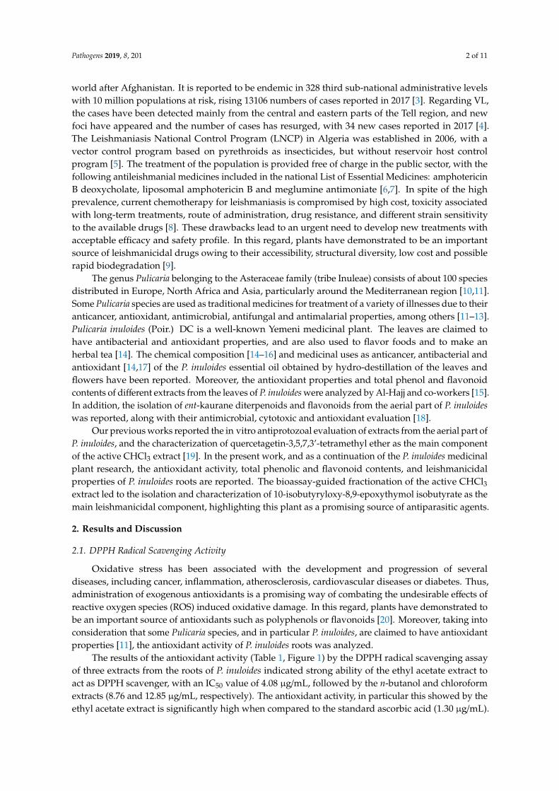
The results of the antioxidant activity (Table 1, Figure 1) by the DPPH radical scavenging assayof three extracts from the roots of P. inuloides indicated strong ability of the ethyl acetate extract toact as DPPH scavenger, with an IC50 value of 4.08 µg/mL, followed by the n-butanol and chloroformextracts (8.76 and 12.85 µg/mL, respectively). The antioxidant activity, in particular this showed by theethyl acetate extract is significantly high when compared to the standard ascorbic acid (1.30 µg/mL).
Pathogens 2019, 8, 201 3 of 11
Therefore, to investigate the correlation between the antioxidant activity and presence of antioxidantscompounds into the different extracts, the total phenolic and flavonoid contents were determined.
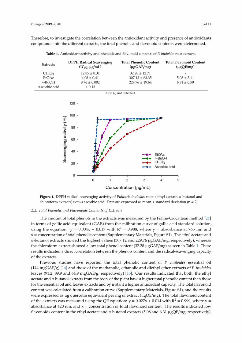
Table 1. Antioxidant activity and phenolic and flavonoid contents of P. inuloides root extracts.
Extracts DPPH Radical Scavenging(IC50, µg/mL)
Total Phenolic Content(µgGAE/mg)
Total Flavonoid Content(µgQE/mg)
CHCl3 12.85 ± 0.31 32.28 ± 12.71 -EtOAc 4.08 ± 0.41 307.12 ± 63.35 5.08 ± 3.11
n-BuOH 8.76 ± 0.002 229.76 ± 19.64 6.31 ± 0.59Ascorbic acid ± 0.13
Key: (-) not detected.
Pathogens 2019, 8, x FOR PEER REVIEW 3 of 11
µg/mL). Therefore, to investigate the correlation between the antioxidant activity and presence of antioxidants compounds into the different extracts, the total phenolic and flavonoid contents were determined.
Table 1. Antioxidant activity and phenolic and flavonoid contents of P. inuloides root extracts.
Extracts DPPH Radical
Scavenging (IC50, µg/mL)
Total Phenolic Content
(µgGAE/mg)
Total Flavonoid Content
(µgQE/mg) CHCl3 12.85 ± 0.31 32.28 ± 12.71 - EtOAc 4.08 ± 0.41 307.12 ± 63.35 5.08 ± 3.11
n-BuOH 8.76 ± 0.002 229.76 ± 19.64 6.31 ± 0.59 Ascorbic acid 1.30 ± 0.13
Key: (-) not detected.
Figure 1. DPPH radical-scavenging activity of Pulicaria inuloides roots (ethyl acetate, n-butanol and chloroform extracts) versus ascorbic acid. Data are expressed as mean ± standard deviation (n = 2).
2.2. Total Phenolic and Flavonoids Contents of Extracts
The amount of total phenols in the extracts was measured by the Foline-Ciocalteau method [21] in terms of gallic acid equivalent (GAE) from the calibration curve of gallic acid standard solution, using the equation: y = 0.004x + 0.017 with R² = 0.988, where y = absorbance at 765 nm and x = concentration of total phenolic content (Supplementary Materials, Figure S1). The ethyl acetate and n-butanol extracts showed the highest values (307.12 and 229.76 µgGAE/mg, respectively), whereas the chloroform extract showed a low total phenol content (32.28 µgGAE/mg) as seen in Table 1. These results indicated a direct correlation between the phenols content and the radical-scavenging capacity of the extracts.
Previous studies have reported the total phenolic content of P. inuloides essential oil (144 mgGAE/g) [14] and those of the methanolic, ethanolic and diethyl ether extracts of P. inuloides leaves (91.2, 89.9 and 64.9 mgGAE/g, respectively) [15]. Our results indicated that both, the ethyl acetate and n-butanol extracts from the roots of the plant have a higher total phenolic content than those for the essential oil and leaves extracts and by instant a higher antioxidant capacity. The total flavonoid content was calculated from a calibration curve (Supplementary Materials, Figure S1), and the results were expressed as µg quercetin equivalent per mg of extract (µgQE/mg). The total flavonoid
Figure 1. DPPH radical-scavenging activity of Pulicaria inuloides roots (ethyl acetate, n-butanol andchloroform extracts) versus ascorbic acid. Data are expressed as mean ± standard deviation (n = 2).
2.2. Total Phenolic and Flavonoids Contents of Extracts
The amount of total phenols in the extracts was measured by the Foline-Ciocalteau method [21]in terms of gallic acid equivalent (GAE) from the calibration curve of gallic acid standard solution,using the equation: y = 0.004x + 0.017 with R2 = 0.988, where y = absorbance at 765 nm andx = concentration of total phenolic content (Supplementary Materials, Figure S1). The ethyl acetate andn-butanol extracts showed the highest values (307.12 and 229.76 µgGAE/mg, respectively), whereasthe chloroform extract showed a low total phenol content (32.28 µgGAE/mg) as seen in Table 1. Theseresults indicated a direct correlation between the phenols content and the radical-scavenging capacityof the extracts.
Previous studies have reported the total phenolic content of P. inuloides essential oil(144 mgGAE/g) [14] and those of the methanolic, ethanolic and diethyl ether extracts of P. inuloidesleaves (91.2, 89.9 and 64.9 mgGAE/g, respectively) [15]. Our results indicated that both, the ethylacetate and n-butanol extracts from the roots of the plant have a higher total phenolic content than thosefor the essential oil and leaves extracts and by instant a higher antioxidant capacity. The total flavonoidcontent was calculated from a calibration curve (Supplementary Materials, Figure S1), and the resultswere expressed as µg quercetin equivalent per mg of extract (µgQE/mg). The total flavonoid contentof the extracts was measured using the QE equation: y = 0.027x + 0.014 with R2 = 0.999, where y =
absorbance at 420 nm, and x = concentration of total flavonoid content. The results indicated lowflavonoids content in the ethyl acetate and n-butanol extracts (5.08 and 6.31 µgQE/mg, respectively),
Pathogens 2019, 8, 201 4 of 11
while this type of compounds could not be detected in the chloroform extract (Table 1). A correlationbetween the total phenolic content and the antioxidant capacity of the extracts was observed. Previousreported studies revealed that phenolic compounds are major antioxidant constituents in the leaves ofP. inuloides extracts [15], and the good correlation observed in our study support the hypothesis thatphenolic compounds in P. inuloides roots contribute significantly to their antioxidant properties.
2.3. Bioassay-Guided Fractionation
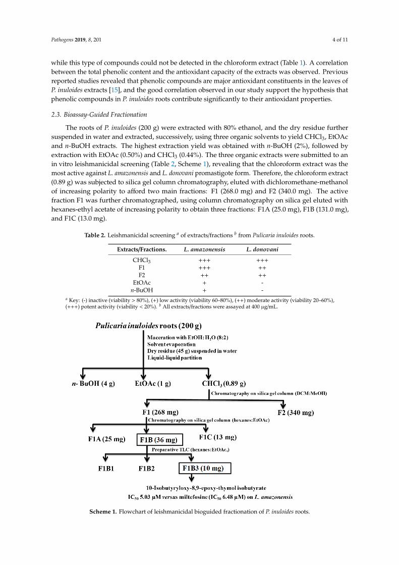
The roots of P. inuloides (200 g) were extracted with 80% ethanol, and the dry residue furthersuspended in water and extracted, successively, using three organic solvents to yield CHCl3, EtOAcand n-BuOH extracts. The highest extraction yield was obtained with n-BuOH (2%), followed byextraction with EtOAc (0.50%) and CHCl3 (0.44%). The three organic extracts were submitted to anin vitro leishmanicidal screening (Table 2, Scheme 1), revealing that the chloroform extract was themost active against L. amazonensis and L. donovani promastigote form. Therefore, the chloroform extract(0.89 g) was subjected to silica gel column chromatography, eluted with dichloromethane-methanolof increasing polarity to afford two main fractions: F1 (268.0 mg) and F2 (340.0 mg). The activefraction F1 was further chromatographed, using column chromatography on silica gel eluted withhexanes-ethyl acetate of increasing polarity to obtain three fractions: F1A (25.0 mg), F1B (131.0 mg),and F1C (13.0 mg).
Table 2. Leishmanicidal screening a of extracts/fractions b from Pulicaria inuloides roots.
Extracts/Fractions. L. amazonensis L. donovani
CHCl3 +++ +++F1 +++ ++F2 ++ ++
EtOAc + -n-BuOH + -
a Key: (-) inactive (viability > 80%), (+) low activity (viability 60–80%), (++) moderate activity (viability 20–60%),(+++) potent activity (viability < 20%). b All extracts/fractions were assayed at 400 µg/mL.
Pathogens 2019, 8, x FOR PEER REVIEW 6 of 11
thymol derivatives have been evaluated as antibacterial, anti-inflammatory, antioxidant, antiprotozoal, cytotoxic, piscicidal, or allelophatic agents [25]. Regarding epoxythymol derivatives, evaluation on Entamoeba histolytica and Giardia lamblia of thymol isobutyrate derivatives revealed a moderate antiprotozoal activity on both protozoa [26]. However, studies on their leishmanicidal or antioxidant activities have not been reported.
To our knowledge, this is the first report of thymol derivative 1 in Pulicaria genus. Previously, the leishmanicidal bioguided fractionation of the active chloroform extract from the aerial part of P. inuloides led to the isolation of quercetagetin-3,5,7,3'-tetramethyl ether as the main bioactive component, exhibiting a moderate leishmanicidal activity with an IC50 value of 0.483 mM on L. amazonensis [19]. Furthermore, the present study reveals that a thymol derivative, the major component from the roots of this species, is responsible for the leishmanicidal activity. This further supports P. inuloides as a source of promising leishmanicidal agents.
Scheme 1. Flowchart of leishmanicidal bioguided fractionation of P. inuloides roots.
3. Materials and Methods
3.1. General Experimental Procedure
Optical rotation was measured on a Perkin Elmer 241 automatic polarimeter in CHCl3 at 20 ⁰C. 1H (600 MHz), 13C (150 MHz) Nuclear Magnetic Resonance (NMR) spectra were recorded on a Bruker Avance 600 spectrometer, the chemical shifts are given in δ (ppm) with residual CDCl3 (δH 7.26, δC 77.0) as internal reference and coupling constants in Hz; the experiments were carried out with the pulse sequences given by Bruker. EIMS and HREIMS were collected with a Micromass Autospec spectrometer. Silica gel 60 (particle size 15-40 and 63-200 μm, Macherey-Nagel) was used for column chromatography, while silica gel 60 F254 (Macherey-Nagel) were used for analytical or preparative TLC. The spots were visualized by UV light and heating silica gel plates sprayed with H2O-H2SO4-AcOH (1:4:20). All solvents used were purchased from Sigma Aldrich.
3.2. Plant Material and Extraction
Scheme 1. Flowchart of leishmanicidal bioguided fractionation of P. inuloides roots.
Pathogens 2019, 8, 201 5 of 11
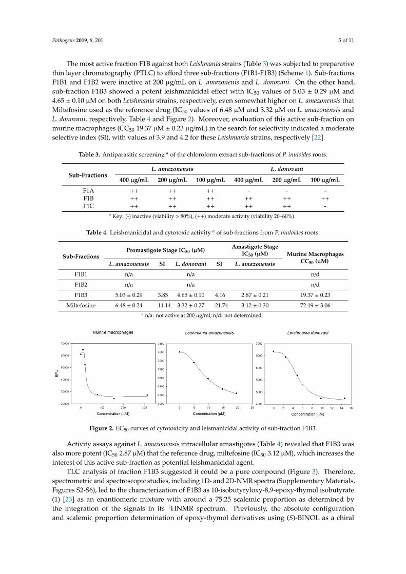
The most active fraction F1B against both Leishmania strains (Table 3) was subjected to preparativethin layer chromatography (PTLC) to afford three sub-fractions (F1B1-F1B3) (Scheme 1). Sub-fractionsF1B1 and F1B2 were inactive at 200 µg/mL on L. amazonenis and L. donovani. On the other hand,sub-fraction F1B3 showed a potent leishmanicidal effect with IC50 values of 5.03 ± 0.29 µM and4.65 ± 0.10 µM on both Leishmania strains, respectively, even somewhat higher on L. amazonensis thatMiltefosine used as the reference drug (IC50 values of 6.48 µM and 3.32 µM on L. amazonensis andL. donovani, respectively, Table 4 and Figure 2). Moreover, evaluation of this active sub-fraction onmurine macrophages (CC50 19.37 µM ± 0.23 µg/mL) in the search for selectivity indicated a moderateselective index (SI), with values of 3.9 and 4.2 for these Leishmania strains, respectively [22].
Table 3. Antiparasitic screening a of the chloroform extract sub-fractions of P. inuloides roots.
Sub-FractionsL. amazonensis L. donovani
400 µg/mL 200 µg/mL 100 µg/mL 400 µg/mL 200 µg/mL 100 µg/mL
F1A ++ ++ ++ - - -F1B ++ ++ ++ ++ ++ ++F1C ++ ++ ++ ++ ++ -
a Key: (-) inactive (viability > 80%), (++) moderate activity (viability 20–60%).
Table 4. Leishmanicidal and cytotoxic activity a of sub-fractions from P. inuloides roots.
Sub-FractionsPromastigote Stage IC50 (µM) Amastigote Stage
IC50 (µM) Murine MacrophagesCC50 (µM)L. amazonensis SI L. donovani SI L. amazonensis
F1B1 n/a n/a n/d
F1B2 n/a n/a n/d
F1B3 5.03 ± 0.29 3.85 4.65 ± 0.10 4.16 2.87 ± 0.21 19.37 ± 0.23
Miltefosine 6.48 ± 0.24 11.14 3.32 ± 0.27 21.74 3.12 ± 0.30 72.19 ± 3.06a n/a: not active at 200 µg/ml; n/d: not determined.
Pathogens 2019, 8, x FOR PEER REVIEW 5 of 11
Table 4. Leishmanicidal and cytotoxic activitya of sub-fractions from P. inuloides roots.
Sub-Fractions Promastigote Stage IC50 (µM) Amastigote Stage
IC50 (µM) Murine Macrophages CC50 (µM) L
amazonensis SI L. donovani
SI L. amazonensis
F1B1 n/a n/a n/d F1B2 n/a n/a n/d F1B3 5.03 ± 0.29 3.85 4.65 ± 0.10 4.16 2.87 ± 0.21 19.37 ± 0.23
Miltefosine 6.48 ± 0.24 11.14 3.32 ± 0.27 21.74 3.12 ± 0.30 72.19 ± 3.06 a n/a: not active at 200 µg/ml; n/d: not determined.
Figure 2. EC50 curves of cytotoxicity and leismanicidal activity of sub-fraction F1B3.
Activity assays against L. amazonensis intracellular amastigotes (Table 4) revealed that F1B3 was also more potent (IC50 2.87 µM) that the reference drug, miltefosine (IC50 3.12 µM), which increases the interest of this active sub-fraction as potential leishmanicidal agent.
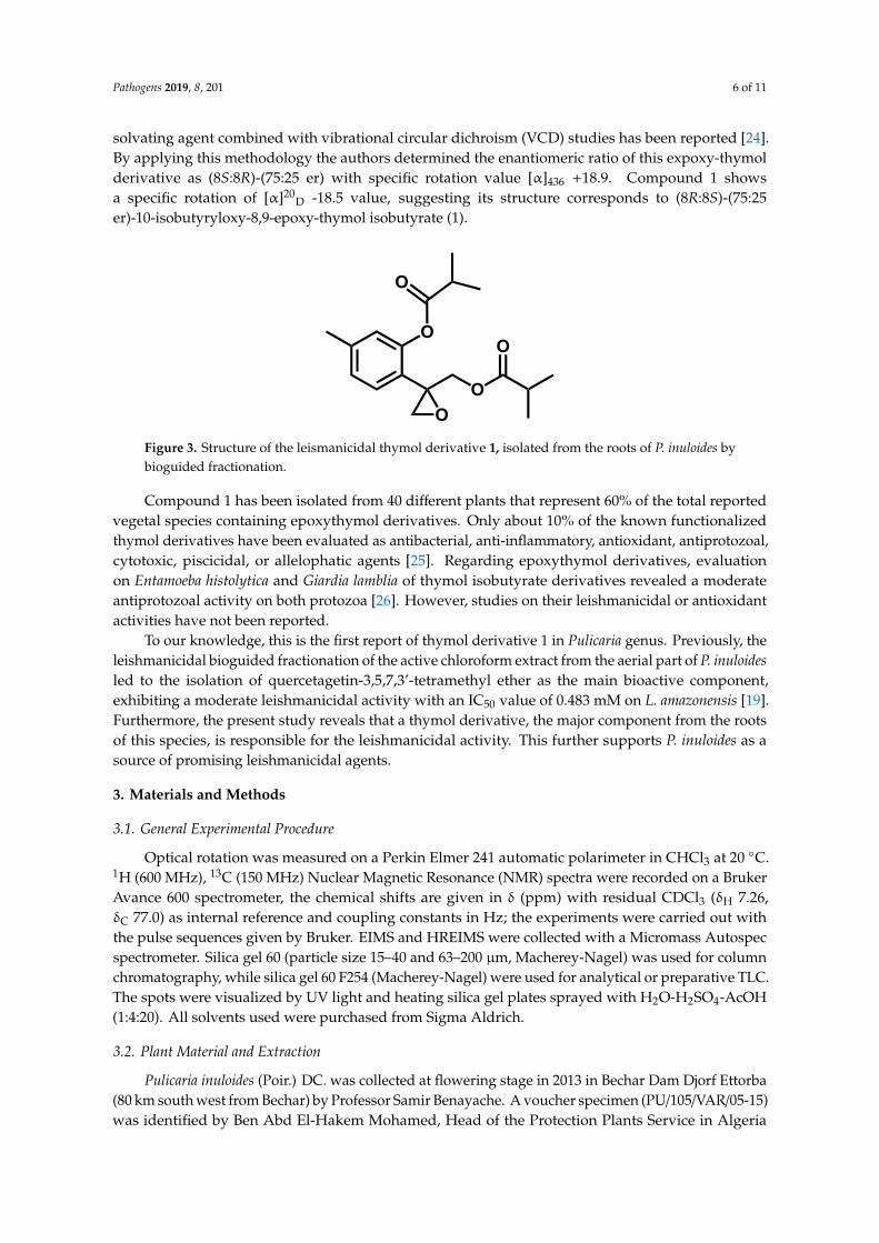
TLC analysis of fraction F1B3 suggested it could be a pure compound (Figure 3). Therefore, spectrometric and spectroscopic studies, including 1D- and 2D-NMR spectra (Supplementary Materials, Figures S2-S6), led to the characterization of F1B3 as 10-isobutyryloxy-8,9-epoxy-thymol isobutyrate (1) [23] as an enantiomeric mixture with around a 75:25 scalemic proportion as determined by the integration of the signals in its 1HNMR spectrum. Previously, the absolute configuration and scalemic proportion determination of epoxy-thymol derivatives using (S)-BINOL as a chiral solvating agent combined with vibrational circular dichroism (VCD) studies has been reported [24]. By applying this methodology the authors determined the enantiomeric ratio of this expoxy-thymol derivative as (8S:8R)-(75:25 er) with specific rotation value [α]436 +18.9. Compound 1 shows a specific rotation of [α]20D -18.5 value, suggesting its structure corresponds to (8R:8S)-(75:25 er)-10-isobutyryloxy-8,9-epoxy-thymol isobutyrate (1).
O
O
OO
O
Figure 3. Structure of the leismanicidal thymol derivative 1, isolated from the roots of P. inuloides by bioguided fractionation.
Compound 1 has been isolated from 40 different plants that represent 60% of the total reported vegetal species containing epoxythymol derivatives. Only about 10% of the known functionalized
Figure 2. EC50 curves of cytotoxicity and leismanicidal activity of sub-fraction F1B3.
Activity assays against L. amazonensis intracellular amastigotes (Table 4) revealed that F1B3 wasalso more potent (IC50 2.87 µM) that the reference drug, miltefosine (IC50 3.12 µM), which increases theinterest of this active sub-fraction as potential leishmanicidal agent.
TLC analysis of fraction F1B3 suggested it could be a pure compound (Figure 3). Therefore,spectrometric and spectroscopic studies, including 1D- and 2D-NMR spectra (Supplementary Materials,Figures S2-S6), led to the characterization of F1B3 as 10-isobutyryloxy-8,9-epoxy-thymol isobutyrate(1) [23] as an enantiomeric mixture with around a 75:25 scalemic proportion as determined bythe integration of the signals in its 1HNMR spectrum. Previously, the absolute configurationand scalemic proportion determination of epoxy-thymol derivatives using (S)-BINOL as a chiral
Pathogens 2019, 8, 201 6 of 11
solvating agent combined with vibrational circular dichroism (VCD) studies has been reported [24].By applying this methodology the authors determined the enantiomeric ratio of this expoxy-thymolderivative as (8S:8R)-(75:25 er) with specific rotation value [α]436 +18.9. Compound 1 showsa specific rotation of [α]20
D -18.5 value, suggesting its structure corresponds to (8R:8S)-(75:25er)-10-isobutyryloxy-8,9-epoxy-thymol isobutyrate (1).
Pathogens 2019, 8, x FOR PEER REVIEW 5 of 11
Table 4. Leishmanicidal and cytotoxic activitya of sub-fractions from P. inuloides roots.
Sub-Fractions Promastigote Stage IC50 (µM) Amastigote Stage
IC50 (µM) Murine Macrophages CC50 (µM) L
amazonensis SI L. donovani
SI L. amazonensis
F1B1 n/a n/a n/d F1B2 n/a n/a n/d F1B3 5.03 ± 0.29 3.85 4.65 ± 0.10 4.16 2.87 ± 0.21 19.37 ± 0.23
Miltefosine 6.48 ± 0.24 11.14 3.32 ± 0.27 21.74 3.12 ± 0.30 72.19 ± 3.06 a n/a: not active at 200 µg/ml; n/d: not determined.
Figure 2. EC50 curves of cytotoxicity and leismanicidal activity of sub-fraction F1B3.
Activity assays against L. amazonensis intracellular amastigotes (Table 4) revealed that F1B3 was also more potent (IC50 2.87 µM) that the reference drug, miltefosine (IC50 3.12 µM), which increases the interest of this active sub-fraction as potential leishmanicidal agent.
TLC analysis of fraction F1B3 suggested it could be a pure compound (Figure 3). Therefore, spectrometric and spectroscopic studies, including 1D- and 2D-NMR spectra (Supplementary Materials, Figures S2-S6), led to the characterization of F1B3 as 10-isobutyryloxy-8,9-epoxy-thymol isobutyrate (1) [23] as an enantiomeric mixture with around a 75:25 scalemic proportion as determined by the integration of the signals in its 1HNMR spectrum. Previously, the absolute configuration and scalemic proportion determination of epoxy-thymol derivatives using (S)-BINOL as a chiral solvating agent combined with vibrational circular dichroism (VCD) studies has been reported [24]. By applying this methodology the authors determined the enantiomeric ratio of this expoxy-thymol derivative as (8S:8R)-(75:25 er) with specific rotation value [α]436 +18.9. Compound 1 shows a specific rotation of [α]20D -18.5 value, suggesting its structure corresponds to (8R:8S)-(75:25 er)-10-isobutyryloxy-8,9-epoxy-thymol isobutyrate (1).
O
O
OO
O
Figure 3. Structure of the leismanicidal thymol derivative 1, isolated from the roots of P. inuloides by bioguided fractionation.
Compound 1 has been isolated from 40 different plants that represent 60% of the total reported vegetal species containing epoxythymol derivatives. Only about 10% of the known functionalized
Figure 3. Structure of the leismanicidal thymol derivative 1, isolated from the roots of P. inuloides bybioguided fractionation.
Compound 1 has been isolated from 40 different plants that represent 60% of the total reportedvegetal species containing epoxythymol derivatives. Only about 10% of the known functionalizedthymol derivatives have been evaluated as antibacterial, anti-inflammatory, antioxidant, antiprotozoal,cytotoxic, piscicidal, or allelophatic agents [25]. Regarding epoxythymol derivatives, evaluationon Entamoeba histolytica and Giardia lamblia of thymol isobutyrate derivatives revealed a moderateantiprotozoal activity on both protozoa [26]. However, studies on their leishmanicidal or antioxidantactivities have not been reported.
To our knowledge, this is the first report of thymol derivative 1 in Pulicaria genus. Previously, theleishmanicidal bioguided fractionation of the active chloroform extract from the aerial part of P. inuloidesled to the isolation of quercetagetin-3,5,7,3’-tetramethyl ether as the main bioactive component,exhibiting a moderate leishmanicidal activity with an IC50 value of 0.483 mM on L. amazonensis [19].Furthermore, the present study reveals that a thymol derivative, the major component from the rootsof this species, is responsible for the leishmanicidal activity. This further supports P. inuloides as asource of promising leishmanicidal agents.
3. Materials and Methods
3.1. General Experimental Procedure
Optical rotation was measured on a Perkin Elmer 241 automatic polarimeter in CHCl3 at 20 ◦C.1H (600 MHz), 13C (150 MHz) Nuclear Magnetic Resonance (NMR) spectra were recorded on a BrukerAvance 600 spectrometer, the chemical shifts are given in δ (ppm) with residual CDCl3 (δH 7.26,δC 77.0) as internal reference and coupling constants in Hz; the experiments were carried out withthe pulse sequences given by Bruker. EIMS and HREIMS were collected with a Micromass Autospecspectrometer. Silica gel 60 (particle size 15–40 and 63–200 µm, Macherey-Nagel) was used for columnchromatography, while silica gel 60 F254 (Macherey-Nagel) were used for analytical or preparative TLC.The spots were visualized by UV light and heating silica gel plates sprayed with H2O-H2SO4-AcOH(1:4:20). All solvents used were purchased from Sigma Aldrich.
3.2. Plant Material and Extraction
Pulicaria inuloides (Poir.) DC. was collected at flowering stage in 2013 in Bechar Dam Djorf Ettorba(80 km south west from Bechar) by Professor Samir Benayache. A voucher specimen (PU/105/VAR/05-15)was identified by Ben Abd El-Hakem Mohamed, Head of the Protection Plants Service in Algeria
Pathogens 2019, 8, 201 7 of 11
and was deposited in the Herbarium of the VARENBIOMOL Research Unit, Université des FrèresMentouri, Constantine, Algeria.
The powdered air-dried roots of P. inuloides (200 g) were extracted three times with 80% ethanol(3 × 1 L) at 26 ◦C. The extracts were filtered through cotton and solvent was evaporated underreduced pressure to dryness. The dry residue (45 g) was further suspended in water and extracted,successively, with chloroform (3 × 100 mL), ethyl acetate (3 × 100 mL) and n-butanol (5 × 100 mL)at room temperature. Each extract was collected, separately, and concentrated by a rotary vacuumevaporator to remove the solvent to yield the CHCl3 (0.89 g, 0.44% w/w on dry plant material), EtOAc(1 g, 0.50% w/w) and n-BuOH (4 g, 2.0% w/w) extracts.
3.3. DPPH Radical Scavenging Assay
The antioxidant activity of the extracts has been evaluated using the DPPH method(2,2-diphenyl-1-picrylhydrazyl) [27]. All extracts have been prepared in methanol at a concentration of8 mg/mL. Then, 30 µL of sample solutions at different concentrations were added to 3 mL of DPPHsolution (0.04 mg/mL). After 30 min of incubation in the dark at room temperature, the absorbance ofthe samples was measured at 517 nm with an UV-Vis spectrophotometer (Thermo Scientific Evolution300). The capability to scavenge the DPPH radical was calculated using the following equation: %inhibition = [(Ac – As)/Ac] x 100; where Ac is the blank control absorbance and is the sample absorbanceat 517 nm. Ascorbic acid was used as a positive control. All determinations were carried out induplicate, and the results were expressed as IC50 values (concentration of sample required to scavenge50% DPPH free radicals) calculated from a calibration curve using Microsoft Excel [28].
3.4. Determination of Total Phenolic Content
The total phenolic content of extracts was measured by the Folin-Ciocalteu method [21] withslight modifications. Briefly, the extracts were prepared at a concentration of 1.0 mg/mL in distilledwater. There were 500 µL aliquot transferred into a test tube and 1.0 mL of Folin-Ciocalteu reagent(1 N) was added. The mixture was allowed to stand at room temperature for 4 min, and afterward,5 mL of 20% sodium carbonate were added to the mixture and was kept at room temperature for 2 hprior to recording the absorbance at 765 nm with an UV-Vis spectrophotometer. All determinationswere carried out in duplicate. The total phenolic content was calculated from a calibration curve,and the results are expressed as µg gallic acid equivalents per mg of extract (µgGAE/mg).
3.5. Determination of Total Flavonoid Content
The total flavonoid content in extracts was determined by the aluminium trichloride method [29],using quercetin as the reference compound. Briefly, a volume of 2 mL of ethanolic solution ofextract (1 mg/mL) was mixed with 2 mL of 2 % AlCl3. After incubation at room temperature for 1 h,the absorbance was measured at 420 nm. All determinations were carried out in duplicate. The totalflavonoid content was calculated from a calibration curve, and the results were expressed as µgquercetin equivalent per mg of extract (µgQE/mg).
3.6. Parasite Strains
The leishmanicidal activity was evaluated against promastigotes of L. amazonensis(MHOM/BR/77/LTB0016) and L. donovani (MHOM/IN/90/GE1F8R). Promastigotes were culturedin Schneider’s medium (Sigma-Aldrich, Madrid, Spain) supplemented with 10% fetal bovine serum at26 ◦C and were grown to the log phase as per previous methods. For some of the assays, the parasiteswere also cultured in RPMI 1640 medium (Gibco), with or without phenol red [30].
Pathogens 2019, 8, 201 8 of 11
3.7. In Vitro Effect on Promastigote Forms of Leishmania spp
The activity of crude extracts and fractions were determined by the modified alamarBlue® assay(Invitrogen/Life Technologies, Madrid, Spain) as previously described [31]. This simple and rapidtest is based on oxido/reduction reaction. Briefly, the oxidized, blue, non-fluorescent Alamar Blue isreduced to a pink fluorescent dye in the medium by cell activity. This reaction could be measuredeither by colorimetric or fluorimetric [32]. Samples were dissolved in dimethyl sulfoxide (DMSO) andfurther dilutions were made in RPMI 1640 medium. The final DMSO concentration never exceeded0.1% (v/v) with no effect on the parasites proliferation or morphology. Promastigotes of L. amazonensisand L. donovani were grown at 26 ◦C in RPMI 1640 modified medium (Gibco) and supplemented with10% heat-inactivated fetal bovine serum. Logarithmic phase cultures were used for experimentalpurposes, and the in vitro susceptibility assay was performed in sterilized 96-wellmicrotiter plates(Corning™). To these wells, 106/well parasites were added, and the samples at the concentrationto be tested. The final volume was 200 µL in each well. After an incubation of 72 h, analysis ofthe plates was carried out visually using an inverted microscope. Subsequently, in the case of thepure compound, the plates were analyzed on an EnSpire multimode plate reader (PerkinElmer, MA,USA) using a test wavelength of 570 nm and a reference wavelength of 630 nm. Miltefosine, kindlyprovided by Æterna Zentaris Inc., was used as reference drug. Percentage of inhibition, 50% inhibitoryconcentrations (IC50) for the active samples, was calculated by linear regression analysis with 95%confidence limit. All experiments were performed three times each in duplicate, and the mean valueswere calculated. A non-parametric regression, adjusting the data to a four-parameter logistic curvewas used for analysis of the data. The obtained inhibition curves statistical analysis was undertakenusing Sigma Plot 12.0 software program (Systat Software Inc.).
3.8. In Vitro Effect on Leishmania Amazonensis Amastigote Stage
Activity assay against intracellular amastigotes was performed according to Jain et al. (2012) [33].Macrophages (J774A.1 cell line) were placed in a 96-well flat bottom plate at a density of 2 × 10⁵/mLin RPMI supplemented with 10% SBF and incubated for an hour at 37 ◦C in 5 % CO2 environment.Additionally, 100 µL of stationary phase promastigotes (7 days old culture) were added in a 10:1 ratio,and plates were re-incubated at 37 ◦C overnight to allow a maximum infection. After incubation,free promastigotes were washed off with the same medium at least 3 times. 50 µL of culturemedium (RPMI-1640 with 10% FBS) were added into each well. Subsequently, a serially dilution oftest compounds was made in a 96-deep well plate with the same medium, and then 50 µL of thisserially-diluted standard were added to each well. The plates were incubated at 37 ◦C, 5% CO2 for24 h. After incubation, the medium from each well was removed, and 30 µL of Schneider (with 0.05%SDS) was added to each well. Plate was shacked for 30 sec. and 170 µL of Schneider medium wereadded to each well. AlamarBlue at 10% was added into each well of the 96-well plates and incubatedat 26 ◦C for 72 h to allow transformation of rescued amastigotes to promastigotes. After incubation,the emitted fluorescence was measured in a Perkin Elmer EnSpire spectrofluorometer at 585 nm.
3.9. Cytotoxicity Assay
Cytotoxicity was evaluated after 24 h incubation of macrophages J774A.1 (ATCC TIB-67) murinemacrophage cell line with different concentrations of samples. The viability of the macrophageswas determined with alamarBlue® assay as previously described [34]. Briefly, the macrophages atconcentration of 105 cells/mL cultured in RPMI medium supplemented with 10% fetal bovine serum at37 ◦C in a 5% CO2 were incubated with different concentrations of sample and AlamarBlue reagent isadded at 10% of the final volume. After 24 h of incubation, the plates were analyzed on an EnSpiremultimode plate reader (PerkinElmer, MA, USA) using a test wavelength of 570 nm and a referencewavelength of 630 nm. Dose response curves were plotted and the CC50 was obtained. The analyseswere performed in triplicate.
Pathogens 2019, 8, 201 9 of 11
3.10. Leishmanicidal Bioguided Fractionation
Fractionation of the active chloroform extract was performed against the promastigote stageof L. amazonensis and L. donovani (Scheme 1). The CHCl3 extract (0.89 g) was subjected to columnchromatography on silica gel and eluted with dichloromethane-methanol of increasing polarity(CH2Cl2-MeOH; 100:0, 70:30, 30:70, 0:100). Four fractions of 50 mL were collected, and then combinedin two fractions on the basis of their thin layer chromatography (TLC) profiles. Based on the resultsof leishmanicidal screening assay, the active fraction F1 (268 mg) was submitted to further silica gelcolumn chromatography eluted with hexanes-ethyl acetate of increasing polarity (100:0 to 0:100).Three sub-fractions (F1A-F1C) were obtained based on their TLC profile, and tested for their activityagainst L. amazonensis and L. donovani. The active F1B fraction (36 mg) was purified on a preparativesilica gel thin layer chromatography (PTLC), using mixtures of hexanes-ethyl acetate (50:50) to obtainsub-fractions F1B1-F1B3. Sub-fraction F1B3 yielded compound 1, as a pale yellow amorphous solid(10 mg), which was identified as 10-isobutyryloxy-8,9-epoxy-thymol isobutyrate by comparison of itsspectroscopic and spectrometric data with those previously reported [23,24].
4. Conclusions
This study aimed to investigate the therapeutic potential of P. inuloides, an Algerian species usedfor traditional medicine. The results reported herein indicated that the ethyl acetate extract of theroots possesses a significant antioxidant profile, in accordance with a high total phenolic content.Moreover, the leishmanicidal bioguided fractionation against L. amazonensis and L. donovani of the activechloroform extract led to the isolation of a thymol derivative as the main bioactive component, showinghigher potency that the reference drug against promastigote and amastigote forms of L. amazonensis.This study, and the previously reported one on the aerial part of this species, underlines this plantas being a potential source of therapeutic agents. Further studies will be conducted in intracellularamastigote form of the parasites to explore the potential of P. inuloides and its metabolites as alternativeor complementary leishmanicidal remedies.
Supplementary Materials: The following are available online at http://www.mdpi.com/2076-0817/8/4/201/s1,Figure S1: Calibration graphs for total phenolic and flavonoid contents, Figures S2–S6: 1D and 2D NMR spectra ofthymol dervative 1.
Author Contributions: Data curation, All authors; Formal analysis, I.S., H.F. and A.L-A.; Funding acquisition,I.L.B., J.E.P. and J.L.-M.; Investigation, all authors.; Methodology, I.A.J., J.E.P. and J.L.-M.; Supervision, I.L.B., I.A.J.,J.E.P., S.B. and J.L.-M.; Writing—original draft, I.L.B., H.F. and J.L.-M.; Writing—review & editing, I.L.B., J.L.-M.and A.L.-A.
Funding: This study was supported by PI18/01380 and RICET (RD16/0027/0001 of the program of Redes Temáticasde Investigación Cooperativa, FIS), Spanish Ministry of Health, Spain, and RTI2018-094356-B-C21 SpanishMINECO co-funded by the European Regional Development Fund (FEDER) projects. HF was supported by astage grant of the Ministry of Higher Education and Scientific Research of Algeria (Exceptional National Program2016–2017). IS thanks the Cabildo de Tenerife (Agustín de Betancourt Program).
Conflicts of Interest: The authors declare no conflict of interest
References
1. WHO. Leishmaniasis. 2019. Available online: https://www.who.int/es/news-room/fact-sheets/detail/leishmaniasis (accessed on 29 July 2019).
2. Aronson, N.; Herwaldt, B.L.; Libman, M.; Pearson, R.; López-Velez, R.; Weina, P.; Carvalho, E.; Ephros, M.;Jeronimo, S.; Magill, A. Guidelines. Diagnosis and treatment of leishmaniasis: Clinical Practice Guidelinesby the Infectious Diseases Society of America (IDSA) and the American Society of Tropical Medicine andHygiene (ASTMH). Am. J. Trop. Med. Hyg. 2017, 96, 24–45. [CrossRef] [PubMed]
3. Khezzani, B.; Bouchemal, S. Demographic and spatio-temporal distribution of cutaneous leishmaniasis in theSouf oasis (Eastern South of Algeria): Results of 13 years. Acta Trop. 2017, 166, 74–80. [CrossRef] [PubMed]
4. WHO. Leishmaniasis. Comprehensive Analytical Profile: Algeria. 2017. Available online: http://www.aho.afro.who.int/profiles_information/index.php/Algeria:Index (accessed on 29 July 2019).
Pathogens 2019, 8, 201 10 of 11
5. Kardjadj, M.; Ben-Mahdi, M.H. Epidemiology of dog-mediated zoonotic diseases in Algeria: A One Healthcontrol approach. New Microbe New Infect. 2019, 28, 17–20. [CrossRef] [PubMed]
6. WHO. Algeria. Leishmaniasis Country Profiles. 2014. Available online: https://www.who.int/leishmaniasis/burden/Leishmaniasis_Algeria/en (accessed on 29 July 2019).
7. Adel, A.; Boughoufalah, A.; Saegerman, C.; Deken, R.D.; Bouchene, Z.; Soukehal, A.; Berkvens, D.; Boelaert, M.Epidemiology of visceral leishmaniasis in Algeria: An update. PLoS ONE 2014, 9, e99207. [CrossRef]
8. Eddaikra, N.; Ait-Oudhia4, K.; Kherrachi1, I.; Oury, B.; Moulti-Mati, F.; Benikhlef, R.; Harrat, Z.; Sereno, D.Antimony susceptibility of Leishmania isolates collected over a 30-year period in Algeria. PLoS Negl. Trop. Dis.2018, 12, e0006310. [CrossRef]
9. Domingues, P.L.F.; Cruz, L.A.; Santos-Gomes, G.; Rodrigues, E.; Laurenti, M.D.; Ghilardi, L.J.H. Conventionalversus natural alternative treatments for leishmaniasis: A review. Curr. Top. Med. Chem. 2018, 18, 1275–1286.
10. Williams, C.A.; Harborne, J.B.; Greenham, J.R.; Grayer, R.J.; Kite, G.C.; Eagles, J. Variations in lipophilic andvacuolar flavonoids among European Pulicaria species. Phytochemistry 2003, 64, 275–283. [CrossRef]
11. Liu, L.L.; Yang, J.L.; Shi, Y.P. Phytochemicals and biological activities of Pulicaria species. Chem. Biodivers.2010, 7, 327–349. [CrossRef]
12. Mothana, R.A.A.; Kriegisch, S.; Harms, M.; Wende, K.; Lindequist, U. Assessment of selected Yemenimedicinal plants for their in vitro antimicrobial, anticancer, and antioxidant activities. Pharm. Biol. 2011,49, 200–210. [CrossRef]
13. Awadh, A.N.A.; Julich, W.D.; Kusnick, C.; Lindequist, U. Screening of Yemeni medicinal plants for antibacterialand cytotoxic activities. J. Ethnopharmacol. 2001, 74, 173–179.
14. Al-Hajj, N.Q.M.; Wang, H.X.; Ma, C.; Lou, Z.; Bashari, M.; Thabit, R. Antimicrobial and antioxidantactivities of the essential oils of some aromatic medicinal plants (Pulicaria inuloides-Asteraceae and Ocimumforskolei-Lamiaceae). Trop. J. Pharm. Res. 2014, 13, 1287–1293. [CrossRef]
15. Al-Hajj, N.Q.M.; Wan, H.; Gasmalla, M.A.A.; Ma, C.; Thabit, R.; Rahman, M.R.T.; Tang, Y. Chemicalcomposition and antioxidant activity of the essential oil of Pulicaria inuloides. J. Food Nutr. Res. 2014,2, 221–227. [CrossRef]
16. Al-Hajj, N.Q.M.; Ma, C.; Thabit, R.; Al-alfarga, A.; Gasmalla, M.A.A.; Musa, A.; Aboshora, W.; Wang, H.Chemical composition of essential oil and mineral contents of Pulicaria inuloides. J. Acad. Ind. Res. 2014,2, 675–678.
17. Al-Hajj, N.Q.M.; Algabr, M.N.; Omar, K.A.; Wang, H. Anticancer, antimicrobial and antioxidant activities ofthe essential oils of some aromatic medicinal plants (Pulicaria inuloides-Asteraceae). J. Food Nutr. Res. 2017,5, 490–495. [CrossRef]
18. Galala, A.A.; Sallam, A.; Abdel-Halim, O.B.; Gedara, S.R. New ent-kaurane diterpenoid dimer from Pulicariainuloides. Nat. Prod. Res. 2016, 30, 2468–2475. [CrossRef] [PubMed]
19. Fadel, H.; Sifaoui, I.; López, A.A.; Reyes, B.M.; Hajaji, S.; Chiboub, O.; Jiménez, I.A.; Bazzocchi, I.L.;Lorenzo, J.M.; Benayache, S.; et al. Assessment of the antiprotozoal activity of Pulicaria inuloides extracts,an Algerian medicinal plant: Leishmanicidal bioguided fractionation. Parasitol. Res. 2018, 117, 531–537.[CrossRef] [PubMed]
20. Xu, D.-P.; Li, Y.; Meng, X.; Zhou, T.; Zhou, Y.; Zheng, J.; Zhang, J.-J.; Li, H.-B. Natural antioxidants in foodsand medicinal plants: Extraction, assessment and resources. Int. J. Mol. Sci. 2017, 18, 96. [CrossRef]
21. Singleton, V.L.; Orthofer, R.; Lamuela-Raventos, R.M. Analysis of total phenols and other oxidation substratesand antioxidants by means of Folin-Ciocalteu reagent. Methods Enzymol. 1999, 299, 152–178.
22. Likhitwitayawuid, K.; Angerhofer, C.K.; Chai, H.; Pezzuto, J.M.; Cordell, G.A.; Ruangrungsi, N. Cytotoxicand antimalarial alkaloids from the tubers of Stephania pierrei. J. Nat. Prod. 1993, 56, 1468–1478. [CrossRef]
23. Mossa, J.S.; El-Feraly, F.S.; Muhammad, I.; Zaw, K.; Mbwambo, Z.H.; Pezzuto, J.M.; Fong, H.H.S. Sesquiterpenelactones and thymol esters from Vicoa pentanema. J. Nat. Prod. 1997, 60, 550–555. [CrossRef]
24. Arreaga-González, H.M.; Pardo-Novoa, J.C.; del Río, R.E.; Rodríguez-García, G.; Torres-Valencia, J.M.;Manríquez-Torres, J.J.; Cerda-García-Rojas, C.M.; Joseph-Nathan, P.; Gómez-Hurtado, M.A. Methodologyfor the absolute configuration determination of epoxythymols using the constituents of Ageratina glabrata. J.Nat. Prod. 2018, 81, 63–71. [CrossRef] [PubMed]
Pathogens 2019, 8, 201 11 of 11
25. Talavera-Alemán, A.; Rodríguez-García, G.; López, Y.; García-Gutiérrez, H.A.; Torres-Valencia, J.M.;del Río, R.E.; Cerda-García-Rojas, C.M.; Joseph-Nathan, P.; Gómez-Hurtado, M.A. Systematic evaluationof thymol derivatives possessing stereogenic or prostereogenic centers. Phytochem. Rev. 2016, 15, 251–277.[CrossRef]
26. Bustos-Brito, C.; Vázquez-Heredia, V.J.; Calzada, F.; Yépez-Mulia, L.; Calderón, J.S.; Hernández-Ortega, S.;Esquivel, B.; García-Hernández, N.; Quijano, L. Antidiarrheal thymol derivatives from Ageratina glabrata.Structure and absolute configuration of 10-benzoyloxy-8,9-epoxy-6-hydroxythymol isobutyrate. Molecules2016, 21, 1132. [CrossRef] [PubMed]
27. Foti, M.C. The use and abuse of the DPPH radical. J. Agric. Food Chem. 2015, 63, 8765–8776. [CrossRef][PubMed]
28. Ozturk, H.; Kolak, U.; Meric, C. Antioxidant, anticholinesterase and antibacterial activities of Jurineaconsanguinea DC. Rec. Nat. Prod. 2011, 5, 43–51.
29. Ordoñez, A.A.L.; Gomez, J.D.; Vattuone, M.A.; Isla, M.I. Antioxidant activities of Sechium edule (Jacq.) Swartextracts. Food Chem. 2006, 97, 452–458. [CrossRef]
30. López-Arencibia, A.; Martín-Navarro, C.; Sifaoui, I.; Reyes-Batlle, M.; Wagner, C.; Lorenzo-Morales, J.;Maciver, S.K.; Piñero, J.E. Perifosine mechanisms of action in Leishmania species. Antimicrob. Agents Chemother.2017, 61, e02127-16. [CrossRef]
31. Cabrera-Serra, M.G.; Lorenzo-Morales, J.; Romero, M.; Valladares, B.; Piñero, J.E. In Vitro activity ofperifosine: A novel alkylphospholipid against the promastigote stage of Leishmania species. Parasitol. Res.2007, 100, 1155–1157. [CrossRef]
32. O’Brien, J.; Wilson, I.; Orton, T.; Pogman, F. Investigation of the Alamar Blue (resazurin) fluorescent dye forthe assessment of mammalian cell cytotoxicity. Eur. J. Biochem. 2000, 267, 5421–5426. [CrossRef]
33. Jain, S.K.; Sahu, R.; Walker, L.A.; Tekwani, B.L. A parasite rescue and transformation assay for antileishmanialscreening against intracellular Leishmania donovani amastigotes in THP1 human acute monocytic leukemiacell line. J. Vis. Exp. 2012, 70, e4054. [CrossRef]
34. Lorenzo-Morales, J.; Martín-Navarro, C.M.; López-Arencibia, A.; Santana-Morales, M.A.; Afonso, L.R.N.;Maciver, S.K.; Valladares, B.; Martínez-Carretero, E. Therapeutic potential of a combination of twogene-specific small interfering. Antimicrob. Agents Chemother. 2010, 54, 5151–5155. [CrossRef] [PubMed]
© 2019 by the authors. Licensee MDPI, Basel, Switzerland. This article is an open accessarticle distributed under the terms and conditions of the Creative Commons Attribution(CC BY) license (http://creativecommons.org/licenses/by/4.0/).


