Embed Size (px)
Citation preview

Nutrition 26 (2010) 449–458
Special article
Erythritol is a sweet antioxidant
Gertjan J.M. den Hartog, Ph.D.a,*, Agnes W. Boots, Ph.D.a, Aline Adam-Perrot, Ph.D.b,y,Fred Brouns, Ph.D.c, Inge W.C.M. Verkooijen, M.Sc.d, Antje R. Weseler, Ph.D.a,
Guido R.M.M. Haenen, Ph.D.a, and Aalt Bast, Ph.D.a
aDepartment of Pharmacology and Toxicology, Faculty of Health, Medicine and Life Sciences, Maastricht University, Maastricht, The NetherlandsbCargill R&D Center Europe, Vilvoorde, Belgium
cDepartment of Human Biology, Faculty of Health, Medicine and Life Sciences, Maastricht University, Maastricht, The NetherlandsdClinical Trial Center Maastricht, Maastricht, The Netherlands
Manuscript received November 21, 2008; accepted May 13, 2009.
Abstract Objective: Hyperglycemia, oxidative stress, and the onset and progression of diabetic complications
www.nutritionjrnl.com
y Current address:
France.
*Corresponding au
E-mail address: gj
0899-9007/10/$ – see
doi:10.1016/j.nut.2009
are strongly linked. Reduction of oxidative stress could be of utmost importance in the long-term treat-
ment of diabetic patients. The chronic nature of the disease calls for a mode of antioxidant intake that
can be sustained easily, e.g., by the diet. Erythritol, a simple polyol, could be such a compound. It is
orally available, well tolerated, and its chemical structure resembles that of mannitol, a well-known
hydroxyl radical (HO�) scavenger.
Methods: We studied the antioxidant properties of erythritol in vitro and subsequently determined its
antioxidant activity and its vasoprotective effect in the streptozotocin diabetic rat.
Results: Erythritol was shown to be an excellent HO� radical scavenger and an inhibitor of 2,20-azo-
bis-2-amidinopropane dihydrochloride–induced hemolysis but inert toward superoxide radicals. High-
performance liquid chromatographic and electron spin resonance spectroscopy studies showed that the
reaction of erythritol with hydroxyl radicals resulted in the formation of erythrose and erythrulose by
abstraction of a carbon-bound hydrogen atom. In the streptozotocin diabetic rat, erythritol displayed an
endothelium-protective effect and, in accordance with the in vitro experiments, erythrose was found in
the urine of erythritol-consuming rats.
Conclusion: Erythritol acts as an antioxidant in vivo and may help protect against hyperglycemia-
induced vascular damage. � 2010 Elsevier Inc. All rights reserved.
Keywords: Erythritol; Diabetes; Oxidative stress; Rat
Introduction
Diabetes mellitus (DM) is a chronic disease with a rapidly
increasing prevalence characterized by hyperglycemia. It is as-
sociated with a high risk of vascular, renal, neuronal, and ocular
damage. Patients with DM often develop diabetic complica-
tions including myocardial infarction, stroke, blindness, and
gangrene [1]. A large body of evidence indicates that these di-
abetic complications are linked to oxidative stress [2]. There is
considerable evidence that hyperglycemia causes formation of
Tate & Lyle Ingredients France, Villeneuve d’Ascq,
thor. Tel.:þ31-43-388-2916; fax:þ31-43-388-4149.
[email protected] (G. J. M. den Hartog).
front matter � 2010 Elsevier Inc. All rights reserved.
.05.004
oxygen radicals (most notably the hydroxyl radical [HO�]) and
lowers antioxidant defenses [3]. Reducing hyperglycemia by
replacing sucrose with sweeteners will probably reduce the
amount of oxidative stress. The remaining oxidative damage
can be prevented by supplementing with antioxidants. Prevent-
ing hyperglycemia and supplying appropriate levels of antiox-
idants seems a rational approach to reduce the amount of
oxidative damage and thus the onset and development of dia-
betic complications in patients with DM [4]. Because of the
chronic nature of the disease, supplementation with antioxi-
dants also has to be long term. Intake of appropriate levels of
antioxidants with food or drink would be an acceptable proce-
dure and easy for the patient. Based on its molecular character-
istics, erythritol may be a good antioxidant to incorporate in
food and beverage formulations. However, its antioxidant
properties have thus far not been studied in detail. Erythritol

G. J. M. den Hartog et al. / Nutrition 26 (2010) 449–458450
is a simple polyol (1,2,3,4-butanetetrol), present in small quan-
tities in melons and peaches, and currently produced in large
quantities for use as a low-calorie, tooth-friendly bulk sweet-
ener [5]. Extensive toxicologic testing has shown that erythritol
is well tolerated and has no toxic effects, even after consump-
tion of large quantities [6,7]. In addition, it has no impact on
blood insulin or glucose levels, which renders it a useful and
safe food component for patients with DM. A compound that
closely resembles erythritol is the therapeutically applied pol-
yol mannitol, a well-known HO� radical scavenger [8,9]. How-
ever, in contrast to mannitol, of which about 75% remains
unabsorbed, erythritol is rapidly and virtually completely (up
to 90%) absorbed from the gut [10], which makes it easy to
reach appropriate systemic concentrations required to neutral-
ize the highly reactive HO� radicals.
The goal of the present study was to evaluate the antioxi-
dant properties of erythritol in vitro and subsequently study
its potential protective effect in vivo in the streptozotocin-in-
duced diabetic rat.
Materials and methods
Hydroxyl radical scavenging
The rate constants for the reaction with HO� radicals of
erythritol and related polyols were determined according to
Halliwell et al. [11]. Erythritol and other polyols (xylitol, sor-
bitol, and mannitol) were tested in concentrations ranging
from 0 to 8 mM.
Superoxide radical scavenging
The superoxide radical scavenging activity of erythritol was
determined according to Kirkova et al. [12] using nitroblue tet-
razolium (NBT; 50 mM) as detector for superoxide radicals
generated by xanthine 0.1 mM and xanthine oxidase 10 mU/
mL. Possible interference by inhibition of xanthine oxidase
by the test compounds was checked by measuring the rate of
uric acid formation spectrophotometrically at 293 nm.
Hemolysis assay
To determine the antioxidant activity of erythritol, its
effect on 2,20-azobis-2-amidinopropane dihydrochloride–
induced hemolysis was investigated according to Vosters
and Neve [13]. In short, a red blood cell suspension was in-
cubated with erythritol (0–50 mM) for 5 min at 37 �C after
which 2,20-azobis-2-amidinopropane dihydrochloride
(50 mM) was added. At regular intervals (0–300 min), 200-
mL aliquots were taken and diluted in 2 mL of 0.9% NaCl so-
lution. The extent of hemolysis was determined by treating
the sample with Hemoglobina TC (potassium hexacyanofer-
rate [III] 2.4 mM, potassium cyanide 3 mM) for hemoglobin
determination. The absorbance of the mixture was measured
at 540 nm. One hundred percent hemolysis was obtained by
adding a 200-mL aliquot to 2 mL of demineralized water. The
percentage of hemolysis was calculated from the ratio of
hemolysis in the test sample to 100% hemolysis. The time
needed to obtain 50% hemolysis was determined for each
concentration by fitting the data with the three-parameter sig-
moid model from SigmaPlot 2001 for Windows (SPSS Inc.,
Chicago, IL, USA).
Electron paramagnetic resonance spectroscopy
The formation of radical intermediates during scavenging
of hydroxyl radicals by erythritol was studied with an elec-
tron spin resonance spectrometer (EMX, Bruker GmbH,
Karlsruhe, Germany) with 5,50-dimethyl pyrroline N-oxide
(DMPO) as a spin trap. The spectrometer settings were:
power, 20 mW; center field, 3488 G; modulation amplitude,
0.5 G; time constant, 20.48 ms.
Identification of oxidation products
Erythritol (100 mM) was treated with a Fenton reagent
(10 mM H2O2, 1 mM Fe[II]SO4, and 1.04 mM ethylenedia-
minetetra-acetic acid) for 30 min. Analysis of oxidation prod-
ucts was carried out according to Nascimento et al. [14]. In
short, samples were treated with 2,4-dinitrophenylhydrazine
(2.5 mM) in the presence of perchloric acid to form the 2,4-
dinitrophenylhydrazones of any present carbonyl compound.
After 60 min of shaking at room temperature the solution was
analyzed by high-performance liquid chromatography
(column Hypersil (Supelco Inc., Bellefonte, PA, USA)
5 mm, eluent 25:75 acetonitrile:0.1% trifluoroacetic acid,
detection at 365 nm).
The identity of the reaction products was determined by
comparing their retention times with those for pure erythrose
(2,3,4-trihydroxybutanal) and erythrulose (1,3,4-trihydroxy-
2-butanone) that underwent the same derivation procedure.
Antioxidant activity of erythritol in the streptozotocin-induced diabetic rat
The study protocol was approved by the ethics committee
for animals of the Universiteit Maastricht. Twenty rats (Wis-
tar, 10 male, 10 female, 200–250 g of body weight) were
supplied by Charles River Nederland BV (Maastricht, Neth-
erlands) and kept under standard conditions. Standard food
(Ssniff Spezialdiaten GmbH, Soest, Germany) and acidified
drinking were provided ad libitum. The study’s protocol
was approved by the ethics committee for animals of the
Universiteit Maastricht.
Induction of DM
Diabetes mellitus was induced according to the method of
Hasselbaink et al. [15]. The rats were anaesthetized with hal-
othane and subsequently treated with 70 mg/kg of streptozo-
tocin in citrate buffer (100 mM, pH 4.5) by intravenous
injection in the tail vein. Controls received citrate buffer
Concentration (mM)
0 2 4 6 8 10
1/A
532
0.6
0.8
1.0
1.2
1.4
1.6
1.8
2.0
2.2
MannitolSorbitolXylitolErythritol
Number of hydroxyl groups
4 5 6
k S(x1
09 M-1
s-1)
1.0
1.2
1.4
1.6
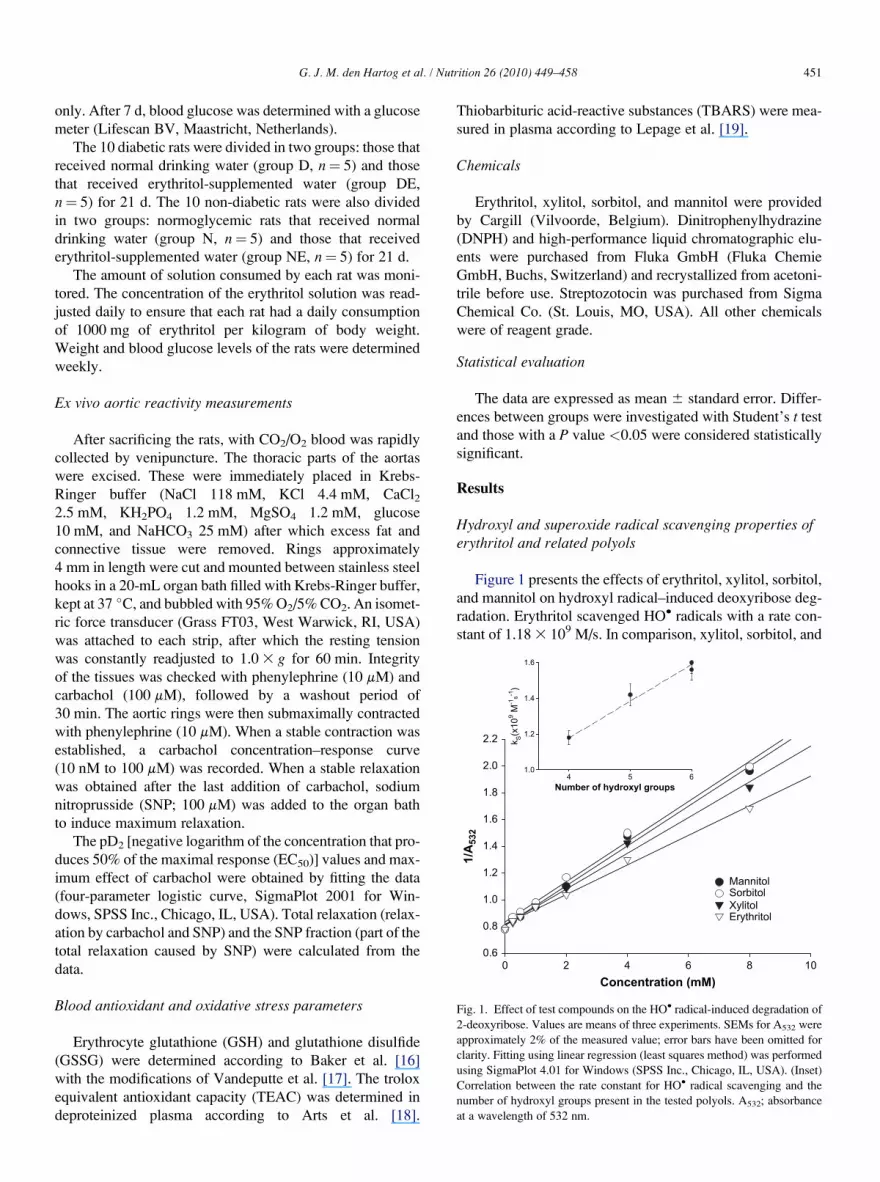
Fig. 1. Effect of test compounds on the HO� radical-induced degradation of
2-deoxyribose. Values are means of three experiments. SEMs for A532 were
approximately 2% of the measured value; error bars have been omitted for
clarity. Fitting using linear regression (least squares method) was performed
using SigmaPlot 4.01 for Windows (SPSS Inc., Chicago, IL, USA). (Inset)
Correlation between the rate constant for HO� radical scavenging and the
number of hydroxyl groups present in the tested polyols. A532; absorbance
at a wavelength of 532 nm.
G. J. M. den Hartog et al. / Nutrition 26 (2010) 449–458 451
only. After 7 d, blood glucose was determined with a glucose
meter (Lifescan BV, Maastricht, Netherlands).
The 10 diabetic rats were divided in two groups: those that
received normal drinking water (group D, n¼ 5) and those
that received erythritol-supplemented water (group DE,
n¼ 5) for 21 d. The 10 non-diabetic rats were also divided
in two groups: normoglycemic rats that received normal
drinking water (group N, n¼ 5) and those that received
erythritol-supplemented water (group NE, n¼ 5) for 21 d.
The amount of solution consumed by each rat was moni-
tored. The concentration of the erythritol solution was read-
justed daily to ensure that each rat had a daily consumption
of 1000 mg of erythritol per kilogram of body weight.
Weight and blood glucose levels of the rats were determined
weekly.
Ex vivo aortic reactivity measurements
After sacrificing the rats, with CO2/O2 blood was rapidly
collected by venipuncture. The thoracic parts of the aortas
were excised. These were immediately placed in Krebs-
Ringer buffer (NaCl 118 mM, KCl 4.4 mM, CaCl22.5 mM, KH2PO4 1.2 mM, MgSO4 1.2 mM, glucose
10 mM, and NaHCO3 25 mM) after which excess fat and
connective tissue were removed. Rings approximately
4 mm in length were cut and mounted between stainless steel
hooks in a 20-mL organ bath filled with Krebs-Ringer buffer,
kept at 37 �C, and bubbled with 95% O2/5% CO2. An isomet-
ric force transducer (Grass FT03, West Warwick, RI, USA)
was attached to each strip, after which the resting tension
was constantly readjusted to 1.0 3 g for 60 min. Integrity
of the tissues was checked with phenylephrine (10 mM) and
carbachol (100 mM), followed by a washout period of
30 min. The aortic rings were then submaximally contracted
with phenylephrine (10 mM). When a stable contraction was
established, a carbachol concentration–response curve
(10 nM to 100 mM) was recorded. When a stable relaxation
was obtained after the last addition of carbachol, sodium
nitroprusside (SNP; 100 mM) was added to the organ bath
to induce maximum relaxation.
The pD2 [negative logarithm of the concentration that pro-
duces 50% of the maximal response (EC50)] values and max-
imum effect of carbachol were obtained by fitting the data
(four-parameter logistic curve, SigmaPlot 2001 for Win-
dows, SPSS Inc., Chicago, IL, USA). Total relaxation (relax-
ation by carbachol and SNP) and the SNP fraction (part of the
total relaxation caused by SNP) were calculated from the
data.
Blood antioxidant and oxidative stress parameters
Erythrocyte glutathione (GSH) and glutathione disulfide
(GSSG) were determined according to Baker et al. [16]
with the modifications of Vandeputte et al. [17]. The trolox
equivalent antioxidant capacity (TEAC) was determined in
deproteinized plasma according to Arts et al. [18].
Thiobarbituric acid-reactive substances (TBARS) were mea-
sured in plasma according to Lepage et al. [19].
Chemicals
Erythritol, xylitol, sorbitol, and mannitol were provided
by Cargill (Vilvoorde, Belgium). Dinitrophenylhydrazine
(DNPH) and high-performance liquid chromatographic elu-
ents were purchased from Fluka GmbH (Fluka Chemie
GmbH, Buchs, Switzerland) and recrystallized from acetoni-
trile before use. Streptozotocin was purchased from Sigma
Chemical Co. (St. Louis, MO, USA). All other chemicals
were of reagent grade.
Statistical evaluation
The data are expressed as mean 6 standard error. Differ-
ences between groups were investigated with Student’s t test
and those with a P value <0.05 were considered statistically
significant.
Results
Hydroxyl and superoxide radical scavenging properties oferythritol and related polyols
Figure 1 presents the effects of erythritol, xylitol, sorbitol,
and mannitol on hydroxyl radical–induced deoxyribose deg-
radation. Erythritol scavenged HO� radicals with a rate con-
stant of 1.18 3 109 M/s. In comparison, xylitol, sorbitol, and

em
olysis (%
)
40
60
80
100
[Erythritol] (mM)
0 10 20 30 40 50
t5
0(m
in
)
120130140150160170180190200
G. J. M. den Hartog et al. / Nutrition 26 (2010) 449–458452
mannitol displayed rate constants of 1.42, 1.56, and
1.62 3 109 M/s, respectively. An interesting correlation
was observed between the number of hydroxyl groups in
the compound under investigation and its rate constant for
the reaction with HO� radicals (Fig. 1, inset).
Figure 2 shows the effect of erythritol on superoxide rad-
ical–induced reduction of NBT. Erythritol at concentrations
of up to 2 mM did not lower the NBT reduction rate. Cate-
chol, which was used as a reference compound, completely
suppressed NBT reduction at 30 mM.
Time (min)
0 50 100 150 200 250
Ha
0
20
0 mM erythritol2.5 mM erythritol5 mM erythritol10 mM erythritol20 mM erythritol50 mM erythritol
Fig. 3. Effect of erythritol (0–50 mM) on 2,20-azobis-2-amidinopropane di-
hydrochloride–induced hemolysis. Data points represent the average of three
separate experiments. Curves were fitted using SigmaPlot (three-parameter
sigmoidal). Erythritol concentration-dependently shifted the curve to the
Hemolysis assay
Figure 3 shows the effect of erythritol (0–50 mM) on 2,20-azobis-2-amidinopropane dihydrochloride–induced hemolysis.
Erythritol caused a concentration-dependent increase in lag time
(the time it takes before hemolysis starts). The lag time was
quantified by determining the time needed to obtain 50% hemo-
lysis. The relation between the increase in lag time and the eryth-
ritol concentration is displayed in the inset of Figure 3.
right. (Inset) Relation between erythritol concentration and t50. t50, timeneeded to obtain 50% hemolysis.
Electron spin resonance
Scavenging of HO� radicals by sugars probably proceeds
by abstraction of a hydrogen atom, as shown in Figure 4. This
mechanism was verified by spin trapping and analysis using
an electron spin resonance spectrometer.
Exposure of erythritol to HO� radicals in the presence of
DMPO yielded a 1:1:1:1:1:1 spectrum (Fig. 5) with hyperfine
splitting AN¼ 15.8 G, AH¼ 22.3 G. This is consistent with
generation of an adduct of a carbon-centered radical with
DMPO (DMPO-R) [20]. Also visible in this spectrum is a sig-
nal from a DMPO-OH adduct (1:2:2:1) with hyperfine split-
ting AN¼ 15.0 G, AH¼ 14.8 G which is the result of
scavenging of HO� radicals by DMPO (AN and AH are split-
ting constants due to nitrogen or hydrogen nuclei).
Concentration (μμM)
0 500 1000 1500 2000 2500
NB
T red
uctio
n (ΔA
560.m
in
-1)
0.00
0.04
0.08
0.12
0.16
MannitolSorbitolXylitolErythritolCatechol
Fig. 2. Effect of test compounds on the rate of NBT reduction in a system
consisting of xanthine and xanthine oxidase. Values are means 6 SEMs of
three experiments. NBT, nitroblue tetrazolium.
Oxidation products
According to the mechanism proposed in Figure 4, expo-
sure of erythritol to HO� radicals results in the formation of
a carbonyl group containing the products erythrose or eryth-
rulose. Figure 6 shows chromatograms of erythritol solution
before (Fig. 6B) and after (Fig. 6A) exposure to HO� radicals
followed by treatment with DNPH. Exposure of erythritol to
HO� radicals resulted in the formation of an at least 5-
carbonyl group containing DNPH reactive oxidation prod-
ucts. The two most prominent products had chromatographic
properties identical to those of the hydrazones of erythrose
[retention time (RT) 3.99 min] and erythrulose (double
peak at RT 5.09 min and RT 5.41 min).
Erythritol in the streptozotocin-induced diabetic rat
General characteristicsBlood glucose levels were increased 7 d after treatment
with streptozotocin (25.0 6 1.4 mM) compared with control
rats (6.7 6 0.2 mM).
Blood antioxidant and oxidative stress parametersAntioxidant levels and status were determined at the end
of the study in the animals by measuring erythrocyte GSH
and GSSG concentrations and the TEAC value. As presented
in Table 1, no differences among the different experimental
groups were observed in GSH and GSSG levels. In diabetic
rats, the erythritol-supplemented rats (DE group) tended to
have slightly higher GSH (172 versus 162 mM) and slightly
lower GSSG (15 versus 14 mM) levels than normoglycemic
rats that drank water (D group). The same effect of erythritol
supplementation was observed in the normoglycemic rats.

CH2OH
CH2OH
OHH
OHH
Erythritol
C
CH2OH
OHH
OHH
OHH
CH2OH
C
CH2OH
OH
OHH
+ O2
+ HO
+ O2
C
CH2OH
OHH
OHH
OHH
CH2OH
C
CH2OH
OH
OHH
O2
O2 -HO2
-HO2
CH2OH
OHH
OHH
OH
HOH2C
CH2OH
O
OHH
Erythrose
Erythrulose
Fig. 4. Proposed mechanism for the scavenging of HO� radicals by erythritol. HO� radicals attack erythritol by abstracting carbon-bound hydrogen atoms, gen-
erating two different primary erythrityl radicals. In the presence of oxygen, these carbon-centered radicals are converted into the corresponding peroxyl radicals,
which after expulsion of a protonated superoxide radical (HO2�), yield erythrose or erythrulose.
G. J. M. den Hartog et al. / Nutrition 26 (2010) 449–458 453
The TEAC did not differ among experimental groups. How-
ever, a trend was observed that the diabetic rats (D and DE
groups) displayed somewhat lower TEAC values than their
normoglycemic control counterparts (N and NE groups). In
addition to the antioxidant parameters, we assessed oxidative
damage in the rats by measuring TBARS levels in plasma.
Similar TBARS levels were found in all four experimental
groups (Table 1).
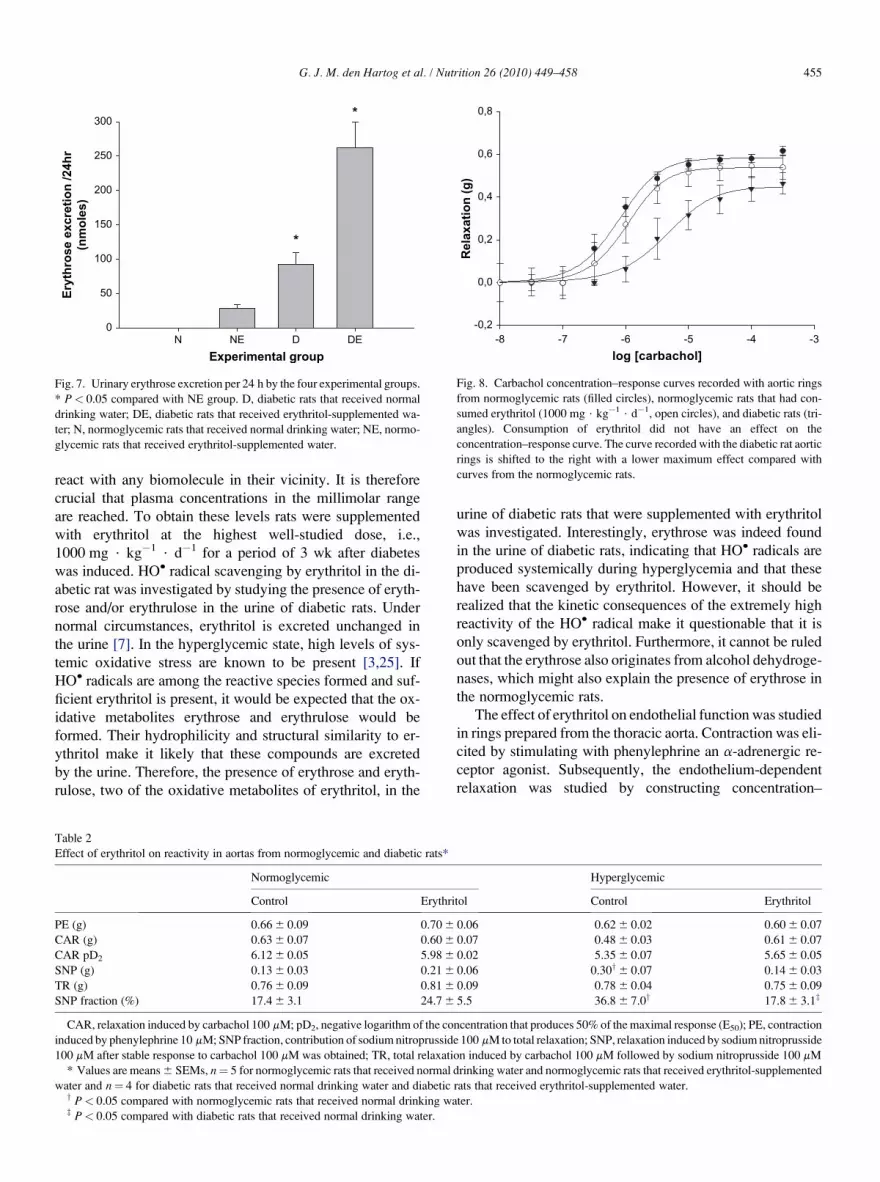
Urine production and analysisUrine from all four groups was collected for 24 h. Samples
were treated with DNPH and the amount of erythrose hydra-
zones was determined by high-performance liquid chroma-
tography. The total amount of erythrose excreted (urinary
erythrose concentration 3 urine production in 24 h) was
higher in the DE group (262 6 37 nmol) compared with
the NE group (28 6 6 nmol, P< 0.05; Fig. 7).
Endothelial functionEndothelial function was determined by measuring the re-
sponse of isolated aortic rings to phenylephrine, carbachol,
and SNP. As presented in Table 2, phenylephrine 100 mM
caused an identical contraction in the aortic rings from all
four groups. Figure 8 shows the carbachol concentration–
response curves recorded with rings from diabetic rats (D
group) and normoglycemic rats (N and NE groups). The car-
bachol concentration–response curve from aortic rings from
diabetic rats differed markedly from the curves obtained
with rings from the normoglycemic rats. The pD2 value for
carbachol was lower in the aortic rings obtained from the di-
abetic animals (Table 2). The curve was shifted to the right
and total relaxation was lowered (Fig. 8, Table 2). Figure 9
presents the carbachol concentration–response curves from
rats in the D and DE groups. Total relaxation in the aortic seg-
ments from group DE was greater, although not significantly.
The pD2 value for carbachol in the aortic rings from group
DE was higher and approached the value found in the aortic
rings of groups N and NE (Table 2).
Discussion
Erythritol is intended for use as a bulk sweetener in, e.g.,
confectionery, chewing gum, beverages, and bakery prod-
ucts. Because it does not affect glucose and insulin levels,
it is safe for diabetics. Its structural properties resemble those
of mannitol, a well-known antioxidant. Because oxidative
damage has been implicated in the pathogenesis and develop-
ment of diabetic complications, the antioxidant activity of
erythritol was investigated in vitro and in vivo.
The hydroxyl radical scavenging activity of erythritol and
other polyols such as xylitol, sorbitol, and mannitol were
evaluated in the deoxyribose assay (Fig.1). The rate constant
for the reaction was found to be close to that of mannitol. In
fact, a good correlation between the number of hydroxyl
groups in a polyol and its rate of HO� radical scavenging
was found (Fig. 1, inset). Erythritol is therefore an appealing
HO� radical scavenger that combines a high scavenging rate
constant with good bioavailability. Erythritol did not affect
superoxide-induced reduction of NBT at concentrations of
up to 2 mM. This did not come as a surprise, because eryth-
ritol does not possess a hydroxyl group attached to an
aromatic moiety, which has been shown to be a major struc-
tural requirement for superoxide scavengers [21].

10 G
X
X X
XX
X
O
O
O
O
Fig. 5. Electron spin resonance spectrum of the mixture containing H2O2
(2 mM), 5,50-dimethyl pyrroline N-oxide (50 mM), erythritol (500 mM),
and FeSO4 (1 mM) 75 s after mixing the components of the incubation. X,
peaks belonging to the carbon-centered radical adduct; O, peaks belonging
to the 5,50-dimethyl pyrroline N-oxide OH adduct.
ErythroseErythrulose
DNPH
A
B
Retention time (min)
0 2 4 6 8 10
Fig. 6. Chromatograms A (erythritol plus Fenton reagent treated with
DNPH) and B (blank, erythritol treated with DNPH). Oxidation products ob-
served in chromatogram A are erythrose (3.99 min), unknown 1 (4.85 min),
erythrulose, double peak (5.09 and 5.41 min), unknown 2 (6.73 min), un-
known 3 (8.11 min), and unknown 4 (9.31 min). The large peak visible in
both chromatograms at time 8.56 min is unreacted DNPH. DNPH, dinitro-
phenylhydrazine.
Table 1
Antioxidant and oxidative stress parameters in blood*
Experimental
group
GSH
(mmol/L)
GSSG
(mmol/L)
TEAC
(mmol/L)
TBARS
(mmol/L)
N 139 6 12 11.9 6 0.7 589 6 34 3.5 6 0.2
NE 153 6 13 10.7 6 1.1 638 6 34 3.8 6 0.2
D 162 6 7 14.8 6 1.8 561 6 53 3.7 6 0.2
DE 172 6 16 14.2 6 0.6 517 6 34 3.9 6 0.5
D, diabetic rats that received normal drinking water; DE, diabetic rats that
received erythritol-supplemented water; GSH, glutathione; GSSG, glutathi-
one disulfide; N, normoglycemic rats that received normal drinking water;
NE, normoglycemic rats that received erythritol-supplemented water;
TBARS, thiobarbituric acid-reactive substances; TEAC, trolox equivalent
antioxidant capacity
* Values are means 6 SEMs, n¼ 5 for groups N and NE and n¼ 4 for
groups D and DE.
G. J. M. den Hartog et al. / Nutrition 26 (2010) 449–458454
Because of their susceptibility to peroxidation, red blood
cells were used as a model to investigate the ability of eryth-
ritol to prevent oxidative damage in biological membranes.
Erythritol inhibited radical-induced hemolysis in a concentra-
tion-dependent manner, which indicates that erythritol is also
able to exert its antioxidant activity in a cellular system.
Schuchmann and von Sonntag [22] thoroughly investi-
gated the reaction between glucose and HO� radicals. If their
findings are applicable to the reaction of erythritol with HO�
radicals, erythrose and erythrulose would be formed as
shown in Figure 4. This scheme is supported by the findings
of Cherqaoui et al. [23] who detected these compounds after
electrolysis of an aqueous erythritol solution with platinum
electrodes. Our study provides strong evidence that exposi-
tion of erythritol to HO� radicals indeed results in the forma-
tion of erythrose and erythrulose, among two or three other,
as yet unidentified, minor oxidation products. The mecha-
nism by which erythrose and erythrulose are formed out of
erythritol appears to be analogous to the findings of
Schuchmann and von Sonntag, as shown by our spin trapping
experiment in which we confirmed the formation of a carbon-
centered radical after abstraction of a carbon-bound hydrogen
atom by a HO� radical.
The antioxidant properties of erythritol in the in vitro ex-
periments prompted us to test its antioxidant activity in dia-
betic rats. Yokozawa et al. [24] studied the effect of
erythritol on glucose metabolism and oxidative stress in
diabetic rats. Erythritol (100, 200, or 400 mg/kg of body
weight per day) showed a beneficial effect through lowering
glucose levels of serum (by 15% at the highest dose) and he-
patic and renal tissues. Our in vivo study focused on the HO�
radical scavenging activity of erythritol and the possible pre-
vention of diabetic complications. HO� radicals are ex-
tremely reactive and because of that almost instantaneously
log [carbachol]
-8 -7 -6 -5 -4 -3
Relaxatio
n (g
)
-0,2
0,0
0,2
0,4
0,6
0,8
Fig. 8. Carbachol concentration–response curves recorded with aortic rings
from normoglycemic rats (filled circles), normoglycemic rats that had con-
sumed erythritol (1000 mg $ kg�1 $ d�1, open circles), and diabetic rats (tri-
angles). Consumption of erythritol did not have an effect on the
concentration–response curve. The curve recorded with the diabetic rat aortic
rings is shifted to the right with a lower maximum effect compared with
curves from the normoglycemic rats.
Experimental group
N NE D DE
Eryth
ro
se excretio
n /24h
r
(n
mo
les)
0
50
100
150
200
250
300*
*
Fig. 7. Urinary erythrose excretion per 24 h by the four experimental groups.
* P< 0.05 compared with NE group. D, diabetic rats that received normal
drinking water; DE, diabetic rats that received erythritol-supplemented wa-
ter; N, normoglycemic rats that received normal drinking water; NE, normo-
glycemic rats that received erythritol-supplemented water.
G. J. M. den Hartog et al. / Nutrition 26 (2010) 449–458 455
react with any biomolecule in their vicinity. It is therefore
crucial that plasma concentrations in the millimolar range
are reached. To obtain these levels rats were supplemented
with erythritol at the highest well-studied dose, i.e.,
1000 mg $ kg�1 $ d�1 for a period of 3 wk after diabetes
was induced. HO� radical scavenging by erythritol in the di-
abetic rat was investigated by studying the presence of eryth-
rose and/or erythrulose in the urine of diabetic rats. Under
normal circumstances, erythritol is excreted unchanged in
the urine [7]. In the hyperglycemic state, high levels of sys-
temic oxidative stress are known to be present [3,25]. If
HO� radicals are among the reactive species formed and suf-
ficient erythritol is present, it would be expected that the ox-
idative metabolites erythrose and erythrulose would be
formed. Their hydrophilicity and structural similarity to er-
ythritol make it likely that these compounds are excreted
by the urine. Therefore, the presence of erythrose and eryth-
rulose, two of the oxidative metabolites of erythritol, in the
Table 2
Effect of erythritol on reactivity in aortas from normoglycemic and diabetic rats*
Normoglycemic
Control Erythri
PE (g) 0.66 6 0.09 0.70 6
CAR (g) 0.63 6 0.07 0.60 6
CAR pD2 6.12 6 0.05 5.98 6
SNP (g) 0.13 6 0.03 0.21 6
TR (g) 0.76 6 0.09 0.81 6
SNP fraction (%) 17.4 6 3.1 24.7 6
CAR, relaxation induced by carbachol 100 mM; pD2, negative logarithm of the co
induced by phenylephrine 10 mM; SNP fraction, contribution of sodium nitroprussid
100 mM after stable response to carbachol 100 mM was obtained; TR, total relaxati
* Values are means 6 SEMs, n¼ 5 for normoglycemic rats that received normal
water and n¼ 4 for diabetic rats that received normal drinking water and diabeticy P< 0.05 compared with normoglycemic rats that received normal drinking waz P< 0.05 compared with diabetic rats that received normal drinking water.
urine of diabetic rats that were supplemented with erythritol
was investigated. Interestingly, erythrose was indeed found
in the urine of diabetic rats, indicating that HO� radicals are
produced systemically during hyperglycemia and that these
have been scavenged by erythritol. However, it should be
realized that the kinetic consequences of the extremely high
reactivity of the HO� radical make it questionable that it is
only scavenged by erythritol. Furthermore, it cannot be ruled
out that the erythrose also originates from alcohol dehydroge-
nases, which might also explain the presence of erythrose in
the normoglycemic rats.
The effect of erythritol on endothelial function was studied
in rings prepared from the thoracic aorta. Contraction was eli-
cited by stimulating with phenylephrine an a-adrenergic re-
ceptor agonist. Subsequently, the endothelium-dependent
relaxation was studied by constructing concentration–
Hyperglycemic
tol Control Erythritol
0.06 0.62 6 0.02 0.60 6 0.07
0.07 0.48 6 0.03 0.61 6 0.07
0.02 5.35 6 0.07 5.65 6 0.05
0.06 0.30y6 0.07 0.14 6 0.03
0.09 0.78 6 0.04 0.75 6 0.09
5.5 36.8 6 7.0y 17.8 6 3.1z
ncentration that produces 50% of the maximal response (E50); PE, contraction
e 100 mM to total relaxation; SNP, relaxation induced by sodium nitroprusside
on induced by carbachol 100 mM followed by sodium nitroprusside 100 mM
drinking water and normoglycemic rats that received erythritol-supplemented
rats that received erythritol-supplemented water.
ter.

log [carbachol]
-8 -7 -6 -5 -4 -3
Relaxatio
n (g
)
-0,2
0,0
0,2
0,4
0,6
0,8
Fig. 9. Carbachol concentration–response curves recorded with aortic rings
from diabetic rats (closed circles) and diabetic rats that had consumed eryth-
ritol (1000 mg $ kg�1 $ d�1, open circles). Consumption of erythritol caused
a leftward shift of the concentration–response curve and an increased maxi-
mum effect.
G. J. M. den Hartog et al. / Nutrition 26 (2010) 449–458456
response curves with the muscarinic receptor agonist carba-
chol. In rat aorta the relaxant response to carbachol is medi-
ated by nitric oxide (NO) produced in the endothelium. NO
diffuses to the vascular smooth muscle cells, where it activates
cyclic guanosine monophosphate formation by soluble gua-
nylate cyclase, which eventually causes relaxation. Maximum
relaxation was induced by the addition of SNP, a compound
that releases NO and thus bypasses endothelial NO produc-
tion. Our in vitro determination of the endothelium-dependent
relaxation of aortic rings showed that the carbachol response
in aortic segments from diabetic rats was markedly attenuated
after 3 wk of hyperglycemia. The pD2 value and the maximum
effect were lowered compared with those in normoglycemic
rats. Total relaxation of the diabetic rings was not less com-
pared with the other groups but a larger part had to be pro-
voked by SNP, indicating that the relaxant capacity of
vascular smooth muscle was not affected in the diabetic
rats. The attenuated response to carbachol in diabetic rats
combined with the unaltered total relaxation provides strong
evidence that NO production is hampered in diabetic animals.
It is known that hyperglycemia is associated with attenuation
or loss of endothelium-dependent relaxation [26] and that re-
active oxygen species mediate this loss [25]. This reduced en-
dothelium-dependent vasodilator capacity of the coronary
and peripheral circulation to acetylcholine or other M3 recep-
tor agonists is commonly referred to as endothelial dysfunc-tion [27]. Endothelial dysfunction is thought to play an
important role in vascular diabetic complications or diabetic
vasculopathy [28] and appears to be a stepping stone for ath-
erosclerosis [29]. In diabetic rats (group D) we found an atten-
uated response to the vasorelaxant compound carbachol. The
total relaxation and the pD2 value were lower in aortic rings
from group D, whereas the total relaxant response (carbachol
plus the NO-donating compound SNP) was unchanged. This
indicates that the endothelium of the diabetic rats was dam-
aged and therefore not capable of generating sufficient NO
to induce maximum relaxation. The smooth muscle that lies
underneath the endothelium, however, appears to be intact
as evidenced by the unaltered contractile response to phenyl-
ephrine and the total relaxant response to the carbachol–SNP
combination. In the diabetic rats that were consuming
erythritol, the endothelium appeared intact, and the response
to carbachol was almost identical to that observed in the nor-
moglycemic rats. Reflecting this uncompromised response to
carbachol is the smaller SNP contribution to the total relaxa-
tion of aortic segments in the erythritol-supplemented diabetic
rats. Our results are in good agreement with those obtained by
Dhein et al. [30] who studied the effect of vitamin E supple-
mentation in the streptozotocin-induced diabetic rat. After 7
mo endothelium-dependent relaxation was found to be de-
creased by 50% in diabetic rats on a medium or high vitamin
E diet, whereas in vitamin E–deprived rats, a complete loss of
endothelium-dependent relaxation was found [30]. From
these results and the results of our study it can be concluded
that dietary antioxidants can play an important role in prevent-
ing vascular damage associated with diabetes. It is neverthe-
less intriguing that two different compounds, vitamin E and
erythritol, which differ greatly in terms of lipophilicity and
structural characteristics such as the presence of an aromatic
function, demonstrate a similar vasoprotective effect in
vivo. The lipid-soluble vitamin E owes its antioxidant activity
to trapping lipid peroxyl radicals in membranes [31]. This
contrasts sharply with the highly hydrophilic erythritol, which
will almost exclusively act in the aqueous compartments. Fur-
ther mechanistic research into the vasoprotective effect of er-
ythritol is therefore indicated. In addition to its potential
vasoprotective action, we investigated the effect of erythritol
supplementation on antioxidant levels and markers of oxida-
tive stress in blood. Surprisingly, the hyperglycemia to which
the diabetic rats were subjected did not cause detectable
changes in GSH, GSSG, and total antioxidant capacity. These
findings contrast with the larger part of data on antioxidant
levels and oxidative stress markers in diabetic patients and
laboratory animals [1,2,30,32]. This discrepancy may result
from the relatively short period (21 d) that our diabetic rats
were hyperglycemic. Their antioxidant reserves may have
been sufficient to prevent systemic oxidative damage for
that period. The observed endothelial dysfunction may be
the result of locally generated HO� radicals, which fits per-
fectly with the observation of Pennathur et al. [33] who found
that glycoxidation reactions in the arterial microenvironment
form the first step toward heart disease in diabetics. Thus, not
the generalized oxidative stress but rather the very local over-
production of HO� radicals could be responsible for the ef-
fects we observed. Pennathur et al. [33] also demonstrated
that a HO� radical-like species is implicated in diabetic vas-
culopathy by comparing oxidation products of amino acids
oxidized in vitro with those obtained from human arteries.
The protective effects of erythritol need not be restricted to
DM. Its unique free radical scavenging properties could be
beneficial in other chronic disorders in which oxygen radicals
are responsible for tissue damage. Efficient scavengers of

G. J. M. den Hartog et al. / Nutrition 26 (2010) 449–458 457
HO� radicals, namely mannitol and dimethylsulfoxide, are
used in the treatment of complex regional pain syndrome I
[34]. In a rat model of cisplatin-induced acute renal failure,
dimethylthiourea attenuated the increase in serum creatinine,
accumulation of malonaldehyde, and reduced the amount of
tubular damage [35]. Li et al. [9] showed that polyols protect
proteins from oxidation, indicating that a significant demand
for compounds that reduce HO� radical-induced damage ex-
ist. For biomedical research purposes, erythritol could be em-
ployed as a marker for endogenous HO� radical formation
during inflammatory diseases in the same fashion as antipy-
rine is currently employed [36].
In summary, the present results demonstrate that in vitro er-
ythritol is an excellent HO� scavenger with membrane-protect-
ing properties. Scavenging of HO� radicals by erythritol results
in the formation of erythrose and erythrulose. In vivo, erythritol
prevented endothelial dysfunction in diabetic rats, although it
remains difficult to attribute this to its HO� radical scavenging
properties. In addition, analytical data are presented that pro-
vide strong, although not conclusive, evidence that erythrose
and erythrulose are among the oxidative metabolites of eryth-
ritol and that erythrose was found in the urine of erythritol-
consuming diabetic rats. Safety studies have indicated that
erythritol is well tolerated and shows no signs of toxicity [7].
It is therefore an excellent sugar substitute for individuals
with DM. In this respect erythritol may help reduce the glyce-
mic impact of a food or beverage, thereby reducing the effects
of hyperglycemia-induced free radical formation. Both are
expected to reduce the onset and progression of painful and
life-threatening diabetic complications.
Conclusion
Erythritol proved to be a good HO� radical scavenger
and inhibitor of diazocompound-induced erythrocyte dam-
age in vitro. In the diabetic rat, erythritol consumption re-
sulted in unaffected endothelial function that was
accompanied by the presence of oxidative erythritol metab-
olites in the urine.
Acknowledgments
The authors gratefully acknowledge Agnieszka Brouns
for assistance with the intravenous injections.
References
[1] Gil-del Valle L, C Milian L de la, Toledo A, Vilaro N, Tapanes R,
Otero MA. Altered redox status in patients with diabetes mellitus
type I. Pharmacol Res 2005;51:375–80.
[2] Assaloni R, Da Ros R, Quagliaro L, Piconi L, Maier A, Zuodar G, et al.
Effects of S21403 (mitiglinide) on postprandial generation of oxidative
stress and inflammation in type 2 diabetic patients. Diabetologia 2005;
48:1919–24.
[3] West IC. Radicals and oxidative stress in diabetes. Diabet Med 2000;
17:171–80.
[4] Yoshida K, Hirokawa J, Tagami S, Kawakami Y, Urata Y, Kondo T.
Weakened cellular scavenging activity against oxidative stress in diabe-
tes mellitus: regulation of glutathione synthesis and efflux. Diabetolo-
gia 1995;38:201–10.
[5] Goossens J, Roper H. Erythritol: a new bulk sweetener. Int Food Ingred
1994;1–2:27–33.
[6] Bernt WO, Borzelleca JF, Flamm G, Munro IC. Erythritol: a review of
biological and toxicological studies. Regul Toxicol Pharmacol 1996;
24:S191–7.
[7] Munro IC, Berndt WO, Borzelleca JF, Flamm G, Lynch BS,
Kennepohl E, et al. Erythritol: an interpretive summary of biochemical,
metabolic, toxicological and clinical data. Food Chem Toxicol 1998;
36:1139–74.
[8] Ching TL, Haenen GR, Bast A. Cimetidine and other H2 receptor an-
tagonists as powerful hydroxyl radical scavengers. Chem Biol Interact
1993;86:119–27.
[9] Li S, Patapoff TW, Nguyen TH, Borchardt RT. Inhibitory effect of
sugars and polyols on the metal-catalyzed oxidation of human relaxin.
J Pharm Sci 1996;85:868–72.
[10] Livesey G. Health potential of polyols as sugar replacers, with empha-
sis on low glycaemic properties. Nutr Res Rev 2003;16:163–91.
[11] Halliwell B, Gutteridge JMC, Aruoma OI. The deoxyribose method:
a simple ‘test-tube’ assay for determination of rate constants for reac-
tions of hydroxyl radicals. Anal Biochem 1987;165:215–9.
[12] Kirkova M, Atanassova M, Russanov E. Effects of cimetidine and its
metal complexes on nitroblue tetrazolium and ferricytochrome c reduc-
tion by superoxide radicals. Gen Pharmacol 1999;33:271–6.
[13] Vosters O, Neve J. Inhibitory effects of thiol-containing drugs on eryth-
rocyte oxidative damages investigated with an improved assay system.
Talanta 2002;57:595–600.
[14] Nascimento RF, Marques JC, Lima Neto BS, De Keukeleire D,
Franco DW. Qualitative and quantitative high-performance liquid
chromatographic analysis of aldehydes in Brazilian sugar cane spirits
and other distilled alcoholic beverages. J Chromatogr A 1997;
782:13–23.
[15] Hasselbaink DM, Glatz JFC, Luiken JJFP, Roemen THM, van der
Vusse GJ. Ketone bodies disturb fatty acid handling in isolated cardio-
myocytes derived from control and diabetic rats. Biochem J 2003;
371:753–60.
[16] Baker MA, Cerniglia GJ, Zaman A. Microtiter plate assay for the mea-
surement of glutathione and glutathione disulfide in large numbers of
biological samples. Anal Biochem 1990;190:360–5.
[17] Vandeputte C, Guizon I, Genestie-Denis I, Vannier B, Lorenzon G. A
microtiter plate assay for total glutathione and glutathione disulfide
contents in cultured/isolated cells: performance study of a new minia-
turized protocol. Cell Biol Toxicol 1994;10:415–21.
[18] Arts MJ, Haenen GR, Voss HP, Bast A. Antioxidant capacity of reac-
tion products limits the applicability of the Trolox Equivalent Antiox-
idant Capacity (TEAC) assay. Food Chem Toxicol 2004;42:45–9.
[19] Lepage G, Munoz G, Champagne J, Roy CC. Preparative steps neces-
sary for the accurate measurement of malondialdehyde by high-perfor-
mance liquid chromatography. Anal Biochem 1991;197:277–83.
[20] Buettner GR. Spin trapping: ESR parameters of spin adducts. Free
Radic Biol Med 1987;3:259–303.
[21] van Acker SABE, van den Berg DJ, Tromp MN, Griffioen DH, van
Bennekom WP, van der Vijgh WJF, et al. Structural aspects of antiox-
idant activity of flavonoids. Free Radic Biol Med 1996;20:331–42.
[22] Schuchmann MN, Von Sonntag C. Radiation chemistry of carbohydrates.
Part 14. Hydroxyl radical induced oxidation of D-glucose in oxygenated
aqueous solution. J Chem Soc Perkins Trans II 1977;2:1958–63.
[23] Cherqaoui A, Takki D, Kokoh KB, Hahn F, Belgsir EM, Leger J-M, et al.
Electro-oxidation of meso-erythritol on platinum in acid medium: analy-
sis of the reaction products. J Electroanalyt Chem 1999;464:101–9.
[24] Yokozawa T, Kim HY, Cho EJ. Erythritol attenuates the diabetic oxi-
dative stress through glucose metabolism and lipid peroxidation in
streptozotocin-induced diabetic rats. J Agric Food Chem 2002;
50:5485–9.
[25] Gross ER, LaDisa JF, Weihrauch D, Olson LE, Kress TT, Hettrick DA,
et al. Reactive oxygen species modulate coronary wall shear stress and

G. J. M. den Hartog et al. / Nutrition 26 (2010) 449–458458
endothelial function during hyperglycemia. Am J Physiol Heart Circ
Physiol 2003;284:H1552–9.
[26] Widlansky ME, Gokce N, Keany JF, Vita JA. The clinical implications
of endothelial dysfunction. J Am Coll Cardiol 2003;42:1149–60.
[27] Pratico D. Antioxidants and endothelium protection. Atherosclerosis
2005;181:215–24.
[28] Leighton F, Miranda-Rottmann S, Urquiaga I. A central role of eNOS
in the protective effect of wine against metabolic syndrome. Cell Bio-
chem Funct 2006;24:291–8.
[29] Halcox JP, Schenke WH, Zalos G, Mincemoyer R, Prasad A,
Waclawiw MA, et al. Prognostic value of coronary vascular endothelial
dysfunction. Circulation 2002;6:653–8.
[30] Dhein S, Kabat A, Olbrich A, Rosen P, Schroder H, Mohr FW. Effect of
chronic treatment with vitamin E on endothelial dysfunction in a type I
in vivo diabetes mellitus model and in vitro. J Pharmacol Exp Ther
2003;305:114–22.
[31] Wang X, Quinn PJ. Vitamin E and its function in membranes. Prog
Lipid Res 1999;38:309–36.
[32] Ceriello A. New insights on oxidative stress and diabetic complications
may lead to a ‘‘causal’’ antioxidant therapy. Diabetes Care 2003;
26:1589–96.
[33] Pennathur S, Wagner JD, Leeuwenburgh C, Litwak KN, Heinecke JW. A
hydroxyl radical-like species oxidizes cynomolgus monkey artery wall
proteins in early diabetic vascular disease. J Clin Invest 2001;107:853–60.
[34] Goris RJA. Treatment of reflex sympathetic dystrophy with hydroxyl
radical scavengers. Unfallchirurg 1985;88:330–2.
[35] Matsushima H, Yonemura K, Ohishi K, Hishida A. The role of oxygen
free radicals in cisplatin-induced acute renal failure in rats. J Lab Clin
Med 1998;131:518–26.
[36] Meijer EP, Coolen SA, Bast A, Westerterp KR. Exercise-induced oxi-
dative stress in older adults as measure by antipyrine oxidation. Metab-
olism 2001;50:1484–8.