Embed Size (px)
Citation preview

1
Antioxidative and immunostimulatory effect of natural clinoptilolite in vivo
Kre�imir Pavelić1, Ma�a Katić1, Neven �arković1, Vi�nja �verko1, Tanja Marotti1, Marijeta
Kralj1, Kamelija �arković2, Berislav Bo�njak1, Tihomir Balog1, Ranko Stojković, Marko
Radačić, Miroslav Čolić3, Marija Poljak Bla�i1
1Rudjer Boskovic Institute, Division of Molecular Medicine, Bijenička 54, Zagreb, Croatia 2 Medical Faculty, Clinical Hospital Centre "Zagreb", Institute of Pathology, Division of
Neuropathology, Ki�patićeva 12, Zagreb, Croatia 3 Molecutec Corporation. 6512 Segovia #317; Goleta, CA 93117, USA
Address for correspondence: Prof. Dr. Kre�imir Pavelić
Ruđer Bo�ković Institute
Division of Molecular Medicine,
Bijenička 54, HR-10000 Zagreb
CROATIA
Phone: (385 1) 456 11 14
Fax: (385 1) 456 10 10
E-mail: [email protected]

2
Abstract
Many biochemical processes are closely related to ion exchange, adsorption and catalysis.
Zeolites reversibly bind small molecules such as oxygen or nitric oxide; they possess size and
shape selectivity, the possibility of metalloenzyme mimicry, and immunomodulatory activity.
These properties make them interesting for pharmaceutical industry and medicine. Our in vitro
experiments showed inhibition of tumor cell proliferation as well as micronized zeolite (MZ) to
be the possible scavenger of 4-hydroxynonenal (HNE). MZ administered by gastric intuabtionto
rats protected specifically stroma, but not tumor cells from oxidative stress caused by
doxorubicin. It also significantly reduced the number of melanoma metastases in mice. In mice
fed MZ lipid peroxidation in liver was decreased. The cells of lymph node of these mice
provoked significantly higher alogeneic graft-versus-host (GVH) reaction than cells of control
mice. After i.p. application of MZ, the number of peritoneal macrophages, as well as their
production of oxide anion, was increased. However, NO generation was totally abolished. At the
same time, translocation of NFκB p65 subunit in splenic cells was observed. Thus, here we
report anticancer, antioxidative and immunostimulatory effect of MZ and we propose a possible
mechanism of in vivo zeolite action.
Key words: micronized zeolite, clinoptilolite, scavenger, oxidative stress, antioxidative effect,
immunostimulation, T-lymphocyte, NFκB
Introduction
Zeolites are hydrated natural and synthetic microporous crystals with well-defined
structures containing AlO4 and SiO4 tetrahedra linked through the common oxygen atoms [1].
Zeolites have properties to act as catalysts, ion-exchangers, adsorbents and detergent builders [2,
3, 4, 5, 6]. Except for being extensively used in different industrial applications it is known that
silicates and aluminosilicates possess also either positive or negative biological activity. Well-
defined structures and catalytic activity make aluminosilicates an attractive model system for
protein and enzyme mimetics [7]. Recent results have demonstrated that it was very effective as a
glucose adsorbent [8] as well as a potential adjuvant in anticancer therapy [9]. Zeolites reversibly
bind small molecules such as oxygen or nitric oxide, they possess size and shape selectivity, the
possibility of metalloenzyme mimicry, and immunomodulatory activity [10].
Accumulating evidence has indicated that zeolites play an important role in modulation of
3
the immune system. It was reported that silica, silicates and aluminosilicates act as non-specific
immunostimulators similarly to superantigens [11]. Superantigens (SAG) are a class of
immunostimulatory and disease-consisting proteins of bacterial and viral origin with the ability
to activate relatively large fractions (5-20%) of the T cell population. Activation requires
simultaneous interaction of the SAG with Vβ domain of T cell complex (MHC) class II
molecules on the surface of antigen presenting cells [11]. Pro-inflammatory macrophages, that
belong to the class II MHC antigen presenting cells, are activated by fibrinogen silicate
particulate [12, 13].
It was shown that exposure of some cells to silicate particles leads to activation of
mitogen activated protein kinases (MAPK), protein kinase C and stress activated protein kinases
(SAPK) [14]. Important transcription factors such as AP-1 and NFκB are also activated and
expression of pro-inflammatory cytokines such as IL-1α, IL-6 or TNF-α was enhanced [15].
Modifications of receptor activation kinetics or activity of integrins may be responsible for the
observed behavior. Alternatively, particles engulfed by phagocytosis were shown to stimulate
production of reactive oxygen species (ROIs) that have been found to be important second
messengers for signal transduction in general [16]. Alterations in the redox homeostasis of cells
may play an important role in modulating immune functions. For example, transmembrane redox
signaling activates NFκB in macrophages and T lymphocytes [17, 18]. Nuclear factor kappa B
(NFκB)/Rel proteins are dimeric, sequence-specific transcription factors involved in the
activation of an exceptionally large number of genes in response to inflammation, viral and
bacterial infections and other stressful situations requiring rapid reprogramming of gene
expression.
Previous results showed that clinoptilolite treatment of mice and dogs suffering from
various tumor types led to improvement of the overall health status, prolongation of life span,
and decrease of tumor size in some cases. In addition, toxicology studies on mice and rats
demonstrated that the same treatment did not have any negative effect [9]. In vitro tissue culture
studies showed that finely ground clinoptilolite inhibited protein kinase B (c-Akt), induced
expression of p21WAF1/CIP1 and p27KIP1 tumour suppressor proteins, and blocked cell proliferation
in several cancer cell lines. Furthermore, tribomechanically activated clinoptilolite increased total
antioxidant status of patients with malignant diseases as well as healthy individuals (Ivković S,
�abčić D, personal communication). Here we present some new in vitro evidence for antitumour
activity of clinoptilolite as well as possible mechanism of in vivo zeolite action. Also, we report a
novel use of clinoptilolite as a potential antioxidant and stimulant of immunological response.

4
Materials and Methods
Natural clinoptilolite. A fine powder of natural clinoptilolites (MZ; micronized zeolite)
from Slovakia was obtained by tribomechanical micronization. Particle size distribution curves
of the MZ were taken by a Mastersize XLB (Malvern) laser light-scattering particle size analyzer.
Tribomechanically treated natural clinoptilolite contained approximately 80 wt % clinoptilolite.
The remaining 20% consisted of silica, montmorollonite and mordenite zeolites. Chemical
composition of clinoptilolite was SiO2 70.06%, Al2O3 12.32%, Fe2O3 1.48%, CaO 3.42% MgO
0.96%, TiO2 0.71%, P2O5 0.05%, MnO 0.02%, Na2O 0.68%, K2O 2.38%, SO3 0.17%, H2O
7.3%. Humidity at 105°C was max. 6%, pH 6.9-7.1, specific mass 2.39 g/cm3, specific area 360-
390 m2/g and NH4+ substitution capacity 8500 mg NH4
+/kg. Particle size analysis of the
clinoptilolite showed that maximum frequency of particles appeared at 1 µm.
Animals and treatment. The following animals were used: 6-month-old male Wistar rats
bearing Walker carcinoma W256, weighting 308 ± 25 g, were treated with MZ (in a single dose
of 2g/kg, by gastric intubation). C57Bl/6 4-month-old male mice bearing lung metastases were
used for antimetastatic effect of MZ. Healthy CBA mice, 3.5-month-old females, were fed
(12.5% or 25% MZ) over 28 days and used for oxidative stress parameters measurement. These
mice were afterwards donors of lymph node cells that were tested on RFM mice for cellular
immune response. CBA mice bearing mammary carcinoma were fed 14 days before tumor cell
(1x10⁵ cells/mouse) injections into the right hinder leg and were fed for additional 28 days.
Three-month-old male RFM mice were used for i.p. administration of MZ (3 mg/0.3 ml/mouse).
Control mice were injected with Hank�s solution.
Mice were bred in the Animal Facility of the Ruđer Bo�ković Institute. Food (Dom�ale,
Slovenia) and tap water were given ad libitum. Animals were kept in conventional
circumstances: slight dark rhythms 12/12 hours, temperature 22oC and humidity 55%.
In vitro assay for tumor growth. Effect of MZ on in vitro cell proliferation was studied on
several human cell lines: MiaPaCa-2 (pancreatic carcinoma), HeLa (cervical carcinoma), CaCo-2
(colon carcinoma), MCF-7 (breast cancer) and WI 38 (diploid fibroblasts). The cells were
maintained in DMEM supplemented with 10% fetal bovine serum (FBS; Sigma, USA) in
standard conditions. Thirty thousand cells/ml were plated in standard medium onto 96 microwell
plates (200 µl/well) (Greiner, Germany). After overnight incubation the medium was replaced
with the fresh DMEM containing different concentrations (0.05-0.5 mg/ml) of MZ. After 72
hours of incubation the cells were washed with PBS in order to remove MZ. The cell viability
was determined using MTT assay, which detects dehydrogenase activity in viable cell [9].

5
Statistical analysis was performed using One-way ANOVA test (p<0.05).
MTT Assay for interference with 4-hydroxynonenal (HNE). HeLa cells were cultured
under standard conditions in RPMI 1640 medium supplemented with 5% fetal calf serum (FCS;
Sigma). The cells were seeded at 2x10⁴/well density into the plastic 96-microwell plates
(Greiner). They were incubated for 2 hours before treatment with either plain medium, MZ-pre-
treated medium (0.5 µg/ml), medium with plasma at final concentration of 1% and 10% (in
saline) and HNE (10 µM) that was added 30 minutes after pre-treatment of medium, or different
combinations of already mentioned components. Concentration of HNE (10 µM) corresponds to
the aldehyde values generated during oxidative stress in vivo. The experiment was performed in
triplicate. After further 24h incubation the viability of cells (metabolic activity) was measured
using MTT assay. Substrate (10 µl) was added per well and the cells were further incubated for 2
hours when intensity of colored reaction was determined with plate reader (Anthos, Germany) at
450 nm wavelength (with 620 nm as reference).
Chemical induction of oxidative stress by doxorubicin in vivo. Rats were divided into
four groups, each comprising 4-6 animals: 1) control tumor-bearing rats; 2) tumor-bearing rats
treated with MZ p.o.; 3) tumor-bearing rats treated with doxorubicin i.p. 4) tumor-bearing rats
treated with MZ p.o. and doxorubicin i.p. Walker carcinoma tumor cells (107 live cells/rat) were
injected i.m. in the hind limb of male Wistar rats. MZ was applied six days later, when tumors
developed to 3-4 cm diameter on the average. One-hour later rats were injected i.p. with
doxorubicin (Sigma) at 10 mg/kg dose to induce oxidative stress. After three hours, animals were
killed, tumors were removed and stored in 10% buffered formalin to be used for
immunohistochemistry of HNE-protein adducts in tumour tissue.
Immunohistochemistry of HNE-protein adducts in W256 carcinoma tissue. Tissue
sections (5 µ) of the formalin-fixed paraffin-embedded tumors were used. For
immunohistochemical detection (with peroxidase-antiperoxidase method) of HNE-protein
adducts, we used specific monoclonal antibodies raised against HNE-histidine conjugate, as
described before [19]. Sections were pre-treated with normal rabbit serum and H2O2 using 1%
BSA solution as scavenger for slide washing (three times). Secondary rabbit-anti-mouse
antibodies were obtained from Dako, USA.
Evaluation of antimetastatic effect of MZ. Ten mice (C57Bl/6) were injected i.v.
with 7.5 x 10⁴ melanoma B16 cells. For the next 16 days, they were treated daily with MZ (100
mg/ml distilled H2O per mouse) by gastric intubation. Controls (6 mice) were intubated daily

6
with distilled H2O. Mice were killed and lungs were removed and fixed in Bouen. Metastases
were counted and statistical analysis was performed by Student's t-test.
Isolation of peritoneal macrophages. Peritoneal macrophages were aseptically collected
from the peritoneal cavities of mice 24 hours after i.p, or 7, 14, 21 and 28 days after per os
administration of MZ. Macrophages were resuspended in RPMI 1640 (without phenol red;
Sigma) and erythrocytes were removed by NH4Cl lysis. The remaining cells were washed three
times, suspended in RPMI 1640 supplemented with antibiotics and 10% fetal calf serum (FCS;
Sigma) and adjusted to 2 x 10⁶ cells/ml.
Assay for superoxide anion (O2-) release. In macrophages, superoxide release was
measured as superoxide dismutase (SOD) inhibitable reduction of ferricytochrome C using a
modification of the method of Johnston et al [20]. Samples contained 1 ml of cytochrome C (1
mg/ml) in phenol-free Hank's balanced salt solution and 2 x 10⁶ cells in 100 µl of medium. The
specificity of the reaction was tested by the addition of 60 IU SOD per ml of the reaction
mixture. The reactivity of the cells was tested by the addition of cytochrome C in phenol-free
Hank's solution for 30 min at 37oC. After incubation, the reaction mixture was centrifuged for 5
min at 800xg, and the absorbance of the supernatant was determined spectrophotometrically at
550 nm. The concentration of reduced cytochrome C was calculated using the formula E550nm =
2.1 x 10⁴ M-1 cm-1. Experiments were performed in duplicate and the results were expressed as
nmol O2- (106 cells)-1 (30 min)-1.
Measurement of nitrite production. The measurement of nitric oxide (NO) from
macrophages was assayed according to Naslund et al [21]. Briefly, cultures of isolated peritoneal
macrophages were incubated in plastic 24-well flat-bottom microplates (Falcon, USA) for 48
hours at 37oC and 5% CO2. Aliquots (800 µl) of each supernatant were placed in tubes and
mixed with 800 µl of GRIESS reagent (1% sulfanilamide in 2.5% phosphoric acid and 0.5%
naphthylethylenediamine in 2.5% phosphoric acid; 1:1). The resulting colorimetric reaction was
measured spectrophotometrically at 540 nm. Nitrite concentration was calculated from a standard
curve using sodium nitrite (0-100 µM) as standard.
Measurement of lipid-bound sialic acid (LSA) in serum, total sialic acid (TSA) in spleen
and assay for lipid peroxidation (LPO) in liver. After exanguination, sera from fed mice were
collected and prepared for LSA measurement according to Katopodis et al [22]. The spleen and
liver were removed from i.p. and per os treated mice. Concentration of TSA in the spleen was
determined according to Had�ija et al [23] and expressed as mg/106 spleen cells. Lipid
peroxidation (LPO) was estimated according to the presence of thiobarbituric acid reactive

7
substances (TBARS) in the liver as reported by Ohkawa et al [24]. Protein concentration was
measured by the method of Lowry, using bovine serum albumin (BSA; Sigma) as standard.
Local alogeneic graft versus host reaction. A modified version of LXGVHR described by
Shohat and Trainin [25] was used. In our experiment, LAGVHR was done on allegoric mice
instead of rats. For each experiment, 10 control mice (fed conventional food) and 10 mice in each
experimental group (mice fed 12.5% or 25% MZ, during period of 21 or 28 days) were used.
Mice were anesthetized and killed by exsanguination. Pooled lymphocytes from lymph nodes of
3 to 5 treated or control mice were washed two times with Hank's by centrifugation. Cells of
lymph nodes (2x10⁷) were injected intradermally into the shaved abdominal skin of RFM mice
(irradiated with 7Gy, 24 hours before) where they provoked the GVH reaction and damage of
skin. On day 5, the treated mice were injected intravenously with 0.4 ml of 0.5 % Evans blue.
Five hours later the entire abdominal skin was excised and two perpendicular diameters of the
blue stained area were measured with a caliper. A mean diameter of each area was calculated.
Statistical analysis was performed by the Student�s t-test.
NFκB activation in spleen. Twenty-four hours after i.p. injection of MZ into
experimental, and Hank's solution into control mice, animals were killed. For preparation of
cytoplasmic and nuclear fractions, spleens were isolated and crude splenic extracts prepared.
Erythrocytes were removed by ammonium chloride lysis. The nuclear and cytosolic fractionation
procedure was a modification of the protocol of Lernbecher et al [26]. Cells were washed twice
with phosphate-buffered saline without calcium and magnesium and resuspended in buffer A (10
mM HEPES, pH 7.9, 1.5 mM MgCl2, 10 mM KCl, 0.5 mM PMSF). After lysis on ice for 60
minutes, nuclei were spun down, and the supernatant, after additional centrifugation at 17500 g,
was stored as the cytoplasmic fraction. The nuclear pellet was suspended in buffer C (20 mM
HEPES, pH 7.9, 0.42 M NaCl, 1.5 mM MgCl2, 0.2 mM EDTA, 0.5 mM PMSF, 25% glycerol),
vortexed, and incubated on ice for 45 minutes. Centrifugation at 17500 g was performed to
remove insoluble debris. The supernatant was used as nuclear extract.
Western blot (immunoblot) analysis. Concentrations of proteins in nuclear and cytoplasm
fractions were determined by Bradford assay. Equal amounts of nuclear and cytoplasmic protein
lysates (20 µg and 80 µg respectively) were separated by 9% - SDS PAGE. Proteins were
transferred onto PVDF membrane (Immobilon-P, Millipore). Comparable loading of each protein
sample was checked by Ponceau S and Commassie blue staining. Membranes were blocked
overnight with TBS/2.5% BSA at 4oC. After that, they were incubated for 90 minutes with
primary antibodies (anti-p50, anti-RelB, and anti-p65), washed in TBS/0.05% Triton X-100, and

8
then incubated for 1 hour with appropriate secondary antibody. Following further washes,
immunoblots were visualized using enhanced chemiluminiescence reagent (POD; Boehringer-
Mannheim, Germany). For immunoblots, polyclonal antibodies against p50 and RelB (Santa
CruzTM, USA) and monoclonal antibody against p65 (Transduction Laboratories, USA) were
used. Secondary antibodies were peroxide-conjugated rabbit anti-mouse immunoglobulin
(Amersham/Pharmacia, Sweden) and peroxide-conjugated protein A from Kierkegaard and Perry
Laboratories.
Results
Effect of MZ on tumor cell growth in vitro. The results, showing cell growth inhibition of
HeLa, MiaPaCa-2, CaCo-2, MCF-7 and WI 38 cells, are presented in Figure 1. The growth of all
tumor cell lines was significantly inhibited. The strongest and dose-dependent inhibition was
observed on HeLa and MiaPaCa-2 cells (30-50% and 15-65%, respectively). CaCo-2 and MCF-7
cells were inhibited only at the highest MZ concentration. The growth of WI 38 normal (diploid)
fibroblast cell line was even stimulated (about 20%).
Effect of MZ on 4-hydroxinonenal. The results of the experiment evaluating influence of
MZ on the biological effects of HNE in vitro are presented in Table 1. MZ increased MTT values
for the cells cultured in the absence of plasma (p<0.05), but not if the cells were cultured also
with HNE.
MZ did not influence stimulation of the cell growth caused by 1% plasma addition. On
the other hand, in the presence of 1% plasma it abolished inhibiting effects of HNE (p<0.05). In
the presence of 10% plasma neither MZ nor HNE showed any effects.
Effects of MZ on chemically induced oxidative stress of W256 carcinoma in vivo. Results
of pathohistological evaluation of W256 carcinoma treated either with doxorubicin or MZ, or
combined treatment are summarized in Table 2 and presented in Figure 2.
Although there were no particular differences in general appearance of differently treated
tumors, doxorubicin induced prominent lipid peroxidation (generation of HNE-protein
conjugates), and both in malignant cells as well as in normal stromal components of tumor tissue.
MZ itself did not change immunohistochemical appearance of 4-hydroxynonenal (HNE-protein
conjugates) compared to untreated tumour-bearing rats. However, it had selective, tissue specific
effects on doxorubicin-induced oxidative stress (lipid peroxidation within tumour). Namely,
when MZ was applied, it did not influence doxorubicin-induced oxidative stress (HNE-protein
conjugates) in malignant cells, while it completely prevented formation of HNE-protein

9
conjugates in tumor stroma. Specificity of the anitoxidative effects of MZ for tumor stroma was
also supported by the finding that MZ did not influence the presence of HNE-protein conjugates
in necrotic tumor tissue.
Antimetastatic effect of MZ. MZ strongly reduced the number of lung metastasis in treated
mice. While control mice had 5.2 ± 1.64, treated mice had 0.7 ± 1.06 metastasis. Statistical
significance was p<0.001.
Influence of MZ on macrophage O2- production, lipid peroxidation (LPO) in liver and
lipid-bound sialic acid (LSA) in serum. The results are shown in Table 3. Concentration of O2-
(in peritoneal macrophages) started to change slightly 14 days after administration of 12.5% MZ.
However, TBARS concentration (in liver) started to change significantly after 21 days regardless
of MZ concentration. Significant differences regarding concentration of LSA (in serum) was
obtained with 12.5% MZ on day 21 and 25% MZ on day 28.
In the experiment with mammary carcinoma, CBA mice were divided into four groups: 1)
control; 2) tumor-bearing mice (T); 3) tumor-bearing mice fed 12.5% MZ and 4) tumor-bearing
mice fed 25% MZ. Each group consisted of 16 mice. Neither 12.5 nor 25% MZ reduced tumor
volume during 28 days after tumour injection. Concentration of TBARS (measured in 5 of
survived tumor-bearing mice) was significantly increased compared to healthy control. However,
administration of 25% MZ for 28 days decreased TBARS to the control value (Figure 3A). LSA
concentration was significantly increased in tumor-bearing mice compared to controls and MZ
did not influence it additionally (Figure 3B).
Effect of MZ on local alogeneic graft versus host reaction. Results of two separately
prepared experiments with healthy mice 21 and 28 days after administration are shown in Figure
4. The cells of lymph node of mice fed 28 days with 25% MZ provoked significantly higher
GVH reaction than cells of control group mice. A treatment with lower dose (12,5%) of MZ for
21 or 28 days showed also the higher reaction than control group, but was not significant.
Effect of i.p. administration of MZ on peritoneal macrophages, ROIs generation and
oxidative stress (OS) parameters. Intraperitoneal administration of MZ at doses higher than 3 mg
was lethal for mice and dose of 3 mg was sublethal but proinflammatory (data not shown). In our
experiments, dose of 3 mg was used and the number of macrophages, production of O2- and NO
as well as measurement TSA in spleen and TBARS concentration in liver was performed 24
hours after intraperitoneal injection of MZ.
MZ provoked accumulation of macrophages in peritoneum. The number of peritoneal
macrophages (PM) after treatment was 7 times higher than in control mice (Figure 5A). The

10
concentration of O2- was 10 times higher in macrophages of treated mice than in controls (Figure
5B). Since O2- release was calculated to 106 cells, the increased release was not the result of
increased number of macrophages, but represents truly increased activity. Production of NO by
peritoneal macrophages isolated from treated mice, and cultivated for another 24 hours ex vivo,
was strongly decreased (Figure 5C). There was no change in liver TBARS concentrations
(expressed in nmol/mg protein) between control (0.907 ± 0.17) and treated (0.886 ± 0.16) group.
Also, TSA concentration (expressed as µg/106 splenocytes) was not changed after treatment with
MZ. The value of control group was 5.25 ±0.73, and 5.58±0.78 for treated group.
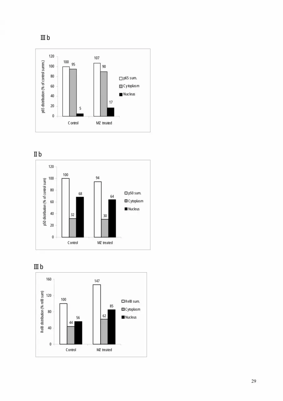
Effect of MZ on NFκB/Rel proteins. The spleens of MZ treated animals were slightly
(11%) heavier than spleens of control animals (data not shown). Effect of MZ on NFκB/Rel
proteins could be seen after preparation of cytoplasm and nuclear fractions, electrophoresis and
Western blotting. MZ treatment increased translocation of p65 subunit into nucleus (Figure 6)
suggesting that NFκB containing p65 subunit had been activated. However, MZ treatment did
not have any effect on RelB or p50 subunit concerning translocation into the nucleus. However,
total amount of RelB subunit was increased by about 40% and total amount of p50 was slightly
decreased compared to controls.
Discussion
Antitumour effect of MZ in vitro. Previous in vitro and in vivo animal studies showed that
MZ was a non-toxic compound that acted as an adjuvant in anticancer treatment [9]. Previous in
vivo results showed diverse effects, ranging from no antitumour response, to normalization of
biochemical parameters and prolongation of life span.
Our previous in vitro experiments [9] showed 30-50% inhibition of proliferation (by
MTT as well as 3H-tymidine test) of several cell lines after incubation with MZ-pre-treated
medium. Now we used another approach: instead of MZ-pre-treated medium, we added MZ
directly to the culture medium. The cell proliferation was also inhibited in a dose-dependent
manner. The best and the most consistent results were obtained with HeLa and MiaPaCa-2.
However, the proliferation of WI 38 cell line was even slightly stimulated by MZ. According to
these experiments, our conclusion is that MZ response was cell line dependent. We previously
analyzed mitogenic and survival signaling pathways in tumor cells [9]. The most significant
results were observed in the activity of Akt protein that was highly inhibited after MZ treatment
of cancer cells. That resulted in growth inhibition and increase in apoptosis of cancer cells, but
only in the presence of serum. We suppose that the same mechanism could be responsible for

11
results obtained with direct application of MZ. However, since MZ was constantly present in the
medium, it probably enabled lower concentrations of MZ to have the same (or even better)
inhibitory effects on tumor cells.
Absorption of serum components, at least in vitro, could be one of the mechanisms of MZ
action. In favor of this assumption are also preliminary findings of an increased HNE-protein
conjugate formation in the presence of MZ (data not presented). MZ might further act as HNE-
scavenger but HNE binding to MZ might also affect activity of MZ. HNE has high affinity of
binding to proteins so plasma (serum) or albumin can attenuate its toxicity. HNE can also act as a
bifunctional growth regulating factor, probably by physical or biological interference of the
aldehyde and MZ as well as interfering with the activity of humoral growth factors [27]. This
might be also relevant for its in vivo effects.
Since our in vitro results referred to indirect effect on tumor cell lines, orally
administrated MZ is not absorbed into blood, and that which does get into the body will be
phagocytized by macrophages, the MZ effect in vivo cannot be due to a direct biochemical
interaction. We speculate that MZ may induce certain immunological responses. To try to reveal
a possible mechanism of MZ in vivo effect, we used different ways of application of MZ either to
healthy or tumor-bearing mice as well as to tumor-bearing rats.
Antioxidative effect of MZ in vivo. Cancer and inflammation can be considered as
interfering processes that share two common pathophysiological mechanisms: cytokine network
and oxidative stress. Both of these processes involve lipid peroxidation and are linked by tumour
stroma. TNF-α and TGF-β are among cytokines that are likely to be mediators of this complex
network system, those interfering with lipid peroxidation and consequently biological effects of
HNE (known as a second messenger of free radicals). Growth modulating effects of HNE
involve signaling pathways affecting c-fos expression and might interfere with activity of EGF
and PDGF [28]. HNE as second messenger of radical oxygen intermediates (ROIs) activates AP-
1, followed by further TGF-β synthesis and fibrogenesis (spread of connective tissue). Moreover,
low-density lipoprotein modified by lipid peroxidation (oxLDL) is known as a potent cytotoxic,
immunogenic and pro-inflammatory factor, which activates production of cytokines by stromal
cells (fibroblasts, endothelium, and macrophages). This leads to further oxidative stress and
spread of inflammation associated by further lipid peroxidation and production of HNE [29].
Doxorubicin is a very potent inducer of hydroxyl radical and consequential lipid peroxidation
(resulting in HNE production), the effects of which could be demonstrated in vivo already half an
hour after administration [30, 31]. Thus, we assume that applying MZ in water by gastric

12
intubation could in a relatively short period of time (several hours as done in experiment
presented) provoke response of the gastrointestinal system, which might further induce a
systemic response. Regarding specificity of MZ antioxidant effect on stromal cells in
doxorubicin-treated W256 carcinoma, we assume that differential response of malignant and
normal cells to antioxidative effects of MZ could be due to: 1) different patterns of oxygen
metabolism between these cells (internal oxidative stress), 2) the difference in their response to
cytokines involved in determination of tumor host relationship, 3) systemic response to MZ
application and doxorubicin-induced oxidative stress.
Tumor cells have an abnormal lipid composition and level of enzymes of the cytochrome
P450 system which can initiate and propagate LPO and thus may cause alterations in LPO level
[32]. In our experiment, the statistically increased LPO in mice with mammary carcinoma was
abolished in animals that were fed a diet supplemented with 25% of MZ. According to some
data, the level of oxyradical scavenging enzymes is reduced in tumor cells [33]. Thus, the
reduced LPO level in MZ treated tumor-bearing mice might be the result of the MZ antioxidant
potential. These data show, to the best of our knowledge, the first time this effect on lipid
peroxidation.
Immunostimulatory effect of MZ in vivo. Immunostimulators are associated with an
increase of serum sialic acid level [34]. Sialic acid also may be a useful indicator for the
diagnosis and staging of malignancy, and it may have a regulatory role in immunological
processes, particularly masking the antigen site and being a marker for inflammation. The data
presented here demonstrated that application of MZ increased the serum LSA concentration in
healthy mice, which is probably associated with inflammatory process, i.e. activation of
macrophages. This was confirmed by our data of elevated O2- in MZ fed mice. We suspect that
factors activating and influencing the proliferation or increasing the synthetic capacity of the
phagocyte system might cause a change in the serum LSA level. It is also possible that
macrophages participate in this process indirectly by releasing TNF-α and interleukin-1 or is
connected with an elevation of other acute phase proteins.
Although parentheral route of application is not suitable, to confirm immunomodulatory
effect of MZ we examined the processes that followed MZ intraperitoneal application. For that
purpose, different amounts of MZ were injected in normal, healthy mice. It was shown that effect
of MZ was dose dependent. Doses higher than 3mg/mouse were lethal. However, since lower
doses (3mg/mouse) had non-toxic, but proinflammatory effect, some immunological parameters
were measured. In the acute phase of an inflammatory process, large number of

13
polymorphonuclear leukocytes (PMNs) migrates from the blood and accumulates in the exudate
[35]. In our experiments, 24 hours after MZ administration, high accumulation of macrophages
was found in the peritoneum of treated animals. Our results confirmed that macrophages of
treated mice were activated since they generated 9 times higher amount of O2- than those of
control mice.
Normally, NO react rapidly with O2- and the reaction is completed in less than 1 µsec
[36]. Therefore, any NO produced under aerobic conditions is converted rapidly to peroxynitrite
anion. Peroxynitrite anion is a strong oxidant with bactericidal activity. At physiological pH it is
protonated to form peroxinitrous acid, a relatively long-lived strong oxidant that could initiate
oxidation of lipids. This could explain the toxic (at higher doses) and inflammatory (at lower
doses) effect of MZ administrated in peritoneum. Also, the observed depletion of NO from
peritoneal macrophages might have significantly increased superoxide generation and in this way
could have intensified the effect of MZ. Since concentration of TBARS in liver and TSA in
spleen of treated mice remained the same; this is obviously a local reaction.
Phagocytosis per se or reactive oxygen species (ROS) can stimulate macrophages to
secrete TNF-α and other cytokines that normally stimulate immunological response [37]. One
ubiquitous transcription factor of particular importance in immune and inflammatory responses is
nuclear factor kappa B (NFκB) [38]. Therefore, we wanted to examine the activation of NFκB in
splenocytes of MZ treated mice. Our results showed MZ induced translocation of p65 to the
nucleus of RFM mice spleen cells. This finding suggests that MZ acts as immunoactivator,
activating NFκB, and therefore inducing transcription of genes regulated with NFκB.
Decreased amount of p50 and increased amount of RelB proteins in treated compared to
control mice could be due to changed number and/or ratio of B- and T-lymphocytes. The fact that
spleens of treated mice were 11% heavier contributes to that assumption. B-lymphocytes have a
basal level of p50 homodimer that is not inducible upon stimulation [39] and/or serve as
regulators of NFκB activity [40]. Decreased total amount of p50 protein, as well as it not being
translocated into the nucleus, could imply that B-lymphocytes were not stimulated by MZ. Also,
RelB/p50 heterodimer is constitutively active in primary lymphoid cells and its presence
correlates with constitutive lymphoid-specific transgene expression of genes involved in B- and
T-cell development [26, 41]. This explains our finding that MZ treatment did not have any (or
had only a slight) effect on translocation of RelB subunit into the nucleus of RFM mice spleen
cells. These facts also explain a relatively high basal amount of p50 and RelB proteins in nuclei
of control splenocytes.

14
While B-lymphocytes and several other cells exhibit both constitutive and stimulated
NFκB activation, only inducible NFκB activity has been described in T-cells or T cell lines [42].
However, as complete T-cell activation requires at least two signals provided by the T-cell
receptor (TCR) complex and another stimulatory molecule, optimal NFκB activation in the T-
cell is also dependent on dual signaling mechanisms [43]. Many agents have been shown to
promote activation of NFκB in T-cells including TNF-α [44], calcium ionophores [18] and H2O2
[42]. However, maximal NFκB activation has been observed in response to combinations of
stimulants which fulfil the dual signaling requirements of T-cells [43, 45]. ROS have been found
to act as second messengers in activation of NFκB/Rel proteins [42] and oxidative stress can
modulate the activity of NFκB in T-cells [18]. In addition, previous results have shown that
NFκB is activated in inflammatory diseases. Therefore, according to all these as well as to our
results, we concluded that MZ, in our experiments, activated T-cell immunological (cellular)
response, that could be involved in anticancer effect of MZ in vivo.
Possible mechanism of MZ in vivo action. We propose a mechanism for MZ in vivo
action (Figure 7). MZ causes local inflammation at the place of application, e.g. peritoneum.
Macrophages will be attracted and activated, which has been shown with increased O2-
production. We suggest that activated macrophages produce TNF-α that, together with some
other stimulants (e.g. other cytokines, ROS or changed calcium concentration), stimulate splenic
T-cells. Since products of the genes that are regulated by NFκB also cause its activation, this type
of positive regulatory loop may amplify and perpetuate the local inflammatory response. Our
hypothesis is that MZ will act the same way after per os administration, affecting intestinal
macrophages. Results of experiments with local alogeneic graft versus host reaction, as well as
strong reduction of immunogenic melanoma B16 lung metastasis, support this hypothesis. This is
in agreement with accumulating evidence that zeolites could play an important role in modifying
the immune system as well as with the report that silica, silicates and aluminosilicates act as non-
specific immunomodulators similarly to superantigens.
To additionally confirm the hypothesis, TNF-α in serum should be measured, as well as
activation of NFκB in macrophages, and B- and T-lymphocytes separately.

15
References
1. Breck DW (1964) Christalline molecular sieves. J Chem Educ 41: 678-689
2. Flanigen EM (1980) Molecular sieve zeolite technology-the first twenty-five years. In: Rees
LVC (ed) Proc 5th Int Conf Zeolites. Heyden, London, Philadelphia, Rheine, pp 760-780
3. Sersale R (1985) Natural zeolites processing, present and possible applications. Stud Sturf Sci
Catal 24: 503-512
4. Naber JE, De Jong KP, Stork WHJ, Kuipres HPCE, Post MFM (1994) Industrial application
of zeolite catalysis. Stud Sturf Sci Catal 84C: 2197-2220
5. Garces JM (1999) Observations of zeolite applications. In: Treacz MMJ, Marcus BK, Misher
ME, Higgins JB (eds) Proc 12th Int Conf Zeolites. Materials Research Society, Warrendale, PA,
pp 551-566
6. Colella C (1999) Natural zeolites in environmentally friendly processes and applications. Stud
Sturf Sci Catal 125: 641-655
7. Bedioui F (1995) Zeolite-encapsulated and clay-intercalated metal phorphyrin phthalocyanine
and schiff-base complexes as models for biomimetic oxidation catalysts: an overview. Coord
Chem Rev 144: 39-68
8. Concepcion-Rosabal B, Rodrigues-Fuentes G, Simon-Carballo R (1997) Development and
featuring of the zeolitic active principle FZ: a glucose adsorbent. Zeolites 19: 47-50
9. Pavelic K, Hadzija M, Bedrica Lj, Pavelic J, Dikic I, Katic M, Kralj M, Herak Bosnar M,
Kapitanovic S, Poljak-Blazi M, Krizanac S, Stojkovic R, Jurin M, Subotic B and Colic M (2001)
Mechanically treted natural clinoptilolite zeolite - new adjuvant agent in anticancer therapy. J
Mol Med, in press
10. Ozesmi M, Karlsson-Parra A, Hillerdal G and Forsum V (1986) Phenotypic characterisation
of peripheral blood lymphoid cells in people exposed to fibrous zeolite Br J Ind Med 43: 830-833
11. Ueki A, Yamguchi M, Ueki H, Watanabe Y, Ohsawa G, Kinugawa K, Kawakami Y, Hyodoh
F (1994) Polyclonal human T cell activation by silicate in vitro. Immunology 82: 332-335
12. Drumm K, Oettinger R, Smolarski R, Bay M, Kienast K (1998) In vitro study of human
alveolar macrophages inflammatory mediator transcriptions and releases induced by soot FR 101,
Printex 90, titandioxide and chrysotile B. Eur J Med Res 3: 432-438
13. Allison AC, Harrington JS, Birbeck M (1996) An examination of the cytotoxic effects of
silica on macrophages. J Exp Med 124: 141-154

16
14. Lim Y, Kim SH, Kim KA, Oh MW, Lee KH (1997) Involvement of protein kinase C,
phospholipase C, and protein tyrosine kinase pathways in oxygen radical generation by asbestos-
stimulated alveolar macrophages. Environ Health Perspect 105 (Suppl. 5): 1325-1327
15. Simeonova P, Torium W, Kommineni C, Erkan M, Muson AE, Rom WN, Luster MI (1997)
Molecular regulation of IL-6 activation by asbestos in lung epithelial cell-role of reactive oxygen
species. J Immunol 159: 3921-3928
16. Martin LD, Krunkosky TM, Dye JA, Fischer BM, Jiang NF, Rochelle LG, Akley NJ, Dreher
KL, Adler KB (1997) The role of reactive oxygen and nitrogen species in the response of airway
epithelium to particulates. Environ Health Perspect 105 (Suppl. 5): 1301-1307
17. Kaul N, Choi J, Forman HJ (1998) Transmembrane redox signalling activates NF-κB in
macrophages. Free Radic Biol Med 24: 202-207
18. Gin-Pease ME, Whisler RL (1998) Redox signals and NF-κB activations in T cells. Free
Radic Biol Med 25: 346-361
19. �arković K, �arković N, Schlag G, Redl H, Waeg G (1997) Histological aspects of sepsis-
induced brain changes in a baboon model. In: Schlag G, Redl H, Traber DL (eds.) Shock, Sepsis
and Organ Failure, 5th Wiggers Bernard Conference. Springer-Verlag, Heidelberg, pp 146-160
20. Johnston RB, Godzik CA, Cohn ZA (1978) Increased superoxide anion production by
immunologically actived and chemically elicited macrophages. J Exp Med 148: 115-119
21. Naslund PK, Miller WC, Granger DL (1995) Cryptococcus neoformans fails to induce nitric
oxide synthase in primed murine macrophage-like cells. Infec Immun 63: 1298-1304
22. Katopodis N, Hirshaut Y, Geller NL, Stock C (1982) Lipid-associated sialic acid test for the
detection of human cancer. Cancer Res 42: 5270-5275
23. Had�ija M, Lipovac K, Gavella M, Ročić B, Slijepčević M (1992) Concentration of sialic
acid in rats with diabetes. Cell Mol Biol 38: 613-619
24. Ohkawa H, Ohiski N, Yagy K (1979) Assay for lipid peroxides in animal tissues by
thiobarbituric acid reaction. Anal Biochem 95:351-358
25. Shohat B, Trainin N (1980) The local xenogeneic graft-versus-host reaction as a clinical test
for immunocompetence of human T lymphocytes. Thymus 2: 93-105
26. Lernbecher T, Muller U, Wirth T (1993) Distinct NFκB/Rel transcription factors are
responsible for tissue-specific and inducible gene activation. Nature 365: 767-770
27. Zarkovic N, Schaur RJ, Puhl H, Jurin M, Esterbauer H (1994) Mutual dependence of growth
modifying effects of 4-hydroxy-nonenal and fetal calf serum in vitro. Free Radic Biol Med 16:

17
877-884
28. Kreuzer T, Grube R, �arković N, Schaur RJ (1998) 4-Hydroxynonenal modifies the effects of
serum growth factors on the expression of the c-fos proto-oncogene and the proliferation of HeLa
carcinoma cells. Free Radic Biol Med 25: 42-49
29. Poli G, Parola M (1997) Oxidative damage and fibrogenesis. Free Radic Biol Med 22: 287-
305
30. Floyd RA, Henderson R, Watson JJ, Wong PK (1986) Use of salycilate with high pressure
liquid chromatography and electrochemical detection (LCED) as a sensitive measure of hydroxyl
free radicals in adriamycin treated rats. J Free Radiol Biol Med 2: 13-17
31. Myres C (1987) Anthracyclines. In: Pined HM, Longo DL, Chabner BA (eds.) Cancer
chemotherapy and biological response modifiers. Annual 9, Elsevier, Amsterdam, pp 36-49
32. Gonzales MJ (1992) Lipid peroxidation and tumor growth: an inverse relationship. Medical
Hypothesis 38: 106-110
33. Sun Y (1990) Free radicals, antioxidant enzymes, and carcinogenesis. Free Rad Biol Med 8:
583-599
34. Sydow G, Sydow H, Rucker K (1989) Factors affecting serum sialic acid levels. Biomed
Biochim Acta 48: 365-369
35. Hambleton P, Miller P (1989) Studies on the carrageenin air pouch inflammation in the rat.
Br J Exp Path 70: 425-443
36. Huie RE, Padmaja S (1993) The reaction of NO with superoxide. Free Rad Res Comns 18:
195-199
37. Chaudhri G, Clark IA (1989) Reactive oxygen species facilitate in vitro and in vivo
lipopolysaccharide-induced release of tumour necrosis factor. J Immunol 143: 1290-1294
38. Kopp EB, Ghosh S (1995) NF-kappa B and rel proteins in innate immunity. Adv Immunol
58: 1-27
39. Liou H-C, Sha WC, Scott ML, Baltimore D (1994) Sequential induction of NFκB/Rel family
proteins during B-cell termianl differentiation. Mol Cell Biol 14: 5349-5359
40. Kang SM, Tran AC, Grill M, Leonardo MJ (1992) NFκB subunit regulation in
nontransformed CD4+ T lymphocytes. Science 256: 1452-1456
41. Lernbecher T, Kistler B, Wirth T (1994) Two distinct mechanisms cotribute to the
constitutive activation of Rel B in lymphoid cells. EMBO J 13: 4060-4069

18
42. Schreck R, Rieber P, Bauerle PA (1991) Reactive oxygen intermediates as apparently widely
used messengers in the activation of the NF-kappa B transcription factor and HIV-1. EMBO J.
10: 2247-2258
43. Crabtree GR, Clipstone NA (1994) Signal transmission between the plasma membrane and
nucleus of T lymphocytes. Ann Rev Biochem 63: 1045-1083
44. Menon SD, Guy GR, Tan YH (1995) Involvement of a putative protein-tyrosine phosphatase
and I kappa B-alpha serine phosphorylation in nuclear factor kappa B activation by tumor
necrosis factor. J Biol Chem 270: 18881-18887
45. Kanno T, Siebenlist U (1996) Activation of nuclear factor kappa B via T cell receptor
requires a Raf kinase and Ca2+ influx. Functional synergy between Raf and calcineurin. J
Immunol 157: 5277-5283
Acknowledgment Dr. Georg Waeg, Institute of Biochemistry, Graz, Austria, kindly provided monoclonal antibodies for HNE-immunohistochemistry.

19
Figure legend
Figure 1. The effect of MZ treatment on growth of different human cell lines: 0.05 mg/ml, 0.25
mg/ml and 0.5 mg/ml. The results are presented as percentage of the growth of control cells.
* indicates significant difference compared to control cells (ANOVA, p<0.05).
Figure 2. Influence of MZ on oxidative stress in vivo. Immunohistological distribution of HNE-
protein conjugates in W256 carcinoma. Control tumor (upper left panel): anaplastic tumor
infiltrates skeletal muscle; only occasionally, near muscle infiltration, malignant cells show mild
(yellow) HNE presence in cytoplasm, while stroma is negative. MZ treated tumour (upper right
panel): while necrosis of tumor tissue (right and left top side) contains several slightly HNE-
positive cells, stromal as well as tumor cells remote from necrosis are HNE-negative.
Doxorubicin treated tumour (lower left panel): almost all tumour as well as stromal cells show
very strong membrane and cytoplasmic HNE immunopositivity. Doxorubicin and MZ treated
tumour (lower right panel): while all tumor cells show diffuse HNE immunopositivity, stroma is
entirely negatively stained. Magnification 400 x.
Figure 3. Concentration of TBARS in liver and LSA in serum of control and tumour-bearing
mice as well as tumor-bearing mice fed with either 12.5% or 25% MZ.
Figure 4. Effect of MZ on alogeneic graft versus host reaction. The GVH reaction of lymph node
cells of control (empty column), mice fed with 12.5% (gray column) and 25% (black column)
MZ were tested.
Figure 5. Number of peritoneal macrophages per mouse (A), superoxide generation in
macrophages (B) and nitric oxide concentration (C) in peritoneal macrophages ex vivo 24 hours
after i.p. treatment with 3 mg MZ/mouse. There were 18 control and 21 mice treated with MZ in
two experiments.
Figure 6. Effect of MZ on level and distribution of p65 (I), p50 (II) and RelB (III) subunits of
NFκB. (a) Western blot analysis of NFκB subunits in nuclear and cytoplasm fractions. C
indicates control groups of animals, whereas MZ indicates treated animals. (b) Densitometric
quantification of signal intensity. Total level indicates the sum of signal intensities of cytoplasm
and nuclear fraction in each group (C and MZ). Results are expressed as percentage of each
subunit compared to their total level in controls.
Figure 7. Clinoptilolite-induced stimulation of cellular immune response; proposed mechanism
of MZ action in vivo.

20
Table 1. Influence of MZ on the biological effects of HNE in vitro.
Without plasma 1% plasma 10% plasma
HNE - MZ + MZ - MZ + MZ - MZ + MZ - 24 51a 90 81 100 108 + 42 a 38 a,b 70 a 91 c 98 105
The results are expressed as percentages of the viability of the control group (with 10%
plasma, without MZ and HNE). Significant (Student's t-test, p<0.05) if compared (absolute
values) with the cells cultured: awithout HNE and without MZ, bwithout HNE, but with MZ, cwithout MZ but with HNE, in experiment where plasma was absent as well as in experiments
with 1% or 10% plasma.

21
Table 2. Immunohistochemical findings of HNE distribution in Walker 256 carcinoma tissue.
HNE positivity in:
Treatment
Animal No.
tumor cells
tumor cells
near necrosis
tumor cells within zone
of tumor spreading
stroma
necrosis
1 + ++ - ++ + 2 - + - - +
Saline control 3 - + - - + 4 - + - ++ - 5 - - - - - 6 - - + - ++ 7 + ++++ + + ++
Doxorubicin 8 +++ ++++ +++ +++ ++ 9 +++ ++++ +++ +++ +++ 10 ++ +++ ++ ++ ++ 11 - +++ ++ - ++ 12 - ++ - - +
MZ 13 + + +++ - ++ 14 - - ++ - ++ 15 - - - ++ - 16 + + + - +++
MZ + 17 ++ ++ +++ - +++ Doxorubicin 18 +++ +++ ++++ - ++
19 ++ ++ +++ - ++ (-) negative; (+) weak positivity (<5 % of the cells); (++) moderate staining approximately 25 % of the cells); (+++) strong staining (>50-75 % of the cells)

22
Table 3.
Influence of MZ on O2- production, lipid peroxidation (TBARS) and lipid bound sialic acid
(LSA) in healthy mice
Days 7 14 21 28
O2 Control 3.10 ± 0.14 1.50 ± 0.23 0.45 ± 0.07 1.20 ± 0.9
nmol/2x106 12.5% MZ 3.95 ± 0.8 0.68 ± 0.25* 2.55 ± 0.21* 0.55 ± 0.07*
macrophages 25% MZ 3.20 ± 0.56 0.95 ± 0.35 0.57 ± 0.39 1.10 ± 0.14
TBARS Control 0.97 ± 0.11 0.94 ± 0.22 1.89 ± 0.18 1.78 ± 0.09
nmol/mg 12.5% MZ 0.95 ± 0.11 0.72 ± 0.04 1.20 ± 0.29* 1.16 ± 0.08*
liver proteins 25% MZ 0.94 ± 0.13 0.61 ± 0.09 0.85 ± 0.11* 0.85 ± 0.12*
LSA Control 0.18 ± 0.07 0.26 ± 0.11 0.29 ± 0.03 0.27 ± 0.03
nmol/l 12.5% MZ 0.29 ± 0.13 0.25 ± 0.04 0.42 ± 0.06* 0.46 ± 0.20
25% MZ 0.28 ± 0.06 0.28 ± 0.08 0.34 ± 0.16 0.49 ± 0.15*
The values are ξ ± SD; * indicates significant (t-test, p<0.05) result compared to relevant
control

23
b,c
a
0
0,5
1
1,5
2
2,5
control T T+MZ 12.5 % T+MZ 25 %
TBA
RS
(nm
ol/m
g pr
otei
n)
aa
a
0
0,1
0,2
0,3
0,4
0,5
control T T+MZ 12.5 % T+MZ 25 %
LSA
(mm
ol/L
)
B
A

24
MZ jedan reprezentativni pokuscontrol 0.05 mg/ml 0.25 mg/ml 0.5 mg/ml devijacije
HeLa 100 67 60 49 6 2 4 2MiaPaCa-2 100 85 74 37 4 1 7 8CaCo-2 100 100 115 82 4 7 7 9MCF-7 100 103 100 84 2 12 10 8WI 38 100 125 121 106 11 7 7 12

25
b,c
a
0
0,5
1
1,5
2
2,5
control T T+MZ 12.5 % T+MZ 25 %
TBA
RS
(nm
ol/m
g pr
otei
n)
aa
a
0
0,1
0,2
0,3
0,4
0,5
control T T+MZ 12.5 % T+MZ 25 %
LSA
(mm
ol/L
)
B

26
0
3
6
9
12
15
18
21 28
Days
Dia
met
er o
f rea
ctio
n (m
m)
*

27
0
6
12
18
Control MZ
No.
of m
acro
phag
es x
10
6
0
15
30
45
Control MZ
O2- (n
mol
/2 x
10
6 mac
roph
ages
)
0
12
24
36
Control MZ
NO
( µ µµµm
ol/1
06 cel
ls)

I a
II a
III a
C
nucleus
C
c
cytoplasm
MZ C MZ
p65
ytoplas
WB: anti-p65
MZ C MZ
m nucleus
p50
WB: anti-p50
nucleus
C MZ C MZ
cytoplasm
WB: anti-RelB
RelB
28
29
III b
100107
95 90
175
0
20
40
60
80
100
120
Control MZ treated
p65 d
istrib
ution
(% of
contr
ol su
mm.)
p65 sum.
Cy toplasm
Nucleus
II b
94100
3032
6864
0
20
40
60
80
100
120
Control MZ treated
p50 d
istrib
ution
(% of
contr
ol su
m)
p50 sum.
Cytoplasm
Nucleus
III b
100
147
4462
85
56
0
40
80
120
160
Control MZ treated
RelB
distr
ibutio
n (%
relB
sum)
RelB sum.
Cytoplasm
Nucleus


