Embed Size (px)
Citation preview

bEditor-in-Chief
Luc Pieters, Antwerp, Belgium
Senior Editor
Adolf Nahrstedt, Münster, Germany
Review Editor
Matthias Hamburger, Basel, Switzerland
Editors
Wolfgang Barz, Münster, GermanyRudolf Bauer, Graz, AustriaVeronika Butterweck, Gainesville FL, USAJoão Batista Calixto, Florianopolis, BrazilThomas Efferth, Heidelberg, GermanyJerzy W. Jaroszewski, Copenhagen,DenmarkIkhlas Khan, Oxford MS, USAWolfgang Kreis, Erlangen, GermanyIrmgard Merfort, Freiburg, GermanyKurt Schmidt, Graz, AustriaThomas Simmet, Ulm, GermanyHermann Stuppner, Innsbruck, AustriaYang-Chang Wu, Kaohsiung, TaiwanYang Ye, Shanghai, China
Editorial Offices
Claudia Schärer, Basel, SwitzerlandTess De Bruyne, Antwerp, Belgium
Advisory Board
Giovanni Appendino, Novara, ItalyJohnT. Arnason, Ottawa, CanadaYoshinori Asakawa, Tokushima, JapanLars Bohlin, Uppsala, SwedenGerhard Bringmann, Würzburg, GermanyReto Brun, Basel, SwitzerlandMark S. Butler, Singapore, R. of SingaporeIhsan Calis, Ankara, TurkeySalvador Cañigueral, Barcelona, SpainHartmut Derendorf, Gainesville, USAVerena Dirsch, Vienna, AustriaJürgen Drewe, Basel, SwitzerlandRoberto Maffei Facino, Milan, ItalyAlfonso Garcia-Piñeres, Frederick MD, USARolf Gebhardt, Leipzig, GermanyClarissa Gerhäuser, Heidelberg, GermanyJürg Gertsch, Zürich, SwitzerlandSimon Gibbons, London, UKDe-An Guo, Shanghai, ChinaLeslie Gunatilaka, Tuscon, USASolomon Habtemariam, London, UKAndreas Hensel, Münster, GermanyWerner Herz, Tallahassee, USAKurt Hostettmann, Geneva, SwitzerlandPeter J. Houghton, London, UKJinwoong Kim, Seoul, KoreaGabriele M. König, Bonn, GermanyUlrich Matern, Marburg, GermanyMatthias Melzig, Berlin, GermanyDulcie Mulholland, Guildford, UKEduardo Munoz, Cordoba, SpainKirsi-Maria Oksman-Caldentey, Espoo,FinlandAna Maria de Oliveira, São Paulo, BrazilNigel B. Perry, Dunedin, New ZealandJoseph Pfeilschifter, Frankfurt, GermanyPeter Proksch, Düsseldorf, GermanyThomas Schmidt, Münster, GermanyVolker Schulz, Berlin, GermanyHans-Uwe Simon, Bern, SwitzerlandLeandros Skaltsounis, Athens, GreeceHan-Dong Sun, Kunming, ChinaBenny K. H. Tan, Singapore, R. of SingaporeRen Xiang Tan, Nanjing, ChinaDeniz Tasdemir, London, UKNunziatina de Tommasi, Salerno, ItalyArnold Vlietinck, Antwerp, BelgiumAngelika M. Vollmar, München, GermanyHeikki Vuorela, Helsinki, FinlandJean-Luc Wolfender, Geneva, SwitzerlandDe-Quan Yu, Beijing, China
Publishers
Georg Thieme Verlag KGStuttgart · New YorkRüdigerstraße 14D-70469 StuttgartPostfach 301120D-70451 Stuttgart
Thieme Publishers333 Seventh AvenueNew York, NY 10001, USAwww.thieme.com
Reprint
© Georg Thieme Verlag KGStuttgart ·New York
Reprint with the permissionof the publishers only
Planta MedicaJournal of Medicinal Plant and Natural Product Research
www.thieme.de/fz/plantamedica l www.thieme-connect.com/ejournals
Thisis
aco
pyof
theau
thorʼs
person
alreprint
Thisis
aco
pyof
theau
thorʼs
person
alreprint

bAbstract!
Five compounds representative of major structur-al classes of lichen polyketides, viz. (+)-usnic (1),salazinic (2), vulpinic (3), gyrophoric (4), andevernic acids (5), were investigated for their abil-ity to affect cell proliferation or wound healing,two functional targets of relevance for researchon cancer or tissue regeneration. The experi-ments were carried out on MM98 malignant me-sothelioma cells, A431 vulvar carcinoma cells, andHaCaT keratinocytes. The NRU and CV cytotoxic-ity assays showed high toxicity for (+)-usnic acid,intermediate toxicity for vulpinic acid, and lowtoxicity for salazinic, gyrophoric and evernicacids. Scratch wounding experiments on HaCaTmonolayers, in the presence of subtoxic doses oflichen compounds, showed strong wound closureeffects by (+)-usnic and gyrophoric acid, an inter-mediate effect by vulpinic and salazinic acids, and
no effect by evernic acid. A combination of(+)-usnic and gyrophoric acids gave a further in-crease in the wound closure rates. The results ofa cell migration test correlated with the woundhealing data. In conclusion, (+)-usnic acid mightbe a particularly interesting compound for theprevention of hyperproliferation syndromes,while (+)-usnic and gyrophoric acids qualify as in-teresting leads in the promotion of tissue regen-eration.
Abbreviations!
CV: crystal violet assayNRU: neutral red assay
Supporting information available online athttp://www.thieme-connect.de/ejournals/toc/plantamedica
Antiproliferative Effects on Tumour Cells andPromotion of Keratinocyte Wound Healing byDifferent Lichen Compounds
Authors Bruno Burlando1, Elia Ranzato1, Andrea Volante1, Giovanni Appendino2, Federica Pollastro2, Luisella Verotta3
Affiliations 1 Dipartimento di Scienze dellʼAmbiente e della Vita, Università del Piemonte Orientale, Alessandria, Italy2 Dipartimento di Scienze Chimiche, Alimentari, Farmaceutiche e Farmacologiche, Università del Piemonte Orientale,Novara, Italy
3 Dipartimento di Chimica Organica e Industriale, Università di Milano, Milano, Italy
Key wordsl" lichen compoundsl" HaCaT keratinocytesl" cancer cellsl" cytotoxicity assayl" wound healingl" cell migration
received Sept. 9, 2008revised Dec. 7, 2008accepted Dec. 11, 2008
BibliographyDOI 10.1055/s-0029-1185329Published online February 6,2009Planta Med 2009; 75: 607–613© Georg Thieme Verlag KGStuttgart · New York ·ISSN 0032‑0943
CorrespondenceProf. Bruno BurlandoDipartimento di ScienzedellʼAmbiente e della VitaUniversità del PiemonteOrientale “Amedeo Avogadro”Via Bellini 25 G15100 AlessandriaItalyPhone: + 390131360274Fax: + [email protected]
607Original Paper
Thisis
aco
pyof
theau
thorʼs
person
alreprint
Thisis
aco
pyof
theau
thorʼs
person
alreprint
Introduction!
Lichens are symbiontic associations between fun-gi and algae. These living beings showa secondarymetabolism characterised by the accumulation ofketides derived from oxidative and/or acylativecoupling of alkylated resorcinols. Lichen com-pounds are believed to play an ecological role, act-ing as antimicrobial agents against pathogens andas feeding deterrents against herbivores, but theycan also display activities of biomedical relevance[1]. Despite early isolation and structure elucida-tion of many of their constituents [2], lichens aresubstantially uncharacterised in terms of bioac-tivity and underexploited in terms of clinical ap-plications [3,4].Many reasons can be given for the scarce atten-tion paid to lichens in the biomedical area, a ma-jor one being the lack of a renewable source. Thus,since lichens are very difficult to cultivate, scale-
Burlando B et al.
up by collection is not amenable to the large vol-umes required for commercial exploitation. Onthe other hand, the possibility of obtaining lichencompounds by tissue culture [4] and the continu-ous progresses in synthetic chemistry have pavedthe way to alternative sources of these com-pounds, providing a strong rationale for a system-atic reinvestigation of the biological potential oflichen compounds.As part of research efforts aiming at the discoveryof new natural product leads, we have evaluatedlichen compounds representing five major struc-tural classes and isolated in experimentally suffi-cient amounts from common lichens. The diben-zofuran derivative (+)-usnic acid (1), the depsidylether salazinic acid (2), the tetronate vulpinic acid(3), the trimeric depside gyrophoric acid (4), andthe dimeric depside evernic acid (5) were investi-gated (l" Fig. 1). Usnic acid has been muchstudied, and various biological properties of its
Antiproliferative Effects on… Planta Med 2009; 75: 607–613

Fig. 1 Chemical structures of (+)-usnic acid (1),salazinic acid (2), vulpinic acid (3), gyrophoric acid(4), and evernic acid (5).
608 Original Paper
Thisis
aco
pyof
theau
thorʼs
person
alreprint
Thisis
aco
pyof
theau
thorʼs
person
alreprint
b(+) and (−) enantiomers have been highlighted, including cyto-toxicity [5], antitumorigenicity/non-genotoxicity [6–8], antibiot-ic activity [9], gastroprotective effects [10], anti-inflammatory ac-tivity [11], and hepatotoxicity onwhole animals, hepatocytes andliver mitochondria [12].By contrast, the other compounds have been poorly investigatedso far. Salazinic acid has been studied for antimycobacterial andapoptotic effects [13,14], vulpinic acid for antibacterial and anti-proliferative activities [15], gyrophoric acid for antimicrobial andcytotoxic activities [16,17], and evernic acid for antifungal effects[18]. Hence, by considering the potentials of usnic acid in termsof biological activities, and the limited information about the oth-er compounds, we have adopted a broad-spectrum strategy, byexploring the ability of these compounds to affect cell prolifera-tion, or to promote motility and wound healing.The inhibition of cell growth is a main target of drug discovery incancer research [19], while the stimulation of cell migration andwound healing is pursued for regenerative therapies [20]. Anti-proliferative properties have been reported for lichen com-pounds (see above), whereas, despite the use of lichens for thetreatment of skin lesions and infections [21], their potential forwound healing is still totally unexplored.Two human tumour cell lines, A431carcinoma and MM98 malig-nant mesothelioma, were used to investigate antiproliferative ef-fects. A431 human epidermoid carcinoma cells are considered asa good model to study the effects of cytotoxic compounds [22].Malignant mesothelioma is a lethal tumour caused by exposureto asbestos for which new therapeutic approaches are urgentlyneeded [23], and hence, the aggressive and chemoresistantMM98 cells are a goodmodel in the search for alternative chemo-therapeutics. HaCaT keratinocytes are a good in vitro model ofthe skin epidermal layer [24], and were used here to assess possi-ble effects of lichen compounds on tissue regeneration.Antiproliferative properties of lichen compounds were exploredby two cytotoxicity endpoints, viz. the crystal violet and the neu-tral red uptake, while tissue regeneration was studied using ascratch wound test on cell monolayers and an in vitro migrationtest on isolated cells.
Burlando B et al. Antiproliferative Effects on… Planta Med 2009; 75: 607–613
Materials and Methods!
Cell lines, chemicals and biochemicalsMM98 cells were obtained from pleural effusion of a patient withsarcomatous mesothelioma [25], A431 vulvar carcinoma cellswere a kind gift of Prof. Mauro Patrone (University of PiemonteOrientale, Alessandria, Italy), and HaCaT keratinocytes were akind gift of Prof. Stefano Biffo (S. Raffaele Hospital, Milano, Italy).Cells were maintained at 37°C, 5% CO2, in DMEM supplementedwith 10% foetal bovine serum (FBS; Euroclone) and 1% antibioticmixture (Gibco). Sodium dodecyl sulphate (SDS, purity ≥ 99%)was from USB Corporation, while all other reagents were fromSigma Chemical Co.
Lichen compoundsDetails about lichen specimens and identification are provided asSupporting Information. Compounds were isolated from lichensby extraction with acetone, concentration and crystallisation(compounds 2–5) or by gravity column chromatography of themother liquors (compound 1). The isolation of (+)-usnic acid (1)and salazinic acid (2) from Xanthoparmelia somloensis is given asan example of the isolation protocol. Dried powdered material(800 g) was extracted with acetone (2 × 12 L) at room tempera-ture. Concentration of the extract yielded a copious precipitateof salazinic acid (2) (6.22 g) that was recrystallised from hot ace-tone to afford 4.89 g (0.61%) of 2 as a white powder. The pooledacetone mother liquors were purified over a silica gel (40 g) col-umn (1.5 × 20 cm, petroleum ether:EtOAc 1:9, fraction volume:10mL) to afford 1.77 g, 0.22%, (+)-usnic acid (1) from fractions7–13. Vulpinic acid (3) was obtained from Letharia vulpina, gyro-phoric acid (4) from Lassalia pustulata, and evernic acid (5) fromEvernia prunastri, in yields of 0.81%, 1.12%, and 8.23%, respec-tively, by direct precipitation from the acetone extracts. Com-pounds were identified by comparing their 1D- and 2D‑NMRspectra with published data [26]. The purity of all assayed com-poundswas higher than 95% as judged by their proton NMR spec-tra.
Cytotoxicity assaysCells were seeded (20000 cells/well) in 96-well plates, grown for24 h, and exposed to lichen compounds for 24 h. Thereafter, cellviability and proliferation were assessed by the crystal violet as-say (CV) [27], and by the neutral red uptake (NRU) [28].

Table 1 EC50 values extrapolated from crystal violet (CV) and neutral red uptake (NRU).
Salazinic Gyrophoric Evernic Vulpinic (+)-Usnic SDS
MM98 CV 159*** (60–415) 264*** (201–346) 489* (461–519) 645* (372–1117) 23** (13–40) 293 (282–305)
NRU 1925* (1538–2410) 134*** (86–208) 569* (522–621) 503* (487–520) 64** (60–67) 212 (203–221)
A431 CV 2870* (2216–3717) 544* (474–623) 100** (69–144) 473* (412–544) 39** (35–43) 300 (295–306)
NRU 1913* (1695–2160) 911* (857–969) 466* (440–493) 76** (64–90) 72** (63–82) 289 (272–306)
HaCaT CV 48** (42–54) 2454* (1527–3942) 406* (331–499) 150** (128–176) 35** (19–67) 217 (207–226)
NRU 907* (825–997) 2172* (2153–2191) 680* (629–735) 160** (141–183) 76** (63–91) 222 (213–231)
Data are concentrations expressed in µM; 95% confidence intervals are given in parentheses. Based on overlapping or not overlapping confidence intervals, EC50 values are classified
as follows: * significantly higher than SDS; ** significantly lower than SDS; *** not significantly different from SDS
Fig. 2 Concentration-response curves of differentlichen compounds on MM98, A431, and HaCaTcells, extrapolated by CV (lefthand panels) and NRU(righthand panels) indexes (three experimentsdone in triplicate). Curves were obtained by regres-sion analysis of experimental data (not shown).Concentrations are expressed as log10 of µM. EC50
and EC05 values are reported in l" Tables 1 and 2,respectively.
609Original Paper
Thisis
aco
pyof
theau
thorʼs
person
alreprint
Thisis
aco
pyof
theau
thorʼs
person
alreprint
bLichen compounds were dissolved in DMSO, and stock solutionswere properly diluted in order to obtain a final concentration of0.1% DMSO (v/v). In some experiments with the highest doses oflichen compounds the final concentration of DMSO was 0.5%.Controls were always run in the presence of 0.1% DMSO. The cy-totoxicity of DMSO was evaluated on HaCaT using the NRU assay,finding an EC05 (toxicity threshold) of 2.6% (95% CI: 2.5–2.7), i.e.,about 5-fold higher than the highest concentration used in ourexperiments.
Scratch wound and cell migration testsScratch wound experiments and cell migration assays were per-formed as reported by Ranzato et al. [27]. Confluent HaCaT layerswere mechanically scratch wounded, exposed to lichen com-pounds for 24 h, fixed with 3.7% formaldehyde, and stained with0.1% toluidine blue. Wound closure was estimated by measuringthe distance betweenwound edges at 0 and 24 h. In these experi-ments, cell exposure to a platelet lysate used in clinical practicewas included as positive control. In a previous study such a plate-
let lysate has been shown to induce a strong wound closure effectin HaCaT [27]. Details about platelet lysate preparation are pro-vided as Supporting Information.The cell migration assay was performed in Transwell plates (8 µmpore size; Costar). Briefly, 105 HaCaT cells per well were seeded inthe upper compartment of filters in the presence or absence oflichen compounds. After 24 h of migration, filters were removedand stained with crystal violet solution. The upper side of filterswas scraped using a cotton swab to remove cells that had at-tached but not migrated. Following PBS washing of filters, thedye was extracted as in the crystal violet assay and measured at540 nm.
Statistical analysisDatawere analysed by ANOVA, Tukeyʼs, and Dunnettʼs tests, usingthe Instat software (GraphPad Software, Inc.). Median (EC50) andminimum (EC05) effect concentrations were determined by adownhill logistic concentration-response curve.
Burlando B et al. Antiproliferative Effects on… Planta Med 2009; 75: 607–613
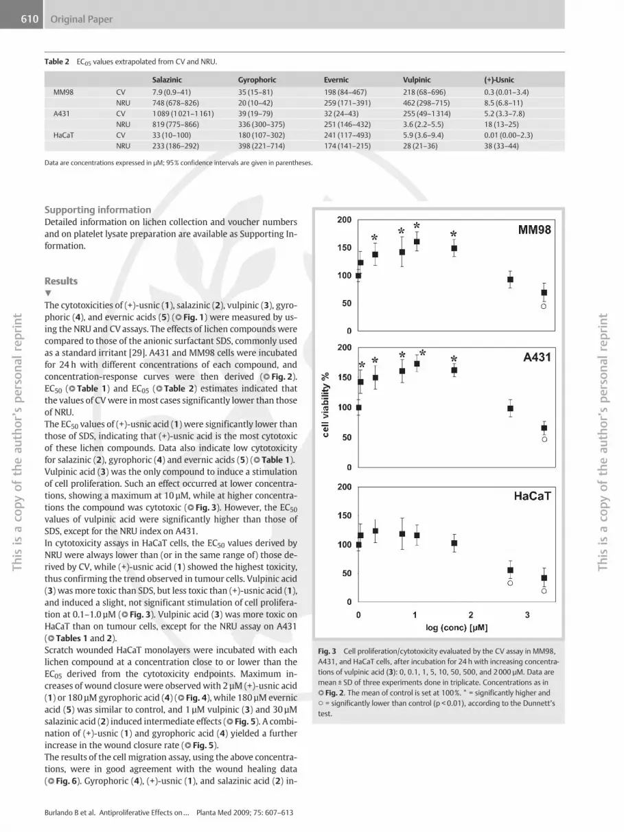
Table 2 EC05 values extrapolated from CV and NRU.
Salazinic Gyrophoric Evernic Vulpinic (+)-Usnic
MM98 CV 7.9 (0.9–41) 35 (15–81) 198 (84–467) 218 (68–696) 0.3 (0.01–3.4)
NRU 748 (678–826) 20 (10–42) 259 (171–391) 462 (298–715) 8.5 (6.8–11)
A431 CV 1089 (1021–1161) 39 (19–79) 32 (24–43) 255 (49–1314) 5.2 (3.3–7.8)
NRU 819 (775–866) 336 (300–375) 251 (146–432) 3.6 (2.2–5.5) 18 (13–25)
HaCaT CV 33 (10–100) 180 (107–302) 241 (117–493) 5.9 (3.6–9.4) 0.01 (0.00–2.3)
NRU 233 (186–292) 398 (221–714) 174 (141–215) 28 (21–36) 38 (33–44)
Data are concentrations expressed in µM; 95% confidence intervals are given in parentheses.
610 Original PaperTh
isis
aco
pyof
theau
thorʼs
person
alreprint
Supporting informationDetailed information on lichen collection and voucher numbersand on platelet lysate preparation are available as Supporting In-formation.
Fig. 3 Cell proliferation/cytotoxicity evaluated by the CV assay in MM98,A431, and HaCaT cells, after incubation for 24 h with increasing concentra-tions of vulpinic acid (3): 0, 0.1, 1, 5, 10, 50, 500, and 2000 µM. Data aremean ± SD of three experiments done in triplicate. Concentrations as inl" Fig. 2. The mean of control is set at 100%. * = significantly higher and○ = significantly lower than control (p < 0.01), according to the Dunnettʼstest.
Thisis
aco
pyof
theau
thorʼs
person
alreprint
bResults!
The cytotoxicities of (+)-usnic (1), salazinic (2), vulpinic (3), gyro-phoric (4), and evernic acids (5) (l" Fig. 1) were measured by us-ing the NRU and CV assays. The effects of lichen compounds werecompared to those of the anionic surfactant SDS, commonly usedas a standard irritant [29]. A431 and MM98 cells were incubatedfor 24 h with different concentrations of each compound, andconcentration-response curves were then derived (l" Fig. 2).EC50 (l" Table 1) and EC05 (l" Table 2) estimates indicated thatthe values of CVwere inmost cases significantly lower than thoseof NRU.The EC50 values of (+)-usnic acid (1) were significantly lower thanthose of SDS, indicating that (+)-usnic acid is the most cytotoxicof these lichen compounds. Data also indicate low cytotoxicityfor salazinic (2), gyrophoric (4) and evernic acids (5) (l" Table 1).Vulpinic acid (3) was the only compound to induce a stimulationof cell proliferation. Such an effect occurred at lower concentra-tions, showing a maximum at 10 µM, while at higher concentra-tions the compound was cytotoxic (l" Fig. 3). However, the EC50values of vulpinic acid were significantly higher than those ofSDS, except for the NRU index on A431.In cytotoxicity assays in HaCaT cells, the EC50 values derived byNRU were always lower than (or in the same range of) those de-rived by CV, while (+)-usnic acid (1) showed the highest toxicity,thus confirming the trend observed in tumour cells. Vulpinic acid(3) wasmore toxic than SDS, but less toxic than (+)-usnic acid (1),and induced a slight, not significant stimulation of cell prolifera-tion at 0.1–1.0 µM (l" Fig. 3). Vulpinic acid (3) was more toxic onHaCaT than on tumour cells, except for the NRU assay on A431(l" Tables 1 and 2).Scratch wounded HaCaT monolayers were incubated with eachlichen compound at a concentration close to or lower than theEC05 derived from the cytotoxicity endpoints. Maximum in-creases of wound closurewere observedwith 2 µM (+)-usnic acid(1) or 180 µM gyrophoric acid (4) (l" Fig. 4), while 180 µM evernicacid (5) was similar to control, and 1 µM vulpinic (3) and 30 µMsalazinic acid (2) induced intermediate effects (l" Fig. 5). A combi-nation of (+)-usnic (1) and gyrophoric acid (4) yielded a furtherincrease in the wound closure rate (l" Fig. 5).The results of the cell migration assay, using the above concentra-tions, were in good agreement with the wound healing data(l" Fig. 6). Gyrophoric (4), (+)-usnic (1), and salazinic acid (2) in-
Burlando B et al. Antiproliferative Effects on… Planta Med 2009; 75: 607–613

bFig. 4 Scratch wound healing of HaCaT under control condition (A) orupon incubation with 2 µM (+)-usnic acid (1) (B) or a combination of 2 µM(+)-usnic acid (1) and 180 µM gyrophoric acid (4) (C). Scale bar, 150 µm.
Fig. 5 Wound healing rates of HaCaT monolayers in the presence or ab-sence (cont) of lichen compounds. Gyr: gyrophoric acid (4) (180 µM); Usn:(+)-usnic acid (1) (2 µM); Sal: salazinic acid (2) (30 µM); Vul: vulpinic acid (3)(1 µM); Eve: evernic acid (5) (180 µM); PL: treatment with a platelet lysate(20% v/v) used as positive control (see Methods for further details). Dataare mean ± SD (n = 8–13) of wound closure rates expressed as the differ-ence between wound width at 0 h and 24 h. The mean of control is set at100%. Letters on bars indicate significant difference (p < 0.01) by the Tu-keyʼs multiple comparison test.
Fig. 6 Effect of different lichen compounds on HaCaT cell migration eval-uated by the transwell assay (see Methods). Data are mean ± SD (n = 6) ofthe numbers of migrating cells. Bar labels and statistics as in l" Fig. 5.
611Original Paper
Thisis
aco
pyof
theau
thorʼs
person
alreprint
Thisis
aco
pyof
theau
thorʼs
person
alreprint
duced the highest increase in the number of migrating cells,while the effect of vulpinic acid (3) was intermediate, and evernicacid (5) did not differ from the control. The combination of(+)-usnic (1) and gyrophoric acid (4) resulted in a further signifi-cant stimulation of cell migration.
Discussion!
Cytotoxicity assays showed that compounds 1–5 display a broadrange of activities, while the comparisonwith SDS allowed an ex-trapolation of the potential toxicity in vivo of lichen compounds.
The cytotoxicity profile of (+)-usnic acid (1) in keratinocytes is inagreement with previous data [30,31], and indicates potentialapplications in hyperproliferative skin disorders like psoriasis,while the strong antiproliferative effect on MM98 malignantmesothelioma is of particular interest due to the resistance of thistumour to chemotherapy [32]. Gyrophoric acid (4) showed a low-er cytotoxicity than previously reported [30], but in this latterstudy cell proliferation was inferred from cell counts rather thanfrom cytotoxicity markers.Wound healing is of great relevance for the treatment of skin le-sions and a particular focus is set on natural compounds [33–35].Lichen compounds had never been tested before in a woundhealing model, and our scratch wound experiments highlightedvarious degrees of wound closure stimulation. (+)-Usnic acid (1),the most toxic compound, was also the most effective in promot-ing wound healing. However, wound healing properties occurredat a low, subtoxic dose. Another compound showing diverging ac-tivities was vulpinic acid (3), which induced proliferation or cyto-
Burlando B et al. Antiproliferative Effects on… Planta Med 2009; 75: 607–613

612 Original Paper
thorʼs
person
alreprint
Thisis
aco
pyof
theau
thorʼs
person
alreprint
toxicity on tumour cells depending on the concentration em-ployed, while it did not show a strong effect on wound closure.Diverging properties occurring at different concentrations havealready been reported for natural compounds. For instance, ithas been shown that a hop extract induces prolactin release inhumans at low doses, while an opposite effect is observed athigher doses [36].The effects of gyrophoric acid (4) observed in different tests weremore consistent, since this compound induced the lowest toxicityon HaCaT cells, while it was a strong promoter of wound healing.Therefore, gyrophoric acid (4) seems to be of particular interestto treat wounds, since it combines a substantial wound closureeffect with an extremely low toxicity on keratinocytes.In summary, the effects of lichen compounds on wound healingwere correlated to those on cell migration, while they were un-correlated to those on proliferation/cytotoxicity, thus suggestingthat wound healing resulted from stimulation of motility ratherthan of mitosis. Also, lichen compounds seem less potent thanmedical preparations used in wound healing [37,38]. However,the combination of (+)-usnic acid (1) and gyrophoric acid (4) in-duced an about fourfold increase of HaCaT wound closure rate,thus reaching an activity comparable to that of the platelet lysateused as a positive control. A combination of lichen compoundscan therefore act synergistically, suggesting that crude lichen ex-tracts might reach levels of activity comparable to those obtainedwith clinically relevant preparations.In conclusion, we have shown that lichen compounds include in-teresting leads for the prevention of hyperproliferation syn-dromes and the promotion of tissue regeneration. While the mo-lecular target(s) underlying these activities remain elusive, themarked differences observed within the investigated compoundssuggest the existence of structure-activity relationships worthpursuing.
Thisis
aco
pyof
theau
bAcknowledgements!
We thank Simona Martinotti for her help with in vitro activities.This work was funded by the Università del Piemonte Orientale“Amedeo Avogadro”.
References1 Boustie J, Grube M. Lichens – a promising source of bioactive secondarymetabolites. Plant Genet Resources 2005; 3: 273–287
2 Bebert J. Sur une nouvelle substance découverte dans le lichen vulpi-nus. J Pharm Sci 1831; 17: 696–700
3 Huneck S. The significance of lichens and their metabolites. Naturwis-senschaften 1999; 86: 559–570
4 Muller K. Pharmaceutically relevant metabolites from lichens. Appl Mi-crobiol Biotechnol 2001; 56: 9–16
5 Bazin MA, Le Lamer AC, Delcros JG, Rouaud I, Uriac P, Boustie J, Corbel JC,Tomasi S. Synthesis and cytotoxic activities of usnic acid derivatives.Bioorg Med Chem 2008; 16: 6860–6866
6 Koparal AT, Tuylu BA, Turk H. In vitro cytotoxic activities of (+)-usnic ac-id and (−)-usnic acid on V79, A549, and human lymphocyte cells andtheir non-genotoxicity on human lymphocytes. Nat Prod Res 2006;20: 1300–1307
7 Mayer M, OʼNeill MA, Murray KE, Santos-Magalhaes NS, Carneiro-LeaoAM, Thompson AM, Appleyard VC. Usnic acid: a non-genotoxic com-pound with anti-cancer properties. Anticancer Drugs 2005; 16: 805–809
8 Han D, Matsumaru K, Rettori D, Kaplowitz N. Usnic acid-induced ne-crosis of cultured mouse hepatocytes: inhibition of mitochondrialfunction and oxidative stress. Biochem Pharmacol 2004; 67: 439–451
Burlando B et al. Antiproliferative Effects on… Planta Med 2009; 75: 607–613
9 Elo H, Matikainen J, Pelttari E. Potent activity of the lichen antibiotic(+)-usnic acid against clinical isolates of vancomycin-resistant entero-cocci and methicillin-resistant Staphylococcus aureus. Naturwissen-schaften 2007; 94: 465–468
10 Odabasoglu F, Cakir A, Suleyman H, Aslan A, Bayir Y, Halici M, Kazaz C.Gastroprotective and antioxidant effects of usnic acid on indometha-cin-induced gastric ulcer in rats. J Ethnopharmacol 2006; 103: 59–65
11 Jin J, He L, Li C. Effect of usnic acid on TNF-alpha and NO production inlipopolysaccharide-stimulatedmacrophages. Academic J Xiʼan JiaotongUniv 2006; 18: 153–156
12 Pramyothin P, Janthasoot W, Pongnimitprasert N, Phrukudom S, Ruan-grungsi N. Hepatotoxic effect of (+)-usnic acid from Usnea siamensisWainio in rats, isolated rat hepatocytes and isolated rat liver mitochon-dria. J Ethnopharmacol 2004; 90: 381–387
13 Ingolfsdottir K, Chung GA, Skulason VG, Gissurarson SR, Vilhelmsdottir M.Antimycobacterial activity of lichen metabolites in vitro. Eur J PharmSci 1998; 6: 141–144
14 Correche ER, Enriz RD, Piovano M, Garbarino J, Gomez-Lechon MJ. Cyto-toxic and apoptotic effects on hepatocytes of secondary metabolitesobtained from lichens. Altern Lab Anim 2004; 32: 605–615
15 Nadir MT, Rashan LJ, Ayoub MT, Awni LT. Antibacterial and antiprolifer-ative activities of vulpinic acids in vitro. Farmaco 1992; 47: 643–647
16 Candan M, Yilmaz M, Tay T, Kivanc M, Turk H. Antimicrobial activity ofextracts of the lichen Xanthoparmelia pokornyi and its gyrophoric andstenosporic acid constituents. Z Naturforsch 2006; 61: 319–323
17 Kumar KC, Muller K. Lichen metabolites. 2. Antiproliferative and cyto-toxic activity of gyrophoric, usnic, and diffractaic acid on human kera-tinocyte growth. J Nat Prod 1999; 62: 821–823
18 Halama P, Van Haluwin C. Antifungal activity of lichen extracts and li-chenic acids. Biocontrol 2004; 49: 95–107
19 Singh Y, Palombo M, Sinko PJ. Recent trends in targeted anticancer pro-drug and conjugate design. Curr Med Chem 2008; 15: 1802–1826
20 Harding KG, Morris HL, Patel GK. Science, medicine and the future:healing chronic wounds. BMJ 2002; 324: 160–163
21 Zopf W. Die Flechtenstoffe in chemischer, botanischer, pharmacolo-gischer und technischer Beziehung. Jena: Editorʼs Note; 1907
22 Giard DJ, Aaronson SA, Todaro GJ, Arnstein P, Kersey JH, Dosik H, ParksWP. In vitro cultivation of human tumors: establishment of cell linesderived from a series of solid tumors. J Natl Cancer Inst 1973; 51:1417–1423
23 Tsiouris A, Walesby RK. Malignant pleural mesothelioma: current con-cepts in treatment. Nat Clin Pract Oncol 2007; 4: 344–352
24 Boukamp P, Petrussevska RT, Breitkreutz D, Hornung J, Markham A, Fu-senig NE. Normal keratinization in a spontaneously immortalizedaneuploid human keratinocyte cell line. J Cell Biol 1988; 106: 761–771
25 Orengo AM, Spoletini L, Procopio A, Favoni RE, De Cupis A, Ardizzoni A,Castagneto B, Ribotta M, Betta PG, Ferrini S, Mutti L. Establishment offour new mesothelioma cell lines: characterization by ultrastructuraland immunophenotypic analysis. Eur Respir J 1999; 13: 527–534
26 Huneck S, Yoshimura I. Identification of lichen substances. Berlin:Springer; 1996
27 Ranzato E, Patrone M, Mazzucco L, Burlando B. Platelet lysate stimulateswound repair of HaCaT keratinocytes. Br J Dermatol 2008; 159: 537–545
28 Burlando B, Parodi A, Volante A, Bassi AM. Comparison of the irritationpotentials of Boswellia serrata gum resin and of acetyl-11-keto-beta-boswellic acid by in vitro cytotoxicity tests on human skin-derived celllines. Toxicol Lett 2008; 177: 144–149
29 Le M, Schalkwijk J, Siegenthaler G, van de Kerkhof PCM, Jacques H, Veer-kamp JH, van der Valk PGM. Changes in keratinocyte differentiation fol-lowing mild irritation by sodium dodecyl sulphate. Arch Dermatol Res2008; 288: 684–690
30 Kumar KC, Muller K. Lichen metabolites. 2. Antiproliferative and cyto-toxic activity of gyrophoric, usnic, and diffractaic acid on human kera-tinocyte growth. J Nat Prod 1999; 62: 821–823
31 Ingolfsdottir K. Usnic acid. Phytochemistry 2002; 61: 729–73632 Zervos MD, Bizekis C, Pass HI.Malignant mesothelioma. Curr Opin Pulm
Med 2008; 14: 303–30933 Khanna S, Venojarvi M, Roy S, Sharma N, Trikha P, Bagchi D, Bagchi M,
Sen CK. Dermal wound healing properties of redox-active grape seedproanthocyanidins. Free Radic Biol Med 2002; 33: 1089–1096
34 Shukla A, Rasik AM, Jain GK, Shankar R, Kulshrestha DK, Dhawan BN. Invitro and in vivo wound healing activity of asiaticoside isolated fromCentella asiatica. J Ethnopharmacol 1999; 65: 1–11
613Original PaperTh
isis
aco
pyof
theau
thorʼs
person
alreprint
35 Udupa S, Udupa A, Kulkarni D. Anti inflammatory and wound healingproperties of Aloe vera. Fitoterapia 1994; 65: 141–145
36 Merz PG, Gorkow C, Schrodter A, Rietbrock S, Sieder C, Loew D, Ricks-TanJS, Taubert HD. The effects of a special Agnus castus extract (BP1095E1)on prolactin secretion in healthy male subjects. Exp Clin EndocrinolDiabet 1996; 104: 447–453
37 Mazzucco L, Medici D, Serra M, Panizza R, Rivara G, Orecchia S, Libener R,Cattana E, Levis A, Betta PG, Borzini P. The use of autologous platelet gel
b
to treat difficult-to-heal wounds: a pilot study. Transfusion 2004; 44:1013–101838 Knighton DR, Ciresi K, Fiegel VD, Schumerth S, Butler E, Cerra F. Stimula-tion of repair in chronic, nonhealing, cutaneous ulcers using platelet-derived wound healing formula. Surg Gynecol Obstet 1990; 170: 56–60
Burlando B et al. Antiproliferative Effects on… Planta Med 2009; 75: 607–613
Thisis
aco
pyof
theau
thorʼs
person
alreprint


