Embed Size (px)
Citation preview

Attenuated Food Anticipatory Activity and AbnormalCircadian Locomotor Rhythms in Rgs16 Knockdown MiceNaoto Hayasaka1*, Kazuyuki Aoki2, Saori Kinoshita3, Shoutaroh Yamaguchi2, John K. Wakefield4¤a,
Sachiyo Tsuji-Kawahara3, Kazumasa Horikawa2, Hiroshi Ikegami5, Shigeharu Wakana6, Takamichi
Murakami7, Ram Ramabhadran4¤b, Masaaki Miyazawa3, Shigenobu Shibata2
1 Department of Anatomy and Neurobiology, Kinki University School of Medicine, Osaka-Sayama, Osaka, Japan, 2 Department of Physiology and Pharmacology, School of
Advanced Science and Engineering, Waseda University, Shinjuku-ku, Tokyo, Japan, 3 Department of Immunology, Kinki University School of Medicine, Osaka-Sayama,
Osaka, Japan, 4 Tranzyme Pharma, Durham, North Carolina, United States of America, 5 Department of Endocrinology, Metabolism and Disease, Kinki University School of
Medicine, Osaka-Sayama, Osaka, Japan, 6 Technology and Development Team for Mouse Phenotype Analysis: Japan Mouse Clinic, RIKEN Bioresource Center, Tsukuba,
Ibaraki, Japan, 7 Department of Radiology, Kinki University School of Medicine, Osaka-Sayama, Osaka, Japan
Abstract
Regulators of G protein signaling (RGS) are a multi-functional protein family, which functions in part as GTPase-activatingproteins (GAPs) of G protein a-subunits to terminate G protein signaling. Previous studies have demonstrated that the Rgs16transcripts exhibit robust circadian rhythms both in the suprachiasmatic nucleus (SCN), the master circadian light-entrainable oscillator (LEO) of the hypothalamus, and in the liver. To investigate the role of RGS16 in the circadian clock invivo, we generated two independent transgenic mouse lines using lentiviral vectors expressing short hairpin RNA (shRNA)targeting the Rgs16 mRNA. The knockdown mice demonstrated significantly shorter free-running period of locomotoractivity rhythms and reduced total activity as compared to the wild-type siblings. In addition, when feeding was restrictedduring the daytime, food-entrainable oscillator (FEO)-driven elevated food-anticipatory activity (FAA) observed prior to thescheduled feeding time was significantly attenuated in the knockdown mice. Whereas the restricted feeding phase-advanced the rhythmic expression of the Per2 clock gene in liver and thalamus in the wild-type animals, the above phaseshift was not observed in the knockdown mice. This is the first in vivo demonstration that a common regulator of G proteinsignaling is involved in the two separate, but interactive circadian timing systems, LEO and FEO. The present study alsosuggests that liver and/or thalamus regulate the food-entrained circadian behavior through G protein-mediated signaltransduction pathway(s).
Citation: Hayasaka N, Aoki K, Kinoshita S, Yamaguchi S, Wakefield JK, et al. (2011) Attenuated Food Anticipatory Activity and Abnormal Circadian LocomotorRhythms in Rgs16 Knockdown Mice. PLoS ONE 6(3): e17655. doi:10.1371/journal.pone.0017655
Editor: Paul Bartell, Pennsylvania State University, United States of America
Received January 12, 2011; Accepted February 4, 2011; Published March 9, 2011
Copyright: � 2011 Hayasaka et al. This is an open-access article distributed under the terms of the Creative Commons Attribution License, which permitsunrestricted use, distribution, and reproduction in any medium, provided the original author and source are credited.
Funding: This study was supported by Grant-in-Aid for Scientific Research: No. 19590235 and 21590264 from the Japan Society for the Promotion of Science,Grants-in-Aid from the Ministry of Education, Culture, Sports, Science and Technology of Japan including the High-Tech Research Center and Anti-aging Centergrants, those from the Ministry of Health, Labor and Welfare of Japan for Research on HIV/AIDS, and those from the Japan Health Sciences Foundation. Thefunders had no role in study design, data collection and analysis, decision to publish, or preparation of the manuscript.
Competing Interests: Authors include employees at Tranzyme Pharma and have an affiliation to Tranzyme Pharma using the material TranzVector for the study.This does not alter the authors’ adherence to all the PLoS ONE policies on sharing data and materials.
* E-mail: [email protected]
¤a Current address: Thermo Scientific/Open Biosystems, Huntsville, Alabama, United States of America¤b Current address: Integrated Systems Toxicology Division, NHEERL, ORD, United States Environmental Protection Agency, Research Triangle Park, NorthCarolina, United States of America
Introduction
The circadian timing system in mammals exerts control over a
wide range of physiology and behavior, including daily environ-
mental changes, the circadian system can be reset by external time
cues (Zeitgeber) such as light and metabolic correlates of feeding
[1–3]. Two separate, but coupled oscillators, LEO and FEO, are
involved in circadian system(s) in mammals. Whereas the master
LEO is located in the SCN of the hypothalamus [4,5], localization
of the FEO(s) still remains to be determined [6–8]. An output of a
putative FEO is FAA, which can be assessed by restricted feeding
(RF), daily limiting food availability to a restricted time window.
SCN lesion studies demonstrated that FEO-driven FAA persists
without the LEO, suggesting that FEO resides outside of the SCN
[6,9,10]. It is also reported that FEO dominates LEO under RF in
the entrainment of activity phase [4]. Moreover, disruption of the
known circadian clock genes resulted in mostly partial or little
alterations of FAA [11]. These data suggest that the FEO, which
functions under limited nutrient availability, is a dominant
circadian oscillator independent of LEO.
To elucidate molecular machinery driving circadian clock(s) in
mammals, a list of candidate circadian clock/clock-controlled
genes have been identified by microarray [8,12,13]. Among these
was Rgs16, a member of RGS family. RGS proteins regulate G
protein-coupled receptor (GPCR)-mediated signaling by negative-
ly or positively interacting with downstream effectors [14]. In the
brain, Rgs16 is predominantly expressed in the SCN and thalamus
[15,16]. RGS16 exhibits robust circadian rhythms both in the
SCN and in liver with its peak at 4–6 (zeitgeber time, ZT4-6) and
8–10 (ZT8-10) hours after light-on, respectively, suggesting that
PLoS ONE | www.plosone.org 1 March 2011 | Volume 6 | Issue 3 | e17655

RGS16 is involved in the central and peripheral circadian clocks
and/or their outputs [16]. Interestingly, a previous study
demonstrated that Rgs16 expression in liver was up-regulated
during fasting and rapidly down-regulated by re-feeding [17]. In
addition, expression of Rgs16 in liver was restricted to periportal
hepatocytes, the predominant locations of gluconeogenesis and
lipolysis [17]. Considering that a number of GPCR ligands
including vasoactive intestinal peptide (VIP; [18]), glutamate
[19,20], melatonin [21,22], orexin [23,24] and ghrelin [25,26]
have been implicated in the central or peripheral circadian clocks,
these data raise the possibility that RGS proteins function in the
circadian systems by modulating signaling through as yet unknown
GPCR/GPCR ligand(s).
Here, to elucidate the possible role of RGS16 in the circadian
clock in vivo, we generated two independent knockdown (KD)
mouse lines using lentiviral vectors expressing two separate short
hairpin RNAs (shRNAs) targeting the Rge16 mRNA.
Results
Generation of the Rgs16 knockdown miceWe designed different shRNAs against different regions of
Rgs16. Each shRNA expression vectors was transfected into
NIH3T3 cell line and levels of the Rgs16 mRNA were quantified
by quantitative RT-PCR (qPCR). The two shRNAs silencing
endogenous Rgs16 mRNA with the greatest efficiency (#41 and
#53) were selected for producing two independent transgenic
mouse lines (Fig. S2). We then produced separate high-titer
lentiviral vector lots encoding the two shRNA expression cassettes
as well as the GFP protein, and introduced them into fertilized
one-cell stage mouse embryos by microinjection into the perivitel-
line space [27,28]. The transgenic mice were selected by detecting
GFP expression and the presence of the transgene confirmed by
PCR-based genotyping.
Expression of the Rgs16 mRNA in the KD and wild-typebrain and liver
We first examined the spacio-temporal expression patterns of
the Rgs16 mRNA in brain and liver of the KD and wild-type
(WT) mice by in situ hybridization (ISH) and quantitative PCR
(qPCR). In the WT brain, predominant expression of Rgs16 was
observed in the SCN, and at a lower level in thalamus (Fig. 1A).
We also confirmed that Rgs16 transcription exhibits robust
circadian rhythms in the SCN (Fig. 1B). As shown in Fig. 1C,
average Rgs16 expression level was reduced the KD SCN
(Fig. 1C). In the WT liver, robust circadian rhythms were
observed peaking at ZT11 (Fig. 1D, F = 21.7, P,0.001). In the
KD liver, average Rgs16 expression level was reduced, and
circadian changes were weakened (Fig. 1D, F = 6.06, P,0.05)
relative to the controls (F = 21.7, P,0.001). In thalamus, no daily
expression rhythms were observed in both WT (F = 0.65, P.0.05)
and KD (F = 0.36, P.0.05) mice (Fig. 1E). The average
expression level of Rgs16 in the KD thalamus was lower than
that of WT control, however, the knockdown efficiency was lower
than that in liver (Fig. 1D, E).
Free-running period of locomotor activity rhythm wasshorter in the KD mice
To study the possible involvement of RGS16 in the central
circadian clock, we examined locomotor activity rhythms of the
KD and control mice. Wheel-running activities of individual mice
were monitored under 12 hr light and 12 hr dark (LD) and
constant dark (DD) conditions (Fig. 2A, B). In DD, The KD mice
showed an average free-running period significantly shorter than
that of the controls (23.8460.05 hr in WT vs. 23.4560.07 hr in
KD, P,0.01, Fig. 2C). After locomotor activity rhythms were
assessed, brains were sampled at ZT5 from individual KD and
WT mice and quantitative ISH was performed on the brain
sections. Average Rgs16 mRNA level in the SCN was significantly
decreased in the KD mice relative to controls (Fig. 1C).
Total amount of locomotor activity was reduced in theRgs16 KD mice
We next compared total amount of locomotor activity of the
Rgs16 KD mice with that of controls. Total activity counts were
assessed by an infrared sensor of KD and WT mice averaged every
30 minutes. The average activity of the KD mice was significantly
lower than that of the controls regardless of day or night (Fig. 3A,
B), whereas averaged day/night ratio of locomotor activity was
comparable between the two genotypes (Fig. 3C).
Figure 1. Spatial and temporal expression patterns of the Rgs16mRNA in brain and its reduction in the KD mice. A, B, in situhybridization performed on brain sections. A, Specific and intensesignals were observed in the SCN and thalamus (Th). B, Diurnal rhythmsof the Rgs16 transcript were observed in the SCN. C, ISH quantificationof the Rgs16 mRNA in the KD and WT SCN (n = 8 each) at ZT5. **P,0.01vs. WT (Student’s t-test). D, Expression of Rgs16 in the KD and WT liverobserved by qPCR. **P,0.01 vs. WT (Student’s t-test). ##, P,0.01circadian gene expression profile by 2-way ANOVA (WT vs. KD mice;n = 3–4). E, Expression of Rgs16 in thalamus of the KD and WT mice(n = 3–4). *P,0.05 vs. WT.doi:10.1371/journal.pone.0017655.g001
In Vivo Knockdown of Rgs16
PLoS ONE | www.plosone.org 2 March 2011 | Volume 6 | Issue 3 | e17655

Attenuated FAA in the Rgs16 KD miceThe Rgs16 mRNA in liver is not only circadianly regulated, but is
also up-regulated by fasting and down-regulated by re-feeding [17].
These data raise the possibility that RGS16 is involved in metabolism
in liver and in the food-driven behavior (FAA) regulated by a certain
brain region(s) and/or liver. To test this, we next examined the effects
of RF, daytime scheduled feeing for 4 hours (ZT6-ZT10), which is
usually a resting period for nocturnal rodents. When mice are food-
restricted during the day, FAA is observed about 3–4 hour prior to the
feeding time [12]. In WT mice, a remarkable increase in the
locomotor activities was observed for about 4 hours before the
scheduled feeding time (Fig. 4A, C). On the other hand, the FAA of
the KD mice was significantly attenuated in comparison with the
controls (Fig. 4B, C). The reduction in the average percent FAA in the
KD animals compared to that in the controls was statistically
significant (Fig. 4D, P,0.01). Fig. 4E demonstrates FAA counts of
individual KD and WT mice under RF as compared with baseline
activity counts under free feeding (FF, as measured activity during
ZT2-6). Bouts of locomotor activity during daytime was strongly
reduced in KD group by RF, however, those in nighttime were
comparable between WT and KD mice under FF or RF (Compare
Fig. 3A and 4C). It is of note that, under the RF schedule, both WT
and KD mice obtained almost the same amount of food during ZT6-
ZT10 (0.8860.08/10 g body weight/day for the WT; 0.8060.05/
10 g body weight/day for KD). Although body weight was slightly
reduced by RF in both WT (33.162.8 g prior to and 30.061.6 g after
RF) and KD mice (30.761.6 g prior to and 27.660.8 g after RF),
there was no significant difference in the amount of food intake or the
body weight between the two genotypes (P.0.05, Student’s t-test).
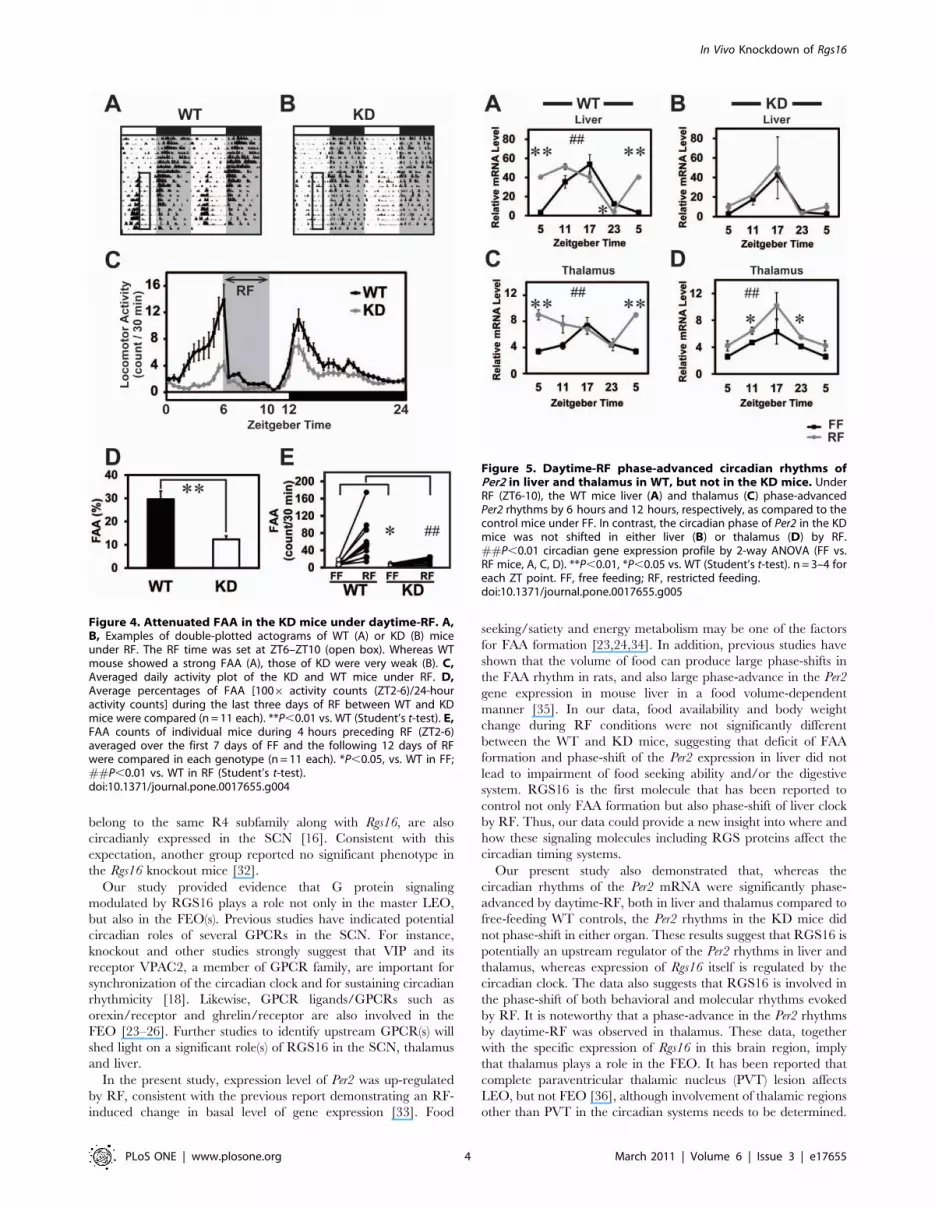
Daytime-RF phase-advanced the Per2 mRNA rhythm inWT, but not in KD
Reduced FAA has been reported in several knockout mice, but
the mechanism underlying such behavioral change remains to be
elucidated. To examine whether RF elicits FAA by affecting
expression profiles of circadian clock gene and/or Rgs16 in brain
and liver, we sampled KD and WT mouse tissues at 4 time points
(ZT5, 11, 17, and 23) and performed qPCR. Fig. 5 demonstrates
the circadian rhythms in the Per2 clock gene expression in liver
and thalamus in both the WT and the KD mice, where Rgs16
transcripts are abundantly expressed. Under FF, the Per2 mRNA
exhibited circadian rhythms both in liver (F = 21.7 P,0.01 for
WT, F = 16.1 P,0.05 for KD, one-way ANOVA) and thalamus
(F = 5.1 P,0.05 for WT) with a peak at ZT17 in both genotypes.
Under RF in WTs, the Per2 rhythms were significantly phase-
advanced, peaking at ZT11 and ZT5 in liver and thalamus,
respectively (Fig. 5A, C; F = 25.2 P,0.01 for liver, F = 4.1 P,0.05
for thalamus). In contrast, the phase of the Per2 rhythms in the KD
mice was unaffected by RF either in liver or thalamus (Fig. 5B, D).
Discussion
In the present study, we generated transgenic Rgs16 KD mice
using lentiviral vectors carrying a shRNA expression cassette and a
modified microinjection method, i.e., perivitelline injection
[27,28]. This gene silencing technique enabled us to efficiently
generate sufficient numbers of transgenic mice within a few
months, because of the higher rate of transgenesis by lentiviral
vector injection compared with the conventional pronuclear DNA
microinjection [29–31]. Moreover, this in vivo KD strategy could
be a method of choice when disruption of the gene of interest is
likely to cause lethality in vivo, or leads to a lack of phenotype
because redundant functionalities from within the gene family or
from related gene(s) result in compensation for the gene loss. We
anticipated that Rgs16 knockout could potentially be compensated
by other Rgs genes (Rgs2 and/or Rgs4), because these genes, which
Figure 2. Short free-running period of locomotor activityrhythm in the KD mice. A, B, Locomotor activity rhythms of theWT (A) and KD (B) mice were entrained to 12:12 LD. But in DD, the KDmice exhibited circadian period of locomotor activity shorter than thatof WTs. The vertical axis in each graph indicates the day path.Horizontal open and closed bars indicate the light and dark periods,respectively. C, The difference in the circadian period between the KDand WT mice (n = 15 each) was statistically significant **P,0.01 vs. WT(Student’s t-test).doi:10.1371/journal.pone.0017655.g002
Figure 3. Reduced amount of locomotor activity in the Rgs16KD mice. A, Averaged daily activity plot of the KD and WT mice underLD condition. B, Average daytime or nighttime locomotor activity wassignificantly decreased in the KD mice as compared to that of WTcontrols (n = 21 each). **P,0.01 vs. WT (Student’s t-test). C Averagedday/night ratio of locomotor activity was comparable between the KDand WT mice (n = 21 each).doi:10.1371/journal.pone.0017655.g003
In Vivo Knockdown of Rgs16
PLoS ONE | www.plosone.org 3 March 2011 | Volume 6 | Issue 3 | e17655
belong to the same R4 subfamily along with Rgs16, are also
circadianly expressed in the SCN [16]. Consistent with this
expectation, another group reported no significant phenotype in
the Rgs16 knockout mice [32].
Our study provided evidence that G protein signaling
modulated by RGS16 plays a role not only in the master LEO,
but also in the FEO(s). Previous studies have indicated potential
circadian roles of several GPCRs in the SCN. For instance,
knockout and other studies strongly suggest that VIP and its
receptor VPAC2, a member of GPCR family, are important for
synchronization of the circadian clock and for sustaining circadian
rhythmicity [18]. Likewise, GPCR ligands/GPCRs such as
orexin/receptor and ghrelin/receptor are also involved in the
FEO [23–26]. Further studies to identify upstream GPCR(s) will
shed light on a significant role(s) of RGS16 in the SCN, thalamus
and liver.
In the present study, expression level of Per2 was up-regulated
by RF, consistent with the previous report demonstrating an RF-
induced change in basal level of gene expression [33]. Food
seeking/satiety and energy metabolism may be one of the factors
for FAA formation [23,24,34]. In addition, previous studies have
shown that the volume of food can produce large phase-shifts in
the FAA rhythm in rats, and also large phase-advance in the Per2
gene expression in mouse liver in a food volume-dependent
manner [35]. In our data, food availability and body weight
change during RF conditions were not significantly different
between the WT and KD mice, suggesting that deficit of FAA
formation and phase-shift of the Per2 expression in liver did not
lead to impairment of food seeking ability and/or the digestive
system. RGS16 is the first molecule that has been reported to
control not only FAA formation but also phase-shift of liver clock
by RF. Thus, our data could provide a new insight into where and
how these signaling molecules including RGS proteins affect the
circadian timing systems.
Our present study also demonstrated that, whereas the
circadian rhythms of the Per2 mRNA were significantly phase-
advanced by daytime-RF, both in liver and thalamus compared to
free-feeding WT controls, the Per2 rhythms in the KD mice did
not phase-shift in either organ. These results suggest that RGS16 is
potentially an upstream regulator of the Per2 rhythms in liver and
thalamus, whereas expression of Rgs16 itself is regulated by the
circadian clock. The data also suggests that RGS16 is involved in
the phase-shift of both behavioral and molecular rhythms evoked
by RF. It is noteworthy that a phase-advance in the Per2 rhythms
by daytime-RF was observed in thalamus. These data, together
with the specific expression of Rgs16 in this brain region, imply
that thalamus plays a role in the FEO. It has been reported that
complete paraventricular thalamic nucleus (PVT) lesion affects
LEO, but not FEO [36], although involvement of thalamic regions
other than PVT in the circadian systems needs to be determined.
Figure 4. Attenuated FAA in the KD mice under daytime-RF. A,B, Examples of double-plotted actograms of WT (A) or KD (B) miceunder RF. The RF time was set at ZT6–ZT10 (open box). Whereas WTmouse showed a strong FAA (A), those of KD were very weak (B). C,Averaged daily activity plot of the KD and WT mice under RF. D,Average percentages of FAA [1006 activity counts (ZT2-6)/24-houractivity counts] during the last three days of RF between WT and KDmice were compared (n = 11 each). **P,0.01 vs. WT (Student’s t-test). E,FAA counts of individual mice during 4 hours preceding RF (ZT2-6)averaged over the first 7 days of FF and the following 12 days of RFwere compared in each genotype (n = 11 each). *P,0.05, vs. WT in FF;##P,0.01 vs. WT in RF (Student’s t-test).doi:10.1371/journal.pone.0017655.g004
Figure 5. Daytime-RF phase-advanced circadian rhythms ofPer2 in liver and thalamus in WT, but not in the KD mice. UnderRF (ZT6-10), the WT mice liver (A) and thalamus (C) phase-advancedPer2 rhythms by 6 hours and 12 hours, respectively, as compared to thecontrol mice under FF. In contrast, the circadian phase of Per2 in the KDmice was not shifted in either liver (B) or thalamus (D) by RF.##P,0.01 circadian gene expression profile by 2-way ANOVA (FF vs.RF mice, A, C, D). **P,0.01, *P,0.05 vs. WT (Student’s t-test). n = 3–4 foreach ZT point. FF, free feeding; RF, restricted feeding.doi:10.1371/journal.pone.0017655.g005
In Vivo Knockdown of Rgs16
PLoS ONE | www.plosone.org 4 March 2011 | Volume 6 | Issue 3 | e17655

Interestingly, the report also indicated that PVT ablation
increased total daily activity, suggesting that this thalamic region
is involved in regulating behavioral output. Altogether, our data
along with previous studies raise a possibility that RGS16 in
thalamus modulates light-entrained and/or food-entrained behav-
ior by regulating activity levels and/or circadian behavioral
rhythms under different environmental conditions.
Our study provided for the first time evidence suggesting that
two distinct regions of the body, thalamus and liver, where Rgs16
mRNA is abundantly expressed, are involved in the regulation of
the FAA under restricted food availability. The data raise the
question of whether liver or thalamus plays a lead role in the FEO-
operated FAA, or both of these regions regulate the behavior,
either independently or cooperatively. Further studies in search of
upstream GPCRs/ligands and downstream signaling molecules
will elucidate the mechanism of how RGS16 specifically regulates
food-entrained behavior in different loci.
Materials and Methods
AnimalsC57BL/6J inbred mice were purchased from CREA Japan and
used for generating transgenic mice and all other experiments.
Animals were provided with food and water ad libitum, and
entrained to 12 hr light: 12 hr dark condition (12:12 LD) for at
least 2 weeks at 2362uC, All animals were cared for in accordance
with the Law (No. 105) and Notification (No. 6) of the Japanese
Government, and all experiments were conducted under permis-
sion of the Experimental Animal Welfare Committees of Kinki
University (Permission #KDMS-16-002) and Waseda University
(Permission #08A36).
shRNASix shRNAs were designed from the Rgs16 cDNA sequence, and
annealed double-stranded oligomers were subcloned into the
pSilencer 1.0-U6 siRNA Expression Vector (Ambion). Knock-
down efficiencies of the expression vectors carrying individual
shRNAs were evaluated by transient transfection of the vectors
into mouse NIH3T3 (RIKEN Cell Bank) fibroblast cell line by
Lipofectamine 2000 (Life Technologies) according to manufac-
turer’s instruction. After 48 hours of incubation, cells were lysed
using Sepazol-RNA I (Nacalai Tesque), RNA was extracted, and
quantitative PCR (qPCR) was performed using Rgs16 primers.
Two shRNAs (#41, 53), which significantly suppressed the
endogenous Rgs16 expression levels, were used for transgenesis.
The sequences of annealed oligonucleotides for expressing #41 or
#53 shRNA were as follows:
#41: 59-GCGAGGAGTTCAAGAAGATTTCAAGA-
GAATCTTCTTGAACTCCTCGCTTTTTT-39 and 59-AAT-
TAAAAAAGCGAGGAGTTCAAGAAGATTCTCTTGAAAT-
CTTCTTGAACTCCTCGCGGCC-39
#53: 59-GAGAACTGACCAAGACAAATTCAAGA-
GATTTGTCTTGGTCAGTTCTCTTTTTT-39 and 59-AAT-
TAAAAAAGAGAACTGACCAAGACAAATCTCTTGAATT-
TGTCTTGGTCAGTTCTCGGCC-39
Lentiviral vector construction and production of high-titer lentiviral vector particles
The two shRNA sequences (#41 and #53) targeting Rgs16
along with the mouse U6 promoter were PCR-amplified from the
pSilencer vectors and cloned into the pTZV TranzVectorTM
(Tranzyme) as depicted in Fig.S1. An eGFP fluorescent marker
driven from the human immediate early cytomegalovirus virus
(CMV) promoter is co-expressed with the shRNA hairpin
sequence. High-titer lentiviral vector particles were generated
using the Trans-LentiviralTM Vector Packaging system [37]. Viral
particles were concentrated by ultracentrifugation. Functional
titers were determined by transducing HEK293T (ATCC) cells
with limiting dilutions of virus and counting GFP-positive colonies.
Generation of Rgs16 KD miceFertilized oocytes were harvested from super-ovulated C57BL/6J
female mice. Viral vectors at a concentration of 16109/ml were
microinjected into the perivitelline space of the oocytes using a
FemtoJet injector (Eppendorf), and then reimplanted into the oviduct
of pseudo-pregnant recipient females after 2–4 hours of microinjec-
tion. Injections were performed under 4006 magnification (DM
IRE2, Leica), and injection volume was approximately 100 nl.
Genotyping and evaluation of the KD miceFor genotyping, genomic DNA was extracted from a tail tip of
each mouse by a standard protocol using proteinase K and
following phenol/chloroform extraction. Using the genomic DNA
as a template, PCR was performed using GFP primers. To
confirm expression of the GFP protein, we observed tail tips under
fluorescent microscopy.
qPCRTotal RNA was extracted from tail tip, liver, and thalamus of each
KD or WT control mouse using RNeasy Mini Kit (Qiagen). The
extracted RNA was reverse-transcribed into cDNA using High
Capacity cDNA Reverse Transcription Kit (Applied Biosystems). For
qPCR reaction, SYBR GREEN Premix ExTaq (Takara) was used
following manufacturer’s instruction. The sequences of primers used
in this study are as follows. GFP primers: 59-AGCAAAGACCC-
CAACGAGA-39 and 59-GGCGGCGGTCACGAA-39; Rgs16 prim-
ers: 59-TGCTTGTGAACAGGGCTAACTG-39 and 59-CTCC-
CTCCTTAGACCCCATCTT-39; Per2 primers: 59-TGTGTCT-
TACACGGGTGTCCTA-39 and 59-ACGTTTGGTTTGCGCA-
TGAA-39, 18S rRNA primers: 59-CGGCTACCACATCCAAG-
GAA-39, 59-GCTGGAATTACCGCGGCT-39.
The value of the PCR product of the target gene was
normalized to that of 18S rRNA.
Measurement of locomotor activity rhythmMice were individually housed in translucent polypropylene
cages under 12:12 LD (200 lux) and DD, and locomotor activity
was assessed either by a running wheel (Fig. 2) or an area sensor
(F5B, Omron; Fig. 3, 4). Activity was continuously monitored and
analyzed using ClockLab software (Actimetrics).
Schedules for RFIndividually housed KD mice and WT controls were main-
tained in a 12:12 LD cycle under FF for at least 7 days. The RF
experiment for the measurement of anticipatory activity was
performed as previously described [38]. Briefly, after 1 day of food
deprivation starting at ZT12 (day 0), food was restricted to ZT6-
ZT10 for 12 days (day 1–12). From day 13 to 14, food was again
withdrawn for the entire day to record motor activity under food
deprivation. Another RF experiment for the measurement of clock
gene expression was performed. After application of the same RF
conditions, animals were sacrificed at ZT5, 11, 17, and 23 on day
13 under food deprivation.
In situ hybridization (ISH)Riboprobe was labeled with 35S-UTP (Amersham/GE Health-
care) by in vitro transcription using either T7 or SP6 polymerase
In Vivo Knockdown of Rgs16
PLoS ONE | www.plosone.org 5 March 2011 | Volume 6 | Issue 3 | e17655

(Promega). Frozen mouse brain sections (40 mm thickness) were
hybridized with riboprobe for overnight and apposed to Kodak
film (BioMax MR). The Rgs16 cDNA (entire ORF length 606 bp)
was amplified by PCR and subcloned into pGEM-T Easy Vector
(Promega). The plasmids were linealized with NcoI to synthesize
riboprobe.
StatisticsResults were expressed as the mean 6 SEM. One-way ANOVA
was applied to evaluate significant difference of circadian
rhythmicity of gene expression, and 2-way ANOVA was applied
to evaluate significance between KD and control mice. The
significance of the differences between groups was determined by
the Student’s t-test. Statistical analysis software (StatView version
5.0, SAS Institute) was used.
Supporting Information
Figure S1 Schematic representation of the shRNAlentiviral vectors. The shRNA hairpin sequences (#41 and
#53) targeting mouse Rgs16 is cloned into pTZV, a HIV1-based
self-inactivating (SIN) lentiviral transfer vector containing the
central polypurine tract/termination sequence (FLAP) and the
Woodchuck hepatitis virus post-transcriptional regulatory element
(WPRE) for enhanced gene expression. A PCR-amplified
fragment containing the mouse U6 promoter, shRNA hairpin,
and pol III termination sequence (TTTTT) is cloned into pTZV
between 59 – ClaI and 39 – BamHI sites. A GFP marker is co-
expressed with the shRNA from a CMV promoter to enable
visualization of transduced cells and transgenic embryos.
(EPS)
Figure S2 In vitro knockdown of the Rgs16 mRNA byshRNAs used in this study. 2 mg of the pSilencer 1.0 vector
expressing either of the two shRNAs (#41 or #53) was transfected
into the NIH3T3 mouse fibroblast cell line with Lipofectamine
2000. RNA was extracted from each of the transfected cells two
days after the transfection, and expression levels of Rgs16 were
compared by qPCR (see Materials and Methods). Both #41 and
#53 shRNAs significantly reduced the average Rgs16 mRNA level
(**P,0.01, n = 4).
(EPS)
Acknowledgments
We thank Ralph Mistlberger for critical reading, and Takahiro Moriya,
Yuka Miyoshi, Yuka Sugahara, Shinsuke Noso, Naru Babaya, Ikuko
Yamada, Tomohiro Suzuki, Tamio Furuse and Hiroshi Takemori for
technical support and helpful discussion.
Author Contributions
Conceived and designed the experiments: NH. Performed the experiments:
NH KA SK SY STK KH. Analyzed the data: NH HI SW TM SS.
Contributed reagents/materials/analysis tools: JKW RR MM. Wrote the
paper: NH SS.
References
1. Lamont EW, James FO, Boivin DB, Cermakian N (2007) From circadian clock
gene expression to pathologies. Sleep Med 8: 547–556.
2. Mendoza J (2007) Circadian clocks: setting time by food. J Neuroendocrinol 19:
127–137.
3. Dibner C, Schibler U, Albrecht U (2010) The mammalian circadian timing
system: organization and coordination of central and peripheral clocks. Annu
Rev Physiol 72: 517–549.
4. Schibler U, Ripperger J, Brown SA (2003) Peripheral circadian oscillators in
mammals: time and food. J Biol Rhythms 18: 250–260.
5. Escobar C, Cailotto C, Angeles-Castellanos M, Delgado RS, Buijs RM (2009)
Peripheral oscillators: the driving force for food-anticipatory activity.
Eur J Neurosci 30: 1665–1675.
6. Krieger DT, Hauser H, Krey LC (1977) Suprachiasmatic nuclear lesions do not
abolish food-shifted circadian adrenal and temperature rhythmicity. Science
197: 398–399.
7. Rashotte ME, Stephan FK (1996) Coupling between light- and food-entrainable
circadian oscillators in pigeons. Physiol Behav 59: 1005–1010.
8. Stephan FK (2001) Food-entrainable oscillator in mammals. Circadian Clocks
12: 223–246.
9. Stephan FK, Swann JM, Sisk CL (1979) Entrainment of circadian rhythms by
feeding schedules in rats with suprachiasmatic lesions. Behav Neural Biol 25:
545–554.
10. Stephan FK, Swann JM, Sisk CL (1979) Anticipation of 24-hr feeding schedules
in rats with lesions of the suprachiasmatic nucleus. Behav Neural Biol 25:
346–363.
11. Challet E, Mendoza J, Dardente H, Pevet P (2009) Neurogenetics of food
anticipation. Eur J Neurosci 30: 1676–1687.
12. Mistlberger RE (1994) Circadian food-anticipatory activity: formal models and
physiological mechanisms. Neurosci Biobehav Rev 18: 171–195.
13. Mistlberger RE (2009) Food-anticipatory circadian rhythms: concepts and
methods. Eur J Neurosci 30: 1718–1729.
14. Willars GB (2006) Mammalian RGS proteins: multifunctional regulators of
cellular signalling. Semin Cell Dev Biol 17: 363–376.
15. Grafstein-Dunn E, Young KH, Cockett MI, Khawaja XZ (2001) Regional
distribution of regulators of G-protein signaling (RGS) 1, 2, 13, 14, 16, and
GAIP messenger ribonucleic acids by in situ hybridization in rat brain. Brain
Res Mol Brain Res 88: 113–123.
16. Ueda HR, Chen W, Adachi A, Wakamatsu H, Hayashi S, et al. (2002) A
transcription factor response element for gene expression during circadian night.
Nature 418: 534–539.
17. Huang J, Pashkov V, Kurrasch DM, Yu K, Gold SJ, et al. (2006) Feeding and
fasting controls liver expression of a regulator of G protein signaling (Rgs16) in
periportal hepatocytes. Comp Hepatol 5: 8.
18. Maywood ES, O’Neill JS, Chesham JE, Hastings MH (2007) Minireview: The
circadian clockwork of the suprachiasmatic nuclei–analysis of a cellular oscillator
that drives endocrine rhythms. Endocrinology 148: 5624–5634.
19. Hannibal J (2002) Neurotransmitters of the retino-hypothalamic tract. Cell
Tissue Res 309: 73–88.
20. Gillette MU, Mitchell JW (2002) Signaling in the suprachiasmatic nucleus:
selectively responsive and integrative. Cell Tissue Res 309: 99–107.
21. Pevet P, Agez L, Bothorel B, Saboureau M, Gauer F, et al. (2006) Melatonin in
the multi-oscillatory mammalian circadian world. Chronobiol Int 23: 39–51.
22. Dubocovich ML (2007) Melatonin receptors: role on sleep and circadian rhythm
regulation. Sleep Med 8(Suppl 3): 34–42.
23. Akiyama M, Yuasa T, Hayasaka N, Horikawa K, Sakurai T, et al. (2004)
Reduced food anticipatory activity in genetically orexin (hypocretin) neuron-
ablated mice. Eur J Neurosci 20: 3054–3062.
24. Mieda M, Williams SC, Sinton CM, Richardson JA, Sakurai T, et al. (2004)
Orexin neurons function in an efferent pathway of a food-entrainable circadian
oscillator in eliciting food-anticipatory activity and wakefulness. J Neurosci 24:
10493–10501.
25. LeSauter J, Hoque N, Weintraub M, Pfaff DW, Silver R (2009) Stomach
ghrelin-secreting cells as food-entrainable circadian clocks. Proc Natl Acad
Sci U S A 106: 13582–13587.
26. Blum ID, Patterson Z, Khazall R, Lamont EW, Sleeman MW, et al. (2009)
Reduced anticipatory locomotor responses to scheduled meals in ghrelin
receptor deficient mice. Neuroscience 164: 351–359.
27. Lois C, Hong EJ, Pease S, Brown EJ, Baltimore D (2002) Germline transmission
and tissue-specific expression of transgenes delivered by lentiviral vectors.
Science 295: 868–872.
28. Shaughnessy L, Chamblin B, McMahon L, Nair A, Thomas MB, et al. (2004)
Novel approaches to models of Alzheimer’s disease pathology for drug screening
and development. J Mol Neurosci 24: 23–32.
29. Park F (2007) Lentiviral vectors: are they the future of animal transgenesis?
Physiol Genomics 31: 159–173.
30. Singer O, Verma IM (2008) Applications of lentiviral vectors for shRNA delivery
and transgenesis. Curr Gene Ther 8: 483–488.
31. Gama Sosa MA, De Gasperi R, Elder GA (2010) Animal transgenesis: an
overview. Brain Struct Funct 214: 91–109.
32. Bansal G, Druey KM, Xie Z (2007) R4 RGS proteins: regulation of G-protein
signaling and beyond. Pharmacol Ther 116: 473–495.
33. Oishi K, Kasamatsu M, Ishida N (2004) Gene- and tissue-specific alterations of
circadian clock gene expression in streptozotocin-induced diabetic mice under
restricted feeding. Biochem Biophys Res Commun 317: 330–334.
34. Stephan FK (1997) Calories affect zeitgeber properties of the feeding entrained
circadian oscillator. Physiol Behav 62: 995–1002.
In Vivo Knockdown of Rgs16
PLoS ONE | www.plosone.org 6 March 2011 | Volume 6 | Issue 3 | e17655

35. Hirao A, Tahara Y, Kimura I, Shibata S (2009) A balanced diet is necessary for
proper entrainment signals of the mouse liver clock. PLoS One 4: e6909.36. Landry GJ, Yamakawa GR, Mistlberger RE (2007) Robust food anticipatory
circadian rhythms in rats with complete ablation of the thalamic paraventricular
nucleus. Brain Res 1141: 108–118.37. Wu X, Wakefield JK, Liu H, Xiao H, Kralovics R, et al. (2000) Development of
a novel trans-lentiviral vector that affords predictable safety. Mol Ther 2: 47–55.
38. Wakamatsu H, Yoshinobu Y, Aida R, Moriya T, Akiyama M, et al. (2001)
Restricted-feeding-induced anticipatory activity rhythm is associated with a
phase-shift of the expression of mPer1 and mPer2 mRNA in the cerebral cortex
and hippocampus but not in the suprachiasmatic nucleus of mice. Eur J Neurosci
13: 1190–1196.
In Vivo Knockdown of Rgs16
PLoS ONE | www.plosone.org 7 March 2011 | Volume 6 | Issue 3 | e17655


