Embed Size (px)
Citation preview

This article appeared in a journal published by Elsevier. The attachedcopy is furnished to the author for internal non-commercial researchand education use, including for instruction at the authors institution
and sharing with colleagues.
Other uses, including reproduction and distribution, or selling orlicensing copies, or posting to personal, institutional or third party
websites are prohibited.
In most cases authors are permitted to post their version of thearticle (e.g. in Word or Tex form) to their personal website orinstitutional repository. Authors requiring further information
regarding Elsevier’s archiving and manuscript policies areencouraged to visit:
http://www.elsevier.com/authorsrights

Author's personal copy
Knockdown of receptor for advanced glycation end products attenuate17α-ethinyl-estradiol dependent proliferation and survival of MCF-7breast cancer cells
Kusum Lata a, Tapan K. Mukherjee a,b,⁎a Department of Biology, Indian Institute of Science Education and Research (IISER) Mohali Sector 81, SAS Nagar, Manauli, 140306, Punjab, Indiab Department of Biotechnology, Maharishi Markendeshwar University (A deemed University), Mullana, Haryana-133203, India
a b s t r a c ta r t i c l e i n f o
Article history:Received 13 June 2013Received in revised form 25 September 2013Accepted 9 November 2013Available online 16 November 2013
Keywords:MCF-7 breast cancer cells proliferation and survival17-alpha-ethinyl estradiolEstrogen receptor related receptor gammaReceptor for advanced glycation productReactive oxygen species
Background: 17α-ethinyl-estradiol (17α-EE), a synthetic estrogen is the world's most widely and commonlyused orally bioactive estrogen. Currently, 17α-EE is in use in all formulations of contraceptive pills and is impli-cated in the complication of breast cancer. Receptor for advanced glycation end products (RAGE) is a cell surfaceimmunoglobulin class of molecule. RAGE is involved in the complication of various cancers.Methods and results: This study indicates that treatment of MCF-7 breast cancer cells with 17α-EE enhances theexpression of estrogen receptor related receptor gamma (ERRγ), followed by enhanced level of oxidative stressand subsequent activation of the transcription factor, nuclear factor kappa-B (NF-кB), leading to increase in RAGEexpression. RAGE thus expressed by 17α-EE treatment causes further enhancement of the oxidative stresswhich, in turn, activates expression of cell cycle protein cyclin D1 and subsequent induction of MCF-7 breastcancer cell proliferation. RAGE also enhanced phosphorylation of prosurvival protein AKT and increased expres-sion of Bcl2, an antiapoptotic protein.Conclusion: InMCF-7 breast cancer cells, 17α-EE-ERRγ interaction induces the expression of RAGE,which in turn,enhances the number of MCF-7 breast cancer cells through a multiprong action on the divergent molecules likecyclin D1, AKT and Bcl2.General significance: This is thefirst reportwhich explains the intermediate role of ERRγ in the 17α-EE dependentRAGE expression in MCF-7 breast cancer cells. This report for the first time explains that RAGE is important notonly for MCF-7 breast cancer cell proliferation but also for its survival and anti-apoptotic activities.
© 2013 Elsevier B.V. All rights reserved.
1. Introduction
Estrogens are a group of female steroid hormones, having a role in awide range of physiological processes like development of female sexorgans, maintaining bone density andmaintenance of sexual reproduc-tion [1]. Currently, estrogens are in use as constituent of contraceptivepills [2], and in post menopausal hormone replacement therapy (HRT)[3]. 17alpha-ethinyl-estradiol (17α-EE), a synthetic estrogen is theworld's most commonly used bioactive estrogen which is in use in allformulations of oral contraceptive pills [4]. In spite of having a pivotalrole in normal physiology of females, estrogens are implicated in thevarious types of female cancers including breast cancer [5]. Estrogen re-ceptors (ERs) [6], and estrogen receptor related receptors (ERRs), a
nuclear hormone receptor superfamily [7], are involved in estrogen de-pendent breast cancer complication and drug resistance [8].
Recentlywehave shown that in endothelial cells, interaction of 17α-EE with ERα induces the expression of receptor for advanced glycationend products (RAGE) [9]. RAGE is a cell surface immunoglobulin class ofmolecule [10]. RAGE interacts with divergent ligands including ad-vanced glycation end products (AGE) [11] and S100 or calgranulin[12,13]. Thus, RAGE is amultiligand receptormolecule. RAGE is involvedin proinflammatory and prooxidative reactions [13]. One recent studyindicates that interaction of RAGE with S100 induces the proliferationof MCF-7 breast cancer cells [14]. However, the specific mechanism in-volved in the RAGE dependent MCF-7 breast cancer cells proliferationis not known. More importantly, it is not known whether 17α-EE isequally potent to induce the expression of RAGE inMCF-7 breast cancercells as observed in endothelial cells [9]. The importance of 17α-EE gen-erated RAGE on MCF-7 breast cancer cells proliferation and the specificmechanism involved needs to be evaluated. Moreover, the role of RAGEon AKT phosphorylation and Bcl2 expression is not known. Of note,while AKT is a prosurvival protein [15], Bcl2 attenuates apoptosis [16].
Biochimica et Biophysica Acta 1840 (2014) 1083–1091
⁎ Corresponding author at: Department of Biology, Indian Institute of Science Educationand Research (IISER) Mohali Sector 81, SAS Nagar, Manauli, 140306, Punjab, India.Tel.: +91 9876722596.
E-mail address: [email protected] (T.K. Mukherjee).
0304-4165/$ – see front matter © 2013 Elsevier B.V. All rights reserved.http://dx.doi.org/10.1016/j.bbagen.2013.11.014
Contents lists available at ScienceDirect
Biochimica et Biophysica Acta
j ourna l homepage: www.e lsev ie r .com/ locate /bbagen

Author's personal copy
Since in a number of recent observations ERRs are implicated in anties-trogen resistance in ERpositive breast cancer cells [8], the role of ERRs in17α-EE dependent RAGE expression needs to be evaluated.
The present study, therefore, was undertaken to understand the roleof 17α-EE in the expression of RAGE in the ER positive MCF-7 breastcancer cells and to evaluate the role of ERRs in thismechanism. The spe-cific mechanism through which 17α-EE generated RAGE may enhancethe number of MCF-7 breast cancer cells, was another important areaof observations. In this study, we have shown that 17α-EE inducesRAGE expression in theMCF-7 breast cancer cells through the activationof ERRγ. RAGE is responsible for 17α-EE mediated MCF-7 breast cancercells survival and proliferation by activating cyclin D1, AKT and Bcl2. Wehave also shown that reactive oxygen species (ROS) play a very impor-tant intermediate role in the RAGE dependentMCF-7 breast cancer cellsproliferation and survival.
2. Materials and methods
Minimum essential medium (MEM), estrogens (17β-estradiol, 17α-ethinyl estradiol and 17α-epiestriol), N-acetyl cysteine (NAC), sRAGE(Santa Cruz Biotechnology), siRNA for RAGE, ERα, ERβ, ERRγ, MTT cellproliferation assay kit, antibodies for cyclin D1, Bcl2, anti-IκBα, andHRP-conjugated anti-mouse and anti-rabbit secondary antibodieswere purchased from Sigma. ICI182780 was obtained from TocrisCookson Ltd. Antibodies for RAGE, ERα, ERβ, NF-κB, P-IκBα werepurchased from Millipore Corporation Ltd. Catalase and AKT (phosphoS473) were procured from Calbiochem and Abcam respectively.
Glutathione peroxidase 4 (GPx-4) and ERRγ antibodies were purchasedfrom Abcam.
2.1. Culture of MCF-7 breast cancer cells in minimum essential medium
MCF-7 breast cancer cells (human) were procured from ATCC andmaintained in minimum essential medium (MEM) supplementedwith 5% fetal bovine serum (FBS), 1 mM L-glutamine, 0.01 mg/ml insu-lin, 1 mM sodium pyruvate, 2.2 g/ml sodium bicarbonate, 1% antibioticand antimycotic at 37 °C and 5% CO2. For experiments cells were grownup to 70–75% confluency.
2.2. Treatment of MCF-7 breast cancer cells with various drugs of interest
Prior to treatment, MCF-7 breast cancer cells were washed twicewith PBS and pre-incubated for 12 hrs with experimental medium con-taining MEM without phenol red, 2% charcoal-dextran treated FBS,2.2 g/ml sodium bicarbonate and 1% antibiotic/antimycotic. After12 hrs of preincubation with the experimental medium, MCF-7 breastcancer cells were treated with various experimental drugs. For treat-ment, MCF-7 breast cancer cells were treated with various concentra-tions of 17α-EE for 6 hrs. Since estrogens have a half life of onlyaround3 hrs [17], fresh 17α-EEwas added to the cellswithout changingthe medium and treated for another 6 hrs. This 12 hrs (6 hrs + 6 hrs)treatment time for 17α-EE was maintained for all the experiments inthis study. The specific time point and treatment conditions for otherdrugs are discussed in their results section.
Fig. 1.A: RepresentativeWestern blot analysis showing the effect of various concentrations of 17α-EE on the expression of RAGE inMCF-7 cells: MCF-7 breast cancer cells were incubatedwith 17α-EE for 12 hrs. 50 μg of proteins (total cell lysate) were loaded into each lane. A 1: RAGE (top) and β-actin (bottom, internal control) were analyzed using specific monoclonalantibodies. Lane 1, untreated control; Lanes 2–4, treatment with various concentrations of 17α-EE. A 2: Densitometric plot of the above experiments. Maximum intensity of the RAGEexpression was noticed when MCF-7 breast cancer cells were treated with 10 nM of 17α-EE alone for 12 hrs (lane 4). B: Representative Western blot analysis showing the effect ofpreincubation of ICI182780 on the 17α-EE dependent expression of RAGE in theMCF-7 cells: MCF-7 cells were incubatedwith ICI182780 for 6 hrs prior to treatmentwith 10 nM concen-tration of 17α-EE for 12 hrs. B 1: RAGE (top) andβ-actin (bottom, internal control) were analyzed using specific monoclonal antibodies. Lane 1, untreated control; Lane 2, treatmentwith10 nM concentration of 17α-EE alone for 12 hrs; Lane 3-4 incubation of MCF-7 cells with 1 μM and 3 μM of ICI182780 alone for 2 hrs; Lane 5–6 incubation of MCF-7 cells with 1 μM and3 μM of ICI182780 for 6 hrs prior to 10 nM of 17α-EE for 12 hrs. B 2. Densitometric plot of the above experiment.
1084 K. Lata, T.K. Mukherjee / Biochimica et Biophysica Acta 1840 (2014) 1083–1091
Author's personal copy
2.3. Determination of MCF-7 breast cancer cell proliferation by MTT cellproliferation assay
MCF-7 breast cancer cells were grown in 96 well plates and treatedwith various drugs of interest as described in Results and discussion sec-tion. Following treatment, 10 μl of MTT solution from a freshly preparedstock (5 mg/ml) was added to 90 μl of culture medium per well and in-cubated at 37 °C for 4–5 hrs. Following incubation, the MTT containingmedium was replaced with 100 μl solvent in each well, incubated at37 °C for 15 min with constant shaking. The absorbance was measuredon an ELISA plate reader with a wavelength of 570 nm.
2.4. Transient siRNA transfection of the MCF-7 breast cancer cells
MCF-7 breast cancer cells were grown up to 60–70% confluency in 6well plates. N-Ter nano particle based siRNA transfection reagent(Sigma) was used for transfection. Before transfection the plates werewashed with phenol red free and antibiotic/antimycotic free MEM con-taining 2% charcoal-dextran treated FBS and incubated for 30 min at37 °C. After incubation, MCF-7 breast cancer cells were subjected totransfection with various siRNA according to the standard protocol ofthe manufacturer.
2.5. Preparation of cytoplasmic and nuclear extracts of the MCF-7 breastcancer cells
Cytosolic extracts (CE) and nuclear extracts (NE) were isolated asdescribed previously withmodification. [18]. Very briefly, MCF-7 breastcancer cells were isolated by cell scraper, washed with ice cold PBS andresuspended with 100 μl ice cold CE buffer containing 10 mM HEPES(pH 7.9), 1.5 mM MgCl2, 10 mM KCl, 0.5 mM DTT, 0.3 mM sucrose,
0.1 mM EGTA, protease inhibitor cocktail, incubated on ice for 10–15 min. 3 μl of 10% NP-40 was added, vigorously vortexed for 10–15 sec and centrifuged at 13000 rpm, 4 °C for 30 sec. The supernatantobtained was CE and pellet was nuclear material. The nuclear materialwas washed with ice cold CE buffer without detergent for two timesat 13,000 rpm, 4 °C for 1 min. The pellet was resuspended in ice cold100 μl of NE buffer containing 20 mM HEPES (pH 7.9), 25% glycerol,0.42 M NaCl, 1.5 mM MgCl2, 0.2 mM EDTA, 0.5 mM DTT, proteaseinhibitor cocktail, rocked gently for 30 min, centrifuged at 13000 rpm,4 °C for 30 min. The supernatant obtained was NE. Protein measure-ment was done by Bradford assay. 50 μg of protein was used for West-ern blot analysis.
2.6. Preparation of total cell lysate of the MCF-7 breast cancer cells andWestern blot analysis
For Western blot analysis, MCF-7 breast cancer cells were grown in100 mmpetriplates and 17α-EE treatmentwas done on 70–75% conflu-ent cells as mentioned above. The Western blot was performed as de-scribed previously [18]. Very briefly, after the treatment, MCF-7 breastcancer cells were trypsinized, pelleted, washedwith ice cold PBS and re-suspended in 100 μl of lysis buffer containing 5 mM HEPES (pH 7.5),1 mM DTT, 150 mM NaCl, 1 mM EDTA, 0.5% tween-20, 10% glycerol,and protease inhibitor cocktail (Sigma). After 1 hr, the cell lysate wascentrifuged, supernatant was collected and protein concentration wasmeasured by Bradford reagent. 50 μg of protein was subjected to SDS-PAGE and separated proteins were transferred to PVDF membrane(BIORAD) by semi-dry transfer apparatus (BIORAD), membranes weretreated with primary and secondary antibodies, and developed by ECLdeveloping reagents (Amersham).
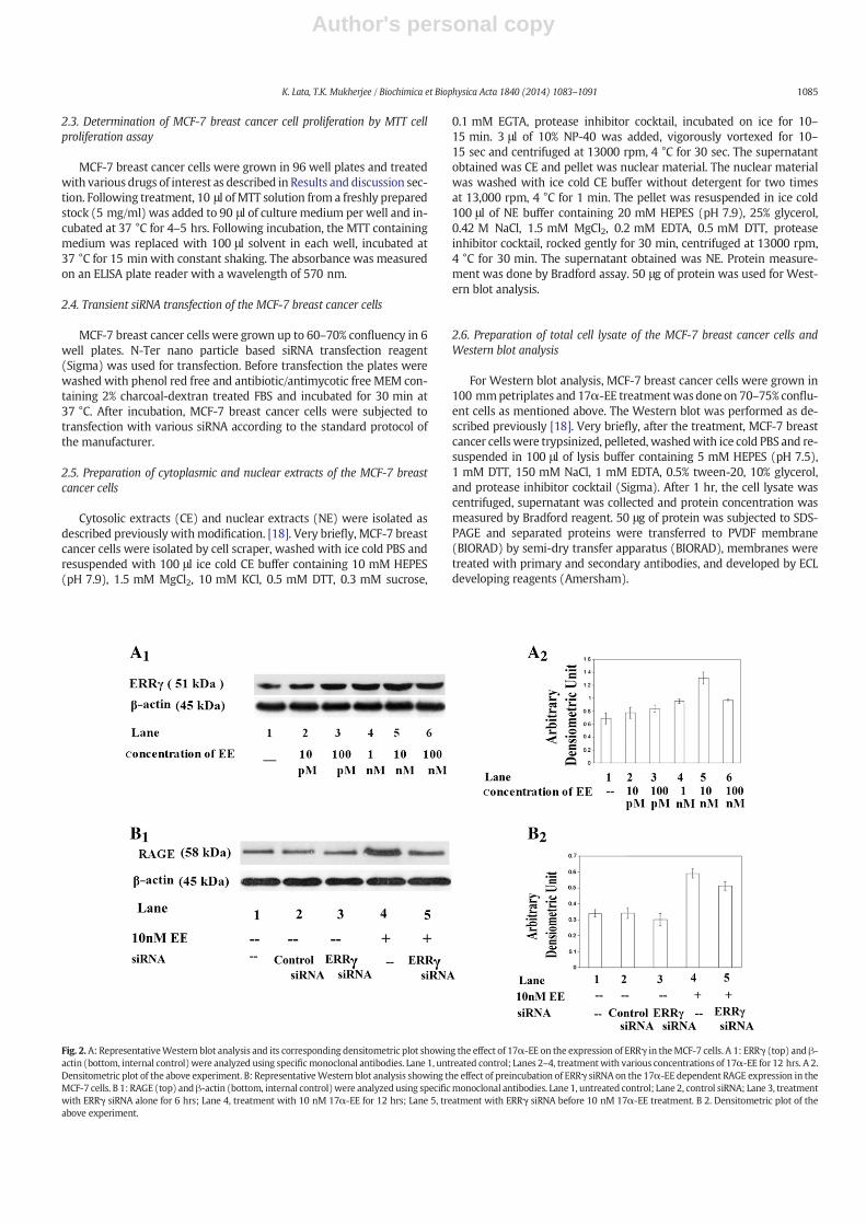
Fig. 2.A: RepresentativeWestern blot analysis and its corresponding densitometric plot showing the effect of 17α-EE on the expression of ERRγ in theMCF-7 cells. A 1: ERRγ (top) and β-actin (bottom, internal control)were analyzed using specificmonoclonal antibodies. Lane 1, untreated control; Lanes 2–4, treatmentwith various concentrations of 17α-EE for 12 hrs. A 2.Densitometric plot of the above experiment. B: RepresentativeWestern blot analysis showing the effect of preincubation of ERRγ siRNA on the 17α-EE dependent RAGE expression in theMCF-7 cells. B 1: RAGE (top) andβ-actin (bottom, internal control)were analyzed using specificmonoclonal antibodies. Lane 1, untreated control; Lane 2, control siRNA; Lane 3, treatmentwith ERRγ siRNA alone for 6 hrs; Lane 4, treatment with 10 nM 17α-EE for 12 hrs; Lane 5, treatment with ERRγ siRNA before 10 nM 17α-EE treatment. B 2. Densitometric plot of theabove experiment.
1085K. Lata, T.K. Mukherjee / Biochimica et Biophysica Acta 1840 (2014) 1083–1091

Author's personal copy
2.7. Determination of the generation of reactive oxygen species by 2′,7′dichloro-fluorescence di-acetate
The level of ROS generated by MCF-7 breast cancer cells was mea-sured as described previously [19]. Very briefly, to measure the levelof ROS generation, MCF-7 breast cancer cells were treated overnightwith the experimentalmediumas described previously. Next day, treat-ment with 10 nM of 17α-EE was done at various time points from15 min to 12 hrs. After completion of the treatment with 17α-EE, themedium was aspirated, MCF-7 breast cancer cells were washed andplaced in Hanks balanced salt solution (HBSS) at 37 °C incubator for15 min. 2′,7′-dichlorofluorescin diacetate (DCFDA) solution was pre-pared fresh by dissolving in 100%DMSO. TheMCF-7 cellswere incubatedin the dark with 50 μM DCFDA (Invitrogen) for 15 minutes prior to themeasurement of the level of ROS. Following incubation, MCF-7 breast
cancer cells were scraped and re-suspended in 2 ml of HBSS and ROSwere measured using a fluorescence spectroflourimeter (Perkin Elmer)at 534 nm emission with an excitation wavelength of 488 nm.
3. Results and discussion
Receptor for advanced glycation end products (RAGE) is a cell surfaceimmunoglobulin class of molecule [10]. RAGE binds with various ligandsand therefore RAGE is recognized as a multiligand receptor molecule[11,12]. Interaction of RAGE with its ligands namely advanced glycationend products (AGE) [11] and S100 or calgranulin [12] might enhancethe proinflammatory and prooxidative reactions of the cells [11,13]. En-gagement of RAGE with its various ligands namely S100 complicatesbreast cancer [14,20]. Based on these observations, it is assumed thatRAGE might be involved in the complication of breast cancer, althoughthe exact mechanism is not known. Indeed, the use of RAGE deficientmice (RAGE−/−) in well established mouse models of inflammation-associated carcinogenesis such as chemically induced carcinogenesis[21], and colitis associated cancer [22] provided direct genetic evidencefor a novel role of RAGE in cancer.
It is now generally accepted that estrogens can complicate breastcancer [5]. Estrogens have major roles in cancer cell proliferation in ERpositive breast cancer cells [23], and ERRs are held responsible for ta-moxifen (an antagonist of ERs) resistance [8]. Previously, we haveshown that in endothelial cells, 17α-EE and other estrogens stimulatethe expression of RAGE through the activation of ERα [9]. However, itis not known whether 17α-EE is equally potent to stimulate RAGE ex-pression in various cancer cells as observed in endothelial cells. More-over, the role of ERRs (if any) in this mechanism is not known. Since17α-EE is the most widely used orally bioactive estrogen, it is impera-tive to check the role of 17α-EE-ERs and 17α-EE-ERRs interactions inthe expression of RAGE in ER positive breast cancer cells.
Fig. 3. Representative Western blot analysis showing the effect of 17α-EE on the translo-cation of NF-кB-p65 and phosphorylation of IкBα in theMCF-7 cells. Figures are in the fol-lowing order: A. NF-кB-p65 in cytosolic extracts (CE). B. NF-кB-p65 in nuclear extracts(NE). C. Phosphorylated IкBα in total cell lysate. Lane 1, untreated control; Lanes 2–4,treatment with various concentrations of 17α-EE.
Fig. 4. A: Representative plot showing the effect of 10 nM concentration of 17α-EE at different time points on the generation of ROS in the MCF-7 cells. B. Representative Western blotanalysis showing the effect of 17α-EE on the expression of catalase and GPx-4. Lane 1, untreated control; Lane 2-4, treatment with various concentrations of 17α-EE. C. RepresentativeWestern blot showing the effect of NAC on the expression of catalase, GPx-4 and RAGE. MCF-7 cells were incubated with NAC for 6 hrs prior to treatment with 10 nM of 17α-EE for12 hrs. Lane 1, untreated control. Lane 2, treatment with 10 nM of 17α-EE alone for 12 hrs; Lanes 3–4 incubation of cells with 3 mM and 10 mM of NAC alone for 6 hrs; Lane 5-6, incu-bation of MCF-7 cells with 3 mM and 10 mM of NAC alone for 6 hrs prior to 10 nM of 17α-EE for 12 hrs.
1086 K. Lata, T.K. Mukherjee / Biochimica et Biophysica Acta 1840 (2014) 1083–1091

Author's personal copy
It is also essential to examine the exact mechanism through which17α-EE possibly enhances RAGE expression in cancer cells. Our previ-ous study indicates that in endothelial cells, TNFα generated ROS in-duces RAGE expression through the activation of transcription factorNF-кB [24]. Estrogens can also enhance the generation of ROS in cancercells [25]. Therefore ROS may be the possible downstream moleculesthrough which 17α-EE enhances RAGE expression in cancer cells.
It is also essential to understand the exact mechanism throughwhich RAGE could affect breast cancer cells proliferation and survival.A number of recent studies indicate that interaction of RAGE with itsligands might enhance the ROS level in the cells [11]. Therefore, whileRAGE is the product of enhanced oxidative stress, once expressed,RAGE itself could further enhance oxidative stress and thus might cycli-cally cause its own expression by a positive feedback mechanism [24].ROS is one of the molecules that might affect cell cycle progressionand increase the cell number [26]. In a separate independent study, itwas also shown that estrogens affect the cell cycle progression by en-hanced expression of cyclin D1, a G1 specific cell cycle protein [27].ROS might also affect the cell survival and the expression of pro- orantiapoptotic proteins [28]. Thus, it is highly essential to check the effectof RAGE generated ROS on MCF-7 breast cancer cells proliferation, sur-vival and apoptosis.
ER positive MCF-7 breast cancer cells are widely in use to examinethe effect of estrogens on breast cancer complication. Therefore, thepresent study was undertaken to check the followings: (a) Role of
17α-EE, ERs and ERRs in the expression of RAGE inMCF-7 breast cancercells, (b) Role of 17α-EE generated ROS and consequent oxidative stresson controlling the expression of RAGE in MCF-7 breast cancer cells, (c)Intermediate role of RAGE and RAGE generated ROS and subsequent ox-idative stress in the 17α-EE dependentMCF-7 breast cancer cells prolif-eration, survival and apoptosis. The following paragraphs represent ourexperimental observation:
3.1. Role of 17α-ethinyl-estradiol and estrogen receptor related receptorson the expression of receptor for advanced glycation end products inMCF-7 breast cancer cells
Initially, we have checked whether estrogens can stimulate RAGEexpression inMCF-7 breast cancer cells. ER positiveMCF-7 breast cancercells are widely in use to study the effects of estrogens on variousestrogen dependent cell signaling pathways. In our previous studieson endothelial cells we have shown the comparative effect of 17α-EE(a synthetic estrogen and a major component of contraceptive pills),17β-estradiol (major estrogen present in the female blood) and 17α-epiestriol (a metabolite of estrogen present in female blood) on the ex-pression of RAGE. Of note, while 17α-EE predominantly works throughERα, 17β-estradiol is equally potent to both ERα and ERβ and 17α-epiestriol predominantlyworks through ERβ and shows similarity in ac-tion with genestin, a phytoestrogen [18]. Moreover, in endothelial cells17α-EE is comparatively more potent in stimulating the expression of
Fig. 5.A1: Representativefigure showing the effect of 17α-EE on theproliferation ofMCF-7 cells byMTT cell proliferation assay. A2. Representativefigure showing the effect of RAGE siRNAon 17α-EE induced proliferation ofMCF-7 cells byMTT cell proliferation assay. B. RepresentativeWestern blot analysis showing the effect of 17α-EE on the expression of cyclinD1 inMCF-7 cells. Lane 1, untreated control; Lanes 2–6, treatmentwith various concentration of 17α-EE. C. RepresentativeWestern blot analysis showing the effect of pretreatmentwith RAGE siRNAon the 17α-EE dependent expression of cyclin D1 onMCF-7 cells. Lane 1, untreated control; Lane 2, control siRNA; Lane 3, treatment with RAGE siRNA alone for 6 hrs; Lane 4, treatmentwith 10 nM 17α-EE for 12 hrs; Lane 5, treatment with RAGE siRNA before 10 nM 17α-EE treatment.
1087K. Lata, T.K. Mukherjee / Biochimica et Biophysica Acta 1840 (2014) 1083–1091

Author's personal copy
RAGE in comparison to 17β-estradiol and 17α-epiestriol [9]. In thepresent study usingMCF-7 breast cancer cells 17α-EE is themost potentestrogen to stimulate RAGE expression when compared its effect with17β-estradiol and 17α-epiestriol (data not shown). Thus, we have cho-sen 17α-EE as our experimental estrogen of interest.
The results show a dose dependent induction of RAGE expression inMCF-7 breast cancer cells by 17α-EE (Fig. 1A) whichwas attenuated byICI182780 (fulvestran) (Fig. 1B), a nonspecific ER antagonist. Further re-sults confirmed that ERα is responsible for 17α-EE dependent RAGE ex-pression in MCF-7 breast cancer cells (data not shown). These resultsconfirm our speculation that both in endothelial cells and in MCF-7breast cancer cells 17α-EE is equally potent in inducing the expressionof RAGE.
In this context, in a number of recent observations it is claimed thatERRs, a family of nuclear orphan receptors are equally potent to bindvarious estrogens and show similar kind of effects as observed with es-trogens [7]. It is further noticed that ICI182780 is able to attenuate ERRsbinding with estrogens [29]. Additionally, tamoxifen (a nonspecific ERantagonist and breast cancer drug) dependent drug resistance is medi-ated at least partially through the binding of estrogens with ERRs [8].Therefore, we examined the involvement of various ERRs in 17α-EE de-pendent RAGE expression inMCF-7 breast cancer cells. The results showdose dependent induction of ERRγ (Fig. 2A), but not of ERRα and ERRβ(data not shown). Further, treatment of MCF-7 breast cancer cells withsiRNA against ERRγ prior to the treatment with 17α-EE attenuated the17α-EE dependent RAGE expression (Fig. 2B), confirming the importantcontributory role of ERRγ in 17α-EE dependent RAGE expression.
3.2. Role of 17α-ethinyl-estradiol activated NF-κB on the expression ofreceptor for advanced glycation end products in MCF-7 breast cancer cells
Estrogens regulate the expression of various genes by binding topromoters of the responsive genes through either estrogen response el-ements (ERE), or via activation of various transcription factors namely,NF-κB and SP1 [9]. The proinflammatory and prooxidative transcriptionfactors being activated by oxidative and inflammatory stresses regulatethe expression of various proinflammatory and prooxidative genes [30].While RAGE promoter sequence shows binding sites for transcriptionfactors NF-κB and SP1, no ERE are observed in the RAGE promoter[31]. Compelling evidence from our two independent studies on endo-thelial cells indicate that proinflammatory and prooxidative cytokineTNFα and female steroids (estrogens) activate RAGE expression bytwo different transcription factors. While TNFα generated ROS induces
Fig. 6. A: RepresentativeWestern blot analysis showing the effect of 17α-EE on the phos-phorylation of AKT and expression of Bcl2. Lane 1, untreated control; Lanes 2–4, treatmentwith various concentration of 17α-EE. B. Representative Western blot analysis showingthe effect of pretreatment with RAGE siRNA on the 17α-EE dependent expression ofphospho Akt and the expression of Bcl2, in MCF-7 cells. Lane 1, untreated control; Lane2, control siRNA; Lane 3, treatment with RAGE siRNA alone for 6 hrs; Lane 4, treatmentwith 10 nM 17α-EE for 12 hrs; Lane 5, treatment with RAGE siRNA before 10 nM 17α-EE treatment.
Fig. 7. A. Representative plot showing the effect of RAGE siRNA on ROS generation in the 17α-EE treated MCF-7 cells. B. Representative Western blot analysis showing the effect of pre-treatment of RAGE siRNA on the 17α-EE dependent expression of catalase and Gpx-4 in the MCF-7 cells. Lane 1, untreated control; Lane 2, control siRNA; Lane 3, treatment with RAGEsiRNA alone for 6 hrs; Lane 4, treatment with 10 nM 17α-EE for 12 hrs; Lane 5, treatment with RAGE siRNA before 10 nM 17α-EE treatment. C. Representative Western blot analysisshowing the effect of NAC prior to 17α-EE treatment on the expression of cyclin D1, Bcl2 and the phosphorylation of AKT in MCF-7 cells. Lane 1, untreated control; Lane 2, treatmentwith 10 nM concentration of 17α-EE alone for 12 hrs; Lane 3-4, incubation of cells with 3 mM and 10 mM of NAC alone for 6 hrs; Lane 5-6, incubation of cells with 3 mM and 10 mMof NAC for 6 hrs prior to 10 nM of 17α-EE for 12 hrs.
1088 K. Lata, T.K. Mukherjee / Biochimica et Biophysica Acta 1840 (2014) 1083–1091

Author's personal copy
RAGE expression via the activation of NF-κB [24], estrogens induceRAGE expression through the activation of transcription factor SP1and the involvement of NF-κB in this mechanism is negligible [9].Other studies also confirmed a role of ROS in the activation of NF-κB[32].
Like TNFα, estrogens are known inducers of ROS generation andsubsequent oxidative stress, particularly in various cancer cells [33]. Infact, estrogen dependent oxidative stress has a key role in breast cancercell proliferation and survival [34]. In the present study, we have exam-ined whether SP1 or NF-κB is responsible for 17α-EE dependent RAGEexpression inMCF-7 breast cancer cells. The results indicate that follow-ing treatment of theMCF-7 breast cancer cells with 17α-EE, SP1 activitybecomes negligible (data not shown) and NF-κB becomes activated andgets translocated to the nucleus (Fig. 3A and B). IкBα is the inhibitorprotein which binds with NF-кB and prevents translocation of NF-кBfrom the cytoplasm to the nucleus. Translocation of NF-кB needs phos-phorylation followed by degradation of IкBα [24]. Increased level phos-phorylation of IкBα further confirmed the involvement of NF-кB in17α-EE dependent RAGE expression in the MCF-7 breast cancer cells(Fig. 3C).
These results indicate that endothelial cells behave differently incomparison to MCF-7 breast cancer cells in terms of 17α-EE dependent
expression of RAGE. In endothelial cells, SP1 is predominantly responsi-ble for the estrogen dependent RAGE expression [9], and in MCF-7breast cancer cells, NF-κB is responsible for 17α-EE dependent RAGE ex-pression.While the exact reason is yet to be determine, most possibly ahigh level of ROS generation and subsequent oxidative stress in MCF-7breast cancer cells favored stimulation of NF-κB by 17α-EE treatment.Of note, endothelial cells show a comparatively low level of ROS gener-ation [35], in comparison to cancer cells [36].
To check whether the treatment of MCF-7 breast cancer cells with17α-EE, in fact generated ROS and induced oxidative stress, we havemeasured the level of ROS and the expression of some of the markersof oxidative stress namely GPx-4 and catalase. 17α-EE treatment notonly generated ROS (Fig. 4A), but also enhanced the expression ofGPx-4 and catalase (Fig. 4B). Catalases are utilized to neutralize the ex-cess amount of H2O2 [37]. Similarly, a high level of GPx-4 is necessary toneutralize the excess level of H2O2 [38]. Thus, in our experiments highlevel expression of GPx-4 and catalase indicates enhanced level of oxi-dative stress following treatment of MCF-7 breast cancer cells with17α-EE.
To check whether 17α-EE dependent oxidative stress is really re-sponsible for RAGE expression in MCF-7 breast cancer cells, we havetreated them with antioxidant NAC prior to treatment with 17α-EE
Fig. 8.A schematic diagram depicting themechanism throughwhich 17α-EE generated receptor for advanced glycation end products affect the proliferation and survival of MCF-7 breastcancer cells.
1089K. Lata, T.K. Mukherjee / Biochimica et Biophysica Acta 1840 (2014) 1083–1091

Author's personal copy
and checked the level of expression of GPx-4, catalase and RAGE. Ofnote, in a number of investigations the antioxidant NAC is used to neu-tralize the excess amount of ROS [39]. Very recently it has been shownthat NAC can attenuate RAGE expression, indicating the role of oxidativestress on RAGE expression [40]. In our experiments, the dose dependentattenuation of the expression of GPx-4, catalase and RAGE by NAC indi-cate the important contributory role of oxidative stress on 17α-EE de-pendent RAGE expression in the MCF-7 breast cancer cells (Fig. 4C).These results confirm that treatment of MCF-7 breast cancer cells with17α-EE induces the generation of ROSwhich in turn activates RAGE ex-pression through the activation of the transcription factor NF-кB.
3.3. Role of receptor for advanced glycation end products on 17α-ethinyl-estradiol dependent MCF-7 breast cancer cells proliferation and survival
Estrogens regulate cell signalingmolecules involved in breast cancercells proliferation [27]. However, role of RAGE in estrogen dependentbreast cancer cells proliferation is yet to be evaluated. The presentstudy reveals the intermediary role of 17α-EE generated RAGE on theMCF-7 breast cancer cells proliferation and the mechanism involved.Initially, we checked MCF-7 breast cancer cells proliferation by MTTcell proliferation assay following treatment with 17α-EE in presenceand absence of RAGE siRNA or sRAGE. The results show that 17α-EEfails to induce MCF-7 breast cancer cells proliferation in presence ofRAGE siRNA (Fig. 5A1 and A2) or sRAGE (data not shown), indicatingthe involvement of RAGE in the 17α-EE dependentMCF-7 breast cancercell proliferation.
To understand the mechanism through which RAGE is involved inMCF-7 breast cancer cells proliferation,we have checked the level of cy-clin D1, following treatment ofMCF-7 breast cancer cellswith 17α-EE inpresence or absence of RAGE siRNA. In this study, cyclin D1 level waschecked taking into consideration previous report which mentionedthat estrogens regulate cell cycle progression by enhancing the expres-sion of cyclin D1 [27]. The results of the present experiments indicatethe involvement of cyclin D1 in RAGE dependent cell cycle progression(Fig. 5B and C). Besides activating cell cycle, estrogens also regulate cellsurvival by regulating the expression as well as phosphorylation / de-phosphorylation of prosurvival protein AKT [41]. The pro-survival roleof AKT is widely accepted [15]. Estrogens also induce Bcl2, anantiapoptotic protein [16]. The role of RAGE (if any) in 17α-EE depen-dent phosphorylation of AKT and expression of Bcl2 is not known. Inthe present study, RAGE siRNA attenuated 17α-EE dependent AKTphosphorylation and Bcl2 expression (Fig. 6A and B), indicating impor-tant contributory role of 17α-EE generated RAGE in MCF-7 cells surviv-al. Treatment of theMCF-7 breast cancer cells with sRAGE show similarkind of results (data not shown). Thus, 17α-EE induces the MCF-7breast cancer cells proliferation through a multiprong mechanism byactivating G1 specific cell cycle protein cyclin D1, prosurvival proteinAKT and antiapoptotic protein Bcl2.
3.4. Role of reactive oxygen species in receptor for advanced glycation endproducts dependent activation of cyclin D1 and Bcl2 expression and thephosphorylation of AKT in MCF-7 breast cancer cells
The specific mechanism throughwhich RAGEmight affect cyclin D1,AKT and Bcl2 levels in MCF-7 breast cancer cells needs to be evaluated.One of the possible explanations is RAGE dependent activation of cellsignaling molecule MAPKs. Of note, MAPKs are one of the cell signalingmolecules affecting cell cycle progression through activation of cyclinD1 [42]. Since in a number of investigations MAPKs are reported to beactivated by RAGE [43], including direct binding of RAGE with MAPKs[44], this study focused on additional molecules which may affectRAGE dependent activation of cyclin D1.
Recent studies indicate that ROS could be one of the moleculeswhich affect cell cycle progression [26]. Similarly, both AKT [45] andBcl2 [46] are sensitive to ROS. It is also reported that estrogen generated
ROSmay affect AKT andBcl2 [47,48]. Therefore, ROSmight be suitable in-termediatemolecules of RAGE dependent AKT phosphorylation and Bcl2activation. Interestingly, in a number of nontransformed cells interactionof RAGEwith its various ligands induces generation of ROS [11]. Howev-er, it is highly essential to check whether the ROS generated by RAGE issufficient enough to affect cell cycle progression. Additionally, it is essen-tial to distinguish RAGE generated ROS from the RAGE independent ROSgenerationwhich could be generated due to direct action of estrogens onthe ROS generating system (e.g. mitochondria) of the cells [49].
In our experiments, treatment with RAGE siRNA shows no effect onthe level of 17α-EE generated ROS in the time point between 1 and4 hrs. However, there is a substantial decrease of 17α-EE dependentROS generation within 4-5 hrs following treatment with 17α-EE(Fig. 7A). We speculate that the 1st peak of ROS generation originatesfrom MCF-7 breast cancer cells activation by 17α-EE. This ROS genera-tion might have started due to direct action of 17α-EE on the ROS gen-erating system of cells [49]. However, estrogens have a half life of onlyaround 3 hrs (17). So, there should be a substantial decrease of 17α-EE dependent ROS generation following 3 hrs of 17α-EE treatment.Consequently, RAGE is expressed which causes further generation ofROS (24), leading to the gradual formation of the 2nd peak of ROS.RAGE siRNA also attenuates the expression of catalase and GPx-4, indi-cating most possible involvement of RAGE in oxidative stress (Fig. 7B).Finally, treatment of theMCF-7 breast cancer cells with NAC attenuatesRAGE dependent AKT phosphorylation and cyclin D1 and Bcl2 expres-sion (Fig. 7C). This result confirms the role of RAGE generated oxidativestress in AKT phosphorylation and Bcl2 expression.
While in the present investigation 17α-EE generated ROS is responsi-ble forNF-κB activation and subsequent activation of RAGEexpression,wepredict that 17α-EE dependent activation of highmobility group box pro-tein 1 (HMGB1) is also involved in the MCF-7 breast cancer cells prolifer-ation and survival. This prediction is based on our observation that in thein vitro culturedMCF-7 breast cancer cells estrogens stimulateHMGB1 ex-pressionwhichwas attenuated by RAGE siRNA (data not shown). Of note,HMGB1 is one of themajor ligands of RAGE [50]. HMGB1 is involved in theprogressionof various cancers [51]. RAGE-HMGB1axis is implicated in thecomplication of various types of cancers [52,53]. A detailed role of RAGE-HMGB1 axis in the estrogen dependent breast cancer cells proliferationand survival needs to be further elaborated.
Similarly, in a recent study it was shown that under hypoxic condi-tion MCF-7 breast cancer cells acquire a proinflammatory phenotypewith anupregulation of inflammatory response genes. Proinflammatorymolecule RAGE and TLRs are induced in hypoxic condition and activateNF-kB and hypoxia inducing factor (HIF-1) translocation [54]. Impor-tance of this signaling pathway in terms of estrogen dependent breastcancer cell proliferation need to be further evaluated.
In conclusion, it appears that inMCF-7 breast cancer cells, interactionof 17α-EEwith ERRγ generates ROSwhich activatesNF-kB andultimate-ly leads to the activation of RAGE expression. RAGE thus expressed,causes further generation of ROS and enhances oxidative stress whichincreases MCF-7 breast cancer cells number in a multiprong mechanismby affecting cell proliferation (cyclin D1), cell survival (AKT) and anti-apoptotic proteins (Bcl2). A schematic diagramdepicting themechanismthrough which 17α-EE generated RAGE might enhance the number ofMCF-7 breast cancer cells (Fig. 8). While RAGE dependent activation ofROS generation assumes a very crucial role in MCF-7 breast cancercells proliferation and survival, other signaling pathways includingRAGE-HMGB1 axis or hypoxia might also assume important contributo-ry roles in themaintenance of the overall inflammatory status of the can-cer cells. Further studies in this area will shed new light on themechanism of RAGE dependent breast cancer cells proliferation andsurvival.
Conflict of interest statement
No conflict of interest.
1090 K. Lata, T.K. Mukherjee / Biochimica et Biophysica Acta 1840 (2014) 1083–1091

Author's personal copy
Acknowledgments
This work is supported by a grant proposal (grant # 5/13/55/2008-NCD-III) to Dr. Tapan k Mukherjee from Indian Council of MedicalResearch (ICMR), Government of India. Kusum Lata is a recipient ofsenior research fellowship (SRF) from Council of Scientific and Industri-al Research (CSIR) Government of India.
References
[1] M.E. Wierman, Sex steroid effects at target tissues: mechanisms of action, Adv.Physiol. Educ. 31 (2007) 26–33.
[2] L.P. Shulman, The state of hormonal contraception today: benefits and risks of hor-monal contraceptives: combined estrogen and progestin contraceptives, Am. J.Obstet. Gynecol. 205 (2011) S9–S13.
[3] A.L. Rogerio, Benefits and risks of estrogen replacement therapy, Am. J. Obstet.Gynecol. 173 (1995) 982–989.
[4] K.L. Goa, G.T. Warne, S.E. Easthope, Transdermal ethinylestradiol/norelgestromin: Areview of its use in hormonal contraception, Treat. Endocrinol. 2 (2003) 191–206.
[5] J. Russo, L.M. Hasan, G. Balogh, S. Guo, I.H. Russo, Estrogen and its metabolites arecarcinogenic agents in human breast epithelial cells, J. Steroid Biochem. Mol. Biol.87 (2003) 1–25.
[6] J. Matthews, J.A. Gustafsson, Estrogen signaling: a subtle balance between ER alphaand ER beta, Mol. Interv. 3 (2003) 281–292.
[7] N. Jichi, T. Shigekawa, K. Ikeda, K. Horie-Inoue, T. Fujimura, H. Tsuda,Estrogen-related receptor γ modulates cell proliferation and estrogen signaling inbreast cancer, J. Steroid Biochem. Mol. Biol. 123 (2011) 1–7.
[8] R.B. Riggins, J.P. Lan, Y. Zhu, U. Klimach, A. Zwart, L.R. Cavalli, ERRγmediates Tamox-ifen resistance in novel models of invasive lobular breast cancer, Cancer Res. 68(2008) 8908–8917.
[9] T.K.Mukherjee, P.R. Reynolds, J.R. Hoidal, Differential effect of estrogen receptor alphaand beta agonists on the receptor for advanced glycation end product expression inhumanmicrovascular endothelial cells, Biochim. Biophys. Acta 1745 (2005) 300–309.
[10] M. Neeper, A.M. Schmidt, J. Brett, S.D. Yan, F. Wang, Y.C. Pan, Cloning and expressionof a cell surface receptor for advanced glycosylation end products of proteins, J. Biol.Chem. 267 (1992) 14987–14997.
[11] S.D. Yan, A.M. Schmidt, G.M. Anderson, J. Zhang, J. Brett, Y.S. Zou, Enhanced cellularoxidant stress by the interaction of advanced glycation end products with theirreceptors/binding proteins, J. Biol. Chem. 269 (1994) 9889–9897.
[12] E. Leclerc, G. Fritz, S.W. Vetter, C.W. Heizmann, Binding of S100 proteins to RAGE:An update, Biochim. Biophys. Acta 1793 (2009) 993–1007.
[13] M.A. Hofmann, S. Drury, C. Fu, W. Qu, A. Taguchi, Y. Lu, C. Avila, N. Kambham, A.Bierhaus, P. Nawroth, M.F. Neurath, T. Slattery, D. Beach, J. McClary, M. Nagashima, J.Morser, D. Stern, A.M. Schmidt, RAGE mediates a novel proinflammatory axis: a cen-tral cell surface receptor for S100/calgranulin polypeptides, Cell 97 (1999) 889–901.
[14] S. Ghavami, I. Rashedi, B.M. Dattilo, M. Eshraghi, W.J. Chazin, M. Hashemi, S.Wesselborg, C. Kerkhoff, M. Los, S100A8/A9 at low concentration promotes tumorcell growth via RAGE ligation and MAP kinase-dependent pathway, J. Leukoc. Biol.83 (2008) 1484–1492.
[15] S.R. Datta, A. Brunet, M.E. Greenberg, Cellular survival: a play in three Akts, GenesDev. 13 (1999) 2905–2927.
[16] P.N. Kelly, A. Strasser, The role of Bcl-2 and its pro-survival relatives intumourigenesis and cancer therapy, Cell Death Differ. 9 (2011) 1414–1424.
[17] E.S. Ginsburg, X. Gao, B.F. Shea, R.L. Barbieri, Half-life of estradiol in postmenopausalwomen, Gynecol. Obstet. Invest. 45 (1998) 45–48.
[18] T.K. Mukherjee, L. Nathan, H. Dinh, S.T. Reddy, Chaudhuri G17-epiestriol, an estro-gen metabolite, is more potent than estradiol in inhibiting vascular cell adhesionmolecule 1 (VCAM-1) mRNA expression, J. Biol. Chem. 278 (2003) 11746–11752.
[19] P.G. Gunasekar, A.G. Kanthasamy, J.L. Borowitz, G.R. Isom, Monitoring intracellularnitric oxide formation by dichlorofluorescein in neuronal cells, J. Neurosci. Methods61 (1995) 15–21.
[20] M.W. Nasser, Z. Qamri, Y.S. Deol, J. Ravi, C.A. Powell, P. Trikha, R.A. Schwendener, X.F.Bai, K. Shilo, X. Zou, G. Leone, R. Wolf, S.H. Yuspa, R.K. Ganju, S100A7 enhancesmammary tumorigenesis through upregulation of inflammatory pathways, CancerRes. 72 (2012) 604–615.
[21] C. Gebhardt, A. Riehl, M. Durchdewald, J. Nemeth, G. Furstenberger, K.Muller-Decker, A. Enk, B. Arnold, A. Bierhaus, P.P. Nawroth, J. Hess, P. Angel, RAGEsignaling sustains inflammation and promotes tumor development, J. Exp. Med.205 (2008) 275–285.
[22] O. Turovskaya, D. Foell, P. Sinha, T. Vogl, R. Newlin, J. Nayak, M. Nguyen, A. Olsson,P.P. Nawroth, A. Bierhaus, N. Varki, M. Kronenberg, H.H. Freeze, G. Srikrishna,RAGE, carboxylated glycans and S100A8/A9 play essential roles in colitis-associated carcinogenesis, Carcinogenesis 29 (2008) 2035–2043.
[23] S.F. Doisneau-Sixou, C.M. Sergio, J.S. Carroll, R. Hui, E.A. Musgrove, R.L. Sutherland,Estrogen and antiestrogen regulation of cell cycle progression in breast cancercells, Endocr. Relat. Cancer 10 (2003) 179–186(Review).
[24] T.K. Mukherjee, S. Mukhopadhyay, J.R. Hoidal, The role of reactive oxygen species inTNFα dependent expression of the receptor for advanced glycation end products inhuman umbilical vein endothelial cells, Biochim. Biophys. Acta 1744 (2000) 213–223.
[25] J. Frasor, J.M. Danes, B. Komm, K.C. Chang, C.R. Lyttle, B.S. Katzenellenbogen, Profilingof estrogen up- and down-regulated gene expression in human breast cancer cells:insights into gene networks and pathways underlying estrogenic control of prolifer-ation and cell phenotype, Endocrinology 144 (2003) 4562–4574.
[26] H. Sauer, M. Wartenberg, J. Hescheler, Reactive oxygen species as intracellular mes-sengers during cell growth and differentiation, Cell. Physiol. Biochem. 11 (2001)173–186.
[27] L. Altucci, R. Addeo, L. Cicatiello, S. Dauvois, M.G. Parker, M. Truss, M. Beato, V. Sica, F.Bresciani, A. Weisz, 17beta-Estradiol induces cyclin D1 gene transcription,p36D1-p34cdk4 complex activation and p105Rb phosphorylation during mitogenicstimulation of G(1)-arrested human breast cancer cells, Oncogene 12 (1996)2315–2324.
[28] J.L. Martindale, N.J. Holbrook, Cellular response to oxidative stress: signaling for sui-cide and survival, J. Cell. Physiol. 192 (2002) 1–15.
[29] M. Gao, L.H. Wei, P.M. Sun, J.L. Wang, D. Zhao, C. Zhao, Z.Q. Wang, Regulation of or-phan receptor ERR alpha by estrogen and progesterone in endometrial carcinomacell line, Beijing Da Xue Xue Bao 37 (2005) 281–283.
[30] M. Karin, Y. Ben-Neriah, Phosphorylation meets ubiquitination: the control ofNF-[kappa]B activity, Annu. Rev. Immunol. 18 (2000) 621–663.
[31] N. Tanaka, H. Yonekura, S. Yamagishi, H. Fujimori, Y. Yamamoto, H. Yamamoto, Thereceptor for advanced glycation end products is induced by the glycation productsthemselves and tumor necrosis factor-alpha through nuclear factor-kappa B, andby 17beta-estradiol through Sp-1 in human vascular endothelial cells, J. Biol.Chem. 275 (2000) 25781–25790.
[32] M.J. Morgan, Z.G. Liu, Crosstalk of reactive oxygen species and NF-κB signaling, CellRes. 21 (2011) 103–115.
[33] D. Roy, Q. Cai, Q. Felty, S. Narayan, Estrogen-induced generation of reactive oxygenand nitrogen species, gene damage, and estrogen-dependent cancers, J. Toxicol. En-viron. Health B Crit. Rev. 10 (2007) 235–257.
[34] J. Sastre-Serra, A. Valle, M.M. Company, I. Garau, J. Oliver, P. Roca, Estrogendown-regulates uncoupling proteins and increases oxidative stress in breast cancer,Free Radic. Biol. Med. 48 (5) (2010) 06–512.
[35] D.X. Zhang, D.D. Gutterman, Mitochondrial reactive oxygen species-mediated sig-naling in endothelial cells, Am. J. Physiol. Heart Circ. Physiol. 292 (2007) 2023–2031.
[36] T.P. Szatrowski, C.F. Nathan, Production of large amounts of hydrogen peroxide byhuman tumor cells, Cancer Res. 51 (1991) 794–798.
[37] D.K. Benjamin, C.S. Melpo, D.S. Thomas, M. Silvia, G.B. Donald, C.S. Charalambos,Immuno-targeting of catalase to the pulmonary endothelium alleviates oxidative stressand reduces acute lung transplantation injury, Nat. Biotechnol. 21 (2003) 392–398.
[38] A. Seiler, M. Schneider, H. Förster, S. Roth, E.K. Wirth, C. Culmsee, N. Plesnila, E.Kremmer, O. Rådmark, W. Wurst, G.W. Bornkamm, U. Schweizer, M. Conrad, Gluta-thione peroxidase 4 senses and translates oxidative stress into 12/15-lipoxygenasedependent- and AIF-mediated cell death, Cell Metab. 8 (2008) 237–248.
[39] M. Zafarullah, W.Q. Li, J. Sylvester, M. Ahmad, Molecular mechanisms ofN-acetylcysteine actions, Cell. Mol. Life Sci. 60 (2003) 6–20.
[40] Y. Lu, W. Qin, T. Shen, L. Dou, Y. Man, S. Wang, C. Xiao, J. Li, The antioxidantN-acetylcysteine promotes atherosclerotic plaque stabilization through suppressionof RAGE, MMPs and NF-κB in ApoE-deficient mice, J. Atheroscler. Thromb. 18 (2011)998–1008.
[41] G.E. Stoica, T.F. Franke, A. Wellstein, F. Czubayko, H.J. List, R. Reiter, E. Morgan, M.B.Martin, A. Stoica, Estradiol rapidly activates AKT via the erbB2 signaling pathway,Mol. Endocrinol. 17 (2003) 818–830.
[42] Y. Terada, O. Nakashima, S. Inoshita, M. Kuwahara, S. Sasaki, F. Marumo, Mitogen-activated protein kinase cascade and transcription factors: the opposite role ofMKK3/6-p38KandMKK1-MAPK,Nephrol. Dial. Transplant. 14 (Suppl. 1) (1999)45–47.
[43] A. Schmidt, B. Kuhla, K. Bigl, G. Münch, T. Arendt, Cell cycle related signaling inNeuro2a cells proceeds via the receptor for advanced glycation end products, J. Neu-ral Transm. 114 (2007) 1413–1424.
[44] K. Ishihara, K. Tsutsumi, S. Kawane, M. Nakajima, T. Kasaoka, The receptor for ad-vanced glycation end-products (RAGE) directly binds to ERK by a D-domain-likedocking site, FEBS Lett. 550 (2003) 107–113.
[45] X. Wang, J.Z. Liu, J.X. Hu, H. Wu, Y.L. Li, H.L. Chen, H. Bai, C.X. Hai, ROS-activated p38MAPK/ERK-Akt cascade plays a central role in palmitic acid-stimulated hepatocyteproliferation, Free Radic. Biol. Med. 51 (2011) 539–551.
[46] X.Q. Tang, J.Q. Feng, J. Chen, P.X. Chen, J.L. Zhi, Y. Cui, R.X. Guo, H.M. Yu, Protection ofoxidative preconditioning against apoptosis induced by H2O2 in PC12 cells: mecha-nisms via MMP, ROS, and Bcl-2, Brain Res. 1057 (2005) 57–64.
[47] Q. Felty, D. Roy, Mitochondrial signals to nucleus regulate estrogen-induced cellgrowth, Med. Hypotheses 64 (2005) 133–141.
[48] M. Subramanian, C. Saha, Up regulation of Bcl2 through ERK phosphorylation is as-sociated with human macrophage survival in an estrogen microenvironment, J.Immunol. 179 (2007) 2330–2338.
[49] D. Roy, Q. Felty, S. Narayan, P. Jayakar, Signature of mitochondria of steroidalhormones-dependent normal and cancer cells: potential molecular targets for can-cer therapy, Front. Biosci. 12 (2007) 154–173.
[50] G.P. Sims, D.C. Rowe, S.T. Rietdijk, R. Herbst, A.J. Coyle, HMGB1 and RAGE in inflam-mation and cancer, Annu. Rev. Immunol. 28 (2010) 367–388.
[51] D. Tang, R. Kang, H.J. Zeh III, M.T. Lotze, High-mobility Group Box 1 [HMGB1] andcancer, Biochim. Biophys. Acta 1799 (2010) 131.
[52] L. Lin, K. Zhong, Z. Sun, G. Wu, G. Ding, Receptor for advanced glycation end prod-ucts (RAGE) partially mediates HMGB1-ERKs activation in clear cell renal cell carci-noma, J. Cancer Res. Clin. Oncol. 138 (2012) 11–22.
[53] J. Choi, M.K. Lee, K.H. Oh, Y.S. Kim, H.Y. Choi, S.K. Baek, K.Y. Jung, J.S. Woo, S.H. Lee,S.Y. Kwon, Interaction effect between the receptor for advanced glycation end prod-ucts (RAGE) and high-mobility group box-1 (HMGB-1) for the migration of a squa-mous cell carcinoma cell line, Tumori 97 (2011) 196–202.
[54] M. Tafani, M. Di Vitro, P. Sale, L. Pellegrini, L. Schito, S. Gentileschi, R. Bracaglia, F.Marandino, E. Garaci, Upregulation of Proinflammatory genes as adaptation to hyp-oxia in MCF7 cells and in human mammary invasive carcinoma microenvironment,Cancer Sci. 101 (2010) 1014–1023.
1091K. Lata, T.K. Mukherjee / Biochimica et Biophysica Acta 1840 (2014) 1083–1091


![[Aging: role and control of glycation]](https://img.pdfslide.net/doc/110x75/635f0913ac6942764f03d5ce/aging-role-and-control-of-glycation.jpg)


