Embed Size (px)
Citation preview
BioacousticsThe International Journal of Animal Sound and its Recording, 2008, Vol. 17, pp. 293–3230952-4622/08 $10© 2008 AB Academic Publishers
PART 10
EXPOSURE TO INTENSE SOUNDS, PATHOLOGY AND INJURY


295
ATYPICAL BEAKED WHALE MASS STRANDING IN ALMERIA´S COASTS: PATHOLOGICAL STUDY
MANUEL ARBELO, YARA BERNALDO DE QUIRÓS, EVA SIERRA, MARIÑA MÉNDEZ, ANA GODINHO, GUSTAVO RAMÍREZ, MARIA JOSÉ CABALLERO, AND ANTONIO FERNÁNDEZ1
Unit of Histology and Veterinary Pathology, Institute for Animal Health, Veterinary School, University of Las Palmas de Gran Canaria (ULPGC), Trasmontaña s/n. 35416 Arucas. Gran Canaria. Spain. [email protected]
INTRODUCTION
A pathological study was carried out in beaked whales (BWs) massively stranded on the coast of Almería (Southeast Spain) in January 2006. This work has provided new data about the relationship between naval exercises and the stranding and death of marine mammals. Two animals were stranded alive on January 26, and the other two were founded dead on January 27. Four Cuvier’s BWs (Ziphius cavirostris), two juvenile females and two adult males, were examined postmortem and studied histopathologically; three of the four animals were in a very fresh state.
METHODS
Tissues were collected and stored in buffered 10% formalin immediately or were frozen and later formalin fixed before processing for routine light microscopy. Tissues from stranded animals included brain, hypophysis, choroid plexus, cervical spinal cord, liver, lung, kidney, heart, lymph nodes, digestive tracts, reproductive tracts, and perilaryngeal tissues including the trachea and thyroid. Histologic stains used on frozen, formalin-fixed tissues to detect fat emboli were oil red O stain 22 (ORO) and Sudan black B 23 stain. Osmium tetroxide postfixation and paraffin embedding were also used to detect fat emboli. A picric acid technique was used to reduce pigment artefacts when necessary. The best technique for demonstrating fat emboli was postfixation with osmium of paraffin-embedded tissue. Frozen sections of fixed tissue stained with ORO and Sudan black B stained fat emboli well, but tissue morphology was inferior to osmium postfixation-stained, paraffin-embedded sections. The picric acid technique removes formalin pigment from fixed tissues and demonstrated fat better, but it washed out some of the lipid from tissue sections.

296
RESULTS
All animals were in a good body condition; no inflammatory or neoplastic processes were noted; and no pathogens were identified. Macroscopically, the whales had a severe venous gas embolism, diffuse congestion, and haemorrhage, especially around the acoustic jaw fat, ears, brain, and kidneys. Gas bubble-associated lesions and fat embolisms were observed in the vessels and parenchyma of vital organs. Severely injured whales died or became stranded and died due to cardiovascular collapse during beaching.
DISCUSSION
Based on current scientific knowledge and the pathological findings in this study, the most likely primary cause of this type of beaked whale mass-stranding event is naval exercises, most probably antisubmarine active midfrequency sonar used during the military naval exercises (Cox et al. 2006; Fernandez et al. 2004, 2005; Jepson et al. 2003). No official information about sonar activities has been released, but naval activities have been reported spatially and temporally related to that atypical beaked whale mass stranding.
ACKNOWLEDGMENTS
This research was possible due to the efforts and collaboration of PROMAR-Almería and supported by the Ministerio de Educación y Ciencia (Spain´s Government).
REfERENCES
Cox, T. M., Ragen, T. J., Read, A. J., Vos, E., Baird, R. W., Balcomb, K., Barlow, J., Caldwell, J., Cranford, T., Crum L., D’amico, A., D’Spain, G., Fernández, A., Finneran, J., Gentry, R., Gerth, W., Gulland, F., Hildebrand, J., Houser, D., Hullar, T., Jepson, P. D., Ketten, D., Macleod, C. L., Miller, P., Moore, M., Mountain, D. C., Palka, D., Ponganis, P., Rommel, S., Rowles, T., Taylor, B., Tyack, P., Wartzok, D., Gisiner, R., Meads, J., & Benner, L. (2006). Understanding the impacts of anthropogenic sound on beaked whales. J. Cetacean Res. Manage. 7, 177–187.
Fernández, A., Arbelo, M., Deaville, R., Patterson, I. A. P., Castro, P., Baker, J. R., Degollada, E., Ross, H. M., Herráez, P., Pocknell, A. M., Rodríguez, F., Howie, F. E., Espinosa, A., Reid, R. J., Jaber, J. R., Martin, V., Cunningham, A. A., & Jepson, P. D. (2004). Beaked whales, sonar and decompression sickness (reply). Nature 428, doi,10.1038/nature 02528.
Fernandez, A., Edwards, J. F., Rodriguez, F., Espinosa de los Monteros, A., Herraez, P., Castro, P., Jaber, J. R., Martin, V., & Arbelo, M. (2005). Gas and fat embolic syndrome involving a mass stranding of beaked whales (family Ziphiidae) exposed to anthropogenic sonar signals. Vet. Pathol. 42, 446–457.

297
Jepson, P. D., Arbelo, M., Deaville, R., Patterson, I. A. P., Castro, P., Baker, J. R., Degollada, E., Ross, H. M., Herráez, P., Pocknell, A. M., Rodríguez, F., Howie, F. E., Espinosa, A., Reid, R. J., Jaber, J. R., Martin, V., Cunningham, A. A., & Fernández, A. (2003). Gas-bubble lesions in stranded cetaceans. Nature 435, 575-576.
PATHOLOGICAL ASSESSMENT Of ACOUSTIC NOISE EffECTS IN STRANDED DOLPHINS
DANIEL F. COWAN
Department of Pathology, The University of Texas Medical Branch, Galveston, TX 77555-0555 and State Director, Texas Marine Mammal Stranding Network, Galveston, TX 77551, USA. [email protected]
Because marine mammals are protected by the laws of most nations, assessment of important pathological events in free-ranging dolphins depends almost entirely on the examination of stranded or by-caught animals. A current focus of interest is the potential effect of anthropogenic acoustic injury. However, ascribing a cause-effect relationship between anthropogenic noise sources and marine mammal strandings is both complex and difficult.
Experiments on the effects of acoustic injury or noise effects on marine mammals cannot be done because they are protected both by law and by public sentiment. This places a premium on the assessment of spontaneously stranded dolphins and whales, who present almost as many problems in the interpretation of findings as opportunities for learning new information. The observations reported here are based on detailed necropsy of several hundred spontaneously stranding or fishery by-caught odontocetes, which were examined in detail in a necropsy laboratory.
Necropsy findings relate to three conceptually distinct events or situations, which may overlap.
(1) Recognition of the event that was the cause of the stranding. This may be overt or subtle and acute or chronic. One cause is trauma inflicted by boats, fishing gear, or other marine animals. This constitutes about 20% of strandings along the coast of Texas. Other causes include natural disease, including parasitism, bacterial or viral infection, biotoxicity, as from algal blooms, and events relating to parturition. In our dolphins, heavy parasitic infestation and inflammation of the acoustic apparatus is a major issue. Evolution of lesions is strongly time dependent, and the observer must be able to recognize the lesion and its stage of development.
(2) Events related to the process of stranding, including such things as wounds and abrasions from tumbling over rocks or debris, or in a live animal, the wounds and effects of extreme stressors, that

298
is, the process of going ashore of a live, conscious, perhaps terrified stressed-out animal. These may produce fatal lesions not related to the first cause of the stranding or pneumonia. Animals may be scavenged by sharks or other sea creatures. Little studied in rehabilitated stranded cetaceans is the effect of various drugs, in particular antibiotics, some of which are ototoxic, producing deafness.
(3) Changes occurring on the beach that kill the animal, such as hyperthermia from disabling of the water-dependent thermoregulatory system, increasing respiratory distress from chest compression, and congestive heart failure. In survivors later dying during attempts at rehabilitation, these ‘stress’ lesions and their time-dependent evolution must also be recognized. Postmortem decomposition and putrefaction as well as scavenging by birds or land animals must be recognized for what they are. Putrefaction from bacterial action may be accompanied by the formation of gas bubbles in tissues and in blood vessels, which must not be confused with antemortem gas or fat emboli. Preservation of a carcass by freezing introduces artefacts that could be confounding if unrecognized. Freezing, especially the slow freezing of an intact carcass or head, causes cell lysis from the formation of large ice crystals, hemolysis and blood staining of tissues, and rupture of very small vessels by ice crystals, which may simulate haemorrhage, especially around the brain. Because cetaceans are well insulated by a blubber layer, freezing may be slow enough to permit decomposition or putrefaction to occur to some degree even during the slow cooling process that might be feasible on a beach. Slow thawing and processing of tissues, especially the stringent decalcification needed for examination of the auditory apparatus, themselves introduce additional artefacts.
Barotrauma, such as a blast injury, produces characteristic lesions that are not easily replicated by natural events, but lesser degrees of injury, especially to the auditory apparatus as might be produced by high-intensity sonar impulses or air cannon, may be very difficult to distinguish from the confounding factors cited. Blast injury ruptures gas-filled viscera, including haemorrhage into the lungs, air sinuses, and the middle ear. Blast injury occurs through body walls by differential rates of acceleration of tissues by the blast pressure wave, which disrupts the transmitting tissues. They are literally torn apart. Pressure waves move through water at varying velocities. Near the point of detonation, velocity is very high and related to pressure. At some point about 20 charge diameters distant from the detonation, the velocity of pressure waves reaches that of sound in water (5,000 ft/s); beyond that point, pressure waves follow the laws of sound in water. Therefore, at some point, the difference between blast injury and sonic injury becomes a matter of degree and may be recognizable only by injury to the difficult-to-examine auditory apparatus, which must be distinguished from all the other variables cited above.

299
EffECTS Of UNDERWATER SOUND fIELDS ON TISSUES CONTAINING GAS
DIANE DALECKI
Department of Biomedical Engineering and the Rochester Center for Biomedical Ultrasound, University of Rochester, Rochester, NY, 14627, USA. [email protected]
Underwater sound over a broad frequency range can be produced from a variety of sources including sonar systems and underwater blasts. An understanding of the interaction of underwater sound fields with biological systems is necessary to develop safe exposure guidelines for marine mammals, fish, and humans exposed to these acoustic fields. Tissues containing gas are particularly sensitive to underwater sound exposure. Over the years, our laboratory has been working to quantify the thresholds for sound-induced damage to tissues containing gas and identify the physical mechanisms for tissue damage.
For much of our work, we have employed the murine lung as a model to study the response of gas cavities in vivo to underwater sound exposure. The high compressibility and low speed of sound of the lung compared to the surrounding tissues result in a complex dynamic response of the lung to sound exposure. We have investigated the response of the murine lung to underwater sound exposure for frequencies spanning over four orders of magnitude (i.e., ~100 Hz to 1,000 kHz). Using several different acoustic systems, we have developed the capability to generate acoustic fields at frequencies of ~100 Hz to 10 MHz within the laboratory setting. To generate low-frequency underwater sound fields, we used a G40 inertial calibrator (~100-500 Hz) and a specially designed travelling wave tube (~100–6,000 Hz). At higher frequencies (i.e., ≥10 kHz), we employ either horn transducers or single element piezoceramic transducers as acoustic sources. All systems are designed to easily accommodate exposure of small lab animals to the sound fields in vivo. We have developed both analytical and computational models to predict the acoustic fields within the exposure chambers of our acoustic systems.
When the intact, air-filled lung is exposed to sound at frequencies where the wavelength is much greater than the radius of the lung, we have demonstrated that the whole lung oscillates radially in response to exposure to this spatially uniform sound field. Using both an acoustic scattering technique and a pulse-echo ranging technique, we have shown that the response of the lung is maximized for exposure at the resonance frequency of the lung. At the resonance frequency of the lung, the threshold for damage to the lung and surrounding tissues is lowest. In the adult mouse, the resonance frequency of the lung is ~325 Hz and the threshold

300
for lung damage at the resonance frequency is ~2 kPa. At exposure amplitudes near threshold, haemorrhage appeared grossly as distinct red lesion areas. The extent and area of lung damage increased with increasing exposure amplitude. In some mice exposed at high pressures, air was external to the lung in the pleural cavity. In these instances, areas of liver located near the diaphragm showed localized sites of haemorrhage. Evidence indicates that effects on the liver do not result from the direct action of the sound field on the liver but rather from the indirect effect of the oscillation of the lung. We have performed studies to provide insight into the dependence of sound-induced tissue damage on exposure duration and pulsing parameters. Through several experimental tests, we have demonstrated that sound-induced lung haemorrhage can occur for exposure durations as short as 1 s. For exposure at the lung resonance frequency, the threshold for lung haemorrhage does not differ greatly for exposure durations ranging from 10 s to 3 min. However, the extent of damage and likelihood of rupture of the pleural cavity can increase with increasing exposure duration.
Although the response of the lung is maximized at the resonance frequency, sound-induced lung damage can occur for frequencies significantly greater than lung resonance. Pressure thresholds for sound-induced lung haemorrhage increase markedly as the difference between exposure frequency and lung resonance frequency increases. At 500 Hz, the threshold is approximately twice the threshold at the
Figure 1. Measured pressure thresholds for adult murine lung haemorrhage produced by exposure to underwater sound as a function of frequency. The line represents a best-fit power law to the data over the 2.5-1,000 kHz range. The equation of this line is Pthresh = 0.01f0.64 where Pthresh is the threshold pressure in MPa and f is the acoustic exposure frequency in kHz. The resonance frequency for adult murine lung is ~325 Hz.
Thre
shol
d Pr
essu
re (
Pa)

301
resonance frequency. At ultrasonic frequencies, the wavelength of the sound field becomes significantly small compared to the dimensions of the lung. The hemorrhagic lung lesions produced by ultrasound exposure are confined to the acoustic beam width.
Figure 1 presents a summary of our measured pressure thresholds for lung haemorrhage in adult mice as a function of acoustic frequency. A variety of acoustic sources were used for these experiments. In all cases in Figure 1, the sound exposure duration is 3 min. As Figure 1 indicates, at the resonance frequency (~325 Hz), the response of the lung to sound exposure is maximized and the threshold for lung haemorrhage is lowest. The threshold for lung haemorrhage increases for frequencies above and below the resonance frequency. At frequencies significantly greater than lung resonance (2.5–1,000 kHz), the equation Pthresh = 0.01f0.64 approximates our lung threshold data, where Pthresh is the threshold pressure in MPa and f is the acoustic exposure frequency in kHz.
ACKNOWLEDGMENTS
I acknowledge many helpful discussions with Edward Cudahy of the U.S. Naval Submarine Medical Research Laboratory and Sheryl Gracewski and Edwin Carstensen of the University of Rochester. This research was supported, in part, by the U.S. Navy.
AUDITORY EffECTS Of INTENSE SOUNDS ON ODONTOCETES: CONTINUOUS, INTERMITTENT, AND IMPULSIVE EXPOSURES
JAMES J. FINNERAN
U.S. Navy Marine Mammal Program, Space and Naval Warfare Systems Center, San Diego, Code 2351, 49620 Beluga Rd., San Diego, CA 92152, USA. [email protected]
INTRODUCTION
One of the most familiar consequences of noise exposure is a noise-induced threshold shift, i.e., an increase in threshold (i.e., a reduction in hearing sensitivity) that persists after the cessation of the noise. If thresholds recover after some period of time, the shift is called a temporary threshold shift (TTS); if thresholds remain elevated after several weeks, the effect is classified as a permanent threshold shift (PTS). Since the 1950s, a large number of TTS experiments have been conducted with humans and other terrestrial mammals,

302
with the resulting data used to establish permissible exposure guidelines for people working in noisy environments. More recently, TTS experiments have also been carried out with marine animals, including bottlenose dolphins and belugas at the U.S. Navy Marine Mammal Program (MMP).
METHODS
Marine mammal TTS experiments use the same experimental approach as those with terrestrial mammals: hearing thresholds measured before and after exposure to intense sounds are compared to determine the amount of TTS. Experiments are repeated at a variety of amplitudes, frequencies, and durations to determine the relationships between sound exposure and the resulting TTS at specific times postexposure.
Most TTS experiments at the MMP have used behavioural methods, where subjects are trained using operant conditioning techniques. A novel paradigm, originally developed to test the hearing of belugas in the open ocean at depths to 300 m (Ridgway et al. 2001), is used wherein the subjects vocalize (whistle) in response to hearing test tones and remain quiet otherwise. Stimuli are presented in staircase fashion, where the stimulus level decreases after a vocal response to a tone and increases if there is no response to a tone. Reinforcement is withheld until the conclusion of variable-length trial blocks and the amount of fish reward is scaled to the subject’s performance. The combination of a whistle response, which odontocetes can produce in a few hundred milliseconds, multiple trials per block, and a short intertrial interval allows thresholds to be consistently obtained in four minutes or less after an exposure. More recently, auditory-evoked potential (AEP) techniques have been used to measure thresholds before and after exposure. These methods do not require specific training and are not influenced by the subject’s response bias, making AEPs attractive for work with marine mammals. A particular type of evoked potential, called the envelope following response or auditory steady-state response, offers great potential for TTS measurements because it allows thresholds (and thus the effects of noise) to be assessed at multiple frequencies simultaneously.
The majority of TTS data from the MMP has come from exposure to short-duration tones. Early experiments conducted with five dolphins and two belugas used 1-s tones at a variety of sound pressure levels and frequencies but were conducted in San Diego Bay where ambient noise levels were relatively high. For this reason, broadband masking noise was presented to provide a “floor” effect and keep thresholds consistent over time despite fluctuations in background noise. Later
303
tests were performed in a quiet pool using three dolphins exposed to single and intermittent pure tones at a variety of sound pressures and durations. In addition to tonal exposures, experiments have also been performed with two dolphins and a white whale exposed to single impulsive sounds, i.e., transient sounds with short durations and relatively high peak pressures similar to those produced from underwater explosions or seismic air guns.
MAJOR fINDINGS
As in terrestrial mammals, the amount of TTS induced by an intense sound depends on the sound pressure, duration, frequency content, and temporal pattern of the exposure. Sounds of very short durations require much larger sound pressure levels (SPLs) to induce TTS compared to sounds of longer duration. The amount of TTS will generally increase with exposure SPL and duration; however, SPL or duration alone are poor predictors for TTS. This concept has been adopted somewhat slowly, in part because of the simplicity of SPL measurements and the ease with which received SPLs can be converted into distances from the source. This allows the creation of fixed “impact zones” around a source within which animals would be at risk of TTS. Accounting for the duration of an exposure is more difficult and requires some manner in which to accumulate the effects of sound pressure over time. The increased complexity of metrics that incorporate both pressure and duration, and thus better predict auditory effects, has therefore delayed their widespread use.
For steady-state sounds with durations greater than a few milliseconds, the onset of TTS can be predicted reasonably well using an equal energy relationship. This means that exposure conditions just sufficient to cause TTS may be extrapolated based on a 3-dB change in SPL for every doubling/halving of exposure duration. In terms of predicting the actual amount of TTS, the equal energy relationship appears to break down when the exposure duration exceeds 16-32 s. For intermittent sounds, less TTS occurs than from the same exposure without intervening quiet periods. The actual reduction in TTS is a function of the exposure duty cycle; if the quiet intervals between exposures are short relative to the amount of time required for recovery, there will be a cumulative increase in TTS and the overall amount may not be significantly different than that of a single, continuous exposure.
Behaviourally measured TTS as large as 33 dB has completely recovered (after several days) in dolphins. Recovery curves reveal a more rapid initial rate of recovery for larger shifts and a slower recovery rate for smaller shifts and/or at longer times postthreshold. Over the limited range of exposures and recovery times that have

304
been tested in dolphins, recovery data are fit well with functions that are linear (in dB) with the logarithm of time.
One of the most interesting new findings concerns the relationship between TTS measured behaviourally and electrophysiologically (using the auditory steady-state response). In the only study to directly compare the two, striking differences in TTS magnitude were observed for moderate shifts (20-40 dB) in response to pure-tone exposures. In these cases, TTS measured with AEPs was always larger, sometimes dramatically, than that measured behaviourally, raising questions about the appropriateness of pooling behavioral and AEP TTS data from moderate to large shifts.
CONCLUDING REMARKS
Much progress has been made over the last 10 years in determining the potential auditory effects of intense sounds on small odontocetes (dolphins and belugas) through the TTS paradigm. The resulting data remain one of the few direct measures of the effects of anthropogenic sound on marine mammals.
ACKNOWLEDGMENTS
The research described here was pioneered by Sam Ridgway, Don Carder, and Carolyn Schlundt. Financial support was provided by the U.S. Office of Naval Research.
REfERENCES
Ridgway, S. H., Carder, D. A., Kamolnick, T., Smith, R. R., Schlundt, C. E., & Elsberry, W. R. (2001). Hearing and whistling in the deep sea: depth influences whistle spectra but does not attenuate hearing by white whales (Delphinapterus leucas) (Odontoceti, Cetacea). J. Exp. Biol. 204, 3829-3841.

305
EffECTS Of EXPOSURE TO PILE-DRIVING SOUNDS ON fISH
MICHELE B. HALVORSEN1, THOMAS CARLSON2, AND ARTHUR N. POPPER1,3
1,3Department of Biology and Center for the Comparative and Evolutionary Biology of Hearing, University of Maryland, College Park, MD 20742, USA. [email protected], [email protected] 2Battelle Memorial Institute, Pacific Northwest Division, Richland, WA 99352, USA. [email protected]
INTRODUCTION
There is concern that the intense sounds produced during pile-driving operations may have detrimental effects on the health and survival of nearby fish. In a recent review, Hastings & Popper (2005) concluded that very few studies have successfully tested the effects of pile-driving sound on fish. Thus, a wide range of data is needed to help investigators, regulators, and the pile-driving industry understand the potential effects of these sounds on fish and to develop exposure-response models to estimate effects based upon exposure variables (discussed below).
A number of technical issues make obtaining these data very difficult. One issue is that pile-driving signals are very complex and intense impulses. Thus, they are not easily replicated outside of pile-driving activity. Second, safety and logistic concerns at a pile-driving operation severely restricts the scope of experiments that may be conducted at such sites.
Recently, an experimental device has been developed to allow testing of the effects of pile-driving sounds on fish in the laboratory. This “high-intensity controlled-impedance fluid-filled wave tube” (HICI-FT or “fishabrator”) will enable us to simulate a wide range of pile-driving and other impulsive sounds, including their reproduction with high fidelity of both their particle motion and pressure components. Data sets of impulsive sounds acquired during pile-driving activities will be used to determine the characteristics of impulsive sound to be used for our experiments. Test fish will be exposed to the selected sounds using the HICI-FT, and the consequences of exposure will be evaluated.
APPROACH
The HICI-FT is a thick-walled, 20-liter-volume steel tube with heavy shakers at either end that drive large-diameter pistons. The shakers are operated to produce pile-driving (or other types of) signals up to at
306
least 210 dB re 1 μPaRMS and high particle motions. The pistons are individually driven under computer control to permit exact simulation of all features of impulsive sound signals. In this way, both near-field and far-field impulsive-sound field conditions can be created in the tube.
Fish will be placed into the HICI-FT and exposed to pile-driving signals. After exposure, test fish will be examined for external signs of barotrauma followed by tests of their hearing. Measures of hearing loss will be made using the auditory brainstem (ABR) technique (e.g., Popper et al. 2007). Experimental protocols call for testing fish at different times postexposure to determine recovery and/or increases in effects to fish hearing. Following completion of hearing tests, all test fish will ultimately be necropsied for examination of internal barotrauma injury and physical damage to ear tissue.
The effects on fish could include shifts in hearing thresholds, barotrauma, and, potentially, death. Recent experiments exposing fish to navy sonar in the far field (i.e., low particle motion) have demonstrated threshold shifts in hearing generalists (Popper et al. 2007). These observations lead us to believe that fish exposed to the severe impulsive sound in the HICI-FT will likely exhibit hearing impacts and, in some cases, barotrauma. In addition, mortality is possible, particularly for physoclistous fish.
Exposure variables of interest include (a) characteristics of individual impulsive sounds; (b) duration of exposure; (c) the time interval between impulsive sounds, and (d) the species, size, and physiological state at the time of exposure of test fish. Response variables include measures of (a) hearing impacts, (b) barotrauma, and (c) mortality. Our experimental design accommodates assessment of many factors beyond primary response variables such as physiological recovery in hearing as a function of time delay between exposure to individual impulsive sounds.
It is also important to note that although our initial studies will examine effects on hearing in young or adult fishes, the HICI-FT could also be used to examine effects of pile-driving or other intense impulsive or other sounds on eggs and larvae.
ACKNOWLEDGMENTS
This work is being sponsored by the American Association of State Highway and Transportation Officials in cooperation with the U.S. Federal Highway Administration.

307
REfERENCES
Hastings, M. C., & Popper, A. N. (2005). Effects of Sound on Fish. California Department of Transportation Contract 43A0139 Task Order, 1. http://www4.trb.org/trb/crp.nsf/reference/boilerplate/Attachments/$file/EffectsOfSoundOnFish1-28-05(FINAL).pdf
Popper, A. N., Halvorsen, M. B., Kane, E., Miller, D. D., Smith, M. E., Stein, P., & Wysocki, L. E. (2007). The effects of high-intensity, low-frequency active sonar on rainbow trout. J. Acoust. Soc. Am. 122, 623-635.
BAROTRAUMA IN AQUATIC ANIMALS
MARDI HASTINGS1 AND THOMAS CARLSON2
1Pennsylvania State University, State College, PA 16804, USA. [email protected] 2Pacific Northwest National Laboratory, Richland, WA 99352, USA. [email protected]
INTRODUCTION
Barotrauma is physiological damage to nonauditory tissue associated with changes in pressure, which manifests itself as physical injury or modified behaviour resulting from damage to neural tissue or processes. It may be transient or cause immediate or delayed mortality. Risk of injury from barotrauma is a function of many biological and physical variables. Trauma occurs when tissue stress and strain exceed limits beyond which membranes at the cellular or organ level overextend, tear, or rupture. These injuries depend on the physiological state of the animal, size of its body and anatomical structures relative to the acoustic wavelength, and temporal characteristics of the pressure wave relative to the time it takes for tissue to respond to forces created by differences in pressure.
MECHANISMS AND MODELS fOR INJURY AND MORTALITY
Studies of lung injury caused by blast waves in terrestrial animals indicate that tissue damage occurs because of relatively large motion at the interface when sound passes from soft tissue to a gas void. Stuhmiller et al. (1996) developed a model to predict the incidence and severity of injury from blast based on intensity of the compression wave within the lung. Their model showed that normalized work done on the lung correlated with 50% lethality for all animal tests reported in the literature. They concluded that the compression wave generated by chest wall motion under blast loading results in the barotraumas and lethality observed in laboratory studies.

308
Prediction of barotrauma underwater is not as straightforward. Yelverton et al. (1975) developed an empirical model to predict mortality in fish as a function of body mass based on the results of experiments in which they exposed multiple sizes of different species to underwater blasts. By controlling peak pressure, pulse width, and surface reflections, they determined that injury and mortality depended on the acoustic impulse (integral of pressure over time), another measure of work done on the tissue. However, Wiley et al. (1981) found that the impulse metric applied only when swim bladder oscillation was limited to one cycle, which occurs when the fish and/or charge is at a very shallow depth. They modelled the swim bladder as a spherical air bubble in water, applied an arbitrary damping factor, and correlated their model with data obtained from exposing spot and white perch to blasts. Their analysis indicated that cumulative injury probability was a function of not only the work done on the tissue but also the phase between swim bladder oscillations and the pressure wave.
Shear is another mechanism for damage in soft tissues. Shear wave speed is a fraction of the longitudinal wave speed, and particle velocity is inversely proportional to the wave speed. So in soft tissue, shearing motion is much larger than longitudinal motion. Krysl et al. (2006) included this mechanism in a finite-element model to evaluate the response of tissues in the head of a neonate beaked whale to a 3,500-Hz continuous wave stimulus. They found that regions of highest energy dissipation were also regions of maximum strain, but the computational mesh was not fine enough to resolve actual shear levels.
Another mechanism for tissue damage is in vivo bubble growth by static diffusion. Gas dissolved in the blood and other body tissues leaves solution and expands during a decrease in pressure to form gas bubbles (decompression sickness). Recent theoretical modelling and pathological evidence suggest that deep-diving marine mammals may be susceptible to this condition (Houser et al. 2001; Jepson et al. 2005). Exposure to intense sound can also cause in vivo bubble growth by rectified diffusion. Bubbles grow because more gas diffuses inside during expansion than outside during contraction, primarily because bubble surface area is much greater when expanded than contracted. Crum and Mao (1996) developed a model for rectified diffusion that predicted bubble growth at 500 Hz for received underwater sound pressure levels above 190 dB re 1 µPa. Growth of bubbles in tissue can cause inflammation and cellular damage and, in extreme cases, death from arterial gas embolism.

309
EXPERIMENTAL STUDIES WITH fISH
In 2004, Pacific Northwest National Laboratory initiated an ongoing series of studies for the U.S. Army Corps of Engineers Portland District to investigate impacts of simulated turbine passage pressure on fish permitted access to air while held for acclimation at absolute pressures greater than atmospheric (Carlson & Abernathy 2005). The objective was to acquire pilot scale data to aid the design of subsequent studies to examine effects of rapid pressure changes on juvenile salmonids that were neutrally buoyant at various depths. Samples from two size groups of juvenile Chinook salmon, 80-100 and 125-140 mm, were held for 22-24 hours in hyperbaric chambers at pressures equivalent to 15-, 30-, and 60-foot depths with access to an air bubble within an acclimation chamber. At the end of the acclimation period, test fish were exposed to a pressure time history simulating passage through a mainstem Columbia River Kaplan turbine. Control fish were handled, acclimated, and held in the same manner as test fish; however, they were not exposed to the pressure time history. Results of these tests indicate that juvenile salmonids acclimated to pressures greater than atmospheric, with access to air so that neutral buoyancy can be achieved, may be at greater risk of injury and death than fish that are acclimated to pressures very near atmospheric.
REfERENCES
Carlson, T. C., & Abernethy, C. S. (2005). Pilot Study of the Effects of Simulated Turbine Passage Pressure on Juvenile Chinook Salmon Acclimated With Access to Air at Absolute Pressures Greater Than Atmospheric. PNNL-15011, Pacific Northwest National Laboratory, Richland, WA.
Crum, L. A., & Mao, Y. (1996). Acoustically enhanced bubble growth at low frequencies and its implications for human diver and marine mammal safety. J. Acoust. Soc. Am. 99, 2898-2907.
Houser, D. S., Howard, R., & Ridgway, S. (2001). Can diving-induced tissue nitrogen supersaturation increase the chance of acoustically driven bubble growth in marine mammals? J. Theor. Biol. 213, 183-205.
Jepson, P. D., Deaville, R., Patterson, I. A. P., Pocknell, A. M., Ross, H. M., Baker, J. R., Howie, F. E., Reid, R. J., Colloff, A., & Cunningham, A. A. (2005). Acute and chronic gas bubble lesions in cetaceans stranded in the United Kingdom. Vet. Pathol. 42, 291-305.
Krysl, P., Cranford, T. W., Wiggins, S. M., & Hildebrand, J. A. (2006). Simulating the effect of high-intensity sound on cetaceans: modeling approach and a case study for Cuvier’s beaked whale (Ziphius cavirostris). J. Acoust. Soc. Am. 120, 2328-2339.
Stuhmiller, J. H., Ho, K. H.-H., Vander Vorst, M. J., Dodd, K. T., Fitzpatrick, T., & Mayorga, M. (1996). A model of blast overpressure injury to the lung, J. Biomech. 29, 227-234.
Wiley, M. L., Gaspin, J. B., & Goertner, J. F. (1981). Effects of underwater explosions on fish with a dynamical model to predict fishkill. Ocean Sci. Eng. 6, 223-284.

310
Yelverton, J. T., Richmond, D. R., Hicks, W., Saunders, K., & Fletcher, E. R. (1975). The Relationship Between Fish Size and Their Response to Underwater Blast. Report DNA 3677T, Director, Defense Nuclear Agency, Washington, D.C.
ULTRASOUND INSPECTION fOR INTRAVASCULAR BUBBLES IN A REPETITIVELY DIVING DOLPHIN
DORIAN S. HOUSER1, LOIS A. DANKIEWICZ2, TORRE K. STOCKARD3, AND PAUL J. PONGANIS4
1Biomimetica, 7951 Shantung Drive, Santee, CA 92071, USA. [email protected] 2Science Applications International Corporation, 4065 Hancock Street, San Diego, CA 92110, USA. [email protected] 3,4Scripps Institution of Oceanography, University of California, San Diego, 9500 Gilman Drive, La Jolla, CA 92093, USA. [email protected], [email protected]
INTRODUCTION
Nitrogen (N2) bubbles resulting from behavioural responses to sonar exposure have been postulated as a mechanism contributing to beaked whale strandings. According to this hypothesis, sequential deep diving of beaked whales produces tissue N2 gas supersaturation that manifests when the animal is near the surface. During certain sonar exposure conditions, altered dive behaviour is postulated to produce a degree of supersaturation that results in intravascular N2 bubble formation and symptoms similar to those observed in decompression sickness (DCS). Under normal diving conditions, this supersaturation is mitigated via behavioural (e.g., diving) or physiological means (e.g., blood flow redistribution) and is insufficient to produce symptomatic N2 bubbles.
Although intravascular bubble formation is unlikely to be evaluated in beaked whales, such experiments are feasible in captive bottlenose dolphins. Because N2 uptake is limited to the depths above which lung collapse occurs, dolphins can be trained to perform dive profiles that permit N2 uptake to approximate that expected to occur in diving beaked whales. Therefore, in this study, postdive ultrasound scans and blood sampling were used to evaluate intravascular bubble formation and blood N2 levels in bottlenose dolphins trained to perform dive sequences that purposely increased tissue N2 saturation.
METHODS
Two bottlenose dolphins Tursiops truncatus were trained to perform sequences of 10-12 dives to 30, 50, 70, or 100 m. An ~1-min surface

311
interval occurred between dives. An ~90-s wait on a biteplate placed at depth was performed on each dive to maximize time at depth. Following the dive sequence, a 1- to 5-min ultrasound bubble inspection of the portal or innominate vein was conducted with a Sonosite Titan and a C15 (4-2 MHz) transducer while the animal rested on a padded mat or in the water at the side of a boat.
At the end of one dive sequence to 50, 70, or 100 m, serial blood samples were taken from the fluke over a 20-min period to assess blood N2 levels. Nitrogen partial pressures (PN2) were determined with a Van Slyke technique (Ponganis et al. 1999). Postdive blood oxygen and carbon dioxide partial pressures (PO2 and PCO2, respectively) were determined with an I-stat portable blood analyzer after the 70- and 100-m dive series.
RESULTS
No vascular bubbles were observed in any of the postdive ultrasound inspections. Postdive blood PN2 values were not significantly elevated above those of dolphins at rest. Average PO2 and PCO2 of blood samples were 48 + 4.1 and 59 + 2.6 mmHg, respectively.
DISCUSSION
Ultrasound results suggest that for bouts of repetitive, prolonged dives up to 100-m depth, the accumulation of N2 is insufficient to generate asymptomatic intravascular bubbles in bottlenose dolphins. The postdive PN2 data similarly reflect minimal N2 accumulation in fluke tissue during dives. The blood PO2 and PCO2 data, consistent with arterialized venous blood samples, also suggest efficient pulmonary clearance of blood N2 at the surface. If the depth of lung collapse in beaked whales is similar to that in dolphins, then the lack of bubble formation and elevated PN2 observed in this study do not support the hypothesis that alveolar N2 uptake is sufficient to be an aetiology for beaked whale strandings coincident with sonar exposure. However, because blood samples were only obtainable from arterialized venous blood in the periphery (the fluke), definitive N2 washout curves from the body core (central organs and muscle) still await postdive central venous blood sampling. Furthermore, the impact of slow-exchanging, high N2 solubility compartments (e.g., bone marrow) on the retention of N2 in lower solubility compartments is undetermined and may be an important aspect of N2 kinetics for beaked whales diving below the depth of lung collapse.
312
ACKNOWLEDGMENTS
This research was supported by United States Office of Naval Research, Marine Mammal Science and Technology Program Grant N0001405WX20723, NOAA Contracts DG133F-05-SE-6593 and DG133F-06-SE-4766, and International Association of Oil & Gas Producers Contract JIP 2206/07.
REfERENCES
Ponganis, P. J., Kooyman, G. L., van Dam, R., & LeMaho, Y. (1999). Physiological responses of king penguins during simulated diving to 136 m depth. J. Exp. Biol., 202, 2819-2822.
UNDERWATER EARS AND THE PHYSIOLOGY Of IMPACTS: COMPARATIVE LIABILITY fOR HEARING LOSS IN SEA TURTLES, BIRDS, AND MAMMALS
DARLENE R. KETTEN
Woods Hole Oceanographic Institution, Biology Department, Woods Hole, MA 02543, USA. [email protected]
INTRODUCTION
The problem of underwater noise is a hydra: manifold, complex, and mutable. It cannot be addressed simply, locally, or with data culled from just one species or region. A recent U.S. Ocean Studies Board panel found anthropogenic noise is doubling per decade, whereas another recent Marine Mammal Commission panel was unable to reach consensus on research and mitigation priorities because of insufficient data. We lack data on noise trends in any marine habitat, and we have audiometric data on fewer than 100 marine species, with even less data on noise-induced threshold shifts. Despite considerable recent research, we are far from understanding the mechanisms and scope of underwater noise impacts. Essentially, we do not know what, where, or how sound is having an impact on any marine organism.
Our knowledge base for larger marine vertebrate hearing is spotty at best. We have behavioural or electrophysiological audiograms for fewer than 22 marine mammal species, all obtained from smaller odontocete and pinniped vertebrates (Nachtigall et al., this volume; Finneran et al., this volume). There has been considerable progress on some noise impacts (e.g., threshold shifts and masking) in captive

313
studies, but conventional audiometric techniques are simply not yet employable with very large marine vertebrates because sheer size creates substantial obstacles on measuring hearing in some species. Virtually nothing is known about the majority of marine mammals and even less about the incidence or aetiologies of hearing loss in wild populations. The data gap is even more acute for basic hearing and hearing loss in sea turtles and sea birds, for which fewer than five species have been tested (Bartol, this volume). However, by combining conventional audiometry from captives with modelling and biomechanical measures of ear tissues, we have obtained a much broader picture of hearing as well as a better understanding of critical features of underwater ears.
This paper summarizes what is known about underwater hearing of sea turtles, sea birds, and marine mammals from auditory system anatomy and modelling and, contextually, what is their relative liability for hearing loss from underwater sound exposures.
APPROACHES
Functional models can fill the broadest gaps in our current knowledge and provide insights into important hearing mechanisms. An important concept in neuroethology is the Umwelt, i.e., an animal’s perceived world is a species-specific model constructed of blocks of data that sensory systems capture, which are in turn tuned by evolution. Functional modelling builds on this concept, analyzing sensory system elements in the context of the operational medium. Thus, an important aspect of modelling is that it not only provides sensory ability estimates but also examines the evolutionary habitat-anatomy push-pull.
Major modelling techniques for auditory systems combine biomechanical measures of middle and inner ear stiffness and mass with high-resolution morphometry (see Mountain et al., this volume). Recently, finite element and finite difference models have also been brought to bear on the problem. Much of the data for these techniques derive from another recent development, computerized tomography, which can image simultaneously entire whale heads and the inner ear (Figure 1).
SOUND CONDUCTION
Dolphins and whales lack conventional external ear canals; seal ear canals vary widely in size and patency; and sound-reception mechanisms in seabirds and turtles are poorly understood. Nevertheless, all are believed to hear underwater, and some have better frequency

314
resolution and localization than most land mammals. Computerized tomography reveals well-organized bundles of coherent fatty tissues (Figure 1) connected to the middle ear in all sea mammal, turtle, and sea birds examined. Densities of these fats are similar across all species and are consistent with sound speeds of sea water. Three-dimensional reconstructions of scan data show that in turtles and birds, the fats are single lobes attaching to the tympanum. In seals, the fats align with the external auditory canal. In whales, the fats form one to three distinct lobes along the mandible (see also Yamata et al., this volume). These data suggest that all these taxa evolved in parallel specialized soft tissues as low-impedance sound-channels to the ear.
MIDDLE AND INNER EARS
Marine vertebrates have ears that fundamentally resemble those of their land counterparts but may have broader hearing ranges and better acuity, Odontocetes, like bats, are excellent echolocators, with hearing between 200 Hz and 200 kHz. The upper functional range for mysticetes is predicted to be 20-30 kHz. Most pinnipeds have peak sensitivities between 1 and 20 kHz. Good lower frequency hearing appears to be confined to the largest cetaceans and pinnipeds. No mysticete hearing has been directly tested, but functional models indicate that their hearing extends infrasonically, with several species hearing as low as 10-15 Hz. Among pinnipeds tested, only elephant seals have good hearing below 1 kHz. Some pinnipeds may hear adequately in both air and water but are not acute in either.
Figure 1. Three-dimensional reconstructions of a beaked whale (Mesopolodon densirostris) head showing jaw fats and the skull (left) and its inner ear based on tissue specific CT attenuation values (reprint permission D. Ketten www.whoi.edu/csi).

315
Like marine mammals, sea turtles are endangered and potentially impacted by human activities. Little is known about their hearing or dependency on sounds. Interestingly, at each life stage, sea turtles have substantial differences in the size, shape, and possibly function of their ears. Currently, their hearing can be broadly summarized as limited to relatively few octaves with best sensitivities near 400-1,000 Hz (see Bartol, this volume).
HEARING LOSS
Although marine ears are impressive, they are not invincible. Some captive animal and postmortem histologic studies suggest that some test animals had substantial preexisting high-frequency hearing loss. We do not know whether these findings represent natural processes or losses exacerbated by anthropogenic underwater sounds.
Virtually all forms of trauma and disease are found in marine mammal ears in all species and age ranges. Ears from older dolphins and seals often have inner ear neuropathy consistent with age-related or presbycusic changes. Damage tends to be more acute in seals, suggesting odontocetes may be less subject to noise-related loss, possibly because their hearing is more sensitive at frequencies higher than are common in ocean ambient noise.
ACKNOWLEDGMENTS
Research related to this paper was supported by ONR, N45, NIH, NOPP, and WHOI OLI.
MARINE INVERTEBRATES, INTENSE ANTHROPOGENIC NOISE, AND SQUID RESPONSE TO SEISMIC SURVEY PULSES
R. D. McCAULEY1 AND J. FEWTRELL2
1Centre for Marine Science and Technology and 2Aquatic Sciences, Curtin University, G.P.O. Box U 1987, Perth 6845, Western Australia, Australia. [email protected], [email protected]
INTRODUCTION
Marine invertebrates have been poorly studied for sound reception, behavioural response to intense sound, and ecological implications of anthropogenic noise. Despite the proliferation of known invertebrate

316
sounds and associated specialised organs for producing these, until recently, there has been little evidence to suggest that invertebrates hear. With no direct evidence, cephalopods have been postulated as capable of sensing sound (Hanlon & Budelmann 1987), and some decapod crustaceans are known to respond to sound (Popper et al. 2001).
To perceive sound, an animal must convert a sound wave’s backward and forward motion of its medium into a nervous response. In vertebrates, this is achieved by hair cells that are mechanically coupled so that sound energy bends sensory hairs that produce appropriately coded nervous signals. In marine vertebrates, hair cells may be driven by sound pressure or by particle motion via differential movement of hair cells coupled to a mass with a different acoustic impedance to water or surrounding tissue. Assuming analogous systems, for an invertebrate sensory system to detect “sound,” some form of sensory hairs coupled to a gas bubble or object denser than water is required. Notable for the latter are some cnidaria, crustaceans, and molluscs that have statocysts containing fluid, a dense mass (statolith), and usually hair cells. Statocyst organs are known to sense vestibular orientation and are long speculated as capable of detecting sound particle motion (Popper et al. 2001). This proposed ability of the statocyst to detect sound was recently confirmed in prawns by Lovell et al. (2005), who used the ABR technique to produce a prawn audiogram comparable to that of generalist fishes. By appropriately ablating the statocyst organ, Lovell et al. showed that the statocyst was the hearing receptor.
There are many ecologically and commercially important invertebrates in adult and larval forms that possess statocyst organs. Notable among these are krill and squid, key prey species. Despite the reliance of top-end trophic species on invertebrate prey, little work has been directed at studying invertebrate response to intense sounds. McCauley (1994) reviewed such works up to 1993. Since then, further studies include caged squid exposed to air gun pulses (McCauley et al. 2003), correlation of giant squid strandings in the Mediterranean with marine petroleum exploration (Guerra & Gonzales 2006), and no catch rate changes of south eastern Australian rock lobsters subjected to seismic exploration (Parry & Gason 2006). Here we elaborate on McCauley et al. (2003), who exposed caged squid to air gun pulses.
METHODS
Squid were captured by jig and maintained in a 10- × 6- × 3-m (depth) cage floated in an enclosed bay of 9- to 10-m depth. Three experiments were run in which squid were acclimated for at least 10 days, then exposed to a Bolt 600B air gun with a 20 cui chamber

317
operating at 10- to 11-MPa pressure, 10-s repetition rate, and 5-m depth. Monitoring equipment included hydrophones inside the cage and video cameras. In trial 1, squid were suddenly exposed to the air gun starting at a 30-m range, with this range held constant; in later trials, the air gun was started at around 300 m and cycled to and from the cage.
RESULTS
At the precise time of receiving the first air gun pulse in trial 1, three squid fired their ink sacs and jetted directly away from the air gun. In the latter two trials in which the first signal received was of lower level and the signal was ramped up through time, squid did not fire their ink sacs.
On air gun approach, squid showed a noticeable increase in alarm responses. The proportional number of alarm responses at or above different sound thresholds increased as the received air gun level increased (see squid section in McCauley et al. 2003 for figures). This correlation of increased alarm responses with air gun level was significant using t-tests and lumped data (control vs. air gun periods, P < 0.005).
General squid swimming behaviour and location in the cage was less consistent than the alarm measure, with squid in the first trial all moving to within the top 1 m of the cage furthest from the air gun. In the latter trials, many squid initially responded to the approaching air gun by swimming faster, then slowing and lying motionless near the surface, although some animals did remain spread throughout the 3-m cage depth. A 12-dB reduction in air gun level was measured at 0.5-m compared to 3-m depth, indicating the trend of squid moving near the surface may have been to exploit this sound shadow.
DISCUSSION
There is enough evidence to imply that at least some invertebrates, presumably those with well-developed sensory systems, are responsive to intense anthropogenic sounds. How such responses translate to ecological implications is speculative. Guerra & Gonzales (2006) suggested that intense signals caused some squid to change behaviour in such a way that ultimately led to their death; although there were correlations, there was no direct evidence. It must be recognized that base-level invertebrate species in the marine environment have naturally high turnover rates, high mortality, and often wildly fluctuating population sizes. Although it is easy to envision negative responses for higher order predators from anthropogenic sound

318
influences on invertebrates, there may just as likely be positive influences (i.e., debilitating prey, making them easier to capture). Until we can gain a better idea of the types and scale of invertebrate responses to intense anthropogenic signals, we really have little idea of the wide-scale implications of many marine activities.
ACKNOWLEDGMENTS
The Australian Petroleum Production and Exploration Association supported this work.
REfERENCES
Guerra, A., & Gonazales, A. F. (2006). Severe injuries in the giant squid Architeuthis dux stranded after seismic exploration. In International Workshop, Impacts of Seismic Survey Activities on Whales and Other Marine Biota, Dessau, September 2006, http://www.umweltbundesamt.de.
Hanlon, R. T., & Budelmann, B. U. (1987). Why cephalopods are probably not deaf. Am. Nat. 129, 312-317.
Lovell, J. M, Findlay, M. M., Moate, R. M., & Yan, H. Y. (2005). The hearing abilities of the prawn, Palaemon serratus. Comp. Biol. Physiol. A 140, 89-100.
McCauley, R. D. (1994). Seismic surveys. In Environmental Implications of Offshore Oil and Gas Development in Australia: The Findings of an Independent Scientific Review (Ed. by J. M. Swan, J. M. Neff, & P. C. Young), pp. 19-122. Sydney: Australian Petroleum Exploration Association.
McCauley, R. D., Fewtrell, J., Duncan, A. J., Jenner, C., Jenner, M.-N., Penrose, J. D., Prince, R. I .T., Adhitya, A., Murdoch, J., & McCabe, K. (2003). Marine seismic surveys: analysis and propagation of air-gun signals; and effects of exposure on humpback whales, sea turtles, fishes and squid. In Environmental Implications of Offshore Oil and Gas Development in Australia: Further Research, pp. 364-521. Canberra: Australian Petroleum Production Exploration Association.
Parry, G. D., & Gason, A. (2006). The effect of seismic surveys on catch rates of rock lobsters in western Victoria, Australia. Fish. Res. 79, 272-284.
Popper, A. N., Salmon, M., & Horch, K. W. (2001) Acoustic detection and communication by decapod crustaceans. J. Comp. Physiol. A 187, 83-89.

319
EffECTS Of INTENSE ULTRASOUND ON ATLANTIC COD, GADUS MORHUA
HENRIETTE B. SCHACK, HANS MALTE, AND PETER T. MADSEN
Department of Biological Sciences, Aarhus University, C. F. Møllers Alle Building 1131, DK-8000 Aarhus C, Denmark. [email protected]
INTRODUCTION
Astrup & Møhl (1993) showed that Atlantic cod could be conditioned to detect ultrasound. They hypothesized that this ability could have evolved to avoid echolocating toothed whales. Ultrasound may therefore be perceived as a threat by cod, and it can be hypothesized that the extensive use of echo sounders in modern fisheries could represent a possible stress factor for exposed cod. Furthermore, in 1983, Norris & Møhl suggested that very high intensities of ultrasound may have a debilitating effect on fish. This was initially proposed as a feeding mechanism in odontocetes, but it may also have implications for echo sounders that employ similar source levels.
Here we investigate (1) the effect of intense ultrasound on unconditioned Atlantic cod using heart rate as a measure of short-term stress and (2) the possible debilitating effects of very intense ultrasound.
METHODS
For heart rate measurements, steel electrodes were inserted on either side of the heart in 10 cod that were left to recover for 15-24 h. The fish were exposed to 50-kHz 10-ms sound pulses with sound pressure levels of 214 ± 2 dB re 1 μPa (peak) and a repetition rate of 10 pulses/s generated using a Simrad EK-38/22E echo sounder.
Another 10 Atlantic cod were placed, one at a time, in a flow chamber with a flow of 0.4 m/s and exposed to sound pressures of 214 ± 3 dB re 1 µPa (peak) and tracked with a 2-D video system.
RESULTS
The maximum heart rate interval from 30 s preexposure and the 30-s exposure periods is compared for all 10 fish, with no significant difference between periods (Wilcoxon test, P = 1, df = 10).
Figure 1 shows the mean swimming speed of 10 cod in a 180-s window around the exposure from t = 0 s to t = 5 s. In the case that

320
debilitation occurs, a sudden change in swim speed is expected but such a change does not occur (Figure 1).
DISCUSSION
The results show no reaction in heart rate in unconditioned Atlantic cod when exposed to ultrasound, implying that ultrasound does not elicit antipredator responses nor is it perceived as a predatory threat in unconditioned fish. Cod must therefore rely on other sensory cues for detection of echolocating predators. In addition, we did not find any signs of debilitating effect on cod from very high intensities of ultrasound. This is in agreement with the findings of Benoit-Bird et al. (2006), suggesting that debilitation may not be a feeding mechanism in odontocetes after all.
Echo sounders do not, therefore, pose a source of stress to cod populations, but low-frequency noise from the fishing vessels themselves could induce stress and thereby present a challenge to conservation.
Figure 1. Mean swimming speed for the 10 cod. The marked area is the exposure period.
mea
n sw
imm
ing
spee
d m
/sec
time sec.

321
ACKNOWLEDGMENTS
We thank the North Sea Museum for providing both fish and facilities.
REfERENCES
Astrup, J., & Møhl, B. (1993). Detection of intense ultrasound by the cod Gadus morhua. J. Exp. Biol. 182, 71-80.
Benoit-Bird, K. J., Au, W. W. L., & Kastelein, R. (2006). Testing the odontocete acoustic prey debilitation hypothesis: no stunning results. J. Acoust. Soc. Am. 120, 1118-1123.
Norris, K. S., & Møhl, B (1983). Can odontocetes debilitate prey with sound?. Am. Nat. 122, 85-104.
BIG BANG? INTENSE ULTRASOUND DOES NOT HAVE ANY DETECTABLE EffECTS ON THE SQUID LOLIGO PEALEII
MARIA WILSON1, ROGER T. HANLON2, PETER L. TYACK3, AND PETER T. MADSEN1,3
1Department of Biological Sciences, University of Aarhus, Building 1131, 8000 Aarhus C, Denmark. [email protected] 2Marine Biological Laboratory, Woods Hole, MA 02543, USA. 3Woods Hole Oceanographic Institution, Woods Hole, MA 02543, USA
INTRODUCTION
There are two important sources of ultrasound in the aquatic environment. One is the anthropogenic source in the form of echosounders and the other is the biosonar system of toothed whales. Both systems are very powerful, with source sound pressure levels of more than 220 dB re 1 μPa (peak to peak) (Au 1993). Their widespread use means that many fish and cephalopods often are exposed to intense ultrasound, but only a few studies have been conducted on the effects of these exposures. In this experiment (Wilson et al. 2007), we exposed the squid, Loligo pealeii, to intense ultrasonic signals to test for behavioural responses and to test if toothed whales may use intense echolocation signals to debilitate their prey.
METHODS
Twelve squid were exposed to ultrasonic click types at two repetition rates (16 and 125 clicks/s) with received sound pressure levels of 199-
322
226 dB re 1 μPa (peak to peak), mimicking the sound exposure from a nearby toothed whale or echosounder. Video recordings of the squids were analyzed by comparing a 3-s preexposure with the 3-s exposure scoring for the following behaviours: change in chromatophore patterning of the skin, inking presence, and fast forward or backward jetting presence.
RESULTS
The Loligo pealeii did not show any apparent behavioural response when stimulated. They did not reveal antipredator behaviour as parameterized above irrespective of click type and repetition rate. When exposed to clicks with received levels in excess of 222 dB re 1 μPa (peak to peak), there were no signs of acoustic debilitation.
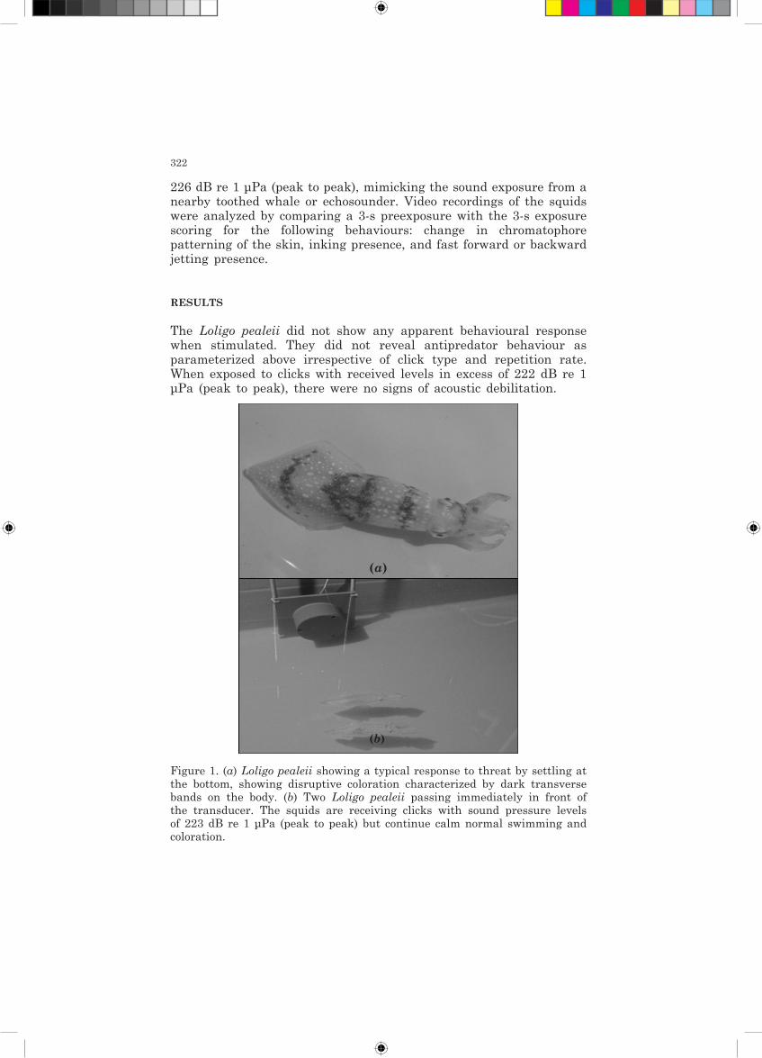
Figure 1. (a) Loligo pealeii showing a typical response to threat by settling at the bottom, showing disruptive coloration characterized by dark transverse bands on the body. (b) Two Loligo pealeii passing immediately in front of the transducer. The squids are receiving clicks with sound pressure levels of 223 dB re 1 μPa (peak to peak) but continue calm normal swimming and coloration.
(a)
(b)

323
DISCUSSION
There were no signs of the antipredator behaviours that could be expected if the squid detected and perceived the ultrasonic sound pulses as coming from an approaching predator. Approaching predators provide several other sensory cues to their prey, and squids might have evolved other ways of detecting an approaching toothed whale. Cephalopods can detect low-frequency particle motions, and it is conceivable that they may detect the low-frequency vortices that a swimming toothed whale creates for every fluke stroke. We demonstrate that intense ultrasonic clicks with received levels up to 226 dB re 1 μPa (peak to peak) do not acoustically debilitate this cephalopod species. Therefore, Loligo pealeii do not seem to detect echolocating toothed whales by their very intense echolocation clicks, and we conclude that echosounders do not affect cephalopods either.
ACKNOWLEDGMENTS
This research was supported by Oticon, Faculty of Science, AU, & FNU.
REfERENCES
Au, W. W. L. (1993). Sonar of Dolphins. New York: Springer-Verlag.Wilson, M., Hanlo, R. T., Tyack, P. L., & Madsen, P. T. (2007). Intense ultrasonic clicks
from echolocating toothed whales do not elicit anti-predator responses or debilitate the squid Loligo pealeii. Biol. Lett. 3, 225-227 (doi:10.1098/rsbl.2007.0005).


