Embed Size (px)
Citation preview
Autophagy is a cell death mechanism in Toxoplasma gondii
Debasish Ghosh1, Julia L. Walton1, Paul D. Roepe2, and Anthony P. Sinai1,*
1Department of Microbiology, Immunology and Molecular Genetics; University of KentuckyCollege of Medicine, Lexington KY 40536, USA2Departments of Chemistry, Biochemistry and Cellular and Molecular Biology, GeorgetownUniversity, Washington DC. 20057, USA
SummaryNutrient sensing and the capacity to respond to starvation is tightly regulated as a means of cellsurvival. Among the features of the starvation response are induction of both translationalrepression and autophagy. Despite the fact that intracellular parasite like Toxoplasma gondiiwithin a host cell predicted to be nutrient rich, they encode genes involved both in translationalrepression and autophagy. We therefore examined the consequence of starvation, a classic triggerof autophagy, on intracellular parasites. As expected, starvation results in the activation of thetranslational repression system as evidenced by elevation of phosphorylated TgIF2α (TgIF2α-P).Surprisingly, we also observe a rapid and selective fragmentation of the single parasitemitochondrion that leads irreversibly to parasite death. This profound effect was dependentprimarily on the limitation of amino acids and involved signaling by the parasite TOR homolog.Notably, the effective blockade of mitochondrial fragmentation by the autophagy inhibitor 3-methyl adenine (3-MA) suggests an autophagic mechanism. In the absence of a documentedapoptotic cascade in T. gondii, the data suggest that autophagy is the primary mechanism ofprogrammed cell death in T. gondii and potentially other related parasites.
KeywordsToxoplasma; starvation; autophagy; mitophagy; eIF2α; TOR
IntroductionAutophagy is a catabolic process in eukaryotic cells involved in the targeted degradation ofcellular organelles and the cytoplasm (Inoue et al., 2010, Mizushima et al., 2010).Autophagy is widely regarded to be critical for cell survival under starvation conditions andfor the turnover of defunct organelles (Kroemer et al., 2010, Kristensen et al., 2008). Themolecular machinery driving this process exhibits a remarkable level of conservation fromyeast to mammals (Yorimitsu et al., 2005). The process of autophagy is triggered by theformation of unique double membranes that mature into autophagosomes and eventuallyfuse with lysosomes, resulting in the degradation of captured components (Weidberg et al.,2011). The execution of autophagy is dependent on 31 ATG proteins described in yeast thatare largely conserved through mammals (Klionsky et al., 2010). Initial bioinformatic studiesin several parasitic protozoa suggest that consistent with their evolutionary antiquity,homologs of many but not all the yeast ATG genes are evident (Duszenko et al., 2011,Herman et al., 2006). This suggests that parasitic protozoa may possess a progenitor ofautophagy as defined in yeast, or, alternatively, use the machinery for parasite specific
*Corresponding author: tel: +1-859-323-6680, fax: +1-859-257-8994, [email protected].
NIH Public AccessAuthor ManuscriptCell Microbiol. Author manuscript; available in PMC 2013 April 1.
Published in final edited form as:Cell Microbiol. 2012 April ; 14(4): 589–607. doi:10.1111/j.1462-5822.2011.01745.x.
NIH
-PA Author Manuscript
NIH
-PA Author Manuscript
NIH
-PA Author Manuscript

functions (Duszenko et al., 2011, Denninger et al., 2008, Luder et al., 2010). With this studywe sought to establish the response of Toxoplasma gondii to starvation stress in order tocharacterize the response of the parasite to what is considered a classic trigger of autophagy.
The protozoan parasite Toxoplasma gondii is an important opportunistic pathogen inimmune compromised individuals as well as a significant cause of morbidity and mortalitywhen vertically transmitted during pregnancy (Tenter et al., 2000, Montoya et al., 2008).Within the mammalian host the parasite exhibits rapid growth as a tachyzoite defining theacute phase of the infection before transforming into the slow growing tissue cyst form(Tenter et al., 2000). The specific triggers for differentiation appear to be linked to theestablishment of immunity although recent evidence points also to physiological andmetabolic triggers (Weiss et al., 2000, Radke et al., 2006). Thus while the intracellular nichethat T. gondii establishes is not typically considered to be nutrient limiting, specialized casessuch as tryptophan limitation in activated macrophages or interferon stimulated fibroblastsare known to limit parasite growth (Pfefferkorn, 1984, Pfefferkorn et al., 1986, Silva et al.,2002). Under such conditions, induction of autophagy within the parasite could serve aprotective role as seen in other systems (Kroemer et al., 2010). Alternatively, activation ofautophagy may promote death by triggering the targeted degradation of critical cellularcomponents (Kourtis et al., 2009, Levine et al., 2009). The notion of autophagy as amechanism of both cell survival and death is emerging in studies from yeast to mammals(Kourtis et al., 2009, Levine et al., 2009, Gozuacik et al., 2007).
In this study we directly examine the impact of nutrient limitation on the induction ofautophagy in T. gondii. We were specifically interested in determining whether potentialautophagy-related processes contribute to parasite survival or death. The absence of a bonafide apoptotic death pathway in protozoa and yeasts suggests autophagy may play a role inprogrammed cell death (Luder et al., 2010). We also probe how nutrient limitation andconsequent translational repression induce the cell biological consequences of starvation.We find that starvation induces the rapid and selective degradation of the single parasitemitochondrion culminating in parasite death. Our findings represent the first functionaldescription of autophagy in an Apicomplexan parasite and integrate key elements of theparasite response to nutrient limitation. These findings effectively establish an autophagymediated programmed cell death pathway in T. gondii, and potentially other protozoa. Thestudy of autophagy in parasites as a potential mechanism of death addresses a vital questionin the development of new drugs (Brennand et al., 2011, Paguio et al., 2011).
ResultsIntracellular Starvation activates stress-induced translational arrest in T. gondii
Extracellular T. gondii are reportedly able to activate the translational arrest response (Weket al., 2006) defined by an increase in TgIF2α phosphorylation (Joyce et al., 2010, Sullivanet al., 2004). As this pathway is activated in diverse systems in response to nutrientlimitation (Wek et al., 2006), we examined the effect of starvation on intracellular T. gondiiby monitoring TgIF2α phosphorylation (TgIF2α-P) levels as complete media (CM, seeexperimental procedures) was progressively diluted with glucose free HBSS (see legend,Figure1). HFF cells infected for 24 hours in complete media were exposed to diluted mediafor 8 hours. Consistent with other cell types, levels of TgIF2α-P for parasite infected HFFcells increased in response to nutrient dilution (Figure 1A). While maximal TgIF2α-P levelsare evident in media diluted to 2% (not shown), the loss of the cell monolayer at this dilutionindicated toxicity to the host cell (Supplemental Figure 1). We therefore selected mediadiluted to 6% (6% Starvation Medium, 6%-SM) as our standard treatment (SupplementalFig 1A). Furthermore, with 6 % SM, we do not observe activation of autophagy in host cellsas defined by appearance of LCIII (lipid modified ATG8) (Mizushima et al., 2010) (data not
Ghosh et al. Page 2
Cell Microbiol. Author manuscript; available in PMC 2013 April 1.
NIH
-PA Author Manuscript
NIH
-PA Author Manuscript
NIH
-PA Author Manuscript

shown). Finally, the level of host cell death, measured by the release of lactatedehydogenase (LDH) was identical for both complete and 6%-SM media in uninfected andinfected cells respectively (Supplemental Figure1B).
The kinetics of phospho-TgIF2α generation were established following exposure of parasiteinfected cells as above in 6%-SM. TgIF2α-P levels are apparent as early as 3-hour poststarvation induction and continued to increase in a time-dependent manner (Figure 1B).
Starved parasites exhibit defective invasion and growth retardationTo assess the consequence of starvation in terms of parasite infection and growth ability, weexamined the competence of starved parasites to infect new host cells and their growththereafter. HFF cells infected for 24 hours in complete media were incubated in 6%-SM foreither 4 or 8 hours. The intracellular parasites were liberated by syringe passage andenumerated. Equal numbers of parasites were added to fresh monolayers grown on coverslips and allowed to infect for 24 hours in CM. The number of vacuoles per field was usedas a measure of the invasive capacity. In addition, the distribution of parasite number pervacuole served as a measure of parasite growth.
Infection of fresh HFF monolayers with parasites maintained throughout in CM resulted inthe establishment of on average 7.2 vacuoles per field (defined as 100%; Figure 1C). Theeffect of 6%-SM treatment for 4 and 8 hours results in 25% and 80% reduced vacuolecounts, respectively (Figure 1C). In addition, vacuoles established by starved parasitesexhibited growth retardation relative to those grown in CM throughout (Figure 1D). Bothdefects are likely due at least in part to translational arrest indicated by elevated TgIF2α-P(Figure 1B) (Joyce et al., 2010). Additionally, starvation may induce cellular andphysiological changes outside of translational arrest that also promote these defects.
Starvation results in selective mitochondrial fragmentation in T. gondiiIn addition to translational arrest, the selective degradation of expendable organellesprovides a means of coping with starvation (Kanki et al., 2008, Kristensen et al., 2008). Toaddress whether starvation triggered organelle degradation in T. gondii, we examined themorphology of key parasite organelles following 8 hours in 6%-SM. Since significantinhibition of parasite invasion was a consequence of intracellular starvation (Figure 1C), weexamined the morphology of rhoptries and micronemes (Carruthers et al., 2007).Interestingly, the organization of these organelles were unaffected by exposure to 6%-SMfor 8 hours (Figure 2A), even though they play a critical role in parasite attachment andinvasion (Carruthers et al., 2007). Similarly the apicoplast, a relict plastid-derived organelle(Lim et al., 2010, Waller et al., 2005) appeared unaffected in starved parasites (Figure 2A).
The single parasite mitochondrion (Melo et al., 2000, Seeber et al., 1998, Vivier et al.,1972), detected with an antibody against the F1β–subunit of the mitochondrial H+ ATPase,normally assumes a ring-shaped appearance when T. gondii are grown in CM (Figure 2B,F1β). In contrast, a substantial number of vacuoles in 6%-SM displayed punctate orotherwise abnormal mitochondrial morphology (Figure 2B and see below). Mitochondrialfragmentation was observed to increase dramatically as a function of dilution of the culturemedia (Supplemental Figure1A). Increased fragmentation correlated with increased TgIF2α-P levels (Figure 1A) suggesting the effect was also a consequence of reduced nutrientavailability. In addition, starvation induced mitochondrial starvation was seen not only in theType I RHΔHX strain used here but also in the avirulent cyst forming Type II ME49 (datanot shown). Finally, RH parasites expressing HXGPRT responded identically as does thedeletion strain (data not shown).
Ghosh et al. Page 3
Cell Microbiol. Author manuscript; available in PMC 2013 April 1.
NIH
-PA Author Manuscript
NIH
-PA Author Manuscript
NIH
-PA Author Manuscript

Starvation induced mitochondrial fragmentation occurs sequentiallyWe next examined the kinetics of mitochondrial fragmentation (Figure. 3). Infected cellmonolayers were fixed at hourly intervals and the morphology of the mitochondrionquantified (see legend, Figure 3). Progression from normal, ring-shaped morphology topuncta occurred via intermediate forms. These forms appeared, in turn, as linearmitochondria, patterns reminiscent of beads on a string, and then as dumbbell shapes withbulbous ends connected by thin segments (Figure 3A, F1β top to bottom).
Normal (ring shaped), punctate, and intermediate morphologies were quantified. As shownin Figure 3B, the progression from normal morphology to puncta occurs via the intermediateforms. The changes in mitochondrial morphology are evident as early as 3 hours followingtransfer to 6%-SM, with the first appearance of punctate mitochondrial fragments at 4 hourspost starvation. By 6 - 7 hrs even punctate forms disappear altogether (Figure3A bottomF1β, Figure3B diamond symbols),
We next tested whether parasites with morphologically abnormal mitochondria wereinvasion competent. Parasites exposed to 6%-SM for 8 hours harvested from the firstmonolayer, comprised the inoculum for a second fresh monolayer (Figure 3C). As expected,this inoculum exhibited a distribution of normal and altered mitochondrial morphologiesessentially identical to those in the original monolayer (Figure 3C, cytospin). Notably, while70% of the inoculum had abnormal mitochondria, all of the invasion competent parasitesestablishing vacuoles with GRA3 positive PVM’s had mitochondria with normalmorphology following a 2 hour invasion assay (Figure 3C). Restoration of normalmorphology for those observed to be fragmented prior to harvesting, was not observed uponthe addition of CM, even after a 20 hour incubation (data not shown). Taken together, thesedata indicate that fragmentation is irreversible and that the integrity of the mitochondrion isvital for invasion. This is intriguing since localized glycolysis at the site of the motorcomplex is believed to power the invasion machinery (Pomel et al., 2008), although theability of T. gondii to use glutamine to power the tricarboxilic cycle provides a bypasspathway in the absence of glucose (Blume et al., 2009).
A limitation of immunofluorescence analysis to monitor the integrity of the mitochondrionis the possibility that starvation results in the reorganization and loss of the marker F1β butnot the organelle itself. We therefore examined the organization of mitochondria byconventional transmission electron microscopy. Cross sections of the parasite mitochondrionare easily recognized based on their possession of the annular cristae (Melo et al., 2000)(Figure 4A). Also evident are rhoptries and micronemes (Dubey et al., 1998) (Figure 4A).Consistent with the IFA data, no significant changes were observed in the organization ofthe rhoptries or micronemes in 6%-SM treated cells (Figure 4B).
In contrast to the secretory organelles, dramatic changes consistent with the IFA results(Figure 3A) were noted for the mitochondrion by TEM (Figure 4B). Among these changeswere mitochondrial profiles with marked constrictions and bulbous ends (Figure 4B, top 3panels) as well as mitochondria that appear to have lost their integrity evident from thediscontinuity in their limiting membranes as well as an apparent “discharge” of cristae intothe cytoplasm (Figure 4B, brackets, lower panels). The ultrastructural data providecompelling evidence that starvation results in a selective fragmentation of the T. gondiimitochondrion but does not directly address the underlying mechanism for the phenotype.
Starvation-induced mitochondrial fragmentation occurs by an autophagic mechanismTo test whether starvation induced mitochondrial fragmentation in T. gondii involves anautophagic mechanism we investigated the effect of 3-methyl adenine (3-MA). 3-MA is anestablished selective inhibitor of the Class III PI3 kinase Vps34 and is a reversible inhibitor
Ghosh et al. Page 4
Cell Microbiol. Author manuscript; available in PMC 2013 April 1.
NIH
-PA Author Manuscript
NIH
-PA Author Manuscript
NIH
-PA Author Manuscript

of autophagy that blocks the recruitment of lipid to the autophagosome (Abeliovich et al.,2001, Seglen et al., 1982, Yue et al., 2010). The T. gondii genome encodes Vps34 homolog(TGGT1_126620) (Supplemental Table 1). 3-MA treatment of infected cells in CM had noeffect on the integrity of mitochondria (Supplemental Figure 2A). In contrast, while 6%-SMresulted in mitochondrial fragmentation, the addition to 3-MA to 6%-SM effectivelyblocked fragmentation (Figure 5A,B). This blockade of starvation induced mitochondrialfragmentation by a selective inhibitor of autophagy indicates that mitochondrialfragmentation occurs by an autophagic mechanism. Notably, the presence of 3-MA had noimpact on the morphology of the rhoptries, micronemes or apicoplast in either complete(data not shown) or starvation media (Supplemental Figure 3).
In order to establish the kinetics of the 3-MA mediated blockade of starvation inducedmitophagy we staggered the addition of the drug to parasites in 6%SM. As expected,incubation of infected cells in 6%SM + 3-MA blocked for the 8 hour duration of theexperiment completely blocked mitophagy. This blockade was effectively instituted even if3-MA was added 3 hours into the 8-hour incubation. Notably, a 3-hour incubation with6%SM does not significantly trigger the starvation-induced phosphorylation of TgIF2α(Figure 1B). In contrast, the addition of 3-MA following 4 or 5 hours of starvation results inthe accumulation of the intermediate morphology without significant progression to thepunctate form. Effectively, the addition of 3-MA at these time points freezes the progressionof mitochondrial fragmentation at levels similar to what is seen at these time points (Figure5B). This pattern is observed when 3-MA is added 6 and 7 hours post starvation as well(Figure 5C). Together these data indicate 2 key points: 1. 3-MA effectively blocks theprogression of starvation induced mitophagy arresting further fragmentation at the time ofits addition. 2. Both the initial formation of the “intermediate” forms and their progression tothe punctate forms are sensitive to 3-MA suggesting an autophagic basis for both events.
While blocking mitophagy, 3-MA had no significant effect on the phosphorylation ofTgIF2α in 6%-SM media indicating that it does not interfere with translational repression(Supplementary Figure 2B). Finally, the addition of 3-MA to 6%SM completely rescued theinvasion defect observed in 6%SM starved parasites (Figure 5D). Taken together, these dataindicate that loss of mitochondrial integrity, but not starvation itself or starvation inducedtranslational repression are the key determinants responsible for the invasion defect whichfor T. gondii is a lethal event.
The effect of 3-MA on parasite growth also includes partial rescue of growth retardationfollowing infection of a fresh monolayer in complete media relative to starvation mediaalone (Figure 5E). This is not entirely surprising as 3-MA has recently been shown to causea reversible cell cycle arrest in T. gondii (Wang et al., 2010). In addition the potential for 3-MA mediated inhibition of host autophagy could further limit amino acid availability for theparasite further exacerbating the starvation condition (Wang et al., 2009).
Morphological evidence of autophagosomes in T. gondiiAutophagy progresses in a highly regulated manner that is initiated by the de novo formationof the phagophore and isolation membrane in the cytoplasm (Figure 6A) (Weidberg et al.,2011). The isolation membrane is comprised of 2 bilayers which form the autophagic cupthat corrals both cytoplasm and organellar components into an enclosed autophagsome(Weidberg et al., 2011). These fuse with hydrolytic compartments resulting in thedegradation of the contents and the generation of the building blocks to be recycled duringnutrient limitation (Weidberg et al., 2011).
We examined TEM sections of parasites incubated in 6%-SM to establish whether thecharacteristic double membrane autophagosomal intermediates could be detected. One
Ghosh et al. Page 5
Cell Microbiol. Author manuscript; available in PMC 2013 April 1.
NIH
-PA Author Manuscript
NIH
-PA Author Manuscript
NIH
-PA Author Manuscript

confounding issue in identifying such structures is the presence in T. gondii of the 4-membrane containing apicoplast (Kohler, 2005) and the absence of specific autophagicmarkers for immune-electron microscopy. Notwithstanding these caveats, our ability todetect what appear to be double and/or multiple membrane containing structures, often inthe vicinity of aberrant and fragmented mitochondria that appear to be engulfing thecytoplasm are highly suggestive of autophagosomes (Figure 6). While several of thesestructures appear to possess 3-4 membranes, a feature of the apicoplast, they could alsorepresent a putative double membrane autophagosome that has engulfed a mitochondrialfragment, itself possessing 2 membranes. Confirmation of these structures asautophagosomes will require the generation of specific markers, as the molecular machineryis unraveled.
Loss of energy metabolites are not the primary triggers for mitophagyMitophagy in yeast occurs through a selective mechanism and is triggered by the limitationof both energy metabolites and amino acids (Kanki et al., 2008, Youle et al., 2011, Singh etal., 2011). Given the selective targeting of mitochondria by nutrient limitation we examinedwhether starvation for glucose and pyruvate were sufficient to trigger mitophagy in T.gondii. Cells infected for 24 hours in complete CM were shifted to glucose-free, pyruvate-free or glucose and pyruvate free media for 4 or 8 hours. We monitored the effect on theintegrity of mitochondria using immunofluorescence. Starvation for energy metabolites didresult in changes in the organization of the mitochondrion, which were considerably lessextreme that treatment with 6%-SM (Figure 7A). Specifically, mitochondrial fragmentationwas largely arrested at the intermediate stages and few vacuoles displayed punctate forms(Figure 7A).
While the impact on mitochondrial integrity was modest, limitation of energy metaboliteswas a stress as TgIF2α-P levels were elevated suggesting translational inhibition (Figure7B). The slower progression of mitophagy following starvation for energy metabolitessuggests that the effect may be secondary due to an impact on ATP-dependent processessuch as amino acid transport.
The primary mechanism involved in sensing energy flux involves the cyclic AMP-activatedprotein kinase (AMPK) an activity that has been extensively studied in higher eukaryotes(Hardie, 2008, Rutter et al., 2003) but not in parasitic protozoa. The Toxoplasma genomeencodes a potential AMPK homolog (TGGT1_113440, a 46 KD putative serine/ threonineprotein kinase with 58% identity to Human AMPK alpha). This led us to examine the effectof 2 AMPK agonists - metformin and AICAR (Hardie, 2008, Lee et al., 2011). Treatmentwith these drugs at high concentrations for up to 24 hours had a limited if any effect onmitochondrial integrity (Supplemental Figure 4), reinforcing the interpretation that the effectof glucose/pyruvate starvation on mitochondrial integrity is secondary as is likely energymetabolism in general.
Similar starvation experiment where we removed micronutrients (vitamins and lipoic acid)failed to trigger any significant changes in the morphology of the parasite mitochondrion(Supplementary Figure 5). The absence of a requirement for micronutrients (SupplementaryFigure 5) and the modest impact of energy metabolites and drugs affecting energymetabolism (Figure 7, Supplemental Figure 4) suggest that starvation-induced mitophagy islinked to amino acid levels.
Mitophagy is triggered by amino acid starvation and is TgTOR dependentAmino acid deprivation has been established as a primary trigger of autophagy, includingmitophagy (Wang et al., 2011), in both yeast and mammalian cells (Mortimore et al., 1977,
Ghosh et al. Page 6
Cell Microbiol. Author manuscript; available in PMC 2013 April 1.
NIH
-PA Author Manuscript
NIH
-PA Author Manuscript
NIH
-PA Author Manuscript

Meijer, 2008, Onodera et al., 2005). Addition of amino acid free media to HFF cells infectedwith T. gondii for 24 hours resulted in a rapid induction of mitophagy with changes in themitochondrial architecture being evident as early as 1 hour following amino acid deprivation(Figure 8A). The progression to punctuate mitochondria was similarly rapid and accelerated(Figure 8A) relative to incubation in 6%-SM (Figure 3B). This rapid mitochondrialfragmentation occurred in all parasites within 8 hours following transfer to amino aciddeprived media, with approximately 10% lacking a detectable mitochondrion (Figure 8A).Consistent with other systems (Wek et al., 2006, Kroemer et al., 2010), amino aciddeprivation resulted in a robust induction of TgIF2α-P levels indicating the activation of thetranslational repression system (Figure 8B).
Phosphorylation of TgIF2α is mediated by one or both of the two homologs of eIF2α-kinase,which are homologs of the yeast kinase GCN2 (Sullivan et al., 2004, Narasimhan et al.,2008). In yeast GCN2 itself is repressed by TOR mediated phosphorylation (Figure 8C)(Wek et al., 2006). The target of rapamycin (TOR) kinase is a vital component of the aminoacid sensing mechanism in eukaryotic cells (Proud, 2007, Dann et al., 2006). TheToxoplasma genome (www.toxodb.org) is reported to encode a single TOR homolog(TgTOR, TGGT1_094250, a 263 KD putative fkbp-rapamycin associated protein with 44%identity with Human mTOR kinase (Brown et al., 1994)). Recent studies in mammaliancells and yeast establish TOR-kinase serves as a signaling hub that integrates both theautophagic response and translational repression in response to amino acid starvation(Figure 8C) (Kim et al., 2011b, Meijer, 2008, Egan et al., 2011, Kroemer et al., 2010,Kundu, 2011).
Rapamycin is a well established inhibitor of the TOR-complex both in yeast (Chang et al.,2009, Kunz et al., 1993) and mammalian cells (Huo et al., 2011). It has been shown to begrowth inhibitory in the extracellular Trypanosoma brucei (Barquilla et al., 2008). Wereasoned that if TgTOR was in fact an integral component of the amino acid sensingmachinery in T. gondii, then rapamycin treatment should mimic amino acid starvation. Wetherefore incubated infected cells in CM with increasing levels of rapamycin for 8 hours.Treatment at < 500nM had a limited effect on mitochondrial integrity (Figure 8D), however,exposure to ≥ 1μm resulted in the detection of punctuate mitochondria in approximately80% of the vacuoles (Figure 8D). Typically, 50 – 500nM rapamycin induces autophagy inmammalian cells, but this requires incubation times of 24-72 hours as opposed to the 8 hoursused here (Huo et al., 2011). In neurons an absence of rapamycin toxicity at doses as high as3μM for 72 hours has been reported (Pan et al., 2009). We were nonetheless concerned thatthe effects seen on the T. gondii mitochondrion 1μM may reflect non-specific toxicity andnot autophagy. To address this point we tested whether 3-MA could block the rapamycininduced mitochondrial fragmentation (Figure 8D, right hand side). The observation thatinhibition of autophagy with 3-MA effectively blocks rapamycin induced mitophagyconfirms that the T. gondii TOR pathway directly integrates amino acid sensing and theparasite’s autophagic response.
Starvation induced mitochondrial fragmentation correlates with the loss of membranepotential
Involvement of autophagy in mitochondrial depolarization has been evident in both yeastand mammalian systems upon nutrient starvation (Rodriguez-Enriquez et al., 2009,Rodriguez-Enriquez et al., 2006, Okamoto et al., 2011, Zhang et al., 2007). To establishwhether starvation-induced changes in mitochondrial mitochondrial morphology areassociated with the loss of the membrane potential (ΔΨm) we used the membrane potentialsensitive dye MitoTracker. Consistent with growth in complete media, accumulation ofMitoTracker is evident in both the parasite mitochondrion and host mitochondria (Figure9A,B, CM). The replacement of CM with 6%SM results in changes in the morphology of
Ghosh et al. Page 7
Cell Microbiol. Author manuscript; available in PMC 2013 April 1.
NIH
-PA Author Manuscript
NIH
-PA Author Manuscript
NIH
-PA Author Manuscript

the parasite mitochondrion. While the ‘intermediate ”phenotype mitochondria continue toretain the ΔΨm noted by the accumulation of Mitotracker (Figure 9A second row), thepunctate mitochondria exhibit only diffuse staining with the dye indicative of a loss of themembrane potential. This is not surprising as the TEM evidence indicates that while theintermediate-stage mitochondrion is morphologically altered, it still retains the integrity ofits membranes (Figure 4). In contrast, the apparently punctate mitochondria exhibit abreakdown of membrane integrity precluding the establishment of the requisiteelectrochemical gradient. This is also evident in row 4 of Figure 9A where a vacuole with anormal mitochondrial morphology is adjacent to one with punctate mitochondria. Finally,the loss of host mitochondrial ΔΨm was observed in monolayers treated with 6%SM for 8hours (Figure 9A, row 3,4). The loss of the host mitochondrial potential could be to thedepletion of energy metabolites (glucose/pyruvate) or amino acids.
In light of the mitophagy phenotype being linked to amino acid limitation we investigatedthe effect of amino acid starvation on the mitochondrial membrane potential. Consistentwith the observations with 6%SM (Figure 9A), the membrane potential was retained inintermediate stage mitochondria (Figure 9B row 2) but depleted in parasites containingpunctate mitochondria. In these cases, mitotracker staining was weak and diffuse within theparasites (Figure 9B rows 3,4). In contrast to what was observed in 6%SM however, aminoacid deprivation had no impact on the ability of host mitochondria to accumulateMitoTracker even following an 8-hour incubation (Figure 9B row 3,4). These data suggestthat the loss of host ΔΨm in 6%SM is a consequence of the depletion of energy metaboliteswhile the loss of parasite ΔΨm is associated with the loss of mitochondrial integrity, aphenotype triggered by amino acid starvation.
Bioinformatic evidence reveals the presence and absence of ATG genesThe genes associated with autophagy have been studied in considerable detail in the yeastSaccharomyces cerevisiae (Klionsky et al., 2010, Mizushima, 2007). We interrogated the T.gondii genome (www.toxodb.org) to identify potential homologs of key autophagy related(Atg) genes. What is immediately evident is that while several homologs can be readilyidentified, others appear to be missing (Supplemental Table 1).
Several of the putative homologs, including TgATG1, TgATG3, TgATG7, TgATG8,TgATG18, TgATG20 and TgVPS34 have respectable identity (similarity) scores and maybe considered to be true homologs (Supplemental Table 1). In contrast the assignment ofseveral other genes with identity scores < 30% must be considered provisional assignmentssubject to experimental confirmation (Supplemental Table 1). Notably, a block of apparentlymissing activities include key members of the cytoplasm to vacuole (Cut) pathway (Lynch-Day et al., 2010, Teter et al., 2000) and components of the ATG12-ATG5 conjugationmachinery (Inoue et al., 2010). While a potential homolog of TgATG5 was identified, thepoor identity score complicates a definitive assignment (Supplemental Table 1).
The bioinformatic analysis does however indicate that genes for conventional autophagy areevident. Furthermore the finding that several of the putative TgATG genes that shouldpossess key enzymatic activities (e.g. protein kinase –TgATG1, lipid kinase -TgVPS34,lipid conjugation systems -TgATG7, -TgATG5) are predicted to be substantially larger thantheir yeast counterparts (Supplemental Table 1) suggests additional parasite specificfunctions that could be independent of “classical” starvation induced autophagy. Futurestudy of these important genes may therefore uncover novel aspects of parasite biology.
Ghosh et al. Page 8
Cell Microbiol. Author manuscript; available in PMC 2013 April 1.
NIH
-PA Author Manuscript
NIH
-PA Author Manuscript
NIH
-PA Author Manuscript

DiscussionOur understanding of autophagy and its role in cellular homeostasis is evolving (Mizushima,2007, Kroemer et al., 2010). What was once seen primarily as a cellular catabolicmechanism to deal with nutrient limitation is now seen to be integrated into the centralmetabolic regulation of the cell (Kroemer et al., 2010). The role of autophagy in normalcellular homeostasis centers on the elimination of defective organelles (most notablymitochondria) and excess cytoplasm (Sakai et al., 2006, Wang et al., 2011, Weidberg et al.,2011, Kristensen et al., 2008). Under conditions of nutrient limitation, the autophagiccascade assumes a cell survival function. This is achieved by selectively targeting excesscapacity in the cell for degradation thereby increasing pools of nutrients to sustain cellularfunctions (Weidberg et al., 2011, Kristensen et al., 2008). Cellular degradation pathways areunder considerable control as degradation taken to an extreme results in the breakdown ofcritical components leading to cell death (Assuncao Guimaraes et al., 2004, Blommaart etal., 1997, Gozuacik et al., 2007). Thus autophagy, while a cell survival mechanism incertain contexts, is a mediator of cell death in others. An autophagy mediated cell death isclassified as a Type II death pathway that has a distinct progression from that of the Type Iapoptotic death (Gozuacik et al., 2007, Kourtis et al., 2009). In the absence of clear evidencefor apoptotic pathways in early branching eukaryotes, autophagy presents a potentialmechanism for programmed cell death (Luder et al., 2010, Duszenko et al., 2011). Such anassignment must however be made with caution as a death pathway caused by autophagy isoften difficult to distinguish from a death mechanism that involves autophagy as acomponent of its execution (Galluzzi et al., 2009, Galluzzi et al., 2011).
The extent of mechanistic similarity of autophagy, between yeast and mammalian systemspoints to a high degree of functional conservation (Inoue et al., 2010, Mizushima et al.,2010). Much less is known about the autophagic pathways in truly early branchingeukaryotes (Duszenko et al., 2011, Odronitz et al., 2007, Herman et al., 2006). We chose tofunctionally investigate whether or not the protozoan parasite Toxoplasma gondii possessesan autophagy related pathway by investigating the response of the parasite to nutrientlimitation, a classical autophagic trigger.
Nutritional stress triggered by incubation of infected cells in diluted complete growth media(CM) results in the activation of the translational repression pathway defined by theincreases in levels of TgIF2α-P (Figure 1A,B) (Joyce et al., 2010, Sullivan et al., 2004).Interestingly, starvation for as few as 4 hours, rather than establishing a state of stasis,results in parasite death. This is defined by a marked defect in their ability to invade a freshmonolayer (Figure 1C). Despite exhibiting an invasion defect, the integrity of the invasionrelated organelles (micronemes and rhoptries) (Carruthers et al., 2007) was not affected.Rather we observed a rapid and sequential fragmentation of the single parasitemitochondrion (Figure 3A). The kinetics of fragmentation proceeds from the normal ringshape (Melo et al., 2000), via linear intermediates, to puncta all the way to the loss of theorganelle (Figure 3A,B). Evidence based on the incorporation of Mitotracker, amitochondrial membrane potential (ΔΨm) sensitive dye, suggests that mitochondria retaintheir ΔΨm despite morphological alteration, losing it only when the mitochondrion is trulyfragmented (Figure 9).
It is easy to envision the loss of viability in a parasite that has lost its mitochondrion, ourinability to observe invasion by parasites exhibiting any mitochondrial defects wassurprising (Figure 1C). This is remarkable given the fact that invasion is believed to bepowered by glycolysis (Pomel et al., 2008, Polonais et al., 2010). Furthermore, our findingthat once fragmentation is initiated, it is irreversible (Figure 5C) suggests loss ofmitochondrial integrity is a marker of parasite death.
Ghosh et al. Page 9
Cell Microbiol. Author manuscript; available in PMC 2013 April 1.
NIH
-PA Author Manuscript
NIH
-PA Author Manuscript
NIH
-PA Author Manuscript

The systematic fragmentation and elimination of the mitochondrion is reminiscent ofstarvation-induced mitophagy observed in yeast and mammalian cells (Bhatia-Kissova et al.,2010, Kim et al., 2007, Youle et al., 2011). A critical difference lies in the fact that yeastand mammalian cells possess multiple mitochondria representing an excess capacity that canbe regenerated upon the return of nutrient replete conditions (Bhatia-Kissova et al., 2010,Youle et al., 2011). With its single mitochondrion (Melo et al., 2000), T. gondii does nothave the luxury of dispensing with this critical organelle.
Autophagy is a sequential process that is initiated by a cascade of events initiated by the denovo formation of a unique double membrane entity termed the phagophore whichsurrounds cytoplasm and organelles destined for clearance into a autophagosome that fuseswith a lysosomal compartment resulting in the breakdown of the contents (Figure 6A)(Weidberg et al., 2011, Kristensen et al., 2008). A key activity in yeast (and othereukaryotes) is Vps34, a class III PI3 kinase that is involved in membrane recruitment to theautophagosome (Yue et al., 2010). This enzyme is selectively and competitively inhibited by3-methyl adenine (3-MA), which is viewed as a selective inhibitor of autophagy (Yue et al.,2010, Abeliovich et al., 2001, Seglen et al., 1982). 3-MA is well tolerated by T. gondii as itis reversible and can be used to synchronize the parasite cell cycle (Wang et al., 2010). Ourfinding that the addition of 3-MA effectively blocked the starvation-induced fragmentationof the mitochondrion (Figure 5A,B) strongly implicates an autophagic mechanism in theselective mitophagic response. More importantly, while the presence of 3-MA did notimpact the starvation-induced induction of translational repression (Supplemental Figure 2),it did rescue the starvation-induced inhibition of parasite invasion (Figure 5D).
A morphological signature of autophagy is a double (or multiple) membrane structureengulfing either organelles or the cytoplasm (Abeliovich et al., 2000, Hamasaki et al., 2010,Weidberg et al., 2011). A confounding issue in T. gondii and other Apicomplexa is theexistence of the 4-membraned apicoplast, a relict non-photosynthetic plastid derivedendosymbiont (Kohler, 2005). It should be noted, that the frequency of double/multi-membrane structures in starved cells was qualitatively higher by TEM (Figure 6, data notshown). However, the organization and number of apicoplasts in starved parasites wasindistinguishable from those grown in complete media by IFA analysis (Figure 2A). Whilenot confirmatory, these observations are suggestive of the double/multi-membrane structuresseen by electron microcopy representing distinct stages of autophagosome formation. Thefact that several of these entities are not entirely membrane enclosed further suggest they arenot apicoplasts (Figure 6B).
The sheer size of the single mitochondrion evident both by IFA and TEM (Melo et al., 2000,Seeber et al., 1998) suggests that the intact organelle is an unlikely target for the autophagy-mediated breakdown. Consistent with this view, we observe a sequential breakdown in thecourse of starvation-induced mitochondrial fragmentation (Figure 3AB), which itself isblocked by 3-MA (Figure 5A). This suggests that activities involving PI3K/Vps34 play arole in mitochondrial fission consistent with what has been recently reported in mammaliancells (Kim et al., 2011a). In cells undergoing mitophagy, mitochondria that elongate evadeautophagic destruction assuring a pool of protected organelles (Gomes et al., 2011). Giventhe fragmentation of the single parasite mitochondrion, a protective role during starvationdoes not appear to be likely.
The mitophagy response to starvation appeared to be directly linked to amino acid sensing(Figure 8) and not a response to energy metabolites (Figure 7) or micronutrients in the media(Supplemental Figure 5). Amino acid sensing in higher eukaryotes, including S. cerevisiae,is regulated by the TOR-complex (target of rapamycin) (Dann et al., 2006, Goberdhan et al.,2009, Noda et al., 1998) which recently has been shown to integrate glucose sensing (via
Ghosh et al. Page 10
Cell Microbiol. Author manuscript; available in PMC 2013 April 1.
NIH
-PA Author Manuscript
NIH
-PA Author Manuscript
NIH
-PA Author Manuscript

AMPK) and amino acid sensing to autophagy and translational repression (Figure 8C)(Hardie, 2008, Kim et al., 2011b). A homolog of TOR has been identified in theToxoplasma genome (TGGT1_094250) suggesting susceptibility to rapamycin. Indeed at ahigh concentration of rapamycin fragmentation of the parasite mitochondrion is evident, aphenotype that is largely blocked in the presence of 3-MA (Figure 8D), thereby linking thepredicted TOR cascade to the autophagic response (Kim et al., 2011b).
The presence of a sensitive switch responsive to amino acid starvation that leads to a rapiddeath presents an enigma. The intracellular pools of amino acids derived from synthesis andturnover, coupled with transport from the extracellular environment suggest amino acids arenot likely to be limiting under normal conditions (Sinai, 2008, Laliberte et al., 2008, Sinai etal., 1997a). Quite to the contrary, an increasing literature indicates that T. gondii and otherintracellular pathogens actively induce autophagic pathways in the infected host cell,potentially as a means of increasing free amino acid pools (Wang et al., 2009, Subauste,2009, Orlofsky, 2009, Halonen, 2009).
One amino acid, for which the parasite is auxotrophic is tryptophan (Pfefferkorn, 1984,Pfefferkorn et al., 1986). Tryptophan is known to become limiting in an activatedmacrophage where it contributes to parasite killing (Pfefferkorn, 1984, Pfefferkorn et al.,1986). This may provide a clue to why such a sensitive programmed cell death pathway,triggered by amino acid limitation may be retained within the parasite. Recent work fromseveral laboratories has demonstrated that the capacity to present both endogenous as well asmodel antigens is dependent on the context in which the antigen is delivered (Jordan et al.,2010, Blanchard et al., 2010, Dzierszinski et al., 2007, Dzierszinski et al., 2008). Thepresentation of antigens detected only in the context of dead parasites (with epitopes inintracellular proteins) could present a selective advantage in the course of infection bydistracting the immune response from away from viable organisms. Accelerating death bythe activation of an autophagy based parasite death pathway may increase the pool of non-protective decoy epitopes.
Nutrient-limitation induced autophagy may also play a role in the transmission stages of theparasites, the tissue cyst and oocysts (Dubey, 1998, Dubey et al., 1998). One may postulatethat nutrient limitation within a cyst may be alleviated by the activation of a bulk proteinturnover mechanism like autophagy-although no direct evidence for this has been reported.Finally, programmed death mechanisms in protozoa have been suggested to be triggered as ameans to controlling cell density in the host (Luder et al., 2010). Whether such a strategyapplies to an intravacuolar pathogen is not known.
The pharmacological evidence for the presence of an autophagic pathway (Figure 5,8) issupported for by the bioinformatic analysis that reveals homologs of several key autophagygenes are encoded in the parasite genome, while others are not evident (Supplemental Table1).
In summation, we find that autophagy related processes in T. gondii are activated inresponse to amino acid limitation with rapid kinetics leading to selective mitophagy anddeath. As an activatable and irreversible cell death pathway, the autophagic cascade presentsa very attractive pathway for the development of drugs (Brennand et al., 2011, Paguio et al.,2011). Investigations into this pathway in an earlier branching eukaryote has the potential toreveal novel aspects in parasite biology while enriching our understanding of autophagy andits evolution into a highly specialized pathway.
Ghosh et al. Page 11
Cell Microbiol. Author manuscript; available in PMC 2013 April 1.
NIH
-PA Author Manuscript
NIH
-PA Author Manuscript
NIH
-PA Author Manuscript

Experimental ProceduresReagents
Base cell culture media include α-MEM and Glucose and Pyruvate-free MEM (Invitrogen,Carlsbad, CA). MEM Vitamin solution was purchased from GIBCO (Life Technologies/Invitrogen, Carlsbad, CA). 3-methyladenine (3-MA), Metformin (1, 1 – Dimethylbiguanidehydrochloride), Lipoic acid and AICAR (5-Aminoimidazole-4-carboxamide riboside) werepurchased from Sigma (St. Louis, MO). Rapamycin was purchased from EMD Biosciences(Calbiochem, Gibbstown, NJ).
Cell Lines, Parasite and starvation mediumLow passage Human foreskin fibroblast (HFF) cells (ATCC) and Vero cells (ATCCCCL-81) were maintained in α-MEM supplemented with 7% heat inactivated FBS (GeminiBio-Products, West Sacramento), 100 U/ml penicillin, 100 mg/ml streptomycin, and 2 mML-Glutamine (complete medium) (CM) (Gibco BRL, Rockville, MD). All cell lines weregrown as monolayers at 37°C in an atmosphere of 5% CO2. The type I RHΔHX strain ofToxoplasma gondii (NIH-AIDS Resource Center) were used throughout the study andmaintained by serial passage in Vero cells as previously described (Carmen et al., 2006). Allexperiments to investigate the role of starvation were conducted in infected HFF cells(passage <8) allowed to be confluent for at least 1 week. Starvation media (SM) comprisedcomplete αMEM (CM) diluted in 1X Hanks Balanced Salts Solution (HBSS) to a finalconcentration from 2% - 50%. Glucose and pyruvate free (Gibco) was supplemented asabove for complete αMEM with the exception of the use of dialyzed FBS. Reconstitution ofglucose, pyruvate or both compounds was achieved by the addition of cell culture gradeglucose (5.56mM) and/or pyruvate (1mM) respectively. Amino acid starvation medium wasmade by adding 1 mM sodium pyruvate, 5.56 mM cell culture grade glucose, vitaminsolution (1X), and lipoic acid (0.2 mg/ L) to 1X HBSS solution, without supplemental L-Glutamine. Dialyzed serum was added as above.
Induction of starvationA confluent HFF cell monolayer in either a 6-well plate (for immunoblot) or on a cover slipin a 24-well tissue culture dish (for IFA) (Falcon) was infected with 5×10⁵ or 2×10⁵ freshlypassaged parasites for 24 hours. The infected cells were washed with 1X PBS, and eitherfresh complete media or SM added for up to 8 hours. For experiments involvingpharmacological treatments (3-MA, Metformin, AICAR or rapamycin) the drug treatmentswere added in either CM (3-MA, Metformin, AICAR, rapamycin) or SM (3-MA) media forthe time points and at the concentrations indicated in the figure legends. 3-MA was used at aconcentration of 10mM.
Impact of starvation on invasion and growthHFF cell monolayer in a 10 cm dish was infected with freshly passaged 1×10⁷ parasites for24 hours. Infected monolayers were washed with 1X PBS, and transferred to the 6%-SM for4 and 8 hours with or without 3-MA. Control plates were returned to complete media.Intracellular parasites were harvested by syringe passage and enumerated. 2×10⁵ parasiteswere allowed to infect fresh HFF monolayers grown on the cover slips for 24 hours incomplete media. Infected HFF monolayers were fixed and the number of vacuoles per field(defined staining with the PVM marker GRA3) as well as the distribution of parasites pervacuole (defined using the surface marker SAG1) determined. These serve as measures ofparasite invasive ability and replication respectively. In addition, a portion of the inoculumwas pelleted onto a glass slide using a cytospin centrifuge, fixed and examined for theintegrity of the mitochondrion as described below. In addition the integrity of the
Ghosh et al. Page 12
Cell Microbiol. Author manuscript; available in PMC 2013 April 1.
NIH
-PA Author Manuscript
NIH
-PA Author Manuscript
NIH
-PA Author Manuscript

mitochondrion in the newly infected cell was established using anti-F1β. Specific conditionsfor immunofluorescence analyses are described below. At least 50 independent fields fromthe control, 4 and 8-hour starvation treatments were randomly chosen for the quantificationof invasion efficiency while at least 100 PV’s were counted for their parasite burden- ameasure of parasite growth.
Host viability assayNecrosis was examined by measuring the LDH activity as a marker for cellular necrosisusing LDH cytotoxicity assay kit (Cayman Chemical, Ann Arbor MI), according tomanufacturer’s instructions. Briefly, HFF monolayers seeded in a 96-well plate wereinfected with 2 × 10⁴ no. of RH parasites for 24 hours. Following infection, the completemedia were switched to 6% starvation media for 8 hours, and subjected to the measurementof LDH activity in the supernatant. Non-infected HFF cells starved for 8 hours and cellstreated with 0.1% Triton X-100 for 10 minutes were used as control.
Immunoblot analysisFollowing induction of starvation and/ or drug treatment in a 6-well plate, both adherent anddetached cells from the normal and starvation-induced population were collected byscraping and pelleted by centrifugation using a standard tabletop centrifuge at 1000×g for 5minutes. The cell pellets were lysed directly in 4x SDS-PAGE sample buffer and subjectedto SDS-PAGE followed by immunoblot analysis as previously described. immunoblots wereprobed overnight at 4°C using anti-TgIF2α-P (rabbit polyclonal, 1:500, Dr. W. Sullivan),anti-TgIF2α (rabbit polyclonal, 1:7000, Dr. W. Sullivan) (Joyce et al., 2010) and anti-TgTubulin (rabbit polyclonal 1: 2000, Dr. N. Morrissette) (Ma et al., 2010). Following washes,the blots were incubated with goat anti-rabbit-HRP (1:2000, JAX) and detected usingenhanced chemiluminescence (Pierce) using X-ray film (Kodak). Immunoblot analysesexperiments were performed at least 3 times with a representative experiment shown.
Immunofluorescence analysis and image processingConfluent HFF cells growing on sterile 12 mm glass cover slips in 24-well plates wereinfected with freshly harvested parasites for 24 hours in complete medium (CM). Themedium was replaced with either CM, or 6%-SM or media with the pharmacologicaltreatments for the time points as noted in the figure legends. The infected cell monolayerswere washed with PBS and fixed in methanol at -20°C. Primary antibodies used were mouseanti-TgROP7 (1:2000, rhoptry marker) (Bradley et al., 2005), mouse anti-TgF1β (1:2000,mitochondrial marker)(Bradley and Morrissette, submitted), mouse anti-TgAtrx1 (1:2000,apicoplast marker) (DeRocher et al., 2008), mouse anti-TgMIC2 (1:1000, micronememarker) (Huynh et al., 2006), rabbit anti-TgSAG1 (1:5000, parasite surface) (Lekutis et al.,2001) and mouse anti-GRA3 (1:3000, PVM) (Bermudes et al., 1994) as appropriate.Proteins were localized using species-specific Oregon Green or Texas Red conjugatedsecondary antibodies diluted at 1:2000 (Molecular Probes) and visualized using a ZeissAxiovision Fluorescence Microscope with 100X oil immersion objective. Images wereacquired as multiple Z-stacks and deconvoluted using the iterative algorithm using a high-resolution grayscale camera (Zeiss). The merged images were generated by pseudo-coloringthe constituent grayscale images and merging them using Adobe Photoshop software. In allcases, adjustments for brightness and contrast, and for color balance in the merged images,were applied uniformly on the entire image. For quantification purposes at least threeindependent replicates were used. Data were plotted and subjected to statistical analysesGraph-pad Prism (version 5.0).
Ghosh et al. Page 13
Cell Microbiol. Author manuscript; available in PMC 2013 April 1.
NIH
-PA Author Manuscript
NIH
-PA Author Manuscript
NIH
-PA Author Manuscript

Mitochondrial membrane potential assayMitotracker ® Red CM – H2X Ros (Molecular Probes; Invitrogen detection technologies,Carlsbad CA) dye was used to detect the membrane potential of mitochondria as previouslydescribed (Sinai et al., 1997b). Infected cells were incubated in media as indicated in thefigure legend. MitoTracker labeled cells, were formalin fixed, and subjected toimmunofluorescence analysis. Mouse TgF1ß antibody was used as a marker for parasitemitochondrion.
Electron MicroscopyHFF monolayers growing on a 10cm dish were infected with freshly passaged RH parasitesfor 24 hours, and switched to CM, 6%SM or 3-MA containing 6%SM supplemented with 3-MA (data not shown). Infected monolayers were processed essentially as previouslydescribed (Sinai et al., 1997b). The EM-grids were visualized using a Phillips Tecnai 12transmission electron microscope and images were digitally acquired.
Supplementary MaterialRefer to Web version on PubMed Central for supplementary material.
AcknowledgmentsThe authors would like to thank Drs. John Boothroyd, Peter Bradley, Vern Carruthers Jean-Francois Dubremetz,Francesc Marti, Naomi Morrissette and William Sullivan for their gifts of antibodies and reagents. The assistance ofthe University of Kentucky College of Medicine Electron Microscopy and Imaging facility with the preparation ofthe EM samples is gratefully acknowledged. We would like to thank Dr. William Sullivan for critically reading themanuscript. Finally, we acknowledge members of the Sinai laboratory for their insights and Ms Sourita Das forassistance in putting together the figures. This work was supported by NIH RO1AI049367 to APS. Work in theRoepe laboratory is supported by NIH RO1 AI045957 awarded to PDR.
Note: While our work was in its final revision a manuscript making a link between the autophagy protein Atg3 andthe maintenance of mitochondrial integrity was published in PLoS Pathogens. This work which is supportive of ourstudy can be found at: Besteiro et. al. (2011), PLoS Pathog. 7(12): e1002416.
Literature CitedAbeliovich H, Dunn WA Jr, Kim J, Klionsky DJ. Dissection of autophagosome biogenesis into distinct
nucleation and expansion steps. J Cell Biol. 2000; 151:1025–1034. [PubMed: 11086004]Abeliovich H, Klionsky DJ. Autophagy in yeast: mechanistic insights and physiological function.
Microbiol Mol Biol Rev. 2001; 65:463–479. [PubMed: 11528006]Assuncao Guimaraes C, Linden R. Programmed cell deaths. Apoptosis and alternative deathstyles. Eur
J Biochem. 2004; 271:1638–1650. [PubMed: 15096203]Barquilla A, Crespo JL, Navarro M. Rapamycin inhibits trypanosome cell growth by preventing TOR
complex 2 formation. Proc Natl Acad Sci U S A. 2008; 105:14579–14584. [PubMed: 18796613]Bermudes D, Dubremetz JF, Achbarou A, Joiner KA. Cloning of a cDNA encoding the dense granule
protein GRA3 from Toxoplasma gondii. Mol Biochem Parasitol. 1994; 68:247–257. [PubMed:7739670]
Bhatia-Kissova I, Camougrand N. Mitophagy in yeast: actors and physiological roles. FEMS YeastRes. 2010; 10:1023–1034. [PubMed: 20629757]
Blanchard N, Shastri N. Topological journey of parasite-derived antigens for presentation by MHCclass I molecules. Trends Immunol. 2010; 31:414–421. [PubMed: 20869317]
Blommaart EF, Luiken JJ, Meijer AJ. Autophagic proteolysis: control and specificity. Histochem J.1997; 29:365–385. [PubMed: 9184851]
Blume M, Rodriguez-Contreras D, Landfear S, Fleige T, Soldati-Favre D, Lucius R, Gupta N. Host-derived glucose and its transporter in the obligate intracellular pathogen Toxoplasma gondii are
Ghosh et al. Page 14
Cell Microbiol. Author manuscript; available in PMC 2013 April 1.
NIH
-PA Author Manuscript
NIH
-PA Author Manuscript
NIH
-PA Author Manuscript

dispensable by glutaminolysis. Proc Natl Acad Sci U S A. 2009; 106:12998–13003. [PubMed:19617561]
Bradley PJ, Ward C, Cheng SJ, Alexander DL, Coller S, Coombs GH, et al. Proteomic analysis ofrhoptry organelles reveals many novel constituents for host-parasite interactions in Toxoplasmagondii. J Biol Chem. 2005; 280:34245–34258. [PubMed: 16002398]
Brennand A, Gualdron-Lopez M, Coppens I, Rigden DJ, Ginger ML, Michels PA. Autophagy inparasitic protists: unique features and drug targets. Mol Biochem Parasitol. 2011; 177:83–99.[PubMed: 21315770]
Brown EJ, Albers MW, Shin TB, Ichikawa K, Keith CT, Lane WS, Schreiber SL. A mammalianprotein targeted by G1-arresting rapamycin-receptor complex. Nature. 1994; 369:756–758.[PubMed: 8008069]
Carmen JC, Hardi L, Sinai AP. Toxoplasma gondii inhibits ultraviolet light-induced apoptosis throughmultiple interactions with the mitochondrion-dependent programmed cell death pathway. CellMicrobiol. 2006; 8:301–315. [PubMed: 16441440]
Carruthers V, Boothroyd JC. Pulling together: an integrated model of Toxoplasma cell invasion. CurrOpin Microbiol. 2007; 10:83–89. [PubMed: 16837236]
Chang YY, Juhasz G, Goraksha-Hicks P, Arsham AM, Mallin DR, Muller LK, Neufeld TP. Nutrient-dependent regulation of autophagy through the target of rapamycin pathway. Biochem Soc Trans.2009; 37:232–236. [PubMed: 19143638]
Dann SG, Thomas G. The amino acid sensitive TOR pathway from yeast to mammals. FEBS Lett.2006; 580:2821–2829. [PubMed: 16684541]
Denninger V, Koopmann R, Muhammad K, Barth T, Bassarak B, Schonfeld C, et al. Kinetoplastida:model organisms for simple autophagic pathways? Methods Enzymol. 2008; 451:373–408.[PubMed: 19185733]
DeRocher AE, Coppens I, Karnataki A, Gilbert LA, Rome ME, Feagin JE, et al. A thioredoxin familyprotein of the apicoplast periphery identifies abundant candidate transport vesicles in Toxoplasmagondii. Eukaryot Cell. 2008; 7:1518–1529. [PubMed: 18586952]
Dubey JP. Advances in the life cycle of Toxoplasma gondii. Int J Parasitol. 1998; 28:1019–1024.[PubMed: 9724872]
Dubey JP, Lindsay DS, Speer CA. Structures of Toxoplasma gondii tachyzoites, bradyzoites, andsporozoites and biology and development of tissue cysts. Clin Microbiol Rev. 1998; 11:267–299.[PubMed: 9564564]
Duszenko M, Ginger ML, Brennand A, Gualdron-Lopez M, Colombo MI, Coombs GH, et al.Autophagy in protists. Autophagy. 2011; 7:127–158. [PubMed: 20962583]
Dzierszinski F, Pepper M, Stumhofer JS, LaRosa DF, Wilson EH, Turka LA, et al. Presentation ofToxoplasma gondii antigens via the endogenous major histocompatibility complex class I pathwayin nonprofessional and professional antigen-presenting cells. Infect Immun. 2007; 75:5200–5209.[PubMed: 17846116]
Dzierszinski FS, Hunter CA. Advances in the use of genetically engineered parasites to studyimmunity to Toxoplasma gondii. Parasite Immunol. 2008; 30:235–244. [PubMed: 18194347]
Egan DF, Shackelford DB, Mihaylova MM, Gelino S, Kohnz RA, Mair W, et al. Phosphorylation ofULK1 (hATG1) by AMP-activated protein kinase connects energy sensing to mitophagy. Science.2011; 331:456–461. [PubMed: 21205641]
Galluzzi L, Aaronson SA, Abrams J, Alnemri ES, Andrews DW, Baehrecke EH, et al. Guidelines forthe use and interpretation of assays for monitoring cell death in higher eukaryotes. Cell DeathDiffer. 2009; 16:1093–1107. [PubMed: 19373242]
Galluzzi L, Vitale I, Abrams JM, Alnemri ES, Baehrecke EH, Blagosklonny MV, et al. Moleculardefinitions of cell death subroutines: recommendations of the Nomenclature Committee on CellDeath 2012. Cell Death Differ. 2011
Goberdhan DC, Ogmundsdottir MH, Kazi S, Reynolds B, Visvalingam SM, Wilson C, Boyd CA.Amino acid sensing and mTOR regulation: inside or out? Biochem Soc Trans. 2009; 37:248–252.[PubMed: 19143641]
Gomes LC, Di Benedetto G, Scorrano L. During autophagy mitochondria elongate, are spared fromdegradation and sustain cell viability. Nat Cell Biol. 2011; 13:589–598. [PubMed: 21478857]
Ghosh et al. Page 15
Cell Microbiol. Author manuscript; available in PMC 2013 April 1.
NIH
-PA Author Manuscript
NIH
-PA Author Manuscript
NIH
-PA Author Manuscript

Gozuacik D, Kimchi A. Autophagy and cell death. Curr Top Dev Biol. 2007; 78:217–245. [PubMed:17338918]
Halonen SK. Role of autophagy in the host defense against Toxoplasma gondii in astrocytes.Autophagy. 2009; 5:268–269. [PubMed: 19139630]
Hamasaki M, Yoshimori T. Where do they come from? Insights into autophagosome formation. FEBSLett. 2010; 584:1296–1301. [PubMed: 20188731]
Hardie DG. AMPK: a key regulator of energy balance in the single cell and the whole organism. Int JObes (Lond). 2008; 32(Suppl 4):S7–12. [PubMed: 18719601]
Herman M, Gillies S, Michels PA, Rigden DJ. Autophagy and related processes in trypanosomatids:insights from genomic and bioinformatic analyses. Autophagy. 2006; 2:107–118. [PubMed:16874069]
Huo Y, Iadevaia V, Proud CG. Differing effects of rapamycin and mTOR kinase inhibitors on proteinsynthesis. Biochem Soc Trans. 2011; 39:446–450. [PubMed: 21428917]
Huynh MH, Carruthers VB. Toxoplasma MIC2 is a major determinant of invasion and virulence.PLoS Pathog. 2006; 2:e84. [PubMed: 16933991]
Inoue Y, Klionsky DJ. Regulation of macroautophagy in Saccharomyces cerevisiae. Semin Cell DevBiol. 2010; 21:664–670. [PubMed: 20359542]
Jordan KA, Hunter CA. Regulation of CD8+ T cell responses to infection with parasitic protozoa. ExpParasitol. 2010; 126:318–325. [PubMed: 20493842]
Joyce BR, Queener SF, Wek RC, Sullivan WJ Jr. Phosphorylation of eukaryotic initiationfactor-2{alpha} promotes the extracellular survival of obligate intracellular parasite Toxoplasmagondii. Proc Natl Acad Sci U S A. 2010; 107:17200–17205. [PubMed: 20855600]
Kanki T, Klionsky DJ. Mitophagy in yeast occurs through a selective mechanism. J Biol Chem. 2008;283:32386–32393. [PubMed: 18818209]
Kim I, Lemasters JJ. Mitochondrial degradation by autophagy (mitophagy) in GFP-LC3 transgenichepatocytes during nutrient deprivation. Am J Physiol Cell Physiol. 2011a; 300:C308–317.[PubMed: 21106691]
Kim I, Rodriguez-Enriquez S, Lemasters JJ. Selective degradation of mitochondria by mitophagy.Arch Biochem Biophys. 2007; 462:245–253. [PubMed: 17475204]
Kim J, Kundu M, Viollet B, Guan KL. AMPK and mTOR regulate autophagy through directphosphorylation of Ulk1. Nat Cell Biol. 2011b; 13:132–141. [PubMed: 21258367]
Klionsky DJ, Codogno P, Cuervo AM, Deretic V, Elazar Z, Fueyo-Margareto J, et al. Acomprehensive glossary of autophagy-related molecules and processes. Autophagy. 2010; 6
Kohler S. Multi-membrane-bound structures of Apicomplexa: I. the architecture of the Toxoplasmagondii apicoplast. Parasitol Res. 2005; 96:258–272. [PubMed: 15895255]
Kourtis N, Tavernarakis N. Autophagy and cell death in model organisms. Cell Death Differ. 2009;16:21–30. [PubMed: 19079286]
Kristensen AR, Schandorff S, Hoyer-Hansen M, Nielsen MO, Jaattela M, Dengjel J, Andersen JS.Ordered organelle degradation during starvation-induced autophagy. Mol Cell Proteomics. 2008;7:2419–2428. [PubMed: 18687634]
Kroemer G, Marino G, Levine B. Autophagy and the integrated stress response. Mol Cell. 2010;40:280–293. [PubMed: 20965422]
Kundu M. ULK1, mammalian target of rapamycin, and mitochondria: linking nutrient availability andautophagy. Antioxid Redox Signal. 2011; 14:1953–1958. [PubMed: 21235397]
Kunz J, Hall MN. Cyclosporin A, FK506 and rapamycin: more than just immunosuppression. TrendsBiochem Sci. 1993; 18:334–338. [PubMed: 7694398]
Laliberte J, Carruthers VB. Host cell manipulation by the human pathogen Toxoplasma gondii. CellMol Life Sci. 2008; 65:1900–1915. [PubMed: 18327664]
Lee H, Kang R, Bae S, Yoon Y. AICAR, an activator of AMPK, inhibits adipogenesis via the WNT/beta-catenin pathway in 3T3-L1 adipocytes. Int J Mol Med. 2011; 28:65–71. [PubMed: 21491080]
Lekutis C, Ferguson DJ, Grigg ME, Camps M, Boothroyd JC. Surface antigens of Toxoplasma gondii:variations on a theme. Int J Parasitol. 2001; 31:1285–1292. [PubMed: 11566296]
Ghosh et al. Page 16
Cell Microbiol. Author manuscript; available in PMC 2013 April 1.
NIH
-PA Author Manuscript
NIH
-PA Author Manuscript
NIH
-PA Author Manuscript

Levine B, Kroemer G. Autophagy in aging, disease and death: the true identity of a cell deathimpostor. Cell Death Differ. 2009; 16:1–2. [PubMed: 19079285]
Lim L, McFadden GI. The evolution, metabolism and functions of the apicoplast. Philos Trans R SocLond B Biol Sci. 2010; 365:749–763. [PubMed: 20124342]
Luder CG, Campos-Salinas J, Gonzalez-Rey E, van Zandbergen G. Impact of protozoan cell death onparasite-host interactions and pathogenesis. Parasit Vectors. 2010; 3:116. [PubMed: 21126352]
Lynch-Day MA, Klionsky DJ. The Cvt pathway as a model for selective autophagy. FEBS Lett. 2010;584:1359–1366. [PubMed: 20146925]
Ma C, Tran J, Gu F, Ochoa R, Li C, Sept D, et al. Dinitroaniline activity in Toxoplasma gondiiexpressing wild-type or mutant alpha-tubulin. Antimicrob Agents Chemother. 2010; 54:1453–1460. [PubMed: 20145086]
Meijer AJ. Amino acid regulation of autophagosome formation. Methods Mol Biol. 2008; 445:89–109.[PubMed: 18425444]
Melo EJ, Attias M, De Souza W. The single mitochondrion of tachyzoites of Toxoplasma gondii. JStruct Biol. 2000; 130:27–33. [PubMed: 10806088]
Mizushima N. Autophagy: process and function. Genes Dev. 2007; 21:2861–2873. [PubMed:18006683]
Mizushima N, Yoshimori T, Levine B. Methods in mammalian autophagy research. Cell. 2010;140:313–326. [PubMed: 20144757]
Montoya JG, Remington JS. Management of Toxoplasma gondii infection during pregnancy. ClinInfect Dis. 2008; 47:554–566. [PubMed: 18624630]
Mortimore GE, Schworer CM. Induction of autophagy by amino-acid deprivation in perfused rat liver.Nature. 1977; 270:174–176. [PubMed: 927529]
Narasimhan J, Joyce BR, Naguleswaran A, Smith AT, Livingston MR, Dixon SE, et al. Translationregulation by eukaryotic initiation factor-2 kinases in the development of latent cysts inToxoplasma gondii. J Biol Chem. 2008; 283:16591–16601. [PubMed: 18420584]
Noda T, Ohsumi Y. Tor, a phosphatidylinositol kinase homologue, controls autophagy in yeast. J BiolChem. 1998; 273:3963–3966. [PubMed: 9461583]
Odronitz F, Kollmar M. Drawing the tree of eukaryotic life based on the analysis of 2,269 manuallyannotated myosins from 328 species. Genome Biol. 2007; 8:R196. [PubMed: 17877792]
Okamoto K, Kondo-Okamoto N. Mitochondria and autophagy: Critical interplay between the twohomeostats. Biochim Biophys Acta. 2011 in press.
Onodera J, Ohsumi Y. Autophagy is required for maintenance of amino acid levels and proteinsynthesis under nitrogen starvation. J Biol Chem. 2005; 280:31582–31586. [PubMed: 16027116]
Orlofsky A. Toxoplasma-induced autophagy: a window into nutritional futile cycles in mammaliancells? Autophagy. 2009; 5:404–406. [PubMed: 19305153]
Paguio MF, Bogle KL, Roepe PD. Plasmodium falciparum resistance to cytocidal versus cytostaticeffects of chloroquine. Mol Biochem Parasitol. 2011; 178:1–6. [PubMed: 21470564]
Pan T, Rawal P, Wu Y, Xie W, Jankovic J, Le W. Rapamycin protects against rotenone-inducedapoptosis through autophagy induction. Neuroscience. 2009; 164:541–551. [PubMed: 19682553]
Pfefferkorn ER. Interferon gamma blocks the growth of Toxoplasma gondii in human fibroblasts byinducing the host cells to degrade tryptophan. Proc Natl Acad Sci U S A. 1984; 81:908–912.[PubMed: 6422465]
Pfefferkorn ER, Eckel M, Rebhun S. Interferon-gamma suppresses the growth of Toxoplasma gondiiin human fibroblasts through starvation for tryptophan. Mol Biochem Parasitol. 1986; 20:215–224.[PubMed: 3093859]
Polonais V, Soldati-Favre D. Versatility in the acquisition of energy and carbon sources by theApicomplexa. Biol Cell. 2010; 102:435–445. [PubMed: 20586726]
Pomel S, Luk FC, Beckers CJ. Host cell egress and invasion induce marked relocations of glycolyticenzymes in Toxoplasma gondii tachyzoites. PLoS Pathog. 2008; 4:e1000188. [PubMed:18949028]
Proud CG. Amino acids and mTOR signalling in anabolic function. Biochem Soc Trans. 2007;35:1187–1190. [PubMed: 17956308]
Ghosh et al. Page 17
Cell Microbiol. Author manuscript; available in PMC 2013 April 1.
NIH
-PA Author Manuscript
NIH
-PA Author Manuscript
NIH
-PA Author Manuscript

Radke JR, Donald RG, Eibs A, Jerome ME, Behnke MS, Liberator P, White MW. Changes in theexpression of human cell division autoantigen-1 influence Toxoplasma gondii growth anddevelopment. PLoS Pathog. 2006; 2:e105. [PubMed: 17069459]
Rodriguez-Enriquez S, Kai Y, Maldonado E, Currin RT, Lemasters JJ. Roles of mitophagy and themitochondrial permeability transition in remodeling of cultured rat hepatocytes. Autophagy. 2009;5:1099–1106. [PubMed: 19783904]
Rodriguez-Enriquez S, Kim I, Currin RT, Lemasters JJ. Tracker dyes to probe mitochondrialautophagy (mitophagy) in rat hepatocytes. Autophagy. 2006; 2:39–46. [PubMed: 16874071]
Rutter GA, Da Silva Xavier G, Leclerc I. Roles of 5’-AMP-activated protein kinase (AMPK) inmammalian glucose homoeostasis. Biochem J. 2003; 375:1–16. [PubMed: 12839490]
Sakai Y, Oku M, van der Klei IJ, Kiel JA. Pexophagy: autophagic degradation of peroxisomes.Biochim Biophys Acta. 2006; 1763:1767–1775. [PubMed: 17005271]
Seeber F, Ferguson DJ, Gross U. Toxoplasma gondii: a paraformaldehyde-insensitive diaphoraseactivity acts as a specific histochemical marker for the single mitochondrion. Exp Parasitol. 1998;89:137–139. [PubMed: 9603501]
Seglen PO, Gordon PB. 3-Methyladenine: specific inhibitor of autophagic/lysosomal proteindegradation in isolated rat hepatocytes. Proc Natl Acad Sci U S A. 1982; 79:1889–1892. [PubMed:6952238]
Silva NM, Rodrigues CV, Santoro MM, Reis LF, Alvarez-Leite JI, Gazzinelli RT. Expression ofindoleamine 2,3-dioxygenase, tryptophan degradation, and kynurenine formation during in vivoinfection with Toxoplasma gondii: induction by endogenous gamma interferon and requirement ofinterferon regulatory factor 1. Infect Immun. 2002; 70:859–868. [PubMed: 11796621]
Sinai AP. Biogenesis of and activities at the Toxoplasma gondii parasitophorous vacuole membrane.Subcell Biochem. 2008; 47:155–164. [PubMed: 18512349]
Sinai AP, Joiner KA. Safe haven: the cell biology of nonfusogenic pathogen vacuoles. Annu RevMicrobiol. 1997a; 51:415–462. [PubMed: 9343356]
Sinai AP, Webster P, Joiner KA. Association of host cell endoplasmic reticulum and mitochondriawith the Toxoplasma gondii parasitophorous vacuole membrane: a high affinity interaction. J CellSci. 1997b; 110(Pt 17):2117–2128. [PubMed: 9378762]
Singh R, Cuervo AM. Autophagy in the cellular energetic balance. Cell Metab. 2011; 13:495–504.[PubMed: 21531332]
Subauste CS. Autophagy in immunity against Toxoplasma gondii. Curr Top Microbiol Immunol.2009; 335:251–265. [PubMed: 19802569]
Sullivan WJ Jr, Narasimhan J, Bhatti MM, Wek RC. Parasite-specific eIF2 (eukaryotic initiationfactor-2) kinase required for stress-induced translation control. Biochem J. 2004; 380:523–531.[PubMed: 14989696]
Tenter AM, Heckeroth AR, Weiss LM. Toxoplasma gondii: from animals to humans. Int J Parasitol.2000; 30:1217–1258. [PubMed: 11113252]
Teter SA, Klionsky DJ. Transport of proteins to the yeast vacuole: autophagy, cytoplasm-to-vacuoletargeting, and role of the vacuole in degradation. Semin Cell Dev Biol. 2000; 11:173–179.[PubMed: 10906274]
Vivier E, Petitprez A. Donnes ultrastructurales complementaires, morphologiques et cytochimiques,sur Toxoplasma gondii. Protistologica. 1972; 8:199–221.
Waller RF, McFadden GI. The apicoplast: a review of the derived plastid of apicomplexan parasites.Curr Issues Mol Biol. 2005; 7:57–79. [PubMed: 15580780]
Wang K, Klionsky DJ. Mitochondria removal by autophagy. Autophagy. 2011; 7:297–300. [PubMed:21252623]
Wang Y, Karnataki A, Parsons M, Weiss LM, Orlofsky A. 3-Methyladenine blocks Toxoplasmagondii division prior to centrosome replication. Mol Biochem Parasitol. 2010; 173:142–153.[PubMed: 20609430]
Wang Y, Weiss LM, Orlofsky A. Host cell autophagy is induced by Toxoplasma gondii andcontributes to parasite growth. J Biol Chem. 2009; 284:1694–1701. [PubMed: 19028680]
Weidberg H, Shvets E, Elazar Z. Biogenesis and cargo selectivity of autophagosomes. Annu RevBiochem. 2011; 80:125–156. [PubMed: 21548784]
Ghosh et al. Page 18
Cell Microbiol. Author manuscript; available in PMC 2013 April 1.
NIH
-PA Author Manuscript
NIH
-PA Author Manuscript
NIH
-PA Author Manuscript

Weiss LM, Kim K. The development and biology of bradyzoites of Toxoplasma gondii. Front Biosci.2000; 5:D391–405. [PubMed: 10762601]
Wek RC, Jiang HY, Anthony TG. Coping with stress: eIF2 kinases and translational control. BiochemSoc Trans. 2006; 34:7–11. [PubMed: 16246168]
Yorimitsu T, Klionsky DJ. Autophagy: molecular machinery for self-eating. Cell Death Differ. 2005;12(Suppl 2):1542–1552. [PubMed: 16247502]
Youle RJ, Narendra DP. Mechanisms of mitophagy. Nat Rev Mol Cell Biol. 2011; 12:9–14. [PubMed:21179058]
Yue Z, Zhong Y. From a global view to focused examination: understanding cellular function of lipidkinase VPS34-Beclin 1 complex in autophagy. J Mol Cell Biol. 2010; 2:305–307. [PubMed:20846953]
Zhang Y, Qi H, Taylor R, Xu W, Liu LF, Jin S. The role of autophagy in mitochondria maintenance:characterization of mitochondrial functions in autophagy-deficient S. cerevisiae strainsAutophagy. 2007; 3:337–346.
Ghosh et al. Page 19
Cell Microbiol. Author manuscript; available in PMC 2013 April 1.
NIH
-PA Author Manuscript
NIH
-PA Author Manuscript
NIH
-PA Author Manuscript

Figure 1.Nutrient limitation induces translational repression impacting both invasion and growth. (A)Dilution of complete media (CM) results in starvation-induced stress detected by theinduction of increasing levels phosphorylated TglF2α (TgIF2-P), an indicator oftranslational repression. Cells were exposed for 8 hours to 100% CM (left), 50 % CM / 50 %glucose free HBSS (2nd lane) or 30 % / 70 %, 20% / 80 %, 12 % / 88 %, 6 % / 94 %, and 3% / 97 %, respectively, western blots were run as described [methods], and samples probedwith antibody specific to TgIF2α-P (top) TgIF2α (middle) or TgTubulin (bottom) (B)Kinetics of TgIF2α phosphorylation in 6% starvation media (6%-SM) reveal the onset oftranslational repression between 3 and 8 hours after transfer to nutrient deficient conditions.(C) Control parasites and parasites subjected to starvation for 4 or 8 hours were liberatedfrom the host cells by syringe passage and used to infect new cell monolayers (2×10⁵/coverslip). The infected monolayers were fixed and stained with SAG1 and GRA3 (seeFigure2 following for representative images). The mean number of vacuoles (based onGRA3 staining of the PVM) per field was used as a measure of invasion potential (n=>20/sample). Starvation for as little as 4 hours was found to reduce the invasion capacity by 33%while a reduction of invasion efficiency of 85% was observed following 8 hours undernutrient limiting conditions. Mean number of vacuoles per field established by controlparasites was 7.22 +/-0.23 (D) Previously starved parasites that productively infected a freshmonolayer exhibit growth inhibition proportional to the duration of starvation. Thedistribution of parasite number per vacuole, a reliable measure of intracellular growth, wasdetermined in untreated parasites (CM, solid squares) and parasites previously exposed to6%-SM for 4 (blue triangles) and 8 (red diamonds) hours. A minimum of 100 vacuoles perexperiment were counted for each sample. The data in C and D are presented as the mean ±S.D. from three independent experiments.
Ghosh et al. Page 20
Cell Microbiol. Author manuscript; available in PMC 2013 April 1.
NIH
-PA Author Manuscript
NIH
-PA Author Manuscript
NIH
-PA Author Manuscript

Figure 2.Impact of starvation on parasite organelle integrity. A) Incubation of established vacuoles inCM (left) or 6% SM (right) for 8 hours does not have any effect on the integrity ororganization of the rhoptries (ROP7), micronemes (MIC2) or the apicoplast (anti-Atrx1) (B)Under normal growth conditions (CM) the tachyzoite mitochondrion possess a characteristicring shape visualized using anti-F1β (far left). Treatment with 6% SM for 8 hours results inmitochondrial fragmentation (right hand side). The surface marker SAG1 is used as acounter-stain. Scale bars:(A-2 μm, B-5 μm).
Ghosh et al. Page 21
Cell Microbiol. Author manuscript; available in PMC 2013 April 1.
NIH
-PA Author Manuscript
NIH
-PA Author Manuscript
NIH
-PA Author Manuscript

Figure 3.Sequential and progressive starvation induced mitochondrial fragmentation. (A)Mitochondrial fragmentation from normal ring morphology to punctate occurs viaintermediate forms. Intermediate forms include linear mitochondria (2nd from top), a beadson a string appearance (3rd from top), and linear forms with bulbous ends (4th from top).Upon continued starvation parasites showed punctate mitochondria (5th from top) and thenoften lost their mitochondrion entirely (bottom). Scale bar each panel = 2 μm. (B)Quantification and kinetics of starvation induced mitochondrial fragmentation indicate thatthe intermediate forms precede the detection of punctate mitochondria which in turn precedethe apparent loss of the organelle. Notably the mitochondrial morphology within individualparasites in a given vacuole all share the same morphology. (C) Parasites with abnormalmitochondrial are unable to invade a fresh monolayer. Parasites harvested from infectedcells incubated in either complete medium (CM) or 6%-starvation medium (6% SM) weresyringe passaged and served as the inoculum for a fresh monolayer. Roughly 75% of the 6%SM – incubated parasites possessed disrupted mitochondria. Notably, only parasitescontaining intact mitochondria were able to invade a fresh monolayer 2 hours followingaddition (see text). Establishment of the PV was confirmed using the PVM marker GRA3 aminimum of 50 vacuoles were scored for each of 3 replicates.
Ghosh et al. Page 22
Cell Microbiol. Author manuscript; available in PMC 2013 April 1.
NIH
-PA Author Manuscript
NIH
-PA Author Manuscript
NIH
-PA Author Manuscript

Figure 4.Ultrastructural evidence for starvation induced selective mitochondrial fragmentation (A)Representative transmission electron microscopy (TEM) images of T. gondii maintained inCM exhibit normal morphology for all organelles including the mitochondrion (M),rhoptries (ROP), micronemes (MIC) (B) Representative TEM images of T. gondii starvedfor 8 exhibit an unusual “dumbbell” shaped mitochondrial morphology with club shapedends and constricted linkers as well (B, top 3 panels). In addition, more profoundmorphological changes defined by the apparent disruption of the mitochondrial outermembrane accompanied by the “discharge” of cristae are consistent with the loss ofmitochondrial integrity (B, bottom 3 panels). TEM analysis also demonstrated no change inthe structural integrity of other parasite cellular organelles upon starvation as the rhoptriesand micronemes are indistinguishable from control parasites. The yellow brackets outlinethe loss of mitochondrial integrity.
Ghosh et al. Page 23
Cell Microbiol. Author manuscript; available in PMC 2013 April 1.
NIH
-PA Author Manuscript
NIH
-PA Author Manuscript
NIH
-PA Author Manuscript

Figure 5.Starvation-induced mitochondrial fragmentation occurs by an autophagic mechanism. (A)Addition of the autophagy inhibitor 3-methyl adenine (3-MA) to starvation mediaeffectively blocks the starvation-induced fragmentation of the parasite mitochondrion. Scalebar = 5 μm. (B) Quantification of the kinetics of mitochondrial fragmentation in 6%-SM islargely blocked by 3-MA as virtually all vacuoles contain intact mitochondria evenfollowing 8 hours in 6%-SM supplemented with 3-MA (filled green circle). (C).Quantification and kinetics of 3-MA reversal experiment. RH infected HFF cells werestarved for 8 hours with an addition of 3-MA at hourly intervals. Addition of 3-MA upto 3hours post starvation completely blocked mitochondrial fragmentation. Addition of 3-MA 5or 5 hours following the induction of starvation arrested mitochondrial fragmentation at theintermediate stage. Like wise addition of 3-MA at later time points blocked furtherfragmentation effectively freezing the extent of fragmentation to the levels at the time of theaddition of the drug. Notably, addition of 3-MA at any time point did not trigger thereassembly of the mitochondrion indicating fragmentation is irreversible. (D) 3-MAmediated blockade of mitochondrial fragmentation is sufficient to rescue the starvation-induced defect in parasite invasion. Invasion of fresh monolayers by parasites released frominfected cells incubated for 8 hours in CM, 6%-SM for 4 or 8 hours, or 6%-SM + 3-MA for8 hours were scored for the number of vacuoles per field following infection with 2×10⁵
parasites. Incubation of infected cells in 6%-SM supplemented with 3-MA (green bar)completely overcomes the invasion defect (red bar). (E) Partial rescue of the parasite growth(distribution of parasite load per vacuole) 24 hours following infection in parasites pre-treated with 6%-SM supplemented with 3-MA relative to those in starvation medium alone.Failure to completely reverse the growth defect may be associated with the documentedblock by 3-MA of the parasite cell cycle (Wang et al., 2010). All graphs were derived fromat least 3 independent experiments with mean ± S.D. depicted. In D. the number of vacuolesper field for the CM control was 6.90+/- 0.28.
Ghosh et al. Page 24
Cell Microbiol. Author manuscript; available in PMC 2013 April 1.
NIH
-PA Author Manuscript
NIH
-PA Author Manuscript
NIH
-PA Author Manuscript

Figure 6.Detection of putative starvation induced “autophagosome”-like structures in T. gondii. (A)Cartoon depicting the basic steps of autophagy in higher eukaryotes including the formationof the open isolation membrane and autophagosome formation. The specific step inhibitedby 3-MA is indicated for a point of reference. (B) Double and multiple membranecontaining structures consistent with the isolation membrane and autophagosomes (solidarrows) are observed in the parasites subjected to 6%-SM for 8 hours. These structuresappear to engulf the cytoplasm and are often in the vicinity of altered and fragmentedmitochondria (asterisk). The vacuolar compartment in the bottom right corner may contain afragment of the mitochondrion as the internal tubules could be cristae. These structuresrepresent putative autophagosomes since T. gondii possess a 4 membrane relict plastid, theapicoplast, which complicates definitive confirmation (see text). However, such structureswere not typically observed in control parasites (data not shown).
Ghosh et al. Page 25
Cell Microbiol. Author manuscript; available in PMC 2013 April 1.
NIH
-PA Author Manuscript
NIH
-PA Author Manuscript
NIH
-PA Author Manuscript

Figure 7.Energy metabolism is only partially responsible for starvation-induced mitophagy in T.gondii. (A) Quantification of the mitochondrial morphology phenotype following exposureto CM or media lacking glucose (glu), pyruvate (pyr) or both (glu+pyr) indicates inductionof mitochondrial fragmentation with very few vacuoles possessing punctate mitochondria,thus, starvation for energy metabolites is not the primary trigger for mitochondrialfragmentation (see text). (B) Starvation of parasite infected cells for glu or pyr is sufficientto trigger the phosphorylation of TgIF2α (TgIF2-P) indicating the activation of thetranslational repression pathway. Panel A shows the mean ± S.D. from 3 independentexperiments. At least 100 vacuoles were counted for each treatment in each experiment.
Ghosh et al. Page 26
Cell Microbiol. Author manuscript; available in PMC 2013 April 1.
NIH
-PA Author Manuscript
NIH
-PA Author Manuscript
NIH
-PA Author Manuscript
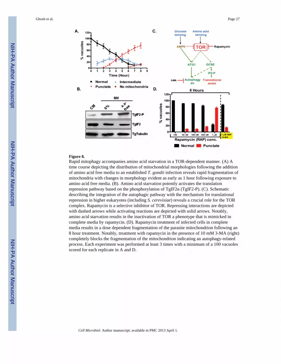
Figure 8.Rapid mitophagy accompanies amino acid starvation in a TOR-dependent manner. (A) Atime course depicting the distribution of mitochondrial morphologies following the additionof amino acid free media to an established T. gondii infection reveals rapid fragmentation ofmitochondria with changes in morphology evident as early as 1 hour following exposure toamino acid free media. (B). Amino acid starvation potently activates the translationrepression pathway based on the phosphorylation of TgIF2α (TgIF2-P). (C). Schematicdescribing the integration of the autophagic pathway with the mechanism for translationalrepression in higher eukaryotes (including S. cerevisiae) reveals a crucial role for the TORcomplex. Rapamycin is a selective inhibitor of TOR. Repressing interactions are depictedwith dashed arrows while activating reactions are depicted with solid arrows. Notably,amino acid starvation results in the inactivation of TOR a phenotype that is mimicked incomplete media by rapamycin. (D). Rapamycin treatment of infected cells in completemedia results in a dose dependent fragmentation of the parasite mitochondrion following an8 hour treatment. Notably, treatment with rapamycin in the presence of 10 mM 3-MA (right)completely blocks the fragmentation of the mitochondrion indicating an autophagy-relatedprocess. Each experiment was performed at least 3 times with a minimum of a 100 vacuolesscored for each replicate in A and D.
Ghosh et al. Page 27
Cell Microbiol. Author manuscript; available in PMC 2013 April 1.
NIH
-PA Author Manuscript
NIH
-PA Author Manuscript
NIH
-PA Author Manuscript

Figure 9.Effect of starvation induced mitophagy on the mitochondrial membrane potential. (A)Accumulation of the membrane potential sensitive dye MitoTracker in both the parasitemitochondrion and host mitochondria is observed in CM (top panel). The 6%SM starvationmediated intermediate mitochondrial morphology does not result in the loss of themitochondrial membrane potential (second panel). Notably, at 4-hour time point presented,host mitochondria retain their membrane potential (second panel). In contrast, mitochondrialfragmentation, seen here in 6%SM for 8 hours, results in a failure to accumulateMitoTracker which now appears as diffuse staining (third panel). Loss of the membranepotential is not a function of length of incubation in 6%SM but rather mitochondrialfragmentation as indicated by adjacent vacuoles at the 8 hour time point (fourth panel,punctate mitochondria bracketed). A loss of discrete MitoTracker labeling in hostmitochondria following exposure to 6%SM for 8 hours is observed (third and fourth panel).(B) The effect of amino acid starvation on the membrane potential of parasite mitochondrionis similar to that seen with 6%SM whereby normal (top panel) and intermediate form(second panel) mitochondria retain the membrane potential while punctate mitochondria(third and fourth panel- bracketed) do not. In contrast to what was observed with regard tohost mitochondria incubated for 8 hours in 6%SM (A), incubation in amino acid freemedium does not result in the loss of the membrane potential of host mitochondria (third andfourth panels). (Scale bars in A and B: Top panel-5 μm, other panels-2μm).
Ghosh et al. Page 28
Cell Microbiol. Author manuscript; available in PMC 2013 April 1.
NIH
-PA Author Manuscript
NIH
-PA Author Manuscript
NIH
-PA Author Manuscript


