Embed Size (px)
Citation preview

of September 3, 2015.This information is current as
-Dependent MechanismγIFN-B7-DC Regulates Asthmatic Response by an
Azuma and Yoichi NakanishiAkiba, Drew Pardoll, Nobuyuki Hara, Hideo Yagita, Miyuki Tsushima, Tomoaki Hoshino, Hisamichi Aizawa, HisayaMiyuki Tsuda, Yuki Yoshiura, Satoru Fukuyama, Fumihiko Koichiro Matsumoto, Hiromasa Inoue, Takako Nakano,
http://www.jimmunol.org/content/172/4/2530doi: 10.4049/jimmunol.172.4.2530
2004; 172:2530-2541; ;J Immunol
Referenceshttp://www.jimmunol.org/content/172/4/2530.full#ref-list-1
, 26 of which you can access for free at: cites 50 articlesThis article
Subscriptionshttp://jimmunol.org/subscriptions
is online at: The Journal of ImmunologyInformation about subscribing to
Permissionshttp://www.aai.org/ji/copyright.htmlSubmit copyright permission requests at:
Email Alertshttp://jimmunol.org/cgi/alerts/etocReceive free email-alerts when new articles cite this article. Sign up at:
Print ISSN: 0022-1767 Online ISSN: 1550-6606. Immunologists All rights reserved.Copyright © 2004 by The American Association of9650 Rockville Pike, Bethesda, MD 20814-3994.The American Association of Immunologists, Inc.,
is published twice each month byThe Journal of Immunology
by guest on September 3, 2015
http://ww
w.jim
munol.org/
Dow
nloaded from
by guest on September 3, 2015
http://ww
w.jim
munol.org/
Dow
nloaded from

B7-DC Regulates Asthmatic Response by an IFN-�-DependentMechanism1
Koichiro Matsumoto,* Hiromasa Inoue,2* Takako Nakano,* Miyuki Tsuda,* Yuki Yoshiura,*Satoru Fukuyama,* Fumihiko Tsushima,† Tomoaki Hoshino,‡ Hisamichi Aizawa,‡
Hisaya Akiba,§ Drew Pardoll,¶ Nobuyuki Hara,* Hideo Yagita,§ Miyuki Azuma, † andYoichi Nakanishi*
B7-H1 (PD-L1) and B7-DC (PD-L2) are the ligands for programmed death-1 (PD-1), which is a member of the CD28/CTLA-4family and has been implicated in peripheral tolerance. We investigated the roles of B7-H1 and B7-DC in a murine OVA-inducedallergic asthma model. B7-H1 was constitutively expressed on dendritic cells, macrophages, B cells, and T cells in the lungs of naivemice, and its expression could be dramatically increased after allergen challenge. In contrast, B7-DC expression was scarcelyexpressed on dendritic cells in naive mice, but was up-regulated after allergen challenge, although the up-regulation of B7-DCexpression on macrophages was minimal. Treatment of mice with anti-B7-DC mAb at the time of allergen challenge, but not atthe time of sensitization, significantly increased their airway hyper-reactivity and eosinophilia. Such treatment also resulted in theincreased production of IL-5 and IL-13, and decreased IFN-� production in the lungs and draining lymph node cells. Thesechanges were diminished when mice were depleted of IFN-� by anti-IFN- � mAb pretreatment. Interestingly, treatment withanti-B7-H1 or anti-PD-1 mAb did not significantly affect the asthmatic response. These results suggest a unique role for B7-DCin the regulation of asthmatic response through an IFN-�-dependent, but PD-1-independent, mechanism.The Journal of Im-munology, 2004, 172: 2530–2541.
R apid advances in immunobiology, particularly the role ofTh1/Th2 cells, have helped to improve our understandingof the inflammatory processes that mediate allergic
asthma (1). Interactions between APCs and T cells appear to be thefirst steps in airway sensitization that ultimately lead to the gen-eration of a Th2-type response. These T cell/APC interactions arecharacterized by the binding of an array of costimulatory mole-cules, as determined using various experimental models (2, 3). Inmurine asthma and rhinitis models, CD80/CD86 and CD28 wereshown to be required for initial T cell activation (4–8). In contrast,the interaction between CD80/CD86 and CTLA-4 was reported tolead to the generation of an inhibitory signal that prevented ex-cessive T cell activation (9, 10). Moreover, it has been suggestedthat inducible costimulator and its ligand (B7RP-1) may be asso-ciated with Th2 commitment and the induction of tolerance (11,12). Accumulating evidence suggests that costimulatory moleculesare critical elements for the promulgation of Th2-skewed allergicresponses.
Programmed death-1 (PD-1)3 is a transmembrane protein orig-inally identified in a T cell line that was undergoing activation-induced cell death (13). Subsequent studies revealed that its ex-pression was associated with cell activation rather than cell death(14, 15). PD-1 was expressed on activated T and B cells and on asubset of thymocytes (14). Structurally, PD-1 belongs to theCD28/CTLA-4 subfamily of the Ig superfamily and, as such, con-tains a single Ig V-like domain in its extracellular region (2, 16).PD-1-deficient C57BL/6 mice were shown to develop glomerulo-nephritis and lupus-like arthritis (17), whereas PD-1-deficientBALB/c mice were shown to develop autoantibody-mediated di-lated cardiomyopathy (18). Additionally, PD-1-deficient mice thatwere crossed with H-2d-specific TCR transgenic mice on an H-2b/d
background exhibited growth retardation, splenomegaly, and lethalgraft-vs-host disease (17). These studies support the idea that PD-1plays a critical role in peripheral tolerance induction and autoim-mune disease prevention.
Recently, two new members of the B7 family, i.e., B7-H1 (PD-L1) and B7-DC (PD-L2), were identified as ligands for PD-1 (19–22). The binding of PD-1 to B7-H1 or B7-DC reportedly inhibitedTCR-mediated T cell proliferation and cytokine production, sug-gesting that the cross-linking of PD-1 by B7-H1 or B7-DC maylead to the down-regulation of T cell responses (20, 21). However,it was also shown that when resting T cells were stimulated withanti-CD3 mAb and B7-DC-Ig, they exhibited enhanced prolifera-tion and IFN-� production (22). Another group also reported en-hanced proliferation and IFN-�, GM-CSF, and IL-10 productionby T cells that were stimulated with low doses of anti-CD3 mAband B7-H1-Ig (19, 23). These conflicting results have made it dif-ficult to decipher the biological significance of B7-H1 and B7-DC
*Research Institute for Diseases of the Chest, Graduate School of Medical Sciences,Kyushu University, Fukuoka, Japan; †Department of Molecular Immunology, Grad-uate School, Tokyo Medical and Dental University, Tokyo, Japan; ‡Department ofInternal Medicine 1, Kurume University School of Medicine, Kurume, Japan; §De-partment of Immunology, Juntendo University School of Medicine, Tokyo, Japan;and ¶Sidney Kimmel Cancer Center, Johns Hopkins University School of Medicine,Baltimore, MD 21205
Received for publication September 15, 2003. Accepted for publication November26, 2003.
The costs of publication of this article were defrayed in part by the payment of pagecharges. This article must therefore be hereby marked advertisement in accordancewith 18 U.S.C. Section 1734 solely to indicate this fact.1 This work was supported in part by a grant-in-aid for Scientific Research from theMinistry of Education, Science, and Culture of Japan.2 Address correspondence and reprint requests to Dr. Hiromasa Inoue, Research In-stitute for Diseases of the Chest, Graduate School of Medical Sciences, Kyushu Uni-versity, 3-1-1 Maidashi, Higashi-ku, Fukuoka 812-8582, Japan. E-mail address:[email protected]
3 Abbreviations used in this paper: PD-1, programmed death-1; AHR, airway hyper-reactivity; BAL, bronchoalveolar lavage; BALF, BAL fluid; DC, dendritic cell; DLN,draining lymph node.
The Journal of Immunology
Copyright © 2004 by The American Association of Immunologists, Inc. 0022-1767/04/$02.00
by guest on September 3, 2015
http://ww
w.jim
munol.org/
Dow
nloaded from

vis-a-vis PD-1. We theorized that a better understanding of theroles of these molecules might be obtained by using well-charac-terized disease models.
B7-H1 mRNA has been detected in various organs, includingthe heart, lung, thymus, spleen, kidney, and liver, and was shownto be up-regulated by IFN-� in monocytes and dendritic cells(DCs) (19, 20). B7-DC mRNA has been detected in liver, lung,and spleen and was shown to be preferentially expressed in bone-marrow-derived and splenic DCs (21, 22). Recently, mAb specificfor B7-H1 and B7-DC have become available (24, 25), which wereused to show that B7-H1 was constitutively expressed on T cells,B cells, macrophages, and DCs and that it was up-regulated on Tcells by CD3 ligation and on macrophages and DCs by IFN-�,GM-CSF, or IL-4. In contrast, B7-DC expression was found to beinducible only on macrophages and DCs after their stimulationwith IFN-�, GM-CSF, or IL-4. The fact that cytokines can inducethe expression of B7-H1 and B7-DC on APCs suggests that theseligands might play a role in regulating the activity of Th1/Th2 cells(26). This possibility prompted us to investigate the roles of thesetwo ligands in a murine asthma models. Our study revealed distinctexpression patterns of B7-H1 and B7-DC on a variety of immunecells in the lungs of these animals and provided a new scenario thatB7-DC, but not B7-H1, might attenuate Th2-skewed allergic re-sponse by an IFN-�-dependent, but PD-1-independent,mechanism.
Materials and MethodsAnimals
Male BALB/c mice, 6–7 wk of age, were purchased from Charles RiverJapan (Kanagawa, Japan) and were housed in environmentally controlledspecific pathogen-free conditions. All animals were maintained in-housefor 1 wk before their use in this study. All procedures and protocols wereapproved by the animal research ethics committee at Kyushu University.
Sensitization and challenge
Mice were sensitized by i.p. injection of 10 �g of chicken OVA (Sigma-Aldrich, St. Louis, MO) and 0.3 mg of aluminum potassium sulfate(SERVA Electrophoresis, Heidelberg, Germany) on days 1 and 11, andthen challenged with 5% OVA in saline mist for 20 min on days 19, 21, and23. The OVA mist was generated using an ultrasonic nebulizer (NE-U17;Omron, Mie, Japan). Control mice were subjected to 0.9% saline sensiti-zation and challenges, and were referred to as naive mice. Subgroups ofanimals received i.p. injections (250 �g/animal) of anti-mouse B7-H1 mAb(MIH6, rat IgG2a), anti-mouse B7-DC mAb (TY25, rat IgG2a), or controlrat IgG (Sigma-Aldrich) 6 h before each sensitization and challenge. Othersubgroups received i.p. injections (250 �g/animal) of anti-mouse PD-1mAb (J43, hamster IgG) (14) or control hamster IgG (BD Biosciences, SanJose, CA) 6 h before each challenge. Two additional anti-mouse PD-1mAbs, RMP1–14 (rat IgG2a) and RMP1–30 (rat IgG2b), were newly gen-erated against mouse PD-1 transfectants. RMP1–14, but not RMP1–30,blocked the binding of both B7-H1-Ig and B7-DC-Ig to PD-1 transfectantsjust like J43 (27). These Abs or control rat IgG (Sigma-Aldrich) wereadministered to subgroups of mice i.p. (250 �g/animal) 6 h before eachallergen challenge. In some experiments mice received i.p. injections of500 �g/animal of anti-mouse IFN-� mAb (R4-6A2, rat IgG1) or control ratIgG 8 h before their OVA challenges on days 19 and 21 to deplete theirendogenous IFN-�. The total dose of mAb was determined according to theprevious study (28). All rat mAbs were purified from ascites by standardprocedures using caprylic acid extraction (25). The anti-PD-1 mAb waspurified from hybridoma supernatant using protein G columns. The purityof these mAbs was verified by SDS-PAGE analysis. Endotoxin levels werefound to be �10 ng/ml in 1 mg/ml of the mAb solutions. Measurements ofairway reactivity to inhaled acetylcholine, bronchoalveolar lavage, and col-lection of immune cells from the lungs and draining lymph nodes (DLN)were conducted 24 h after the last OVA challenge.
Collection of lung cells and lymph node cells
Enzymatic digestion of the lungs was performed as previously described(29). Briefly, mice were exanguinated under a lethal dose of pentobarbital,and the lungs were carefully removed, cut, and minced. Two milliliters ofcomplete Dulbecco’s PBS containing 0.4 mg/ml collagenase type 1A, 330
U/ml hyaluronidase, 50 U/ml DNase, 10% FBS (all additives from Sigma-Aldrich), 100 U/ml penicillin, and 100 mg/ml streptomycin (Life Technol-ogies, Grand island, NY) were added to the minced lungs, and the suspen-sion was incubated for 60 min at 37°C. The samples were then filteredthrough a 52-�m pore size nylon mesh. The single-cell suspensions werewashed once with completed Dulbecco’s PBS, and the erythrocytes wereremoved by lysis with NH4Cl-Tris buffer. Cells were suspended in com-plete RPMI 1640 medium (Sigma-Aldrich) and were adjusted to a densityof 2 � 106 cells/ml. DLN cells were collected from paratracheal and me-diastinal lymph nodes and were dissociated into a single-cell suspension incomplete RPMI 1640.
Flow cytometric analysis
Cells (106) were incubated with mAbs against CD3 (145-2C11, hamsterIgG), CD4 (RM4-5, rat IgG2a), CD8� (53-6.7, rat IgG2a), CD11c (HL3,hamster IgG1), CD19 (ID3, rat IgG2a), CD86 (GL1, rat IgG2a), PD-1 (J43,hamster IgG), and F4/80 (rat IgG2b; Caltag Laboratories, An Der Grub,Austria). All fluorochrome-labeled mAbs and isotype control IgGs werepurchased from BD PharMingen (San Diego, CA) unless otherwise noted.Biotinylated anti-B7-H1 (MIH6, rat IgG2a) and anti-B7-DC mAbs (TY25,rat IgG2a) were used in combination with allophycocyanin-labeled strepta-vidin. In some experiments, PE-labeled anti-B7-H1 mAb was used in com-bination with biotinylated anti-B7-DC mAb plus allophycocyanin-labeledstreptavidin. Cells were first incubated with unlabeled anti-CD16/32 mAbfor 20 min to prevent nonspecific binding via Fc�R and were then incu-bated with FITC-, PE-, and PerCP-labeled mAbs and biotinylated mAb for30 min. After washing with PBS containing 1% FBS, 0.02% NaN3, and0.02% EDTA, referred to as PBS�, the cells were incubated with allophy-cocyanin-labeled streptavidin for 20 min. After washing with PBS�, thecells were fixed with 4% paraformaldehyde (medium A; Caltag Laborato-ries) for 20 min, washed again with PBS�, and stored at 4°C untilanalyzed.
Intracellular cytokine staining was performed as previously reported(29). DLN cells (5 � 106 cells/ml in complete RPMI 1640) were stimulatedwith OVA (0.4 mg/ml) for 24 h or with PMA (10 ng/ml; Sigma-Aldrich)and 2 �g/ml of ionomycin (Sigma-Aldrich) for 5 h; monensin (0.7 �l/well;GolgiStop; BD PharMingen) was added for the final 4 h of stimulation. Thecells were then washed, and cell surface staining was performed usingFITC-labeled anti-CD4, PerCP-labeled anti-CD8�, and allophycocyanin-labeled anti-CD3 mAbs, as described above. After fixation with 10% para-formaldehyde (medium A), cells were permeabilized with 0.1% saponin-containing solution (medium B; Caltag) for 10 min, after which they werestained with PE-labeled anti-IFN-� mAb (XMG1.2, rat IgG1) or isotypecontrol IgG1 for 30 min. After several washes, the cells were suspended inPBS containing 1% paraformaldehyde and analyzed.
It is known that a small number of DCs are double positive for CD11cand F4/80 (30, 31). This might make it difficult to for us distinguish DCsfrom lung macrophages. Thus, the assessment of costimulatory moleculeson APCs in the lung samples was focused on two populations. One wascomposed of small CD8��CD11c�cells (determined by forward scatter)and was regarded as being a myeloid DC-dominant population; our pre-liminary experiments confirmed that one-third of these cells were also pos-itive for F4/80. The other was composed of large F4/80� cells, 90% ofwhich were negative for CD11c. This population was regarded as a mac-rophage-dominant population. Examination of CD8��CD11c� cells,which were thought to be lymphoid DCs, was beyond the scope of ourpresent study because they were extremely scarce in our lung (0.1–0.2% oftotal digested lung cells) and DLN (0.2–0.3% of total DLN cells) samples.
Measurement of airway hyper-reactivity (AHR)
Measurement of AHR was performed according to our previously de-scribed protocol (32). Briefly, animals were anesthetized with a mixture ofketamine and sodium pentobarbital i.p., and their tracheas were cannulatedvia tracheotomy. The animals were ventilated mechanically (model 687;Harvard Apparatus, South Natick, MA), with a tidal volume of 0.3 ml andat a frequency of 120 breaths/min. The airway opening pressure was mea-sured with a differential pressure transducer (model TP-603T; Nihon Ko-hden, Tokyo, Japan) and was recorded continuously with a pen recorder(Nihon Kohden RJG-4124). Stepwise increases in acetylcholine (diluted in0.9% saline; Sigma-Aldrich) concentrations (0.6–10 mg/ml/120 breaths)were administered with an ultrasonic nebulizer (Omron NE-U07). The datawere expressed as the provocative concentration 200 (PC200), i.e., the con-centration at which airway pressure was 200% of its baseline value, whichwas calculated by log-linear interpolation for individual animals. Values ofPC200 were expressed as log (PC200 � 100).
2531The Journal of Immunology
by guest on September 3, 2015
http://ww
w.jim
munol.org/
Dow
nloaded from

Bronchoalveolar lavage (BAL) and cytokine measurements
Mice were exanguinated with a lethal dose of pentobarbital, and their lungswere gently lavaged with 1 ml of 0.9% saline via a tracheal cannula. Totaland differential BAL cell counts were performed as previously described(32). Samples were centrifuged at 2000 rpm for 10 min, and the superna-tants were stored at �80°C. Mouse IL-4, IL-5, IL-10, IL-13, and IFN-�were quantified using ELISA kits (BioSource International, Camarillo,CA) according to the manufacturer’s protocols.
Data analysis
Values were expressed as the mean � SEM. Differences among groupswere analyzed using unpaired t tests or an ANOVA together with a posthoc Bonferroni analysis. Nonparametric data were analyzed using theKruskal-Wallis test, followed by the Mann-Whitney U test. A value of p �0.05 was considered significant.
ResultsDifferential expression of B7-H1 and B7-DC on lung leukocytesand DLN cells
The total cell numbers of DCs (CD8��CD11c� cells) in the DLNand DCs, macrophages (F4/80� cells), and T cells (CD3� cells) inthe lung were significantly elevated in allergen-challenged mice(Fig. 1A, upper panels). Allergen challenge also significantly in-creased the percentages of B7-H1� cells within the DC, macro-phage, and B cell (CD19�) populations in the lungs (Fig. 1, A andB). In contrast, the percentage of B7-H1� cells in the DC popu-lation in the DLNs was significantly reduced in allergen-chal-lenged animals, although the absolute number of B7-H1� cellswas elevated (data not shown). In contrast to the abundant expres-sion of B7-H1, B7-DC expression in the lungs of naive mice wasminimal. Significant B7-DC expression was only observed on DCsin the DLNs. Allergen challenge significantly elevated the expres-sion of B7-DC on lung and DLN DCs and macrophages, but thiselevated expression was limited, especially on lung macrophages.B7-DC expression was not evident on B or T cells.
In the DLN cells of naive mice, a higher percentage of DCs wasfound to coexpress CD86 and B7-H1, but the overall percentagesof CD86�B7-H1� and CD86�B7-H1� cells were reduced in theOVA-challenged mice (Fig. 1B). In contrast, CD86�B7-DC� DCsin the DLNs were elevated in allergen-challenged mice. As shownin Fig. 1C, one-third of the B7-H1� DCs coexpressed B7-DC,whereas one-half of the B7-DC� DCs coexpressed B7-H1. In lungDCs, CD86� cells were increased after OVA challenge, but onlya subpopulation of B7-H1� and B7-DC� DC were found to co-express CD86 (Fig. 1B). An even lower percentage of DCs coex-pressed both B7-H1 and B7-DC in the lung (Fig. 1C). As forCD8�� DCs in DLNs, the expression patterns of B7-H1 andB7-DC were similar to those of CD8�� DCs (data not shown).
B7-H1 was constitutively expressed on lung macrophages innaive mice, and this expression was further enhanced after OVAchallenge. B7-DC expression on lung macrophages in naive micewas minimal, but was somewhat enhanced by allergen challenge.Interestingly, CD86 expression on lung macrophages was dimin-ished after allergen challenge. In sum, our results showed thatB7-H1 was abundantly expressed on DCs and macrophages, andthat there was limited enhancement of B7-DC expression on lungDCs and macrophages in response to allergen challenge.
Expression of PD-1 on T cells in the lungs and on DLN cells
The total number of CD4� T cells was significantly increased inboth the lungs and DLNs, whereas the number of CD8� T cellswas increased only in the DLNs (Fig. 2). PD-1 expression onCD4� and CD8� T cells was significantly increased in the DLNcells of OVA-challenged mice. PD-1 was rarely expressed onCD4� and CD8� T cells in the lungs of naive mice. The expres-
sion of PD-1 on lung CD4� T cells was increased in OVA-chal-lenged mice, but PD-1 expression on lung CD8� T cells was lim-ited. Collectively, these data showed that allergen challengeinduced a significant expansion and activation in both CD4� andCD8� T cells in the DLNs and the preferential infiltration ofCD4� T cells expressing PD-1 in the lung.
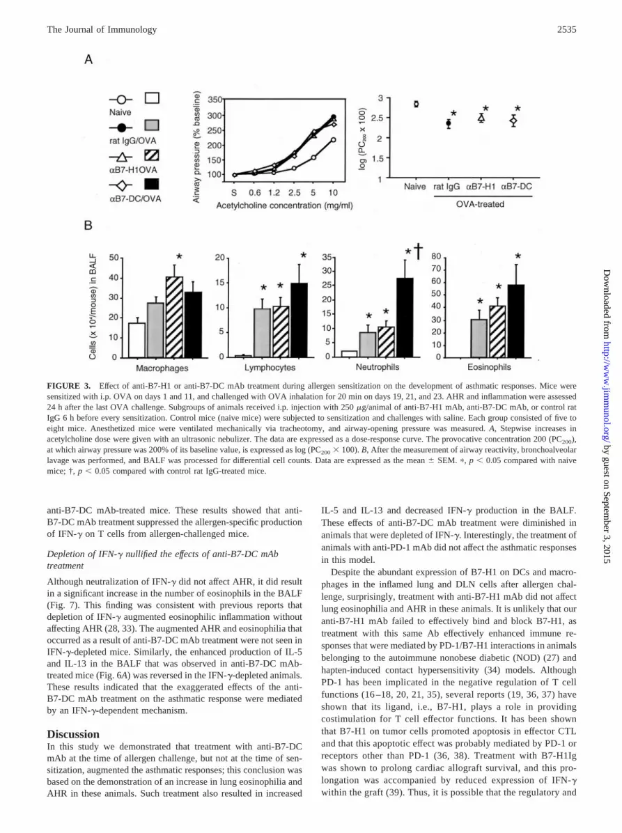
Treatment with anti-B7-H1 or anti-B7-DC mAb during allergensensitization did not affect asthmatic response
Flow cytometric analysis revealed that allergen challenge in-creased B7-H1 and B7-DC expression on CD8�� DCs and mac-rophages, as well as the expression of PD-1 on T cells, in the lungsand DLNs. Airway reactivity to inhaled acetylcholine was signif-icantly higher in OVA-challenged and control IgG-treated micecompared with naive mice. In assessing the effects of treatmentwith neutralizing anti-B7-H1 and anti-B7-DC mAbs during thesensitization phase on the development of allergen-induced asth-matic responses, we found that such treatment did not affect AHR(Fig. 3A). The numbers of lymphocytes, neutrophils, and eosino-phils in the BAL fluid (BALF) of OVA-challenged and controlIgG-treated mice were significantly increased compared with thoseof naive mice (Fig. 3B). Although treatment with anti-B7-H1 mAbdid not affect this inflammatory cell profile, treatment with anti-B7-DC mAb resulted in an elevation in the number of neutrophilsin the BALF.
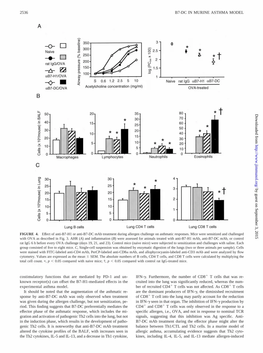
Treatment with anti-B7-DC mAb, but not anti-B7-H1 mAbduring allergen challenge accelerated the asthmatic response
Treatment with anti-B7-H1 mAb had no effect on AHR (Fig. 4A)or the BALF inflammatory cell profile (Fig. 4B). However, treat-ment with anti-B7-DC mAb during OVA challenge significantlyaugmented AHR and BALF eosinophilia compared with those incontrol IgG-treated mice. The effects of mAb treatment on theinflammatory responses were further evaluated by enumerating theT and B cells in the digested lungs. OVA challenge significantlyincreased the number of CD4�, but not CD8�, T cells; this in-crease was not affected by either anti-B7-H1 or anti-B7-DC mAbtreatment. Interestingly, the number of CD8� T cells was signif-icantly reduced in response to anti-B7-DC mAb treatment. Thenumber of B cells was not affected by OVA challenge and/or eachmAb treatment.
Treatment with anti-PD-1 mAb during the allergen challengephase did not affect the asthmatic response
Consistent with the observations in Fig. 4, airway reactivity andthe lymphocyte, neutrophil, and eosinophil counts in the BALFwere significantly enhanced in the OVA-challenged mice. Surpris-ingly, these asthmatic responses were not affected by treatmentwith any of three anti-PD-1 mAbs, J43, RMP1–14, or RMP1–30(Fig. 5).
Treatment with anti-B7-DC mAb enhanced IL-5 and IL-13 andreduced IFN-� production
OVA challenge dramatically increased IL-4, IL-5, and IL-13;moderately increased IL-10; and slightly increased IFN-� produc-tion in the BALF (Fig. 6A). IL-5 and IL-13 production in theBALF was further enhanced by the treatment with anti-B7-DCmAb, but not control IgG. The production of IL-4 and IL-10 pro-duction was not affected by anti-B7-DC mAb treatment. Interest-ingly, IFN-� production in the BALF was substantially suppressed
2532 B7-DC IN MURINE ASTHMA MODEL
by guest on September 3, 2015
http://ww
w.jim
munol.org/
Dow
nloaded from

FIGURE 1. Expression of B7-H1 and B7-DC on immune cells in the lung and DLN. A single-cell suspension was prepared from enzymatically digestedlungs and DLNs of naive mice or OVA-sensitized/challenged mice (two to four animals per sample). Mice were sensitized with i.p. OVA on days 1 and11 and were challenged with OVA inhalation on days 19, 21, and 23. Samples were obtained 24 h after the last OVA challenge. Cells were stained withFITC-labeled anti-CD86 mAb; PE-labeled anti-CD11c, anti-F4/80, or anti-CD19 mAb; PerCP-labeled anti-CD3 or anti-CD8� mAb; and biotinylatedanti-B7-H1 or anti-B7-DC mAb, followed by allophycocyanin-labeled streptavidin. A, The absolute numbers and percentages of B7-H1- or B7-DC-positivecells within CD8��CD11c� (DCs), F4/80� (macrophages), CD19� (B cells), and CD3� (T cells) lymphocytes are shown, respectively. The columnsindicate the mean � SEM from five to seven mice. B, An electronic gate of flow cytometry was set on each immune cell type, and the expression of theindicated costimulatory molecules is shown as a dot plot. Each dot plot represents three to five independent experiments. The quadrant markers were setso that �99% of control Ig-stained cells were included in the lower left quadrant. The numbers indicate the mean � SEM percentages of cells with B7-H1or B7-DC single- or double-positive for CD86. C, The expression of B7-H1 and B7-DC was shown by dual staining with PE-labeled anti-B7-H1 mAb andbiotinylated anti-B7-DC mAb in combination with allophycocyanin-labeled streptavidin (C). �, p � 0.05 compared with naive mice.
2533The Journal of Immunology
by guest on September 3, 2015
http://ww
w.jim
munol.org/
Dow
nloaded from

by anti-B7-DC mAb treatment, which was further confirmed bythe reduced intracellular staining of T cells for IFN-� of DLN Tcells that were stimulated with OVA in vitro. The expression ofIFN-� by both DLN CD4� and CD8� T cells from mice treated
with anti-B7-DC mAb was significantly reduced compared withthat seen in control IgG-treated mice (Fig. 6B). In contrast, theexpression of IFN-� by both CD4� and CD8� DLN T cells thatwere stimulated with PMA and ionomycin was not inhibited in the
FIGURE 2. Expression of PD-1 on T cells in the lung and DLN. Single-cell suspension was prepared from enzymatically digested lungs and DLNs ofnaive mice or OVA-sensitized/challenged mice (two to four animals per sample). Mice were sensitized with i.p. OVA on days 1 and 11 and were challengedwith OVA inhalation on days 19, 21, and 23. Samples were obtained 24 h after the last OVA challenge. Cells were stained with FITC-labeled anti-CD4mAb, PE-labeled anti-PD-1 mAb, PerCP-labeled anti-CD8� mAb, and allophycocyanin-labeled anti-CD3 mAb. A, The absolute numbers CD4 T cells(CD4�CD8�CD3� cells) and CD8 T cells (CD4�CD8�CD3� cells) and the percentages of PD-1-positive cells in each subset. The columns indicate themean � SEM from four or five mice. B, An electronic gate of flow cytometry was set on CD3� cell, and then PD-1 expression is shown as dot plots withreference to CD4 expression. The numbers indicate the mean � SEM percentages of PD-1-positive cells for CD4�-CD3� cells or CD4�CD3� cells. Eachdot plot represents four to five independent experiments. �, p � 0.05 compared with naive mice.
2534 B7-DC IN MURINE ASTHMA MODEL
by guest on September 3, 2015
http://ww
w.jim
munol.org/
Dow
nloaded from
anti-B7-DC mAb-treated mice. These results showed that anti-B7-DC mAb treatment suppressed the allergen-specific productionof IFN-� on T cells from allergen-challenged mice.
Depletion of IFN-� nullified the effects of anti-B7-DC mAbtreatment
Although neutralization of IFN-� did not affect AHR, it did resultin a significant increase in the number of eosinophils in the BALF(Fig. 7). This finding was consistent with previous reports thatdepletion of IFN-� augmented eosinophilic inflammation withoutaffecting AHR (28, 33). The augmented AHR and eosinophilia thatoccurred as a result of anti-B7-DC mAb treatment were not seen inIFN-�-depleted mice. Similarly, the enhanced production of IL-5and IL-13 in the BALF that was observed in anti-B7-DC mAb-treated mice (Fig. 6A) was reversed in the IFN-�-depleted animals.These results indicated that the exaggerated effects of the anti-B7-DC mAb treatment on the asthmatic response were mediatedby an IFN-�-dependent mechanism.
DiscussionIn this study we demonstrated that treatment with anti-B7-DCmAb at the time of allergen challenge, but not at the time of sen-sitization, augmented the asthmatic responses; this conclusion wasbased on the demonstration of an increase in lung eosinophilia andAHR in these animals. Such treatment also resulted in increased
IL-5 and IL-13 and decreased IFN-� production in the BALF.These effects of anti-B7-DC mAb treatment were diminished inanimals that were depleted of IFN-�. Interestingly, the treatment ofanimals with anti-PD-1 mAb did not affect the asthmatic responsesin this model.
Despite the abundant expression of B7-H1 on DCs and macro-phages in the inflamed lung and DLN cells after allergen chal-lenge, surprisingly, treatment with anti-B7-H1 mAb did not affectlung eosinophilia and AHR in these animals. It is unlikely that ouranti-B7-H1 mAb failed to effectively bind and block B7-H1, astreatment with this same Ab effectively enhanced immune re-sponses that were mediated by PD-1/B7-H1 interactions in animalsbelonging to the autoimmune nonobese diabetic (NOD) (27) andhapten-induced contact hypersensitivity (34) models. AlthoughPD-1 has been implicated in the negative regulation of T cellfunctions (16 –18, 20, 21, 35), several reports (19, 36, 37) haveshown that its ligand, i.e., B7-H1, plays a role in providingcostimulation for T cell effector functions. It has been shownthat B7-H1 on tumor cells promoted apoptosis in effector CTLand that this apoptotic effect was probably mediated by PD-1 orreceptors other than PD-1 (36, 38). Treatment with B7-H1Igwas shown to prolong cardiac allograft survival, and this pro-longation was accompanied by reduced expression of IFN-�within the graft (39). Thus, it is possible that the regulatory and
FIGURE 3. Effect of anti-B7-H1 or anti-B7-DC mAb treatment during allergen sensitization on the development of asthmatic responses. Mice weresensitized with i.p. OVA on days 1 and 11, and challenged with OVA inhalation for 20 min on days 19, 21, and 23. AHR and inflammation were assessed24 h after the last OVA challenge. Subgroups of animals received i.p. injection with 250 �g/animal of anti-B7-H1 mAb, anti-B7-DC mAb, or control ratIgG 6 h before every sensitization. Control mice (naive mice) were subjected to sensitization and challenges with saline. Each group consisted of five toeight mice. Anesthetized mice were ventilated mechanically via tracheotomy, and airway-opening pressure was measured. A, Stepwise increases inacetylcholine dose were given with an ultrasonic nebulizer. The data are expressed as a dose-response curve. The provocative concentration 200 (PC200),at which airway pressure was 200% of its baseline value, is expressed as log (PC200 � 100). B, After the measurement of airway reactivity, bronchoalveolarlavage was performed, and BALF was processed for differential cell counts. Data are expressed as the mean � SEM. �, p � 0.05 compared with naivemice; †, p � 0.05 compared with control rat IgG-treated mice.
2535The Journal of Immunology
by guest on September 3, 2015
http://ww
w.jim
munol.org/
Dow
nloaded from
costimulatory functions that are mediated by PD-1 and un-known receptor(s) can offset the B7-H1-mediated effects in theexperimental asthma model.
It should be noted that the augmentation of the asthmatic re-sponse by anti-B7-DC mAb was only observed when treatmentwas given during the allergen challenge, but not sensitization, pe-riod. This finding suggests that B7-DC preferentially mediates theeffector phase of the asthmatic response, which includes the mi-gration and activation of pathogenic Th2 cells into the lung, but notin the induction phase, which results in the development of patho-genic Th2 cells. It is noteworthy that anti-B7-DC mAb treatmentaltered the cytokine profiles of the BALF, with increases seen inthe Th2 cytokines, IL-5 and IL-13, and a decrease in Th1 cytokine,
IFN-�. Furthermore, the number of CD8� T cells that was re-cruited into the lung was significantly reduced, whereas the num-ber of recruited CD4� T cells was not affected. As CD8� T cellsare the dominant producers of IFN-�, the diminished recruitmentof CD8� T cell into the lung may partly account for the reductionin IFN-� seen in that organ. The inhibition of IFN-� production byCD4� and CD8� T cells was only observed in the response to aspecific allergen, i.e., OVA, and not in response to nominal TCRsignals, suggesting that this inhibition was Ag specific. Anti-B7-DC mAb treatment during the effector phase might alter thebalance between Th1/CTL and Th2 cells. In a murine model ofallergic asthma, accumulating evidence suggests that Th2 cyto-kines, including IL-4, IL-5, and IL-13 mediate allergen-induced
FIGURE 4. Effect of anti-B7-H1 or anti-B7-DC mAb treatment during allergen challenge on asthmatic responses. Mice were sensitized and challengedwith OVA as described in Fig. 3. AHR (A) and inflammation (B) were assessed for animals treated with anti-B7-H1 mAb, anti-B7-DC mAb, or controlrat IgG 6 h before every OVA challenge (days 19, 21, and 23). Control mice (naive mice) were subjected to sensitization and challenges with saline. Eachgroup consisted of five to eight mice. C, Single-cell suspension was obtained by enzymatic digestion of the lungs (two or three animals per sample). Cellswere stained with FITC-labeled anti-CD4 mAb, PerCP-labeled anti-CD8� mAb, and allophycocyanin-labeled anti-CD3 mAb and were analyzed by flowcytometry. Values are expressed as the mean � SEM. The absolute numbers of B cells, CD4 T cells, and CD8 T cells were calculated by multiplying thetotal cell count. �, p � 0.05 compared with naive mice; †, p � 0.05 compared with control rat IgG-treated mice.
2536 B7-DC IN MURINE ASTHMA MODEL
by guest on September 3, 2015
http://ww
w.jim
munol.org/
Dow
nloaded from

airway eosinophilia and AHR, whereas IFN-� and IL-12 down-regulate these responses (40). Allergen-induced airway eosino-philia was shown to be augmented by treatment with anti-IFN-�mAb during allergen challenge (28) and in IFN-�-deficient mice(33). Therefore it is possible that blockade of B7-DC might ini-tially suppress IFN-� production, resulting in enhanced Th2 cell
activation and eosinophil recruitment into the lung. In support ofthis idea, we found that the increased severity of the asthmaticresponse that was seen in anti-B7-DC mAb-treated mice dimin-ished after the neutralization of endogenous IFN-�. These datasuggest an initial and critical role of IFN-� in the B7-DC-mediatedregulation of the asthmatic response.
FIGURE 5. Effect of anti-PD-1 mAb treatment during allergen challenge on asthmatic responses. Mice were sensitized and challenged with OVA asdescribed in Figs. 3 and 4. AHR (A and C) and inflammation (B and D) were assessed for animals treated with anti-PD-1 mAbs (J43, RMP1–14, orRMP1–30) or control IgG (hamster IgG for J43 or rat IgG for RMP1–14/RMP1–30) 6 h before every OVA challenge (days 19, 21, and 23). Control mice(naive mice) were subjected to the sensitization and challenges with saline. Each group consisted of nine mice. Values are expressed as the mean � SEM.�, p � 0.05 compared with naive mice.
2537The Journal of Immunology
by guest on September 3, 2015
http://ww
w.jim
munol.org/
Dow
nloaded from

In contrast to the abundant expression of B7-H1, B7-DC expres-sion in the inflamed lung was limited, especially on lung macro-phages. Consistent with previous reports (6, 41–43), we observedreduced expression of CD86 and B7-DC on lung macrophages com-pared with other peripheral tissues. Similar findings were reported inthe CNS of mice with experimental autoimmune encephalomyelitis(44). Interestingly, treatment with anti-B7-DC mAb, but not anti-B7-H1 mAb similarly augmented disease in the experimental auto-immune encephalomyelitis model.
We cannot completely rule out the possibility that the anti-B7-DC mAb (TY25) might have acted as an activator of B7-DCand that it activated Th2 cells. Recent reports suggested that theligation of B7-DC by the cross-linking of IgM Abs may activateDCs and promote T cell effector functions (45, 46). However, bothintact TY25 and Fab of TY25 were found to block the B7-DC-mediated cellular responses in a similar manner in vitro (H. Yagita,unpublished observations). In addition, the in vivo treatment ofNOD mice (27) and mice with contact hypersensitivity (34) with
FIGURE 6. Effect of anti-B7-DC mAb treatment during allergen challenge on cytokine profile in BALF. A, Concentrations of IL-4, IL-5, IL-10, IL-13,and IFN-� in BALF were measured by ELISA for naive and OVA-challenged mice. The OVA-challenged mice received i.p. injection of 250 �g/animalof either anti-B7-DC mAb or control rat IgG 6 h before every OVA challenge (days 19, 21, and 23). Control mice (naive mice) were subjected tosensitization and challenges with saline. Values are expressed as the mean � SEM. Each group consisted of five to eight mice. �, p � 0.05 compared naivemice; †, p � 0.05 compared with control rat IgG-treated mice. B, IFN-�-producing CD4 T cells in DLN were assessed by intracellular cytokine staining.Single-cell suspensions of DLN were stimulated with OVA (0.4 mg/ml) for 24 h or with PMA (10 ng/ml) plus ionomycin (2 �g/ml) for 5 h. Monensinwas added during the last 4 h of stimulation. Cells were stained with FITC-labeled anti-CD4 mAb, PerCP-labeled anti-CD8� mAb, and allophycocyanin-labeled anti-CD3 mAb, fixed and permeabilized with 0.1% saponin, then stained with PE-labeled anti-IFN-� mAb or isotype-matched IgG1. The columnsindicate the mean � SEM of the percentage of IFN-�-positive cells from four mice. �, p � 0.05 compared with naive mice; †, p � 0.05 compared withcontrol rat IgG-treated mice.
2538 B7-DC IN MURINE ASTHMA MODEL
by guest on September 3, 2015
http://ww
w.jim
munol.org/
Dow
nloaded from

TY25 failed to affect their pathological outcomes. These resultssupport the idea that TY25 treatment can block the interaction ofB7-DC with its receptor.
B7-DC is known to provide a negative signal, via an immuno-receptor tyrosine-based inhibitory motif in the PD-1 cytoplasmicregion (21). A recent study has shown that blockade of B7-DC onDCs augmented IFN-� production by alloreactive T cells (47).Thus, the augmentation of asthmatic response by anti-B7-DC mAbmight be due to blocking of a negative signal provided by B7-DCon IL-5 and IL-13 production. However, this speculation does notexplain the reciprocal decrease in IFN-� in BALF by anti-B7-DCmAb treatment, whereas mouse B7-DC transfectants inhibitedIFN-� production by OVA peptide-stimulated CD4� T cells fromDO11.10 transgenic mice in vitro (21). In addition, the diminishedaugmentation of IL-5 and IL-13 production by anti-B7-DC mAb inmice pretreated with anti-IFN-� mAb renders it difficult to fullyexplain the whole phenomena in the context of inhibitory functionof B7-DC.
Given the original concept that PD-1 ligation delivers an inhib-itory signal, a remaining question is how B7-DC can stimulateIFN-� production. Intriguingly, the treatment with anti-PD-1mAbs did not affect airway eosinophilia and AHR. Previous stud-ies using the same anti-mouse PD-1 mAb (J43), effectivelyblocked PD-1-mediated regulation of experimental models for au-toimmune encephalomyelitis, autoimmune diabetes, graft-vs-host
response, and contact hypersensitivity (27, 34, 44, 48). We in-cluded newly established anti-PD-1 mAb, RMP1–14, whichblocks the binding of both B7-H1-Ig and B7-DC-Ig to PD-1 invitro. We have recently found that RMP1–14 treatment exacer-bated experimental autoimmune encephalomyelitis (H. Yagita, un-published observation), as did J43 (44). Tseng et al. (22) showedthat naive T cells that were costimulated with B7-DC-Ig in thepresence of anti-CD3 mAb enhanced their production of IFN-�.but not that of IL-4 and IL-10. A recent report demonstrated thata B7-DC mutant with impaired PD-1 binding capacity was stillable to provide costimulation for the production of IFN-� by Tcells (49). Furthermore, it was shown that DCs from B7-DC-de-ficient mice diminished their ability to costimulate proliferationand IFN-� production by CD4� T cells and that this costimulationwas independent of PD-1 (50). These reports provide strong evi-dence of the existence of a second receptor, in addition to PD-1,which is capable of delivering a costimulatory signal. In ourmodel, B7-DC might function through such a novel receptor in-dependently of PD-1.
In the IFN-�-depleted mice, the treatment with anti-B7-DCmAb failed to enhance IL-5 and IL-13 production. If it is the casethat B7-DC could deliver costimulatory signals to Th2 cells forproduction of IL-5 and IL-13 as well as to Th1 cells (and CTL) forIFN-� production, and the ability for costimulation would be morepotent for Th1 cells, the regulation of asthmatic responses by
FIGURE 7. Effect of anti-B7-DC mAb in IFN-�-depleted mice. OVA-sensitized mice received i.p. injection of 500 �g/animal of anti-IFN-� mAb orcontrol rat IgG 8 h before the first and second OVA challenges (days 19 and 21) for depleting endogenous IFN-�. The mice subsequently received i.p.injection of 250 �g/animal of either anti-B7-DC mAb or control rat IgG 6 h before every OVA challenge (days 19, 21, and 23). These mice were usedfor assessment of AHR (A), eosinophil count in BALF (B), and IL-5 and IL-13 ELISA (C) in BALF. Data are represented as the mean � SEM of five toseven mice in each group. *, p � 0.05 compared with naive mice.
2539The Journal of Immunology
by guest on September 3, 2015
http://ww
w.jim
munol.org/
Dow
nloaded from

IFN-� could overcome the potential enhancement of IL-5 andIL-13 production. To clarify this issue, further studies are nowunderway using B7-DC�/� and IFN-��/� mice.
Two unique properties of B7-DC have been suggested by theabove findings. First, as suppression of Th2 responses is specific toB7-DC, but not to B7-H1, the disparate effects of B7-H1 andB7-DC blockade on different immune responses and disease mod-els raise the possibility that B7-H1 and B7-DC (or PD-1 and anunknown receptor) may preferentially regulate Th1 and Th2 re-sponses (26). Second, the B7-DC blockade is observed in the ef-fector phase of the immune response. These findings suggest thatthe regulation of B7-DC activity may have therapeutic implica-tions for the control of asthma in humans. Further investigation ofthe molecular and cell biology of B7-H1 and B7-DC will undoubt-edly lead to a broadening of our understanding of the mechanismsof immune diseases, including asthma.
AcknowledgmentsWe thank Laboratory Core, Kyushu University Hospital, for technicalassistance.
References1. Umetsu, D. T., J. J. McIntire, O. Akbari, C. Macaubas, and R. H. DeKruyff. 2002.
Asthma: an epidemic of dysregulated immunity. Nat. Immunol. 3:715.2. Carreno, B. M., and M. Collins. 2002. The B7 family of ligands and its receptors:
new pathways for costimulation and inhibition of immune responses. Annu. Rev.Immunol. 20:29.
3. Coyle, A. J., and J. C. Gutierrez-Ramos. 2001. The expanding B7 superfamily:increasing complexity in costimulatory signals regulating T cell function. Nat.Immunol. 2:203.
4. Haczku, A., K. Takeda, I. Redai, E. Hamelmann, G. Cieslewicz, A. Joetham,J. Loader, J. J. Lee, C. Irvin, and E. W. Gelfand. 1999. Anti-CD86 (B7.2) treat-ment abolishes allergic airway hyperresponsiveness in mice. Am. J. Respir. Crit.Care Med. 159:1638.
5. Keane-Myers, A. M., W. C. Gause, F. D. Finkelman, X. D. Xhou, andM. Wills-Karp. 1998. Development of murine allergic asthma is dependent uponB7-2 costimulation. J. Immunol. 160:1036.
6. Mathur, M., K. Herrmann, Y. Qin, F. Gulmen, X. Li, R. Krimins, J. Weinstock,D. Elliott, J. A. Bluestone, and P. Padrid. 1999. CD28 interactions with eitherCD80 or CD86 are sufficient to induce allergic airway inflammation in mice.Am. J. Respir. Cell Mol. Biol. 21:498.
7. Okano, M., M. Azuma, T. Yoshino, H. Hattori, M. Nakada, A. R. Satoskar,D. A. Harn, Jr., E. Nakayama, T. Akagi, and K. Nishizaki. 2001. Differential roleof CD80 and CD86 molecules in the induction and the effector phases of allergicrhinitis in mice. Am. J. Respir. Crit. Care Med. 164:1501.
8. Tsuyuki, S., J. Tsuyuki, K. Einsle, M. Kopf, and A. J. Coyle. 1997. Costimulationthrough B7-12 (CD86) is required for the induction of a lung mucosal T helpercell 2 (TH2) immune response and altered airway responsiveness. J. Exp. Med.185:1671.
9. Oosterwegel, M. A., D. A. Mandelbrot, S. D. Boyd, R. B. Lorsbach, D. Y. Jarrett,A. K. Abbas, and A. H. Sharpe. 1999. The role of CTLA-4 in regulating Th2differentiation. J. Immunol. 163:2634.
10. Bour-Jordan, H., J. L. Grogan, Q. Tang, J. A. Auger, R. M. Locksley, andJ. A. Bluestone. 2003. CTLA-4 regulates the requirement for cytokine-inducedsignals in T(H)2 lineage commitment. Nat. Immunol. 4:182.
11. Akbari, O., G. J. Freeman, E. H. Meyer, E. A. Greenfield, T. T. Chang,A. H. Sharpe, G. Berry, R. H. DeKruyff, and D. T. Umetsu. 2002. Antigen-specific regulatory T cells develop via the ICOS-ICOS-ligand pathway and in-hibit allergen-induced airway hyperreactivity. Nat. Med. 8:1024.
12. Guo, J., M. Stolina, J. V. Bready, S. Yin, T. Horan, S. K. Yoshinaga, andG. Senaldi. 2001. Stimulatory effects of B7-related protein-1 on cellular andhumoral immune responses in mice. J. Immunol. 166:5578.
13. Ishida, Y., Y. Agata, K. Shibahara, and T. Honjo. 1992. Induced expression ofPD-1, a novel member of the immunoglobulin gene superfamily, upon pro-grammed cell death. EMBO J. 11:3887.
14. Agata, Y., A. Kawasaki, H. Nishimura, Y. Ishida, T. Tsubata, H. Yagita, andT. Honjo. 1996. Expression of the PD-1 antigen on the surface of stimulatedmouse T and B lymphocytes. Int. Immunol. 8:765.
15. Vibhakar, R., G. Juan, F. Traganos, Z. Darzynkiewicz, and L. R. Finger. 1997.Activation-induced expression of human programmed death-1 gene in T-lym-phocytes. Exp. Cell Res. 232:25.
16. Nishimura, H., and T. Honjo. 2001. PD-1: an inhibitory immunoreceptor in-volved in peripheral tolerance. Trends Immunol. 22:265.
17. Nishimura, H., M. Nose, H. Hiai, N. Minato, and T. Honjo. 1999. Developmentof lupus-like autoimmune diseases by disruption of the PD-1 gene encoding anITIM motif-carrying immunoreceptor. Immunity 11:141.
18. Nishimura, H., T. Okazaki, Y. Tanaka, K. Nakatani, M. Hara, A. Matsumori,S. Sasayama, A. Mizoguchi, H. Hiai, N. Minato, et al. 2001. Autoimmune dilatedcardiomyopathy in PD-1 receptor-deficient mice. Science 291:319.
19. Dong, H., G. Zhu, K. Tamada, and L. Chen. 1999. B7–H1, a third member of theB7 family, co-stimulates T-cell proliferation and interleukin-10 secretion. Nat.Med. 5:1365.
20. Freeman, G. J., A. J. Long, Y. Iwai, K. Bourque, T. Chernova, H. Nishimura,L. J. Fitz, N. Malenkovich, T. Okazaki, M. C. Byrne, et al. 2000. Engagement ofthe PD-1 immunoinhibitory receptor by a novel B7 family member leads tonegative regulation of lymphocyte activation. J. Exp. Med. 192:1027.
21. Latchman, Y., C. R. Wood, T. Chernova, D. Chaudhary, M. Borde, I. Chernova,Y. Iwai, A. J. Long, J. A. Brown, R. Nunes, et al. 2001. PD-L2 is a second ligandfor PD-1 and inhibits T cell activation. Nat. Immunol. 2:261.
22. Tseng, S. Y., M. Otsuji, K. Gorski, X. Huang, J. E. Slansky, S. I. Pai, A. Shalabi,T. Shin, D. M. Pardoll, and H. Tsuchiya. 2001. B7-DC, a new dendritic cellmolecule with potent costimulatory properties for T cells. J. Exp. Med. 193:839.
23. Tamura, H., H. Dong, G. Zhu, G. L. Sica, D. B. Flies, K. Tamada, and L. Chen.2001. B7–H1 costimulation preferentially enhances CD28-independent T-helpercell function. Blood 97:1809.
24. Ishida, M., Y. Iwai, Y. Tanaka, T. Okazaki, G. J. Freeman, N. Minato, andT. Honjo. 2002. Differential expression of PD-L1 and PD-L2, ligands for aninhibitory receptor PD-1, in the cells of lymphohematopoietic tissues. Immunol.Lett. 84:57.
25. Yamazaki, T., H. Akiba, H. Iwai, H. Matsuda, M. Aoki, Y. Tanno, T. Shin,H. Tsuchiya, D. M. Pardoll, K. Okumura, et al. 2002. Expression of programmeddeath 1 ligands by murine T cells and APC. J. Immunol. 169:5538.
26. Loke, P., and J. P. Allison. 2003. PD-L1 and PD-L2 are differentially regulatedby Th1 and Th2 cells. Proc. Natl. Acad. Sci. USA 100:5336.
27. Ansari, M. J., A. D. Salama, T. Chitnis, R. N. Smith, H. Yagita, H. Akiba,T. Yamazaki, M. Azuma, H. Iwai, S. J. Khoury, et al. 2003. The programmeddeath-1 (PD-1) pathway regulates autoimmune diabetes in nonobese diabetic(NOD) mice. J. Exp. Med. 198:63.
28. Iwamoto, I., H. Nakajima, H. Endo, and S. Yoshida. 1993. Interferon gammaregulates antigen-induced eosinophil recruitment into the mouse airways by in-hibiting the infiltration of CD4� T cells. J. Exp. Med. 177:573.
29. Winterrowd, G. E., and J. E. Chin. 1999. Flow cytometric detection of antigen-specific cytokine responses in lung T cells in a murine model of pulmonaryinflammation. J. Immunol. Methods. 226:105.
30. Gonzalez-Juarrero, M., and I. M. Orme. 2001. Characterization of murine lungdendritic cells infected with Mycobacterium tuberculosis. Infect. Immun.69:1127.
31. Leenen, P. J., K. Radosevic, J. S. Voerman, B. Salomon, N. van Rooijen,D. Klatzmann, and W. van Ewijk. 1998. Heterogeneity of mouse spleen dendriticcells: in vivo phagocytic activity, expression of macrophage markers, and sub-population turnover. J. Immunol. 160:2166.
32. Kibe, A., H. Inoue, S. Fukuyama, K. Machida, K. Matsumoto, H. Koto,T. Ikegami, H. Aizawa, and N. Hara. 2003. Differential regulation by glucocor-ticoid of interleukin-13-induced eosinophilia: hyperresponsiveness, and gobletcell hyperplasia in mouse airways. Am. J. Respir. Crit. Care Med. 167:50.
33. Yoshida, M., R. Leigh, K. Matsumoto, J. Wattie, R. Ellis, P. M. O’Byrne, andM. D. Inman. 2002. Effect of interferon-� on allergic airway responses in inter-feron-�-deficient mice. Am. J. Respir. Crit. Care Med. 166:451.
34. Tsushima, F., H. Iwai, N. Otsuki, M. Abe, S. Hirose, T. Yamazaki, H. Akiba,H. Yagita, Y. Takahashi, K. Omura, et al. 2003. Preferential contribution ofB7–H1 to programmed death-1-mediated regulation of hapten-specific allergicinflammatory responses. Eur. J. Immunol. 33:2773.
35. Nishimura, H., N. Minato, T. Nakano, and T. Honjo. 1998. Immunological stud-ies on PD-1 deficient mice: implication of PD-1 as a negative regulator for B cellresponses. Int. Immunol. 10:1563.
36. Dong, H., S. E. Strome, D. R. Salomao, H. Tamura, F. Hirano, D. B. Flies,P. C. Roche, J. Lu, G. Zhu, K. Tamada, et al. 2002. Tumor-associated B7–H1promotes T-cell apoptosis: a potential mechanism of immune evasion. Nat. Med.8:793.
37. Dong, H., S. E. Strome, E. L. Matteson, K. G. Moder, D. B. Flies, G. Zhu,H. Tamura, C. L. Driscoll, and L. Chen. 2003. Costimulating aberrant T cellresponses by B7–H1 autoantibodies in rheumatoid arthritis. J. Clin. Invest.111:363.
38. Iwai, Y., M. Ishida, Y. Tanaka, T. Okazaki, T. Honjo, and N. Minato. 2002.Involvement of PD-L1 on tumor cells in the escape from host immune system andtumor immunotherapy by PD-L1 blockade. Proc. Natl. Acad. Sci. USA 99:12293.
39. Ozkaynak, E., L. Wang, A. Goodearl, K. McDonald, S. Qin, T. O’Keefe,T. Duong, T. Smith, J. C. Gutierrez-Ramos, J. B. Rottman, et al. 2002. Pro-grammed death-1 targeting can promote allograft survival. J. Immunol. 169:6546.
40. Pernis, A. B., and P. B. Rothman. 2002. JAK-STAT signaling in asthma. J. Clin.Invest. 109:1279.
41. Balbo, P., M. Silvestri, G. A. Rossi, E. Crimi, and S. E. Burastero. 2001. Dif-ferential role of CD80 and CD86 on alveolar macrophages in the presentation ofallergen to T lymphocytes in asthma. Clin. Exp. Allergy 31:625.
42. Fischer, H. G., R. Dorfler, B. Schade, and U. Hadding. 1999. Differential CD86/B7-12 expression and cytokine secretion induced by Toxoplasma gondii in mac-rophages from resistant or susceptible BALB H-2 congenic mice. Int. Immunol.11:341.
2540 B7-DC IN MURINE ASTHMA MODEL
by guest on September 3, 2015
http://ww
w.jim
munol.org/
Dow
nloaded from

43. Rogler, G., M. Hausmann, T. Spottl, D. Vogl, E. Aschenbrenner, T. Andus,W. Falk, J. Scholmerich, and V. Gross. 1999. T-cell co-stimulatory molecules areupregulated on intestinal macrophages from inflammatory bowel disease mucosa.Eur. J. Gastroenterol. Hepatol. 11:1105.
44. Salama, A. D., T. Chitnis, J. Imitola, H. Akiba, F. Tushima, M. Azuma,H. Yagita, M. H. Sayegh, and S. J. Khoury. 2003. Critical role of the programmeddeath-1 (PD-1) pathway in regulation of experimental autoimmune encephalo-myelitis. J. Exp. Med. 198:71.
45. Radhakrishnan, S., L. T. Nguyen, B. Ciric, D. R. Ure, B. Zhou, K. Tamada,H. Dong, S. Y. Tseng, T. Shin, D. M. Pardoll, et al. 2003. Naturally occurringhuman IgM antibody that binds B7-DC and potentiates T cell stimulation bydendritic cells. J. Immunol. 170:1830.
46. Nguyen, L. T., S. Radhakrishnan, B. Ciric, K. Tamada, T. Shin, D. M. Pardoll,L. Chen, M. Rodriguez, and L. R. Pease. 2002. Cross-linking the B7 familymolecule B7-DC directly activates immune functions of dendritic cells. J. Exp.Med. 196:1393.
47. Brown, J. A., D. M. Dorfman, F. R. Ma, E. L. Sullivan, O. Munoz, C. R. Wood,E. A. Greenfield, and G. J. Freeman. 2003. Blockade of programmed death-1ligands on dendritic cells enhances T cell activation and cytokine production.J. Immunol. 170:1257.
48. Blazar, B. R., B. M. Carreno, A. Panoskaltsis-Mortari, L. Carter, Y. Iwai,H. Yagita, H. Nishimura, and P. A. Taylor. 2003. Blockade of programmeddeath-1 engagement accelerates graft-versus-host disease lethality by an IFN-�-dependent mechanism. J. Immunol. 171:1272.
49. Wang, S., J. Bajorath, D. B. Flies, H. Dong, T. Honjo, and L. Chen. 2003.Molecular modeling and functional mapping of B7–H1 and B7-DC uncouplecostimulatory function from PD-1 interaction. J. Exp. Med. 197:1083.
50. Shin, T., G. Kennedy, K. Gorski, H. Tsuchiya, H. Koseki, M. Azuma, H. Yagita,L. Chen, J. Powell, D. Pardoll, et al. 2003. Cooperative B7-1/2 (CD80/CD86) andB7-DC costimulation of CD4� T cells independent of the PD-1 receptor. J. Exp.Med. 198:31.
2541The Journal of Immunology
by guest on September 3, 2015
http://ww
w.jim
munol.org/
Dow
nloaded from


