Embed Size (px)
Citation preview

Further Studies on the Epitopes of HLA-B7 Defined by Murine Monoclonal Antibodies
P. Parham, P. Antonelli , L. A. Herzenberg, T. J. Kipps, A. Fuller, and F. E. Ward
ABSTRACT: Monoclonal antibodies reactive with polymorphic epitopes of HLA-B7 were analyzed by direct and indirect cytotoxicity assays on established panels of HLA typed lymphocytes. This permitted further refinement of their specificity and the identification of various novel reactions. The topographic relationship of polymorphic epitopes on the surface of the B7 molecule was assessed with various serological assays using cell surface B7 or papain solubilized B7 as the antigenic target. These studies focused on monoclonal antibodies recognizing B27 and B7. The results, in combination with those of previously published studies, are used to provide a current assessment of the epitope map of HLA-B7 as defined with mouse monoclonal antibodies. This is compared to the results obtained with alloantisera.
ABBREVIATIONS DTT dithiothreitol RAM HPLC high-pressure liquid
chromatography MGH Massachusetts General TMC
Hospital CREG NIH National Institutes of Health MoAb
F(ab')z fragments of rabbit anti-mouse immuno- globulin G
tetramolecular complexes cross-reacting group monoclonal antibody(s)
I N T R O D U C T I O N
An important question in HLA-A,B,C serology concerns the number of alloan- tigenic sites on each allelic product. With the realization that HLA-A,B,C mol- ecules are structurally similar to immunoglobulins and that they function in an- tigen recognition by cytotoxic T lymphocytes, the arrangement of variability in these molecules has become an issue of more general interest.
Analysis with alloantisera has shown that different HLA-A,B,C molecules have both unique and cross-reactive antigenic specificities. Direct lymphocytotoxicity studies of this type for the HLA-B7 and HLA-B40 molecules have recently been reported by Darke [1,2]. From this alone one cannot distinguish whether the cross-reactions are the result of complexity at the level of the antigen or the antibody. More extensive absorption analysis of selected cross-reactive alloan- tisera has revealed distinct, separable populations of alloantibodies against "pub-
From the Department of Cell Biology, Sherman Fairchild Building, Stanford University School of Medicine, Stanford, California; Genetics Systems Corporation, Seattle, Washington; Department of Genetics, Stanford University, Stanford, California; Histocompatibility Laboratory, Massachusetts General Hospital, Boston, Massachusetts; Division of lmmunology, Duke University Medical Center, Durham, North Carolina.
Address reprint requests to Dr. Peter Parham, Department of Cell Biology, Sherman Fairchild Building, Stanford University School of Medicine, Stanford, CA 94305.
Received November 23, 1984; accepted May 13, 1985.
44 0198-8859/86/$3.50
Human Immunology 15, 44-67 (1986) © Elsevier Science Publishing Co., Inc., 1986
52 Vanderbilt Ave., New York, NY 10017

Epitopes of HLA-B7 45
lic" epitopes that are shared by cross-reactive groups of HLA-A,B locus products, as well as unique distinct antibodies to specific, "private" epitopes [3-5] .
Such antigenic dissection of histocompatibility molecules can more rigorously and exhaustively be carried out using monoclonal antibodies. This is because pure reagents against single epitopes can be obtained in large amounts and their serological monospecificity need never be in doubt. A greater range of serological and immunochemical procedures is therefore possible. At present, monoclonal antibody technology is, for practical purposes, restricted to rat and mouse anti- bodies. Early doubts raised as to the intrinsic capacity of the mouse immune system to respond to the alloantigenic determinants of HLA-A,B,C molecules should by now have been allayed. The quantitative specificity of monoclonal antibodies can in fact far exceed that ever obtained with alloantisera and all of the serological complexities and idiosyncracies documented for alloantisera can be found reiterated with individual monoclonal antibodies [6-8] . It is clear from affinity measurements that some epitopes are shared by distinct HLA-A,B mol- ecules [9,10]. In other cases the affinities of a monoclonal antibody for different molecules are quite different and here the relationships are better described as cross-reactions [8]. For some HLA-A,B,C molecules there may be unique epi- topes that permit a complete definition with a single monoclonal antibody. For others it appears as if there are no unique epitopes and that precise definition requires combinations of monoclonal antibodies against different shared and cross-reactive epitopes [ 11,12].
The work described here is concerned with defining the epitopes of the HLA- B7 molecule with mouse monoclonal antibodies. Previous studies have shown that: (i) B7 has a highly specific epitope that is distinct from a cluster of Bw6 related epitopes [13], (ii) there are two epitopes shared by B7 and molecules in the B40 group [14]; and (iii) B7 can undergo conformational changes with as- sociated changes in certain epitopes [15]. In this paper we focus on analyzing the relationship between B7 and B27, on refining the specificities of informative monoclonal antibodies, and on a current assessment of the epitope map of the B7 molecule.
M A T E R I A L S A N D M E T H O D S
Monoclonal antibody producing cell lines were grown in vitro and passaged as ascitic tumors in mice as described [6]. Switch variants of the ME1 hybridoma cell secreting IgG2a and IgG2b monoclonal antibodies were selected and analyzed by the procedures described for Genox 3.53 [16]. Their production, character- ization, and capacity to mediate antibody-dependent cellular cytotoxicity reac- tions (ADCC) is described in Kipps et al. [17]. Monoclonal immunoglobulins were purified from ascitic fluids and radioiodinated as described [7]. The inter- chain disulphides of monoclonal antibodies (MoAb) were broken by reduction for 1 hr with 4 mM dithiothreitol followed by addition of 10 mM iodoacetamide. These reactions were carried out in phosphate or tris buffers at pH 7.3-8.0. Radioimmune cell binding assays were based on the methods of Williams and colleagues [ 18,19] as adapted for human B lymphoblastoid cell targets [6,7,11]. B cell lines were JY (A2,2;B7,7), MST (A3,3;B7.7), MICH (A2,32;B27,15), SWEIG (A29,29;B40,40), and RI (A24;B39,40). Solid phase radioimmune assays were as described [15]. Formation of immune complexes and their analyses by size exclusion high pressure liquid chromatography were as described [20]. Pu- rified papain solubilized B7 from theJY cell line was obtained from Dr. D. Wiley (Harvard).

46 P. Parham et al.
Complement-mediated antibody-dependent lymphocytotoxicity assays were by the standard method as described by Amos et al. [21] or by Mittal [22]. Cytotoxicity assays augmented with affinity-purified goat anti-mouse IgG (Tago) were done by the methods of Fuller et al. [23] or Johnson et al. [24]. This latter assay is comparable to a binding assay as both complement fixing and nonfixing monoclonal antibodies can be detected in a sensitive, reproducible manner.
SFR8-B6 [25] and B27M1 [26], B27M2 [27] MoAb were generously provided by Drs. S. Radka (Duke) and F. Grumet (Stanford), respectively. The ME1 hybridoma cell line [28] was provided by Dr. A. McMichael (Oxford), the RI cell line was provided by Dr. N. Reinsmoen (Minnesota).
RESULTS
Specificity of MoAb
The specificities of most of the MoAb used in these studies of HLA-B7 were previously assessed by radioimmune cell binding assay with relatively small panels of B cell lines [6]. To further define their specificity, they were analyzed for reactivity with panels of well-characterized peripheral blood lymphocytes using a goat anti-mouse immunoglobulin augmented cytotoxicity assay. This assay was used because most of the monoclonal antibodies are of the IgG, class and do not fix complement. It is unlikely that it introduces any artifactual reactions because IgG2 MoAb that do fix complement gave identical reactions in both the direct and indirect cytotoxicity assays. MoAb were tested at 12 concentrations ranging from 4 ~g/ml to 2 ng/ml. Forty-four HLA-A,B,C specificities as defined by alloantisera were represented in this analysis. The revised specificities of the monoclonal antibodies are summarized in Table 1.
BB7.1 reacted with 8/8 cells expressing B7 and with three out of four cells typed as Bw42. The positive cells were from black individuals whereas the neg- ative cell was of Caucasian origin and typed as Bw73 (KA) with 9th International Workshop reagents. Strong reactions were seen at all antibody concentrations above 10 ng/ml and there were no reactions with cells not expressing B7 or Bw42. This shows that BB7.1, which was originally thought to be highly specific for B7 [6], also reacts with Bw42 in the black population.
MB40.2 reacted strongly at concentrations > 100 ng/ml with all 8/8 cells ex- pressing B7, 5/5 cells expressing Bw60, and 3/3 cells expressing Bw41. The black cells expressing Bw42 were also strongly reactive. Of interest were the negative reactions of 3/3 cells expressing Bw61 and 2/2 cells expressing Bw47. It was suspected from our previous analysis that MB40.2 split the B40 antigen because of its negative reaction with the SWEIG cell [14] and it is shown here that it reacts with Bw60 but not Bw61. The titrations suggested a slightly stronger reaction with Bw60 than with B7, Bw41, and Bw42. The MoAb was in fact made by immunization with Bw60.
MB40.3 gave strong reactions with all cells typed as B7 and Bw60. It was unreactive with 3/3 cells typed as B41, as well as two cells typed Bw47, three cells typed Bw42, and the single Bw73 cell (KA). Three out of four cells typed as Bw61, including the SWEIG line, reacted with MB40.3 to concentrations of 0.5 ~g/ml.
In binding analysis with B cell lines, MB40.3 reacted with all cells typed as B40. This was in contrast to MB40.2 which was negative on SWEIG. Subsequent experiments with a panel of B40 positive cell lines obtained from Drs Reinsmoen and Bach (University of Minnesota) revealed a second B cell line RI with the same MB40.3 positive, MB40.2 negative phenotype as SWEIG. Quantitative

Epitopes of HLA-B7 47
TABLE 1 HLA-A,B specificity of polymorphic MoAb reactive with HLA-B7 ~
>.7-
e~
~ -- HLA-B specificity
Site 1 {
Site 2 {
Site 3
Site 4 {
strong weak BB7.1 6 ~/1 B7,Bw42 MB40.2 14 ~'1 B7,Bw41,Bw42,Bw60
B7,B27,Bw22 (w54,w55,w56) } B w46 ,B 14 .2>B 14 .1 . -B w62 ME1 28 Yl,3'2a'Y2b Bw42
B27.M1 26 /z B27(Bw42) B7,Bw46,B14 B7,B27,Bw22, (w54,w55,w56)
P5.3, P7.1 29 N D B17,(w57,w58) ~ Bw46,B14
MB40.3 14 "y~ B7,Bw60 B w 6 1 > B 2 7 > B 1 4
SFR8-B6 25 rat~/2b Bw6 MB40.7 13 % Bw6 related BB7.6 13 5'l Bw6 related
~The assignments shown here represent a compilation of previously published results [8,25-27,29] with those described here. Results from binding, direct, and indirect cytotoxicity assays are pooled. The reactions of MB40.3 with B27 and B14 were only observed with binding and not with indirect cytotoxicity assays. The antibodies MB40.7 and BBT.6 show differential reactivity with many HLA-B molecules that has not been examined in detail. They appear to bind strongly to Bw6 associated molecules and weakly to Bw4 associated molecules. Their target epitopes on B7 have been related to the Bw6 epitope as defined by SFR8-B6 [13]. ND indicates not determined.
analysis of the interaction of MB40.3 with SWEIG showed that the epitope recognized on this cell was significantly different from that on Bw60 and B7 molecules [8]. Therefore the molecule typed as B40 on SWEIG is different from Bw60 and B7 in at least two distinct epitopes. Recent HLA typing of SWEIG during the Ninth International Workshop shows that it is not Bw60 and may be Bw61 or possibly a novel antigen of the B40 group. The reactions of MB40.2 and MB40.3 with peripheral blood cells described here show that both antibodies react strongly with B7 and Bw60. In addition, MB40.2 reacts strongly with Bw41 and black Bw42 and shows no reactions with Bw61. On the other hand, MB40.3 showed weaker reactions with most Bw61 expressing cells but did not react with Bw41,Bw42, or additional HLA-B molecules.
The rat MoAb SFR8-B6 reacts in the lymphocytotoxicity assay with an epitope that correlates well with the Bw6 specificity defined with alloantisera [25]. BB7.6 and MB40.7 give positive reactions with all cells but absorption and inhibition analysis indicated differences in the avidity of the reactions that showed some correlation with Bw6. Competition binding assays between SFR8-B6, MB40.7, and BB7.6 using B7 as the antigenic target showed that the three epitopes closely overlapped. For these reasons the epitopes defined by MB40.7 and BB7.6 were designated as Bw6 related [13]. As discussed below, weak interactions between SFR8-B6 and the B27 molecule, which is negative for Bw6, can be detected by antibody blocking.

48 P. Parham et al.
Switch Variants of ME1
Ellis et al. [28] derived the ME1 antibody and showed by binding assay that it reacted with B7, B27, and Bw22. To further investigate the specificity of this IgG1 antibody we derived IgG2 class switch variants [17]. The three ME1 an- tibodies of different isotypes were compared for reactivity in the standard (NIH) cytotoxicity assay and in the anti-globulin augmented cytotoxicity assay. An initial comparison by radioimmune cell binding assay revealed no significant differences in either titer or specificity of the three antibodies [17]. As expected, only the IgG2a and IgG2b antibodies gave complement-dependent cytotoxicity whereas all three preparations produced cytotoxicity in the anti-immunoglobulin assay. For the IgG2 antibodies there was no difference in their HLA specificity in the two assays and for convenience we have shown results of the anti-globulin assay in Table 2 so that a comparison with IgGl can also be made. This comparison used the MGH cell panel. In addition, a panel of cells from Duke University was analyzed and this provided results for HLA specificities such as Bw55 not rep- resented in the MGH panel (data not shown). There were no significant differ- ences in the reactions obtained with the three ME1 isotypes. Strong reactions were seen with B7, B27, Bw54, Bw55, Bw56, and Bw42, weaker reactions with Bw46 and B14. Heterogeneity in B 14 was detected. Out of three B14 positive cells tested, one, RSM, which subtyped as B14.2, gave a strong reaction with ME1. The two B14.1 cells gave weaker reactions. This suggests that the ME1 antibody may have the potential to detect subtypes of the B14 antigen. At high concentrations it was also possible to find positive reactions with B15. In that case the Bw62 subtype was reactive but Bw63 was not. This contrasts with antibodies P5.3 and P7.1 which have a related specificity to ME1 but are only reactive with the Bw63 split of B15 [29].
Epitopes Shared by B7 and B27
Saturation binding titrations. Our goal in these experiments was to compare the reactions of the ME1 and B27M1 polymorphic monoclonal antibodies which both react with B7 and B27. B27M1 was identified and studied by Grumet et al. [26] with the complement-dependent lymphocytotoxicity assay. It reacted with all of 39 cells typed as HLA-B27 and in addition had reactions with cells expressing either Bw42, homozygous B7, Bw46, or B14. Measurement of the binding of B27M1 to a panel of human B cell lines in an indirect trace assay showed that in comparison to other monoclonal anti-HLA the binding was rel- atively low. As expected from the results of the cytotoxicity assay, the reaction with B27 was stronger than that with B7 (data not shown).
As described above, Ellis et al. [28] produced the ME1 antibody and showed that in contrast to B27M1, it bound with similar high avidity to cell surface B7 and B27. We have confirmed these results by indirect trace binding against a panel of human B cell lines. The titrations of MEI-IgG,-F(ab')2 and -Fab' in cell binding assays were found to be similar showing that the affinity of ME1 is also high.
The difference in the binding of ME1 and B27M1 to cells is shown in Figure 1 and is compared to that of other antibodies against polymorphic epitopes. The binding to a homozygous B7 expressing cell is shown in panel A and to a het- erozygous B27 expressing cell in panel B. The assay was carried out using sat- urating concentrations of ~25I-RAM so that the amount of radioactivity bound is proportional to the numbers of molecules of MoAb bound. Consider first the reactions with B7 (panel A). MB40.5 is a high affinity monomorphic antibody [30] that shows saturation of all HLA-A,B molecules down to concentrations of

TA
BL
E
2 C
yto
tox
ic
rea
cti
on
s o
f M
EI-
IgG
o
f d
iffe
ren
t is
oty
pe
w
ith
p
eri
ph
era
l b
loo
d
lym
ph
oc
yte
s in
th
e
an
ti-g
lob
uli
n
ass
ay
a
Rel
evan
t H
LA
-B
B7
spec
ific
ity
Tot
al
nu
mb
er
8 o
f ce
lls
test
ed
Iso
typ
e 1
2a
B27
B
w5
6
Bw
54
Bw
42
B
14
Bw
46
B
40
2 2
1 2
3 1
7
2b
1
2a
2b
1 2a
2b
1
2a
2b
1 2a
2
b
1 2a
2
b
1 2a
2b
1
2a
2b
Dil
utio
n 1
8 8
8 2
2 2
2 2
2 0
1 1
2"
2 2
3 3
3 1
1 1
0 0
2 8
8 8
2 2
2 2
2 2
1 1
1 2
2 2
3 3
3 1
1 1
0 0
3 8
8 8
2 2
2 2
2 2
1 1
1 2
2 2
2 3
2 1
1 1
0 0
4 8
8 8
2 2
2 2
2 2
1 1
1 2
2 2
2 3
1 1
1 1
0 0
5 8
8 8
2 2
2 2
2 2
1 1
1 2
2 2
2 2
1 1
1 1
0 0
6 8
7 8
2 2
2 2
2 2
0 1
1 2
2 2
2 1
1 1
1 1
0 0
7 8
8 6
2 2
2 2
2 2
1 1
0 2
2 1
1 1
1 1
1 0
0 0
8 8
8 7
2 2
2 2
2 1
1 1
2 2
2 0
0 0
0 0
0 0
0 0
9 8
8 4
2 2
2 1
2 2
1 1
0 2
2 2
0 0
0 0
0 0
0 0
10
8 8
3 2
2 1
1 2
2 l
1 0
2 2
1 0
0 0
0 0
0 0
0 11
5
5 1
2 0
0 2
2 2
0 0
0 2
1 0
0 0
0 0
0 0
0 0
12
4 2
1 1
0 0
1 1
2 0
0 0
1 0
0 0
0 0
0 0
0 0
0
~Dilu
tions
wer
e tw
ofol
d w
here
1 c
orre
spon
ds t
o 4/
zg/m
l. Fo
rty-
two
cells
wer
e sc
reen
ed i
n th
is e
xper
imen
t. T
hese
res
ults
for
26
cells
are
sho
wn;
the
oth
er 1
6 ce
lls w
ere
not
lyse
d by
ME
I an
d di
d no
t ty
pe f
or a
ny o
f th
e de
sign
ated
ant
igen
s. T
he n
umbe
r of
cel
ls o
f a
give
n sp
ecif
icity
that
are
lys
ed t
o 10
0% w
ith a
giv
en a
ntib
ody
dilu
tion
are
indi
cate
d.

50 P. Parham et al.
A 70,000 i ~,000
50,000
40,000
30,00@ 20,000 10,000
FIGURE 1
I I ! I I I (A)
500 167 56 19 S 2
- t 4 0 , 0 0 0 - I I I l I (B)I q
120,000 -
~ , ~ 0
- 40 ,000
20 ,000
0 SO0 187 56 19 6 2
CONCENTRATION OF MONOCLONAL ANTIBODY (ug,/ml)
Titration of MoAb against JY cells expressing HLA-B7 and MICH cells expressing HLA-B27 in an indirect binding assay. 105 JY cells (panel A) or MICH cells (panel B) were incubated in 25 ~l with MoAb; MB40.5 (O), ME1 (A), BB7.6 (V), MB40.2 (&), MB40.3 (D), BB7.1 (m), B27M1 (©) for 2 hr at 4°C. The cells were washed three times by centrifugation and then resuspended in 25 ~l (668,000 cpm) of ~25I-RAM at 0.50 ~g/ml. These were conditions for saturation with 125I-RAM. After 2 hr at 4°C, the cells were washed four times and assayed for bound radioactivity.
10 ~g/ml. The plateau value of 6 × 10⁴ cpm corresponds to 6.7 × 10⁶ molecules of 125I-RAM bound/cell. This is consistent with the known site numbers of HLA- A,B molecules on B cell lines (0.5-5 x 10⁶) [31] and with the possibility of one molecule of MoAb binding more than one RAM molecule. The three poly- morphic antibodies BB7.1, MB40.2, and MB40.3 are of relatively low affinity and bind in a bivalent mode. They reach saturation at higher concentrations than MB40.5, with plateau values that are about half that of MB40.5, consistent with the reduction in the number of target molecules from all HLA-A,B,C to just HLA-B. Although ME1 reacts with the same population of target molecules as BB7.1, MB40.2, and MB40.3, its binding curves are quite different. ME 1 shows higher levels of binding, probably resulting from its high affinity and the con- sequent proper ty that it can bind strongly with one or two site attachment. It may also bind more molecules of 125I-RAM/molecule of MoAb. In contrast, B27M1 binds significant but low levels of lzSI-RAM that are not consistent with saturation of B7 sites. The apparent plateau of binding cannot be taken as a reliable indication of saturation at the point in time when the radioactivity bound is assayed. It could, for example, show that saturation existed before washing of the cells and that equivalent amounts of antibody dissociated from the various assay points during washing.
For the B27 expressing cell line M I C H there was no binding of BB7.1 and MB40.2 as expected from their previously determined specificity (Table 1). MB40.3 shows a weak cross-reaction with B27 and this can be seen as a slight reaction in Figure lB. ME1 binds to B27 with comparable strength to that found with B7. B27M 1 shows a significantly stronger binding to B27 than to B7. The plateau of binding is about half that obtained with ME1 and indicates that saturation of sites was approximated.
Further evidence for a large difference in the affinities of ME1 and B27M1 for B7 was obtained from measuring their binding to papain solubilized B7 that had been absorbed to the wells o f microti ter plates. This is shown in Figure 2.

Epitopes of HLA-B7 51
C~ Z
~E er
W~
r -
30,000
20,000
10,000
1 I I I
o r
. - - o ME1 ...z>-.-----
~ BB7.$
/~ ..~ ~- _~ MB40.§ - ~ BB7.1
~ ~-J MB40.3
¢.- c __ ~ MB40.2
- - m m B27.MI 16 32 63 125
CONCENTRATION OF MONOCLONAL ANTIBODY (~B/ml)
FIGURE 2 Binding of MoAb to papain solubilized HLA-B7 in a solid phase radioim- mune assay. Purified papain soLubilized B7 was ~bsorbed by the wells of microtiter plates by addition of 50/zl of a 100/zg/ml solution for 4 hr at 4°C. Unbound B7 was removed and the wells filled with a solution of BSA at 5 mg/ml in PBS. After incubation for 1 hr at room temperature, the wells were washed with PBS containing 2 mg/ml BSA (assay buffer). 50 ~l of dilutions of MoAb ME1 (O), BB7.6 (V), MB40.5 (A), BB7.1 (V), MB40.3 ([]), MB40.2 (O), and B27.M1 ( . ) in assay buffer were added to the wells and incubated at 4°C for 1 hr. The wells were exhaustively washed and 3 × 10⁵ cpm of 125I-F(ab')2 rabbit anti-mouse IgG (125I-RAM) added for 1 hr at 4°C. After washing with assay buffer the wells were separated and assayed for bound radioactivity. Background binding obtained with dilutions of X63 [39], a nonspecific myeloma protein (1000-2000 cpm), have been subtracted.
O f seven antibodies known to bind cell surface B7 molecules, ME1 gave the highest binding and B27M1 gave no specific binding. In this assay bivalent at- tachment of B27M1 to the solid phase could not occur due to the lack of lateral mobili ty of the antigen, thus explaining the lack of a d e t e c t ~ l e reaction.
Cell blocking assays. The capacity of various monoclonal ant i-HLA-A,B reagents to block the binding of ME1 and B27M1 to B7 and B27 expressing cells was tested. The most informative curves are shown in Figure 3 and the results are summarized in Table 3. N o antibody b~ocked the binding o¢ ~251-ME1 to either B7 or B27 with a capacity comparable to that o f ME1 (Figure 3B,D). SFR8-B6 was the only other antibody to block the binding of ME 1 to background values and this was with a potency that was about 80-fold lower than ME1. It should be noted that the specificity of SFR8-B6 as assessed by ant ibody-dependent cytotoxicity is for the Bw6 supertypic determinant [25] which is not found on B27. However , the antibody clearly interacts with B27 as shown by the inhibition of ME1 binding to B27. Different patterns were seen with the other Bw6 as- sociated antibodies; BB7.6 and MB40.7 that are of broader specificity [13]. MB40.7 gave a partial blocking of ME 1 (Figure 1 D) and BB7.6 gave no blocking (data not shown). B27M1 partially blocked ME1 with a capacity that was about
52 P. Parham et al.
8 A m E
10
6
8
4
6
, °
x x 0
~ 2 u
o , , , , , , , i , i I = ~ 25
'~ C =_=,= 2O
s
4 10
3 5
2 0 = I I I I I I i I t 1 2 3 4 5 6 7 8 9 10 11 1 2 3 4 5 6 7 8 9 10
D I L U T I O N N U M B E R OF B L O C K I N G A N T I B O D Y
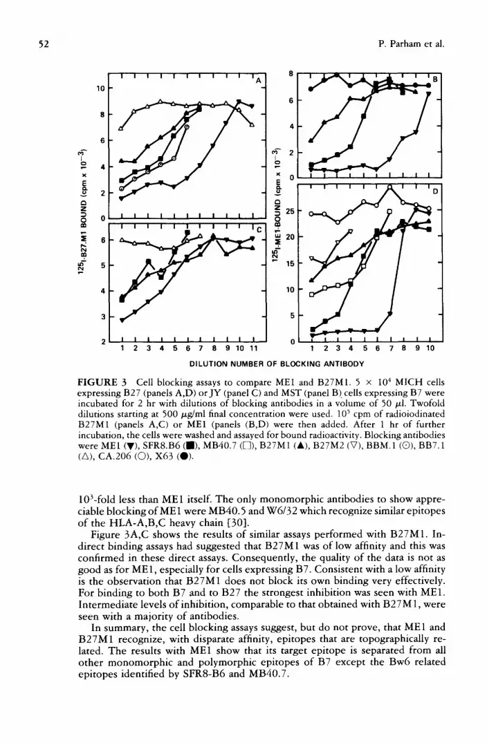
FIGURE 3 Cell blocking assays to compare ME1 and B27M1. 5 × 10⁴ MICH cells expressing B27 (panels A,D) orJY (panel C) and MST (panel B)cells expressing B7 were incubated for 2 hr with dilutions of blocking antibodies in a volume of 50 ~l. Twofold dilutions starting at 500 tzg/ml final concentration were used. 105 cpm of radioiodinated B27M1 (panels A,C) or ME1 (panels (B,D) were then added. After 1 hr of further incubation, the cells were washed and assayed for bound radioactivity. Blocking antibodies were ME1 (V), SFR8.B6 (J) , MB40.7 (D), B27M1 (&), B27M2 (V), BBM.1 (Q), BB7.1 (A), CA.206 (Q), X63 (Q).
10Lfold less than ME1 itself. The only monomorphic antibodies to show appre- ciable blocking of ME1 were MB40.5 and W6/32 which recognize similar epitopes of the HLA-A,B,C heavy chain [30].
Figure 3A,C shows the results of similar assays per formed with B27M1. In- direct binding assays had suggested that B27M1 was of low affinity and this was confirmed in these direct assays. Consequently, the quality of the data is not as good as for ME1, especially for cells expressing B7. Consistent with a low affinity is the observation that B27M1 does not block its own binding very effectively. For binding to both B7 and to B27 the strongest inhibition was seen with ME1. Intermediate levels of inhibition, comparable to that obtained with B27M 1, were seen with a majority of antibodies.
In summary, the cell blocking assays suggest, but do not prove, that ME1 and B27M1 recognize, with disparate affinity, epitopes that are topographically re- lated. The results with ME1 show that its target epitope is separated from all o ther monomorph ic and polymorphic epitopes of B7 except the Bw6 related epitopes identified by SFR8-B6 and MB40.7.

Epitopes of HLA-B7 53
TABLE 3 Summary of the results of cell-based blocking assays a
Radioactive antibody ME1 MB40.2 MB40.3 B27M1
Target HLA molecule B7 B7 B27 B40 B7 B7 B7 B27
o .m ~
BB7.1 B7,w42 6 0 0 0 2 3 2 0 1 MB40.2 B7,w41,w42,w60 14 0 0 0 4 4 0 1 1 MB40.3 B7,w60 14 2 2 0 0 0 4 1 1 B27M1 27,(7,w42,w46,14) 26 1 2 2 0 2 2 2 2 B27M2 27,7 27 0 0 1 3 ME1 7,w22,w27,w42 28 4 4 4 0 4 3 4 SFR8-B6 Bw6 25 3 3 3 2 2 1 2 3 BB7.6 Bw6 related 6,13 0 0 2 2 2 0 0 1 MB40.7 Bw6 related 13,14 2 0 2 0 0 0 0 3 MB40.1 broad 32 0 0 3 3 0 0
polymorphic MB40.4 broad 14 1 0 0 1 1 3 0 1
polymorphic PA2.2 broad 11,33 2 3
polymorphic BBM. 1 /32-m 34,35 0 4 0 2 3 BB7.7 [ J monomorphic [ J 6 , 3 0 0 0 4 4 4 0 2 BB7.5 J ~ J ] c o m b i n a t o r i a l 6,30 0 0 2 2 4 0 2 W6/32 } { .} { 3 1 , 3 6 1 2 0 2 PA2.6 monomorphic 30 1 3 0 2 MB40.5 30 2 2 3 4 0 2 Q 1/28 monomorphic 37 1 1 CA.206 H L A - D R 38 0 0 0 0 1 1
monomorphic X63 nonspecific 39 0 0 0 0 0 1 1
~0 indicates no blocking as defined by the control antibodies X63 and CA.206. 4 is blocking to background at antibody concentrations similar to that given by the test antibody. 3 is blocking to background at antibody concentrations greater than required by the test antibody. 2 is blocking to intermediate levels, often with a plateau being reached at a value significantly greater than background. 1 is slight inhibition of binding (10-20% of total) at high concentrations of antibody. No figure indicates not determined. The binding of ME1 was examined on two different cell lines expressing B7. All antibodies are directed against epitopes of HLA-A,B,C molecules, with the exception of × 63 and CA.206.
Size exclusion HPLC analysis of immune complexes formed by MEI. ME1 is of suf- ficiently high affinity that complexes formed with papain solubilized B7 can be analyzed by size exclusion high pressure liquid chromatography (HPLC). A prep- aration of complexes was formed by incubation of MEI-IgG1 with a slight molar excess of B7 over antibody combining sites. On HPLC analysis this gave two complex peaks corresponding to IgG with one or two molecules of B7 bound. Addition of more B7 did not change the distribution of material between these two peaks, indicating that a proportion of the IgG molecules had only a single active combining site. This we attribute to the presence of nonspecific light chain derived from the NS1 myeloma parent [28]. To determine whether other anti-

54 P. Parham et al.
bodies could bind to B7 at the same time as ME1, we incubated them with aliquots of the ME1/B7 complex and analyzed the mixture by HPLC. The results are described in Table 4. Four different patterns were observed. MB40.3 induces a dissociation of B7 from ME1 that is due to a conformational change and is discussed in [15]. This effect is most readily observed with Fab' fragments of MB40.3 because it results in a large change in the complex size ( - 2 5 0 k and - 2 0 0 k to 10Ok). However, equivalent effects can be detected with IgG and F(ab')2 as indicated in Table 4. Some antibodies had no affect on the elution position of the complex peaks. This could result either from the low affinity of the added antibody so that a stable complex is not formed or from a competition for the part of the B7 molecule seen by ME1. The third and fourth patterns both involved an increase in molecular weight of the complex peaks and, therefore, show that both ME1 and the added antibody can simultaneously bind to a B7 molecule. This can clearly be interpreted to mean that the two epitopes are topographically distinct. In some cases e.g., MB40.5, PA2.6, W6/32, and mostly for antibodies of high affinity, a sharp peak at the excluded volume of the column is seen. This is due to catenary o r circular complexes involving more than three antibody molecules. In other cases circular tetramolecular complexes (TMC)
T A B L E 4 Effects of addition of MoAb to the complex of MEI-IgG and papain-solubilized HLA-B7 a
Strong Weak N o Strong Weak excluded excluded Displaces
effect TMC TMC peak peak ME 1
Added antibody polymorphic BB7.1 + MB40.2 MB40.2-F(ab')2 reduced MB40.2 + MB40.2-Fab' + MB40.3 MB40.3-F(ab')2 MB40.3-Fab' B27.M1 +
SFR8-B6 + BB7.6 + MB40.7 + MB40.1 + MB40.4 +
monomorphic BBM. 1 BB7.7 BB7.5 MB40.5 PA2.6 W6/32
+
+
+
q- +
+
+ +
q-
+ + +
~The size of immune complexes was measured by size exclusion chromatography on a TSK3000 SW column. Procedures were as described in [13,20].

Epitopes of HLA-B7 55
containing two antibody and two B7 molecules are preferentially formed e.g., BBT.6. For the high affinity monomorphic antibodies: BB7.7, MB40.5, and W6/32, both types of complex were formed. MB40.2-1gG and -F(ab~)~ gave small but reproducible excluded peaks with ME 1/B7 complexes. This was judged significant as mild treatment of the MB40.2 F(ab')2 complex with dithiothreitol resulted in loss of the excluded peak and generation of a chromatogram that was similar to that given when MB40.2-Fab' was added to the ME1/B7 complex.
These results confirm those from cellular blocking assays in showing that the ME 1 epitope is topographically separate from all the monomorphic epitopes and the polymorphic epitopes defined by MB40.2, BB7.6, and MB40.7. As seen in the cellular blocking experiments, a splitting of the Bw6 associated epitopes was observed. SFR8-B6 had no effect on the complex of ME1 and BT, whereas MB40.7 and BB7.6 both formed TMC.
A recent study by Jackson et al. [40] has shown that formation of closed or ring complexes can be favored by mild reduction of the interheavy chain disul- phide bonds in the hinge region of IgG. This presumably results from an increase in the flexibility of the antibody and a consequent reduction in strain when changing the angle between the Fab arms. We compared the immune complexes formed by reduced and mock reduced MEI-IgGI in combination with B7 and other monoclonal antibodies. Only in the case of MB40.2 was a qualitative dif- ference observed. With reduced ME1, a TMC was formed whereas with mock treated or untreated antibody long chain complexes or larger cyclic complexes were formed. A similar effect was seen if the MB40.2-I~G was reduced (Table 4). These results are consistent with reduction of the interheavy chain disulphides increasing antibody flexibility. Similar experiments were done with the ME1- I~G~a and I~G2b switch variants. No complexes greater than IgG with two B7 molecules were observed with the native antibodies in combination with MB40.2. After reduction, IgG~a but not IgG~b gave a TMC with B7 and MB40.2. The interpretation of these results is not clear.
Relationship of ME1 and MB40.3 epitopes. The capacity of ME1 to block the binding of other MoAb to cell surface and soluble B7 was examined. A strong blocking of MB40.3 was seen. However, in the reciprocal experiment, there was no significant blocking of ME1 by MB40.3. Further examination showed that Fab' and F(ab')2 fragments of MB40.3, but not IgG, were effective in blocking ME1 binding to cell surface B7. A direct comparison of MB40.3-IgG,-F(ab')2 and -Fab' as blocking reagents is shown in Figure 4.
Quantitative comparisons of the kinetics of MB40.3-1gG,-F(ab')2 and -Fab' showed that the forward rate constant of the IgG was less than F(ab')2 and Fab' [8]. (A smiliar difference between IgG and Fab was also found for W6/32 [41]). This results in a lowering of the binding constant for IgG which in part contributes to the lack of blocking. As the rate of association of MB40.3-IgG is relatively slow its capacity to block is sensitive to the length of time of preincubation with blocking antibodies before addition of radioactive second antibody. For illustra- tive purposes no preincubation of the cells with blocking antibody was performed in the experiment depicted in Figure 4. With preincubation (2-6 hr), a weak but significant blocking of ME1 by MB40.3-IgG could be seen.
From Figure 4 we see that MB40.3 is a poor inhibitor of 125I-ME1 compared with ME1 itself. It is also seen that antibody bivalency has little effect, a fact substantiated by quantitative analysis [8]. To further characterize the blocking relationship between ME1 and MB40.3, we simplified the system by using only Fab' fragments. Reciprocal blocking was clearly observed and was dependent upon the time of preincubation with blocking antibodies. This is because ME1

56 P. Parham et al.
40
I o
30 x
E v E3 Z
o 20 >- I,-
w I,--
<~ O 10
n,"
i , i ~ . . j ~ ' i
I
9 q"- 2 3 4 5 6
NUMBER OF D I L U T I O N OF B L O C K I N G R E A G E N T
FIGURE 4 Inhibition of binding of ME1 to cell surface B7 by MB40.3-IgG,-F(ab')~ and -Fab'. Aliquots of ~251-ME1-IgG (2 × 10⁵ cpm in 25/A) were added to 25 ~l of dilutions of MB40.3-IgG (O),-F(ab')~ (A),-Fab' (m), and MEI-IgG (©). The dilutions were twofold starting at 1 mg/ml. 2 × 10⁵ JY cells in 25 /zl were added and incubated for 1 hr at 4°C with shaking. The cells were cen- trifuged and the unbound antibody removed by aspiration. The cells were then washed four times and assayed for bound radioactivity.
has a fast and MB40.3 a slow rate of association as illustrated by the times of 10 min and - 6 hr required for equivalent concentrations of ME 1 and MB40.3
to saturate cells. With a relatively long preincubation, reciprocal blocking of ME 1 and MB40.3 Fab' fragments was seen as shown in Figure 5.
A kinetic analysis o f the interaction of MB40.3 and ME1 with papain solu- bilized B7 showed that these two antibodies are against topographically distinct epitopes which are associated with different conformations of the B7 molecule [ 15 ]. Compet i t ion or blocking is seen between MB40.3 and ME 1 because binding of one of the antibodies to B7 locks it into a conformation with lower affinity for the second antibody. Although more difficult to directly demonstrate it is believed that the same relationships hold at the cell surface. It is therefore im- portant to point out that the strong reciprocal blocking seen in Figure 5 between two Fab' fragments is not the result of direct competi t ion for an epi tope but of a conformational change relating two topographically distinct epitopes.
Previous analysis showed that when MB40.3 binds to B7, a conformational change causes the epitopes recognized by ME1 and BB7.1 to be lost. Conversely, when ME1 or BB7.1 bind to B7, the MB40.3 epitope is lost. This causes blocking of L25I-B7 to solid phase MB40.3. The only antibodies giving blocking in this assay were M B 4 0 . 3 > M E l > > B B 7 . 1 - B 2 7 M 1 . This provides another example where ME1 and B27M1 are showing qualitatively similar but quantitatively dif- ferent behavior.
Relationship of ME1 andBB7.1 epitopes. ME1 and BB7.1 did not cross-inhibit in direct binding assays at the cell surface (data not shown). In a solid phase ra- d io immune assay (Figure 6), ME1 enhanced the binding of radioiodinated B7 to solid phase BB7.1. The dose response observed in the enhancement assay is characteristic [20]. The decrease in enhancement at high concentration of ME1 is because B7 becomes limiting and the number of complexes that provide en- hancement i.e., ME1 with two B7 molecules bound, decreases. This is similar to

Epitopes of HLA-B 7 57
z 0 I--
z
I - Z uJ ¢.J
uJ D.
60
2t1
I I I I I ( A ) 1251.ME1_Fa b, _ --
/ / /
p¢l
I I I I I (B) 1251.MB40.3_ Fab,
-
A - - & MIB40.3 Fab' ¢ -" ME1-Fab ' 11---41 MA2.1-Fab
I I I l i P " l i m B | J J 4 16 64 250 1000 4 10 64 250 1000
CONCENTRATION OF BLOCKING Fab" (t,~g/ed)
FIGURE 5 Reciprocal blocking between ME1 and MB40.3: antibodies against topo- graphically distinct epitopes of HLA-B7. JY cells that express HLA-A2 and HLA-B7 molecules were preincubated for 6 hr with different concentrations of monovalent frag- ments of MB40.3, ME1, or MA2.1 MoAb. MB40.3 and ME1 only bind to the HLA-B7 molecule; MA2.1 is the control and only binds to HLA-A2. Radioiodinated preparations of ME1-Fab' (panel A) or MB40.3-Fab' (panel B) were then added for 1 hr after which the cells were washed and counted for bound radioactivity. Although they are against distinct epitopes, strong reciprocal blocking is seen between ME1 and MB40.3 because they bind to different confirmations of the B7 molecule and when one antibody is bound the affinity for the second antibody is greatly reduced. The monovalent fragments of MB40.3 and ME1 were made by pepsin digestion and are designated Fab'. The monovalent fragments of MA2.1 were made by papain digestion and are designated Fab.
the prozone at antibody excess seen in classical precipitin assays. This result shows that ME1 and BB7.1 antibodies can bind to a molecule o r B 7 at the same time.
However , when BB7.1 and ME1 were mixed in solution with B7 and the resulting complexes analyzed by size exclusion HPLC, no long chain or tetra- molecular complexes were detected (Table 4). The complex peaks were identical to those obtained with ME1 and B7 in the absence of BB7.1. We interpret this to mean that although the complexes formed by ME1 and BB7.1 are strong enough to give enhancement on the solid phase, they are not stable to the extensive and prolonged dilution during HPLC. These results are all consistent with ME1 and BB7.1 having specificity for topographically distinct sites o f B7.
As shown in Figure 6 for BB7.1, solid phase "enhancement" radioimmune assays with two antibodies can be useful for determining topographical relation- ships between epitopes for low affinity antibodies. This was not, however, true for B27M1. N o significant binding of 125I-B7 to solid phase B27M1 was observed in the presence or absence of antibodies against other ep~topes of the B7 molecule.

58 P. Parham et al.
I 1 I I ! 1 I 1 ] I
v
e ~
Z
g p ~ I n
2000
1000
. . . . .a
0 I I I J I I I I I I 5 0 0 167 56 19 6 2 0.7 0.2 0.01B 0.03
C O N C E N T R A T I O N (pg /m l ) OF S E C O N D A N T I B O D Y
FIGURE 6 ME1 and BB7.1 are against topographically separate epitopes. BB7.t-lgG was absorbed to the wells of microtiter plates by incubation with 50/xl well at 100/zg/ml for 4 hr. The unabsorbed antibody was then removed, the wells filled with BSA at 5 mg/ml in PBS and incubated for 1 hr at room temperature. The wells were washed and 25/~l of dilution of ME I-IgG (second antibody) added. 40,000 cpm of 125I-B7 were then added. Incubation was overnight at 4°C after which the wells were washed, separated, and assayed for bound radioactivity. The binding with X63 [39], a nonspecific IgG1 myeloma protein absorbed to the solid phase is given by level a. The binding with no second antibody is given by level b. The mean binding with W6/32 [36], a high affinity mono- morphic anti-HLA-A,B,C on the plate was 9250 cpm.
Relationship between SRF8-B6 and ME1 and B27MI epitopes. Cell blocking assays showed that SFR8-B6 was the only antibody to give complete blocking of ~sI- ME1 binding (Figure 3). Proximity of the ME1 and SFR8-B6 epitopes was also inferred f rom HPLC analysis (Table 4).
To see if this was a reciprocal relationship, the effect of ME1 and B27M1 on the binding of *~5I-B7 to solid phase SFR8-B6 was examined. Significant blocking was seen with both antibodies (Figure 7) although it was weak compared to that given by SFR8-B6 (data not shown). Consistent with their relative affinities, the blocking with B27M1 was weaker than with ME1.
An analogous exper iment was performed with ME1 as the solid phase antibody. In this case SRF8-B6 gave blocking down to background values but with a reduced titration compared to ME1 itself. N o blocking of MEt by B27M1 was observed. This probably results from the large affinity difference between the two antibodies.
In conclusion, the results suggest that of the three Bw6 associated epitopes that recognized by SFR8-B6 is topographically closer to the ME1 and B27M1 epitopes.
Relationship of BB7.1 and MB40.2 sites. BB7.1 and MB40.2 reciprocally compete for binding to B7 when it is at the cell surface, on a solid phase, or in solution [13]. An example is shown in Figure 8 where the effect of second antibodies on

Epitopes of HLA-B7 59
3OOO
i 2000
1000
L b
a
i
0 500 167 56 19 6 2 0.7 0.2 0.0!
CONCENTRATION OF SECOND ANTIBODY (/Jg/ml)
FIGURE 7 ME1 and B27M1 block the binding of SFR8-B6 to B7 in a solid phase radioimmune assay. The solid phase radioim- mune assay was as described in the legend to Figure 6. SFRS-B6-IgG was absorbed to the solid phase and B27M1 (O) and ME1 (A) were titrated as second antibodies. Lev- els a and b are as in Figure 6.
the binding of 125I-B7 to solid phase MB40.2 is shown. Inhibition to background levels was seen with BB7.1, MB40.2 and no other antibody. N o effect was observed with MB40.7 and B27M1 and enhancement was seen with SFR8-B6, MB40.5, and ME1. Illustrative results for SFRS-B6, B27M1, BB7.1, and MB40.2 are shown in Figure 8. In combination with the HPLC analysis these results show that the MB40.2 epi tope is topographically separate from the MB40.3, ME1, B27M1, and Bw6 associated epitopes.
Experiments were done to distinguish between the two possibilities that re- ciprocal blocking between MB40.2 and BB7.1 was: (i) a result of direct com- petit ion for an epi tope or (ii) a result of conformational changes between distinct epitopes, as in the case of ME1 and MB40.3 ([15], Figure 5). The results from cell based and solid phase assays are consistent with direct competition. (Data not shown and [15].) Similar conclusions were reached from experiments using solid phase radioimmunoassays. We therefore conclude that the BB7.1 and MB40.2 epitopes, one specific to B7 and Bw42 and the other shared by B7, Bw41, Bw42, and Bw60 are topographically close.
D I S C U S S I O N
Cor re l a t i ons w i t h A l loan t i s e r a
Since the first description of MoAb that discriminate between HLA-A,B products [33] there has been considerable debate as to the suitability and potential of mouse MoAb as HLA typing reagents. A major reservation concerns whether

60 P. Parham et al.
~ 1 ~ 1 I I I l I I I
80O0
7000
8O00 A
4OOO i n
2000
1000
128 64 32 16 8 4 2 1
CONCENTRATION OF SECOND ANTIBODY (,ug/ml)
FIGURE 8 Blocking of BB7.1 binding to B7 by MB40.2 in a solid phase radioimmune assay. The solid phase was as described in the legend to Figure 6 with MB40.2-IgG on the plate and SFR8.B6 (V), B27M1 (©) BB7.1 (O), and MB40.2 ( . ) as second antibodies. A difference from Figure 6 is that level a gives the binding in the absence of second antibody, level b, gives the binding with nonspecific antibody in the solid phase.
the xenogeneic immune system of the mouse has the same ability to differentiate human polymorphisms as that o f a multiparous woman. B7 and its cross-reacting group of molecules provide a system in which we can consider this question by comparing our results with those obtained by recent studies with alloantisera [1 -5] . The first conclusion to be drawn is that all of the reactions of the MoAb with polymorphic specificity for B7 can be accommodated within the cross- reacting group defined by alloantisera. These need not be the only cross-reactions as the MoAb were for the most part selected because of their concordance with established reaction patterns. However , this clearly demonstrates that polymor- phic mouse MoAb can see and discriminate the same structural elements of HLA- A,B molecules as the alloantibodies.
A second and related issue is the relative specificity of MoAb and alloanti- serum. This can be determined by assessing the concentration range over which a given level o f discrimination between HLA-A,B molecules is maintained. For alloantisera this is rarely more than a single order of magnitude due to the low concentration of antibodies. For MoAb such as BB7.1 and MB40.2, the specificity is maintained over a concentration range that is greater than 1000. Therefore one can conclude that the operational specificity of MoAb can often be bet ter

Epitopes of HLA-B7 61
than that of alloantisera. We have also shown here that noncytotoxic IgG~ MoAb can be rendered cytotoxic by use of an appropriate anti-immunoglobulin reagent. Our experience is that the specificity of the MoAb is not changed in the process.
The third question concerns the discriminatory potential of MoAb. The split- ting of B40 into Bw60 and Bw61 represents one of the more difficult differences to detect, as evidenced by the "W" workshop designations. The MoAb MB40.2 only reacts with Bw60 and not with Bw61 over a wide concentration range. This provides just one of a number of examples where the most subtle distinctions obtained with alloantibodies have been possible with MoAb. However, like many of the alloantisera that permit detection of "splits" this MoAb is of broader specificity and reacts with B7, Bw41 and Bw42 in addition to Bw60. These results show that xenogeneic MoAb are capable of as fine a distinction of human poly- morphisms as the alloantisera. A view to the future is provided by the splitting at the Ninth International Histocompatibility Workshop of A28 into Aw68 and Aw69 solely on the basis of monoclonal reactions [42].
Rodey and colleagues [3] found that alloantibodies prepared from an anti-B7, B27 serum by absorbtion and elution from B7 negative, B27 positive platelets behaved in a monospecific fashion, reacting with B7, Bw22, B27, and B40 mol- ecules. They interpreted these results to mean that a single epitope, which they called X, was shared by these four molecules and was recognized by the alloan- tibodies. However, their results could have been obtained with a mixture of antibodies against multiple epitopes. All the antibodies would have to be absorbed by B27 but different combinations could be responsible for the cytotoxic reac- tions on the other HLA-B molecules. In this respect it should be noted that low affinity antibodies can be absorbable by cells for which they are not cytotoxic. So far, no MoAb has been derived with anti-X activity. However a mixture of MB40.2 or MB40.3 and ME1 has essentially the specificity of anti-X, i.e., B7, B40, plus B7, B27, and Bw22, and would be absorbable by B27.
Darke has recently reported "A reanalysis of the HLA-B7 cross-reactive group" using 60 alloantisera of broad reactivity in direct lymphocytotoxicity assays [1]. The results did not support the model of a single epitope shared by B7, Bw22, B27, and B40 but instead favored three different public determinants having reactivities: (i) B27 and B7; (ii) B40, B7, and Bw48; (iii) Bw22, Bw42, and B7. From the results with MoAb discussed here it is clear that multiple distinct epitopes of B7 contribute to the CREG. We have been able to define five distinct contributory specificities in four topographically distinct sites. Within a given site one can identify overlapping epitopes for which the antibodies have related but differing specificity. This is illustrated by a comparison of B27M1 and ME1 or BB7.1 and MB40.2. Such series of overlapping epitopes could accommodate the differences in specificity between the three determinants of Darke (see above) and those described in Table I.
Our results suggest that the epitopes shared by B7 and B40 are quite distinct from the epitopes shared by B7, B27, and Bw22. This agrees with Darke's observation [ 1] that alloantisera resulting from stimulation with Bw60 failed to react with Bw22, Bw27, Bw42, or Bw54 and our own experience that poly- morphic MoAb raised against Bw60 rarely react with B27.
Epitope Map of B7 Epitope mapping of cell surface antigens is commonly done by antibody com- petition or blocking assays. In these experiments inhibition of one antibody by another is commonly interpreted to mean that the target epitopes are overlapping or closely juxtaposed. In no case has this been verified by independent and direct

62 P. Parham et al.
structural methods. From our analysis o fHLA-B7, we have found examples where blocking is not the result of direct competition for an epitope. This is particularly true for assays using cells as the antigenic target and the causative mechanisms are varied. Figure 9 provides an experiment which illustrates this point and will serve as an aid in enumerating the epitopes of B7. In this experiment a strict competi t ion assay was performed in which '25I-MB40.3 was mixed with dilutions of cold competitors and then incubated with B7 expressing cells for 1 hr. In Figure 5 it is shown that ME1 blocks the binding of MB40.3. This is because it "locks" the B7 molecule into a conformation in which the affinity for MB40.3 is greatly reduced. It has, however, been shown that B7 can bind to both ME1 and MB40.3 at the same time and that their target epitopes are topographically separate [15]. A similar relationship exists between the epitopes recognized by BB7.1 and MB40.3. Prebinding of BB7.1 stabilizes cell-surface B7 in a confor- mation that binds MB40.3 with lower affinity which results in blocking as shown in Figure 9.
For these examples binding of one antibody reduces the strength of interaction with a second antibody. The complementary phenomenon in which binding of
FIGURE 9 Competition for 'zSI-MB40.3 binding to cell surface B7. 10 /~1 of 12~I- MB40.3-Fab' containing 105 cpm were added to 50 gtl of dilutions of cold competitors in microtiter wells. 5 x 10⁴ MST cells were then added in 25/~l and incubated for 1 hr at 4°C. The plates were centrifuged and the unbound antibody removed. Then the cells were washed three times by resuspension and centrifugation before assaying for bound radioactivity. Cold competitors were MB40.3-IgG (&), BB7.1-IgG ([~), BB7.6-1gG (V), MB40.2-IgG (g), MB40.2-F(ab')2 (C)), BB7.7 (O), MB40.5-F(ab')2 (~).
I I I I I I
30,000 ~ ~ --
20,000 - . o " " " " c'}
[3
g
10,000
~ . . < ~ , . ~ ..O. ,,.O--<3PM~ 5_ F,a b.} 2 . I I I I I I
1000 250 63 16 4 1 CONCENTRATION OF COMPETITOR (lug/ml)

Epitopes of HLA-B7 63
one antibody increases the interaction with a second antibody can also occur. An example shown in Figure 9 is the effect that MB40.2 has on the binding of MB40.3. Such enhancement can be due to conformational changes that increase the affinity for the second antibody or to the formation of bivalently attached complexes that facilitate bivalent attachment of the second antibody. For the case shown in Figure 9 the effect is probably produced by a conformational change as it is seen with Fab' (data not shown) and F(ab')2 fragments.
A second and more common cause of antibody blocking may result from steric and other nonspecific effects due to the closeness of HLA-A,B antigenic deter- minants to the cell membrane. In such cases blocking is only observed with assays using cell surface HLA-A,B as the antigenic target. With assays using soluble HLA-A,B molecules it can be shown that both antibodies can bind simultaneously to the same B7 molecule. An example of this is shown in Figure 9 where the binding of ~5I-MB40.3 to cell surface B7 can be strongly blocked to background values by BB7.7. However, in solution or by solid phase radioimmune assay, one can demonstrate the formation of a stable complex between MB40.3, B7, and BB7.7. This is also true for MB40.5, B7, and MB40.3, despite the observation that MB40.5 is the most efficient inhibitor of MB40.3 binding to cells seen in Figure 9.
Experiments that show in a positive way that two antibodies can bind to the same antigen molecule are simple to interpret in comparison to inhibition of binding. If two antibodies bind simultaneously, then they are clearly against topographically distinct epitopes. For this reason we emphasize this approach, as a complementary analysis to that by inhibition assay. To gain some idea of the limits of resolution of this approach, we performed experiments to count the number of different MoAb that could bind to a single HLA-B7 molecule at one time. To do this we analyzed the size of immune complexes by size exclusion HPLC. Four different MoAb could be simultaneously bound to a B7 molecule. Two of the antibodies were against monomorphic determinants; L368 against/32- microglobulin [43] (provided by Dr. F. Brodsky, Becton Dickinson Monoclonal Center) and MB40.5 against a heavy chain determinant [30]. The other two antibodies were MB40.2 and MB40.3 against polymorphic determinants. This result only provides a minimal estimate, because the resolving capacity of the column and availability of high affinity antibodies did not permit analysis of larger complexes. It was of interest that the order in which antibodies were bound to B7; MB40.5/L368/MB40.3/MB40.2 was critical. When MB40.3 was first added to the complex of MB40.5 and B7 subsequent addition of L368 did not result in binding.
At least four serologically distinct and spatially separate polymorphic epitopes of B7 have been defined in these studies. The first site is defined by the MoAb BB7.1 and MB40.2 that reciprocally cross-block. There is no evidence for con- formational changes producing these blocking reactions, and we therefore con- sider these two epitopes as being closely juxtaposed or overlapping. It is clear from the HLA specificity of the two antibodies that they recognize distinct ep- itopes. BB7.1 reacts with B7 and Bw42 whereas MB40.2 recognizes B7, Bw41, Bw42, and Bw60. The relationship between these two epitopes of B7 is analagous to that found between the epitopes of HLA-A2 defined by MoAb PA2.1 and MA2.1. These antibodies reciprocally cross-block though their HLA-A,B spec- ificity is quite different; MA2.1 binding A2 and B17, PA2.1 binding A2 and Aw69 (A28") [11].
The second site is defined by a group of antibodies, SFR8-B6, BB7.6, and MB40.7, that recognize epitopes with a distribution related to that of the Bw6 specificity defined with alloantisera. They define a cluster with differences in both

64 P. Parham et al.
serological specificity and patterns of cross-blocking. For example, a hierarchy of blocking of ME1 (SFR8-B6>MB40.7>BB7.6 -- no blocking) an antibody against an epitope in the third polymorphic site, was observed.
The third site is defined by a group of antibodies including B27M1, ME1 (and probably P5.3 and P7.1) in which reactions with both B7 and B27 are charac- teristic features. It is again likely that this site consists of a number of overlapping epitopes with related but distinguishable distributions among HLA-B allelic products.
The fourth site is defined by the MoAb MB40.3 and is a second site of similarity between B7 and Bw60 (MB40.2 defining the first).
In addition to the four separate sites of polymorphic variation there are at least two clusters of monomorphic epitopes that are topographically independent. One is defined by antibodies such as W6/32, PA2.6, MB40.5, and BB7.5; the second by antibodies BB7.7 and BBM. 1 [11].
For reasons of practicality our epitope mapping studies have used a small number of target cell lines and purified HLA-B7 derived from a single cell line,
JY. As the study of HLA-A,B molecules with cloned T cells, MoAb and D N A probes progresses, it is apparent that much undiscovered diversity in HLA-A,B molecules exists. It is therefore possible that different results in any given assay could be obtained with subtypes of B7 or other HLA-B locus molecules that differ from the ones we have studied.
The results presented here agree with previous conclusions that there are many overlapping and different alloantigenic sites on an extensive polymorphic surface [14]. This is compatible with a similar analysis of a rat class I antigen [44]. The trends observed with the antibodies studied here suggest that one will be able to make many additional MoAb with different combinations of reactivity within the B7 cross-reacting group of molecules. This distribution of alloantigenic determinants is not consistent with the concept that polymorphic regions of HLA- A,B molecules form a localized site that is functionally active in binding of foreign antigens, at least in a manner that is analagous to that of the immunoglobulin combining site.
A C K N O W L E D G M E N T S
This research was in part supported by grants from the US Public Health Services GM10356 (FEW), AI17892 (PP), and AI 08917 (LAH). We acknowledge the technical assistance of K. Tadlock, A. Martin, and J. Trevithick. We thank Dr. T.C. Fuller for a critical reading of the manuscript.
REFERENCES 1. Darke C: A reanalysis of the HLA-B7 cross-reactive group. Tissue Antigens 22:326,
1983.
2. Darke C: The HLA-B12 and -B40 cross-reactive groups and their serological rela- tionships. Tissue Antigens 21:281, 1983.
3. Schwartz BD, Leuhrman LK, Rodey GE: Public antigenic determinant on a family of HLA-B molecules. J Clin Invest 64:938, 1979.
4. Schwartz BD, Leuhrman LK, Lee J, Rodey GE: A public antigenic determinant in the HLA-B5 cross-reacting group--a basis for cross-reactivity and a possible link with Behcet's disease. Hum Immunol 1:37, 1980.
5. Schwartz BD, Leuhrman LK, Lee J, Rodey GE: HLA serological cross-reactivity: HLA-B15 has two public antigens. Hum Immunol 2:331, 1980.

Epitopes of HLA-B7 65
6. Brodsky FM, Parham P, Barnstable CJ, Crumpton MJ, Bodmer WF: Monoclonal antibodies for analysis of the HLA system. Immunol Rev 47:3, 1979.
7. Parham P, Androlewicz MJ, Brodsky FM, Holmes NJ, Ways JP: Monoclonal anti- bodies: Purification and application to structural and functional studies of class I MHC antigens. J Immunol Methods 53:133, 1982.
8. Parham P: The binding of monoclonal antibodies to cell surface molecules: quanti- tative analysis of the reactions and cross-reactions of an antibody (MB40.3) with four HLA-B molecules. J Biol Chem 259:13077, 1984.
9. McMichael AJ, Parham P, Rust N, Brodsky F: A monoclonal antibody that recognizes an antigenic determinant shared by HLA-A2 and B17. Hum Immunol 1:121, 1980.
10. Ways JP, Parham P: The binding of monoclonal antibodies to cell surface molecules: A quantitative analysis with IgG against two alloantigenic determinants of HLA-A2. Biochem J 216:423, 1983.
11. Ways JP, Parham P: The antigenic structure of HLA-A2: An analysis with competitive binding assays and monoclonal antibodies. J Immunol 131:856, 1983.
12. Holmes N, Parham P: Molecular characterization of HLA-A28*, a novel HLA prod- uct, and its relationship to HLA-A28 and HLA-A2. Immunogenetics 20:103, 1984.
13. Parham P: Antigenic determinants of the B7 molecule; Bw6 and B7 specific deter- minants are spatially separate. Immunogenetics 18:1, 1983.
14. Parham P: Monoclonal antibodies against two separate alloantigenic sites of HLA- B40. Immunogenetics 13:509, 1981.
15. Parham P: Changes in conformation with loss of alloantigenic determinants of a histocompatibility antigen (HLA-B7) induced by monoclonal antibodies. J Immunol 132:856, 1983.
16. Parham P, Kipps TJ, Ward FE, Herzenberg LA: Isolation of heavy chain class switch variants of a monoclonal anti-DC1 hybridoma cell line. Effective conversion of non- cytotoxic IgG~ antibodies to cytotoxic IgG2 antibodies. Hum Immunol 8:141, 1983.
17. Kipps TJ, Punt J, Parham P, Herzenberg LA: The importance of immunoglobulin isotype in human antibody dependent cell-mediated cytotoxicity directed by murine antibodies. J Exp Med 161:1, 1985.
18. Williams AF: Differentiation antigens of the lymphocyte cell surface. In: RR Porter, GL Ada, Eds. Contemporary topics in molecular immunology. New York, Plenum Press, 1977.
19. Mason DW, Williams AF: The kinetics of antibody binding to membrane antigens in solution and at the cell surface. Biochem J 187:1, 1980.
20. Holmes NJ, Parham P: Enhancement of monoclonal antibodies against HLA-A2 is due to antibody bivalency. J Biol Chem 258:1580, 1982.
21. Amos DB, Pool P, Grier J: Manual of clinical immunology, Washington D.C., Amer- ican Society for Microbiology, 1980, pp. 978-981.
22. Mittal K.K: Standardization of the HLA typing method and reagents. Vox Sang 34:58, 1978.
23. Fuller TC, Phelan D, Gebel H, Rodey G: Antigenic specificity of antibody reactive in the antiglobulin--augmented lymphocyctotoxicity test. Transplantation 34:24, 1982.
24. Johnson AH, Rossen RD, Butler WT: Detection of alloantibodies using a sensitive antiglobulin microcytotoxicity test. Identification of low levels of preformed anti- bodies in accelerated graft rejection. Tissue Antigens 2:215. 1972.

66 P. Parham et al.
25. Radka SF, Kostyu DD, Amos DB: A monoclonal antibody directed against the HLA- Bw6 epitope. J Immunol 128:2804, 1982.
26. Grumet FC, Fendly BM, Engleman EG: Monoclonal anti-HLA-B27 antibody (B27M1): Production and lack of detectable typing difference between patients with ankylosing spondylitis, Reiter's syndrome and normal controls. Lancet ii:174, 1981.
27. Grumet FC, Fendly BM, Fish L, Foung S, Engleman EG: A monoclonal antibody (B27M2) subdividing HLA-B27. Hum Immunol 5:61, 1982.
28. Ellis SA, Taylor C, McMichael A: Recognition of HLA-B27 and related antigens by a monoclonal antibody. Hum Immunol 5:49, 1982.
29. Antonelli P, Nisperos B, Gladstone P, Hansen J: Recognition of a unique HLA~ B27,7,w22,17,14,w63,w46 common epitope by two independently derived B ALB/c monoclonal antibodies. Hum Immunol 8:296, 1983.
30. Brodsky FM, Parham P: Monomorphic Anti-HLA-A,B,C Monoclonal Antibodies Detecting Molecular Subunits and Combinatorial Determinants. J Immunol 128:129, 1982.
31. Parham P, Barnstable CJ, Bodmer WF: Use of monoclonal antibody (W6/32) in structural studies of HLA-A,B,C antigens. J Immunol 123:342, 1979.
32. Parham P, McLean J: Characterization, Evolution and Molecular Basis of a Poly- morphic Antigenic Determinant Shared by HLA-A and B products. Hum Immunol 1:131, 1980.
33. Parham P, Bodmer WF: Monoclonal antibody to a human histocompatibility alloan- tigen, HLA-A2. Nature 276:397, 1978.
34. Brodksy FM, Bodmer WF, Parham P: Characterization of a monoclonal anti ~2- microglobulin and its use in the genetic and biochemical analysis of Major Histo- compatibility antigens. EurJ Immunol 9:536, 1979.
35. Parham P, Androlewicz MJ, Holmes NJ, Rothenberg BE: Arginine 45 is a major part of the antigenic determinant of human/32-microglobulin recognized by mouse mono- clonal antibody BBM.1. J Biol Chem 258:6179, 1983.
36. Barnstable CJ, Bodmer WF, Brown G, Galfre MC, Williams AF, Ziegler A: Pro- duction of monoclonal antibodies to Group A eyrthrocytes, HLA and other human cell surface antigens--new tools for genetic analysis. Cell 14:9, 1978.
37. Quaranta V, Walker LE, Ruberto G, Pellegrino MA, Ferrone S: The free and the/32- microglobulin-associated heavy chains of HLA-A,B alloantigens share the antigenic determinant recognized by the monoclonal antibody Q1/28. Immunogenetics 13:285, 1981.
38. Charron DJ, McDevitt HO: Analysis of HLA-D region-associated molecules with monoclonal antibody. Proc Natl Acad Sci USA 76:6567, 1979.
39. Kohler G, Milstein C: Continuous cultures of fused cells secreting antibody of pre- defined specificity. Nature 256:495, 1975.
40. Jackson AP, Siddle K, Thompson RJ: A monoclonal antibody to human brain-type creatine kinase. Increased avidity with mercaptans. Biochem J 215:505, 1983.
41. Parham P, Orr HT, Golden SG. In: Monoclonal antibodies for analysis of HLA antigens: further studies with the W6/32 antibody. Houston Symposium 1982: Hy- bridomas and cellular immortality. B Tom, JP Allison, Eds. New York, Plenum Press, 1983, pp. 101-123.
42. Bodmer WF, Albert ED, BodmerJG, Dausset J, Kissmeyer-Nelson F, Mayr W, Payne

Epitopes of HLA-B7 67
43.
44.
R, van Rood JJ, Trnka Z, Walford RL: Nomenclature for factors of the HLA systems. Tissue Antigens 24:73, 1984.
Swanson RA, Tracy RP, Katzmann JA, Wilson DM, Young DS: /32-microglobulin determined by radioimmunoassay with monoclonal antibody. Clin Chem 28:2033, 1982.
Diamond AG, Larkins AP, Wright B, Ellis ST, Butcher GW, Howard JC: The al- loantigenic organization of RT1A a, a class I major histocompatibility complex mol- ecule of the rat. EurJ Immunol 14:405, 1984.


