Embed Size (px)
Citation preview
Minireview
Bacterial genotoxicity bioreporters
Alva Biran,1 Sharon Yagur-Kroll,1 Rami Pedahzur,1
Sebastian Buchinger,2 Georg Reifferscheid,2
Hadar Ben-Yoav,3 Yosi Shacham-Diamand3 andShimshon Belkin1*1Institute of Life Sciences, The Hebrew University ofJerusalem, Jerusalem 91904, Israel.2Division of Qualitative Hydrology, Federal Institute ofHydrology (BfG), Koblenz 56068, Germany.3Department of Physical Electronics, School of ElectricalEngineering, Faculty of Engineering, Tel Aviv University,Tel-Aviv 69978, Israel.
Summary
Ever since the introduction of the Salmonella typhi-murium mammalian microsome mutagenicity assay(the ‘Ames test’) over three decades ago, there hasbeen a constant development of additional genotox-icity assays based upon the use of genetically engi-neered microorganisms. Such assays rely either onreversion principles similar to those of the Ames test,or on promoter–reporter fusions that generate aquantifiable dose-dependent signal in the presence ofpotential DNA damaging compounds and the induc-tion of repair mechanisms; the latter group is thesubject of the present review. Some of these assayswere only briefly described in the scientific literature,whereas others have been developed all the way tocommercial products. Out of these, only one, theumu-test, has been fully validated and ISO- and OECDstandardized. Here we review the main directionsundertaken in the construction and testing ofbacterial-based genotoxicity bioassays, including theattempts to incorporate at least a partial metabolicactivation capacity into the molecular design. We listthe genetic modifications introduced into the testerstrains, compare the performance of the differentassays, and briefly describe the first attempts toincorporate such bacterial reporters into actual geno-toxicity testing devices.
Introduction
The increasing need to assay and monitor the potentialgenotoxic effects of an ever-growing number of chemicalsand environmental samples is countered by the logistic,economical and ethical constraints imposed by the use ofanimal-based test systems. Consequently, ever since theintroduction of the revolutionary Salmonella typhimuriummammalian microsome mutagenicity assay (the ‘Amestest’) over three decades ago (Ames et al., 1973), con-tinuous efforts have been directed towards the develop-ment, improvement and implementation of additionalbacterial-based genotoxicity assays. One group of theseassays is based on the same principle of the Ames test, inthat they quantify the reversion rate from a defined muta-tion back to the wild-type (Biran et al., 2009; Reifferscheidand Buchinger, 2009). The present review will not discussthis group but rather concentrate on assays that employgenetically engineered microorganisms, ‘tailored’ to gen-erate a quantifiable signal that reflects the genotoxicpotency of the tested sample. Such assays possessseveral significant advantages including rapid responsetimes, high reproducibility, facility of use and low opera-tional cost. Yet, bacterial-based assays cannot carry outthe complex biochemical reactions collectively known as‘metabolic activation’; these take place mainly in mamma-lian liver cells, in which xenobiotics may be transformedinto genotoxic forms. Herein, we review the main direc-tions undertaken in the construction and testing ofbacterial-based genotoxicity bioassays, including theattempts to incorporate at least a partial metabolic activa-tion capacity into the molecular design.
The promoter–reporter concept
As will be discussed below, several genetic engineeringapproaches have been employed over the years in theconstruction of bacterial reporter strains that respond tothe presence of genotoxic compounds. Many of theseshare the same basic principle: the fusion of a genepromoter, known to be activated by the presence of geno-toxic chemicals, to a gene or a group of genes the activityof which can be monitored quantitatively, preferably inreal time (Belkin, 2003). The gene promoter acts asthe sensing element, which – upon activation – drivesthe transcription of the downstream reporter gene(s).
Received 5 October, 2009; accepted 7 November, 2009. *For corre-spondence. E-mail [email protected]; Tel. (+972) 26584192;Fax (+972) 26585559.
Microbial Biotechnology (2010) 3(4), 412–427 doi:10.1111/j.1751-7915.2009.00160.x
© 2009 The AuthorsJournal compilation © 2009 Society for Applied Microbiology and Blackwell Publishing Ltd
Consequently, the gene promoter will dictate the responsespectrum of the construct and, to some extent, its sensi-tivity. The reporter genes determine the nature of thegenerated signal (bioluminescence, fluorescence, etc.)and thus also the instrumentation required for its acquisi-tion. The host cell, the third major component in the con-struction of a genotoxicity reporter strain, is selected forease of genetic manipulation, for its relevance, and –most importantly – for its effects on detection sensitivityand threshold.
Sensing elements
In the selection of sensing elements to be used for theconstruction of genotoxicity reporter systems, the mostpromising candidates are promoters of genes involved inDNA repair. Such genes, induced in response to eitheractual DNA damage or to the presence of DNA damagingagents, are mainly part of one of two inducible systems:the recA-dependent, lexA-controlled SOS response andthe recA-independent, ada-controlled adaptive systeminduced in response to alkylation damage of DNA. Thelatter system responds to the presence of methylatedphosphotriesters generated by DNA alkylation that acti-vate the ada gene product which, in turn, triggers thetranscription of genes such as ada, alkA, alkB and aid(Volkert, 1988; Volkert et al., 1989).
The SOS response is under the control of the LexAprotein that binds to the SOS box in the promoter regionof the regulon genes, repressing their expression.De-repression occurs when the RecA protein binds tosingle-stranded DNA at replication forks that are blockedby DNA damage, forming RecA-ssDNA nucleoprotein fila-ments (Courcelle and Hanawalt, 2003; Janion, 2008).Once bound to DNA, the RecA protein changes confor-mation and acts as a co-protease in the cleavage of LexA,thus allowing transcription of the SOS genes (Little, 1991;Janion, 2001; Giese et al., 2008). Among these are genessuch as uvrA, recA, recN or umuDC, responsible for DNArepair, and others such as sulA, that couple DNA damageto cell division (D’Ari, 1985; Janion, 2001). Expression ofa given SOS gene depends on the specific LexA-bindingproperties of its promoter, determined by the sequence ofthe LexA-binding sites (SOS boxes), their number andtheir arrangement (Lewis et al., 1994; Fernández deHenestrosa et al., 2000; Norman et al., 2005).
SOS promoters
Several gene promoters from over 30 known SOS regulongenes induced upon DNA damage were used for theconstruction of genotoxicity sensors, as is brieflydescribed below.
umuDC. The two proteins coded by this operon, UmuDand UmuC, are induced under DNA damage conditions bythe LexA- and RecA-dependent transcriptional upregula-tion of the SOS regulon. They first form a UmuD2Ccomplex, which acts as a checkpoint inhibitor of cell divi-sion until repair can address the original DNA damagesignal. After RecA/ssDNA-mediated UmuD cleavage,these proteins form a (UmuD′)2C complex (DNA poly-merase V), which carries out the error-prone replication ofdamaged DNA (SOS mutagenesis; for review see Suttonet al., 2000).
The first description of a umuC′–lacZ fusion coded onplasmid pSK1002 in S. typhimurium for the detection ofgenotoxic agents was published by Oda and colleagues(1985), and has since been recognized as the ‘umu-test’.The S. typhimurium strain carrying this fusion (TA1535)has undergone several modifications including excisionrepair deficiency (uvrB), an rfa deletion which increasespermeability to many chemicals, and a deletion of thenatural lac operon. The umu-test was standardizedaccording to the German Institute of Standardization(DIN 38415-3) and the International StandardizationOrganization (ISO/CD 13829). It is now a part of the setof tools available to authorities and researchers for theinvestigation and monitoring of genotoxicity of environ-mental samples. The system was adapted to a 96-wellmicrotiter plate format (Reifferscheid et al., 1991) andhas been used, for example, to detect a wide range ofcarcinogenic mutagens (Oda et al., 1985; Nakamuraet al., 1987; Shimada and Nakamura, 1987; McDanielset al., 1990; Reifferscheid and Heil, 1996), as well asgenotoxic activity in disinfectants (Sakagami et al.,1988), complex mixtures (Whong et al., 1986; Hameret al., 2000), environmental pollutants (Bihari et al.,1990), river waters and industrial wastewaters (Reiffer-scheid et al., 1991; Ehrlichmann et al., 2000; Dizer et al.,2002).
sulA (sfiA). The SulA protein, produced in large amountsduring the SOS response, halts cell division in Escheri-chia coli by binding to the tubulin-like GTPase, FtsZ(Higashitani et al., 1995). It has been used as a sensingelement for genotoxicity detection in several cases, mostnotably in the colorimetric SOS chromotest (Quillardetet al., 1982), commercialized in 1984. The E. coli PQ37tester strain used in the SOS chromotest harbours asfiA′::lacZ fusion and carries a deletion of the normal lacregion, so that b-galactosidase activity is strictly depen-dent on sfiA expression. Similarly to the umu-test bacte-rium it is mutated in the uvr-system (uvrA) to hinder DNArepair, and in rfa to increase cell wall permeability(Quillardet and Hofnung, 1985). A different colorimetricassay based on a plasmid-borne sulA′::lacZ fusion inS. typhimurium TA1538 was proposed by El Mzibri and
Bacterial genotoxicity bioreporters 413
© 2009 The AuthorsJournal compilation © 2009 Society for Applied Microbiology and Blackwell Publishing Ltd, Microbial Biotechnology, 3, 412–427
colleagues (1996), who also described a procedure thatincludes metabolic activation based on S9-mix.
recN. Another E. coli SOS gene promoter fusion that hasbeen developed into a commercial product (VITOTOXTM)is based on the recN gene, coding for a protein that isinvolved in double stranded DNA break repair. The E. coliand several S. typhimurium strains (TA 98, TA 100 andTA104) are used as bacterial hosts. A multi-copy plasmidharbouring a fusion of the recN promoter to the Vibriofischeri luxCDABE genes drives the emission of light inresponse to the presence of DNA damaging agents (vander Lelie et al., 1997), allowing real-time monitoring of thebacterial response. The VITOTOXTM test strains wereevaluated with a variety of chemicals (van der Lelie et al.,1997; Verschaeve et al., 1999; Westerink et al., 2009) aswell as river water (Vijayashree et al., 2005), ground water(Verschaeve, 2002) and air samples.
recA. The promoter of the RecA recombinase gene,which plays a key role in the SOS response by itsco-protease activity on the LexA repressor, has served asthe basis of several genotoxicity sensors. Nunoshiba andNishioka (1989; 1991) described the colorimetric E. coli‘Rec-lac test’, based on the GE94 strain that carries therecA–lacZ fusion, and its DNA repair-deficient derivativestrains such as KY946 (uvrA), KY945 (recA) and KY943(lexA). The system was tested against 4-Nitroquinoline-N-oxid (4-NQO), N-methyl-N′-nitro-N-nitrosoguanidine(MNNG), mitomycin C (MMC) and UV radiation, as well aswith hydrogen peroxide, formaldehyde, tert-butyl hydro-peroxide, cumene hydroperoxide and streptonigrin.
A different reporter system, the V. fischeri luxCDABEoperon, was used by Vollmer and colleagues (1997) togenerate several E. coli reporter strains, one of them(DPD2794) carrying a recA′::luxCDABE fusion on themulti-copy plasmid pUCD615 (Vollmer et al., 1997). Asin other constructs carrying this 5-gene complement,these bioluminescent fusions allowed real-time visualiza-tion of the transcriptional responses induced by DNAdamage, without the need for cell-free enzyme assaysor the exogenous addition of luciferase substrates. Tomake full use of these advantages, Polyak and col-leagues (2000) have alginate-immobilized a similarrecA′::lux harbouring strain to the tip of an optic fibre,the other end of which was connected to a photoncounter. The instrument allowed a real-time determina-tion of genotoxicity by dipping the bacteria-clad fibre endinto a sample.
The recA gene promoter of the radiation resistantbacterium Deinococcus radiodurans, characterized byan extremely efficient DNA repair capabilities, was fusedto the EGFP gene, generating a genotoxicity and radio-activity bioreporter that can persist in extremely geno-
toxic conditions (Gao et al., 2008). A real-timeconcentration-dependent fluorescent response tog-radiation and to MMC was demonstrated.
cda. The colicin D gene cda, a constituent of the ColDplasmid (Frey et al., 1986), also served as a basis for abioluminescent genotoxicity sensor using Photobacteriumleiognathi luxCDABE as a reporter. This ‘SOS lux’ testresponded sensitively to diverse genotoxins such asMMC, MNNG, nalidixic acid (NA), dimethylsulfate (DMS),H2O2, CH2O, UV and g-radiation (Ptitsyn et al., 1997).This assay has later been combined with the GFPuv-based Lac-Fluoro test to generate a combined toxicity-genotoxicity sensor (Baumstark-Khan et al., 2001).
Four different SOS promoters (recA, umuCD, sulA andcda) were compared by Norman and colleagues (2005)using the same fluorescent reporter (gfpmut3*, in plasmidpANO1). The differences between the constructs wereevaluated after exposure of cells harbouring thefusion plasmids (MG1655/pANO1::SOS promoter) to theknown genotoxicant N-methyl-N′-nitro-N-nitrosoguanidine(MNNG). A tolC mutation enhanced the sensitivity to thischemical, the only genotoxic agent tested in this study.Performance of the cda-based sensor in response toMNNG clearly surpassed the other three with respect to theSOS-induction factor, as a result of high rates of geneexpression combined with a low background activity of thecda promoter. Thus, cda promoter was selected for thefurther development of the GenoTox test (Østergaardet al., 2007).
Non-SOS promoters
alkA. Several bacterial DNA protection and repairsystems that are independent of the SOS regulon havebeen described, one of which, most efficiently induced byalkylating agents, has been generally termed the ‘adap-tive response’. Several genes of this system have beencharacterized, including alkA, which encodes a repair gly-cosylase (N3-methyladenine DNA glycosylase II; Volkert,1988). A promoter of this gene has been fused by Vollmerand colleagues (1997) to the V. fischeri luxCDABE genes.The construct displayed a very strong response to thealkylating agent MNNG, the magnitude of which wasenhanced by very low background bioluminescence; theresponses of an equivalent lacZ fusion were much moremoderate.
nrdA. The expression of the E. coli nrdA gene, whichencodes for a ribonucleoside diphosphate reductase, isstrongly affected by DNA damage, induced, for example,by UV exposure, but is independent of LexA (Courcelleet al., 2001). The nrdA promoter was fused by Hwangand colleagues (2008) to Photorhabdus luminescens
414 A. Biran et al.
© 2009 The AuthorsJournal compilation © 2009 Society for Applied Microbiology and Blackwell Publishing Ltd, Microbial Biotechnology, 3, 412–427
luxCDABE genes. The E. coli strain BBTNrdA carryingthis plasmid-borne fusion responded to the DNA damag-ing agents NA, MMC, MNNG, 4-NQO and hydrogen per-oxide, but not to other oxidants or phenolic compounds(Hwang et al., 2008).
Reporter systems
The spectrum of reporter systems available for monitoringgene expression by transcriptional fusions is continuouslyexpanding, as is the instrumentation for signal detectionand quantification. Colorimetric, fluorescent, biolumines-cent and electrochemical detection of genotoxicity havebeen described, and are briefly outlined below.
Colorimetric and electrochemical (lacZ, phoA, uidA)
The b-galactosidase gene, lacZ, has been used as agene expression reporter for several decades. The mostcommon substrates employed for assaying the activity ofthis enzyme are o-nitrophenyl b-D-galactopyranoside(ONPG) and 5-bromo-4-chloro-3-indolyl b-D-galactoside(X-gal) for colorimetric detection, 4-methylumbelliferyl-b-D-galactopyranoside (MUG) for fluorimetry, 1,2-dioxetanesubstrates for luminescence, and p-aminophenyl-b-D-galactopyranoside (pAPG) for electrochemical analysis.The advantages of colorimetric assays lie in their simplicityand rapidity, but the need for improved sensitivity, fasterresponse times, a broader dynamic range and the capa-bility of real-time monitoring has led to a continuous searchfor alternatives (Jain and Magrath, 1991). The umu-test(Oda et al., 1985), SOS chromotest (Quillardet et al.,1982), sulA-test (El Mzibri et al., 1996) and Rec-lac test(Nunoshiba and Nishioka, 1991) were all developed usinglacZ as the reporter and ONPG as the substrate. Severalremedies have been proposed to overcome interferencesby colored samples, such as the inclusion of a washingstep after the exposure of the bacteria to the samples(Nakamura et al., 1987; Pal et al., 1992), or a post-treatment dilution and re-incubation (McDaniels et al.,1990; Reifferscheid et al., 1991). The latter procedure wasreported to enhance the sensitivity of the umu-test togenotoxicants in environmental samples in a high-throughput microtiter plate system (Hamer et al., 2000).Oda and colleagues (2004) achieved higher sensitivityby using a different substrate, chlorophenol red-b-D-galactopyranoside (CRPG). The red reaction product fol-lowing cleavage by the b-galactosidase has a longer lifetime than o-nitrophenol, the ONPG reaction product.Similar modifications were also introduced to the SOSchromotest (Ohta et al., 1984). The original colorimetricprocedure of the assay (Quillardet et al., 1982) wassuccessfully changed to a fluorimetric one by usingthe fluorescent substrate 4-methylumbelliferyl-b-D-galactopyranoside (MUG) (Fuentes et al., 2006).
A different approach was proposed by Matsui and col-leagues (2006), who provided the umu-test bacteria(TA1535/pSK1002) with the substrate pAPG, the endproduct of which (p-aminophenol) can be monitored elec-trochemically. This approach, utilized earlier for other bac-terial sensor systems (Biran et al., 1999; 2000; Paitanet al., 2003; Schwartz-Mittelmann et al., 2003), requiresthe addition of an external substrate but does not involvelysis or permeabilization of the cells. Similarly to biolumi-nescence, it is thus suitable for continuous online mea-surement of enzymatic activity, even in turbid solutionsand under anaerobic conditions (Badihi-Mossberg et al.,2007). Matsui and colleagues (2006) have demonstratedthis by scanning electrochemical microscopy (SECM) in aspecialized glass biochip configuration, using 5 nl cell ali-quots immobilized in collagen gel. Overall, lower limitsof detection of 2-aminoflouren (2-AF), MMC and 2-aminoanthracene (2-AA; +S9-mix) were obtained by themicrobial chip as compared with the conventionalumu-test, but it should be noted that the definition of thedetection limit was different and exposure times werelonger. Buchinger and colleagues (2009) demonstratedthe applicability of chrono-amperometric detection usingscreen printed electrodes for assaying the activity of theumu-test bacterial strain (TA1535/pSK1002). The effect ofthe S9-mix on the measurement was evaluated and nointerference was found. The response of the umu-teststrain to IQ, metabolically activated with an S9-mix, wasmonitored both electrochemically and with the ISO-standardized colorimetric detection, revealing a good cor-relation between the induction factors calculated for bothmethods.
Bioluminescence (lux, luc)
The reaction by which photons are released by a biologi-cal reaction is shared by numerous groups of organisms,including bacteria, protozoa, fungi, insects and fish. In allcases the reaction is catalysed by an enzyme genericallyreferred to as luciferase, which oxidizes a substrateknown as luciferin; however, the chemical and enzymaticnature of both entities vary greatly, depending on theorganism from which the system is derived. The two biolu-minescent systems most commonly used as reporters ofgene activation are of bacterial and insect (firefly) origin.Firefly luciferase, coded by the luc gene, is a 62 kDamonomeric protein, and its activity is oxygen- and ATP-dependent. Its luciferin, benzothiazoyl-thiazole, has to beadded externally when luc is used as a reporter gene.Bacterial luciferase catalyses the oxidation of a reducedflavin mononucleotide (FMNH2) by a long-chain fatty alde-hyde to FMN and the corresponding fatty acid, in thepresence of molecular oxygen. All bacterial luciferasesare heterodimeric proteins composed of two subunits, a
Bacterial genotoxicity bioreporters 415
© 2009 The AuthorsJournal compilation © 2009 Society for Applied Microbiology and Blackwell Publishing Ltd, Microbial Biotechnology, 3, 412–427
(40 kDa) and b (37 kDa), encoded by the luxA and luxBgenes of the lux operon. Three other genes in this operon(luxCDE) encode the synthesis and recycling enzymes ofthe fatty acid aldehyde (Meighen, 1993). Constructs car-rying just luxAB are sufficient to generate a biolumines-cent signal, but necessitate the external addition of along-chain aldehyde. The commonly used luciferases ofV. fischeri and Vibrio harveyi have limited upper tempera-tures of 30°C or 37°C respectively. In recent years, Pho-torhabdus luminescens lux genes are thus often used dueto the higher upper temperature limit (45°C) of their geneproducts (Meighen and Szittner, 1992).
The non-invasive protocol using lux fusions allowsreal-time reporting of the transcriptional activation of themonitored gene promoters. As described above, theVITOTOXTM test uses the V. fischeri luxCDABE operonunder the control of recN promoter (van der Lelie et al.,1997; Verschaeve et al., 1999), and the SOS lux testemploys the luxCDABFE operon of P. leiognathi undercontrol of the cda gene promoter of the plasmid ColD(Ptitsyn et al., 1997). Vollmer and colleagues (1997) havefused the E. coli recA, uvrA and alkA gene promoters to V.fischeri luxCDABE. Further modifications to the samesystem (Davidov et al., 2000; Rosen et al., 2000) includedintegration of the recA′::lux fusion into the E. coli chromo-some, a change of the reporter system to P. luminescenslux, and the use of either S. typhimurium or a tolC E. colimutant as alternative hosts. Application of the P. lumine-scens reporter, which allowed a working temperature of37°C, resulted in a more rapid response to various geno-toxic chemicals and UV.
The luxCDABE genes of V. fischeri were also fused tothe recA promoter of Pseudomonas aeruginosa (Elasriand Miller, 1998). As a soil and freshwater bacterium, P.aeruginosa was presented as a good candidate to serveas a sensor for the state of natural bacterial communities
of both pristine and polluted habitats. Light productionin response to UV exposure was monitored in this strainas part of a study of UV effects on natural bacterialpopulations.
To increase the sensitivity of the umu-test and toexpand its detection capabilities, two groups indepen-dently replaced its b-galactosidase reporting gene byeither bacterial (Justus and Thomas, 1998) or insect(Schmid et al., 1997) luciferase. In both cases, improve-ments in performance were reported, including enhancedsensitivity, improved signal to noise ratios, strongersignals and a better neutralization of color interferences.
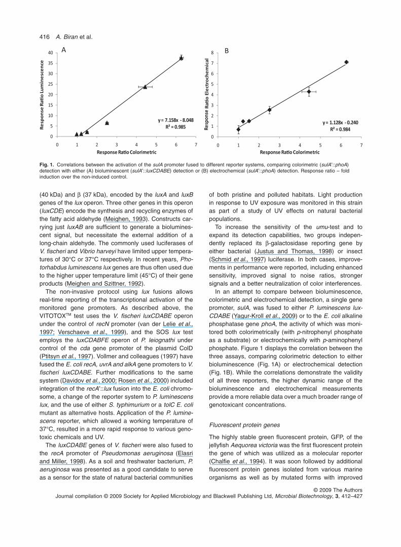
In an attempt to compare between bioluminescence,colorimetric and electrochemical detection, a single genepromoter, sulA, was fused to either P. luminescens lux-CDABE (Yagur-Kroll et al., 2009) or to the E. coli alkalinephosphatase gene phoA, the activity of which was moni-tored both colorimetrically (with p-nitrophenyl phosphateas a substrate) or electrochemically with p-aminophenylphosphate. Figure 1 displays the correlation between thethree assays, comparing colorimetric detection to eitherbioluminescence (Fig. 1A) or electrochemical detection(Fig. 1B). While the correlations demonstrate the validityof all three reporters, the higher dynamic range of thebioluminescence and electrochemical measurementsprovide a more reliable data over a much broader range ofgenotoxicant concentrations.
Fluorescent protein genes
The highly stable green fluorescent protein, GFP, of thejellyfish Aequorea victoria was the first fluorescent proteinthe gene of which was utilized as a molecular reporter(Chalfie et al., 1994). It was soon followed by additionalfluorescent protein genes isolated from various marineorganisms as well as by mutated forms with improved
Fig. 1. Correlations between the activation of the sulA promoter fused to different reporter systems, comparing colorimetric (sulA′::phoA)detection with either (A) bioluminescent (sulA′::luxCDABE) detection or (B) electrochemical (sulA′::phoA) detection. Response ratio – foldinduction over the non-induced control.
416 A. Biran et al.
© 2009 The AuthorsJournal compilation © 2009 Society for Applied Microbiology and Blackwell Publishing Ltd, Microbial Biotechnology, 3, 412–427
performance (Crameri et al., 1996; Tsien, 1998; Matzet al., 1999; Fradkov et al., 2000; Nagai et al., 2002;Wiedenmann et al., 2004; Shaner et al., 2007). The GFPprotein has a high quantum yield and can be expressed inboth prokaryotic and eukaryotic systems with no need fora substrate or cofactor (Kain and Kitts, 1997). To increasethe sensitivity of assays based on the GFP reportersystem, several green fluorescent protein mutants wereconstructed (Cormack et al., 1996; Crameri et al., 1996;Heim and Tsien, 1996; Welsh and Kay, 1997).
Arai and colleagues (2001) have modified the umu-testby replacing the lacZ gene with a DNA fragment encodingfor EGFP (enhanced green fluorescent protein). Thisconstruct was tested in E. coli strain KY706 with3 mg ml-1 4-NQO, a concentration that strongly inducedb-galactosidase activity in the umu-test. Detection sensi-tivity of the GFP reporter system became similar to that ofb-galactosidase only after the introduction of additionalmodifications to the plasmid. These included utilization oftandem lacUV5 and chimeric trp/umu promoters, andcoexpression of the E. coli recA5327 mutant. An addi-tional construct that harbours the fusion of the umuDCpromoter to the gfp gene was generated by Justus andThomas (1999), who reported an overall performanceinferior to that of the lacZ-based assay.
Other fluorescent genotoxicity sensors were con-structed by fusing the gfp and gfpmut3 reporting genes tothe recA promoter (Kostrzynska et al., 2002). GFPmut3 isa mutant that is approximately 20 times more fluorescentthan wild-type GFP and only weakly excited by UV light(Cormack et al., 1996). The use of the wild-type gfpyielded dose-dependent but weak responses, while withgfpmut3, the detection thresholds for MMC, MNNG, NA,hydrogen peroxide and formaldehyde were comparable tothe SOS chromotest (Quillardet et al., 1982), the umu-test(Nakamura et al., 1987) and the SOS lux test (Ptitsynet al., 1997).
The three fluorescent protein genes coding for EGFP,GFPuv and DsRed [a red fluorescent protein derived fromthe sea anemone Discosoma sp. (Matz et al., 1999)] weresimilarly fused to the recA promoter (Sagi et al., 2003),and the responses to nalidixic acid were compared with aluminescent recA′::lux strain. Performance was usuallypoorer compared with bioluminescent recA-based report-ers: lag times were longer and detection thresholds werehigher, unless incubation times were very long. TherecA′::DsRed plasmid, hosted in E. coli UTL2, was used inorder to monitor antigenotoxic activity of plant extractsthat exhibited some protection against MMC, NA andhydrogen peroxide (Bartolome et al., 2006).
The use of fluorescent proteins as reporters has beencharacterized as superior in terms of stability but inferiorto enzyme-based reporters in terms of sensitivity andresponse kinetics (Hakkila et al., 2002; Sagi et al., 2003).
Norman and colleagues (2006) have demonstrated thatthese drawbacks can be circumvented by the use of flowcytometry, displaying a response threshold of straincda′::gfpmut3 to MNNG of 5 nM, 10-fold lower than theminimal detectable concentration (MDC) of the umu-test(Reifferscheid et al., 1991). Moreover, the experimentalprocedure enabled the detection of MMC in spiked soil.
Cytotoxicity controls
As samples or chemicals of suspected DNA damagingactivity are also likely to be cytotoxic, genotoxicity assaysoften incorporate suitable controls to neutralize or correctfor the effects that cell damage or cell death may exert onassay results. One simple measure is an optical determi-nation of cell growth in parallel to assaying reporter geneactivity (Baun et al., 1999). However, this solution islimited, as optical density does not necessarily reflect theviability of a cell suspension. A different approach is basedon the inclusion of an additional, constitutive reportingstrain or enzyme which serves as a ‘light off’ sensor: adecrease in its signal indicates a toxic effect of thesample. This approach, for example, was adopted in theVITOTOXTM test that introduced a constitutive light-producing strain with a lux operon under the control of thestrong promoter, pr1 (Verschaeve et al., 1999).
The SOS lux test similarly incorporated a cytotoxicreporting strain harbouring a constitutive lac-GFPuvplasmid in the same S. typhimurium host strain(Baumstark-Khan et al., 2001). In a further developmentof this system a SWITCH plasmid was added, combiningthe SOS lux plasmid pPLS-1 and the LAC-Fluoro plasmidpGFPuv (Baumstark-Khan et al., 2005).
Using a different approach, the tester strain in the SOSchromotest was made constitutive for alkaline phos-phatase synthesis (Torriani and Rothman, 1961). Thisenzyme, non-inducible by DNA-damaging agents, isassayed in parallel to b-galactosidase and the ratio of thetwo activities is taken as a measure of the specific activityof b-galactosidase (Quillardet et al., 1982).
The toxicity of a sample can also be evaluated withpromoters that are induced by a broad spectrum of envi-ronmental insults and are thus good indicators of toxiccellular stress, such as the promoter of the grpE gene, acomponent of the chaperone network in E. coli (Van Dyket al., 1994; de Marco et al., 2007). The use of two strains,one harbouring the plasmid recA′::GFPuv and the othergrpE′::lux allowed an assessment of the toxicity of thesample along with its genotoxicity (Sagi et al., 2003). Adual-function toxicity/genotoxicity bioreporter system wasreported by Hever and Belkin (2006) who described aplasmid containing both recA′::EGFP and grpE′::DsRedfusions. A somewhat different double reporter conceptwas demonstrated by Mitchell and Gu (2004), who
Bacterial genotoxicity bioreporters 417
© 2009 The AuthorsJournal compilation © 2009 Society for Applied Microbiology and Blackwell Publishing Ltd, Microbial Biotechnology, 3, 412–427
presented a strain containing a fluorescent genotoxicityreporter fusion (recA′::GFPuv4) and a bioluminescent oxi-dative stress reporter (katG′::luxCDABE).
Detection performance
As described in detail above, numerous genotoxicity bio-assays based on genetically engineered bacteria havebeen presented over the years. While some of them, suchas the umu-test and the SOS chromotest, have under-gone intensive validation and were tested against hun-dreds of compounds, others have only been brieflydescribed along with their responses to a very limitedrange of chemicals. Quite clearly therefore pendingfurther validation of the latter group, the validated testsare of a much higher value for routine testing and theirresults merit higher credibility. Detailed reports of anextensive testing of these assays and their comparison tothe Ames test can be found in Nakamura and colleagues(1987), Reifferscheid and Heil (1996), and Quillardet andHofnung (1993). The umu-test has been standardized andaccepted for wastewater quality testing (ISO/CD 13829,DIN 38423-5).
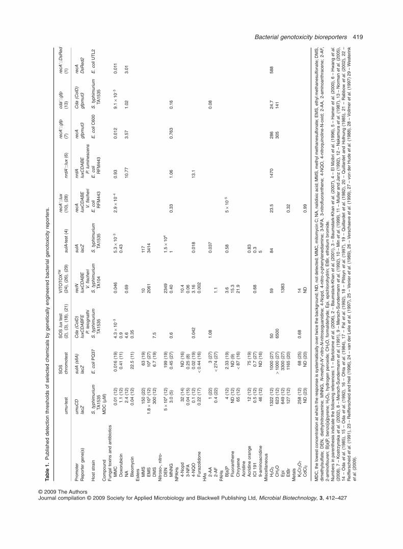
Table 1 summarizes reported detection thresholds ofselected genotoxicants exhibited by many of the assaysdescribed in the present review. Even a brief glance atTable 1 reveals that of all the tested systems, only two, theumu-test and the SOS chromotest, were challenged withthe required spectrum of genotoxic chemicals necessaryto demonstrate their applicability to environmental testing.All others were only preliminarily challenged with a verylimited number of compounds. In fact, Table 1 only listschemicals that have been tested by at least one bioassayin addition to the umu-test and the SOS chromotest; itthus does not contain the detection thresholds for hun-dreds of other compounds that have been reported forthese two tests (Quillardet et al., 1982; 1985; Ohta et al.,1984; Oda et al., 1985; Nakamura et al., 1987; von derHude et al., 1988; Quillardet and Hofnung, 1993; Reiffer-scheid and Heil, 1996). Based on this limited comparisonit may also be observed that detection thresholds varygreatly between the different assays, sometimes byseveral orders of magnitude. Other factors such asresponse times, detection spectra or facility of use thathave not been compared in Table 1 confer additionaladvantages to some of the reporter strains.
Further comparison of the performance of some ofthese bioassays has been performed in several hands-onworkshops conducted in Belgium (Mol TECHNOTOX;Corbisier et al., 2000; Baumstark-Khan et al., 2007) andin the USA (Eilatox-Oregon; Hakkila et al., 2004; Mer-iläinen and Lampinen, 2004; Pancrazio et al., 2004;Pedahzur et al., 2004). In addition to highlighting differ-ences in response characteristics, such workshops help
to emphasize the difficulties encountered when taking anewly developed test out of the lab and into the field andemphasized the merits of utilizing standard testing proto-cols for performance validation.
Sensitivity enhancement and expansion ofresponse spectrum
Very early in the short history of genetically engineeredbacterial reporters, it became apparent that simplepromoter–reporter fusions may be sufficient to demon-strate the applicability of the concept for genotoxicitytesting, but that additional molecular manipulations arerequired in order to turn them into efficient tools for routineuse. Such manipulations have taken several forms,including modification of the sensing elements, introduc-tion of mutations into the reporter strains for enhancedsensitivity and permeability, and the incorporation ofmetabolic activation capabilities.
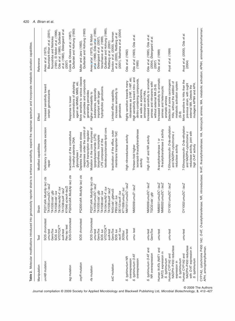
Table 2 lists some of the genetic manipulations intro-duced into the E. coli or S. typhimurium host strains andtheir reported effects. The modifications can be dividedinto two classes: deficiencies that reduce the ability of thecells to defend against DNA damaging agents, and new orenhanced capabilities of bacterial cells to metabolicallyactivate pre-genotoxic compounds, thus at least partiallymimicking the metabolic pathways such compounds mayundergo in mammalian systems.
Introduction of host strain mutations forenhanced sensitivity
As listed in Table 2, several mutations have been intro-duced into genotoxicity reporter strains to enhance theirsensitivity and thus lower their detection thresholds. Whilesome mutations, such as tag or oxyR, have only beenreported once (for the SOS chromotest; Quillardet andHofnung, 1993), others have become almost a pre-requisite in microbial genotoxicity reporters. Most notablein the latter group are uvrAB mutants deficient in excisionrepair, and rfa mutations that, by increasing membranepermeability, allow the build-up of higher intracellular con-centrations of the tested chemicals (Makela et al., 1974;Ames et al., 1975).
Altering the sensing element: manipulation ofregulatory sequences
Molecular manipulations of the DNA fragment harbouringthe sensing promoter element in order to improve thebacterial response to target chemicals have also beendescribed. One of the first examples is the VITOTOXTM
strain (van der Lelie et al., 1997). In addition to the wild-type recN promoter, two different mutant promoters were
418 A. Biran et al.
© 2009 The AuthorsJournal compilation © 2009 Society for Applied Microbiology and Blackwell Publishing Ltd, Microbial Biotechnology, 3, 412–427
Tab
le1.
Pub
lishe
dde
tect
ion
thre
shol
dsof
sele
cted
chem
ical
sby
gene
tical
lyen
gine
ered
bact
eria
lgen
otox
icity
repo
rter
s.
umu-
test
SO
Sch
rom
otes
tS
OS
lux
test
(2),
(3),
(18)
,(2
1)V
ITO
TO
XT
M
(24)
,(2
6),
(29)
sulA
-tes
t(4
)re
cA�:
:lux
(10)
,(2
8)nr
dA�:
:lux
(6)
recA
�::g
fp(7
)cd
a�::g
fp(1
3)re
cA�:
:DsR
ed(1
)
Pro
mot
erum
uCD
sulA
(sfiA
)C
da(C
olD
)re
cNsu
lAre
cAnr
dAre
cAC
da(C
olD
)re
cAR
epor
ter
gene
(s)
lacZ
lacZ
luxC
DA
BF
EP.
leio
gnat
hilu
xCD
AB
EV
.fis
cher
ila
cZlu
xCD
AB
EV
.fis
cher
ilu
xCD
AB
EP.
lum
ines
cens
gfpm
ut3
gfpm
ut3
DsR
ed2
Hos
tst
rain
S.
typh
imur
ium
TA15
35E
.co
liP
Q37
S.
typh
imur
ium
TA15
35S
.ty
phim
uriu
mTA
104
S.
typh
imur
ium
TA15
35E
.co
liR
FM
443
E.
coli
RF
M44
3E
.co
liC
600
S.
typh
imur
ium
TA15
35E
.co
liU
TL2
Com
poun
dM
DC
(mM
)F
unga
ltox
ins
and
antib
iotic
sM
MC
0.01
(12)
0.01
6(1
9)4.
3¥
10-3
0.04
65.
3¥
10-3
2.9
¥10
-40.
930.
012
9.1
¥10
-30.
011
Dox
orub
icin
1.1
(12)
0.41
(11)
0.9
0.43
NA
2.4
(12)
4.6
0.69
10.7
73.
571.
023.
01B
leom
ycin
0.04
(12)
22.5
(11)
0.35
Est
ers
MM
S15
0(2
2)63
(19)
1011
7E
MS
1.8
¥10
3(1
2)10
4(2
7)20
6134
14D
MS
300
(12)
6.7
(19)
7.5
Nitr
oso-
,ni
tro-
DE
N5
¥10
4(1
2)19
9(1
9)23
491.
5¥
104
MN
NG
3.0
(5)
0.45
(27)
0.6
0.40
10.
331.
060.
763
0.16
NP
AH
s4-
Nop
d32
(14)
ND
(16)
10.4
3-N
FA0.
04(1
5)0.
25(8
)0.
064-
NQ
O0.
1(1
2)0.
02(1
9)0.
042
3.16
0.01
813
.1F
uraz
olid
one
0.22
(17)
<0.
44(1
6)0.
002
HA
s 2-A
A1
(22)
3(2
7)1.
080.
037
0.08
2-A
F0.
4(2
2)<
274
(27)
1.1
PA
Hs
B[a
]P4
(12)
2.33
(19)
3.6
0.58
5¥
10-5
Flu
oran
then
eN
D(1
2)N
D(9
)15
.3C
hrys
ene
65(1
2)<
87(1
6)21
.9A
crid
ine
Acr
idin
eor
ange
12(1
2)75
(19)
0.83
ICI
191
0.5
(12)
0.7
(27)
0.68
0.3
9-am
inoa
crid
ine
46(1
2)N
D(1
6)5
Mis
cella
neou
sH
2O2
1322
(12)
1000
(27)
5059
8423
.514
7028
624
.758
8C
H2O
623
(12)
>10
00(2
7)65
0030
514
1E
pi64
9(1
2)33
00(2
7)13
83E
tBr
127
(12)
1165
(20)
0.32
Met
als
K2C
r 2O
725
8(1
2)68
(25)
0.68
14C
dCl 2
ND
(23)
ND
(20)
ND
0.99
MD
C,t
helo
wes
tcon
cent
ratio
nat
whi
chth
ere
spon
seis
syst
emat
ical
lyov
ertw
ice
the
back
grou
nd;N
D,n
otde
tect
ed;M
MC
,mito
myc
inC
;NA
,nal
idix
icac
id;M
MS
,met
hylm
etha
nesu
lfona
te;E
MS
,eth
ylm
etha
nesu
lfona
te;D
MS
,di
met
hyls
ulfa
te;
DE
N,
diet
hyln
itros
amin
e;M
NN
G,
N-m
ethy
l-N′-n
itro-
N-n
itros
ogua
nidi
ne;
4-N
opd,
4-ni
tro-
o-ph
enyl
ened
iam
ine;
3-N
FA,
3-ni
trofl
uora
nthe
ne;
4-N
QO
,4-
nitr
oqui
nolin
e-N
-oxi
d;2-
AA
,2-
amin
oant
hrac
ene;
2-A
F,2-
amin
oflou
ren;
B[a
]P,
benz
o[a]
pyre
ne;
H2O
2,hy
drog
enpe
roxi
de;
CH
2O,
form
alde
hyde
;E
pi,
epic
hlor
ohyd
rin;
EtB
r,et
hidi
umbr
omid
e.N
umbe
rsin
pare
nthe
sis
indi
cate
the
follo
win
gre
fere
nces
:1–
Bar
tolo
me
etal
.(2
006)
,2–
Bau
mst
ark-
Kha
net
al.
(200
1),3
–B
aum
star
k-K
han
etal
.(2
007)
,4–
ElM
zibr
ieta
l.(1
996)
,5–
Ham
eret
al.
(200
0),6
–H
wan
get
al.
(200
8),7
–K
ostr
zyns
kaet
al.
(200
2),8
–M
ersc
h-S
unde
rman
net
al.
(199
1),9
–M
ersc
h-S
unde
rman
net
al.
(199
2),1
0–
Min
etal
.(1
999)
,11
–M
ulle
ran
dJa
nz(1
992)
,12
–N
akam
ura
etal
.(1
987)
,13
–N
orm
anet
al.
(200
5),
14–
Oda
etal
.(1
985)
,15
–O
daet
al.
(199
2),
16–
Oht
aet
al.
(198
4),
17–
Pal
etal
.(1
992)
,18
–P
titsy
net
al.
(199
7),
19–
Qui
llard
etet
al.
(198
2),
20–
Qui
llard
etan
dH
ofnu
ng(1
985)
,21
–R
abbo
wet
al.
(200
2),
22–
Rei
ffers
chei
det
al.
(199
1),2
3–
Rei
ffers
chei
dan
dH
eil(
1996
),24
–va
nde
rLe
lieet
al.
(199
7),2
5–
Ven
ier
etal
.(1
989)
,26
–V
ersc
haev
eet
al.
(199
9),2
7–
von
der
Hud
eet
al.
(198
8),2
8–
Vol
lmer
etal
.(1
997)
29–
Wes
terin
ket
al.
(200
9).
Bacterial genotoxicity bioreporters 419
© 2009 The AuthorsJournal compilation © 2009 Society for Applied Microbiology and Blackwell Publishing Ltd, Microbial Biotechnology, 3, 412–427
Tab
le2.
Mol
ecul
arm
odifi
catio
nsin
trod
uced
into
geno
toxi
city
repo
rter
stra
ins
toen
hanc
ese
nsiti
vity
,ex
pand
the
resp
onse
spec
trum
and
inco
rpor
ate
met
abol
icac
tivat
ion
capa
bilit
ies.
Man
ipul
atio
nTe
stS
trai
nM
odifi
edca
pabi
litie
sE
ffect
Ref
eren
ce
uvrA
Bm
utat
ion
SO
Sch
rom
otes
tP
Q37
/sfiA
::Mud
(Ap
lac)
cts
Defi
cien
cyin
nucl
eotid
eex
cisi
onre
pair
Incr
ease
dse
nsiti
vity
tow
ard
cert
ain
geno
toxi
cant
sA
mes
etal
.(1
973)
,B
aum
star
k-K
han
etal
.(2
001)
,N
unos
hiba
and
Nis
hiok
a(1
991)
,E
lMzi
brie
tal.
(199
6),
Oda
etal
.(1
985)
,Q
uilla
rdet
etal
.(1
982)
,Ø
ster
gaar
det
al.
(200
7)
umu-
test
TA15
35/u
muD
C′::
lacZ
Gen
oTox
TA15
35/c
da′::
gfp
SO
Slu
xTA
1538
/cda
′::lu
xV
ITO
TO
XT
MTA
104/
recN
2-4′
::lux
sulA
-tes
tTA
1538
/sul
A′::
lacZ
Rec
-lac
test
KY
946
j(re
cA–l
acZ
)ta
gm
utat
ion
SO
S-C
hrom
otes
tP
Q24
3/sfi
A::M
ud(A
pla
c)ct
sIn
activ
atio
nof
the
cons
titut
ive
3-m
ethy
l-ade
nine
DN
Agl
ycos
ylas
eI
Res
pons
eto
low
erco
ncen
trat
ions
ofal
kyla
ting
agen
tas
MN
NG
,M
MS
,et
c.
Cos
tade
Oliv
eira
etal
.(1
987)
,Q
uilla
rdet
and
Hof
nung
(199
3)
oxyR
mut
atio
nS
OS
chro
mot
est
PQ
300/
sfiA
::Mud
(Ap
lac)
cts
Dep
lete
sth
eox
idat
ive
stre
ssre
spon
ses
unde
rth
eco
ntro
lof
Oxy
Rtr
ansc
riptio
nre
gula
tor
Mor
ese
nsiti
veto
vario
uscl
asse
sof
pero
xide
san
dco
mpo
unds
gene
ratin
gpe
roxi
des
Mul
ler
and
Janz
(199
2),
Qui
llard
etan
dH
ofnu
ng(1
993)
rfa
mut
atio
nS
OS
Chr
omot
est
PQ
37/s
fiA::M
ud(A
pla
c)ct
sM
utat
ion
inth
eco
reen
zym
esof
lypo
poly
sacc
harid
e(L
PS
)bi
osyn
thes
is.
Inco
mpl
ete
LPS
com
pose
dof
the
keto
deox
yoct
anoa
te-li
pid
core
.
Hig
her
perm
eabi
lity
tosu
bsta
nces
,es
peci
ally
impo
rtan
tw
ithla
rger
hydr
opho
bic
geno
toxi
ns.
Am
eset
al.
(197
3),
ElM
zibr
iet
al.
(199
6),
Oda
etal
.(1
985)
,Ø
ster
gaar
det
al.
(200
7),
Ver
scha
eve
etal
.(1
999)
,Q
uilla
rdet
and
Hof
nung
(198
5),
Ret
tber
get
al.
(200
1)
umu-
test
TA15
35/u
muD
C′::
lacZ
Gen
oTox
TA15
35/c
da′::
gfp
SO
Slu
xTA
1538
/cda
′::lu
xV
ITO
TO
XT
MTA
104/
recN
2-4′
::lux
sulA
-tes
tTA
1538
/sul
A′::
lacZ
tolC
mut
atio
nG
enoT
oxN
43/c
da′::
gfpm
ut3
Inac
tivat
ion
ofth
eef
flux,
oute
rm
embr
ane
tran
spor
ter-
TolC
Lim
ited
efflu
xca
pabi
lity,
incr
ease
sse
nsiti
vity
toge
noto
xins
Dav
idov
etal
.(2
000)
,N
orm
anet
al.
(200
5),
Ret
tber
get
al.
(200
1),
Mae
hana
etal
.(2
004)
SO
Slu
xP
B3/
cda′
::lux
recA
′::lu
xD
E11
2/re
cA′::
lux
SO
Slu
cK
T10
08/u
muD
′::lu
cS
.ty
phim
uriu
mN
Rov
erex
pres
sion
umu-
test
NM
1011
/um
uDC
′::la
cZH
igh
nitr
ored
ucta
seac
tivity
Hig
hly
sens
itive
tow
ards
man
yni
troa
rene
sas
2-N
F,1-
NP,
etc.
Oda
etal
.(1
992)
S.
typh
imur
ium
O-A
Tov
erex
pres
sion
umu-
test
NM
2009
/um
uD′::
lacZ
Thi
rtee
n-fo
ldhi
gher
ison
iazi
d-N
-ace
tyltr
ansf
eras
eac
tivity
Hig
hse
nsiti
vity
tow
ard
nitr
o-an
ddi
nitr
o-co
ntai
ning
com
poun
ds,
asw
ells
asar
ylam
ins,
amin
oanz
oan
dH
As.
Oda
etal
.(1
993)
,O
daet
al.
(199
5)
S.
typh
imur
ium
O-A
Tan
dN
Rov
erex
pres
sion
umu-
test
NM
3009
/um
uDC
′::la
cZH
igh
O-A
Tan
dN
Rac
tivity
Incr
ease
dse
nsiti
vity
toar
omat
icam
ines
and
nitr
oare
nsw
ith/
with
out
exte
rnal
MA
(S-9
)
Oda
etal
.(1
995)
,O
daet
al.
(200
4),
Øst
erga
ard
etal
.(2
007)
Gen
oTox
TG
O2/
cda�
::gfp
Hum
anN
-ATs
(NA
T1
and
NA
T2)
expr
essi
onin
S.
typh
imur
ium
umu-
test
NM
6001
/um
uDC
′::la
cZN
-ace
tyltr
ansf
eras
e1
orN
-ace
tyltr
ansf
eras
e2
activ
ityIn
crea
sed
sens
itivi
tyto
arom
atic
amin
esan
dhe
tero
cycl
icar
omat
icam
ines
Oda
etal
.(1
999)
NM
6002
/um
uDC
′::la
cZ
Hum
anC
YP
1A2
and
NA
DP
H–P
450
redu
ctas
eex
pres
sion
inS
.ty
phim
uriu
m
umu-
test
OY
1001
/um
uDC
′::la
cZ7-
Eth
oxyr
esor
ufin
O-d
e-et
hyla
tion
and
NA
DP
H–c
ytoc
hrom
ec
redu
ctas
eac
tivity
Det
ectio
nof
som
eca
rcin
ogen
icH
As,
with
out
the
addi
tion
ofm
etab
olic
activ
atio
nsy
stem
(S-9
)
Ary
alet
al.
(199
9)
Hum
anC
YP
1A2
and
NA
DP
H–P
450
redu
ctas
ew
ithO
-AT
expr
essi
onin
S.
typh
imur
ium
umu-
test
OY
1002
/um
uDC
′::la
cZ7-
Eth
oxyr
esor
ufin
O-d
e-et
hyla
tion
and
NA
DP
H–c
ytoc
hrom
ec
redu
ctas
eac
tivity
,jo
int
with
high
O-A
Tac
tivity
Mor
ese
nsiti
veto
HA
sth
anth
epr
evio
usst
rain
,w
ithou
ght
exte
rnal
MA
.D
etec
tsth
em
utag
ens
AP
NH
and
AP
H.
Ary
alet
al.
(200
0),
Oda
etal
.(2
004)
CY
P1A
2,cy
toch
rom
eP
450
1A2;
O-A
T,O
-ace
tyltr
ansf
eras
e;N
R,
nitr
ored
ucta
se;
N-A
T,N
-ace
tyltr
ansf
eras
e;H
A,
hetr
ocyc
licam
ines
;M
A,
met
abol
icac
tivat
ion;
AP
NH
,am
inop
heny
lnor
harm
an;
AP
H,
amin
ophe
nylh
arm
an.
420 A. Biran et al.
© 2009 The AuthorsJournal compilation © 2009 Society for Applied Microbiology and Blackwell Publishing Ltd, Microbial Biotechnology, 3, 412–427
constructed and tested alone and in combination: a dele-tion of one of the LexA binding sites, and a ‘promoter up’mutation where a consensus nucleotide was introduced inthe -35 region. Each of the single mutants was superior tothe wild-type in at least one respect, but the double muta-tion resulted in poorer performance. The influence ofaddition/subtraction of lexA binding sites on reporter geneexpression in the SOS promoter::uidA fusion under geno-toxic stress was examined with several SOS promoters(umuD, sulA, recA and recN) (Dreier et al., 2002). Highestsignals were produced by constructs that either har-boured an additional lexA binding site in the sulA promoterthat overlapped the -35 promoter region, or that lackedone of the two recN LexA binding sites. Another success-ful effort was reported by Arai and colleagues (2001), whoimproved the performance of a umuDC′::gfp construct byreplacing the wild-type -35 promoter sequence with the-35 sequence of the highly active trp gene promoter.
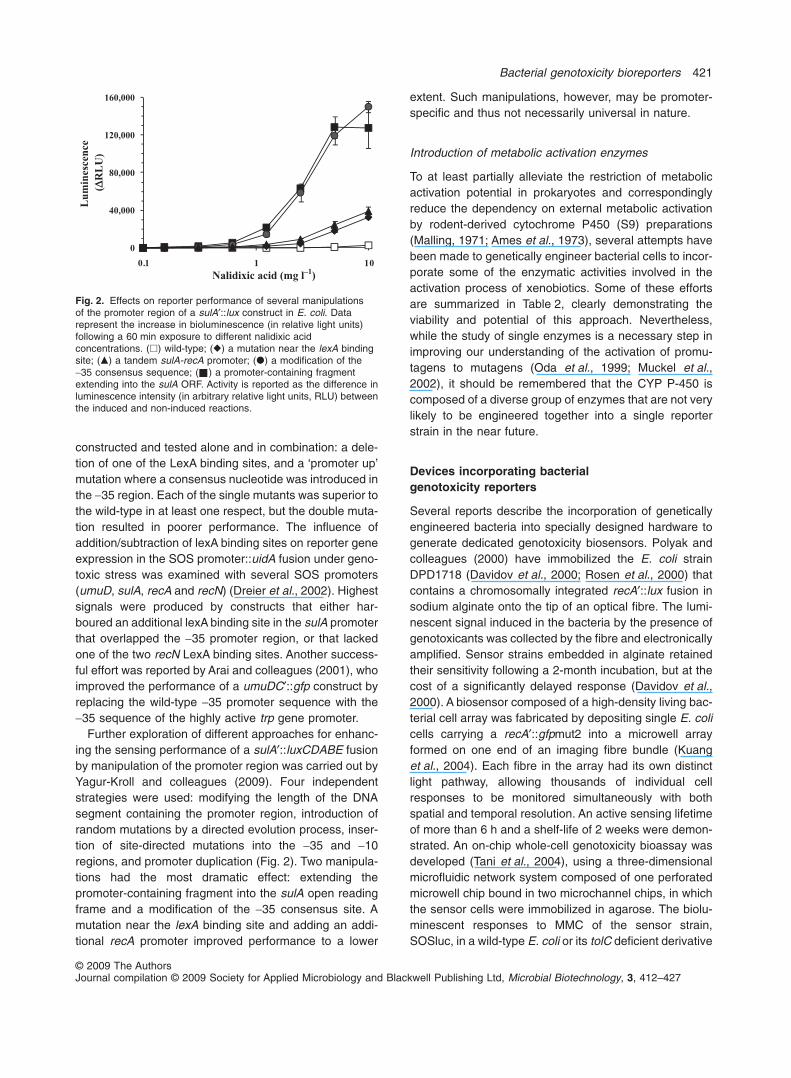
Further exploration of different approaches for enhanc-ing the sensing performance of a sulA′::luxCDABE fusionby manipulation of the promoter region was carried out byYagur-Kroll and colleagues (2009). Four independentstrategies were used: modifying the length of the DNAsegment containing the promoter region, introduction ofrandom mutations by a directed evolution process, inser-tion of site-directed mutations into the -35 and -10regions, and promoter duplication (Fig. 2). Two manipula-tions had the most dramatic effect: extending thepromoter-containing fragment into the sulA open readingframe and a modification of the -35 consensus site. Amutation near the lexA binding site and adding an addi-tional recA promoter improved performance to a lower
extent. Such manipulations, however, may be promoter-specific and thus not necessarily universal in nature.
Introduction of metabolic activation enzymes
To at least partially alleviate the restriction of metabolicactivation potential in prokaryotes and correspondinglyreduce the dependency on external metabolic activationby rodent-derived cytochrome P450 (S9) preparations(Malling, 1971; Ames et al., 1973), several attempts havebeen made to genetically engineer bacterial cells to incor-porate some of the enzymatic activities involved in theactivation process of xenobiotics. Some of these effortsare summarized in Table 2, clearly demonstrating theviability and potential of this approach. Nevertheless,while the study of single enzymes is a necessary step inimproving our understanding of the activation of promu-tagens to mutagens (Oda et al., 1999; Muckel et al.,2002), it should be remembered that the CYP P-450 iscomposed of a diverse group of enzymes that are not verylikely to be engineered together into a single reporterstrain in the near future.
Devices incorporating bacterialgenotoxicity reporters
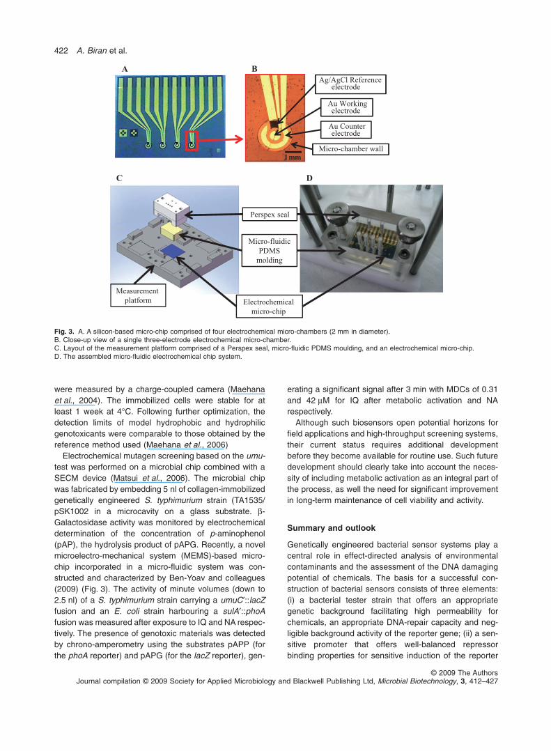
Several reports describe the incorporation of geneticallyengineered bacteria into specially designed hardware togenerate dedicated genotoxicity biosensors. Polyak andcolleagues (2000) have immobilized the E. coli strainDPD1718 (Davidov et al., 2000; Rosen et al., 2000) thatcontains a chromosomally integrated recA′::lux fusion insodium alginate onto the tip of an optical fibre. The lumi-nescent signal induced in the bacteria by the presence ofgenotoxicants was collected by the fibre and electronicallyamplified. Sensor strains embedded in alginate retainedtheir sensitivity following a 2-month incubation, but at thecost of a significantly delayed response (Davidov et al.,2000). A biosensor composed of a high-density living bac-terial cell array was fabricated by depositing single E. colicells carrying a recA′::gfpmut2 into a microwell arrayformed on one end of an imaging fibre bundle (Kuanget al., 2004). Each fibre in the array had its own distinctlight pathway, allowing thousands of individual cellresponses to be monitored simultaneously with bothspatial and temporal resolution. An active sensing lifetimeof more than 6 h and a shelf-life of 2 weeks were demon-strated. An on-chip whole-cell genotoxicity bioassay wasdeveloped (Tani et al., 2004), using a three-dimensionalmicrofluidic network system composed of one perforatedmicrowell chip bound in two microchannel chips, in whichthe sensor cells were immobilized in agarose. The biolu-minescent responses to MMC of the sensor strain,SOSluc, in a wild-type E. coli or its tolC deficient derivative
0
40,000
80,000
120,000
160,000
0111.0
Lum
ines
cenc
e (D
RL
U)
Nalidixic acid (mg l–1)
Fig. 2. Effects on reporter performance of several manipulationsof the promoter region of a sulA′::lux construct in E. coli. Datarepresent the increase in bioluminescence (in relative light units)following a 60 min exposure to different nalidixic acidconcentrations. (�) wild-type; (�) a mutation near the lexA bindingsite; (�) a tandem sulA-recA promoter; (�) a modification of the-35 consensus sequence; ( ) a promoter-containing fragmentextending into the sulA ORF. Activity is reported as the difference inluminescence intensity (in arbitrary relative light units, RLU) betweenthe induced and non-induced reactions.
Bacterial genotoxicity bioreporters 421
© 2009 The AuthorsJournal compilation © 2009 Society for Applied Microbiology and Blackwell Publishing Ltd, Microbial Biotechnology, 3, 412–427
were measured by a charge-coupled camera (Maehanaet al., 2004). The immobilized cells were stable for atleast 1 week at 4°C. Following further optimization, thedetection limits of model hydrophobic and hydrophilicgenotoxicants were comparable to those obtained by thereference method used (Maehana et al., 2006)
Electrochemical mutagen screening based on the umu-test was performed on a microbial chip combined with aSECM device (Matsui et al., 2006). The microbial chipwas fabricated by embedding 5 nl of collagen-immobilizedgenetically engineered S. typhimurium strain (TA1535/pSK1002 in a microcavity on a glass substrate. b-Galactosidase activity was monitored by electrochemicaldetermination of the concentration of p-aminophenol(pAP), the hydrolysis product of pAPG. Recently, a novelmicroelectro-mechanical system (MEMS)-based micro-chip incorporated in a micro-fluidic system was con-structed and characterized by Ben-Yoav and colleagues(2009) (Fig. 3). The activity of minute volumes (down to2.5 nl) of a S. typhimurium strain carrying a umuC′::lacZfusion and an E. coli strain harbouring a sulA′::phoAfusion was measured after exposure to IQ and NA respec-tively. The presence of genotoxic materials was detectedby chrono-amperometry using the substrates pAPP (forthe phoA reporter) and pAPG (for the lacZ reporter), gen-
erating a significant signal after 3 min with MDCs of 0.31and 42 mM for IQ after metabolic activation and NArespectively.
Although such biosensors open potential horizons forfield applications and high-throughput screening systems,their current status requires additional developmentbefore they become available for routine use. Such futuredevelopment should clearly take into account the neces-sity of including metabolic activation as an integral part ofthe process, as well the need for significant improvementin long-term maintenance of cell viability and activity.
Summary and outlook
Genetically engineered bacterial sensor systems play acentral role in effect-directed analysis of environmentalcontaminants and the assessment of the DNA damagingpotential of chemicals. The basis for a successful con-struction of bacterial sensors consists of three elements:(i) a bacterial tester strain that offers an appropriategenetic background facilitating high permeability forchemicals, an appropriate DNA-repair capacity and neg-ligible background activity of the reporter gene; (ii) a sen-sitive promoter that offers well-balanced repressorbinding properties for sensitive induction of the reporter
1mm
Ag/AgCl Reference electrode
Au Working electrode
Au Counter electrode
Micro-chamber wall
BA
Perspex seal
Micro-fluidic PDMS
molding
Electrochemical micro-chip
Measurement platform
DC
Fig. 3. A. A silicon-based micro-chip comprised of four electrochemical micro-chambers (2 mm in diameter).B. Close-up view of a single three-electrode electrochemical micro-chamber.C. Layout of the measurement platform comprised of a Perspex seal, micro-fluidic PDMS moulding, and an electrochemical micro-chip.D. The assembled micro-fluidic electrochemical chip system.
422 A. Biran et al.
© 2009 The AuthorsJournal compilation © 2009 Society for Applied Microbiology and Blackwell Publishing Ltd, Microbial Biotechnology, 3, 412–427
gene; and (iii) a sensitive, fast responding reporter systemwith a broad dynamic range and preferably the capabilityof real-time monitoring.
The discovery and the understanding of the bacterialSOS-system, a regulatory pathway that is mainly respon-sible for inducible DNA repair and induced mutagenesis inbacteria, opened diverse possibilities for genetically tai-loring bacteria for the specific, sensitive and fast detectionof genotoxic contaminants. The fusion of SOS genepromoters to reporter genes that generate quantifiablesignals allow to easily detect the genotoxic potency of asample. This review describes the current stage of devel-opment of the sensing systems by discussing the molecu-lar, biochemical and physico-chemical characteristics ofthe different promoters and suitable reporter genes basedon colorimetric, luminometric, fluorimetric and electro-chemical detection.
The potential of future sensor developments withenhanced external and/or internal metabolic competenceis immense. Against the background of a worldwideincreasing freshwater demand, and the need for reclama-tion of process water as drinking water resource, bacterialsensors can play a crucial role in risk minimization. To fullyreach this objective, additional progress needs to bemade in several directions including enhancement of sen-sitivity, expansion of the response spectrum, stabilizationof the more sensitive reagents and, most importantly, theintroduction of broad-spectrum metabolic activation capa-bilities into the sensor strains.
Acknowledgements
The authors are grateful for funding provided by the GermanBMBF and the Israeli MOST in the framework of projectWT601 (‘DipChip’) of the German/Israeli binational WaterTechnology Program. [Correction added on 26 January 2010,after first online publication: the preceding sentence wasadded after first online publication.]
References
Ames, B.N., Durston, W.E., Yamasaki, E., and Lee, F.D.(1973) Carcinogens are mutagens: a simple test systemcombining liver homogenates for activation and bacteria fordetection. Proc Natl Acad Sci USA 70: 2281–2285.
Ames, B.N., McCann, J., and Yamasaki, E. (1975) Methodsfor detecting carcinogens and mutagens with thesalmonella/mammalian-microsome mutagenicity test.Mutat Res Environ Mutagen Relat Subj 31: 347–364.
Arai, R., Makita, Y., Oda, Y., and Nagamune, T. (2001) Con-struction of green fluorescent protein reporter genes forgenotoxicity test (SOS/umu-test) and improvement ofmutagen-sensitivity. J Biosci Bioeng 92: 301–304.
Aryal, P., Yoshikawa, K., Terashita, T., Guengerich, F.P.,Shimada, T., and Oda, Y. (1999) Development of a new
genotoxicity test system with Salmonella typhimuriumOY1001/1A2 expressing human CYP1A2 and NADPH-P450 reductase. Mutat Res Genet Toxicol EnvironMutagen 442: 113–120.
Aryal, P., Terashita, T., Guengerich, F.P., Shimada, T., andOda, Y. (2000) Use of genetically engineered Salmonellatyphimurium OY1002/1A2 strain coexpressing humancytochrome P450 1A2 and NADPH-cytochrome P450reductase and bacterial O-acetyltransferase in SOS/umuassay. Environ Mol Mutagen 36: 121–126.
Badihi-Mossberg, M., Buchner, V., and Rishpon, J. (2007)Electrochemical biosensors for pollutants in the environ-ment. Electroanalysis 19: 2015–2028.
Bartolome, A., Mandap, K., David, K.J., Sevilla Iii, F., andVillanueva, J. (2006) SOS-red fluorescent protein (RFP)bioassay system for monitoring of antigenotoxic activity inplant extracts. Biosens Bioelectron 21: 2114–2120.
Baumstark-Khan, C., Rode, A., Rettberg, P., and Horneck, G.(2001) Application of the Lux-Fluoro test as bioassay forcombined genotoxicity and cytotoxicity measurements bymeans of recombinant Salmonella typhimurium TA1535cells. Anal Chim Acta 437: 23–30.
Baumstark-Khan, C., Cioara, K., Rettberg, P., and Horneck,G. (2005) Determination of geno- and cytotoxicity ofgroundwater and sediments using the recombinantSWITCH Test*. J Environ Sci Health A Tox Hazard SubstEnviron Eng 40: 245–263.
Baumstark-Khan, C., Rabbow, E., Rettberg, P., and Horneck,G. (2007) The combined bacterial Lux-Fluoro test for thedetection and quantification of genotoxic and cytotoxicagents in surface water: results from the ‘Technical Work-shop on Genotoxicity Biosensing’. Aquat Toxicol 85: 209–218.
Baun, A., Andersen, J.S., and Nyholm, N. (1999) Correctingfor toxic inhibition in quantification of genotoxic response inthe umuC test. Mutat Res Genet Toxicol Environ Mutagen441: 171–180.
Belkin, S. (2003) Microbial whole-cell sensing systemsof environmental pollutants. Curr Opin Microbiol 6: 206–212.
Ben-Yoav, H., Biran, A., Pedahzur, R., Belkin, S., Buchinger,S., Reifferscheid, G., and Shacham-Diamand, Y. (2009) Awhole cell electrochemical biosensor for water genotoxicitybio-detection. Electrochim Acta 54: 6113–6118.
Bihari, N., Vukmirovic, M., Batel, R., and Zahn, R. (1990)Application of the SOS umu-test in detection of pollutionusing fish liver S9 fraction. Comp Biochem Physiol 95:15–18.
Biran, I., Klimentiy, L., Hengge-Aronis, R., Ron, E.Z., andRishpon, J. (1999) On-line monitoring of gene expression.Microbiology 145: 2129–2133.
Biran, I., Babai, R., Levcov, K., Rishpon, J., and Ron, E.Z.(2000) Online and in situ monitoring of environmental pol-lutants: electrochemical biosensing of cadmium. EnvironMicrobiol 2: 285–290.
Biran, A., Pedahzur, R., Buchinger, S., Reifferscheid, G.,and Belkin, S. (2009) Genetically engineered bacteria forgenotoxicity assessment. In Biosensors for EnvironmentalMonitoring of Aquatic Systems, Vol. 5J. Barcelo, D., andHansen, P.-D. (eds). Berlin, Germany: Springer, pp. 161–186.
Bacterial genotoxicity bioreporters 423
© 2009 The AuthorsJournal compilation © 2009 Society for Applied Microbiology and Blackwell Publishing Ltd, Microbial Biotechnology, 3, 412–427
Buchinger, S., Grill, P., Morosow, V., Ben-Yoav, H., Shacham-Diamand, Y., Biran, A., et al. (2009) Evaluation of chrono-amperometric signal detection for the analysis ofgenotoxicity by a whole cell biosensor. Analytica ChimicaActa (in press): doi: 10.1016/j.aca.2009.11.027.
Chalfie, M., Tu, G., Euskirchen, G., Ward, W.W., and Prasher,D.C. (1994) Green fluorescent protein as a marker for geneexpression. Science 263: 802–804.
Corbisier, P., Hansen, P.-D., and Barcelo, D. (2000) Proceed-ings of the BIOSET technical workshop on genotoxicitybiosensing TECHNOTOX. [WWW document]. URL http://wwwa.vito.be/english/environment/pdf/technotox/Proceedings4_00.PDF.
Cormack, B.P., Valdivia, R.H., and Falkow, S. (1996) FACS-optimized mutants of the green fluorescent protein (GFP).Gene 173: 33–38.
Costa de Oliveira, R., Laval, J., and Boiteux, S. (1987) Induc-tion of SOS and adaptive responses by alkylating agents inEscherichia coli mutants deficient in 3-methyladenine-DNAglycosylase activities. Mutat Res DNA Rep 183: 11–20.
Courcelle, J., and Hanawalt, P.C. (2003) RecA-dependentrecovery of arrested DNA replication forks. Annu RevGenet 37: 611–646.
Courcelle, J., Khodursky, A., Peter, B., Brown, P.O., andHanawalt, P.C. (2001) Comparative gene expression pro-files following UV exposure in wild-type and SOS-deficientEscherichia coli. Genet 158: 41–64.
Crameri, A., Whitehorn, E.A., Tate, E., and Stemmer, W.P.C.(1996) Improved green fluorescent protein by molecularevolution using DNA shuffling. Nat Biotech 14: 315–319.
D’Ari, R. (1985) The SOS system. Biochimie 67: 343–347.Davidov, Y., Rosen, R., Smulski, D.R., Van Dyk, T.K., Vollmer,
A.C., Elsemore, D.A., et al. (2000) Improved bacterial SOSpromoter::lux fusions for genotoxicity detection. Mutat ResGenet Toxicol Environ Mutagen 466: 97–107.
Dizer, H., Wittekindt, E., Fischer, B., and Hansen, P. (2002)The cytotoxic and genotoxic potential of surface water andwastewater effluents as determined by bioluminescence,umu-assays and selected biomarkers. Chemosphere 46:225–233.
Dreier, J., Breitmaier, E.B., Gocke, E., Apfel, C.M., and Page,M.G.P. (2002) Direct influence of S9 liver homogenate onfluorescence signals: impact on practical applications in abacterial genotoxicity assay. Mutat Res Genet ToxicolEnviron Mutagen 513: 169–182.
Ehrlichmann, H., Dott, W., and Eisentraeger, A. (2000)Assessment of the water-extractable genotoxic potential ofsoil samples from contaminated sites. Ecotoxicol EnvironSaf 46: 73–80.
El Mzibri, M., De Méo, M.P., Laget, M., Guiraud, H., Séree, E.,Barra, Y., and Duménil, G. (1996) The Salmonella sulA-test: a new in vitro system to detect genotoxins. Mutat ResGenet Toxicol 369: 195–208.
Elasri, M., and Miller, R. (1998) A Pseudomonas aeruginosabiosensor responds to exposure to ultraviolet radiation.Appl Microbiol Biotechnol 50: 455–458.
Fernández de Henestrosa, A.R., Ogi, T., Aoyagi, S., Chafin,D., Hayes, J.J., Ohmori, H., and Woodgate, R. (2000)Identification of additional genes belonging to the LexAregulon in Escherichia coli. Mol Microbiol 35: 1560–1572.
Fradkov, A.F., Chen, Y., Ding, L., Barsova, E.V., Matz, M.V.,and lukyanov, S.A. (2000) Novel fluorescent protein fromDiscosoma coral and its mutants possesses a unique far-red fluorescence. FEBS Lett 479: 127–130.
Frey, J., Ghersa, P., Palacios, P.G., and Belet, M. (1986)Physical and genetic analysis of the ColD plasmid. J Bac-teriol 166: 15–19.
Fuentes, J.L., Alonso, A., Cuétara, E., Vernhe, M., Alvarez,N., Sánchez-Lamar, A., and Llagostera, M. (2006)Usefulness of the SOS Chromotest in the study ofmedicinal plants as radioprotectors. Int J Radiat Biol 82:323–329.
Gao, G., Fan, L., Lu, H., and Hua, Y. (2008) EngineeringDeinococcus radiodurans into biosensor to monitor radio-activity and genotoxicity in environment. Chin Sci Bull 53:1675–1681.
German Institute of Standardization (1996) DIN 38415-3 –German standard methods for the examination of water,waste water and sludge – Sub-animal testing (group T) –Part 3: Determination of the genotoxic potential ofwater with the umu-test (T 3). Berlin, Germany: GermanInstitute of Standardization.
Giese, K.C., Michalowski, C.B., and Little, J.W. (2008) RecA-dependent cleavage of LexA dimers. J Mol Biol 377: 148–161.
Hakkila, K., Maksimow, M., Karp, M., and Virta, M. (2002)Reporter genes lucFF, luxCDABE, gfp, and dsred havedifferent characteristics in whole-cell bacterial sensors.Anal Biochem 301: 235–242.
Hakkila, K., Lappalainen, J., and Virta, M. (2004) Toxicitydetection from EILATox-Oregon Workshop samples byusing kinetic photobacteria measurement: the flashmethod. J Appl Toxicol 24: 349–353.
Hamer, B., Bihari, N., Reifferscheid, G., Zahn, R.K., Müller,W.E.G., and Batel, R. (2000) Evaluation of the SOS/umu-test post-treatment assay for the detection of genotoxicactivities of pure compounds and complex environmentalmixtures. Mutat Res Genet Toxicol Environ Mutagen 466:161–171.
Heim, R., and Tsien, R.Y. (1996) Engineering green fluores-cent protein for improved brightness, longer wavelengthsand fluorescence resonance energy transfer. Curr Biol 6:178–182.
Hever, N., and Belkin, S. (2006) A dual-color bacterialreporter strain for the detection of toxic and genotoxiceffects. Eng Life Sci 6: 319–323.
Higashitani, A., Higashitani, N., and Horiuchi, K. (1995) A celldivision inhibitor SulA of Escherichia coli directly interactswith FtsZ through GTP hydrolysis. Biochem Biophys ResComm 209: 198–204.
von der Hude, W., Behm, C., Gurtler, R., and Basler, A.(1988) Evaluation of the SOS chromotest. Mutat ResEnviron Mutagen Relat Subj 203: 81–94.
Hwang, E.T., Ahn, J.-M., Kim, B.C., and Gu, M.B. (2008)Construction of a nrdA::luxCDABE fusion and its use inEscherichia coli as a DNA damage biosensor. Sensors 8:1297–1307.
International Standardization Organization (2000). ISO/CD13829 – Water quality – Determination of the genotoxicityof water and waste water using the umu-test. Geneva,Switzerland: International Standardization Organization.
424 A. Biran et al.
© 2009 The AuthorsJournal compilation © 2009 Society for Applied Microbiology and Blackwell Publishing Ltd, Microbial Biotechnology, 3, 412–427
Jain, V.K., and Magrath, I.T. (1991) A chemiluminescentassay for quantitation of b-galactosidase in the femtogramrange: application to quantitation of b-galactosidase inlacZ-transfected cells. Anal Biochem 199: 119–124.
Janion, C. (2001) Some aspects of the SOS responsesystem – A critical survey. Acta Biochim Pol 48: 599–610.
Janion, C. (2008) Inducible SOS response system of DNArepair and mutagenesis in Escherichia coli. Int J Biol Sci4: 338–344.
Justus, T., and Thomas, S.M. (1998) Construction of aumuC′-luxAB plasmid for the detection of mutagenic DNArepair via luminescence. Mutat Res Fund Mol MechMutagen 398: 131–141.
Justus, T., and Thomas, S.M. (1999) Evaluation of transcrip-tional fusions with green fluorescent protein versusluciferase as reporters in bacterial mutagenicity tests.Mutagenesis 14: 351–356.
Kain, S.R., and Kitts, P. (1997) Expression and detection ofgreen fluorescent protein (GFP). In Recombinant ProteinProtocols: Detection and Isolation, Vol. 63. Tuan, R.S.(ed.). In Totowa, NJ, USA: Humana Press, pp. 305–324.
Kostrzynska, M., Leung, K.T., Lee, H., and Trevors, J.T.(2002) Green fluorescent protein-based biosensor fordetecting SOS-inducing activity of genotoxic compounds.J Microbiol Methods 48: 43–51.
Kuang, Y., Biran, I., and Walt, D.R. (2004) Living bacterial cellarray for genotoxin monitoring. Anal Chem 76: 2902–2909.
van der Lelie, D., Regniers, L., Borremans, B., Provoost, A.,and Verschaeve, L. (1997) The VITOTOX® test, an SOSbioluminescence Salmonella typhimurium test to measuregenotoxicity kinetics. Mutat Res Genet Toxicol EnvironMutagen 389: 279–290.
Lewis, L.K., Harlow, G.R., Gregg-Jolly, L.A., and Mount, D.W.(1994) Identification of high affinity binding sites for LexAwhich define new DNA damage-inducible genes in Escheri-chia coli. J Mol Biol 241: 507–523.
Little, J.W. (1991) Mechanism of specific LexA cleavage:autodigestion and the role of RecA coprotease. Biochimie73: 411–421.
Maehana, K., Tani, H., Shiba, T., and Kamidate, T. (2004)Effects of using a low-copy plasmid and controlling mem-brane permeability in SOS-based genotoxic bioassay. AnalChim Acta 522: 189–195.
Maehana, K., Tani, H., and Kamidate, T. (2006) On-chipgenotoxic bioassay based on bioluminescence reportersystem using three-dimensional microfluidic network. AnalChim Acta 560: 24–29.
Makela, P.H., Mayer, H., Whang, H.Y., and Neter, E. (1974)Participation of lipopolysaccharide genes in the determina-tion of the enterobacterial common antigen: analysis of Rmutants of Salmonella minnesota. J Bacteriol 119: 760–764.
Malling, H.V. (1971) Dimethylnitrosamine: formation ofmutagenic compounds by interaction with mouse livermicrosomes. Mutat Res Fund Mol Mech Mutagen 13: 425–429.
de Marco, A., Deuerling, E., Mogk, A., Tomoyasu, T., andBukau, B. (2007) Chaperone-based procedure to increaseyields of soluble recombinant proteins produced in E. coli.BMC Biotechnol 7: 32.
Matsui, N., Kaya, T., Nagamine, K., Yasukawa, T., Shiku, H.,and Matsue, T. (2006) Electrochemical mutagen screen-ing using microbial chip. Biosens Bioelectron 21: 1202–1209.
Matz, M.V., Fradkov, A.F., Labas, Y.A., Savitsky, A.P.,Zaraisky, A.G., Markelov, M.L., and Lukyanov, S.A. (1999)Fluorescent proteins from nonbioluminescent Anthozoaspecies. Nat Biotech 17: 969–973.
McDaniels, A.E., Reyes, A.L., Wymer, L.J., Rankin, C.C., andStelma, G.N., Jr (1990) Comparison of the Salmonella(Ames) test, Umu tests, and the sos chromotests fordetecting genotoxins. Environ Mol Mutagen 16: 204–215.
Meighen, E.A. (1993) Bacterial bioluminescence: organiza-tion, regulation, and application of the lux genes. FASEB J7: 1016–1022.
Meighen, E.A., and Szittner, R.B. (1992) Multiple repetitiveelements and organization of the lux operons of lumines-cent terrestrial bacteria. J Bacteriol 174: 5371–5381.
Meriläinen, J., and Lampinen, J. (2004) EILATox-OregonWorkshop: blind study evaluation of vitotox test with geno-toxic and cytotoxic sample library. J Appl Toxicol 24: 327–332.
Mersch-Sundermann, V., Kern, S., and Wintermann, F.(1991) Genotoxicity of nitrated polycyclic aromatic hydro-carbons and related structures on Escherichia coli PQ37(SOS chromotest). Environ Mol Mutagen 18: 41–50.
Mersch-Sundermann, V., Mochayedi, S., and Kevekordes, S.(1992) Genotoxicity of polycyclic aromatic hydrocarbons inEscherichia coli PQ37. Mutat Res Genet Toxicol 278: 1–9.
Min, J., Kim, E.J., LaRossa, R.A., and Gu, M.B. (1999) Dis-tinct responses of a recA::luxCDABE Escherichia colistrain to direct and indirect DNA damaging agents. MutatRes Genet Toxicol Environ Mutagen 442: 61–68.
Mitchell, R.J., and Gu, M.B. (2004) Construction and charac-terization of novel dual stress-responsive bacterial biosen-sors. Biosens Bioelectron 19: 977–985.
Muckel, E., Frandsen, H., and Glatt, H.R. (2002) Heterolo-gous expression of human N-acetyltransferases 1 and 2and sulfotransferase 1A1 in Salmonella typhimurium formutagenicity testing of heterocyclic amines. Food ChemToxicol 40: 1063–1068.
Muller, J., and Janz, S. (1992) Assessment of oxidative DNAdamage in the oxyR-deficient sos chromotest strain escheri-chia coli PQ300. Environ Mol Mutagen 20: 297–306.
Nagai, T., Ibata, K., Park, E.S., Kubota, M., Mikoshiba, K.,and Miyawaki, A. (2002) A variant of yellow fluorescentprotein with fast and efficient maturation for cell-biologicalapplications. Nat Biotech 20: 87–90.
Nakamura, S.-i., Oda, Y., Shimada, T., Oki, I., and Sugimoto,K. (1987) SOS-inducing activity of chemical carcinogensand mutagens in Salmonella typhimurium TA1535/pSK1002: examination with 151 chemicals. Mutat Res Lett192: 239–246.
Norman, A., Hansen, L.H., and Sørensen, S.J. (2005) Con-struction of a ColD cda promoter-based SOS-green fluo-rescent protein whole-cell biosensor with higher sensitivitytoward genotoxic compounds than constructs based onrecA, umuDC, or sulA promoters. Appl Environ Microbiol71: 2338–2346.
Norman, A., Hansen, L.H., and Sørensen, S.J. (2006) A flowcytometry-optimized assay using an SOS-green fluores-
Bacterial genotoxicity bioreporters 425
© 2009 The AuthorsJournal compilation © 2009 Society for Applied Microbiology and Blackwell Publishing Ltd, Microbial Biotechnology, 3, 412–427
cent protein (SOS-GFP) whole-cell biosensor for the detec-tion of genotoxins in complex environments. Mutat ResGenet Toxicol Environ Mutagen 603: 164–172.
Nunoshiba, T., and Nishioka, H. (1989) Genotoxicity of qui-noxaline 1,4-dioxide derivatives in Escherichia coli andSalmonella typhimurium. Mutat Res DNA Repair 217: 203–209.
Nunoshiba, T., and Nishioka, H. (1991) ‘Rec-lac test’ fordetecting SOS-inducing activity of environmental genotoxicsubstances. Mutat Res DNA Repair 254: 71–77.
Oda, Y., Nakamura, S.-i., Oki, I., Kato, T., and Shinagawa, H.(1985) Evaluation of the new system (umu-test) for thedetection of environmental mutagens and carcinogens.Mutat Res Environ Mutagen Relat Subj 147: 219–229.
Oda, Y., Shimada, T., Watanabe, M., Ishidate, M., and Nohmi,T. (1992) A sensitive umu-test system for the detection ofmutagenic nitroarenes in Salmonella typhimurium NM1011having a high nitroreductase activity. Mutat Res EnvironMutagen Relat Subj 272: 91–99.
Oda, Y., Yamazaki, H., Watanabe, M., Nohmi, T., andShimada, T. (1993) Highly sensitive umu test system forthe detection of mutagenic nitroarenes in Salmonellatyphimurium NM3009 having high O-acetyltransferase andnitroreductase activities. Environ Mol Mutagen 21: 357–364.
Oda, Y., Yamazaki, H., Watanabe, M., Nohmi, T., andShimada, T. (1995) Development of high sensitive umu-test system: rapid detection of genotoxicity of promu-tagenic aromatic amines by Salmonella typhimurium strainNM2009 possessing high O-acetyltransferase activity.Mutat Res Environ Mutagen Relat Subj 334: 145–156.
Oda, Y., Yamazaki, H., and Shimada, T. (1999) Role ofhuman N-acetyltransferases, NAT1 or NAT2, in genotoxic-ity of nitroarenes and aromatic amines in Salmonella typh-imurium NM6001 and NM6002. Carcinogenesis 20: 1079–1083.
Oda, Y., Kunihiro, F., Masaaki, K., Akihiko, N., and Taro, Y.(2004) Use of a high-throughput umu-microplate testsystem for rapid detection of genotoxicity produced bymutagenic carcinogens and airborne particulate matter.Environ Mol Mutagen 43: 10–19.
Ohta, T., Nakamura, N., Moriya, M., Shirasu, Y., and Kada, T.(1984) The SOS-function-inducing activity of chemicalmutagens in Escherichia coli. Mutat Res DNA Repair Rep131: 101–109.
Østergaard, T.G., Hansen, L.H., Binderup, M.-L., Norman, A.,and Sørensen, S.J. (2007) The cda GenoTox assay: a newand sensitive method for detection of environmental geno-toxins, including nitroarenes and aromatic amines. MutatRes Genet Toxicol Environ Mutagen 631: 77–84.
Paitan, Y., Biran, D., Biran, I., Shechter, N., Babai, R.,Rishpon, J., and Ron, E.Z. (2003) On-line and in situ bio-sensors for monitoring environmental pollution. BiotechnolAdv 22: 27–33.
Pal, A.K., Rahman, M.S., and Chatterjee, S.N. (1992) On theinduction of Umu gene expression in Salmonella typhimu-rium strain TA1535/pSK1002 by some nitrofurans. MutatRes Genet Toxicol 280: 67–71.
Pancrazio, J.J., McFadden, P.N., Belkin, S., and Marks,R.S. (2004) EILATox-Oregon Biomonitoring Workshop:summary and observations. J Appl Toxicol 24: 317–321.
Pedahzur, R., Polyak, B., Marks, R.S., and Belkin, S. (2004)Water toxicity detection by a panel of stress-responsiveluminescent bacteria. J Appl Toxicol 24: 343–348.
Polyak, B., Bassis, E., Novodvorets, A., Belkin, S., andMarks, R.S. (2000) Optical fiber bioluminescent whole-cellmicrobial biosensors to genotoxicants. Water Sci Technol42: 305–311.
Ptitsyn, L.R., Horneck, G., Komova, O., Kozubek, S.,Krasavin, E.A., Bonev, M., and Rettberg, P. (1997) A bio-sensor for environmental genotoxin screening based on anSOS lux assay in recombinant Escherichia coli cells. ApplEnviron Microbiol 63: 4377–4384.
Quillardet, P., and Hofnung, M. (1985) The SOS chromotest,a colorimetric bacterial assay for genotoxins: procedures.Mutat Res Environ Mutagen Relat Subj 147: 65–78.
Quillardet, P., and Hofnung, M. (1993) The SOS chromotest:a review. Mutat Res Rev Genet Toxicol 297: 235–279.
Quillardet, P., Huisman, O., D’Ari, R., and Hofnung, M. (1982)SOS chromotest, a direct assay of induction of an SOSfunction in Escherichia coli K-12 to measure genotoxicity.Proc Natl Acad Sci USA 79: 5971–5975.
Quillardet, P., de Bellecombe, C., and Hofnung, M. (1985)The SOS chromotest, a colorimetric bacterial assay forgenotoxins: validation study with 83 compounds. MutatRes Environ Mutagen Relat Subj 147: 79–95.
Rabbow, E., Rettberg, P., Baumstark-Khan, C., and Horneck,G. (2002) SOS-LUX- and LAC-FLUORO-TEST for thequantification of genotoxic and/or cytotoxic effects of heavymetal salts. Anal Chim Acta 456: 31–39.
Reifferscheid, G., and Buchinger, S. (2009) Cell-basedgenotoxicity testing: genetically modified and geneticallyengineered bacteria in environmental genotoxicology. AdvBiochem Eng Biotechnol (in press): doi: 10.1007/10-2009-8.
Reifferscheid, G., and Heil, J. (1996) Validation of the SOS/umu-test using test results of 486 chemicals and compari-son with the Ames test and carcinogenicity data. Mutat ResGenet Toxicol 369: 129–145.
Reifferscheid, G., Heil, J., Oda, Y., and Zahn, R.K. (1991) Amicroplate version of the SOS/umu-test for rapid detectionof genotoxins and genotoxic potentials of environmentalsamples. Mutat Res Environ Mutagen Relat Subj 253:215–222.
Rettberg, P., Bandel, K., Baumstark-Khan, C., and Horneck,G. (2001) Increased sensitivity of the SOS-LUX-Test for thedetection of hydrophobic genotoxic substances with Sal-monella typhimurium TA1535 as host strain. Anal ChimActa 426: 167–173.
Rosen, R., Davidov, Y., LaRossa, R.A., and Belkin, S. (2000)Microbial sensors of ultraviolet radiation based onrecA�::lux fusions. Appl Biochem Biotechnol 89: 151–160.
Sagi, E., Hever, N., Rosen, R., Bartolome, A.J., Premkumar,J.R., Ulber, R., et al. (2003) Fluorescence and biolumines-cence reporter functions in genetically modified bacterialsensor strains. Sensor Actuator B Chem 90: 2–8.
Sakagami, Y., Yamazaki, H., Ogasawara, N., Yokoyama, H.,Ose, Y., and Sato, T. (1988) The evaluation of genotoxicactivities of disinfectants and their metabolites by umu-test.Mutat Res Lett 209: 155–160.
Schmid, C., Reifferscheid, G., Zahn, R.K., and Bachmann, M.(1997) Increase of sensitivity and validity of the SOS/umu-
426 A. Biran et al.
© 2009 The AuthorsJournal compilation © 2009 Society for Applied Microbiology and Blackwell Publishing Ltd, Microbial Biotechnology, 3, 412–427
test after replacement of the b-galactosidase reporter genewith luciferase. Mutat Res Genet Toxicol Environ Mutagen394: 9–16.
Schwartz-Mittelmann, A., Neufeld, T., Biran, D., and Rishpon,J. (2003) Electrochemical detection of protein-protein inter-actions using a yeast two hybrid: 17-b-estradiol as a model.Anal Biochem 317: 34–39.
Shaner, N.C., Patterson, G.H., and Davidson, M.W. (2007)Advances in fluorescent protein technology. J Cell Sci 120:4247–4260.
Shimada, T., and Nakamura, S.-i. (1987) Cytochrome P-450-mediated activation of pro-carcinogens and promutagensto DNA-damaging products by measuring expression ofumu gene in Salmonella typhimurium TA1535/pSK1002.Biochem Pharmacol 36: 1979–1987.
Sutton, M.D., Smith, B.T., Godoy, V.G., and Walker, G.C.(2000) THE SOS RESPONSE: recent insights intoumuDC-dependent mutagenesis and DNA damage toler-ance. Annu Rev Genet 34: 479–497.
Tani, H., Maehana, K., and Kamidate, T. (2004) Chip-basedbioassay using bacterial sensor strains immobilized inthree-dimensional microfluidic network. Anal Chem 76:6693–6697.
Torriani, A., and Rothman, F. (1961) Mutants of Escherichiacoli constitutive for alkaline phosphatase. J Bacteriol 81:835–836.
Tsien, R.Y. (1998) The green flourescent protein. Annu RevBiochem 67: 509–544.
Van Dyk, T.K., Majarian, W.R., Konstantinov, K.B., Young,R.M., Dhurjati, P.S., and LaRossa, R.A. (1994) Rapid andsensitive pollutant detection by induction of heat shockgene-bioluminescence gene fusions. Appl Environ Micro-biol 60: 1414–1420.
Venier, P., Montini, R., Zordan, M., Clonfero, E., Paleologo,M., and Levis, A.G. (1989) Induction of SOS response inEscherichia coli strain PQ37 by 16 chemical compoundsand human urine extracts. Mutagenesis 4: 51–57.
Verschaeve, L. (2002) Genotoxicity studies in groundwater,surface waters, and contaminated soil. Scientific World J2: 1247–1253.
Verschaeve, L., Van Gompel, J., Thilemans, L., Regniers, L.,Vanparys, P., and van der Lelie, D. (1999) VITOTOX® bac-terial genotoxicity and toxicity test for the rapid screening ofchemicals. Environ Mol Mutagen 33: 240–248.
Vijayashree, B., Ahuja, Y., Regniers, L., Rao, V., andVerschaeve, L. (2005) Genotoxicity of the Musi River(Hyderabad, India) investigated with the VITOTOX test.Folia Biol (Praha) 51: 133–139.
Volkert, M.R. (1988) Adaptive response of Escherichia colito alkylation damage. Environ Mol Mutagen 11: 241–255.
Volkert, M.R., Gately, F.H., and Hajec, L.I. (1989) Expressionof DNA damage-inducible genes of Escherichia coli upontreatment with methylating, ethylating and propylatingagents. Mutat Res DNA Repair 217: 109–115.
Vollmer, A.C., Belkin, S., Smulski, D.R., Van Dyk, T.K., andLaRossa, R.A. (1997) Detection of DNA damage by use ofEscherichia coli carrying recA′::lux, uvrA′::lux, or alkA′::luxreporter plasmids. Appl Environ Microbiol 63: 2566–2571.
Welsh, S., and Kay, S.A. (1997) Reporter gene expression formonitoring gene transfer. Curr Opin Biotechnol 8: 617–622.
Westerink, W.M., Stevenson, J.C.R., Lauwers, A., Griffioen,G., Horbach, G.J., and Schoonen, W.G.E.J. (2009) Evalu-ation of the VitotoxTM and RadarScreen assays for the rapidassessment of genotoxicity in the early research phase ofdrug development. Mutat Res: Genet Toxicol EnvironMutagen 676: 113–130.
Whong, W.-Z., Wen, Y.-F., Stewart, J., and Ong, T.-m. (1986)Validation of the SOS/Umu test with mutagenic complexmixtures. Mutat Res Lett 175: 139–144.
Wiedenmann, J., Ivanchenko, S., Oswald, F., Schmitt, F.,Röcker, C., Salih, A., et al. (2004) EosFP, a fluorescentmarker protein with UV-inducible green-to-red fluores-cence conversion. Proc Natl Acad Sci USA 101: 15905–15910.
Yagur-Kroll, S., Bilic, B., and Belkin, S. (2009) Strategies forenhancing bioluminescent bacterial sensor performanceby promoter region manipulation. Microb Biotechnol(in press): doi: 10.1111/j.1751-7915.2009.00149.x.
Bacterial genotoxicity bioreporters 427
© 2009 The AuthorsJournal compilation © 2009 Society for Applied Microbiology and Blackwell Publishing Ltd, Microbial Biotechnology, 3, 412–427


![Hematite (Fe2O3) acts by oxydative stress and potentiates benzo[a]pyrene genotoxicity](https://img.pdfslide.net/doc/110x75/6348330df5483db9d306dabb/hematite-fe2o3-acts-by-oxydative-stress-and-potentiates-benzoapyrene-genotoxicity.jpg)


