Embed Size (px)
Citation preview
Genotoxicity of refinery waste assessed by some DNA damage tests
Amit Kumar Gupta, Irshad Ahmad, Masood Ahmad n
Department of Biochemistry, Faculty of Life Sciences, Aligarh Muslim University, Aligarh 202002, Uttar Pradesh, India
a r t i c l e i n f o
Article history:Received 8 January 2014Received in revised form25 March 2014Accepted 27 March 2014
Keywords:Refinery wastewaterGenotoxicityComet assayROS (reactive oxygen species)in vitro DNA damage
a b s t r a c t
Refinery waste effluent is well known to contain polycyclic aromatic hydrocarbons, phenols and heavymetals as potentially genotoxic substances. The aim of the present study was to assess the genotoxicpotential of Mathura refinery wastewater (MRWW) by various in vitro tests including the single cell gelelectrophoresis, plasmid nicking assay and S1 nuclease assay. Treatment of human lymphocytes todifferent MRWW concentrations (0.15� , 0.3� , 0.5� and 0.78� ) caused the formation of comets ofwhich the mean tail lengths increased proportionately and differed significantly from those ofunexposed controls. The toxic effect of MRWW on DNA was also studied by plasmid nicking assay andS1 nuclease assay. Strand breaks formation in the MRWW treated pBR322 plasmid confirmed itsgenotoxic effect. Moreover, a dose dependent increase in cleavage of calf thymus DNA in S1 nucleaseassay was also suggestive of the DNA damaging potential of MRWW. A higher level of ROS generation inthe test water sample was recorded which might be contributing to its genotoxicity. Interaction betweenthe constituents of MRWW and calf thymus DNA was also ascertained by UV–visible spectroscopy.
& 2014 Elsevier Inc. All rights reserved.
1. Introduction
Pollution of water resources is a serious and growing problem.Despite appropriate legislation, the pollution of the aquatic envir-onment by toxic chemicals is still continuing quite menacinglywith domestic and industrial effluents as the main sourcesresponsible for the contamination of aquatic environments(Claxton et al., 1998; White and Rasmussen, 1998).
Nowadays petroleum has become one of the most importantsources of energy on this planet. However, the widespread use ofpetroleum products by industries and automobiles has resulted inthe serious deterioration of water and air quality, and theirexposure to human population is of serious concern for theenvironmental scientists. The individuals who are employed aspetrol-pump workers (PPWs) are constantly exposed to automo-bile exhaust as well as petroleum fumes. Earlier studies demon-strated that PPWs suffer from headaches, sleep disturbance,memory loss, and general weakness (Gupta and Dogra, 2002;Infante et al., 1990). However, no study has so far demonstratedany clear-cut genotoxic effect on the PPWs, nor any study seems tohave been conducted with any human cell line treated with therefinery waste.
Genotoxicity tests of industrial effluents by a variety of bioassaysconfirmed the presence of many unidentified and unregulatedtoxicants that would pose many health risks and carcinogenicity
of unknown magnitudes (Gupta and Ahmad, 2012; Lerda andProsperi, 1996; Ohe et al., 2004; Siddiqui et al., 2011). Among thetests that can be conducted routinely for the genotoxic evaluation ofwater samples, single cell gel electrophoresis (SCGE), plasmidsnicking assay, chromosomal aberration test and micronucleus assayhave gained a reasonable level of popularity.
Among the various components of living cells, DNA is animportant target of environmental stress, and loss of its structuralintegrity may be a reasonable parameter to assess the suscept-ibility of mutations (Avery, 2011; Rocco et al., 2012) includingirreversible toxic effects such as the “genotoxic disease syndrome”of invertebrates (Kurelec, 1993). The level of strand breakage inDNA has been proposed as a sensitive indicator of genotoxicity andserve as an effective biomarker in environmental biomonitoring(Dusinska and Collins, 2008; Mater et al., 2014). Strand breaks area potent biomarker of genotoxicity because they are the commonlesions produced by a wide range of agents and diverse mechan-isms (Mitchelmore et al., 1998).
Comet assay is used as an important tool for monitoringgenotoxicity in aquatic environments. According to Matsumotoet al. (2005, 2003) the comet test is sensitive enough to be used forthe environmental monitoring of waters and is suitable forassessing the quality of water contaminated with effluents con-taining heavy metals. In view of its sensitivity and recommenda-tion for genotoxicity evaluation of water samples, we included thistest in this work.
Under the oxidative stress, reactive oxygen species (ROS) aregenerated and accumulate in the living system, resulting in theoxidation of nucleic acids, lipids and other biomolecules which
Contents lists available at ScienceDirect
journal homepage: www.elsevier.com/locate/ecoenv
Ecotoxicology and Environmental Safety
http://dx.doi.org/10.1016/j.ecoenv.2014.03.0320147-6513/& 2014 Elsevier Inc. All rights reserved.
n Corresponding author.E-mail address: [email protected] (M. Ahmad).
Please cite this article as: Gupta, A.K., et al., Genotoxicity of refinery waste assessed by some DNA damage tests. Ecotoxicol. Environ. Saf.(2014), http://dx.doi.org/10.1016/j.ecoenv.2014.03.032i
Ecotoxicology and Environmental Safety ∎ (∎∎∎∎) ∎∎∎–∎∎∎
ultimately lead to the damage of these biomolecules. Free radicalsinvolved in oxidative stress are small, diffusible species that differfrom most of the biological molecules (Jones, 2008). These ROSinclude superoxide anion (O2
d�), hydroxyl radical (OHd) as well ashydrogen peroxide (H2O2) (Miller et al., 2008). In the presentstudy, certain ROS like O2
d� , OHd and H2O2 were estimated inMRWW samples to examine their role in its genotoxicity.
UV–visible spectroscopy is an effective method to examine thebinding of DNA with water pollutants and other compounds(Rahban et al., 2010). Therefore, this spectroscopic study was alsoperformed in order to provide an evidence for the toxicantscapable of binding to DNA present in MRWW. The aim of thisstudy was to evaluate the genotoxicity of Mathura refinery waste-water (MRWW) with special reference to its DNA damagingpotential in human cells. Moreover, this work was an extensionin support of our previous study on Mathura refinery wastewater.
2. Materials and methods
2.1. Materials
Heparin was purchased from local market. RPMI-1640 medium was purchasedfrom Sigma Chem. Co. (St. Louis, MO) USA.
2.2. Water sampling
The Matura refinery wastewater (MRWW) samples for the present study werecollected from the immediate downstream of the refinery in sterile dark coloredglass bottles as per the method described by APHA (1998), and stored at 4 1C afterfiltration using Whatman no.1 prior to all our studies. This refinery is located inMathura district of Uttar Pradesh in India. Each study was conducted using a freshsample taken from the downstream of the MRWW.
2.3. Physicochemical analysis
Water samples collected from the test area were analyzed for some physico-chemical characteristics (Table 1) such as pH, total phenols, dissolved oxygen (DO),biochemical oxygen demand (BOD) and chemical oxygen demand (COD), usingprocedures described in APHA (1998). Total organic carbon (TOC) was measured onShimadzu TOC analyzer, Japan (TOC-VCSN). Analysis of poly-aromatic hydrocarbon(PAH) concentration present in the test wastewater sample was performed by themethod of Pavlova and Ivanova (2003) with some modification. GC analysis ofwastewater sample was performed using Varian GC, Model CP3800 equipped witha flame ionization detector (FID).
2.4. Isolation of lymphocytes
Heparinized blood samples (2 ml) from a single healthy donor was obtained byvenepuncture and diluted suitably in Ca2þ and Mg2þ free PBS of pH 7.5. In fact, thelymphocytes used in this study were obtained from a single person (first author).Lymphocytes were isolated from blood using Histopaque 1077 and the cells werefinally suspended in RPMI 1640 medium.
2.5. Viability assessment of lymphocytes
The viability of lymphocytes was checked before the start and after the end ofthe reaction using Trypan Blue Exclusion Test (Klein et al., 2006).
2.6. Treatment of lymphocytes
Lymphocytes (�10,000 cells) in 100 ml of RPMI (prepared in phosphate buffersaline) were treated with varying concentration of Mathura refinery wastewater(MRWW) (0.15� , 0.3� , 0.5� and 0.78� ). Incubation was performed for 1 hourbefore conducting the comet assay. Methyl methane sulfonate (MMS) (25 mg/ml)was used as positive control.
2.7. Comet assay (single cell gel electrophoresis)
Comet assay was performed under alkaline conditions according to the procedureof Singh et al. (1988), with slight modifications. Fully frosted microscopic slides pre-coated with 1.0 percent normal melting agarose were used at about 50 1C (dissolvedin Ca2þ and Mg2þ free PBS of pH 7.5). Around 10,000 cells were mixed with 90 ml of1.0 percent low melting agarose to form a cell suspension and pipetted over the firstlayer and covered immediately by a cover slip. The slides were placed on a flat trayand kept on ice for 10 min to solidify the agarose. The cover slips were removed and athird layer of 0.5 percent low melting agarose (80 ml) was pipetted. Cover slips wereplaced over it and it was allowed to solidify on ice for 5 min. The cover slips wereremoved and the slides were immersed in cold lysis solution containing 2.5 M NaCl,100 mM EDTA and 10 mM Tris, pH 10, 1 percent Triton X-100 was added before usefor a minimum of 1 h at 4 1C. After lysis, DNA was allowed to unwind for 30 min inalkaline electrophoretic solution consisting of 300 mM NaOH and 1 mM EDTA,pH413. Electrophoresis was performed at 4 1C in field strength of 0.7 V/cm and300 mA current. The slides were then neutralized with cold 0.4 M Tris, pH 7.5, stainedwith 75 ml ethidium bromide (20 mg/ml) and covered with a cover slip. They werethen placed in a humidified chamber to prevent drying of the gel and analyzed thesame day. Slides were scored using an image analysis system (Komet 5.5; KineticImaging, Liverpool, UK) attached to an Olympus (CX41) fluorescent microscope(Olympus Optical Co., Tokyo, Japan) and a COHU 4910-integrated CC camera(equipped with 510–560 nm excitation and 590 nm barrier filters) (COHU, San Diego,USA). Images from 50 cells (25 from each replicate slide) were analyzed. Tail length(migration of DNA from the nucleus in micrometer used as an index of DNA breakage)was chosen as the parameter taken to assess lymphocytes DNA damage.
2.8. S1 nuclease assay
S1 nuclease assay was performed by the method of Legault et al. (1997), withslight modification. The assay determines the acid soluble nucleotides released fromDNA as a result of treatment. Malondialdehyde formed from deoxyribose radicals wasassayed by addition of thio-barbiturate and resulting adduct was determined bytaking absorbance at 600 nm. 250 μl of DNA (2 mg/ml), 50 μl of Tris (10 mM), varyingconcentrations of MRWW (0.1� , 0.25� , 0.5� and 0.75� ) and distilled water (DW)were taken to make up it 400 μl and incubated for 1 h. After that 100 μl of nucleasebuffer and 30 units of S1 nuclease were added to the tube. DW was added to makefinal volume to 1 ml and incubated for 20 min at 37 1C. 200 μl of BSA (10 mg/ml) and1 ml of perchloric acid (14 percent) was added to the reaction mixture, and was kepton ice for 90 min. The reaction mixture was centrifuged at 2500 rpm for 10 minand its supernatant was collected. 1 ml of supernatant was added to 2 ml of DPA(diphenylamine) reagent, and absorbance was recorded at 600 nm.
2.9. Plasmid nicking assay
This assay was carried out as described by Tabrez and Ahmad (2011), withminor changes. 0.5 mg of covalently closed circular pBR322 DNA was treated withthe test samples (0.3� , 0.5� and 0.78� of MRWW) in a total volume of 20 ml for3 h. After the treatment, 5 ml of 5� tracking dye (40 mM EDTA, 0.05 percentbromophenol blue and 50 percent (v/v) glycerol) were added and loaded on 1percent agarose gel. The gel was run at 50 mA for 2 h and stained with ethidiumbromide (0.5 mg/l) for 30 min at room temperature. After washing, the gel wasvisualized on photodyne UV-transilluminator (USA) and photographed.
Table 1Physicochemical parameters expressed as mean value7SD in Mathura refinerywastewater (MRWW).
S no. Parameters Aqua guard water MRWW
1. pH 6.670.05 8.670.122. BOD (mg/l) 0.870.03 182773. COD (mg/l) 3.270.1 6907154. DO (mg/l) 7.970.02 1.870.15. TC (mg/l) 4.970.1 7807166. TIC (mg/l) 0.970.02 93767. TOC (mg/l) 4.070.1 6877108. Total phenol (mg/l) – 2.870.139. Total nitrogen (mg/l) – 3071.210. Total PAHs (mg/l) – 1570.911. Oil and grease (mg/l) – 3571.712. AHCs(mg/l) – 1.7870.2
BOD: biological oxygen demand.COD: chemical oxygen demand.TC: total carbon.DO: dissolved oxygen.TIC: total inorganic carbon.TOC: total organic carbon.PAHs: polyaromatic hydrocarbons.AHCs: aliphatic hydrocarbons.
A.K. Gupta et al. / Ecotoxicology and Environmental Safety ∎ (∎∎∎∎) ∎∎∎–∎∎∎2
Please cite this article as: Gupta, A.K., et al., Genotoxicity of refinery waste assessed by some DNA damage tests. Ecotoxicol. Environ. Saf.(2014), http://dx.doi.org/10.1016/j.ecoenv.2014.03.032i
2.10. Estimation of ROS generation in the test water samples
2.10.1. Hydrogen peroxideThe amount of H2O2 in the test water sample was estimated by the horse radish
peroxidase (HRPO)-mediated oxidation of phenol red by H2O2 (Pick and Keisari,1980). The reaction mixtures containing 2.8 ml of 0.1 M phenol red, 20 Uml�1
HRPO, 5.5 mM dextrose, 10 mM potassium phosphate buffer, pH 7.0), 100 ml of 1 NNaOH and different concentrations of test water sample (25 ml, 50 ml, 75 ml and100 ml) were incubated at 37 1C for 10 min in fully covered tubes. At the completionof incubation, the reaction mixture was centrifuged for 5 min at 2500 rpm at 4 1C.The supernatant obtained after centrifugation was decanted into a fresh tube and1 mol L�1 NaOH was added to each tube. Absorbance were taken at 610 nm.A separate reaction was also run in the presence of specific scavenger forauthentication.
2.10.2. Hydroxyl radicalsThe estimation of OHd (hydroxyl radical) in MRWW was done by the method
of Richmond et al. (1981). Different volumes of test sample (25 ml, 50 ml, 75 mland 100 ml), 150 μl of Tris–HCl buffer (0.01 M, pH 7.5), 300 μl of DNA (3 mM)were taken and incubated for 1 h at 37 1C. 1.2 ml of 28 percent TCA (trichloroaceticacid) was added to stop the reaction. After that 1.2 ml of 1 percent TBA(thiobarbituric acid) was added and the test tubes were dipped in boiled water-bath for 15 min. Absorbance was taken at 532 nm after cooling the test tubes.A separate reaction was also run in the presence of specific scavenger forauthentication.
2.10.3. Super-oxide radicalThe estimation of O2
d was done by NBT-O2d� determination method. The
reaction mixture contained 300 μl of sodium phosphate buffer (100 mM, pH-8),100 μl of NBT (nitroblue tetrazolium) (1 mM), 300 μl of triton-X-100 (0.6 percent),2.2 ml of DW and different volumes of test water sample (25 ml, 50 ml, 75 ml and100 ml). Absorbance was taken at 560 nm (Nakayama et al., 1983). A separatereaction was also run in the presence of specific scavenger for authentication.
2.11. UV–visible spectrophotometric measurements
The UV absorption spectra of calf thymus DNA solution at 260 nm were takenby the method of Rahban et al. (2010), upon addition of different concentrations ofMRWW. Spectral changes of 4 mM DNA were monitored with different volumes ofMRWW samples viz. 10 μl, 20 μl, 30 μl and 40 μl. Recording of the UV–visibleabsorption (240–300 nm) spectra were made. All experiments were carried outusing Tris-base buffer (0.1 M), pH 7.5 and at 37 1C.
2.12. Free radical scavengers studies
Comet assay and plasmid nicking assay were carried out in presence ofdifferent free radical scavengers at the following doses: superoxide dismutase(SOD) (specific for superoxide radical, 50 mg/ml), mannitol (for hydroxyl radical,4 mM), catalase (CAT) (decomposes hydrogen peroxide, 25 mg/ml).
2.13. Statistical evaluation
Data was expressed as mean7SD of three replicates from three independentexperiments at least and analyzed by one-way ANOVA. Differences among controlsand treatments were determined using Student's t-test. P values of less than 0.05were considered statistically significant.
3. Results
3.1. Physicochemical analysis
Some physicochemical characteristics of water are given inTable 1. The mean values of BOD, COD and TOC in MRWW wererecorded to be 182 mg/l, 690 mg/l and 687 mg/l respectively. Thevalues of total PAHs and phenol observed in the test water samplewere 15 mg/l and 2.8 mg/l respectively. The physico-chemicalparameters obtained in the wastewater were significantly higheras compared to naturally occurring water.
3.2. Comet assay (single cell gel electrophoresis)
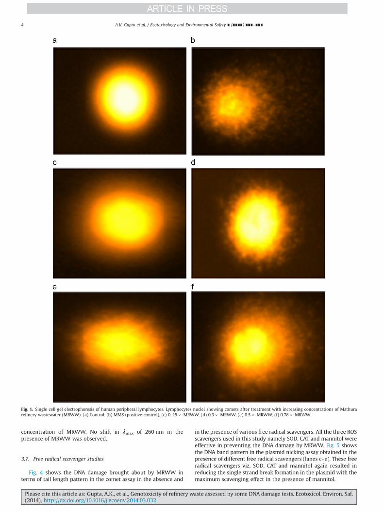
It is noteworthy that when low concentration of MRWW(0.15� ) was used, the small condensed particles at the originallocations of the nuclei with small amounts of fragmented DNAwere observed that depicts a limited degree of nuclear DNAbreakage (Fig. 1c). However, DNA breakage was more pronouncedwhen the concentration was increased and maximum DNA wasobserved at 0.78� concentration of MRWW (Fig. 1f). Similarly, themaximum tail length (an index of DNA breakage) was alsoobserved at 0.78� of concentration of MRWW (Fig. S1).
3.3. Plasmid nicking assay
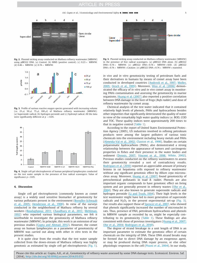
Fig. 2 presents the DNA band profiles of the plasmid nickingassay with MRWW. Three different concentrations (0.23� , 0.46�and 0.74� ) of MRWW were used to analyze the effect of MRWWon pBR322 DNA. 0.23� of MRWW sample resulted in the partialconversion of the supercoiled pBR322 DNA into open circle (lane,2c). 0.46� of this sample caused the conversion of the significantamount of supercoiled pBR322 DNA into two other forms i.e. opencircle and linear (lane, 2d). 0.74� of MRWW resulted in thecomplete loss of supercoiled form of pBR322 DNA with theformation of open circle and linear form.
3.4. Estimation of ROS generation in the test water samples
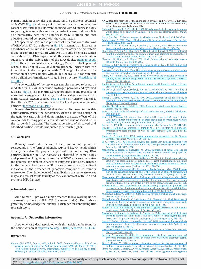
Fig. 3 shows the pattern of ROS generation by the test MRWWsample. There was increasing trend of ROS generation with theincreasing doses of MRWW in the test samples. Thus the highestdose of MRWW invariably exhibited the maximum generation ofindividual ROS i.e. superoxide radical, H2O2 and hydroxyl radical.Fig. 3a presents the dose-dependent pattern of the superoxideradicals generated by the test samples. 100 ml of MRWW in the testsample produced the maximum amount of superoxide radicals(0.48 mmol) being the highest dose. Presence of superoxide dis-mutase (SOD) used as scavenger in the reaction mixture caused adrop in superoxide radical generation from 0.48 to 0.09 mmol incase of 100 ml sample of MRWW. Fig. 3b depicts the profile of H2O2
generated by the increasing concentrations of test water samples.H2O2 production was recorded to be 1.8 mmol in 100 ml of watersample. Presence of H2O2 was confirmed by adding the catalase(CAT) in the reaction mixture which decreased H2O2 level from1.8 to 0.15 mmol with the same amount of MRWW. Fig. 3c showsthe profile of hydroxyl radicals generated by the test samples.100 ml of MRWW sample has shown to produce 1.9 mmol hydroxylradicals. Presence of mannitol in the reaction mixture led to asteep decline in the hydroxyl radical generation from 1.9 to0.10 mmol that authenticates the presence of hydroxyl radicals.
3.5. S1 nuclease assay
Percent hydrolysis in S1 nuclease assay was also found to beenhanced with the increase in dosage of MRWW (Fig. S2). As highas 65 percent hydrolysis of the calf thymus DNAwas recorded with0.75� concentration of MRWW in the reaction mixture under ourexperimental condition.
3.6. UV–visible measurements
Fig. S3 depicts the dose-dependent UV absorption spectra ofDNA exposed to different concentrations of MRWW at 37 1C.The absorption intensity at 260 nm was increased with theincreasing concentrations of MRWW. The changes observed inthe absorption spectra of calf thymus DNA were those in absor-bance at λmax 260 nm up to 30 percent only at the highest
A.K. Gupta et al. / Ecotoxicology and Environmental Safety ∎ (∎∎∎∎) ∎∎∎–∎∎∎ 3
Please cite this article as: Gupta, A.K., et al., Genotoxicity of refinery waste assessed by some DNA damage tests. Ecotoxicol. Environ. Saf.(2014), http://dx.doi.org/10.1016/j.ecoenv.2014.03.032i
concentration of MRWW. No shift in λmax of 260 nm in thepresence of MRWW was observed.
3.7. Free radical scavenger studies
Fig. 4 shows the DNA damage brought about by MRWW interms of tail length pattern in the comet assay in the absence and
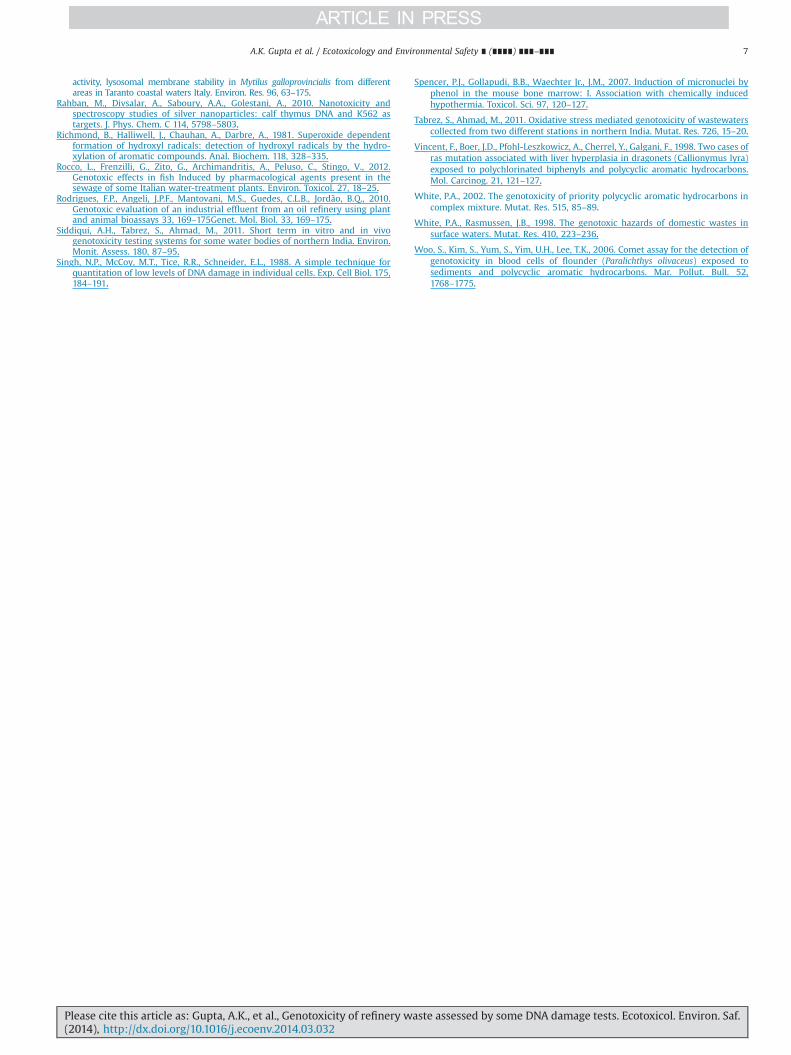
in the presence of various free radical scavengers. All the three ROSscavengers used in this study namely SOD, CAT and mannitol wereeffective in preventing the DNA damage by MRWW. Fig. 5 showsthe DNA band pattern in the plasmid nicking assay obtained in thepresence of different free radical scavengers (lanes c–e). These freeradical scavengers viz. SOD, CAT and mannitol again resulted inreducing the single strand break formation in the plasmid with themaximum scavenging effect in the presence of mannitol.
Fig. 1. Single cell gel electrophoresis of human peripheral lymphocytes. Lymphocytes nuclei showing comets after treatment with increasing concentrations of Mathurarefinery wastewater (MRWW). (a) Control. (b) MMS (positive control). (c) 0. 15� MRWW. (d) 0.3� MRWW. (e) 0.5� MRWW. (f) 0.78� MRWW.
A.K. Gupta et al. / Ecotoxicology and Environmental Safety ∎ (∎∎∎∎) ∎∎∎–∎∎∎4
Please cite this article as: Gupta, A.K., et al., Genotoxicity of refinery waste assessed by some DNA damage tests. Ecotoxicol. Environ. Saf.(2014), http://dx.doi.org/10.1016/j.ecoenv.2014.03.032i
4. Discussion
Single cell gel electrophoresis (commonly known as cometassay) is a widely used sensitive biomarker of genotoxicity forvarious pollutants present in the environment (Brendler-Schwaabet al., 2005; Henderson et al., 1998). In view of the surveysconducted in the neighborhood of Mathura refinery by severalworkers (Boudaghpour, 2011; Chaudhary et al., 2011; Mehlman,1992) who reported various biological parameters, we felt itworthwhile to investigate the genotoxicity of Mathura refinerywastewater (MRWW). In principle, this work is an extension of ourprevious studies (Gupta and Ahmad, 2012). However, the cometassay on human lymphocytes as a parameter of genotoxicity ofMRWW was carried out along with other in vitro tests in thepresent studies.
It is quite clear from the results that the test water samplecollected from the down-stream of Mathura refinery was highlygenotoxic as estimated by single cell gel electrophoresis (Fig. 1).
in vivo and in vitro genotoxicity testing of petroleum fuels andtheir derivatives in humans by means of comet assay have beenconducted in developed countries (Andreoli et al., 1997; Moller,2005; Oesch et al., 1995). Moreover, Woo et al. (2006) demon-strated the efficacy of in vitro and in vivo comet assay in monitor-ing PAHs contamination and assessing the genotoxicity in marineorganisms. Huang et al. (2007) also reported a positive correlationbetween DNA damage in the liver of frogs (Bufo raddei) and dose ofrefinery wastewater by comet assay.
Chemical analysis of the test water indicated that it containedrelatively high levels of phenols, PAHs and hydrocarbons besidesother impurities that significantly deteriorated the quality of waterin view of the remarkably high water quality indices i.e. BOD, CODand TOC. These quality indices were approximately 200 times tothat in negative control (Table 1).
According to the report of United States Environmental Protec-tion Agency (2005), US industries involved in refining petroleumproducts were among the largest polluters of various toxicchemicals into the environment including heavy metals and PAHs(Almeida-Val et al., 2002; Claxton et al., 1998). Studies on certainpolyaromatic hydrocarbons (PAHs), also demonstrated a strongrelationship between the appearance of tumors and carcinogenicproperties in fishes and their presence in the water bodies andsediment (Dennis, 2007; Vincent et al., 1998; White, 2002).Previous studies conducted on the refinery wastewaters to assesstheir genotoxicity revealed a sort of contradictory results.Rodrigues et al. (2010) reported an appreciable amount of primarylesions in rat hepatoma cells exposed to refinery wastewaterwithout any significant genotoxic effect by Allium cepa micronu-cleus assay. Moreover, Huang et al. (2007) found genotoxicity ofpetrochemical pollutants in toad B. raddei. Phenols are alsoimportant organic compounds to have genotoxic effect on livingsystem and are generally present in refinery wastes (Ohe et al.,2004). They are also known to generate superoxide radicals andhydrogen peroxide (Li and Trush, 1994). Their high levels in thetest wastewater might have led to more generation of superoxideradicals and H2O2 in the present experimental set-up (Fig. 3).Our results also support those of Spencer et al., 2007, who showedthat phenols significantly increased the number of micronuclei inrats. Thus, presence of PAH, petroleum hydrocarbons and phenolsin MRWW sample as recorded by us, might be especially con-tributing to its genotoxicity (Table 1). These findings are alsoconsistent with those of previous investigators (Huang et al., 2007;Ohe et al., 2004; Rodrigues et al., 2010).
The degree of strand breakage in a unit length of DNA is animportant parameter to estimate the genotoxic effect of certainchemicals on the integrity of DNA. These DNA strand breaks maybe formed due to direct DNA damage by an exogenous agent,or may be produced during DNA repair process, or else otherphysiologic responses in the cell (Pisoni et al., 2004). In our study,
0
5
10
15
20
25
30
35
40
Control MRWW+SOD MRWW+CAT MRWW+MAN MRWW(0.78x)
Tai
l len
gth
(μ m
eter
)
Fig. 4. Single cell gel electrophoresis of human peripheral lymphocytes conductedon the test water sample in the presence of free radical scavengers. Value ofsignificance po0.05.
Fig. 5. Plasmid nicking assay conducted on Mathura refinery wastewater (MRWW)in the presence of free radical scavengers. (a) pBR322 DNA alone. (b) pBR322DNAþ0.74� MRWW. (c) pBR322 DNAþ0.74� MRWWþSOD. (d) pBR322DNAþ0.74� MRWWþCatalase. (e) pBR322 DNAþ0.74� MRWWþmannitol.
Fig. 2. Plasmid nicking assay conducted on Mathura refinery wastewater (MRWW)using pBR322 DNA. (a) Control. (b) MMS (positive control). (c) 0.23� MRWW.(d) 0.46� MRWW. (e) 0.74� MRWW.
-0.1
0.4
0.9
1.4
1.9
μmol
e
25 μl
50 μl
75 μl
100 μl
Fig. 3. Profile of various reactive oxygen species generated with increasing volume(i.e. 25 ml, 50 ml, 75 ml, 100 ml) of Mathura refinery wastewater (MRWW):(a) Superoxide radical, (b) hydrogen peroxide and (c) hydroxyl radical. All the datawere significantly different at p o0.05.
A.K. Gupta et al. / Ecotoxicology and Environmental Safety ∎ (∎∎∎∎) ∎∎∎–∎∎∎ 5
Please cite this article as: Gupta, A.K., et al., Genotoxicity of refinery waste assessed by some DNA damage tests. Ecotoxicol. Environ. Saf.(2014), http://dx.doi.org/10.1016/j.ecoenv.2014.03.032i
plasmid nicking assay also demonstrated the genotoxic potentialof MRWW (Fig. 2), although it is not as sensitive biomarker ascomet assay. Similar results were obtained with S1 nuclease assaysuggesting its comparable sensitivity under in vitro conditions. It isalso noteworthy here that S1 nuclease assay is simple and costeffective method compared with the comet assay.
UV spectra of DNA in the presence of different concentrationsof MRWW at 37 1C are shown in Fig. S3. In general, an increase inabsorbance at 260 nm is indicative of intercalatory or electrostaticmode of complex formation with DNA of some chemicals whichcan stabilize the DNA duplex, while the existence of a red-shift issuggestive of the stabilization of the DNA duplex (Rahban et al.,2010). The increase in absorbance at λmax 258 nm up to 30 percentwithout any shift in λmax of 260 nm probably indicate that theinteraction of the complexes with DNA might be due to theformation of a new complex with double-helical DNA concomitantwith a slight conformational change in its structure (Magdalena etal., 2006).
The genotoxicity of the test water samples seemed to be partlymediated by ROS viz. superoxide, hydrogen peroxide and hydroxylradicals (Fig. 3). The maxium scavenging effect in the presence ofmannitol is suggestive of the hydroxyl radical generation as themain reactive oxygen species (Figs. 4 and 5e). Hydroxyl radical isthe ultimate ROS that interacts with DNA and promotes geneticdamage (Richmond et al., 1981).
It may also be emphasized that the results presented in thispaper actually reflect the genotoxicity of the dissolved portion ofthe genotoxicants only and do not include the toxic effects of thecompounds forming particulate material or those adsorbed on tothe suspended particles. The cumulative effect of dissolved andadsorbed portions would undoubtedly be much higher.
5. Conclusion
Refinery wastewater is well known to contain genotoxiccompounds in the form of phenols, PAH and heavy metals whichdirectly or indirectly play an important role in causing DNAdamage. Increase in DNA damage as observed in comet assayand plasmid nicking assay caused by MRWW exposure indicatesthe potential for genotoxic hazard at long term exposures. Increasein the percent hydrolysis in S1 nuclease assay is also a directindicative of the presence of genotoxic compounds in refinerywastewater. The higher level of free radicals in the test wastewatermay also account for its toxicity as they can interact with DNA andpromote DNA damage.
Acknowledgments
Amit Kumar Gupta was a junior research fellow working undera research project of U.P. CST, Lucknow (India). The authorsgratefully acknowledge the financial assistance for conducting thisresearch work.
Appendix A. Supporting information
Supplementary data associated with this article can be found inthe online version at http://dx.doi.org/10.1016/j.ecoenv.2014.03.032.
References
Almeida-Val, V.M.F., Duncan, W.P., Val, A.L., 2002. Crude oil effects on fish of theAmazon: current status. In: Val, AL, Almeida-Val, VMF, Mc Kinley, D (Eds.),Tropical Fish: News Reviews. International Congress on the Biology of Fish.University of British Columbia, Vancouver, p. 49.
APHA, Standard methods for the examination of water and wastewater, 20th edn.,1998. American Public Health Association, American Water Works Association,Water Environment Federation, USA.
Andreoli, C., Leopardi, P., Crebelli, R., 1997. DNA damage by hydroquinone in humanwhite blood cells: analysis by alkaline single-cell gel electrophoresis. Mutat.Res. 377, 95–104.
Avery, S.V., 2011. Molecular targets of oxidative stress. Biochem. J. 434, 201–210.Boudaghpour, S., 2011. Effects of leaking out oil in areas around Iran's refineries. Int.
J. Phys. Sci. 6, 3715–3722.Brendler-Schwaab, S., Hartmann, A., Pfuhler, S., Speit, G., 2005. The in vivo comet
assay: use and status in genotoxicity testing. Mutagenesis 20, 245–254.Chaudhary, P., Singh, S.B., Chaudhry, S., Nain, L., 2011. Impact of PAH on biological
health parameters of soils of an Indian refinery and adjoining agricultural area—a case study. Environ. Monit. Assess. 184, 1145–1156.
Claxton, L.D., Houk, V.S., Hugles, T.J., 1998. Genotoxicity of industrial wasteseffluents. Mutat. Res. 410, 237–243.
Dennis, T., 2007. Logan perspective on ecotoxicology of PAHs to fish human andecological risk assessment. Int. J. 13, 2.
Dusinska, M., Collins, A.R., 2008. The comet assay in human biomonitoring: gene–environment interactions. Mutagenesis 23, 191–205.
Gupta, A.K., Ahmad, M., 2012. Assessment of cytotoxic and genotoxic potential ofrefinery waste effluent using plant, animal and bacterial systems. J. Hazard.Mater. 201–202, 92–99.
Gupta, S., Dogra, T.D., 2002. Air pollution human health hazards. Indian J. Occup.Environ. Med. 6, 89–93.
Henderson, L., Wolfreys, A., Fedyk, J., Bourner, C., Windebank, S., 1998. The ability ofthe Comet assay to discriminate between genotoxins and cytotoxins. Mutagen-esis 13, 89–94.
Huang, D., Zhang, Y., Wang, Y., Xi, Z., Ji, W., 2007. Assessment of the genotoxicity intoad Bufo raddei exposed to petrochemical contaminants in Lanzhou Region,China. Mutat. Res. 629, 81–88.
Infante, P.F., Schwartz, E., Cahill, R., 1990. Benzene in petrol: a continuing hazard.Lancet 336, 814–815.
Jones, D.P., 2008. Radical-free biology of oxidative stress. Am. J. Physiol. Cell Physiol.295, 849–868.
Klein, A.B., Witonsky, S.G., Ahmed, S.A., Holladay, S.D., Gogal Jr., R.M., Link, L., Reilly,C.M., 2006. Impact of different cell isolation techniques on lymphocyte viabilityand function. J. Immunoassay Immunochem. 27, 61–76.
Kurelec, B., 1993. The genotoxic disease syndrome. Mar. Environ. Res. 35, 341–348.Legault, J., Tremblay, A., Ramotar, D., Mirault, M.E., 1997. Clusters of S1 nuclease-
hypersensitive sites induced in vivo by DNA damage. Mol. Cell. Biol. 17,5437–5452.
Lerda, D., Prosperi, C.H., 1996. Water mutagenicity toxicology in Río TerceroCordoba, Argentina. Water Res. 30, 819–824.
Li, Y., Trush, M.A., 1994. Reactive oxygen-dependent DNA damage resulting fromthe oxidation of phenolic compounds by a copper-redox cycle mechanism.Cancer Res. 54, 1895–1898.
Magdalena, L., Teresa, S., Gabryelak, M., 2006. Interaction of tannic acid and itsderivatives with calf thymus DNA and bovine serum albumin using spectro-scopic method. J. Phytochem. Photobiol. 82, 72–78.
Mater, N., Geret, F., Castillo, L., Faucet-Marquis, V., Albasi, C., Pfohl-Leszkowicz, A.,2014. in vitro tests aiding ecological risk assessment of ciprofloxacin, tamoxifenand cyclophosphamide in range of concentrations released in hospital waste-water and surface water. Environ. Int. 191, 191–200.
Matsumoto, S.T., Mantovani, M.S., Rigonato, J., Marin-Morales, M.A., 2005. Evalua-tion of the genotoxic potential due to the action of an effluent contaminatedwith chromium, by the comet assay in CHO-K1 cultures. Caryologia 58, 40–46.
Matsumoto, S.T., Mantovani, M.S., Mallaguti, M.I., Marin-Morales, M.A., 2003.Investigation of the genotoxic potential of the waters of a river receivingtannery effluents by means of the in vitro comet assay. Cytologia 68, 395–401.
Mehlman, M.A., 1992. Dangerous and cancer-causing properties of products andchemicals in the oil refining and petrochemical industry: VIII. Health Eff. Mot.Fuels: Carcinog. Gasol.—Sci. Update Environ. Res. 59, 1238–1249.
Miller, G., Shulaev, V., Mittler, R., 2008. Reactive oxygen signaling and abiotic stress.Physiol. Plant. 133, 481–489.
Mitchelmore, C.L., Birmelin, C., Livingstone, D.R., Chipman, J.K., 1998. Detection ofDNA strand breaks in isolated mussel Mytilus edulis L. digestive gland cellsusing the comet assay. Ecotoxicol. Environ. Saf. 41, 51–58.
Moller, P., 2005. Genotoxicity of environmental agents assessed by the alkalinecomet assay. Basic Clin. Pharmacol. Toxicol. 96, 1–42.
Nakayama, T., Kimura, T., Kodama, T., Nagata, C., 1983. Generation of hydrogenperoxide superoxide anion from active metabolites of naphthylamines ami-noazo dyes: its possible role in carcinogenesis. Carcinogenesis 4, 765–769.
Oesch, F., Fuchs, J., Vaupel, J., Hengstler, J.G., 1995. DNA single strand break analysisin mononuclear blood cells of gasoline station attendants. Int. Arch. Occup.Environ. Health 67, 35–39.
Ohe, T., Watanabe, T., Wakabayashi, K., 2004. Mutagens in surface waters: a review.Mutat. Res. 567, 109–149.
Pavlova, A., Ivanova, R., 2003. Determination of petroleum hydrocarbons andpolycyclic aromatic hydrocarbons in sludge from wastewater treatment basins.J. Environ. Monit. 5, 319–323.
Pick, E., Keisari, Y., 1980. A simple colorimetric method for the measurement ofhydrogen peroxide produced by cells in culture. J. Immunol. Methods 38, 161–170.
Pisoni, M., Cogotzi, L., Frigeri, A., Corsi, I., Bonacci, S., Iacocca, A., Lancini, L., Mastrototaro,F., Focardi, S., Svelto, M., 2004. DNA adducts, benzo(a)pyrene monooxygenase
A.K. Gupta et al. / Ecotoxicology and Environmental Safety ∎ (∎∎∎∎) ∎∎∎–∎∎∎6
Please cite this article as: Gupta, A.K., et al., Genotoxicity of refinery waste assessed by some DNA damage tests. Ecotoxicol. Environ. Saf.(2014), http://dx.doi.org/10.1016/j.ecoenv.2014.03.032i
activity, lysosomal membrane stability in Mytilus galloprovincialis from differentareas in Taranto coastal waters Italy. Environ. Res. 96, 63–175.
Rahban, M., Divsalar, A., Saboury, A.A., Golestani, A., 2010. Nanotoxicity andspectroscopy studies of silver nanoparticles: calf thymus DNA and K562 astargets. J. Phys. Chem. C 114, 5798–5803.
Richmond, B., Halliwell, J., Chauhan, A., Darbre, A., 1981. Superoxide dependentformation of hydroxyl radicals: detection of hydroxyl radicals by the hydro-xylation of aromatic compounds. Anal. Biochem. 118, 328–335.
Rocco, L., Frenzilli, G., Zito, G., Archimandritis, A., Peluso, C., Stingo, V., 2012.Genotoxic effects in fish Induced by pharmacological agents present in thesewage of some Italian water-treatment plants. Environ. Toxicol. 27, 18–25.
Rodrigues, F.P., Angeli, J.P.F., Mantovani, M.S., Guedes, C.L.B., Jordão, B.Q., 2010.Genotoxic evaluation of an industrial effluent from an oil refinery using plantand animal bioassays 33, 169–175Genet. Mol. Biol. 33, 169–175.
Siddiqui, A.H., Tabrez, S., Ahmad, M., 2011. Short term in vitro and in vivogenotoxicity testing systems for some water bodies of northern India. Environ.Monit. Assess. 180, 87–95.
Singh, N.P., McCoy, M.T., Tice, R.R., Schneider, E.L., 1988. A simple technique forquantitation of low levels of DNA damage in individual cells. Exp. Cell Biol. 175,184–191.
Spencer, P.J., Gollapudi, B.B., Waechter Jr., J.M., 2007. Induction of micronuclei byphenol in the mouse bone marrow: I. Association with chemically inducedhypothermia. Toxicol. Sci. 97, 120–127.
Tabrez, S., Ahmad, M., 2011. Oxidative stress mediated genotoxicity of wastewaterscollected from two different stations in northern India. Mutat. Res. 726, 15–20.
Vincent, F., Boer, J.D., Pfohl-Leszkowicz, A., Cherrel, Y., Galgani, F., 1998. Two cases ofras mutation associated with liver hyperplasia in dragonets (Callionymus lyra)exposed to polychlorinated biphenyls and polycyclic aromatic hydrocarbons.Mol. Carcinog. 21, 121–127.
White, P.A., 2002. The genotoxicity of priority polycyclic aromatic hydrocarbons incomplex mixture. Mutat. Res. 515, 85–89.
White, P.A., Rasmussen, J.B., 1998. The genotoxic hazards of domestic wastes insurface waters. Mutat. Res. 410, 223–236.
Woo, S., Kim, S., Yum, S., Yim, U.H., Lee, T.K., 2006. Comet assay for the detection ofgenotoxicity in blood cells of flounder (Paralichthys olivaceus) exposed tosediments and polycyclic aromatic hydrocarbons. Mar. Pollut. Bull. 52,1768–1775.
A.K. Gupta et al. / Ecotoxicology and Environmental Safety ∎ (∎∎∎∎) ∎∎∎–∎∎∎ 7
Please cite this article as: Gupta, A.K., et al., Genotoxicity of refinery waste assessed by some DNA damage tests. Ecotoxicol. Environ. Saf.(2014), http://dx.doi.org/10.1016/j.ecoenv.2014.03.032i


