Embed Size (px)
Citation preview
Basic Principles and Technologies for Deciphering the GeneticMap of Cancer
Georgios Voidonikolas, MD1, Stephanie S. Kreml, MD1, Changyi Chen, MD, PhD1, WilliamE. Fisher, MD, FACS1,4, F. Charles Brunicardi, MD, FACS1, Richard A. Gibbs, PhD2, andMarie-Claude Gingras, PhD1,2,*1Michael E. DeBakey Department of Surgery, Baylor College of Medicine, Houston, Texas, USA2Human Genome Sequencing Center; Department of Molecular and Human Genetics, BaylorCollege of Medicine, Houston, Texas, USA3The Center for Medical Ethics and Health Policy, Baylor College of Medicine, Houston, Texas,USA4The Elkins Pancreas Center, Baylor College of Medicine, Houston, Texas, USA
AbstractThe progress achieved in the field of genomics in recent years is leading medicine to adopt apersonalized model in which the knowledge of individual DNA alterations will allow a targetedapproach to cancer. Using pancreatic cancer as a model, we discuss herein the fundamentals thatneed to be considered for the high-throughput and global identification of mutations. Theseinclude patient related issues, sample collection, DNA isolation, gene selection, primer design, andsequencing techniques. We also describe the possible applications of the discovery of DNAchanges to the approach of this disease and cite preliminary efforts where the knowledge has beentranslated into the clinical or preclinical setting.
KeywordsPancreatic cancer; high-throughput sequencing; genotyping; somatic mutations
IntroductionTwenty-eight years after Frederick Sanger made his first attempts to sequence small piecesof DNA, the National Human Genome Research Institute (NHGRI) announced thecompletion of the Human Genome Project [1-3]. After 13 years of internationalcollaborative effort at a cost of $3 billion, an essentially complete human genome sequencewas finished and the world was ushered into a “genomic” era. In 2007, less than 4 yearslater, the first genome sequence of a single individual, James Watson, was deciphered inonly 2 months at a cost of less than $2 million. Project “Jim” was initiated by a privatecompany in collaboration with the Baylor College of Medicine-Human Genome SequencingCenter (BCM-HGSC) [4,5]. This project demonstrated the feasibility of characterizingvirtually all of the small scale variation in a single individual using a next generationsequencing technology thereby advancing the “personalized” genomic medicine model onestep closer to a reality.
*Corresponding author: BCM-Human Genome Sequencing Center One Baylor Plaza, Houston, Texas, 77030 Tel: 713-798-1286 FAX:713-798-5741 [email protected] .
NIH Public AccessAuthor ManuscriptWorld J Surg. Author manuscript; available in PMC 2010 August 19.
Published in final edited form as:World J Surg. 2009 April ; 33(4): 615–629. doi:10.1007/s00268-008-9851-y.
NIH
-PA Author Manuscript
NIH
-PA Author Manuscript
NIH
-PA Author Manuscript
Cancer has long been recognized as fundamentally driven by genetic mutations [6,7].Although opinions vary as to whether a comprehensive inventory of the genetic changes intumor cells can lead to fundamental new insights that will further the diagnosis andtreatment of this cancer, pilots for large-scale projects already have been initiated [8-10].Sjöblom et al have shown that the somatic mutations playing a role in the multistepprogression of carcinogenesis are far from completely identified, even in the most studiedcancer types (breast and colon) [11]. The Tumor Sequencing Project Consortium (TSPC),formed between the NHGRI funded Genome Centers at BCM, Washington University, andthe Broad Institute has initiated a pre-pilot project on non-small cell lung cancer (1,000genes) to demonstrate the potential of a systemic approach to tumor genotyping by DNAsequencing [12]. The benefit of this large-scale project already has been proven by theidentification of a novel candidate proto-oncogene [13]. The Cancer Genome Atlas pilotproject (TCGA), launched by the National Cancer Institute (NCI) and NHGRI, is nowattempting the identification of all genomic alterations significantly associated with cancer.This includes detecting genomic loss or amplification, mutations in coding regions,chromosomal rearrangements, aberrant methylations, and expression profiles. Three tumortypes (glioblastoma multiforme, squamous cell carcinoma of the lung, and ovariancarcinoma) have been selected initially by TCGA, with the scope of expanding to all majorcancer types [14].
Some success in cancer care has already been obtained with the targeting of specific geneticalterations. Among the first effective applications of targeted therapy was the use of imatinibto inhibit the tyrosine kinase activity of the Bcr-Abl fusion protein formed by thechromosomal t(9:22) translocation in chronic myeloid leukemia [15], and the use oftranstuzumab, a recombinant monoclonal antibody against HER2, in women with metastaticbreast cancer with HER2 amplification and overexpression [16]. Other breakthroughs inpersonalized therapies include the treatment of gastrointestinal stromal cell tumors, in whichmutations in KIT and PDGFRA were found to predict a response to imatinib [15,17].Additionally, mutations in EGFR predicted a response to gefitinib and erlotinib in lungadenocarcinoma [18-20]. The fact that specific mutations in multiple loci are a majordeterminant of the response to targeted therapies suggests that DNA sequencing is likely toprovide an effective diagnostic and therapeutic approach to cancer in the future.
This review focuses on the requisites to identify, validate, and confirm mutations, as well asthe possible applications the discovery of new DNA changes can have for thecharacterization and treatment of the disease. Some technical information is provided forthose who may be interested in initiating similar projects, and some examples of preliminaryattempts to apply current knowledge in a clinical setting are given with the goal of attractingnew clinical investigators to bridge the gap between genomics and its application toeveryday patient care.
Identification of mutationsEthical issues
Generating genomic information on a large scale requires strict criteria to protect patient’srights and confidentiality [21]. Institutional Review Board (IRB) oversight and informedconsent are unambiguously required. Genome sequencing should be explained to potentialparticipants in the informed consent process. Specific consent should be obtained for thefuture storage and use of collected samples. For most federally-funded studies, broad datasharing is required. (NHGRI Data sharing policies) Explanation of the anticipated scope ofdata sharing, along with the risks and benefits of broad data release, should be provided.Under some circumstances (e.g., if data sharing is not required to achieve the primary goals
Voidonikolas et al. Page 2
World J Surg. Author manuscript; available in PMC 2010 August 19.
NIH
-PA Author Manuscript
NIH
-PA Author Manuscript
NIH
-PA Author Manuscript
of the project and data release may impede research participation), it may be appropriate tooffer an opt-out provision for public and/or restricted data broadcast [21,22].
Studies involving extensive genome sequencing raise an additional concern about whetherindividualized research results should be shared with study participants. Convincingarguments have been made that if the research reveals validated data of known clinicalrelevance, it should be reported to participants [23-27]. The language used to explain returnof research results should be carefully crafted to avoid potential legal liability.
Management of clinical informationThe patient information collected should include demographics, exposure, family history,symptoms and physical findings at presentation, laboratory values, diagnostic imaging testresults, details of the surgical treatment, histology from preoperative and operativespecimens, pathologic staging data, details of chemotherapy and radiation treatment,response to treatment in terms of follow-up imaging, disease-free survival and overallsurvival, and quality of life survey data.
The data should be entered and stored in a password-protected, HIPPAA-compliantdatabase. To assure patient confidentiality, the specimen should be logged into the databaseand then assigned a new serial number for use in the laboratory. Limited access by cliniciansand biostatisticians will allow later correlation with clinical data. The tubes containing thesamples also should be bar coded to achieve automatic assignment, increase speed ofprocessing, and protection from mixing the samples.
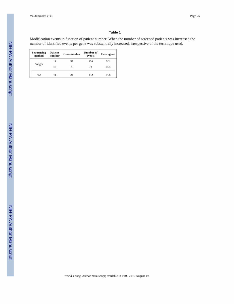
Defining the number of patient samplesThe number of patient samples to be sequenced depends on the number of available samplesand the size and statistical power of the intended study. We have noticed that the number ofmodification event detected increases when a larger panel of patients is used in thediscovery process (11 patients versus > 40 patients) (Table 1). The issue of statistical powerhas been raised in reviews of Sjöblom’s paper [11,28,29]. Much larger samples than theones used in that paper are required to detect cancer genes, since in small sample sizes, somecandidate genes are expected to display false-positive results. The validation and specificityprocesses should also be planned with a statistically valid number of patients.
Sample and DNA preparationIt is necessary to collect both tumor and matched normal tissue from each patient for thecomparison of germline and tumor genotypes in order to verify that any detected mutationsare of somatic origin. Matched normal tissue can consist of surrounding normal tissue and/orblood. Collecting the blood of the patient as the primary source of normal germline DNA ispreferable. If the blood is not available, a sample of “normal” tissue taken from an areaadjacent to the tumor can be used as an alternative. This choice is less attractive because a“normal” sample may not be entirely normal and may contain some invasive neoplastic cellsor normal cells that share alterations with the cancer [30]. In such a case, the sequencingresult might be identical between the tumor and “normal” tissue, representing an inheritedgermline mutation or polymorphism, rather than a somatic mutation.
Samples should be identified as primary tumor tissue or metastasis or abnormal non-cancerous tissue. Strict requirements of quality, quantity, purity, and avoidance of necrotictissue should be met in order to create an ideal tissue bank. Tissue quality issuesencountered by large projects like TCGA can assist in better planning for future projects[31,32]. The degree of homogeneity of the tumor sample to be sequenced is also important.First, it is known that the tumor itself is not homogenous, since it consists of subpopulations
Voidonikolas et al. Page 3
World J Surg. Author manuscript; available in PMC 2010 August 19.
NIH
-PA Author Manuscript
NIH
-PA Author Manuscript
NIH
-PA Author Manuscript
of cells with different phenotypes and genotypes that determine different immunity,aggressiveness and metastatic behavior [33,34]. In addition, the contamination of tumorsamples by surrounding tissue must be considered. This has led to the use of a tumor puritythreshold of at least 80% as a criterion by TCGA and others [13,35,36] and to thedevelopment of Laser Microdissection Technique, especially if the cell population ofinterest is scant [37].
Fresh tissue is the best source of DNA. However, if the DNA cannot be extractedimmediately, freezing remains the best way to preserve tissue. Standard operatingprocedures for the collection of fresh frozen tissue samples have been developed and used inthe European Human Frozen tumor Tissue Bank (TuBaFrost) [38]. Formalin-fixed, paraffin-embedded (FFPE) tissue also can be used, but with limitations [39-41]. In such materialDNA is scant, and cross linking and degradation result in sequencing failure. FFPE tissueconstitutes a valuable source in terms of the number of available samples, diversity, andglobal patient information that has been collected over decades. When using such a source,the DNA has to be whole genome amplified and only short amplicons (~100-200 bases) canbe sequenced. As a consequence, FFPE tissues can be used in the validation process, but arenot preferred for mutation/SNP discovery. Cell lines are not an optimal DNA source sincemutations associated with long term in vitro culture and unrelated to the in vivo developmentof cancer can be acquired over time.
In cases when the amount of the provided human specimen is limited and the extracted DNAis scarce, whole genome amplification (WGA) is required to overcome the problem. By thismethod, the original DNA sample is amplified in a specific way from nanogramconcentrations to microgram, while the sequence representation of the template isconserved. Of the different methods studied, multiple displacement amplification (MDA)results in DNA products of high molecular weight (up to 12kb) and generates the least bias[42-44]. Original unamplified samples can then be saved to validate the identified mutations.
A process overview is illustrated in Fig. 1 and more detailed and technical information canbe found in Table 2.
Candidate gene selectionThere is a wide range of genes that can be chosen for a sequencing study: genes withmutations already known to be associated with the tumor by medical literature and datasources [46-49]; genes with mutations associated with familial genetic syndromes thatincrease the risk of cancer [50,51]; genes known to be differentially expressed in the tumor[52,53]; genes linked to several other cancer types and cellular pathways important in cancer(e.g., oncogenes, tumor suppressor genes, DNA repair genes, cyclins, kinases, phosphatases)[54]. CancerGenes is a valuable source of information for gene selection and prioritizationin the elaboration of a gene list [55].
High-throughput DNA sequencing methods have been used on data sets of 200 to 1000genes in 150-200 patients. In these large scale-up studies, considerable effort is expended infinalizing the gene list [12,14]. In an approach to exon sequencing, Sjöblom et al expandedthe list to 13,023 genes by limiting the study to a single direction in only 11 patients pertumor type [11]. The power to detect rare mutations was therefore limited in this study, andyet 365 mutations in 236 genes were found. As the ability to comprehensively sequence tensof thousands of exons in a single experiment continually increases, the problem of geneselection diminishes.
This sequencing ability has been further improved by another novelty: the replacement ofPCR by capture arrays (Fig. 1). Until now, regions of interest to be sequenced were
Voidonikolas et al. Page 4
World J Surg. Author manuscript; available in PMC 2010 August 19.
NIH
-PA Author Manuscript
NIH
-PA Author Manuscript
NIH
-PA Author Manuscript
selectively sampled through a labor-intensive process whereby each fragment wasindividually amplified using PCR. This required the parallel design and execution ofthousands of reactions. This will now change as NimbleGen (Madison, WI) developed sevencustom arrays that contained 204,000 exons, allowing the entire exonic coding genome to becaptured, enriched, and sequenced on the 454 or Genome Analyzer platform upon release(Fig. 2) [56,57]. Such enrichment will eliminate the need for large numbers of specificprimers, thousands of specific PCR reactions on individual or pooled samples presentlyassociated with the PCR-based large-scale sequencing approaches, thus considerablyreducing cost and time expense. This will give superior results, allowing for theidentification of polymorphisms/mutations on the whole exonic genome.
Primer designCurrently, when performing an exonic study, specific amplification by PCR of the selectedgene exonic regions has to be performed prior to sequencing and specific primer pairs needto be designed (Fig. 1). A pipeline can be established that links several steps for theintegrated automating design of primers (Table 3).
SequencingThe DNA sequencing revolution started in 1977 with Sanger sequencing that soon becameby far the most frequently used sequencing technology (Fig. 4) [1]. The Sanger sequencingmethod, a termination technology, is still widely used to perform individual ampliconsequencing (400-500 bases/read in a 384-well plate = ~150,000 bases/plate). However, theinterest in deciphering the whole genome of organisms and large-scale DNA sequencingprojects recently prompted the development of other technologies and platformsaccommodating high-throughput sequencing.
Discovered in the late 1990s, pyrosequencing technology is based on the real-timemonitoring, by bioluminescence (conversion of luciferase into oxyluciferin), of a 4-enzymesequencing reaction [63]. This technology recently has been adapted to a high throughputsetting by 454 Life Sciences in which whole genome or targeted segments can be analyzedin a single run [64]. The technology is based on emulsion PCR of individual DNA fragmentscaptured on 28-micron beads, at a resolution of one DNA molecule per bead, resulting in a107-fold amplification of the initial DNA copy per bead, followed by pyrosequencing-by-synthesis of each clonally amplified template in a fiber optic slide (Fig. 5). Presently,approximately 500,000 reactions can be performed in parallel in the new FLX system andabout 125,000,000 bases are sequenced per run (with each read currently at 200-300 basepairs long, the expectation is that in late 2008, 500 base pair reads will become available).Whereas patient samples and amplicons are pooled making it less expensive and more rapidfor discovery, the physical segregation of the DNA-carrying beads in an emulsion during thein vitro amplification (clonal amplification) results in the detection of specific mutations inlow tumor content samples without the need for tumor cell enrichment by laboriousmethods. The extreme sensitivity of the 454 technology has been recently demonstrated in astudy in which it enabled the detection of mutations in low tumor content samples for whichconventional Sanger sequencing has failed to detect any of these mutations [65]. The call forsomatic mutation and the percentage of patients carrying a mutation are then estimatedduring validation by genotyping the matching normal and tumor samples individually foreach patient using a different technology.
In addition to the 454 technology, there are several new high-throughput sequencingmethods that are being explored. Among these, the Solexa system (recently acquired byIllumina (San Diego, CA) and renamed Genome Analyzer) is a new, massively parallelsequencing platform in which millions of single molecules are covalently attached to a
Voidonikolas et al. Page 5
World J Surg. Author manuscript; available in PMC 2010 August 19.
NIH
-PA Author Manuscript
NIH
-PA Author Manuscript
NIH
-PA Author Manuscript
planar surface and amplified in situ by a “bridge amplification” process [66-68]. Sequencingby synthesis is then carried out by adding a mixture of four fluorescently labeled reversibleterminators and DNA polymerase to the template on the Solexa flow cell. The nucleotidesequences are determined by the fluorescent signals. After removing the fluorophore andreversing the blockage group and the terminator, the terminator-enzyme mix is added to starta new cycle. The whole process is then repeated until the end of the run (Fig. 6). Onenucleotide sequence is read out for a given molecule at each cycle. One Solexa run, onaverage, generates about 35-45 million reads with read lengths of 36 bases. Between 1.2 and1.5 billion bases are sequenced per run, about 10 fold higher than the 454 technologythroughput. However, the error rate for a single read generated from the Solexa platform isabout 1.5% for 36 cycles. As a result, the detection of mutations in samples with low tumorcontent might be hard to differentiate from the error rate background. The challenges of thetechnology also include a massive data storage and computional load to manage. Presently,due to the short read length, Solexa has better use in a long-range PCR-based approach (>3kb) or a whole genome approach.
Other important characteristics associated with tumor development and progression are thevariation in gene copy number due to heteroploidy and chromosomal loss. This genomicaspect cannot be detected by sequencing but with techniques such as CGH and SNP arrays[69-70]. The SKAP2/SCAP2 gene was found to be amplified and associated with thedevelopment of pancreatic cancer using this technology [71].
What to look forThe data derived from sequencing can be aligned to identify mutations by parallelcomparing of the tumor DNA sequence, the patient normal DNA, and the referencesequence deposited in GenBank (Table 4). This comparison can reveal single nucleotidepolymorphisms (SNPs), germline, and somatic mutations. Modifications from the GenBankreference sequence in the patient samples (sequence identical in the normal and tumorsamples) are germline mutation or SNP. Somatic mutations are modifications specific to thetumor and not found in the blood or other normal tissue of the patient.
It is now believed that polymorphic variations in the DNA sequence also can be related topopulation-attributable cancer heritability. SNP is defined as a genomic locus where two ormore alternative bases occur with a frequency of at least 1% in a population. SNP accountsfor more than 90% of the total variation in the human genome [72]. There are as many as 7million common SNPs with a minor allele frequency of at least 5% [73,74]. SNPs in closechromosomal proximity can be inherited together on haplotype blocks due to underlininglinkage disequilibrium (non random association of alleles from one generation to the next)[75]. SNPs indeed have been associated with predisposition to cancer, prognosis, andresponse and toxicity to chemotherapy or radiotherapy [76-78]. SNPs in xenobioticmetabolizers, hormone metabolizers, DNA repair genes, genes involved in angiogenesis andcell cycle are under scrutiny in several cancers [79].
Discovery of the germline mutations (mutations found in every cell of the individual)predisposing to hereditary cancer syndromes was the trademark of translational cancerresearch in 1990s. Genes such as BRCA1/BRCA2 in breast and ovarian cancer, APC in coloncancer with adenomatous polyposis coli and CDNK2A in melanoma are examples ofmutated genes with high penetrance leading to cancer and studied in family pedigrees[80-82].
Based on observation made by Loeb and others, at least six different metabolic or signalingpathways must be altered to lead to cancer [83-85]. By these alterations cells expressinsensitivity to growth inhibitory signals, escape apoptosis and acquire limitless replicative
Voidonikolas et al. Page 6
World J Surg. Author manuscript; available in PMC 2010 August 19.
NIH
-PA Author Manuscript
NIH
-PA Author Manuscript
NIH
-PA Author Manuscript
potential and sustained angiogenic, invasive and metastatic abilities [86]. As normalmutation rates cannot by themselves account for the multiple mutations found in cancercells, it is believed that special mutations called mutator mutations lead to genetic instabilityand increase the inherent rate of genetic change, thus exhibiting a “mutator phenotype”[85,87].
The whole list of cancer associated genes includes oncogenes, tumor supressor genes andstability genes. Oncogenes can be abnormally activated by intragenic mutation,chromosomal translocation or gene amplification. Tumor suppressor genes can beinactivated by a missense mutation, nonsense mutation, deletion or insertion of varioussizes; by epigenetic silencing; or by amplification of regulatory inhibitors. Finally, thefunction of stability genes including mismatch repair genes, nucleotide excision repair andbase excision repair genes, as well as mitotic recombination and chromosomal segregationgenes also can be impaired by a mutation [48].
These DNA modifications can be found in coding (exon) or non-coding (intron anduntranslated exonic) regions. Exonic mutations in the coding region can be synonymous(silent) in which the change in base does not affect the amino acid call, or non synonymousresulting in a different amino acid (missense) or protein termination (stop codon, non sense).The non-synonymous mutations can have drastic effects on the protein function andstructure. On the other hand, the impact of the silent polymorphisms in the MDR1 and theLamin A genes have proved that synonymous mutations should not be overlooked [88,89].Mutations in non-coding regions located in introns, promoters, splice junctions oruntranslated regions of the gene may also contribute to cancer by changing the regulation,exon splicing, mRNA stability, or conformation of the protein [90-93]. Table 5 illustratesthe different types of modification events we found in pancreatic adenocarcinoma usingSanger and 454 technology.
Although many somatic mutations can be detected in tumors, it is essential to distinguishbetween driver mutations which actually contribute to cancer and passenger mutationswhich randomly happened but are not responsible for cancer [11,94]. In the case where thefunction of the genes is still not clearly known, the impact of the mutated gene on thedevelopment of cancer will have to be evaluated by functional studies.
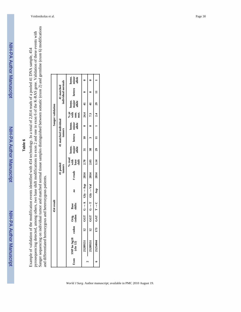
Validation and confirmation of specificityEach sequencing technology has its own limitations and pitfalls. After the completion ofsequencing and the discovery of mutations, a second technique based on different principlesshould be performed in order to validate the obtained genotype. There are a number ofgenotyping methods including TaqMan SNP Genotyping Assay (Applied Biosystem), MIP(ParAllele), SNPStream (Beckman-Coulter), iPlex Gold Assay (Sequenom), GoldenGateAssay (Illumina) and for genome wide studies GeneChip (Affymetrix) and Infinium(Illumina) [95-101]. Our strategy was to use the Sanger or TaqMan SNP Genotyping Assayto validate 454 pyrosequencing and low throughput pyrosequencing (Biotage) to validateSanger results. For example, 454 high throughput pyrosequencing detected basemodifications, but individual sample validation done by Sanger technique allowed thecomparison between tumor and matched normal tissue and thus the differentiation betweengermline and somatic events (Table 6). We also used differential gel migration of ampliconto validate large deletions.
In order to confirm that the identified mutations are specific to the malignant tissue, othertissues should be genotyped. As an example, we intend to genotype pancreatitis,precancerous lesions, benign cystic tumors, and neuroendocrine tumors to discriminate
Voidonikolas et al. Page 7
World J Surg. Author manuscript; available in PMC 2010 August 19.
NIH
-PA Author Manuscript
NIH
-PA Author Manuscript
NIH
-PA Author Manuscript
between mutations associated with pancreas cancer and non-cancerous or other cancerousconditions.
Present knowledge and applicationsThe fact that genomics led to deeper understanding and more targeted approach to CML andsolid cancers like breast, gastrointestinal and lung cancer is indisputable. In the case ofpancreatic cancer, translation of information from the DNA level to the clinical practice isstill preliminary and experimental. Effective screening or diagnostic genomic tools have notbeen established. Targeted therapies based on mutations are just beginning to be tested.
A recurrent pattern of genetic changes associated with pancreatic carcinogenesis has beenidentified [102]. The genetic changes include the inactivation of CDKN2A (p16) and anactivating point mutation of K-RAS in codon 12 as early events, as well as inactivation of theSMAD4 (DPC4), TP53, and BRCA2 genes as later events [103,104]. K-RAS mutation andCDKN2A inactivation are detected in over 90% of the tumors; however, SMAD4 and TP53inactivation can only be found in 55%, and 50-75% of the adenocarcinoma, respectively[105].
Mutations and SNPs associated with pancreatic adenocarcinoma riskWhile tobacco smoking is a high risk factor for the development of pancreaticadenocarcinoma, numerous associations have been found between smoking behavior andgenetic polymorphism in genes responsible for nicotine metabolism [106]. Indirectassociation of K-RAS mutation and activation with occupational exposure to dyes, organicpigments and other agents has been suggested [107,108]. Moreover, a metanalysisevaluating folate intake and genetic polymorphism of 5,10 methylene tetra hydro folatereductase (MTHFR) found a MTHFR variant associated with an increased risk for pancreaticadenocarcinoma [109]. A polymorphism in glutathione S-transferase gene, affectingdetoxification of carcinogens and anticancer agents in the human pancreas, was found toconfer a protective effect against pancreatic adenocarcinoma [110].
Other gene polymorphisms that may determine the risk for pancreatic adenocarcinomainclude Aurora-A and CDKN2A (p16) polymorphisms, which were associated withdiagnosis of pancreatic adenocarcinoma at an early age [111]. Finally, polymorphisms inDNA repair genes XPD and XRCC2 have been studied as genetic risk modifiers forsmoking-related pancreatic adenocarcinoma [112,113], while an XRCC1 polymorphism hada significant interaction with the APE1 or MGMT polymorphism in modifying pancreaticadenocarcinoma risk [114].
A growing body of evidence suggests that some of the aggregation of pancreaticadenocarcinoma in families has a heritable genetic basis and that as many as 10% ofpancreatic adenocarcinoma could be hereditary [51]. Several familial genetic syndromesalready have been associated with an increased risk of pancreatic adenocarcinoma such ashereditary pancreatitis, the hereditary nonpolyposis colorectal cancer syndrome (HNPCC),breast cancer, ataxia-telangiectasia, the familial atypical multiple mole-melanoma syndrome(FAMM), and Peutz-Jeghers syndrome. Already, germline mutations in BRCA2, CDKN2A,STK11, FANCC, PRSS1, and palladin (PALLD) have been shown to predispose to pancreaticadenocarcinoma, although with incomplete penetrance [115-117].
Mutations used for Diagnostics of Pancreatic adenocarcinomaResearchers already have used genetic markers separately or concomitantly to analyzecellular material from pancreatic juice and fine needle aspirates [118-122]. One of thesestudies has indicated that the analysis of K-RAS mutation and TP53 and SMAD4 inactivation
Voidonikolas et al. Page 8
World J Surg. Author manuscript; available in PMC 2010 August 19.
NIH
-PA Author Manuscript
NIH
-PA Author Manuscript
NIH
-PA Author Manuscript
can complement traditional cytology and clarify the diagnosis of patients with atypicalbiopsy samples [119]. EUS-FNA biopsy specimens also are beginning to be examined forsome of the mutations associated with pancreatic adenocarcinoma. In one study, analysis ofK-RAS point mutations improved sensitivity from 44% to 82% [120]. Another similar studyexamined the utility of immunohistochemistry for TP53 expression and found that thesensitivity of EUS-FNA was improved from 76% to 90% [121].
Unfortunately, K-RAS mutations are not specific to pancreatic adenocarcinoma and alsohave been detected in 25% of pancreatitis samples, nonneoplastic exocrine pancreaticlesions of smokers, and pancreatic intraepithelial neoplasia (PanIN) [122-128], which provesthe need for discovering of a broad panel of mutations that would most likely increasediagnostic specificity.
Mutations and SNPs used as prognostic toolsSome studies have shown an association between patient survival and somatic mutations inCDKN2A (p16), TP53, SMAD4 (DPC4), or germline mutations in XRCC2, XRCC3, RecQ1,Rad54L, ATM, and POLB [129-136]. On the other hand, gene copy number of the epidermalgrowth factor receptor (EGFR) did not have prognostic value in pancreatic adenocarcinoma[137].
Mutations and SNPS used as therapeutic tool1. Classic chemotherapy—SNPs have been studied regarding the pharmacodynamicsand pharmacokinetics of gemcitabine (GEM) in the treatment of lung and other cancers[138], but the knowledge is valuable for pancreatic adenocarcinoma as well. Variants in thepromoter region of ENT1, the transporter that brings GEM into the cells influence geneexpression and probably GEM chemosensitivity [139]. A haplotype of cytidine deaminase(CDA), responsible for the detoxification of GEM was found to lead to decreased clearanceand a high incidence of neutropenia, while SNPs in the 5′ regulatory region of thedeoxycytidine kinase gene were found to predict GEM sensitivity [140].
The efficacy and toxicity of other drugs used in treating pancreatic adenocarcinoma like 5-FU and platinum also are affected by polymorphisms. Mutations in dihydropyrimidinedehydrogenase (DPYD) gene, and thimidylate synthetase promoter region, involved in the 5-FU catabolism and pharmaceutical effect respectively, may affect drug toxicity and patientsurvival [141,142]. Moreover, platinum therapy results on survival were found to be affectedby polymorphisms in DNA repair genes like XRCC1, ERCC1, and ERCC2 [143-145].
Finally, in a preclinical study, sensitivity to cross-linking (mitomycin C, cisplatin,chlorambucil, and melphalan) chemotherapeutic agents was affected by the presence ofmutations in BRCA2/Fanconi anemia gene [146].
2. Targeted therapy—K-RAS mutations, which are found in 70-90% of pancreaticadenocarcinoma tissues, have been the target of several therapeutic approaches. K-ras mustbe farnesylated to be active. Although no successful clinical trials have been reported,inhibitors of the enzyme farnesyl-transferase have been developed [147,148]. Animmunotherapy approach to K-RAS mutations has also been proposed. K-RAS mutationscurrently are being studied as a target for immunotherapy with the use of yeast vectorscalled Tarmogens (Targeted Molecular Immunogens) in a phase II clinical trial, in post-resection, pancreatic cancer patients [149]. One other key downstream target of the Rasfamily, the phosphoinositol 3-kinase (PI3K), may play a role in drug resistance. In apreclinical study, treatment with PI3K inhibitors enhanced apoptosis induced by GEM[150].
Voidonikolas et al. Page 9
World J Surg. Author manuscript; available in PMC 2010 August 19.
NIH
-PA Author Manuscript
NIH
-PA Author Manuscript
NIH
-PA Author Manuscript
Another targeted therapeutic approach of pancreatic adenocarcinoma is the inhibition ofEGFR by tyrosine kinase inhibitors like erlotinib. A phase III study for first-line treatment ofadvanced pancreatic adenocarcinoma showed that the addition of erlotinib to GEM offeredsome improvement to survival compared to GEM alone and led to FDA approval [151].Recently, EGFR intron 1 polymorphism was found to influence postoperative patientsurvival and in vitro erlotinib response [152].
ConclusionThe potential impact of gene sequencing studies on cancer treatment is enormous. Resultsfrom large sequencing projects deposited in databases accessible to the scientific communitywill serve as a referral for mutations associated with cancer. As the different cancersequencing projects progress, the importance of mutations in cancer development,progression, and metastasis in unsuspected genes will be uncovered. With these discoveries,the list of genes of interest will expand. It will then be possible to orient such knowledgetoward epidemiology and familial genetic studies to determine the importance of thesemutations in the propensity to develop cancer. Eventually, screening tests and earlydetection for high-risk relatives and population will follow [153,154]. Functional studieswill evaluate the impact on the biologic function of the identified mutated genes. DNAchanges will be evaluated as biologic markers for diagnosis, prognosis (disease-free survivalafter surgery and overall survival), and therapy (including response to or toxicity fromchemotherapy or radiation).
AcknowledgmentsThe study was supported by a grant from the Effie and Wofford Cain Foundation. We would like to acknowledgeMrs. Katie Elsbury for editorial support, Mrs. Sally Hodges for her assistance with patient-related issues and all thepeople at the Human Genome Sequencing Center who made this work possible.
This work was presented at the Molecular Surgeon Symposium on Personalized Genomic Medicine and Surgery atthe Baylor College of Medicine, Houston, Texas, USA, on April 12, 2008. The symposium was supported by agrant from the National Institutes of Health (R13 CA132572 to Changyi Chen).
References1. Sanger F, Nicklen S, Coulson AR. DNA sequencing with chain-terminating inhibitors. Proc Natl
Acad Sci USA 1997;74:5463–5467. [PubMed: 271968]2. International Consortium completes Human Genome Project [National Human Genome Research
Institute web site]. [April 14, 2003]. Available at: http://www.genome.gov/110069293. Guttmacher AE, Collins FS. Welcome to the genomic era. N Engl J Med 2003;349:996–998.
[PubMed: 12954750]4. 454 Life Sciences and Baylor College of Medicine Complete Sequencing of DNA Project. [454 Life
Sciences web site]. [May 31, 2007]. Available athttp://www.454.com/news-events/press-releases.asp?display=detail&id=68
5. Wheeler DA, Srinivasan M, Egholm M, et al. The complete genome of an individual by massivelyparallel DNA sequencing. Nature 2008;452:872–876. [PubMed: 18421352]
6. Kopnin BP. Targets of oncogenes and tumor suppressors: key for understanding basic mechanismsof carcinogenesis. Biochemistry (Mosc) 2000;65:2–27. [PubMed: 10702637]
7. Croce CM. Oncogenes and cancer. N Engl J Med 2008;358:502–511. [PubMed: 18234754]8. Elledge SJ, Hannon GJ. An open letter to cancer researchers. Science 2005;310:439–441. [PubMed:
16239460]9. Heng HH. Cancer genome sequencing: the challenges ahead. Bioessays 2007;29:783–794.
[PubMed: 17621658]10. Varmus H. The new era in cancer research. Science 2006;312:1162–1165. [PubMed: 16728627]
Voidonikolas et al. Page 10
World J Surg. Author manuscript; available in PMC 2010 August 19.
NIH
-PA Author Manuscript
NIH
-PA Author Manuscript
NIH
-PA Author Manuscript
11. Sjöblom T, Jones S, Wood LD, et al. The consensus coding sequences of human breast andcolorectal cancers. Science 2006;314:268–274. [PubMed: 16959974]
12. Cancer Sequencing Projects (CSPs) [National Human Genome Research Institute web site].Available at: http://www.genome.gov/19517442
13. Weir BA, Woo MS, Getz G, et al. Characterizing the cancer genome in lung adenocarcinoma.Nature 2007;450:893–898. [PubMed: 17982442]
14. Collins FS, Barker AD. Mapping the cancer genome. Pinpointing the genes involved in cancer willhelp chart a new course across the complex landscape of human malignancies. Sci Am2007;296:50–57. [PubMed: 17348159]
15. Druker BJ, Talpaz M, Resta DJ, et al. Efficacy and safety of a specific inhibitor of the BCR-ABLtyrosine kinase in chronic myeloid leukemia. N Engl J Med 2001;344:1031–1037. [PubMed:11287972]
16. Slamon DJ, Leyland-Jones B, Shak S, et al. Use of chemotherapy plus a monoclonal antibodyagainst HER2 for metastatic breast cancer that overexpresses HER2. N Engl J Med 2001;344:783–792. [PubMed: 11248153]
17. Heinrich MC, Corless CL, Demetri GD, et al. Kinase mutations and imatinib response in patientswith metastatic gastrointestinal stromal tumor. J Clin Oncol 2003;21:4342–4349. [PubMed:14645423]
18. Paez JG, Jänne PA, Lee JC, et al. EGFR mutations in lung cancer: correlation with clinicalresponse to gefitinib therapy. Science 2004;304:1497–1500. [PubMed: 15118125]
19. Pao W, Miller VA, Politi KA, et al. Acquired resistance of lung adenocarcinomas to gefitinib orerlotinib is associated with a second mutation in the EGFR kinase domain. LoS Med 2005;2:e73.
20. Kobayashi S, Boggon TJ, Dayaram T, et al. EGFR mutation and resistance of non-small-cell lungcancer to gefitinib. N Engl J Med 2005;352:786–792. [PubMed: 15728811]
21. McGuire AL, Gibbs RA. Genetics. No longer de-identified. Science 2006;312:370–371. [PubMed:16627725]
22. McGuire AL, Gibbs RA. Currents in contemporary ethics: meeting the growing demands ofgenetic research. J Law Med Ethics 2006;34:809–812. [PubMed: 17199822]
23. McGuire AL, Caulfield T, Cho MK. Research ethics and the challenge of whole genomesequencing. Nat Rev Gen 2007;9:152–156.
24. Renegar G, Webster CJ, Stuerzebecker S, et al. Returning genetic research results to individuals:points to consider. Bioethics 2006;20:24–36. [PubMed: 16680905]
25. Knoppers BM, Joly Y, Simard J, et al. The emergence of an ethical duty to disclose geneticresearch results: international perspectives. Eur J Human Gen 2006;14:1170–1178.
26. MacNeil S, Fernandez C. Informing research participants of research results: analysis of Canadianuniversity based research ethics board policies. J Med Ethics 2006;32:49–54. [PubMed:16373524]
27. Kohane IS, Mandi KD, Taylor PL, et al. Reestablishing the researcher-patient compact. Science2007;316:836–837. [PubMed: 17495156]
28. Rubin AF, Green P. Comment on “The consensus coding sequences of human breast and colorectalcancers”. Science 2007;317:1500. [PubMed: 17872429]
29. Getz G, Höfling H, Mesirov JP, et al. Comment on “The consensus coding sequences of humanbreast and colorectal cancers”. Science 2007;317:1500. [PubMed: 17872428]
30. Mahadevan D, Von Hoff DD. Tumor-stroma interactions in pancreatic ductal adenocarcinoma.Mol Cancer Ther 2007;6:1186–1197. [PubMed: 17406031]
31. Check E. Cancer atlas maps out sample worries. Nature 2007;447:1036–1037. [PubMed:17597721]
32. Compton C. Getting to personalized cancer medicine: taking out the garbage. Cancer2007;110:1641–1643. [PubMed: 17763370]
33. Tu SM, Lin SH, Logothetis CJ. Stem-cell origin of metastasis and heterogeneity in solid tumours.Lancet Oncol 2002;3:508–513. [PubMed: 12147437]
34. Woodruff MF. Cellular heterogeneity in tumours. Br J Cancer 1983;47:589–594. [PubMed:6342645]
Voidonikolas et al. Page 11
World J Surg. Author manuscript; available in PMC 2010 August 19.
NIH
-PA Author Manuscript
NIH
-PA Author Manuscript
NIH
-PA Author Manuscript
35. Kim JH, Tuziak T, Hu L, et al. Alterations in transcription clusters underlie development ofbladder cancer along papillary and nonpapillary pathways. Lab Invest 2005;85:532–549.[PubMed: 15778693]
36. The Cancer Genome Atlas Biospecimen Selection Process. The Cancer Genome Atlas website.Available at : http://cancergenome.nih.gov/components/hcbcr_process.asp
37. Murray GI. An overview of laser microdissection technologies. Acta Histochem 2007;109:171–176. [PubMed: 17462720]
38. Mager SR, Oomen MH, Morente MM, et al. Standard operating procedure for the collection offresh frozen tissue samples. Eur J Cancer 2007;43:828–834. [PubMed: 17329097]
39. Ruiz, MI Gallegos; Floor, K.; Rijmen, F., et al. EGFR and K-ras mutation analysis in non-smallcell lung cancer: comparison of paraffin embedded versus frozen specimens. Cell Oncol2007;29:257–264. [PubMed: 17452778]
40. Ferrer I, Armstrong J, Capellari S, et al. Effects of formalin fixation, paraffin embedding, and timeof storage on DNA preservation in brain tissue: a BrainNet Europe study. Brain Pathol2007;17:297–303. [PubMed: 17465988]
41. Andreassen CN, Sørensen FB, Overgaard J, et al. Optimisation and validation of methods to assesssingle nucleotide polymorphisms (SNPs) in archival histological material. Radiother Oncol2004;72:351–356. [PubMed: 15450735]
42. Lizardi PM, Huang X, Zhu Z, et al. Mutation detection and single-molecule counting usingisothermal rolling-circle amplification. Nat Genet 1998;19:225–232. [PubMed: 9662393]
43. Dean FB, Nelson JR, Giesler TL, et al. Rapid amplification of plasmid and phage DNA using Phi29 DNA polymerase and multiply-primed rolling circle amplification. Genome Res2001;11:1095–1099. [PubMed: 11381035]
44. Pinard R, de Winter A, Sarkis GJ, et al. Assessment of whole genome amplification-induced biasthrough high-throughput, massively parallel whole genome sequencing. BMC Genomics2006;7:216. [PubMed: 16928277]
45. Genomic DNA Preparation from RNAlater™ Preserved Tissues. [Ambion Web site]. 2008.Available at: www.ambion.com/techlib/misc/genomicDNA_rnalater.html
46. Catalogue of somatic mutations in cancer [Sanger Institute COSMIC Web site]. Available athttp://www.sanger.ac.uk/genetics/CGP/cosmic/
47. Pancreatic Cancer Gene Database [PCGD Web site]. Available athttp://www.bioinformatics.org/pcgdb/
48. Vogelstein B, Kinzler KW. Cancer genes and the pathways they control. Nat Med 2004;10:789–799. [PubMed: 15286780]
49. Futreal PA, Coin L, Marshall M, et al. A census of human cancer genes. Nat Rev Cancer2004;4:177–183. [PubMed: 14993899]
50. Brentnall TA. Management strategies for patients with hereditary pancreatic cancer. Curr TreatOptions Oncol 2005;6:437–445. [PubMed: 16107246]
51. Berger, DH.; Fisher, WE. Inherited Pancreatic Cancer Syndromes. In: Evans, DB.; Pisters, PWT.;Abbruzzese, JL., editors. M.D. Anderson Solid Tumor Oncology Series—Pancreatic Cancer.Springer-Verlag Inc.; New York, NY: 2002. p. 73-82.
52. Logsdon CD, Simeone DM, Binkley C, et al. Molecular profiling of pancreatic adenocarcinomaand chronic pancreatitis identifies multiple genes differentially regulated in pancreatic cancer.Cancer Res 2003;63:2649–2657. [PubMed: 12750293]
53. Buchholz M, Kestler HA, Bauer A, et al. Specialized DNA arrays for the differentiation ofpancreatic tumors. Clin Cancer Res 2005;11:8048–8054. [PubMed: 16299235]
54. Higgins ME, Claremont M, Major JE, et al. CancerGenes: a gene selection resource for cancergenome projects. Nucleic Acids Res 2007;35:D721–d726. [PubMed: 17088289]
55. Cancer Genes Resequencing Resource. Available at : http://cbio.mskcc.org/cancergenes/index.php56. Albert TJ, Molla MN, Muzny DM, et al. Direct selection of human genomic loci by microarray
hybridization. Nat Methods 2007;4:903–905. [PubMed: 17934467]57. Hodges E, Xuan Z, Balija V, et al. Genome-wide in situ exon capture for selective resequencing.
Nat Genet 2007;39:1522–1527. [PubMed: 17982454]
Voidonikolas et al. Page 12
World J Surg. Author manuscript; available in PMC 2010 August 19.
NIH
-PA Author Manuscript
NIH
-PA Author Manuscript
NIH
-PA Author Manuscript
58. Repeat Masker [computer program] Institute for Systems Biology. 2003. Available athttp://www.repeatmasker.org/.
59. Single Nucleotide Polymorphism database. [NCBI Website]. [January 17]. 2008 Available at:http://www.ncbi.nlm.nih.gov/projects/SNP/
60. Primer3. Howard Hughes Medical Institute. [Primer3Web site]. [February 7, 2007]. Available at:http://frodo.wi.mit.edu/.
61. UCSC In-Silico PCR [UCSC Genome Bioinformatics Web site]. Available at:http://genome.ucsc.edu/cgi-bin/hgPcr?command=start
62. Parameswaran P, Jalili R, Tao L, et al. A pyrosequencing-tailored nucleotide barcode designunveils opportunities for large-scale sample multiplexing. Nucleic Acids Res 2007;35:e130.[PubMed: 17932070]
63. Ronaghi M, Uhlén M, Nyrén P. A sequencing method based on real-time pyrophosphate. Science1998;28:363–365. [PubMed: 9705713]
64. Margulies M, Egholm M, Altman WE, et al. Genome sequencing in microfabricated high-densitypicolitre reactors. Nature 2005;37:376–380. [PubMed: 16056220]
65. Thomas RK, Nickerson E, Simons JF, et al. Sensitive mutation detection in heterogeneous cancerspecimens by massively parallel picoliter reactor sequencing. Nat Med 2006;12:852–855.[PubMed: 16799556]
66. Solexa Ltd. Pharmacogenomics 2004;5:433–438. [PubMed: 15165179]67. Bennett ST, Barnes C, Cox A, et al. Toward the 1,000 dollars human genome. Pharmacogenomics
2005;6:373–382. [PubMed: 16004555]68. Solexa sequencing Technology Illumina Website. Available at
http://www.illumina.com/pages.ilmn?ID=20369. Dutt A, Beroukhim R. Single nucleotide polymorphism array analysis of cancer. Curr Opin Oncol
2007;19:43–49. [PubMed: 17133111]70. Calhoun ES, Hucl T, Gallmeier E, et al. Identifying allelic loss and homozygous deletions in
pancreatic cancer without matched normals using high-density single-nucleotide polymorphismarrays. Cancer Res 2006;66:7920–7928. [PubMed: 16912165]
71. Harada T, Chelala C, Bhakta V, et al. Genome-wide DNA copy number analysis in pancreaticcancer using high-density single nucleotide polymorphism arrays. Oncogene 2008;27:1951–1960.[PubMed: 17952125]
72. Cargill M, Altshuler D, Ireland J, et al. Characterization of single-nucleotide polymorphisms incoding regions of human genes. Nat Genet 1999;22(3):231–8. 1999. [PubMed: 10391209] NatGenet 23:373. Erratum in. [PubMed: 10545957]
73. Kruglyak L, Nickerson DA. Variation is the spice of life. Nat Genet 2001;27:234–236. [PubMed:11242096]
74. Hinds DA, Stuve LL, Nilsen GB, et al. Whole-genome patterns of common DNA variation in threehuman populations. Science 2005;307:1072–1079. [PubMed: 15718463]
75. Bernig T, Chanock SJ. Challenges of SNP genotyping and genetic variation: its future role indiagnosis and treatment of cancer. Expert Rev Mol Diagn 2006;6:319–331. [PubMed: 16706736]
76. Abraham J, Earl HM, Pharoah PD, Caldas C. Pharmacogenetics of cancer chemotherapy. BiochimBiophys Acta 2006;1766:168–183. [PubMed: 17141416]
77. Bernig T, Chanock SJ. Challenges of SNP genotyping and genetic variation: its future role indiagnosis and treatment of cancer. Expert Rev Mol Diagn 2006;6:319–331. [PubMed: 16706736]
78. West CM, Elliott RM, Burnet NG. The genomics revolution and radiotherapy. Clin Oncol (R CollRadiol) 2007;19:470–480. [PubMed: 17419040]
79. Imyanitov EN, Togo AV, Hanson KP. Searching for cancer-associated gene polymorphisms:promises and obstacles. Cancer Lett 2004;204:3–14. [PubMed: 14744529]
80. Bodmer WF, Bailey CJ, Bodmer J, et al. Localization of the gene for familial adenomatouspolyposis on chromosome 5. Nature 1987;19(32):614–616. [PubMed: 3039373]
81. Cannon-Albright LA, Goldgar DE, Meyer LJ, et al. Assignment of a locus for familial melanoma,MLM, to chromosome 9p13-p22. Science 1992;258:1148–1152. [PubMed: 1439824]
Voidonikolas et al. Page 13
World J Surg. Author manuscript; available in PMC 2010 August 19.
NIH
-PA Author Manuscript
NIH
-PA Author Manuscript
NIH
-PA Author Manuscript
82. Wooster R, Neuhausen SL, Mangion J, et al. Localization of a breast cancer susceptibility gene,BRCA2, to chromosome 13q12-13. Science 1994;265:2088–2090. [PubMed: 8091231]
83. Armitage P, Doll R. The age distribution of cancer and a multi-stage theory of carcinogenesis. Br JCancer 1954;8:1–12. [PubMed: 13172380]
84. Renan MJ. How many mutations are required for tumorigenesis? Implications from human cancerdata. Mol Carcinog 1993;7:139–146. [PubMed: 8489711]
85. Beckman RA, Loeb LA. Genetic instability in cancer: theory and experiment. Semin Cancer Biol2005;15:423–435. [PubMed: 16043359]
86. Hanahan D, Weinberg RA. The hallmarks of cancer. Cell 2000;100:57–70. [PubMed: 10647931]87. Venkatesan RN, Bielas JH, Loeb LA. Generation of mutator mutants during carcinogenesis. DNA
Repair 2006;5:294–302. [PubMed: 16359931]88. Kimchi-Sarfaty C, Oh JM, Kim IW, et al. A “silent” polymorphism in the MDR1 gene changes
substrate specificity. Science 2007;315:525–528. [PubMed: 17185560]89. McClintock D, Ratner D, Lokuge M, et al. The Mutant Form of Lamin A that Causes Hutchinson-
Gilford Progeria Is a Biomarker of Cellular Aging in Human Skin. PLoS ONE 2007;2:e1269.[PubMed: 18060063]
90. Conne B, Stutz A, Vassalli JD. The 3′ untranslated region of messenger RNA: A molecular‘hotspot’ for pathology? Nat Med 2000;6:637–641. [PubMed: 10835679]
91. Duan J, Wainwright MS, Comeron JM, et al. Synonymous mutations in the human dopaminereceptor D2 (DRD2) affect mRNA stability and synthesis of the receptor. Hum Mol Genet2003;12:205–216. [PubMed: 12554675]
92. Hoogendoorn B, Coleman SL, Guy CA, et al. Functional analysis of human promoterpolymorphisms. Hum Mol Genet 2003;12:2249–2254. [PubMed: 12915441]
93. Pagani F, Baralle FE. Genomic variants in exons and introns: identifying the splicing spoilers. NatRev Genet 2004;5:389–396. [PubMed: 15168696]
94. Greenman C, Stephens P, Smith R, et al. Patterns of somatic mutation in human cancer genomes.Nature 2007;446:153–158. [PubMed: 17344846]
95. Livak KJ. Allelic discrimination using fluorogenic probes and the 5′ nuclease assay. Genet Anal1999;14:143–149. [PubMed: 10084106]
96. Hardenbol P, Yu F, Belmont J, et al. Highly multiplexed molecular inversion probe genotyping:over 10,000 targeted SNPs genotyped in a single tube assay. Genome Res 2005;15:269–275.[PubMed: 15687290]
97. Bell PA, Chaturvedi S, Gelfand CA, et al. SNPstream UHT: ultra-high throughput SNP genotypingfor pharmacogenomics and drug discovery. Biotechniques 2002;74:76–77.
99. Fan JB, Oliphant A, Shen R, et al. Highly parallel SNP genotyping. Cold Spring Harb Symp QuantBiol 2003;68:69–78. [PubMed: 15338605]
100. Genechip Arrays. [Affymetrix Web site]. Available at :http://www.affymetrix.com/products/arrays/index.affx
101. Gunderson KL, Steemers FJ, Lee G, et al. A genome-wide scalable SNP genotyping assay usingmicroarray technology. Nat Genet 2005;37:549–554. [PubMed: 15838508]
102. van der Heijden, MS.; Kern, SE. (200%) Molecular genetic alterations in cancer-associated genes.In: Von Hoff, DD.; Evans, DB.; Hruban, RH., editors. Pancreatic Cancer. 1st Edition. Jones andBartlett Publishers, Inc.; Sandbury, MA: p. 31-41.
103. Moskaluk CA, Hruban RH, Kern SE. p16 and K-ras gene mutations in the intraductal precursorsof human pancreatic adenocarcinoma. Cancer Res 1997;57:2140–2143. [PubMed: 9187111]
104. Hruban RH, Wilentz RE, Kern SE. Genetic progression in the pancreatic ducts. Am J Pathol2000;156:1821–1825. [PubMed: 10854204]
105. Hruban, RH.; Yeo, CJ.; Kem, SE. Pancreatic cancer. In: Vogelstein, B.; Kinzler, KW., editors.The Genetic Basis of Human Cancer. 2nd Edition. McGray-Hill; NY: 2002. p. 659-673.
106. MacLeod SL, Chowdhury P. The genetics of nicotine dependence: relationship to pancreaticcancer. World J Gastroenterol 2006;12:7433–7439. [PubMed: 17167830]
Voidonikolas et al. Page 14
World J Surg. Author manuscript; available in PMC 2010 August 19.
NIH
-PA Author Manuscript
NIH
-PA Author Manuscript
NIH
-PA Author Manuscript
107. Porta M, Malats N, Jariod M, et al. Serum concentrations of organochlorine compounds and K-rasmutations in exocrine pancreatic cancer. PANKRAS II Study Group. Lancet 1999;354:2125–2129. [PubMed: 10609819]
108. Alguacil J, Porta M, Kauppinen T, et al. KRAS II Study Group. Occupational exposure to dyes,metals, polycyclic aromatic hydrocarbons and other agents and K-ras activation in humanexocrine pancreatic cancer. Int J Cancer 2003;107:635–641. [PubMed: 14520703]
109. Larsson SC, Giovannucci E, Wolk A. Folate intake, MTHFR polymorphisms, and risk ofesophageal, gastric, and pancreatic cancer: a meta-analysis. Gastroenterology 2006;131:1271–1283. [PubMed: 17030196]
110. Jiao L, Bondy ML, Hassan MM, et al. Glutathione S-transferase gene polymorphisms and riskand survival of pancreatic cancer. Cancer 2007;109:840–848. [PubMed: 17265526]
111. Chen J, Li D, Wei C, et al. Aurora-A and p16 polymorphisms contribute to an earlier age atdiagnosis of pancreatic cancer in Caucasians. Clin Cancer Res 2007;13:3100–3104. [PubMed:17505013]
112. Jiao L, Hassan MM, Bondy ML, et al. The XPD Asp312Asn and Lys751Gln polymorphisms,corresponding haplotype, and pancreatic cancer risk. Cancer Lett 2007;245:61–68. [PubMed:16458430]
113. Jiao L, Hassan MM, Bondy ML, et al. XRCC2 and XRCC3 Gene Polymorphism and Risk ofPancreatic Cancer. Am J Gastroenterol 2007;16:2379–2386.
114. Jiao L, Bondy ML, Hassan MM, et al. Selected polymorphisms of DNA repair genes and risk ofpancreatic cancer. Cancer Detect Prev 2006;30:284–291. [PubMed: 16844323]
115. Hruban, RH.; Yeo, CJ.; Kem, SE. Pancreatic cancer. In: Vogelstein, B.; Kinzler, KW., editors.The Genetic Basis of Human Cancer. 2nd Edition. McGray-Hill; NY: 2002. p. 659-673.
116. Rogers CD, Couch FJ, Brune K, et al. Genetics of the FANCA gene in familial pancreatic cancer.J Med Genet 2004;41:e126. [PubMed: 15591268]
117. Pogue-Geile KL, Chen R, Bronner MP, Crnogorac-Jurcevic T, et al. Palladin mutation causesfamilial pancreatic cancer and suggests a new cancer mechanism. PLoS Med 2006;3:e516.[PubMed: 17194196]
118. Berthelemy P, Bouisson M, Escourrou J, et al. Identification of K-ras mutations in pancreaticjuice in the early diagnosis of pancreatic cancer. Ann Intern Med 1995;123:188–191. [PubMed:7598300]
119. van Heek T, Rader AE, Offerhaus JA, et al. K-ras, p53, and DPC4 (MAD4) alterations in fine-needle aspirates of the pancreas: a molecular panel correlates with and supplements cytologicdiagnosis. Am J Clin Pathol 2002;117:755–765. [PubMed: 12090425]
120. Takahashi K, Yamao K, Okubo K, et al. Differential diagnosis of pancreatic cancer and focalpancreatitis by using EUS-guided FNA. Gastrointest Endosc 2005;61:76–79. [PubMed:15672060]
121. Itoi T, Takei K, Sofuni A, et al. Immunohistochemical analysis of p53 and MIB-1 in tissuespecimens obtained from endoscopic ultrasonography-guided fine needle aspiration biopsy forthe diagnosis of solid pancreatic masses. Oncol Rep 2005;13:229–234. [PubMed: 15643503]
122. Lohr M, Muller P, Mora J, et al. p53 and K-rasmutations in pancreatic juice samples from patientswith chronic pancreatitis. Gastrointest Endosc 2001;53:734–743. [PubMed: 11375580]
123. Pugliese V, Pujic N, Saccomanno S, et al. Pancreatic intraductal sampling during ERCP inpatients with chronic pancreatitis and pancreatic cancer: cytologic studies and k-ras-2 codon 12molecular analysis in 47 cases. Gastrointest Endosc 2001;54:595–599. [PubMed: 11677475]
124. Berger DH, Chang H, Wood M, et al. Mutational activation of K-ras in nonneoplastic exocrinepancreatic lesions in relation to cigarette smoking status. Cancer 1999;85:326–332. [PubMed:10023699]
125. Caldas C, Hahn SA, da Costa LT, et al. Frequent somatic mutations and homozygous deletions ofthe p16 (MTS1) gene in pancreatic adenocarcinoma. Nat Genet 1994;8:27–32. [PubMed:7726912]
126. Kalthoff H, Schmiegel W, Roeder C, et al. p53 and K-RAS alterations in pancreatic epithelial celllesions. Oncogene 2003;8:289–298. [PubMed: 8426738]
Voidonikolas et al. Page 15
World J Surg. Author manuscript; available in PMC 2010 August 19.
NIH
-PA Author Manuscript
NIH
-PA Author Manuscript
NIH
-PA Author Manuscript
127. Moskaluk CA, Hruban RH, Kern SE. p16 and K-ras mutations in the intraductal precursors ofhuman pancreatic adenocarcinoma. Cancer Res 1997;57:2140–2143. [PubMed: 9187111]
128. Tada M, Omata M, Kawai S, et al. Detection of ras gene mutations in pancreatic juice andperipheral blood of patients with pancreatic adenocarcinoma. Cancer Res 1993;53:2472–2474.[PubMed: 8495407]
129. Ohtsubo K, Watanabe H, Yamaguchi Y, et al. Abnormalities of tumor suppressor gene p16 inpancreatic carcinoma: immunohistochemical and genetic findings compared withclinicopathological parameters. J Gastroenterol 2003;38:663–671. [PubMed: 12898359]
130. Dong M, Ma G, Tu W, et al. Clinicopathological significance of p53 and mdm2 proteinexpression in human pancreatic cancer. World J Gastroenterol 2003;11:2162–2165. [PubMed:15810085]
131. Yokoyama M, Yamanaka Y, Friess H, et al. p53 expression in human pancreatic cancer correlateswith enhanced biological aggressiveness. Anticancer Res 1994;14:2477–2483. [PubMed:7872670]
132. Ghaneh P, Greenhalf W, Humphreys M, et al. Adenovirus-mediated transfer of p53 andp16(INK4a) results in pancreatic cancer regression in vitro and in vivo. Gene Ther 2001;8:199–208. [PubMed: 11313791]
133. Tascilar M, Skinner HG, Rosty C, et al. The SMAD4 protein and prognosis of pancreatic ductaladenocarcinoma. Clin Cancer Res 2001;7:4115–4121. [PubMed: 11751510]
134. Li D, Li Y, Jiao L, et al. Effects of base excision repair gene polymorphisms on pancreatic cancersurvival. Int J Cancer 2007;120:1748–1754. [PubMed: 17230526]
135. Li D, Liu H, Jiao L, et al. Significant effect of homologous recombination DNA repair genepolymorphisms on pancreatic cancer survival. Cancer Res 2006;66:3323–3330. [PubMed:16540687]
136. Li D, Frazier M, Evans DB, et al. Single nucleotide polymorphisms of RecQ1, RAD54L, andATM genes are associated with reduced survival of pancreatic cancer. J Clin Oncol2006;24:1720–1728. [PubMed: 16520463]
137. Lee J, Jang KT, Ki CS, et al. Impact of epidermal growth factor receptor (EGFR) kinasemutations, EGFR gene amplifications, and KRAS mutations on survival of pancreaticadenocarcinoma. Cancer 2007;109:1561–1569. [PubMed: 17354229]
138. Ueno H, Kiyosawa K, Kaniwa N. Pharmacogenomics of gemcitabine: can genetic studies lead totailor-made therapy? Br J 2007;97:145–151.
139. Myers SN, Goyal RK, Roy JD, et al. Functional single nucleotide polymorphism haplotypes inthe human equilibrative nucleoside transporter 1. Pharmacogenet Genomics 2006;16:315–320.[PubMed: 16609362]
140. Kroep JR, Loves WJ, van der Wilt CL, et al. Pretreatment deoxycytidine kinase levels predict invivo gemcitabine sensitivity. Mol Cancer Ther 2002;1:371–376. [PubMed: 12477049]
141. Gross E, Seck K, Neubauer S, et al. High-throughput genotyping by DHPLC of thedihydropyrimidine dehydrogenase gene implicated in (fluoro)pyrimidine catabolism. Int J Oncol2003;22:325–332. [PubMed: 12527930]
142. Pullarkat ST, Stoehlmacher J, Ghaderi V, et al. Thymidylate synthase gene polymorphismdetermines response and toxicity of 5-FU chemotherapy. Pharmacogenomics J 2001;1:65–70.[PubMed: 11913730]
143. Stoehlmacher J, Ghaderi V, Iobal S, et al. A polymorphism of the XRCC1 gene predicts forresponse to platinum based treatment in advanced colorectal cancer. Anticancer Res2001;21:3075–3079. [PubMed: 11712813]
144. Ryu JS, Hong YC, Han HS, et al. Association between polymorphisms of ERCC1 and XPD andsurvival in non-small-cell lung cancer patients treated with cisplatin combination chemotherapy.Lung Cancer 2004;44:311–316. [PubMed: 15140544]
145. Park DJ, Stoehlmacher J, Zhang W, et al. A Xeroderma pigmentosum group D genepolymorphism predicts clinical outcome to platinum-based chemotherapy in patients withadvanced colorectal cancer. Cancer Res 2001;61:8654–8658. [PubMed: 11751380]
Voidonikolas et al. Page 16
World J Surg. Author manuscript; available in PMC 2010 August 19.
NIH
-PA Author Manuscript
NIH
-PA Author Manuscript
NIH
-PA Author Manuscript
146. van der Heijden MS, Brody JR, Dezentje DA, et al. In vivo therapeutic responses contingent onFanconi anemia/BRCA2 status of the tumor. Clin Cancer Res 2005;11:7508–7515. [PubMed:16243825]
147. Sebti SM, Adjei AA. Farnesyltransferase inhibitors. Semin Oncol 2004;31:28–39. [PubMed:14981578]
148. Van Cutsem E, van de Velde H, Karasek P, et al. Phase III trial of gemcitabine plus tipifarnibcompared with gemcitabine plus placebo in advanced pancreatic cancer. J Clin Oncol2004;22:1430–1438. [PubMed: 15084616]
149. GI-4000 for mutated-Ras mediated cancers.[GlobeImmune Web site]. Available at:http://www.globeimmune.com/index.php
150. Ng SSW, Tsao MS, Chow S, et al. Inhibition of phosphatidylinositide 3-kinase enhancesgemcitabine-induced apoptosis in human pancreatic cancer cells. Cancer Res 2000;60:5451–5455. [PubMed: 11034087]
151. Moore MJ, Goldstein D, Hamm J, et al. National Cancer Institute of Canada Clinical TrialsGroup. Erlotinib plus gemcitabine compared with gemcitabine alone in patients with advancedpancreatic cancer: a phase III trial of the National Cancer Institute of Canada Clinical TrialsGroup. J Clin Oncol 2007;25:1960–1966. [PubMed: 17452677]
152. Tzeng CW, Frolov A, Frolova N, et al. Pancreatic cancer epidermal growth factor receptor(EGFR) intron 1 polymorphism influences postoperative patient survival and in vitro erlotinibresponse. Ann Surg Oncol 2007;14:2150–2158. [PubMed: 17453292]
153. Brand RE, Lerch MM, Rubinstein WS, et al. Advances in counselling and surveillance of patientsat risk for pancreatic cancer. Gut 2007;56:1460–1469. [PubMed: 17872573]
154. Canto MI. Strategies for screening for pancreatic adenocarcinoma in high-risk patients. SeminOncol 2007;34:295–302. [PubMed: 17674957]
Voidonikolas et al. Page 17
World J Surg. Author manuscript; available in PMC 2010 August 19.
NIH
-PA Author Manuscript
NIH
-PA Author Manuscript
NIH
-PA Author Manuscript
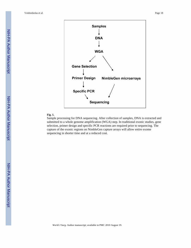
Fig. 1.Sample processing for DNA sequencing. After collection of samples, DNA is extracted andsubmitted to a whole genome amplification (WGA) step. In traditional exonic studies, geneselection, primer design and specific PCR reactions are required prior to sequencing. Thecapture of the exonic regions on NimbleGen capture arrays will allow entire exomesequencing in shorter time and at a reduced cost.
Voidonikolas et al. Page 18
World J Surg. Author manuscript; available in PMC 2010 August 19.
NIH
-PA Author Manuscript
NIH
-PA Author Manuscript
NIH
-PA Author Manuscript
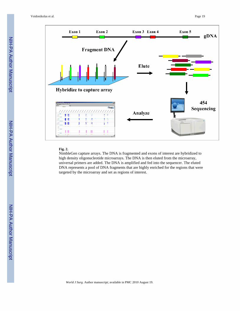
Fig. 2.NimbleGen capture arrays. The DNA is fragmented and exons of interest are hybridized tohigh density oligonucleotide microarrays. The DNA is then eluted from the microarray,universal primers are added. The DNA is amplified and fed into the sequencer. The elutedDNA represents a pool of DNA fragments that are highly enriched for the regions that weretargeted by the microarray and set as regions of interest.
Voidonikolas et al. Page 19
World J Surg. Author manuscript; available in PMC 2010 August 19.
NIH
-PA Author Manuscript
NIH
-PA Author Manuscript
NIH
-PA Author Manuscript
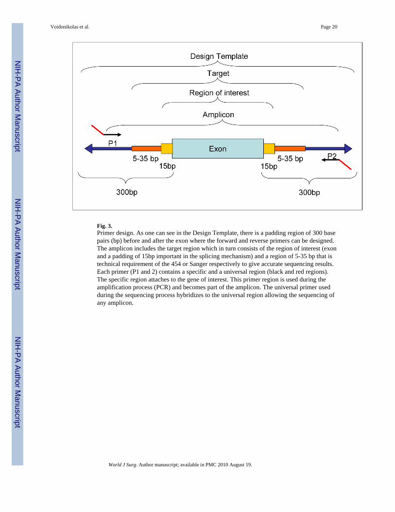
Fig. 3.Primer design. As one can see in the Design Template, there is a padding region of 300 basepairs (bp) before and after the exon where the forward and reverse primers can be designed.The amplicon includes the target region which in turn consists of the region of interest (exonand a padding of 15bp important in the splicing mechanism) and a region of 5-35 bp that istechnical requirement of the 454 or Sanger respectively to give accurate sequencing results.Each primer (P1 and 2) contains a specific and a universal region (black and red regions).The specific region attaches to the gene of interest. This primer region is used during theamplification process (PCR) and becomes part of the amplicon. The universal primer usedduring the sequencing process hybridizes to the universal region allowing the sequencing ofany amplicon.
Voidonikolas et al. Page 20
World J Surg. Author manuscript; available in PMC 2010 August 19.
NIH
-PA Author Manuscript
NIH
-PA Author Manuscript
NIH
-PA Author Manuscript
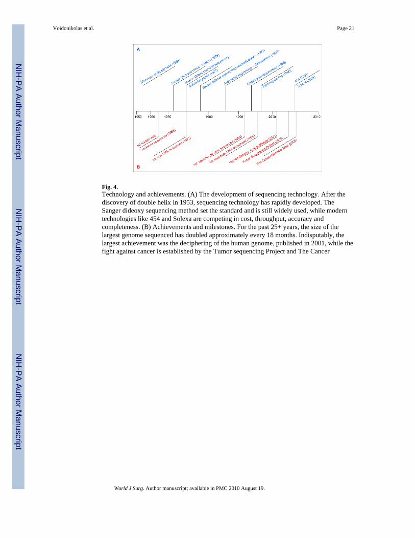
Fig. 4.Technology and achievements. (A) The development of sequencing technology. After thediscovery of double helix in 1953, sequencing technology has rapidly developed. TheSanger dideoxy sequencing method set the standard and is still widely used, while moderntechnologies like 454 and Solexa are competing in cost, throughput, accuracy andcompleteness. (B) Achievements and milestones. For the past 25+ years, the size of thelargest genome sequenced has doubled approximately every 18 months. Indisputably, thelargest achievement was the deciphering of the human genome, published in 2001, while thefight against cancer is established by the Tumor sequencing Project and The Cancer
Voidonikolas et al. Page 21
World J Surg. Author manuscript; available in PMC 2010 August 19.
NIH
-PA Author Manuscript
NIH
-PA Author Manuscript
NIH
-PA Author Manuscript
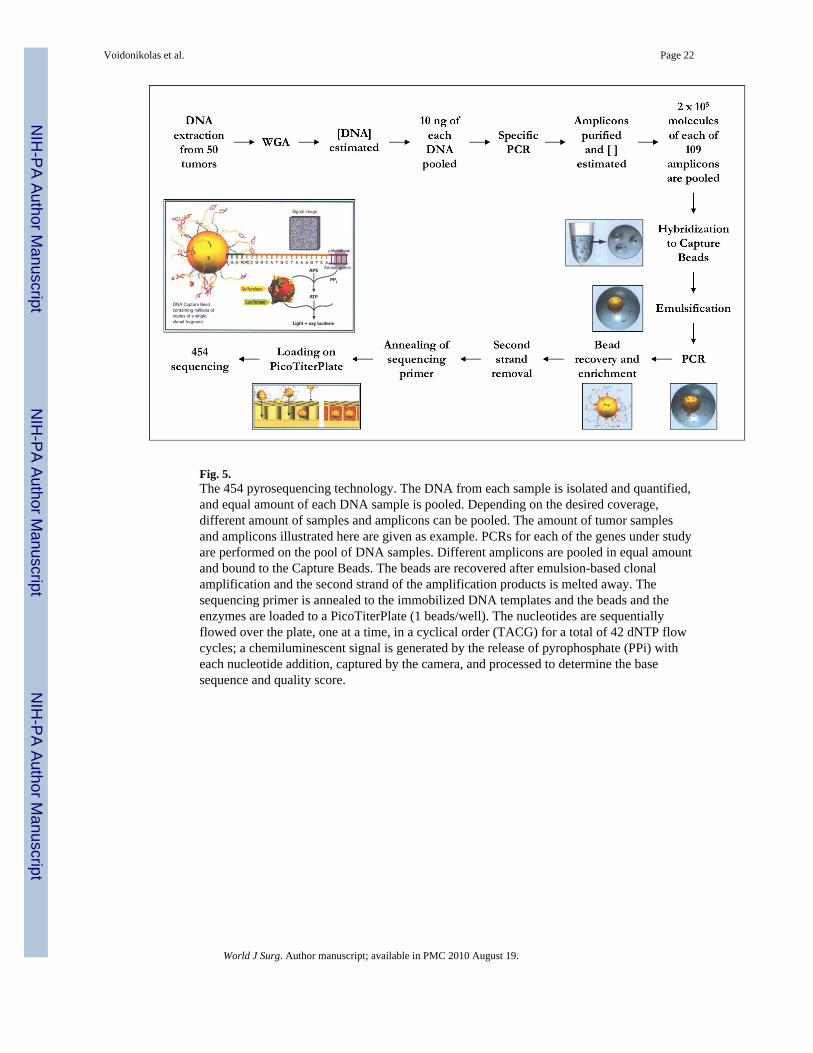
Fig. 5.The 454 pyrosequencing technology. The DNA from each sample is isolated and quantified,and equal amount of each DNA sample is pooled. Depending on the desired coverage,different amount of samples and amplicons can be pooled. The amount of tumor samplesand amplicons illustrated here are given as example. PCRs for each of the genes under studyare performed on the pool of DNA samples. Different amplicons are pooled in equal amountand bound to the Capture Beads. The beads are recovered after emulsion-based clonalamplification and the second strand of the amplification products is melted away. Thesequencing primer is annealed to the immobilized DNA templates and the beads and theenzymes are loaded to a PicoTiterPlate (1 beads/well). The nucleotides are sequentiallyflowed over the plate, one at a time, in a cyclical order (TACG) for a total of 42 dNTP flowcycles; a chemiluminescent signal is generated by the release of pyrophosphate (PPi) witheach nucleotide addition, captured by the camera, and processed to determine the basesequence and quality score.
Voidonikolas et al. Page 22
World J Surg. Author manuscript; available in PMC 2010 August 19.
NIH
-PA Author Manuscript
NIH
-PA Author Manuscript
NIH
-PA Author Manuscript
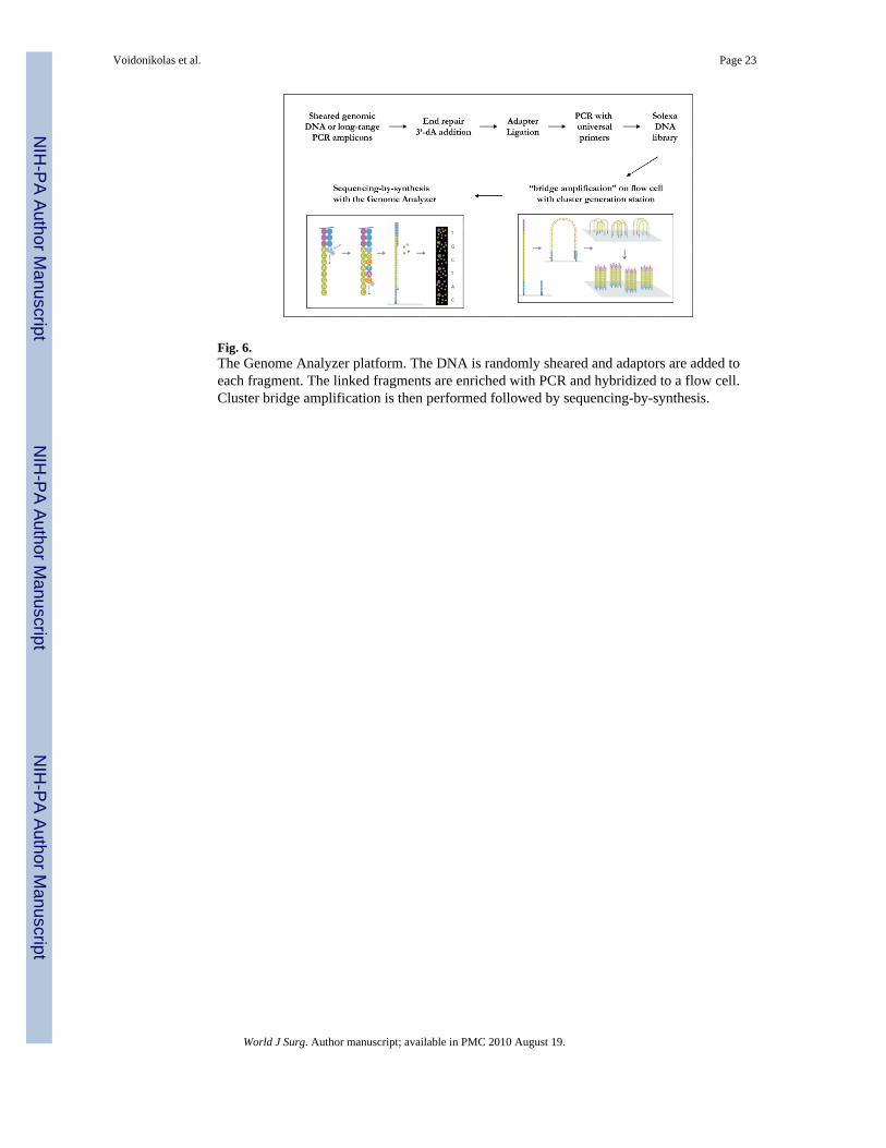
Fig. 6.The Genome Analyzer platform. The DNA is randomly sheared and adaptors are added toeach fragment. The linked fragments are enriched with PCR and hybridized to a flow cell.Cluster bridge amplification is then performed followed by sequencing-by-synthesis.
Voidonikolas et al. Page 23
World J Surg. Author manuscript; available in PMC 2010 August 19.
NIH
-PA Author Manuscript
NIH
-PA Author Manuscript
NIH
-PA Author Manuscript
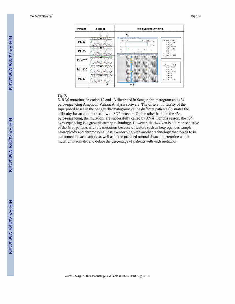
Fig. 7.K-RAS mutations in codon 12 and 13 illustrated in Sanger chromatogram and 454pyrosequencing Amplicon Variant Analysis software. The different intensity of thesuperposed bases in the Sanger chromatograms of the different patients illustrates thedifficulty for an automatic call with SNP detector. On the other hand, in the 454pyrosequencing, the mutations are successfully called by AVA. For this reason, the 454pyrosequencing is a great discovery technology. However, the % given is not representativeof the % of patients with the mutations because of factors such as heterogenous sample,heteroploidy and chromosomal loss. Genotyping with another technology then needs to beperformed in each sample as well as in the matched normal tissue to determine whichmutation is somatic and define the percentage of patients with each mutation.
Voidonikolas et al. Page 24
World J Surg. Author manuscript; available in PMC 2010 August 19.
NIH
-PA Author Manuscript
NIH
-PA Author Manuscript
NIH
-PA Author Manuscript
NIH
-PA Author Manuscript
NIH
-PA Author Manuscript
NIH
-PA Author Manuscript
Voidonikolas et al. Page 25
Table 1
Modification events in function of patient number. When the number of screened patients was increased thenumber of identified events per gene was substantially increased, irrespective of the technique used.
Sequencingmethod
Patientnumber Gene number Number of
events Event/gene
Sanger11 58 304 5.2
47 4 74 18.5
454 41 21 332 15.8
World J Surg. Author manuscript; available in PMC 2010 August 19.

NIH
-PA Author Manuscript
NIH
-PA Author Manuscript
NIH
-PA Author Manuscript
Voidonikolas et al. Page 26
Table 2
Sample and DNA preparation
The blood can be collected in any collection tubes normally used in the hospital setting. However, we have found that the PAXgene BloodDNA kit (PreAnalytiX) offers some advantages. The kit has two constituents that can be separately ordered: the blood collection tubes forthe clinic and the DNA isolation kit for the laboratory. The PAXgene blood collection tubes contain a proprietary blend of reagents optimizedfor standardized blood collection, storage, and isolation of high-quality genomic DNA. These reagents stabilize the cellular bloodconstituents for up to 14 days at room temperature or 28 days at 4°C, allowing pick-up time flexibility. Moreover, we have found that thedesign of the covering cap minimizes blood spillage upon opening, an important concern for blood pathogen exposure.
In order to study the tumor, a punch biopsy should be taken from the specimen. The punch should be longitudinally dissected. Theupper half should be submitted in formalin for paraffin embedding and be used as a mirror image to measure the percentage of viable tumorcells. The lower half should be used for the study. Tissue samples should then be immersed in preservative solutions. We have found thattissues preserved in RNAlater (Ambion) and protease inhibitor (Roche) solution generate high quality DNA, and also allow for parallelexpression studies [45]. Tissue preserved in either solution must be washed several times in PBS before DNA isolation to remove any traceof the stabilizing solution that would otherwise interfere with the DNA extraction protocol.
DNA can be isolated from the tissues and WGA can be performed using different commercial kit. When using FFPE material, theREPLI-g FFPE kit (Qiagen, Valencia, CA), which combines DNA isolation, random ligation, and WGA has enabled us to sequence oldersamples that had previously failed sequencing.
World J Surg. Author manuscript; available in PMC 2010 August 19.

NIH
-PA Author Manuscript
NIH
-PA Author Manuscript
NIH
-PA Author Manuscript
Voidonikolas et al. Page 27
Table 3
Primer design
At BCM-HGSC, the exonic regions of interest are first surrounded with intronic 200-300 base padding sequences in which the primers willbe picked (Fig. 3). The DNA interspersed repeated sequences and SNPs in these padding regions are masked using RepeatMasker [58]and the dbSNP database [59] to prevent the design of primers over these bases. The primer set is then designed with Primer3 software[60] using certain basic criteria (primer length between 18-30 bases, melting temperature between 50-60° Celsius and primer pair withsimilar TM values, GC content ranging between 40% and 60%, 3′ end containing a GC clamp but free of secondary structures, repetitivesequences, palindromes, and highly degenerate sequences, no formation of hairpins in the primer and self or cross dimerization with otherprimers in the reaction). The exclusive specificity of the primers is then checked with PCR software [61] during which, in-silico PCR is usedto simulate the PCR process. If the desired sequence to amplify is large, the pipeline breaks apart the sequence and designs multipleprimer sets (or amplicon tiles) to cover the large sequence. If the pipeline is unable to come up with some primer designs to amplify asequence of interest, then manual primer design is done. The pipeline finally attaches forward and reverse universal sequencing primers tothe specific primers. These universal regions will be used during the sequencing process to obtain forward and reverse sequence reads. Abar code of few nucleotides also can be added to the primer to segregate the individual sequences if the patient samples are pooled, suchas for 454 sequencing [62].
The primers must then be tested on commercial or cell-line human DNA before using them on patient samples. The resulting testamplicons should be run on gel and sequenced to verify the PCR efficacy and specificity, respectively.
World J Surg. Author manuscript; available in PMC 2010 August 19.

NIH
-PA Author Manuscript
NIH
-PA Author Manuscript
NIH
-PA Author Manuscript
Voidonikolas et al. Page 28
Table 4
Mutation call
The call of the mutations in case of Sanger sequencing can be performed in an automated fashion with SNP Detector (created by JinhuiZhang at the NCI) using the corresponding sequence deposited in GenBank as reference. Manual verification (visual study of thechromatograms) can be done in software such as Consed and Sequencher (Gene Codes Corp) (Fig. 7) Technologies such as 454pyrosequencing and Solexa have developed their own analytical software such as Amplicon Variant Analysis (AVA) software (454 LifeSciences) (Fig. 7) Complementary informatic supports also are developed in the different institutions using these technologies forautomated high throughput SNP calling and associated function analysis.
World J Surg. Author manuscript; available in PMC 2010 August 19.
NIH
-PA Author Manuscript
NIH
-PA Author Manuscript
NIH
-PA Author Manuscript
Voidonikolas et al. Page 29
Tabl
e 5
Mod
ifica
tion
even
ts fo
und
in p
ancr
eatic
ade
noca
rcin
oma
usin
g Sa
nger
and
454
tech
nolo
gy. B
ase
shift
s and
bas
e in
serti
ons o
r del
etio
ns (i
ndel
s) w
ere
iden
tifie
d in
bot
h th
e in
troni
c re
gion
and
the
exon
s of s
ever
al a
naly
zed
gene
s. Ex
onic
bas
e sh
ifts w
ere
eith
er sy
nony
mou
s (sy
n) o
r non
syno
nym
ous (
non
syn)
or d
etec
ted
in th
e un
trans
late
d re
gion
(UTR
).
intr
onic
eve
nts
exon
ic e
vent
sA
naly
zed
gene
sT
OT
AL
base
shift
inde
lU
TR
syn.
nons
yn.
inde
l
Sang
er
Ger
mlin
e12
58
5848
171
257
Som
atic
523
2411
292
121
Tot
al17
711
8259
463
6237
8
454
106
4578
103
2133
2
World J Surg. Author manuscript; available in PMC 2010 August 19.
NIH
-PA Author Manuscript
NIH
-PA Author Manuscript
NIH
-PA Author Manuscript
Voidonikolas et al. Page 30
Tabl
e 6
Exam
ple
of v
alid
atio
n of
the
mod
ifica
tion
even
ts id
entif
ied
with
454
tech
nolo
gy. I
n a
tota
l of 2
,814
read
s of a
poo
led
41 D
NA
sam
ple,
454
pyro
sequ
enci
ng d
etec
ted,
am
ong
othe
rs, t
wo
base
shift
mod
ifica
tions
in e
xon
2 an
d on
e in
exo
n 6
of th
e K
-RA
S ge
ne. V
alid
atio
n of
thes
e ev
ents
with
Sang
er se
quen
cing
on
indi
vidu
al tu
mor
and
mat
ched
nor
mal
tiss
ue sa
mpl
es d
istin
guis
hed
betw
een
som
atic
(exo
n 2)
and
ger
mlin
e (e
xon
6) m
odifi
catio
nsan
d di
ffer
entia
ted
hom
ozyg
ous a
nd h
eter
ozyg
ous p
atie
nts.
454
resu
ltSa
nger
val
idat
ion
41 p
oole
dtu
mor
s41
mat
ched
indi
vidu
altu
mor
s41
mat
ched
indi
vidu
al n
orm
als
Exo
nSN
P lo
c hg
18(c
hr 1
2)co
don
Ori
g.co
don
Bas
esh
ifts
aa#
read
s%
rea
dw
ithba
sech
ift
hom
o.m
ajor
alle
lehe
tero
hom
o.m
inor
alle
le
% p
tw
ithso
m.
mut
.
hom
o.m
ajor
alle
lehe
tero
hom
o.m
inor
alle
le
225
2895
5112
GG
TG
→ A
Gly
→ A
sp28
142.
7031
100
24.4
410
0
2528
9551
12G
GT
G →
TG
ly →
Val
2814
2.03
383
07.
341
00
625
2540
4417
3G
AT
T →
CA
sp28
141.
1429
111
2.4
2911
1
World J Surg. Author manuscript; available in PMC 2010 August 19.