Embed Size (px)
Citation preview
12
Briefings in Functional Genomics, 21(1), 2022,12–22
https://doi.org/10.1093/bfgp/elab005Advance Access Publication Date: 5 March 2021Review Paper
From mutation to mechanism: decipheringthe molecular function of genetic variants linkedto human ageing
Maarouf Baghdadi , Helena M. Hinterding, Linda Partridge andJoris DeelenCorresponding author: Joris Deelen, Max Planck Institute for Biology of Ageing, Joseph-Stelzmann-Str. 9b, 50931 Cologne, Germany.Tel.: +49 221 379 70 612; E-mail: [email protected]
Abstract
Many of the leading causes of death in humans, such as cardiovascular disease, type 2 diabetes and Alzheimer’s disease areinfluenced by biological mechanisms that become dysregulated with increasing age. Hence, by targeting theseageing-related mechanisms, we may be able to improve health in old age. Ageing is partly heritable and genetic studies havebeen moderately successful in identifying genetic variants associated with ageing-related phenotypes (lifespan, healthspanand longevity). To decipher the mechanisms by which the identified variants influence ageing, studies that focus on theirfunctional validation are vital. In this perspective, we describe the steps that could be taken in the process of functionalvalidation: (1) in silico characterisation using bioinformatic tools; (2) in vitro characterisation using cell lines or organoids; and(3) in vivo characterisation studies using model organisms. For the in vivo characterisation, it is important to focus ontranslational phenotypes that are indicative of both healthspan and lifespan, such as the frailty index, to inform subsequentintervention studies. The depth of functional validation of a genetic variant depends on its location in the genome andconservation in model organisms. Moreover, some variants may prove to be hard to characterise due to context-dependenteffects related to the experimental environment or genetic background. Future efforts to functionally characterise the(newly) identified genetic variants should shed light on the mechanisms underlying ageing and will help in the design oftargeted interventions to improve health in old age.
Key words: genetic variants; lifespan; healthspan; longevity; functional characterisation; model organisms
IntroductionLife expectancy has been steadily rising in the world, partly dueto treatment of the elderly but mostly due to the reduction ofearly life mortality and treatment of communicable disease [1].The increasing population of elderly individuals will bring a con-comitant increase in multimorbidity [2, 3]. Stagnating birth rates
Maarouf Baghdadi is a PhD student at the Max Planck Institute for Biology of Ageing. His PhD project is focused on understanding the sex-specific effectof genetic interventions related to insulin signalling in mammalian models.Helena M. Hinterding is a PhD student at the Max Planck Institute for Biology of Ageing. Her PhD project is focused on exploring the role of genetic variationin the MAPK/ERK signalling pathway in human longevity.Linda Partridge is the founding director of the Max Planck Institute for Biology of Ageing. Her research is focused on evolutionarily conserved dietary,genetic and pharmacological interventions promoting healthy ageing.Joris Deelen is a research group leader at the Max Planck Institute for Biology of Ageing. His research is focused on the genetics and biomarkers of healthyageing in humans.
© The Author(s) 2021. Published by Oxford University Press.This is an Open Access article distributed under the terms of the Creative Commons Attribution License (http://creativecommons.org/licenses/by/4.0/),which permits unrestricted reuse, distribution, and reproduction in any medium, provided the original work is properly cited.
and a growing percentage of pensioners are posing a seriouschallenge to our economies and will do so to an even greaterextent in the future. Data from the European Union highlighthow age-associated multimorbidity leads to a rise in individualhealthcare expenditure on older people, up to 18% per capitagross domestic product [4]. Furthermore, healthcare costs differ
Dow
nloaded from https://academ
ic.oup.com/bfg/article/21/1/13/6159025 by guest on 25 July 2022
Deciphering the molecular function of genetic variants 13
between sexes, mainly due to differences in multimorbidityand lifespan, with women using a third more resources thanmen and the majority of human healthcare expenditure takingplace in middle to old age [5]. The use of an overwhelmingamount of capital to develop and test therapies targeting age-associated diseases, such as cancer, with only marginal benefitsin quality-adjusted life years (average 2 months) [6], raises thequestion whether resources would be better spent targeting theunderlying biological mechanisms dysregulated with age ratherthan disease-related endpoints [7]. Moreover, by focusing onthe compression of morbidity, societies can benefit from thetremendous social and economic opportunities that come withan active and vibrant older population [8].
The idea of reducing multimorbidity by targeting ageingcomes from the fact that exceptionally long-lived individualsand their family members often present a compression ofmorbidity or a longer lifespan free of disease [9, 10]. Nevertheless,they suffer from the same causes of death at old age (i.e.they do not seem to be immune to disorders but rather havea later onset of disease) [9]. There is evidence suggestingthat the factors contributing to these benefits are partlyheritable, given that longevity [i.e. survival to an exceptionalold age (e.g. top 10% of their respective birth cohort)] can betransmitted as a quantitative genetic trait [11]. On the otherhand, the evidence for a genetic component of lifespan (i.e.number of years lived), an alternative phenotype used to studyageing, is more compelling. In twin studies for lifespan, theheritability has been estimated to be around 25% [12]. However,large genealogical studies for lifespan offer a more modestview of heritability (i.e. below 12%) [13, 14], potentially dueto the diverse population studied (geographically diverse),inclusion of early mortality and accounting for the non-additive genetic component. Taken together, there is enoughsupport for studying ageing-related phenotypes using geneticapproaches. Ultimately, the aim of genetic studies on ageing isto identify genes that can elucidate mechanisms of healthyphysiological ageing, which can subsequently be targetedusing lifestyle and/or pharmacological interventions to reduce(multi)morbidity.
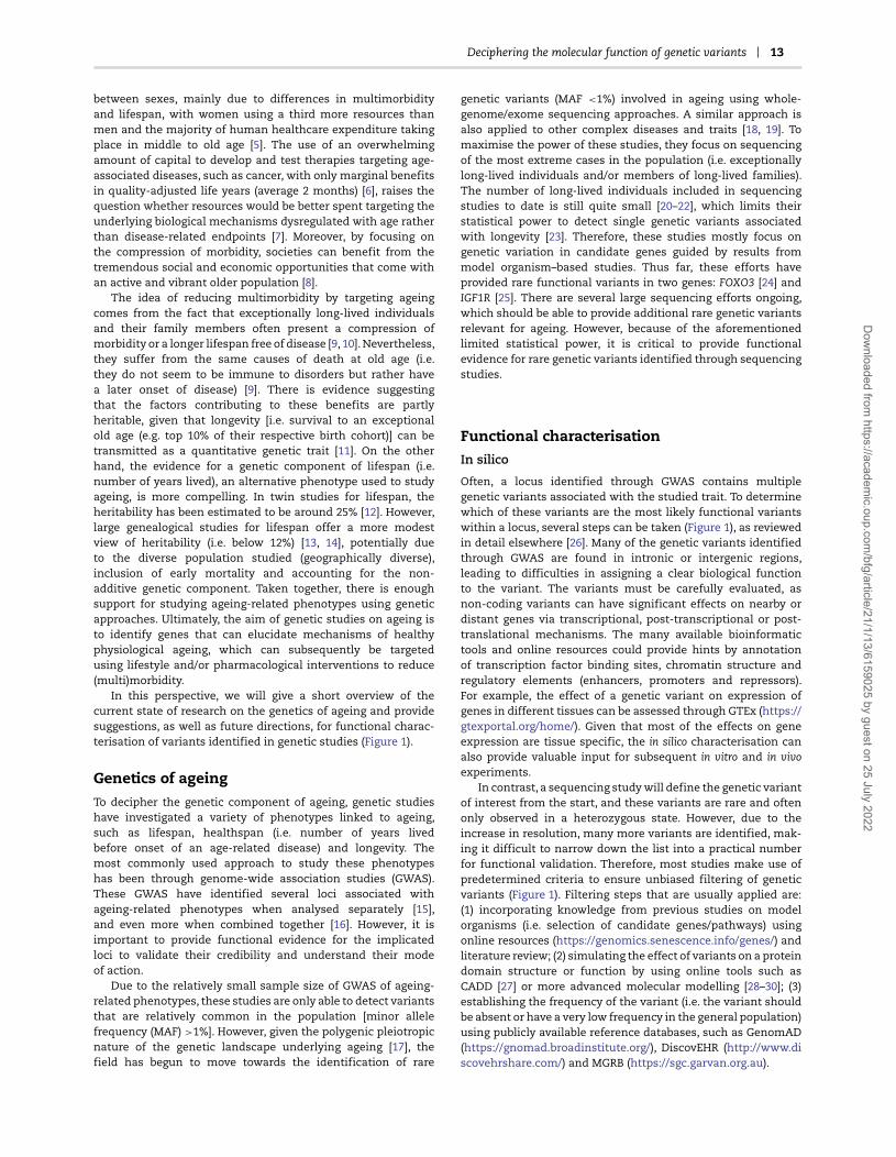
In this perspective, we will give a short overview of thecurrent state of research on the genetics of ageing and providesuggestions, as well as future directions, for functional charac-terisation of variants identified in genetic studies (Figure 1).
Genetics of ageingTo decipher the genetic component of ageing, genetic studieshave investigated a variety of phenotypes linked to ageing,such as lifespan, healthspan (i.e. number of years livedbefore onset of an age-related disease) and longevity. Themost commonly used approach to study these phenotypeshas been through genome-wide association studies (GWAS).These GWAS have identified several loci associated withageing-related phenotypes when analysed separately [15],and even more when combined together [16]. However, it isimportant to provide functional evidence for the implicatedloci to validate their credibility and understand their modeof action.
Due to the relatively small sample size of GWAS of ageing-related phenotypes, these studies are only able to detect variantsthat are relatively common in the population [minor allelefrequency (MAF) >1%]. However, given the polygenic pleiotropicnature of the genetic landscape underlying ageing [17], thefield has begun to move towards the identification of rare
genetic variants (MAF <1%) involved in ageing using whole-genome/exome sequencing approaches. A similar approach isalso applied to other complex diseases and traits [18, 19]. Tomaximise the power of these studies, they focus on sequencingof the most extreme cases in the population (i.e. exceptionallylong-lived individuals and/or members of long-lived families).The number of long-lived individuals included in sequencingstudies to date is still quite small [20–22], which limits theirstatistical power to detect single genetic variants associatedwith longevity [23]. Therefore, these studies mostly focus ongenetic variation in candidate genes guided by results frommodel organism–based studies. Thus far, these efforts haveprovided rare functional variants in two genes: FOXO3 [24] andIGF1R [25]. There are several large sequencing efforts ongoing,which should be able to provide additional rare genetic variantsrelevant for ageing. However, because of the aforementionedlimited statistical power, it is critical to provide functionalevidence for rare genetic variants identified through sequencingstudies.
Functional characterisationIn silico
Often, a locus identified through GWAS contains multiplegenetic variants associated with the studied trait. To determinewhich of these variants are the most likely functional variantswithin a locus, several steps can be taken (Figure 1), as reviewedin detail elsewhere [26]. Many of the genetic variants identifiedthrough GWAS are found in intronic or intergenic regions,leading to difficulties in assigning a clear biological functionto the variant. The variants must be carefully evaluated, asnon-coding variants can have significant effects on nearby ordistant genes via transcriptional, post-transcriptional or post-translational mechanisms. The many available bioinformatictools and online resources could provide hints by annotationof transcription factor binding sites, chromatin structure andregulatory elements (enhancers, promoters and repressors).For example, the effect of a genetic variant on expression ofgenes in different tissues can be assessed through GTEx (https://gtexportal.org/home/). Given that most of the effects on geneexpression are tissue specific, the in silico characterisation canalso provide valuable input for subsequent in vitro and in vivoexperiments.
In contrast, a sequencing study will define the genetic variantof interest from the start, and these variants are rare and oftenonly observed in a heterozygous state. However, due to theincrease in resolution, many more variants are identified, mak-ing it difficult to narrow down the list into a practical numberfor functional validation. Therefore, most studies make use ofpredetermined criteria to ensure unbiased filtering of geneticvariants (Figure 1). Filtering steps that are usually applied are:(1) incorporating knowledge from previous studies on modelorganisms (i.e. selection of candidate genes/pathways) usingonline resources (https://genomics.senescence.info/genes/) andliterature review; (2) simulating the effect of variants on a proteindomain structure or function by using online tools such asCADD [27] or more advanced molecular modelling [28–30]; (3)establishing the frequency of the variant (i.e. the variant shouldbe absent or have a very low frequency in the general population)using publicly available reference databases, such as GenomAD(https://gnomad.broadinstitute.org/), DiscovEHR (http://www.discovehrshare.com/) and MGRB (https://sgc.garvan.org.au).
Dow
nloaded from https://academ
ic.oup.com/bfg/article/21/1/13/6159025 by guest on 25 July 2022

14 Baghdadi et al.
Figure 1. Pipeline for functional characterisation of genetic variants linked to human ageing.
In vitro
Cell lines are a common tool used to explore functional effectsin vitro. As many genetic variants identified through GWAS arefound in non-coding regions, most in vitro studies first focuson measurements that can be used to determine their effecton enhancer/promoter activity, with luciferase and transcriptionfactor binding assays, or transcription of surrounding genes,
using qPCR or more advanced techniques (Figure 1) [31–34]. Anexample of successful in vitro follow-up of a genetic variantcoming from genetic studies of ageing is provided by Grossi andcolleagues, who have shown that a variant in FOXO3, residingwithin an enhancer region, creates a binding site for HSF1 thatresults in increased transcriptional activity of FOXO3 and itstarget genes in response to oxidative stress [35]. Due to thelack of genetic conservation of non-coding regions in model
Dow
nloaded from https://academ
ic.oup.com/bfg/article/21/1/13/6159025 by guest on 25 July 2022

Deciphering the molecular function of genetic variants 15
organisms, many of the genetic variants coming from GWAScannot be moved forward to an in vivo model. Therefore, thein vitro characterisation concludes the functional validation ofthese variants.
While primary cells from long-lived individuals would serveas the gold standard for in vitro characterisation (Figure 1), suchcells are often difficult to obtain and culture. One example ofusing primary cells is by reprogramming them into inducedpluripotent stem cells (iPSCs). This allows for the study of theeffect of a genetic variant on a cellular phenotype, such as stressresistance, in the context of an individual’s genetic background.An advantage of investigating cells from long-lived individu-als is that epistatic effects are also assessed, although it isharder to pinpoint the functional effect to a specific geneticvariant. Moreover, the differentiation potential of iPSCs intovarious cell and tissue types offers the advantage of study-ing tissue-specific functional effects [36]. One should, however,take into account that reprogramming leads to modificationof a cell’s epigenetic landscape, which could influence ageing-related molecular read-outs such as gene expression [37]. 3Din vitro organoid culture is another method to mimic cellularorganization, intercellular communication and crucial extracel-lular matrix interaction, with a closer approximation to thephysiological microenvironment of a given tissue than tradi-tional cell culture methods [38]. To overcome the practical lim-itations of primary cell lines, researchers often turn to cancercell lines and genetic engineering tools to study a variant’s effectin an adjustable environment. This is done by comparing cellswith the introduced genetic variant to its control counterpart toassess gain- or loss-of-function effects (Figure 1). This approachhas, for example, been applied by Tazearslan and colleagues todetermine the functional effect of two genetic variants in IGF1Ridentified by sequencing a cohort of long-lived individuals [25,39]. However, the identified heterozygous variants were onlyassessed in the homozygous state. Therefore, the potential roleof compensatory effects in the presence of the wild-type proteinstill needs to be addressed.
The in vitro functional characterisation and validation ofgenetic variants have advanced immensely since the practicalapplication of the CRISPR/Cas9 gene editing technology. Initiallydiscovered in bacteria as a defence mechanism against viruses[40], scientists have learned to exploit and adapt this techniquein order to create targeted knockouts and site-specific pointmutations in a wide array of cell types. Essentially, only two com-ponents are needed: a complementary guide RNA unique to alocus of interest and a Cas9 enzyme that cuts the DNA sequence.These steer the cell’s repair mechanism towards endogenousnon-homologous end joining or a cleaner version of homology-directed repair by providing a DNA repair template [41]. Recentadvances have also modified the Cas9 protein to be more spe-cific in targeting [42], thereby minimizing the potential of off-target modifications obscuring a variant’s phenotype. Despitethe advantages of cell models for functional characterisation ofgenetic variants, they also pose certain limitations. For example,genetic background effects and inherent genomic instability incell lines could hide the functional effects under investigation.Furthermore, many of the widely used cell lines are derived fromimmortalized cancer cells and possess karyotypic abnormalities,such as polyploidy, that could affect both the genetic engineeringmethods as well as downstream functional analyses [43].
Assessing the effect of the genetic variant on its host pro-tein and direct downstream targets, for example by looking atstability, functional sites (e.g. phosphorylation), expression andtranscription, using, for example, immunohistochemistry, qPCR
or pull-down assays, can serve as a guide for the subsequentin-depth functional characterisation (Figure 1). As a next step,the obtained or created cell lines can be functionally charac-terised using ageing-related cellular and molecular read-outsbased on the hallmarks of ageing [44]. In Table 1, we haveprovided an overview of assays that can be used to study theeffects of a variant on the different hallmarks in vitro. Moreover,stress assays (e.g. H2O2, UV and heat) can be used to determinehallmark-overarching effects [45].
In vivo
In vitro experiments provide invaluable mechanistic informationbut lack critical insight on how different tissues respond toa genetic intervention or how the effect of a genetic variantchanges with age. A unique opportunity of genetic studies inmodel organisms, other than conducting lifespan analyses, isthe possibility of performing longitudinal healthspan assays thatcan shed light on biological mechanisms of resilience used byhealthy ageing individuals to combat the hallmarks of ageing[64] (Table 2). Performing measurements at multiple time pointsacross the life of an animal allows the assessment of progressivedecline in an outcome measure as, for example, shown for motorability and sleep [65, 66]. It is important to include time pointswith enough temporal resolution in an ageing study to preventmisinterpretation of the results, especially if future therapeuticswill be designed to intervene in the age-related trajectory thatis indicative of the function of a gene or pathway [67]. The endpoint for validating a genetic variant associated with ageingis vague and open to discussion. However, it is clear that thisvariant must at least have an effect, either alone or in com-bination with other variants, on healthspan. Currently, we donot have any proxies for overall healthspan in vitro. Given thatageing is a time-dependent cumulative process where diseaserisk increases, this property must ideally be assessed in theprocess of functional validation.
After enough evidence has been obtained from the in vitroexperiments, the natural next step is to introduce the geneticvariant into a model organism in which the ageing process canbe characterised and an investigation can be launched into itsrole on lifespan or healthspan modulation (Figure 1). The modelorganism of choice is variant dependent, as genetic conserva-tion, tissue homology and practical reasons may favour someorganisms over others. Below we will highlight some of the mostcommonly used model organisms in genetic studies of ageing.We have focused on healthspan outcomes that exhibit func-tional decline with age and that can be assessed non-invasively.
Nematode worms
The nematode worm (Caenorhabditis elegans) has been the defacto model organism for the study of the genetics of age-ing, since the field was founded by paradigm shifting work inwhich a single mutation resulted in doubling of the worm’slifespan [68]. The short lifespan of worms coupled with theability to perform genetic screens for traits has led to manyinsights into genetic mechanisms that regulate lifespan, withmany pathways implicated in higher-order organisms [69]. Thebalance between the presence of multiple distinct tissues andsimplicity (lack of redundancy) of genetic pathways has led toan enticing experimental model system. Moreover, the trans-parent nature of worms allows non-invasive imaging to trackthe effect of reporter-tagged genetic modifications throughouttheir lifespan. Additionally, scientists have leveraged the worm’s
Dow
nloaded from https://academ
ic.oup.com/bfg/article/21/1/13/6159025 by guest on 25 July 2022

16 Baghdadi et al.
Table 1. Overview of assays that can be used to study the hallmarks of ageing in vitro
Hallmark of ageing In vitro assays
Genomic instability • Base excision repair capacity [46, 47]• Measuring DNA lesions [48]
Cellular senescence • Beta-galactosidase [49], p53 and p16 stainings to assess mitotic arrest [50]
Mitochondrial dysfunction • Basal mitochondrial respiration [51] or substrate-uncoupler-inhibitor titration protocols [52]• qPCR to asses mtDNA copy number [53]• Assessing the integrated stress response after doxycycline treatment [54, 55]
Loss of proteostasis • LysoTracker [56]• LC3 (immunohistochemical) staining to assess autophagic flux [57]• Citrate synthase activity assay for chaperone activity [58]
Epigenetic alterations • DNA methylation (arrays/bisulfite sequencing) [59]
Stem cell exhaustion • Stemness markers (immunofluorescence) or proliferation assays such as HALO-96 PREP [60]
Telomere attrition • Terminal restriction fragment (TRF) [61]• Quantitative fluorescence in situ hybridization (Q-FISH) [61]• qPCR [61]
Deregulated nutrient-sensing andaltered intercellular communication
• Assessment of IIS/mTOR activity after nutrient deprivation (i.e. serum or amino acidstarvation) or stimulation (e.g. with insulin, IGF-1 or EGF) byimmunohistochemistry/immunoblotting [62, 63]
IIS, insulin/insulin-like growth factor-1 signalling; mTOR, mammalian target of rapamycin; IGF-1, insulin-like growth factor-1; EGF, epidermal growth factor.
transparency to assess different healthspan parameters, suchas age-associated tissue decline, nucleolar size and body bends(Table 2) [70]. Moreover, worms provide a powerful method forperforming gene knockdown in the whole organism as well as ina spatially restricted manner by feeding them RNAi-generatingbacteria for assessment of gene function [71]. Finally, with theadvent of machine learning and accompanying technologicaladvancements, scientists have developed automated methodsfor performing lifespan assays allowing for high-throughputgenetic studies.
Fruit f lies
The fruit fly (Drosophila melanogaster) is another great tool forageing research due to its relatively short lifespan, practicalhusbandry and advanced genetic tools. These genetic tools, forexample, allow elegant and precise spatiotemporal control ofgenetic perturbations. This allows studies to address questionsabout how genetic modifications affect tissue-specific func-tional decline or tissue–tissue interactions during the ageingprocess. Tissues in fruit flies are more homologous to humansthan those in worms but still lack homology in metabolic organssuch as liver and pancreas. However, flies possess a brain with adiversity in cell types similar to mammals. Moreover, the fruit flyis an invertebrate with a robust circadian rhythm that declineswith age, allowing investigation of the association of circadiandysregulation and ageing [66]. Unlike worms, fruit flies possessheteromorphic sex chromosomes and the ability to determinethe sex of individual cells in a cell-autonomous manner, allow-ing the study of important mechanisms of sexual dimorphismwithout confounding effects of circulating sex hormones [72].Large numbers of animals can be assessed in lifespan assaysto investigate the effect of a mutation on the mortality rate,providing greater insight into the gene function than just meanand maximum lifespan. Moreover, the assessment of mortalityrate is important in determining if a genetic intervention leads toa change in age-specific mortality or age-independent mortality,potentially indicative of whether any increase in lifespan isattributable to slowed ageing or a general improvement in health
[73]. The rich history of studies in fruit flies has brought forwardmany well-developed healthspan assays for which ageing trajec-tories have already been described, such as climbing and sleep(Table 2) [65, 66]. The combination of all these advantages andmore (Table 2) makes fruit flies another valuable tool to studythe biological mechanisms of ageing [74].
African turquoise killifish
The African turquoise killifish (Nothobranchius furzeri) is a rela-tively new model organism that is gaining significant popularityin the ageing field. These killifish are an enticing middle groundbetween the short lifespans of invertebrate models and thedeveloped organ system of vertebrates, such as an adaptiveimmune system. Previous invertebrate models possess shortmaximal lifespans (Table 2), but typical vertebrate models have amaximum lifespan of over 4 years, prohibiting repeated results,iteration of experiments and reducing feasibility of studies try-ing to verify novel ageing genes. There has been a great effort todevelop the genetic and genomic toolkit of killifish, opening thedoor to the genetic modification of this unique vertebrate modelorganism [75]. The killifish is a relatively new model organism,and so the healthspan measures are still under developmentand currently limited to visual macroscopic inspection of theanimal (Table 2). However, there are already studies reportingthe development of cognitive and locomotor assays that aremodulated by environmental ageing interventions [76]. Overall,the killifish is an interesting model organism to incorporate intofunctional genetic studies of ageing given its unique properties[77].
Mice
Mice (Mus musculus) are a great model organism for studyinghuman pathology and longevity as 99% of mouse genes have asequence match in the human genome [78]. However, mice havea dramatically shorter lifespan, there are outstanding genetictools available and there is potential to perform invasive assays[67]. Additionally, mice are social animals with a rich behavioural
Dow
nloaded from https://academ
ic.oup.com/bfg/article/21/1/13/6159025 by guest on 25 July 2022

Deciphering the molecular function of genetic variants 17
Tab
le2.
Ove
rvie
wof
adva
nta
ges,
lim
itat
ion
san
dst
ud
ied
hea
lth
outc
omes
ofd
iffe
ren
tm
odel
orga
nis
ms
use
dfo
rth
ein
vivo
char
acte
risa
tion
ofge
net
icva
rian
tsli
nke
dto
agei
ng
Mod
elor
gan
ism
Life
span
(day
s)A
dva
nta
ges
Lim
itat
ion
sH
ealt
hou
tcom
e
Med
ian
Max
Nem
atod
ew
orm
[70]
Cae
norh
abdi
tis
eleg
ans
∼15
∼27
•Lar
gebr
ood
size
•Sh
ort
life
span
and
gen
erat
ion
tim
e•S
ever
ald
isti
nct
tiss
ues
•Tra
nsp
aren
tbo
dy
•Com
pre
hen
sive
gen
etic
tool
box
•Eas
yan
din
exp
ensi
vecu
ltu
rean
dh
and
lin
g•E
asy
stor
age
and
gen
etic
lin
em
ain
ten
ance
•Doe
sn
otre
pli
cate
hu
man
orga
nsy
stem
s•N
oco
nse
rvat
ion
inth
ege
nom
e•P
ost-
mit
otic
adu
ltti
ssu
e(e
xcep
tge
rmli
ne)
•In
vert
ebra
te(n
osk
elet
alsy
stem
)•M
ales
only
hav
eon
ese
xch
rom
osom
e•N
ocl
ear
circ
adia
nrh
yth
m
•Mu
scle
loss
[85]
•Tis
sue
dec
lin
e[8
6,87
]•N
ucl
eola
rsi
ze[8
8]•B
ody
ben
ds
orth
rash
ing
[89]
•Ph
aryn
geal
pu
mp
ing
rate
[89]
Fru
itfl
y[9
0]D
roso
phila
mel
anog
aste
r
∼80
∼10
0•L
arge
broo
dsi
ze•S
hor
tli
fesp
anan
dge
ner
atio
nti
me
•Sev
eral
dis
tin
ctti
ssu
es•C
omp
reh
ensi
vege
net
icto
olbo
x•E
asy
and
inex
pen
sive
cult
ure
and
han
dli
ng
•Sex
-sp
ecif
icst
ud
ies
pos
sibl
e•D
iurn
al
•Doe
sn
otre
pli
cate
allh
um
anor
gan
syst
ems
•Lim
ited
con
serv
atio
nw
ith
hu
man
sin
the
gen
ome
•Pos
t-m
itot
icad
ult
tiss
ue
(exc
ept
inte
stin
e)•I
nve
rteb
rate
(no
skel
etal
syst
em)
•Can
not
bere
cove
red
aliv
efr
omfr
eezi
ng
soti
me-
con
sum
ing
stor
age
and
lin
em
ain
ten
ance
•Neu
rom
usc
ula
r(c
lim
bin
g)[9
1]•C
irca
dia
nrh
yth
md
ysre
gula
tion
(sle
ep)[
66]
•Gu
tin
tegr
ity,
fem
ale
spec
ific
[92]
•Fec
un
dit
y,fe
mal
esp
ecif
ic[9
3]
Afr
ican
turq
uoi
sek
illi
fish
[94]
Not
hobr
anch
ius
furz
eri
∼12
1∼
243
•Lar
gebr
ood
size
•Gen
ome
engi
nee
rin
gp
ossi
ble
[95]
•Ver
tebr
ate
•Lon
gitu
din
alas
sess
men
tof
ind
ivid
ual
anim
als
pos
sibl
e•E
asy
stor
age
and
mai
nte
nan
ceof
lin
es•S
ex-s
pec
ific
stu
die
sp
ossi
ble
•Diu
rnal
•No
stan
dar
diz
edh
ealt
hsp
anp
aram
eter
sye
t•L
imit
edco
nse
rvat
ion
inn
on-p
rote
inco
din
gge
nom
e•S
uff
icie
nt
tiss
ue
hom
olog
y(c
lose
dci
rcu
lato
rysy
stem
/in
nat
ean
dad
apti
veim
mu
nit
y)•R
equ
ires
spec
ialf
acil
ity
for
mai
nte
nan
cean
dfi
ltra
tion
•Lac
kof
dev
elop
edge
net
icto
olbo
x•R
equ
ires
eth
ical
app
rova
l
•Kyp
hos
is(b
ack
arch
ing)
[77]
•Mu
scle
loss
[77]
•Wou
nd
rep
air
[77]
•Col
our
loss
[77]
•Fec
un
dit
y[9
6]
Mou
se[9
7]M
us
mu
scu
lus
∼73
0∼
1460
•Hig
hge
nom
eco
nse
rvat
ion
•Com
pre
hen
sive
gen
etic
tool
box
•Ver
tebr
ate
•Lon
gitu
din
alas
sess
men
tof
ind
ivid
ual
anim
als
pos
sibl
e•E
asy
stor
age
and
mai
nte
nan
ceof
lin
es•S
ex-s
pec
ific
stu
die
sp
ossi
ble
•Diu
rnal
•Pla
cen
talv
ivip
arit
y
•Rel
ativ
ely
lon
gli
fesp
an•L
imit
edco
nse
rvat
ion
inn
on-p
rote
inco
din
gge
nom
e•G
ood
tiss
ue
hom
olog
y•E
xpen
sive
up
keep
•Req
uir
essp
ecia
lfac
ilit
yfo
rm
ain
ten
ance
•Noc
turn
al—
test
sp
erfo
rmed
du
rin
gd
ayti
me
are
not
idea
l•R
equ
ires
eth
ical
app
rova
l
•Cog
nit
ion
(NO
R)[
98]
•Neu
rom
usc
ula
rfu
nct
ion
[83]
•Mot
orco
ord
inat
ion
[83]
•Met
abol
icst
atu
s(e
ner
gyba
lan
ce/b
ody
com
pos
itio
n)[
83]
•Met
abol
ich
ealt
h(G
TT
/IT
T)[
83]
•Car
dia
cfu
nct
ion
[83]
•Gat
esp
eed
[99]
•Non
-in
vasi
vefr
ailt
yin
dex
[100
]
NO
R,n
ovel
obje
ctre
cogn
itio
n;G
TT,
glu
cose
tole
ran
cete
st;I
TT,
insu
lin
tole
ran
cete
st.
Dow
nloaded from https://academ
ic.oup.com/bfg/article/21/1/13/6159025 by guest on 25 July 2022

18 Baghdadi et al.
repertoire, allowing scientists to assess complex social interac-tions, which are known to influence both mortality and morbid-ity throughout life [79, 80]. Because of these unique advantages,mice have provided great insight into the biological mechanismsof ageing [81]. Unfortunately, measuring healthspan is challeng-ing, as multiple organ systems need to be assessed across thelifespan of the organism, especially as ageing is mediated bypleiotropic genes. Typically, studies focus on one or two organsystems and study them in great detail. However, in the field ofgerontology, it is important that overall health is assessed andthat this is done in both sexes, if possible, as differences betweensexes have been observed in the natural ageing process and inresponse to interventions (see Table 2 for examples) [82].
Recently, a great effort has been made by major labs in Europeand the United States to develop a standard operating procedurefor longitudinal healthspan assessment in mice, targeting avariety of organ systems, to increase robustness, reproducibilityand utility [83]. The introduction of the National Institute ofAging’s multi-institutional Interventions Testing Program (ITP),with the aim of investigating lifespan and healthspan extendinginterventions, is a clear effort of cooperation and aspirationtowards reproducible investigation [84].
Translational follow-up studiesOnce the functional characterisation of a genetic variant iscomplete, the next step would be to try to mimic the health-promoting effects of the variant using targeted lifestyle and/orpharmacological interventions. While model organisms areinvaluable for the mechanistic understanding of proposedinterventions, their genetic and environmental characteristicscould reduce the relevance of the experimental results tohumans. For example, the major cause of death for mice iscancer [101], while for humans, it is ischaemic heart disease,followed by stroke [102]. As a result, treatment with agentsthat target age-related diseases in mice, such as rapamycin,which reduces cancer growth, may prove to be less effective inhumans. An approach to address whether the interventions thatimprove healthspan in model organisms are likely to be relevantto humans is the use of species that are more genetically relatedto humans, such as non-human primates, or that share thehuman environment, such as companion dogs. These organismsdisplay many of the age-related phenotypes and diseasesobserved in humans and could therefore provide insight intothe translatability of interventions that show promising resultsin model organisms [103, 104]. Initial short-term studies usingrapamycin have demonstrated healthspan-promoting effectsin the common marmoset (Callithrix jacchus) [105, 106] and incompanion dogs (Canis lupus familiaris) [107], with long-termlifespan and healthspan studies already planned or in progress.A potential disadvantage of using non-human primates is theirrelatively long lifespan. However, recently introduced modelspecies such as the grey mouse lemur (Microcebus murinus) andcommon marmoset are relatively short lived (average lifespanof 7–10 years), which allows longitudinal studies within areasonable time frame.
The final step in the process would be to test the effectivenessof the identified health-promoting interventions in humans.However, before reaching this stage, analysis of data collectedfrom carriers of the functional genetic variants may alreadyprovide insights into specific metabolic profiles associated withhealthy ageing. The depth of these kinds of analyses, oftenreferred to as phenome-wide association studies [108], dependsa lot on the frequency of the variant under investigation and,
hence, such analyses are often only feasible for variants iden-tified through GWAS. It is important that studies in humansinclude individuals from different ancestries to make sure thatthe identified mechanisms are broadly shared and the targetedlifestyle and pharmacological interventions could be applied tothe population as a whole.
ConclusionRecent advances in the field have resulted in the identifica-tion of several genetic variants associated with healthy ageing.Moreover, the availability of affordable sequencing is pushingthe field into the direction of identification of rare variants (incandidate genes/pathways). However, given that genetic stud-ies are not able to provide information about causality, it isimportant to provide functional evidence for such variants usingin silico, in vitro and, ideally, in vivo tools. The point at whicha variant shows enough evidence to be considered causallyinvolved in healthy ageing is still under debate, especially ifin vivo characterisation is not possible due to the absence ofconservation of the variant. We have tried to provide an overviewof outcomes that could be used to determine the functionaleffect of a variant, but some effects may be context specific [i.e.only visible in a certain genetic background (due to epistasis),sex or environmental state]. With the continuous developmentof gene editing tools, we will soon also be able to test multiplevariants at the same time [109], which will at least allow thestudy of additive and epistatic effects. Moreover, the inclusion ofgenetically heterogeneous mice in in vivo studies, as is currentlydone in the ITP [101], will allow the study of genetic variantsin a diverse but reproducible genetic background. Given thepolygenic nature of ageing, we do not expect to find one sharedmechanism among genetic variants ‘explaining it all’, but rathera variety of mechanisms each influenced to a mild extent byone or a few genetic variants. Once health-promoting mech-anisms have been identified, future studies should focus onthe development of lifestyle and pharmacological interventionstargeting these mechanisms. To make sure that findings frommodel organisms can be translated to humans, it is importantto harmonize phenotypes and focus on biomarkers that can beassessed non-invasively. This will allow for quick iteration ofinterventions in humans without having to wait for terminaloutcomes like mortality and (multi)morbidity. Biomarkers of theageing process that translate well across humans and mice, suchas frailty [100], have also proven to respond to health-promotingpharmacological interventions [110]. The most straightforwardtissue to study is blood given that this is easy to collect inhumans and has the unique property that it is in contact withall the organs, including the brain [111], and can thereby providea good overview of an individual’s health status in a non-invasivemanner. Blood can thus be used as a bridge between modelorganism– and human-based studies to investigate the effects ofan intervention on health-promoting mechanisms. Examples ofblood-based biomarkers of ageing that can be included in studiesof model organisms are those coming from studies of the humanepigenome, proteome and metabolome [112–114].
Key Points• By studying the genetic components of ageing, we
may be able to identify mechanisms that could be tar-geted by lifestyle and pharmacological interventionsto improve healthy ageing in the general population.
Dow
nloaded from https://academ
ic.oup.com/bfg/article/21/1/13/6159025 by guest on 25 July 2022

Deciphering the molecular function of genetic variants 19
• Genetic studies of ageing-related phenotypes haveidentified multiple genetic variants associated withageing.
• Functional characterisation of genetic variants isrequired to prove causality and reveal mechanisms.
• The depth and breadth of functional characterisation(i.e. in silico, in vitro and/or in vivo) depend on the con-servation of the genetic variant in model organismsand context-specific effects (e.g. epistasis or environ-mental state).
• In vivo studies in model organisms should focus onphenotypes related to both lifespan and healthspanwith a focus on translational outcomes.
Acknowledgements
Figure 1 was created with BioRender.com.
Conflict of interests
The authors have no conflicts of interest to declare.
Funding
L.P. has received funding from the European Research Coun-cil (ERC) under the European Union’s Horizon 2020 researchand innovation programme (grant agreement No. 741989),the Wellcome Trust (WT098565/Z/12/Z) and the Max PlanckSociety.
References1. Kontis V, Bennett JE, Mathers CD, et al. Future life
expectancy in 35 industrialised countries: projections witha Bayesian model ensemble. The Lancet 2017;389:1323–35.doi: 10.1016/s0140-6736(16)32381-9.
2. Barnett K, Mercer SW, Norbury M, et al. Epidemiology ofmultimorbidity and implications for health care, research,and medical education: a cross-sectional study. The Lancet2012;380:37–43. doi: 10.1016/s0140-6736(12)60240-2.
3. Marengoni A, Angleman S, Melis R, et al. Aging with multi-morbidity: a systematic review of the literature. Ageing ResRev 2011;10:430–9. doi: 10.1016/j.arr.2011.03.003.
4. Williams GA, Cylus J, Roubal T, et al. Sustainable HealthFinancing with an Ageing Population: Will Population Age-ing Lead to Uncontrolled Health Expenditure Growth?Copenhagen: WHO Regional Office for Europe 2019.
5. Alemayehu B, Warner KE. The lifetime distribution ofhealth care costs. Health Serv Res 2004;39:627–42. doi:10.1111/j.1475-6773.2004.00248.x.
6. Fojo T, Mailankody S, Lo A. Unintended consequences ofexpensive cancer therapeutics—the pursuit of marginalindications and a me-too mentality that stifles inno-vation and creativity. JAMA Otolaryngol–Head Neck Surg2014;140:1225–36. doi: 10.1001/jamaoto.2014.1570.
7. Goldman D. The Economic Promise of Delayed Aging.Cold Spring Harbor Perspectives in Medicine 2016;a025072.doi:10.1101/cshperspect.a025072.
8. Niccoli T, Partridge L. Ageing as a risk factor for disease.Curr Biol 2012;22:R741–52. doi: 10.1016/j.cub.2012.07.024.
9. Andersen SL, Sebastiani P, Dworkis DA, et al. Health spanapproximates life span among many supercentenarians:compression of morbidity at the approximate limit of lifespan. J Gerontol A Biol Sci Med Sci 2012;67:395–405. doi:10.1093/gerona/glr223.
10. Christensen K, McGue M, Petersen I, et al. Exceptionallongevity does not result in excessive levels of dis-ability. Proc Natl Acad Sci U S A 2008;105:13274–9. doi:10.1073/pnas.0804931105.
11. van den Berg N, Rodríguez-Girondo M, van Dijk IK, et al.Longevity defined as top 10% survivors and beyond istransmitted as a quantitative genetic trait. Nat Commun2019;10:35. doi: 10.1038/s41467-018-07925-0.
12. van den Berg N, Beekman M, Smith KR, et al.Historical demography and longevity genetics: backto the future. Ageing Res Rev 2017;38:28–39. doi:10.1016/j.arr.2017.06.005.
13. Ruby JG, Graham Ruby J, Wright KM, et al. Estimates of theheritability of human longevity are substantially inflateddue to assortative mating. Genetics 2018;210:1109–24. doi:10.1534/genetics.118.301613.
14. Kaplanis J, Gordon A, Wahl M, et al. Quantitative analysisof population-scale family trees using millions of relatives.Science 2018;360:171–5. doi: 10.1101/106427.
15. Melzer D, Pilling LC, Ferrucci L. The genetics ofhuman ageing. Nat Rev Genet 2020;21:88–101. doi:10.1038/s41576-019-0183-6.
16. Timmers PRHJ, Paul RH, Wilson JF, et al. Multivariategenomic scan implicates novel loci and haem metabolismin human ageing. Nat Commun 2020;11:3570. doi:10.1038/s41467-020-17312-3.
17. Finch CE, Tanzi RE. Genetics of aging. Science 1997;278:407–11. doi: 10.1126/science.278.5337.407.,
18. Flannick J, Mercader JM, Fuchsberger C, et al. Exomesequencing of 20,791 cases of type 2 diabetesand 24,440 controls. Nature 2019;570:71–6. doi:10.1038/s41586-019-1231-2.
19. Van Hout CV, Tachmazidou I, Backman JD, et al.Exome sequencing and characterization of 49,960individuals in the UK biobank. Nature 2020;586:749–56.doi: 10.1038/s41586-020-2853-0.
20. van den Akker EB, Deelen J, Eline Slagboom P, et al.Exome and whole genome sequencing in aging andlongevity. Adv Exp Med Biol 2015;847:127–39. doi:10.1007/978-1-4939-2404-2_6.
21. Nygaard HB, Zeynep Erson-Omay E, Wu X, et al. Whole-exome sequencing of an exceptional longevity cohort.J Gerontol A Biol Sci Med Sci 2019;74:1386–90. doi:10.1093/gerona/gly098.
22. Shen S, Li C, Xiao L, et al. Whole-genome sequencing ofChinese centenarians reveals important genetic variantsin aging WGS of centenarian for genetic analysis of aging.Hum Genomics 2020;14:23. doi: 10.1186/s40246-020-00271-7.
23. Lee S, Abecasis GR, Boehnke M, et al. Rare-variant associa-tion analysis: study designs and statistical tests. Am J HumGenet 2014;95:5–23. doi: 10.1016/j.ajhg.2014.06.009.
24. Flachsbart F, Dose J, Gentschew L, et al. Identificationand characterization of two functional variants in thehuman longevity gene FOXO3. Nat Commun 2017;8:2063.doi: 10.1038/s41467-017-02183-y.
Dow
nloaded from https://academ
ic.oup.com/bfg/article/21/1/13/6159025 by guest on 25 July 2022

20 Baghdadi et al.
25. Suh Y, Atzmon G, Cho M-O, et al. Functionally significantinsulin-like growth factor I receptor mutations in cente-narians. Proc Natl Acad Sci U S A 2008;105:3438–42. doi:10.1073/pnas.0705467105.
26. Edwards SL, Beesley J, French JD, et al. Beyond GWASs:illuminating the dark road from association to function. AmJ Hum Genet 2013;93:779–97. doi: 10.1016/j.ajhg.2013.10.012.
27. Kircher M, Witten DM, Jain P, et al. A general frameworkfor estimating the relative pathogenicity of human geneticvariants. Nat Genet 2014;46:310–5. doi: 10.1038/ng.2892.
28. Caulfield TR, Richter JE, Jr, Brown EE, et al. Protein molec-ular modeling techniques investigating novel TAB2 vari-ant R347X causing cardiomyopathy and congenital heartdefects in multigenerational family. Mol Genet Genomic Med2018;6:666–72. doi: 10.1002/mgg3.401.
29. Vasilescu C, Ojala TH, Brilhante V, et al. Genetic basis ofsevere childhood-onset cardiomyopathies. J Am Coll Cardiol2018;72:2324–38. doi: 10.1016/j.jacc.2018.08.2171.
30. Riahi A, Messaoudi A, Mrad R, et al. Molecular charac-terization, homology modeling and docking studies ofthe R2787H missense variation in BRCA2 gene: associa-tion with breast cancer. J Theor Biol 2016;403:188–96. doi:10.1016/j.jtbi.2016.05.013.
31. Bick AG, Weinstock JS, Nandakumar SK, et al. Inheritedcauses of clonal haematopoiesis in 97,691 whole genomes.Nature 2020;586:763–8. doi: 10.1038/s41586-020-2819-2.
32. Wu S, Zhang M, Yang X, et al. Genome-wide associa-tion studies and CRISPR/Cas9-mediated gene editing iden-tify regulatory variants influencing eyebrow thickness inhumans. PLoS Genet 2018;14:e1007640. doi: 10.1371/jour-nal.pgen.1007640.
33. Roca-Ayats N, Martínez-Gil N, Cozar M, et al. Func-tional characterization of the C7ORF76 genomic region, aprominent GWAS signal for osteoporosis in 7q21.3. Bone2019;123:39–47. doi: 10.1016/j.bone.2019.03.014.
34. Meng F, Yuan G, Zhu X, et al. Functional variantsidentified efficiently through an integrated transcrip-tome and epigenome analysis. Sci Rep 2018;8:2959. doi:10.1038/s41598-018-21024-6.
35. Grossi V, Forte G, Sanese P, et al. The longevity SNPrs2802292 uncovered: HSF1 activates stress-dependentexpression of FOXO3 through an intronic enhancer. NucleicAcids Res 2018;46:5587–600. doi: 10.1093/nar/gky331.
36. Zhu H, William Lensch M, Cahan P, et al. Investigatingmonogenic and complex diseases with pluripotent stemcells. Nat Rev Genet 2011;12:266–75. doi: 10.1038/nrg2951.
37. Hernando-Herraez I, Evano B, Stubbs T, et al. Ageingaffects DNA methylation drift and transcriptional cell-to-cell variability in mouse muscle stem cells. Nat Commun2019;10:4361. doi: 10.1038/s41467-019-12293-4.
38. Kim J, Koo B-K, Knoblich JA. Human organoids: model sys-tems for human biology and medicine. Nat Rev Mol Cell Biol2020;21:571–84. doi: 10.1038/s41580-020-0259-3.
39. Tazearslan C, Huang J, Barzilai N, et al. Impaired IGF1Rsignaling in cells expressing longevity-associatedhuman IGF1R alleles. Aging Cell 2011;10:551–4. doi:10.1111/j.1474-9726.2011.00697.x.
40. Barrangou R, Fremaux C, Deveau H, et al. CRISPR providesacquired resistance against viruses in prokaryotes. Science2007;315:1709–12. doi: 10.1126/science.1138140.
41. Doudna JA, Charpentier E. Genome editing. The newfrontier of genome engineering with CRISPR-Cas9. Science2014;346:1258096. doi: 10.1126/science.1258096.
42. Slaymaker IM, Gao L, Zetsche B, et al. Rationally engi-neered Cas9 nucleases with improved specificity. Science2016;351:84. doi: 10.1126/science.aad5227.
43. Frattini A, Fabbri M, Valli R, et al. High variability of genomicinstability and gene expression profiling in different HeLaclones. Sci Rep 2015;5:1–9. doi: 10.1038/srep15377.
44. López-Otín C, Blasco MA, Partridge L, et al. Thehallmarks of aging. Cell 2013;153:1194–217. doi:10.1016/j.cell.2013.05.039.
45. Murakami S, Salmon A, Miller RA. Multiplex stress resis-tance in cells from long-lived dwarf mice. FASEB J2003;17:1565–6. doi: 10.1096/fj.02-1092fje.
46. Golato T, Brenerman B, McNeill DR, et al. Devel-opment of a cell-based assay for measuring baseexcision repair responses. Sci Rep 2017;7:1–13. doi:10.1038/s41598-017-12963-7.
47. Trzeciak AR, Barnes J, Evans MK. A modified alkaline cometassay for measuring DNA repair capacity in human popu-lations. Radiat Res 2008;169:110–21. doi: 10.1667/RR1101.1.
48. Azqueta A, Slyskova J, Langie SAS, et al. Comet assayto measure DNA repair: approach and applications. FrontGenet 2014;5:288. doi: 10.3389/fgene.2014.00288.
49. Biran A, Zada L, P AK, et al. Quantitative identification ofsenescent cells in aging and disease. Aging Cell 2017;16:661–71. doi: 10.1111/acel.12592.
50. Bernardes de Jesus, B, Blasco MA. Assessing cell andorgan senescence biomarkers. Circ Res 2012;111:97–109. doi:10.1161/CIRCRESAHA.111.247866.
51. Brand MD, Nicholls DG. Assessing mitochondrialdysfunction in cells. Biochem J 2011;435:297–312. doi:10.1042/BJ20110162.
52. Makrecka-Kuka M, Krumschnabel G, Gnaiger E. High-resolution Respirometry for simultaneous measurementof oxygen and hydrogen peroxide fluxes in permeabi-lized cells, tissue homogenate and isolated mitochondria.Biomolecules 2015;5:1319–38. doi: 10.3390/biom5031319.
53. Zhang R, Wang Y, Ye K, et al. Independent impacts of agingon mitochondrial DNA quantity and quality in humans.BMC Genomics 2017;18:890. doi: 10.1186/s12864-017-4287-0.
54. Kühl I, Miranda M, Atanassov I, et al. Transcriptomic andproteomic landscape of mitochondrial dysfunction revealssecondary coenzyme Q deficiency in mammals. ELife.2017;6:e30952 doi:10.7554/eLife.30952.
55. Ozkurede U, Miller RA. Improved mitochondrial stressresponse in long-lived Snell dwarf mice. Aging Cell2019;18:e13030. doi: 10.1111/acel.13030.
56. Saori R, Yoshii NM. Monitoring and measuring autophagy.Int J Mol Sci 2017;18:1865. doi: 10.3390/ijms18091865.
57. Rosenfeldt MT, Nixon C, Liu E, et al. Analysis of macroau-tophagy by immunohistochemistry. Autophagy 2012;8:963–9. doi: 10.4161/auto.20186.
58. Haslbeck M, Buchner J. Assays to characterizemolecular chaperone function in vitro. Methods MolBiol 2015;1292:39–51. doi: 10.1007/978-1-4939-2522-3_3.
59. Stephens KE, Miaskowski CA, Levine JD, et al. Epigeneticregulation and measurement of epigenetic changes. Biol ResNurs 2013;15:373–81. doi: 10.1177/1099800412444785.
60. Rich IN. Measurement of hematopoietic stem cell prolifer-ation, self-renewal, and expansion potential. Methods MolBiol. 2015;1235:7–17. doi: 10.1007/978-1-4939-1785-3_2.
61. Montpetit AJ, Alhareeri AA, Montpetit M, et al. Telomerelength: a review of methods for measurement. Nurs Res2014;63:289–99. doi: 10.1097/NNR.0000000000000037.
Dow
nloaded from https://academ
ic.oup.com/bfg/article/21/1/13/6159025 by guest on 25 July 2022

Deciphering the molecular function of genetic variants 21
62. Ochocki JD, Simon MC. Nutrient-sensing pathways andmetabolic regulation in stem cells. J Cell Biol 2013;203:23–33. doi: 10.1083/jcb.201303110.
63. Kim J, Otto N, Conti CJ, et al. Immunohistochemical Analysisof mTOR Activity in Tissues. Methods Mol Biol. 2012;821:215–25. doi: 10.1007/978-1-61779-430-8_12.
64. Ferrucci L, Gonzalez-Freire M, Fabbri E, et al. Measuring bio-logical aging in humans: a quest. Aging Cell 2020;19:e13080.doi: 10.1111/acel.13080.
65. Brandt T, Mourier A, Tain LS, et al. Changes of mitochondrialultrastructure and function during ageing in mice anddrosophila. Elife 2017;6:e24662. doi: 10.7554/elife.24662.
66. Koh K, Evans JM, Hendricks JC, et al. A Drosophila model forage-associated changes in sleep:wake cycles. Proc Natl AcadSci U S A 2006;103:13843–7. doi: 10.1073/pnas.0605903103.
67. Miller RA. Principles of animal use for Gerontologicalresearch. In: Conn PN (ed.). Handbook of Models forHuman Aging. Burlington: Academic Press 2006;21–31. doi:10.1016/b978-012369391-4/50004-7.
68. Kenyon C, Chang J, Gensch E, et al. A C. elegans mutant thatlives twice as long as wild type. Nature 1993;366:461–4. doi:10.1038/366461a0.
69. Riera CE, Merkwirth C, De Magalhaes FilhoCD, et al. Signaling networks determining lifespan. Annu Rev Biochem 2016;85:35–64. doi:10.1146/annurev-biochem-060815-014451.
70. Mack HID, Heimbucher T, Murphy CT. The nematodeCaenorhabditis elegans as a model for aging research.Drug Discov Today Dis Models 2018;27:3–13. doi:10.1016/j.ddmod.2018.11.001.
71. Qadota H, Inoue M, Hikita T, et al. Establishment of a tissue-specific RNAi system in C. elegans. Gene 2007;400:166–73.doi: 10.1016/j.gene.2007.06.020.
72. Salz HK, Erickson JW. Sex determination in drosophila:the view from the top. Fly (Austin) 2010;4:60–70. doi:10.4161/fly.4.1.11277.
73. Burger JMS. Sex-specific effects of interventions thatextend fly life span. Sci Aging Knowledge Environ2004;2004:pe30. doi: 10.1126/sageke.2004.28.pe30.
74. He Y, Jasper H. Studying aging in Drosophila. Methods2014;68:129–33. doi: 10.1016/j.ymeth.2014.04.008.
75. Cui R, Willemsen D, Valenzano DR. Nothobranchius furzeri(African turquoise killifish). Trends Genet 2020;36:540–1. doi:10.1016/j.tig.2020.01.012.
76. Valenzano DR, Terzibasi E, Cattaneo A, et al. Temperatureaffects longevity and age-related locomotor and cognitivedecay in the short-lived fish Nothobranchius furzeri. AgingCell 2006;5:275–8. doi: 10.1111/j.1474-9726.2006.00212.x.
77. Hu C-K, Brunet A. The African turquoise killifish: a researchorganism to study vertebrate aging and diapause. Aging Cell2018;17:e12757. doi: 10.1111/acel.12757.
78. Boguski MS. Comparative genomics: the mouse that roared.Nature 2002;420:515–6.
79. Razzoli M, Nyuyki-Dufe K, Gurney A, et al. Social stressshortens lifespan in mice. Aging Cell 2018;17:e12778. doi:10.1111/acel.12778.
80. Snyder-Mackler N, Burger JR, Gaydosh L, et al. Socialdeterminants of health and survival in humansand other animals. Science 2020;368:eaax9553. doi:10.1126/science.aax9553.
81. Mitchell SJ, Scheibye-Knudsen M, Longo DL, et al.Animal models of aging research: implications forhuman aging and age-related diseases. Ann Rev
Anim Biosci 2015;3:283–303. doi: 10.1146/annurev-ani-mal-022114-110829.
82. Richardson A, Fischer KE, Speakman JR, et al. Measuresof healthspan as indices of aging in mice—a recommen-dation. J Gerontol A Biol Sci Med Sci 2016;71:427–30. doi:10.1093/gerona/glv080.
83. Bellantuono I, de Cabo R, Ehninger D, et al. A toolbox for thelongitudinal assessment of healthspan in aging mice. NatProtoc 2020;15:540–74. doi: 10.1038/s41596-019-0256-1.
84. Nadon NL, Strong R, Miller RA, et al. Design of aging inter-vention studies: the NIA interventions testing program. Age(Dord) 2008;30:187–99. doi: 10.1007/s11357-008-9048-1.
85. Herndon LA, Schmeissner PJ, Dudaronek JM, et al. Stochas-tic and genetic factors influence tissue-specific decline inageing C. elegans. Nature 2002;419:808–14. doi: 10.1038/na-ture01135.
86. Garigan D, Hsu A-L, Fraser AG, et al. Genetic analysisof tissue aging in Caenorhabditis elegans: a role for heat-shock factor and bacterial proliferation. Genetics 2002;161:1101–12.
87. Herndon LA, Wolkow CA, Driscoll M, et al. Effects of Ageingon the Basic Biology and Anatomy of C. elegans. In: OlsenA, Gill MS (eds.). Ageing: Lessons from C. elegans. Cham:Springer, 2017, 9–39. doi: 10.1007/978-3-319-44703-2_2.
88. Tiku V, Jain C, Raz Y, et al. Small nucleoli are a cellu-lar hallmark of longevity. Nat Commun 2017;8:16083. doi:10.1038/ncomms16083.
89. Huang C, Xiong C, Kornfeld K. Measurements of age-related changes of physiological processes that predictlifespan of Caenorhabditis elegans. Proc Natl Acad Sci U S A2004;101:8084–9. doi: 10.1073/pnas.0400848101.
90. Piper MDW, Partridge L. Protocols to study aging inDrosophila. Methods Mol Biol 2016;1478:291–302. doi:10.1007/978-1-4939-6371-3_18.
91. Gargano JW, Martin I, Bhandari P, et al. Rapid iterativenegative geotaxis (RING): a new method for assessingage-related locomotor decline in Drosophila. Exp Gerontol2005;40:386–95. doi: 10.1016/j.exger.2005.02.005.
92. Rera M, Clark RI, Walker DW. Intestinal barrier dysfunctionlinks metabolic and inflammatory markers of aging todeath in drosophila. Proc Natl Acad Sci U S A 2012;109:21528–33. doi: 10.1073/pnas.1215849110.
93. Barnes AI, Wigby S, Boone JM, et al. Feeding, fecundity andlifespan in female Drosophila melanogaster. Proc R Soc B: BiolSci 2008;275:1675–83. doi: 10.1098/rspb.2008.0139.
94. Valdesalici S, Cellerino A. Extremely short lifespan in theannual fish Nothobranchius furzeri. Proc R Soc B: Biol Sci2003;270:S189–91. doi: 10.1098/rsbl.2003.0048.
95. Harel I, Valenzano DR, Brunet A. Efficient genomeengineering approaches for the short-lived Africanturquoise killifish. Nat Protoc 2016;11:2010–28. doi:10.1038/nprot.2016.103.
96. Api M, Notarstefano V, Olivotto I, et al. Breeders age affectsreproductive success in Nothobranchius furzeri. Zebrafish2018;15:546–57. doi: 10.1089/zeb.2018.1631.
97. Yuan R, Tsaih S-W, Petkova SB, et al. Aging in inbred strainsof mice: study design and interim report on median lifes-pans and circulating IGF1 levels. Aging Cell 2009;8:277–87.doi: 10.1111/j.1474-9726.2009.00478.x.
98. Moore SJ, Deshpande K, Stinnett GS, et al. Conversion ofshort-term to long-term memory in the novel object recog-nition paradigm. Neurobiol Learn Mem 2013;105:174–85. doi:10.1016/j.nlm.2013.06.014.
Dow
nloaded from https://academ
ic.oup.com/bfg/article/21/1/13/6159025 by guest on 25 July 2022

22 Baghdadi et al.
99. Bair W-N, Petr M, Alfaras I, et al. Of aging mice and men:gait speed decline is a translatable trait, with species-specific underlying properties. J Gerontol A Biol Sci Med Sci2019;74:1413–6. doi: 10.1093/gerona/glz015.
100. Whitehead JC, Hildebrand BA, Sun M, et al. A clinical frailtyindex in aging mice: comparisons with frailty index datain humans. J Gerontol A Biol Sci Med Sci 2014;69:621–32. doi:10.1093/gerona/glt136.
101. Miller RA, Harrison DE, Astle CM, et al. Rapamycin,but not resveratrol or simvastatin, extends lifespan of genetically heterogeneous mice. J GerontolA Biol Sci Med Sci 2011;66:191–201. doi: 10.1093/geron-a/glq178.
102. Lozano R, Naghavi M, Foreman K, et al. Global and regionalmortality from 235 causes of death for 20 age groups in1990 and 2010: a systematic analysis for the global bur-den of disease study 2010. Lancet 2012;380:2095–128. doi:10.1016/S0140-6736(12)61728-0.
103. Colman RJ. Non-human primates as a model for aging.Biochim Biophys Acta Mol Basis Dis 2018;1864:2733–41. doi:10.1016/j.bbadis.2017.07.008.
104. Hoffman JM, Creevy KE, Franks A, et al. The companiondog as a model for human aging and mortality. Aging Cell2018;17:e12737. doi: 10.1111/acel.12737.
105. Tardif S, Ross C, Bergman P, et al. Testing efficacy of admin-istration of the antiaging drug rapamycin in a nonhumanprimate, the common marmoset. J Gerontol A Biol Sci Med Sci2015;70:577–87. doi: 10.1093/gerona/glu101.
106. Ross C, Salmon A, Strong R, et al. Metabolic consequencesof long-term rapamycin exposure on common marmosetmonkeys (Callithrix jacchus). Aging (Albany NY) 2015;7:964–73. doi: 10.18632/aging.100843.
107. Urfer SR, Kaeberlein TL, Mailheau S, et al. A randomizedcontrolled trial to establish effects of short-term rapamycintreatment in 24 middle-aged companion dogs. GeroScience2017;39:117–27. doi: 10.1007/s11357-017-9972-z.
108. Bush WS, Oetjens MT, Crawford DC. Unravelling thehuman genome–phenome relationship using phenome-wide association studies. Nat Rev Genet 2016;17:129–45. doi:10.1038/nrg.2015.36.
109. McCarty NS, Graham AE, Studená L, et al. MultiplexedCRISPR technologies for gene editing and transcrip-tional regulation. Nat Commun 2020;11:1281. doi:10.1038/s41467-020-15053-x.
110. Palliyaguru DL, Moats JM, Di Germanio C, et al. Frailty indexas a biomarker of lifespan and healthspan: focus on phar-macological interventions. Mech Ageing Dev 2019;180:42–8.doi: 10.1016/j.mad.2019.03.005.
111. Qi T, Wu Y, Zeng J, et al. Identifying gene targetsfor brain-related traits using transcriptomic and methy-lomic data from blood. Nat Commun 2018;9:2282. doi:10.1038/s41467-018-04558-1.
112. Deelen J, Kettunen J, Fischer K, et al. A metabolic profileof all-cause mortality risk identified in an observationalstudy of 44,168 individuals. Nat Commun 2019;10:1–8. doi:10.1038/s41467-019-11311-9.
113. Lehallier B, Shokhirev MN, Wyss-Coray T, et al. Datamining of human plasma proteins generates a multi-tude of highly predictive aging clocks that reflect dif-ferent aspects of aging. Aging Cell 2020;19:e13256. doi:10.1111/acel.13256.
114. Horvath S. DNA methylation age of human tissuesand cell types. Genome Biol 2013;14:R115. doi:10.1186/gb-2013-14-10-r115.
Dow
nloaded from https://academ
ic.oup.com/bfg/article/21/1/13/6159025 by guest on 25 July 2022


