Embed Size (px)
Citation preview

Introduction
Pancreatic cancer is among the most common caus-es for cancer related deaths in Western countriesand represents about 10% of all gastrointestinal
malignancies [1]. Ductal adenocarcinoma represents 90% of all cases and is often diagnosed at anadvanced stage with a median survival time of lessthan 6 months and a 12-month and 5-year survivalrate of only 10% and less than 3%, respectively, thusrendering pancreatic cancer one of the neoplasmswith the worst prognosis [2, 3]. Only 10–14% ofpatients are eligible for curative surgery which prolongs median survival to 10–20 months [4, 5].Various adjuvant, neo-adjuvant, locoregional or
bcl-2-specific siRNAs restore
Gemcitabine sensitivity in human pancreatic cancer cells
Kinya Okamoto a, b, #, Matthias Ocker a, *, #, Daniel Neureiter c, #, Otto Dietze c,Steffen Zopf a, Eckhart G. Hahn a, Christoph Herold a
aDepartment of Medicine 1, University Hospital Erlangen, Friedrich-Alexander-University Erlangen-Nuernberg, Erlangen, Germany
b Second Department of Internal Medicine, Tottori University School of Medicine, Tottori, Japan c Insitute of Pathology, Salzburger Landeskliniken, Paracelsus Private Medical University, Salzburg, Austria
Received: October 31, 2006; Accepted: December 20, 2006
Abstract
Gemcitabine has been shown to ameliorate disease related symptoms and to prolong overall survival in pan-creatic cancer. Yet, resistance to Gemcitabine is commonly observed in this tumour entity and has been linkedto increased expression of anti-apoptotic bcl-2. We therefore investigated if and to what extend silencing of bcl-2 by specific siRNAs (siBCL2) might enhance Gemcitabine effects in human pancreatic carcinoma cells.siBCL2 was transfected into the pancreatic cancer cell line YAP C alone and 72 hrs before co-incubation withdifferent concentrations of Gemcitabine. Total protein and RNA were extracted for Western-blot analysis andquantitative polymerase chain reaction. Pancreatic cancer xenografts in male nude mice were treated intraperi-toneally with siBCL2 alone, Gemcitabine and control siRNA or Gemcitabine and siBCL2 for 21 days.Combination of both methods lead to a synergistic induction of apoptosis at otherwise ineffective concentrationsof Gemcitabine. Tumour growth suppression was also potentiated by the combined treatment with siBCL2 andGemcitabine in vivo and lead to increased TUNEL positivity. In contrast, non-transformed human foreskin fibrob-lasts showed only minor responses to this treatment. Our results demonstrate that siRNA-mediated silencing ofanti-apoptotic bcl-2 enhances chemotherapy sensitivity in human pancreatic cancer cells in vitro and might leadto improved therapy responses in advanced stages of this disease.
Keywords: siRNA • RNAi • pancreatic cancer • bcl-2 • gemcitabine
J. Cell. Mol. Med. Vol 11, No 2, 2007 pp. 349-361
doi:10.1111/j.1582-4934.2007.00013.x
# The first three authors contributed equally.
*Correspondence to: Dr Matthias OCKERDepartment of Medicine 1, University Erlangen-Nuernberg,Ulmenweg 18, D-91054 Erlangen, Germany.Tel: +49 9131 8535057Fax: +49 9131 8535058E-mail: [email protected]
© 2007 The AuthorsJournal compilation © 2007 Foundation for Cellular and Molecular Medicine/Blackwell Publishing Ltd

350 © 2007 The AuthorsJournal compilation © 2007 Foundation for Cellular and Molecular Medicine/Blackwell Publishing Ltd
radio-chemotherapy strategies could not significantlyimprove overall survival, and still remain palliative [4, 5].
The nucleoside analogue Gemcitabine has beenshown to slightly prolong overall survival and to ame-liorate disease-related symptoms, especially in com-bination with other cytotoxic agents, and has becomethe first-line treatment option for pancreatic cancer[6]. Resistance to Gemcitabine treatment is mainlyattributed to an altered apoptotic threshold in pancre-atic cancer cells due to increased expression of anti-apoptotic members of the bcl-2 family which stabilizethe mitochondrial membrane [7, 8].
Recently, short double-stranded oligoribonoculeotides(siRNAs) have been shown to inhibit the expressionof a corresponding target gene in mammals via thebiologically conserved mechanism of RNA interfer-ence (RNAi) [9,10] where these siRNA (short inter-fering RNA) molecules are separated into single-strands and incorporated into the RNA inducedsilencing complex (RISC) which then cleaves thecorresponding cellular mRNA [11–13].
Several studies investigated the effect of inhibitingthe expression of anti-apoptotic bcl-2 family members(e.g. bcl-xL) by using conventional antisense oligonu-cleotides in different human tumour types (e.g.melanoma, gastric cancer etc.) [14–16] and indicatethat this treatment strategy might enhance the sensi-tivity to established chemotherapeutic agents likeGemcitabine in pancreatic cancer, where bcl-2 is over-expressed in approximately 25% of all cases [17, 18].
We have shown previously that transfectinghuman pancreatic carcinoma cell lines with bcl-2-specific siRNAs specifically inhibits the expression ofthe cognate target gene, reduces cell proliferationand induces apoptosis via the mitochondrial pathwayin vitro and in vivo [19].
In the present study, we investigated if and to whatextend bcl-2-specific siRNAs might enhance the anti-proliferative and pro-apoptotic effects of Gemcitabineon pancreatic cancer cells in vitro and in vivo.
Material and methods
Cell culture
YAP C pancreatic adenocarcinoma cells (moderately differ-entiated, low aggressiveness in xenografts [20]) were cul-tured on 6-well tissue culture plates (Becton Dickinson,Mannheim, Germany) in RPMI-1640 medium (Biochrom,Berlin, Germany) containing 10% foetal calf serum (FCS,Biochrom), penicillin (107 U/l) and streptomycin (10 mg/l) at
37°C and 5% CO2. Human foreskin fibroblasts (HF) servedas non-malignant controls and were cultured in Dulbecco’smodified Eagle’s medium (DMEM, Biochrom) with thesame supplements. All cell lines were obtained from theGerman Collection of Microorganisms and Cell Cultures(DSMZ, Braunschweig, Germany).
Gemcitabine was kindly provided by Lilly ResearchLaboratories (Indianapolis, USA). A stock solution was pre-pared in dimethyl sulfoxide (DMSO, Sigma, Deisenhofen,Germany) and was further diluted to final working concen-trations (0.01–1000 µM) with complete cell culture medium[21]. Cells were assayed as described below afterGemcitabine treatment (0.01–1000 µM) for 24–120 hrs.
Transfection of siRNA
siRNA was purchased from Qiagen (Hilden, Germany).The sequence of the bcl-2-specific siRNA, named siBCL2,is sense: 5�-UGU GGA UGA CUG AGU ACC UGA-3� andantisense: 3�-GGA CAC CUA CUG ACU CAU GGA CU-5�.SilencerTM Negative Control #1 siRNA (Ambion, Austin,Texas, USA) was used as a non-silencing negative controlin vitro. For animal experiments, siRNA against theenhanced green fluorescent protein (eGFP) expressionplasmid was used as a negative control (sense: 5�-CCACAU GAA GCA GCA CGA CUU-3�; antisense: 3�-CUGGUG UAC UUC GUC GUG CUG AA-5�) [19].
Transfections were performed at about 70% confluencyin six-well plates using oligofectamine (Invitrogen,Carlsbad, California, USA) as described previously [19].Final concentration of the siRNA was 10 nM. In each exper-iment, untreated controls and mock-transfected cells,receiving only oligofectamine without siRNA, were includ-ed. For combination experiments, cells were transfectedwith siRNA 72 hrs before Gemcitabine treatment. Cellswere assayed 24–144 hrs after transfection.
Preparation of RNA, cDNA, and quantitative real-time PCR
Total cellular RNA was extracted by use of peqGOLD RNAPure (Peqlab, Erlangen, Germany) according to the manu-facturer’s instructions and reverse transcription (RT) wasperformed as described previously [19].
Relative transcript levels were quantified by real time poly-merase chain reaction (PCR) on a LightCycler system(Roche, Mannheim, Germany) as described previously [20].The sequences of the primers were as follows: bcl-2 forward:5�-CCT GGT GGA CAA CAT CGC C-3�; reverse: 5�- AAT CAAACA GAG GCC GCA TGC-3�; GAPDH forward: 5�-GAA GGTGAA GGT CGG AGT C-3�; reverse: 5�-GAA GAT GGT GATGGG ATT TC-3�. Data were analysed with the LightCycler

J. Cell. Mol. Med. Vol 11, No 2, 2007
351© 2007 The AuthorsJournal compilation © 2007 Foundation for Cellular and Molecular Medicine/Blackwell Publishing Ltd
software using the proportional second derivative maximumoption.To normalise for differences in RNA amounts and vari-able efficacy of the reverse transcription reactions, results forbcl-2 mRNA were normalised by glyceraldehyde-3-phosphatedehydrogenase (GAPDH) mRNA levels.
Western-blot anaysis
Trypsinized and washed cells were lysed by adding 100 µl Jie’sbuffer (20 mM Tris–HCl pH7.4, 10 mM NaCl, 1 mM phenyl-methansulfonylfluoride, 5 mM MgCl2, 0.5% NP40, 10 µg/mlprotease inhibitor mix [Roche]) and subjected to SDS-PAGE on14% pre-cast gels (Invitrogen) as described [20]. The separat-ed proteins were transferred to nitrocellulose or polyvinylidenefluoride membranes (GE Healthcare, UK). After blockingovernight at 4°C in a buffer containing PBS, 0.1% Tween 20 and 5% low fat milk powder, membranes were incubated for90 min with primary antobodies: mouse monoclonal anti-human bcl-2 antibody (1:200, BD Biosciences Pharmingen,San Diego, USA); polyclonal rabbit anti-human bax (1:250,Santa Cruz Biotechnology, Santa Cruz, USA); monoclonalmouse anti-human �-actin (1:5000, Sigma, Taufkirchen,Germany). Membranes were washed three times for 10 min ina buffer containing PBS and 0.1% Tween 20, and then incubat-ed with an appropriate secondary antibody (Sigma) coupled toavidine for 1 hr at room temperature. Reactive bands weredetected with the ECL chemiluminescence reagent(Amersham Pharmacia Biotech, Freiburg, Germany).
Obtained images were analyzed using GelScan 5 soft-ware (BioSciTec, Frankfurt, Germany).
Flow cytometric analysis of apoptosis
For quantification of apoptosis, culture supernatants werecollected and cells washed two times with PBS, trypsinizedand lysed in a hypotonic solution containing 0.1% sodiumcitrate, 0.1% Triton X-100 and 50 µg/ml propidium iodide(Sigma). Analysis of labelled nuclei was performed on aFACSCalibur fluorescence-activated cell sorter (FACS)using CELLQuest software (both from Becton Dickinson).The percentage of apoptotic cells was determined by meas-uring the fraction of nuclei with a sub-diploid DNA content.
Determination of cell viability
Cells were harvested after the incubation period andstained with trypan blue (Biochrom, Germany).The numberof unstained (intact) cells was counted in a Neubauer
chamber and expressed as relative cell numbers comparedto untreated controls (=100%).
Xenograft model
YAP C pancreatic carcinoma cell lines were harvested andresuspended in sterile physiological NaCl solution. 5 � 106
YAP C pancreatic carcinoma cells were injected subcuta-neously into the flank of 4–6-week-old male NMRI mice(Harlan Winkelmann GmbH, Germany). For each cell line,5 animals were used. Animals were kept in a light- and tem-perature-controlled environment and provided with foodand water ad libitum. Tumour size was determined daily by measurement with a caliper square. Intraperitonealtreatment with siRNA (100 µg/kg/day), Gemcitabine (8 mg/kg/every fourth day) [22] or physiological saline solu-tion (daily) was started when subcutaneous tumoursreached a diameter of 7 mm [19, 22]. The Gemcitabineconcentraions used represents 1/10 of the standard dosefor mice. Animals were sacrificed by cervical dislocationand specimens of tumours were either fixed in 10% phos-phate-buffered formalin or snap-frozen in liquid nitrogen.Ethical approval was obtained before the beginning ofexperiments (621-2531.31-20/01, Government of LowerFranconia, Würzburg, Germany).
Immunohistochemistry and TUNEL staining
Tumour tissue was fixed with 10% phosphate-buffered for-malin and embedded in paraffin. Routine histology (hema-toxylin and eosin staining) was performed in order to eval-uate basic histomorphological features of the specimens.
Sections were dewaxed, rehydrated and processed bymicrowave heating in citrate buffer (pH 6.0). Specimenswere incubated with primary antibodies (anti-Ki-67, 1.50;anti-bcl-2, 1:100; both from Dako Germany, Hamburg,Germany) overnight at 4°C and visualized using strepta-vidin–biotin complex (Biogenex, San Ramon, CA, USA)coupled to alkaline phosphatase and developed using 3-hydroxy-2-naphtylacide-2,4-dimethylanilide as substrate.Nuclei were counterstained with hematoxylin. Replacementof primary antibodies by non-immune mouse or rabbitserum (BioGenex, San Ramon, CA, USA) or Tris-bufferedsaline (pH 7.2) served as negative controls.
TUNEL stainings were performed with the In situ CellDeath detection Kit (Roche, Mannheim, Germany) accord-ing to the manufacturer’s instructions. Slides were digitizedand analyzed with the Ce2001 Cell Explorer software(BioSciTec, Frankfurt Germany). Quantification (extensity)

352 © 2007 The AuthorsJournal compilation © 2007 Foundation for Cellular and Molecular Medicine/Blackwell Publishing Ltd
and semi-quantification (intensity and distribution) were performed for four independent high power fields in eachslide, performed with electronic filtering for respective signals.
Statistical analysis
Statistical analyses were performed with Microsoft Excel2003. Significance was calculated using the Student’s t-testfor paired samples. P < 0.05 was regarded as significant.
Results
Sensitivity of pancreatic cancer cells toGemcitabine treatment
Gemcitabine induced apoptosis in YAP C cells atconcentrations greater than 0.1 µM. Here, a linearincrease in sub-diploid events was observed in FACSanalysis, reaching up to 80% after 120 hr for all effec-tive concentrations (0.1–100 µM). Below 0.1 µM,apoptosis levels remained in the range of untreatedcontrols (Fig. 1A). No further increase in sub-G1-events was observed for concentrations higher than100 µM (not shown).
Non-transformed HF showed a greater resistanceto Gemcitabine-induced apoptosis (Fig. 1A). After120 hr, Gemcitabine induced an apoptosis rate of65.8%, 52.6%, 24.1% at 100, 10 or 1 µM, respective-ly, and only 2.4% at 0.1 µM or below.
Incubation with 0.01 µM Gemcitabine did not influ-ence cell number in either YAP C or HF. YAP Cshowed a moderate response to Gemcitabine at 0.1 µM, reducing the number of viable cells to 50% ofuntreated controls after 72 hrs and to 30% after 96and 120 hrs. Higher concentrations (1–100 µM)quickly reduced the cell number to less than 10% ofuntreated controls (Fig. 1B). In HF, cell number wasmoderately reduced after incubation with 0.1 µMGemcitabine until 72 hrs (62.3%) and dropped fur-ther until 120 hrs (32.7%). Higher concentrationslead to a rapid reduction in cell number whichremained stable until 120 hrs (Fig. 1B).
For further experiments, 0.01 and 1 µMGemcitabine were used, as these concentrationswere either ineffective when applied alone (0.01 µM)or lead to a significant increase in apoptosis andreduction of cell number in tumour cells (1 µM)already after 48 hrs.
Bcl-2 specific siRNA down-regulates thecorresponding mRNA and protein in pancreatic carcinoma cells
Quantitative real time PCR revealed a spontaneousdecrease in bcl-2 mRNA levels in mock transfectedYAP C cells over the time course of 120 hrs whilesteady-state levels of the housekeeping geneGAPDH remained relatively unchanged, in accor-dance with cellular senescence during the time of theexperiment [23] (Fig. 2A). bcl-2 specific siRNA(siBCL2) significantly suppressed the level of bcl-2mRNA (Fig. 2A). The silencing effect increased dur-ing the time course of the experiment and reached–142.9 folds of control after 96 hrs. In HF, the maxi-mum silencing effect of siBCL2 reached –3.6 foldsafter 48 hrs and the silencing effect nearly disap-peared after 96 hrs (Fig. 2A).
In YAP C, 1 µM Gemcitabine treatment up-regulat-ed the bcl-2 mRNA level to +8.8 folds of control inmaximum (Fig. 2B). In contrast, bcl-2 mRNA levelsdecreased to –14.5 folds after 120 hrs in HF cellstreated with 1 µM Gemcitabine (Fig. 2B). In both ofYAP C and HF, the bcl-2 transcriptional levels with0.01 µM Gemcitabine treatment were within ± 2.0folds of controls.
Transfection of siBCL2 for YAP C 72 hr beforeaddition of 1 or 0.01 µM Gemcitabine leads to pro-nounced down regulation of bcl-2 mRNA, reaching–143.0 and – 37.0 folds, respectively (Fig. 2C). Incontrast, mock or control siRNA transfections com-bined with 1 or 0.01 µM Gemcitabine showed onlyrelatively small changes of bcl-2 levels that stayedwithin the range from –2.9 to +1.7 folds. In HF, thesilencing effects of the combination treatment wererelatively weaker than in YAP C. siBCL2 with 1 or0.01 µM Gemcitabine showed a decrease of bcl-2levels reaching –6.0 and –2.4 folds, respectively(Fig. 2C). The silencing effect was not observed inother treatment groups (+1.1 to +2.0 folds of control).
The results obtained from Western blot alsorevealed that siBCL2 could inhibit bcl-2 proteinexpression in YAP C (Fig. 3A). The peak of inhibitionwas observed after 120 hrs from transfection reach-ing 37% of control. Pro-apoptotic bax and housekeeping �-actin were stably expressed through 120hrs. In HF there was no distinct difference betweencontrols and treatment groups (Fig. 3A).
In contrast to mRNA levels, 1 µM Gemcitabinetreatment tended to down-regulate bcl-2 protein

J. Cell. Mol. Med. Vol 11, No 2, 2007
353© 2007 The AuthorsJournal compilation © 2007 Foundation for Cellular and Molecular Medicine/Blackwell Publishing Ltd
expression in YAP C reaching 44% of control at 96 hr(Fig. 3B). 0.01 µM Gemcitabine did not influence theexpression of bcl-2 protein. Concerning HF,Gemcitabine treatments did not affect the expressionof bcl-2 (Fig. 3B).
The combination treatment of siBCL2 and 1 µMGemcitabine led to a down regulation of bcl-2 proteinexpression reaching 47% of control (Fig. 3C). ControlsiRNA and 1 µM Gemcitabine treatment tended tosuppress bcl-2 expression, but the expression of baxin this combinative treatment also decreased and didnot lead to change the ratio of bax/bcl-2 towards pro-apoptosis (99% of Control). In HF, the maximal sup-pression of bcl-2 was 61% of control in siBCL2 and 1 µM Gemcitabine treatment and all of other treatment groups were distributed between 73% and93% of control (Fig. 3C).
Transfection of bcl-2-specific siRNAinduces apoptosis and reduces pancreatic cancer cell numbers
siBCL2 induced moderate rates of apoptosis in YAPC pancreatic cancer cells at 10 nM, reaching 15.4%after 120 hrs (Fig. 4A). In HF, siBCL2 lead to a tran-sient increase in apoptosis after 72 and 96 hrs (17.1%and 11.6%, respectively) which was not detectable at120 hrs (Fig. 4A).
YAP C cells showed a constant decrease in cellnumber beginning 48 hrs after transfection of 10 nMsiBCL2. Cell number was reduced to 30.2% ofuntreated controls after 120 hrs (Fig. 4B). In HF,transfection of 10 nM siBCL2 lead to a minordecrease in cell number, reaching 76% of untreated
Fig. 1 Sensitivity of YAP C pancreatic carcinoma cells and non-malignant human foreskin fibroblasts (HF) to Gemcitabinetreatment. (A) Induction of apoptosis after treatment with different concentrations of Gemcitabine. (B) Decrease in cell num-bers relative to untreated control (=100%) after incubation with different concentrations of Gemcitabine. Values are mean ±S.E.M. of three independent experiments. *P < 0.05 versus untreated controls.

354 © 2007 The AuthorsJournal compilation © 2007 Foundation for Cellular and Molecular Medicine/Blackwell Publishing Ltd
controls after 120 hrs. This effect was stable over thetime course of the experiment (Fig. 4B). Mock trans-fections without any siRNA or transfection of theunspecific control siRNA did not significantly altercell numbers as compared to untreated controls inboth cell lines.
Synergistic effects of bcl-2-specific siRNAs and Gemcitabine in pancreatic cancer cells
Transfection of siRNAs 72 hrs before addition of lowconcentrations of Gemcitabine leads to a potentiatedinduction of apoptosis and inhibition of proliferation in
YAP C but not HF. While mock transfected YAP Ccells did not respond with increased apoptosis whentreated with 0.01 µM Gemcitabine, this concentrationwas sufficient to induce apoptosis levels of nearly50% in siBCL2 transfected cells. This apoptosis levelis significantly higher than in untreated control ormock transfected cells treated with 0.01 µMGemcitabine (P < 0.005, P < 0.02, respectively).Compared to Gemcitabine treatment alone, no fur-ther increase in apoptosis was detectable at 1 µM(Fig. 5A). Transfection of the control siRNA beforeadding Gemcitabine lead to increased apoptosis lev-els at 1 µM, but not at 0.01 µM Gemcitabine, indicat-ing that the apoptosis rates observed here are mereGemctiabine effects. In contrast, fibroblasts did not
Fig. 2 bcl-2 mRNA levels (normalized to GAPDH on log tables). (A) siRNA transfection: Bcl-2 mRNA level was down-regu-lated by bcl-2 specific siRNA (siBCL2) in YAP C, but not by control siRNA, with maximum effect 96 hrs after transfection. (B)Gemcitabine treatment: 1 µM but not 0.01 µM Gemcitabine treatment up-regulated bcl-2 mRNA levels in YAP C, but down-regulated it in HF. (C) Combination treatment: Gemcitabine treatment 72 hrs after siBCL2 transfection leads to pronounceddown regulation of bcl-2 mRNA in YAP C compared to control siRNA and Gemcitabine treatment as well as HF controls. Barsrepresent means of two independent experiments with pooled RNA samples from three independent experimental settings.

J. Cell. Mol. Med. Vol 11, No 2, 2007
355© 2007 The AuthorsJournal compilation © 2007 Foundation for Cellular and Molecular Medicine/Blackwell Publishing Ltd
show elevated levels of apoptosis after treatmentwith siBCL2 and Gemcitabine alone or in any combi-nation (Fig. 5A).
Similar results were obtained for inhibition of cellproliferation (Fig. 5B). Pre-treatment of mock trans-fection did not influence Gemcitabine effects in bothcells lines.Transfection of 10 nM siBCL2 72 hrs beforeaddition of 0.01 µM Gemcitabine reduced the num-ber of viable YAP C cells by more than 50%. Thisvalue was significantly smaller than in untreated con-trols (P < 0.002) and 0.01 µM Gemcitabine treatmentand mock transfection (P < 0.002), while HF showedonly a minor response to this combination therapywith no statistically significant differences (Fig. 5B).1 �M Gemcitabine was able to reduce the number of viable cells in both cell lines with either siBCL2 orthe control siRNA to the levels observed forGemcitabine monotherapy, indicating that theobserved effects here are mainly Gemcitabineeffects, too. This view is further corroborated by thefinding that control siRNA and 0.01 µM Gemcitabine didnot lead to a change in cell number in YAP C and HF.
siBCL2 and Gemcitabine lead to a synergistic growth inhibition in vivo
Previous results showed that the intraperitonealapplication of unmodified siRNA suffices to delaygrowth of pancreatic cancer xenografts in nude mice[19]. Here, mice (n = 5/group) were treated with dailyintraperitoneal injections of siBCL2 or control siRNAcombined with application of Gemcitabine at 1/10 ofthe standard dose every fourth day. Gemcitabine orcontrol siRNA alone or animals receiving only physi-ological saline served as controls.
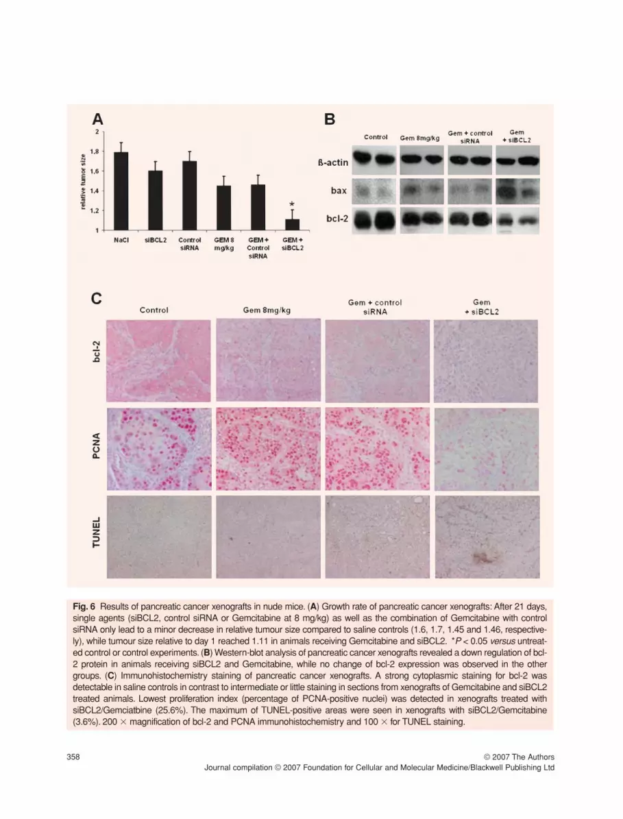
After 21 days, tumours reached a size of 1.8 relativeto day 1 in animals receiving saline only. In animalsreceiving control siRNA, relative tumour size was 1.7,while siBCL2 alone already lead to a reduction intumour size to 1.6. Gemcitabine alone or combinedwith control siRNA lead to a significant tumour growthsuppression compared with saline control (relativetumour sizes are 1.45 and 1.46, P < 0.02 and 0.03,respectively). Tumours of animals receiving
Fig. 3 Expression of the bcl-2 protein determined by Western blotting. (A) siRNA transfection inhibited bcl-2 protein expres-sion of YAP C but not in HF. (B) 1 µM Gemcitabine treatment tended to down-regulate bcl-2 protein expression in YAP C incomparison to 0.01 µM Gemcitabine treated YAPC cell lines or controls. (C) The combination treatments of siBCL2 andGemcitabine lead to a pronounced down-regulation of bcl-2 protein in YAP C but not in HF cells.

356 © 2007 The AuthorsJournal compilation © 2007 Foundation for Cellular and Molecular Medicine/Blackwell Publishing Ltd
Gemcitabine and siBCL2 reached a relative tumoursize of 1.11 (P < 0.04 versus Gemcitabine alone and P < 0.03 versus Gemcitabine and control siRNA;Fig.6A).
These effects were mediated by a down-regulationof bcl-2 protein in animals receiving siBCL2 andGemcitabine, while no change of bcl-2 expressionwas observed in the other groups (Fig. 6B). Thiscombination also lead to increased levels of pro-apoptotic bax, thus shifting the ratio of bax/bcl-2towards pro-apoptosis, whereas this ratio remainedunchanged in the other groups, too.
Immunohistochemistry showed a distinct cytoplas-mic expression of bcl-2 in control animals which wasalmost completely lost in the siBCL2 treated groups andthe intensity of bcl-2 expression significantly decreasedcompared to controls (P < 0.01). Gemcitabine alone orin combination with control siRNA only marginallyreduced bcl-2 immunohistochemistry with no statisticaldifference to controls (Fig. 6C upper panels).
The macroscopically observed growth delay wasalso due to reduced proliferation of tumour cells asdemonstrated by PCNA staining (Fig. 6C middle pan-els). All tumours showed a high proliferation index of49.2% (saline), 56.9% (Gemcitabine alone) and52.9% (control siRNA/Gemcitabine), in contrast to25.6% in the siBCL2/Gemcitabine group. This treat-ment also lead to increased TUNEL positivity, indicat-ing that the diminished tumour size was also due toinduction of cell death (Fig. 6C lower panels). In thesiBCL2/Gemcitabine group, TUNEL-positive areaswere observed in 3.6% of the examined high powerfields compared to 0.8% in saline controls, 1.3% inthe Gemcitabine group and 1.5% in the controlsiRNA/Gemcitabine group, respectively.
Discussion
Gemcitabine, a deoxycytidine analogue, has beenshown to ameliorate disease-related symptoms and toimprove survival time [24], especially in combinationwith other cytotoxic agents (e.g. cisplatin [25] or irinote-can [26]).Yet, the development of an effective treatmentfor pancreatic cancer remains an urgent task. Despiteof detailed knowledge of its molecular pathology, littleprogress has been made regarding overall survival [6].Previously, we have shown that the pancreatic cancercell lines YAP C and DAN G respond to siBCL2 withincreased apoptosis and diminished proliferation invitro and that pancreatic cancer xenografts are suitablefor studying the effect of siBCL2 in vivo [19]. Due to thehigh aggressiveness (early metastases) of DAN G, wenow only investigated YAP C with respect of animalprotection. Furthermore, YAP C represents a moder-ately differentiated pancreatic ductal adenocarcinomawith low aggressiveness in vivo that is still capable of
Fig. 4 Apoptosis rate and numbers of viable cells aftersiRNA transfection. (A) 10 nM siBCL2 lead to a moderateincrease in apoptosis in pancreatic cancer cells and in HFwhile the control siRNA was ineffective in both cell lines. (B)siBCL2 lead to a significant reduction in of viable cells inYAP C, while in HF only a moderate but not significantreduction of cell number was observed. Mock or controlsiRNA transfections did not induce apoptosis or reduce thenumber of viable cells in both cell lines. Values are mean ±S.E.M. of three independent experiments. *P < 0.05 versusmock transfected cells and untreated controls.

J. Cell. Mol. Med. Vol 11, No 2, 2007
357© 2007 The AuthorsJournal compilation © 2007 Foundation for Cellular and Molecular Medicine/Blackwell Publishing Ltd
transdifferentiation [20], rendering YAP C representa-tive for early and still treatable stages of pancreaticcancer in human patients.
Known treatment resistance to Gemcitabine hasbeen correlated with the expression of anti-apoptoticmembers of the bcl-2 protein family in different cancercell lines [27]. It was thus postulated that the effective-ness of these chemotherapeutic regimens could beenhanced by a targeted down-regulation of bcl-2 or bcl-xL [7, 8, 28]. So far, several studies investigated theeffect of conventional DNA antisense oligonucleotideson bcl-2 expression and chemotherapy sensitivity withpromising results [7,14–16]. Due to unspecific effects(e.g. cytokine release), hepatotoxicity and unsatisfacto-ry pharmacokinetic properties (short half-life in vivo,low plasma and tissue concentrations) with the need tointroduce chemical modifications (e.g. locked nucleicacids) [29, 30], we previously applied the recently dis-covered RNA interference (RNAi) pathway to silencethe expression of bcl-2 in human pancreatic cancercells: 10 nM of bcl-2-specific siRNAs inhibited growth ofpancreatic cancer cell lines in vitro and in vivo and lead
to a specific downregulation of bcl-2 mRNA and pro-tein. This lead to the breakdown of the mitochondrialtransmembrane potential ��m and shifted the bax/bcl-2 ratio towards pro-apoptosis [19].
Resistance to the intrinsic, i.e. mitochondrial, path-way of apoptosis induction via up-regulation of anti-apoptotic bcl-2 members has been shown to be themain reason for failure of Gemcitabine treatment inpancreatic cancer [7, 8] and other tumours (e.g.mesothelioma cells [28]). Accordingly, and in additionto our previous findings [19], we have shown here thattransfection of bcl-2-specific siRNAs to pancreatic can-cer cells lowers the apoptotic threshold by decreasinglevels of the corresponding target protein as demon-strated by FACS analysis and Western blotting, shiftingthe bax/bcl-2 ratio towards pro-apoptosis. Co-incuba-tion with low micromolar concentrations ofGemcitabine, which were ineffective when appliedalone, lead to a synergistic increase in apoptosis induc-tion and a decrease in the number of viable cancercells, while non-transformed fibroblasts showed onlyminor responses. At higher concentrations of
Fig. 5 Sensitivity of YAP C pancreatic carcinoma cells and HF to combined siRNA and Gemcitabine treatment. (A) Apoptosisrate of YAP C, but not of HF, significantly increased after addition of 1 or 0.01 µM Gemcitabine 72 hrs after transfection with10 nM siBCL2. In combination with control siRNA, only the high concentration of Gemcitabine leads to significant levels ofapoptosis, indicating the predominant cytotoxic effect of Gemcitabine at this concentration. (B) Addition of 0.01 µMGemcitabine to siBCL2 reduced the number of viable YAP C cells by more than 50%, while HF showed only a minor responseto 0.01 µM Gemcitabine and siBCL2 combination therapy. Combinations with control siRNA and 1 µM Gemcitabine show onlythe Gemcitabine effect but no further reduction in both cell lines.Results are mean ± S.E.M.of three independent experiments.*P < 0.05 versus untreated controls and 0.01 µM Gemcitabine with pre-treatment of mock transfection.
358 © 2007 The AuthorsJournal compilation © 2007 Foundation for Cellular and Molecular Medicine/Blackwell Publishing Ltd
Fig. 6 Results of pancreatic cancer xenografts in nude mice. (A) Growth rate of pancreatic cancer xenografts: After 21 days,single agents (siBCL2, control siRNA or Gemcitabine at 8 mg/kg) as well as the combination of Gemcitabine with controlsiRNA only lead to a minor decrease in relative tumour size compared to saline controls (1.6, 1.7, 1.45 and 1.46, respective-ly), while tumour size relative to day 1 reached 1.11 in animals receiving Gemcitabine and siBCL2. *P < 0.05 versus untreat-ed control or control experiments. (B) Western-blot analysis of pancreatic cancer xenografts revealed a down regulation of bcl-2 protein in animals receiving siBCL2 and Gemcitabine, while no change of bcl-2 expression was observed in the othergroups. (C) Immunohistochemistry staining of pancreatic cancer xenografts. A strong cytoplasmic staining for bcl-2 wasdetectable in saline controls in contrast to intermediate or little staining in sections from xenografts of Gemcitabine and siBCL2treated animals. Lowest proliferation index (percentage of PCNA-positive nuclei) was detected in xenografts treated withsiBCL2/Gemciatbine (25.6%). The maximum of TUNEL-positive areas were seen in xenografts with siBCL2/Gemcitabine(3.6%). 200 � magnification of bcl-2 and PCNA immunohistochemistry and 100 � for TUNEL staining.

J. Cell. Mol. Med. Vol 11, No 2, 2007
359© 2007 The AuthorsJournal compilation © 2007 Foundation for Cellular and Molecular Medicine/Blackwell Publishing Ltd
Gemcitabine (1 µM), siBCL2 did not further increasethe chemotherapeutic efficacy, indicating that especial-ly the very low concentration of 0.01 µM Gemcitabineshould be used for these combination treatments.
1 �M Gemcitabine treatment tended to increasethe bcl-2 mRNA level in YAP C whereas it decreasedbcl-2 protein levels, while 1 µM Gemcitabinedecreased bcl-2 mRNA levels in HF but the changewas not reflected to bcl-2 protein level. Several for-mer reports also have shown the discrepancybetween mRNA and protein levels of bcl-2 in normalor tumour cells [31]. The mechanism has not beenclearly elucidated, Ikeguchi et al. reported that cis-platin treatment significantly increased the expres-sion levels of bcl-2 mRNA in PANC-1 cells [32] andBold et al. suggested that the exposure to a cytotox-ic agent induces bcl-2 levels in pancreas carcinomacells [8]. On the other hand, some reports reveal thepost-transcriptional downregulation of bcl-2 [33]. Forexample, 3’-untranslated region of bcl-2 mRNAwhich contains cis-acting AU-rich elements (ARE)and ARE-binding proteins regulate mRNA stability[34] and Bandyopadhyay et al. showed thatchemotherapeutic agents reduced bcl-2 steady-statelevel through inactivation of ARE-binding proteins[35]. Several reports supported the possibility of thepoor correlation between protein and RNA in eukary-otic systems, where up to 20-fold changes in proteinlevels can be seen without corresponding alterations inmRNA abundance, and up to 30-fold changes in mRNAlevels without reflection on protein levels [36–38].
Corroborating our in vitro results, our in vivoresults show that the combination treatment ofGemcitabine and bcl-2 specific siRNA gives a cleartherapeutic gain over pancreas cancer xenografts.This combination treatment abrogated bcl-2 in pan-creas cancer tissues, reduced proliferation of tumourcells and increased the apoptotic index. This resultsuggests that suppression of bcl-2 by correspondingsiRNA is associated with sensitization of pancreaticcancer to apoptotic cell death in vivo. It is also worthmentioning that this increase of the anti-tumour effectwas not accompanied by an increase of toxicity,since the lack of additive toxicity is an important fac-tor in clinical therapy for advanced cancer patients.The in vivo application of unmodified siRNA still rep-resents an urgent problem for clinical application.Several papers use a hydrodynamic tail vein injectionin mice, which was not used here due to animal pro-tection laws. We have previously established themethod of intraperitoneal application of siRNA dis-solved in RNAse free sterile physiological saline
solution which seems to provide a suitable vehicle forapplying siRNA in vitro [19], especially to different tis-sues of the gastrointestinal tract, while the common-ly used phosphate buffered saline seems to offerunfavourable electrochemical conditions for siRNA.
In summary, our results indicate that silencing anti-apoptotic bcl-2 by siRNA specifically sensitizes humanpancreatic cancer cells to Gemcitabine treatment atlow, otherwise ineffective, concentrations. This treat-ment strategy might contribute to the therapy of other-wise Gemcitabine-resistant pancreatic cancers in vivoand might lower that rate of unwanted chemotherapyeffects in patients with advanced diseases.
Acknowledgements
We are indebted to Gabriele Krumholz for support in ani-mal care and animal experiments, and to Andrea Hartl andSandra Leitner for excellent technical assistance.
This work was supported by grants from the GermanCancer Aid (10-2112-Oc1), the FUTUR-Programme of theState of Bavaria (Project No. 263) and the ELAN-Programme of the Faculty of Medicine, University ofErlangen-Nuernberg (Project 02.08.08.2).
The authors declare no conflict of interest.
References
1. Keighley MR. Gastrointestinal cancers in Europe.Aliment Pharmacol Ther. 2003; 18: 7–30.
2. DiMagno EP, Reber HA,Tempero MA. AGA technicalreview on the epidemiology, diagnosis, and treatmentof pancreatic ductal adenocarcinoma. AmericanGastroenterological Association. Gastroenterology.1999; 117: 1464–84.
3. Simon B, Printz H. Epidemiological trends in pancre-atic neoplasias. Dig Dis. 2001; 19: 6–14.
4. Raraty MG, Magee CJ, Ghaneh P, Neoptolemos JP.New techniques and agents in the adjuvant therapy ofpancreatic cancer. Acta Oncol. 2002; 41: 582–95.
5. Nitecki SS, Sarr MG, Colby TV, van Heerden JA.Long-term survival after resection for ductal adenocar-cinoma of the pancreas. Is it really improving? AnnSurg. 1995; 221: 59–66.
6. Li D, Xie K, Wolff R, Abbruzzese JL. Pancreatic can-cer. Lancet. 2004; 363: 1049–57.
7. Schniewind B, Christgen M, Kurdow R, Haye S,Kremer B, Kalthoff H, Ungefroren H. Resistance ofpancreatic cancer to gemcitabine treatment is depend-ent on mitochondria-mediated apoptosis. Int J Cancer.2004; 109: 182–8.

360 © 2007 The AuthorsJournal compilation © 2007 Foundation for Cellular and Molecular Medicine/Blackwell Publishing Ltd
8. Bold RJ, Chandra J, McConkey DJ. Gemcitabine-induced programmed cell death (apoptosis) of humanpancreatic carcinoma is determined by Bcl-2 content.Ann Surg Oncol. 1999; 6: 279–85.
9. Elbashir SM, Harborth J, Lendeckel W, Yalcin A,Weber K, Tuschl T. Duplexes of 21-nucleotide RNAsmediate RNA interference in cultured mammaliancells. Nature. 2001; 411: 494–8.
10. Elbashir SM, Lendeckel W, Tuschl T. RNA interfer-ence is mediated by 21- and 22-nucleotide RNAs.Genes Dev. 2001; 15: 188–200.
11. Zamore PD, Tuschl T, Sharp PA, Bartel DP. RNAi:double-stranded RNA directs the ATP-dependentcleavage of mRNA at 21 to 23 nucleotide intervals.Cell. 2000; 101: 25–33.
12. Hannon GJ. RNA interference. Nature. 2002; 418:244–51.
13. Martinez J, Patkaniowska A, Urlaub H, LuhrmannR, Tuschl T. Single-stranded antisense siRNAs guidetarget RNA cleavage in RNAi. Cell. 2002; 110: 563–74.
14. Wacheck V, Heere-Ress E, Halaschek-Wiener J,Lucas T, Meyer H, Eichler HG, Jansen B. Bcl-2 anti-sense oligonucleotides chemosensitize human gastriccancer in a SCID mouse xenotransplantation model. JMol Med. 2001; 79: 587–93.
15. Jansen B, Wacheck V, Heere-Ress E, Schlagbauer-Wadl H, Hoeller C, Lucas T, Hoermann M, HollensteinU, Wolff K, Pehamberger H. Chemosensitisation ofmalignant melanoma by BCL2 antisense therapy. Lancet.2000; 356: 1728–33.
16. Xu Z, Friess H, Solioz M, Aebi S, Korc M, Kleeff J,Buchler MW. Bcl-x(L) antisense oligonucleotidesinduce apoptosis and increase sensitivity of pancreat-ic cancer cells to gemcitabine. Int J Cancer. 2001; 94:268–74.
17. Campani D, Esposito I, Boggi U, Cecchetti D,Menicagli M, De Negri F, Colizzi L, Del Chiaro M,Mosca F, Fornaciari G, Bevilacqua G. Bcl-2 expres-sion in pancreas development and pancreatic cancerprogression. J Pathol. 2001; 194: 444–50.
18. Miyamoto Y, Hosotani R, Wada M, Lee JU, KoshibaT, Fujimoto K, Tsuji S, Nakajima S, Doi R, Kato M,Shimada Y, Imamura M. Immunohistochemical analy-sis of Bcl-2, Bax, Bcl-X, and Mcl-1 expression in pan-creatic cancers. Oncology. 1999; 56: 73–82.
19. Ocker M, Neureiter D, Lueders M, Zopf S,Ganslmayer M, Hahn EG, Herold C, Schuppan D.Variants of bcl-2 specific siRNA for silencing antiapoptot-ic bcl-2 in pancreatic cancer. Gut. 2005; 54: 1298–308.
20. Neureiter D, Zopf S, Dimmler A, Stintzing S, HahnEG, Kirchner T, Herold C, Ocker M. Different capabil-ities of morphological pattern formation and its associ-ation with the expression of differentiation markers in a
xenograft model of human pancreatic cancer lines.Pancreatology. 2005; 5: 387–97.
21. Ocker M, Herold C, Ganslmayer M, Hahn EG,Schuppan D. The synthetic retinoid adapalene inhibitsproliferation and induces apoptosis in colorectal can-cer cells in vitro. Int J Cancer. 2003; 107: 453–9.
22. Sun J, Blaskovich MA, Knowles D, Qian Y, OhkandaJ, Bailey RD, Hamilton AD, Sebti SM. Antitumor effi-cacy of a novel class of non-thiol-containing pep-tidomimetic inhibitors of farnesyltransferase and ger-anylgeranyltransferase I: combination therapy with thecytotoxic agents cisplatin, Taxol, and gemcitabine.Cancer Res. 1999; 59: 4919–26.
23. Sasaki M, Kumazaki T, Takano H, Nishiyama M,Mitsui Y. Senescent cells are resistant to death despitelow Bcl-2 level. Mech Ageing Dev. 2001; 122: 1695–706.
24. Burris HA, III, Moore MJ, Andersen J, Green MR,Rothenberg ML, Modiano MR, Cripps MC, PortenoyRK, Storniolo AM, Tarassoff P, Nelson R, Dorr FA,Stephens CD, Von Hott DD. Improvements in survivaland clinical benefit with gemcitabine as first-line therapyfor patients with advanced pancreas cancer: a random-ized trial. J Clin Oncol. 1997; 15: 2403–13.
25. Colucci G, Giuliani F, Gebbia V, Biglietto M, RabittiP, Uomo G, Cigolari S, Testa A, Maiello E, Lopez M.Gemcitabine alone or with cisplatin for the treatment ofpatients with locally advanced and/or metastatic pan-creatic carcinoma: a prospective, randomized phase IIIstudy of the Gruppo Oncologia dell’Italia Meridionale.Cancer. 2002; 94: 902–10.
26. Rocha Lima CM, Savarese D, Bruckner H, Dudek A,Eckardt J, Hainsworth J,Yunus F, Lester E, Miller W,Saville W, Elfring GL, Locker PK, Compton LD,Miller LL, Green MR. Irinotecan plus gemcitabineinduces both radiographic and CA 19-9 tumor markerresponses in patients with previously untreatedadvanced pancreatic cancer. J Clin Oncol. 2002; 20:1182–91.
27. Bergman AM, Pinedo HM, Talianidis I, Veerman G,Loves WJ, van der Wilt CL, Peters GJ. Increased sen-sitivity to gemcitabine of P-glycoprotein and multidrugresistance-associated protein-overexpressing humancancer cell lines. Br J Cancer. 2003; 88: 1963–70.
28. Hopkins-Donaldson S, Cathomas R, Simoes-WustAP, Kurtz S, Belyanskaya L, Stahel RA, Zangemeister-Wittke U. Induction of apoptosis and chemosensitizationof mesothelioma cells by Bcl-2 and Bcl-xL antisensetreatment. Int J Cancer. 2003; 106: 160–6.
29. Lebedeva I, Stein CA. Antisense oligonucleotides:promise and reality. Annu Rev Pharmacol Toxicol.2001; 41: 403–19.
30. Stahel RA, Zangemeister-Wittke U. Antisenseoligonucleotides for cancer therapy-an overview. LungCancer. 2003; 41: S81–8.

J. Cell. Mol. Med. Vol 11, No 2, 2007
361© 2007 The AuthorsJournal compilation © 2007 Foundation for Cellular and Molecular Medicine/Blackwell Publishing Ltd
31. Chleq-Deschamps CM, LeBrun DP, Huie P, BesnierDP,Warnke RA, Sibley RK, Cleary ML. Topographicaldissociation of BCL-2 messenger RNA and proteinexpression in human lymphoid tissues. Blood. 1993;81: 293–8.
32. Ikeguchi M, Nakamura S, Kaibara N. Quantitativeanalysis of expression levels of bax, bcl-2, and survivinin cancer cells during cisplatin treatment. Oncol Rep.2002; 9: 1121–6.
33. Berney CR, Downing SR, Yang JL, Russell PJ, Crowe PJ. Evidence for post-transcriptional down-regulation of the apoptosis-related gene bcl-2 in human colorectal cancer. J Pathol. 2000; 191:15–20.
34. Donnini M, Lapucci A, Papucci L, Witort E,Tempestini A, Brewer G, Bevilacqua A, Nicolin A,Capaccioli S, Schiavone N. Apoptosis is associated
with modifications of bcl-2 mRNA AU-binding proteins.Biochem Biophys Res Commun. 2001; 287: 1063–9.
35. Bandyopadhyay S, Sengupta TK, Fernandes DJ,Spicer EK. Taxol- and okadaic acid-induced destabi-lization of bcl-2 mRNA is associated with decreasedbinding of proteins to a bcl-2 instability element.Biochem Pharmacol. 2003; 66: 1151–62.
36. Fiorentino M, D’Errico A, Barozzi C, Grigioni WF.Discrepancies between detection of Bcl-2 by in situhybridization and immunocytochemistry in humanprostate cancer tissues. Int J Cancer. 1998; 79: 614–8.
37. Rajasekhar VK, Holland EC. Postgenomic globalanalysis of translational control induced by oncogenicsignaling. Oncogene. 2004; 23: 3248–64.
38. Kleijn M, Scheper GC, Voorma HO, Thomas AA.Regulation of translation initiation factors by signaltransduction. Eur J Biochem. 1998; 253: 531–44.


