Embed Size (px)
Citation preview

Ž .Brain Research Reviews 30 1999 27–51www.elsevier.comrlocaterbres
Full-length review
Tapping into spinal circuits to restore motor function
Hugues Barbeau a, David A. McCrea b, Michael J. O’Donovan c, Serge Rossignol d,1,Warren M. Grill e, Michel A. Lemay f
a School of Physical and Occupational Therapy, McGill UniÕersity, Montreal, QC, Canada H3G 1Y5b Department of Physiology, Spinal Cord Research Centre, UniÕersity of Manitoba, Winnipeg, MB, Canada R3E 3J7c Section on DeÕelopmental Neurobiology, Laboratory of Neural Control, NINDS, NIH, Bethesda, MD 20892, USA
d Centre de Recherche en Sciences Neurologiques, UniÕersite de Montreal, Montreal, QC, Canada H3T 1J4´ ´ ´e Department of Biomedical Engineering, Case Western ReserÕe UniÕersity, CleÕeland, OH 44106-4912, USA
f Applied Neural Control Laboratory, Case Western ReserÕe UniÕersity, CleÕeland, OH 44106-4912, USA
Accepted 27 April 1999
Abstract
Motivated by the challenge of improving neuroprosthetic devices, the authors review current knowledge relating to harnessing thepotential of spinal neural circuits, such as reflexes and pattern generators. If such spinal interneuronal circuits could be activated, theycould provide the coordinated control of many muscles that is so complex to implement with a device that aims to address eachparticipating muscle individually. The authors’ goal is to identify candidate spinal circuits and areas of research that might openopportunities to effect control of human limbs through electrical activation of such circuits. David McCrea’s discussion of the ways inwhich hindlimb reflexes in the cat modify motor activity may help in developing optimal strategies for functional neuromuscular
Ž .stimulation FNS , by using knowledge of how reflex actions can adapt to different conditions. Michael O’Donovan’s discussion of thedevelopment of rhythmogenic networks in the chick embryo may provide clues to methods of generating rhythmic activity in the adultspinal cord. Serge Rossignol examines the spinal pattern generator for locomotion in cats, its trigger mechanisms, modulation andadaptation, and suggests how this knowledge can help guide therapeutic approaches in humans. Hugues Barbeau applies the work of
Ž .Rossignol and others to locomotor training in human subjects who have suffered spinal cord injury SCI with incomplete motor functionŽ .loss IMFL . Michel Lemay and Warren Grill discuss some of the technical challenges that must be addressed by engineers to implement
a neuroprosthesis using electrical stimulation of the spinal cord, particularly the control issues that would have to be resolved. q 1999Elsevier Science B.V. All rights reserved.
Keywords: Locomotion; Central pattern generator; Spinal cord injury; Functional neuromuscular stimulation; FNS; FES
Contents
1. Introduction . . . . . . . . . . . . . . . . . . . . . . . . . . . . . . . . . . . . . . . . . . . . . . . . . . . . . . . . . . . . . . . . . . . . . . . . 28
Ž . Ž .2. Can use be made of spinal reflex circuits following spinal cord injury SCI ? David McCrea . . . . . . . . . . . . . . . . . . . . . . . . . . . . 292.1. Monosynaptic excitation by primary muscle spindle afferents . . . . . . . . . . . . . . . . . . . . . . . . . . . . . . . . . . . . . . . . . . . 292.2. Cutaneous reflexes. . . . . . . . . . . . . . . . . . . . . . . . . . . . . . . . . . . . . . . . . . . . . . . . . . . . . . . . . . . . . . . . . . 292.3. Reflexes evoked from spindle secondaries . . . . . . . . . . . . . . . . . . . . . . . . . . . . . . . . . . . . . . . . . . . . . . . . . . . . . 292.4. Reflexes evoked from tendon organs; non-reciprocal group I reflexes. . . . . . . . . . . . . . . . . . . . . . . . . . . . . . . . . . . . . . . 292.5. Reconfiguration of group I reflexes . . . . . . . . . . . . . . . . . . . . . . . . . . . . . . . . . . . . . . . . . . . . . . . . . . . . . . . . . 302.6. Spinal pathways responsible for extension enhancement . . . . . . . . . . . . . . . . . . . . . . . . . . . . . . . . . . . . . . . . . . . . . . 302.7. Summary: altered non-reciprocal group I pathways . . . . . . . . . . . . . . . . . . . . . . . . . . . . . . . . . . . . . . . . . . . . . . . . 322.8. Reconfiguration of motoneuron properties during motor tasks . . . . . . . . . . . . . . . . . . . . . . . . . . . . . . . . . . . . . . . . . . . 322.9. Voltage-dependent conductances during locomotion . . . . . . . . . . . . . . . . . . . . . . . . . . . . . . . . . . . . . . . . . . . . . . . . 322.10. Motoneuron threshold and repetitive firing during locomotion . . . . . . . . . . . . . . . . . . . . . . . . . . . . . . . . . . . . . . . . . . 322.11. Summary . . . . . . . . . . . . . . . . . . . . . . . . . . . . . . . . . . . . . . . . . . . . . . . . . . . . . . . . . . . . . . . . . . . . . . 33
1 To whom inquiries should be addressed.
0165-0173r99r$ - see front matter q 1999 Elsevier Science B.V. All rights reserved.Ž .PII: S0165-0173 99 00008-9

( )H. Barbeau et al.rBrain Research ReÕiews 30 1999 27–5128
Ž .3. Are embryonic motor activity and adult locomotion produced by similar mechanisms? Rhythmic activity in developing spinal cordŽ .Michael J. O’Donovan . . . . . . . . . . . . . . . . . . . . . . . . . . . . . . . . . . . . . . . . . . . . . . . . . . . . . . . . . . . . . . . . . 333.1. Timing of motoneuron discharge during bursts of motor activity . . . . . . . . . . . . . . . . . . . . . . . . . . . . . . . . . . . . . . . . . 343.2. What accounts for the occurrence of spontaneous episodes of activity? . . . . . . . . . . . . . . . . . . . . . . . . . . . . . . . . . . . . . . 343.3. What is the mechanism of rhythmicity? . . . . . . . . . . . . . . . . . . . . . . . . . . . . . . . . . . . . . . . . . . . . . . . . . . . . . . 353.4. Comparison between the genesis of locomotion and embryonic motor activity . . . . . . . . . . . . . . . . . . . . . . . . . . . . . . . . . . 363.5. Conclusion . . . . . . . . . . . . . . . . . . . . . . . . . . . . . . . . . . . . . . . . . . . . . . . . . . . . . . . . . . . . . . . . . . . . . . 36
Ž .4. Triggering, modulating and adapting the spinal pattern generator for locomotion in cats Serge Rossignol . . . . . . . . . . . . . . . . . . . . . 364.1. The central pattern generator . . . . . . . . . . . . . . . . . . . . . . . . . . . . . . . . . . . . . . . . . . . . . . . . . . . . . . . . . . . . 374.2. Triggering the CPG: supraspinal initiation of locomotion . . . . . . . . . . . . . . . . . . . . . . . . . . . . . . . . . . . . . . . . . . . . . 374.3. Pharmacologic triggering of locomotion . . . . . . . . . . . . . . . . . . . . . . . . . . . . . . . . . . . . . . . . . . . . . . . . . . . . . . 374.4. Sensory evoked locomotion . . . . . . . . . . . . . . . . . . . . . . . . . . . . . . . . . . . . . . . . . . . . . . . . . . . . . . . . . . . . . 394.5. Modulating the CPG: supraspinal stimulation . . . . . . . . . . . . . . . . . . . . . . . . . . . . . . . . . . . . . . . . . . . . . . . . . . . 404.6. Pharmacologic modulation . . . . . . . . . . . . . . . . . . . . . . . . . . . . . . . . . . . . . . . . . . . . . . . . . . . . . . . . . . . . . 404.7. Sensory modulation . . . . . . . . . . . . . . . . . . . . . . . . . . . . . . . . . . . . . . . . . . . . . . . . . . . . . . . . . . . . . . . . . 404.8. Adapting the CPG . . . . . . . . . . . . . . . . . . . . . . . . . . . . . . . . . . . . . . . . . . . . . . . . . . . . . . . . . . . . . . . . . . 414.9. Locomotor training . . . . . . . . . . . . . . . . . . . . . . . . . . . . . . . . . . . . . . . . . . . . . . . . . . . . . . . . . . . . . . . . . 414.10. Adaptation to lesions . . . . . . . . . . . . . . . . . . . . . . . . . . . . . . . . . . . . . . . . . . . . . . . . . . . . . . . . . . . . . . . . 414.11. Conclusions . . . . . . . . . . . . . . . . . . . . . . . . . . . . . . . . . . . . . . . . . . . . . . . . . . . . . . . . . . . . . . . . . . . . 41
Ž .5. Can locomotor training enhance the recovery of walking following SCI? Hugues Barbeau . . . . . . . . . . . . . . . . . . . . . . . . . . . . . 415.1. Locomotor training approaches . . . . . . . . . . . . . . . . . . . . . . . . . . . . . . . . . . . . . . . . . . . . . . . . . . . . . . . . . . . 425.2. Pharmacologic approaches to enhance locomotion in people with SCI-IMFL. . . . . . . . . . . . . . . . . . . . . . . . . . . . . . . . . . . 435.3. Locomotor training combined with pharmacologic approaches . . . . . . . . . . . . . . . . . . . . . . . . . . . . . . . . . . . . . . . . . . 435.4. Conclusion . . . . . . . . . . . . . . . . . . . . . . . . . . . . . . . . . . . . . . . . . . . . . . . . . . . . . . . . . . . . . . . . . . . . . . 44
Ž .6. Control issues in using spinal circuits to restore functional movement Michel Lemay . . . . . . . . . . . . . . . . . . . . . . . . . . . . . . . . 446.1. Cyclic vs. static movement . . . . . . . . . . . . . . . . . . . . . . . . . . . . . . . . . . . . . . . . . . . . . . . . . . . . . . . . . . . . . 446.2. Duration of activation . . . . . . . . . . . . . . . . . . . . . . . . . . . . . . . . . . . . . . . . . . . . . . . . . . . . . . . . . . . . . . . . 446.3. Modification of the response during motion . . . . . . . . . . . . . . . . . . . . . . . . . . . . . . . . . . . . . . . . . . . . . . . . . . . . 44
Ž .7. Technical challenges to using spinal circuitry in neural prostheses Warren Grill . . . . . . . . . . . . . . . . . . . . . . . . . . . . . . . . . . . 45
Acknowledgements. . . . . . . . . . . . . . . . . . . . . . . . . . . . . . . . . . . . . . . . . . . . . . . . . . . . . . . . . . . . . . . . . . . . . . 46
References . . . . . . . . . . . . . . . . . . . . . . . . . . . . . . . . . . . . . . . . . . . . . . . . . . . . . . . . . . . . . . . . . . . . . . . . . . 46
1. Introduction
During the past 30 years, researchers have developedmotor prostheses to restore functional movement to para-lyzed limbs by electrically activating skeletal muscle. Thesedevices typically require one electrode, one lead connec-tion between the electrode and the stimulator, and onechannel of a multichannel stimulator for each muscle, andcomplex algorithms to coordinate the excitation of multi-ple muscles for each stereotypical motion. Implementationof these neuroprosthetic systems has been successful andhas created a demand for the restoration of even morefunction. However, more function requires activating moremuscles, and thus, an additional electrode, lead and stimu-lator channel for each added muscle. If ways could befound to harness the potential of spinal neural circuits,such as reflexes and pattern generators, coordinated con-tractions of many muscles could be initiated with far lesshardware and far less surgery to implant it than presentsystems require.
Over the same 30-year span, researchers from the basicscience community have learned a great deal about in-terneuronal circuits in the spinal cord. When activated,these circuits provide stereotyped movements that employcoordinated control of many muscles. Some of thesemovements are executed only once in response to a singlestimulus, and effect novel movements, while others, thatemploy pattern generators, provide rhythmic coordinatedcontractions. Researchers have investigated these move-ments, studied their properties for a wide range of externalstimuli and are beginning to map the anatomical connec-tions of many spinal circuits. Some researchers have alsobegun to think about how such neural circuits might beused in humans and some have even carried out experi-ments on humans with paralysis.
In an effort to share the accumulated knowledge andmotivated by the need to improve the performance ofmotor prostheses, the authors have addressed the problemof gaining access to spinal circuits. Their goal is to identifycandidate spinal circuits and areas of research that might

( )H. Barbeau et al.rBrain Research ReÕiews 30 1999 27–51 29
open opportunities to effect control of human limbs throughelectrical activation of such circuits.
2. Can use be made of spinal reflex circuits following( ) ( )spinal cord injury SCI ? David McCrea
We will examine features of some spinal reflex systemsthat may be useful for movements assisted by functional
Ž .neuromuscular stimulation FNS . The focus will be onrecent observations of cat hindlimb reflexes elicited duringlocomotor activity. While it is obvious that motor activityresults from the combined actions of descending pathways,intrinsic spinal cord circuitry and segmental reflexes, theway in which reflex systems exert their regulatory controlis only beginning to be appreciated. Although it has longbeen recognized that reflex gain is controlled by suchthings as motoneuron excitability and afferent fiber trans-mitter release, reflexes are often incorrectly assumed to be‘‘fixed function’’ modules in which their actions simplyadd to or subtract from centrally generated movements.Rather, spinal reflex systems modify motor activity bothby direct actions on motoneurons and indirectly throughthe interneurons that form the spinal pattern generating
Ž .circuitry for locomotor and other movements. Spinalinterneurons that mediate reflexes are also the targets ofdescending motor control systems. Descending regulationof the excitability of reflexes, according to the currentmotor state, could switch from inhibitory to excitatory
w xreflexes 131 . The result is a flexible control system inwhich not only the gain, but the sign and distribution ofreflex actions can be modified to adapt motor output tochanging conditions. An understanding of how reflex sys-tems function under different conditions is, therefore, es-sential to optimizing FNS strategies. We begin with a briefsurvey of the classical spinal reflexes and outline someproperties for consideration in FNS assisted movements.
2.1. Monosynaptic excitation by primary muscle spindleafferents
The most thoroughly studied spinal reflex is themonosynaptic excitation of homonymous and close syner-gist motoneurons with a disynaptic inhibition of antagonist
Ž .motoneurons evoked by activation of group Ia primarymuscle spindle afferents. Group Ia afferents can be acti-vated by low strength electrical stimulation, and are natu-rally activated by muscle length increases or activity in thegamma motoneuron system that regulates spindle stretch
w xsensitivity 117 . Although these features seem ideal foruse in an FNS strategy, the relatively low gain and the
w xnarrow distribution of excitatory and inhibitory effects 64limit the use of this reflex system for FNS. Furthermore,the concomitant inhibition of antagonists during activationof Ia fibers has the potential to interfere with posturalstability.
2.2. Cutaneous reflexes
Cutaneous reflexes involve several interneuron systemsand complex spinal pathways. This results in a pattern ofreflex actions that is state-dependent, changing in differentmotor tasks and under different conditions, for example,
w xduring locomotion 165 . Without detailed knowledge ofthe factors that can modify the operation of cutaneousreflex pathways, cutaneous FNS is limited to well-con-trolled conditions. Another problem in the use of cuta-neous FNS is that electrical activation of cutaneous affer-ents usually results in weak reflex effects unless repetitivestimulation is used. In patients with intact spinal sensorysystems, this may be painful, limiting the applicability ofcutaneous FNS. Another consideration is that cutaneous
Žreflexes are exerted both locally i.e., to muscles innervat-.ing surrounding areas and extensively throughout the
limb. Under some conditions, cutaneous stimulation mayw xelicit a flexor withdrawal response 65 . Such a general
excitation of flexors may destabilize posture. On the otherhand, there may be certain circumstances in which theactivation of specialized, local cutaneous reflex pathwayscould be useful for FNS. For example, the activity ofcutaneous afferents might be useful to sense foot contactw x104 or cutaneous stimulation could be used to reinforce
w xmuscle activity at particular times during a motor task 74 .
2.3. Reflexes eÕoked from spindle secondaries
Ž .Reflexes evoked from the group II secondary musclespindle afferents remain the least understood lumbar spinalreflex system. Early discussion of their actions noted thesimilarity of effects evoked from group II and cutaneous
w xafferents, i.e., both evoked flexion reflexes 65 . Interneu-rons contacted by group II afferents also have other reflexactions including disynaptic excitation or inhibition of
w xmotoneurons 110 and relays between descending motorw xcommands and motoneurons 129 . Recent studies suggest
that an increase in the gain of group II reflexes is aw xcomponent of human spasticity 67 . This observation is
part of the growing evidence that group II reflex systemsform an essential part of the continuous regulation ofmovements. The ability of group II afferents to evoke theflexion reflex is likely to be of less importance. At present,however, the difficulty in selective activation of group IIafferents and our rudimentary knowledge of their role inproprioception complicates the development of FNS strate-gies targeted to group II afferents. As mentioned, thestate-dependent ability of group II and high thresholdcutaneous afferents to evoke strong limb flexion mayfurther limit the use of these reflex systems when the goalis to activate anti-gravity muscles.
2.4. Reflexes eÕoked from tendon organs; non-reciprocalgroup I reflexes
The final reflex system to be considered here is thatŽ .evoked from Ib tendon organ muscle afferents. This

( )H. Barbeau et al.rBrain Research ReÕiews 30 1999 27–5130
system serves as a good example of multi-sensory conver-gence in spinal reflex systems since low threshold jointand cutaneous afferents, and Ia spindle afferents also acti-
w xvate those reflexes originally ascribed to Ib afferents 109 .In this paper, we will refer to these reflex systems as‘‘group I non-reciprocal’’ reflexes to distinguish themfrom the Ia reciprocal inhibition of antagonists and toemphasize the combined input from group Ia and Ibafferents to the interneurons interposed in the spinal reflexpathways. Non-reciprocal group I reflexes evoked fromankle extensor afferents have received the most attentionbecause, at least in the cat, they are the most widelydistributed. In preparations that are not engaged in an
Žorganized motor program i.e., decerebrate, anaesthetized,.or ‘‘resting’’ preparations , group I strength stimulation
Ždefined as stimulation at intensities twice that recruiting.the lowest threshold fibers in the nerve of ankle extensor
nerves produces mixtures of di- or tri-synaptic latencyreflexes that are predominately inhibitory to ankle, kneeand hip extensor motoneurons and excitatory to bifunc-tional flexors such as posterior biceps. The inhibitoryactions are unlikely to be of great use for FNS designed toincrease weight support by extensor motoneuron recruit-ment. An important recent observation, therefore, is thatnon-reciprocal reflexes are reconfigured during locomotioninto a system that excites extensor motoneurons. As willbe discussed, these actions are a part of a natural strategyfor motor control and appear to be an ideal candidate foruse in FNS.
2.5. Reconfiguration of group I reflexes
Until recently most reflex studies were carried out inthe absence of locomotion where non-reciprocal reflexesevoked from ankle extensor groups Ia and Ib afferents are
w xpredominately inhibitory to extensors 109 . The domi-nance of these actions was the basis for discussing non-re-ciprocal group I reflexes as being organized into a negativefeedback system. Accordingly, muscle activity duringmovements would activate group I afferents which in turn
w xwould evoke a reflex inhibition of extensors 109 . Wenow appreciate that during locomotion, a reorganization ofthese reflexes results in a system that is excitatory toextensors. These excitatory actions can result in a termina-tion of ongoing flexor activity and an initiation of extensoractivity, if extensor group I afferents are activated during
Žthe flexor phase of the step cycle i.e., a resetting of the. w xstep cycle to extension 52,79,88 . More importantly for
the present discussion is the fact that activation of ankleextensor group Ia or Ib afferents during extension in-
w xcreases ongoing extensor activity 52,88,155 . Stimulationof ankle extensor group I afferents results in larger andlonger bursts of activity in ipsilateral hip, knee and ankle
w xextensors 88 .The high sensitivity of groups Ia and Ib afferents to
muscle stretch and tendon tension respectively results in
their rhythmic activity during even low intensity locomo-w xtion on a flat surface 159 . This activity provides the
opportunity for reflexes evoked by these afferents to inter-act with the central locomotor pattern generating circuitryto provide step to step regulation even during normal,unperturbed locomotion. Support for the idea that group Iafferents are an important part of normal step cycle regula-tion comes from a number of studies and in particular
w x w xthose from Hiebert et al. 99 , Pearson 156 , Whelan et al.w x w x198 , and Whelan and Pearson 199 in decerebrate or
Ž .conscious cats also see Rossignol’s section of this paper .In the absence of ankle extensor group I afferent feedback,there is inadequate extensor activity and an increased yield
w xat the knee during treadmill walking 199 . This impliesthat a significant amount of extensor motoneuron driveduring locomotion originates from excitatory group I re-flexes. The term ‘extension enhancement’ has been intro-duced to describe the reflex excitation of extensors pro-
w xduced by group I afferents during locomotion 88 .The ability of ankle extensor group I afferents from one
hindlimb to control stepping behavior depends upon thepreparation and the locomotor task. The largest effects areobserved during fictive locomotion in decerebrate catsw w xxe.g., Ref. 88 . Not surprisingly, with more of the neu-raxis intact, the ability of one source of afferent feedbackto control the entire stepping behavior is diminished. Inconscious animals, stimulation of ankle extensor afferentsis less effective than in decerebrate preparations and alsoless effective during quadrupedal than during bipedal loco-
w xmotion 199 . Evidence suggests, however, that the samecircuitry expressed during locomotion in decerebrate catsis also present in the adult human. Thus, training tore-establish locomotor capabilities following spinal cordlesions is significantly enhanced by body support thatpermits limb loading and presumably an activation of load
Ž . w x Žreceptors i.e., group Ib tendon organ afferents 94 see.Barbeau’s section of this paper . This is an important
observation since it suggests that manipulations such asFNS that are marginally effective in normal subjects, maybe very effective in promoting weight support and locomo-tion following disruption of descending control of thespinal cord.
2.6. Spinal pathways responsible for extension enhance-ment
Animal studies have revealed at least four reflex mecha-nisms contributing to group I evoked enhancement ofipsilateral extensor activity during locomotion. The first isthe monosynaptic reflex described above. While activationof muscle spindles with stretch will monosynaptically ex-cite motoneurons, the limited distribution of this excitationw x64 restricts its contribution to extension enhancement tohomonymous and close synergist motoneurons. Monosy-naptic excitation from ankle extensor afferents is thusunlikely to contribute significantly to the excitation of hip,

( )H. Barbeau et al.rBrain Research ReÕiews 30 1999 27–51 31
w xknee and ankle extensors observed during locomotion 88 .The other mechanisms underlying extension enhancementresult from the reorganization of group I reflex pathwaysduring locomotion. They are: a suppression of the group Ievoked non-reciprocal inhibition that occurs at rest; anactivation of excitatory interneurons that receive inputfrom group I afferents and project directly to extensormotoneurons; and a longer latency group I excitation thatis evoked through extensor portions of the locomotorcircuitry. Fig. 1 summarizes the changes in group I re-flexes to extensor motoneurons during locomotion.
Intracellular studies show that the non-reciprocal, in-Ž .hibitory post-synaptic potentials IPSP produced by exten-
sor nerve stimulation at rest disappear during locomotionw x79,133 . The disappearance of non-reciprocal inhibition iscaused by an unknown mechanism that tonically inhibitsthe interneurons responsible for evoking non-reciprocal
w xinhibition 13,133 . In addition to the inhibition of in-terneurons in non-reciprocal inhibitory pathways, two typesof group I evoked extensor motoneuron excitation emergeduring locomotion. The first is a very short latency excita-
Ž .tion disynaptic, 1.6 ms latency evoked through a pathwayconsisting of a single interneuron between group I affer-ents and extensor motoneurons. Such excitation was origi-nally reported during fictive locomotion induced by L-
w xDOPA in decerebrate, high spinal cats 177 . Disynapticexcitation has been more extensively characterized re-cently during fictive locomotion evoked by midbrain stim-
ulation. These disynaptic excitatory post-synaptic poten-Ž .tials EPSP are evoked from either Ia muscle spindle or Ib
tendon organ afferents and only during the extensor phasew xof locomotion 14,133 . They are evoked most readily
from ankle extensor group I afferents which produce exci-tation of ankle, knee and hip extensor motoneurons. Stimu-lation of hip and knee extensor afferents evokes a more
w xlimited distribution of disynaptic excitation 14 .A population of interneurons has been found in caudal
lumbar segments that are candidates for mediation ofw xdisynaptic extensor excitation during locomotion 13 .
These cells project to motoneurons and are monosynapti-cally activated by group I afferents during the extensionphase but not at rest. They are also rhythmically activeduring extension in the absence of peripheral nerve stimu-lation. No attempt has been made yet to verify that thecandidate interneurons are excitatory. If so, their rhythmiclocomotor activity would contribute to the depolarizationof extensor motoneurons as part of the central locomotornetwork as well as being involved in the reflex regulationof extensor motoneurons. Because locomotion can occur in
w xpreparations without disynaptic group I EPSPs 133 , thedisynaptic excitatory system appears to be organized inparallel to other excitatory outputs from the central pattern
Ž .generator CPG and is not an essential component of thew xCPG 133 . Thus, these interneurons reinforce extensor
motoneuron excitation during locomotion. The regulatorymechanisms that permit expression of disynaptic excitation
Fig. 1. Changes in group I reflexes to extensor motoneurons during locomotion. Inhibitory synapses are indicated by small filled circles and excitatoryŽ .synapses by the forked lines. Extensor group I Ia muscle spindle and group Ib tendon organ afferents excite three populations of spinal interneurons: two
Ž .contacting extensor motoneurons and one within the extensor portion of the spinal locomotor pattern generator CPG . The hatched circle represents thepopulation of interneurons that in the absence of locomotion, mediate the non-reciprocal inhibition of extensor motoneurons. During locomotion these
Ž .interneurons are inhibited and another population of group I activated interneurons open circle is released from inhibition. Phasic excitation of theseŽ .excitatory interneurons by the central locomotor circuitry CPG allows group I afferents to produce disynaptic excitation of hindlimb extensors during the
extension phase of locomotion. Group I excitatory actions mediated though the CPG include the extension enhancement discussed in the text. Duringlocomotion, motoneuron excitability is increased by a reduction in motoneuron threshold and rheobase as well as a reduction in the post-spike afterhyperpolarization which results in faster motoneuron firing rates. The reorganization of reflex pathways and increased motoneuron excitability allowsfeedback from extensor group I afferents to contribute substantially to limb extension during locomotion. The monosynaptic excitation of homonymous
Ž .motoneurons and the disynaptic reciprocal inhibition of flexor motoneurons by group Ia afferents are not shown in the diagram.

( )H. Barbeau et al.rBrain Research ReÕiews 30 1999 27–5132
during extension or prevent its appearance at rest remainunknown.
It is noteworthy that there is both the reduction innon-reciprocal group I inhibition and the presence of ashort latency excitation following the monosynaptic Hreflex in some human subjects during treadmill walkingw x191 . It is thus reasonable to conclude that these pathwayscharacterized during fictive locomotion in decerebrate catsalso operate in humans.
Several observations indicate that extensor group I af-ferents exert powerful control over the locomotor circuitry.The cycle can be reset to extension, i.e., the ongoing flexorphase is terminated and a premature onset of extension isinitiated by group I strength stimulus trains to ankle exten-
w xsor nerves during flexion 52,88 . The same stimulationdelivered during extension increases hindlimb extensormotoneuron discharges and may prolong the step cyclew x52,155,88 . In addition, the step cycle period can be
w xentrained by group I strength stimulation 52,157,155 andcontinued extensor group I afferent activation can prevent
w xthe initiation of flexion 63 . These observations illustratestrong control of the locomotor circuitry by extensor groupI afferents. Intracellular recording shows that, in additionto the disynaptic excitatory pathway, there is another andmore powerful source of extensor excitation during loco-motion. Group I stimulation of ankle extensors also evokesan oligosynaptic excitation with a minimum latency of
w x Ž3.5–4 ms 79 indicating a relatively short but not disy-.naptic pathway from group I afferents to extensor mo-
toneurons. These longer latency effects may be evokedthrough the extension part of the locomotor circuitryw x79,37 . This suggestion is strongly supported by the simul-taneous distribution of this excitation to all the hindlimb
w xextensors 88 . Simultaneous intracellular recordings frompairs of motoneurons show that extensor group I stimula-tion produces depolarization of extensors and hyperpolar-
w xization of flexor motoneurons with identical latencies 89 .This, in turn, indicates that group I afferents can accessflexor and extensor portions of the locomotor circuitrythrough equal length pathways.
2.7. Summary: altered non-reciprocal group I pathways
Clearly, the powerful effects of extensor group I actionson locomotion are produced by relatively direct access tolocomotor circuitry, and with latencies suggesting rela-tively few interneurons between the afferents and mo-
w xtoneurons 52,79,88,89 . The reconfiguration of group Ireflex pathways during locomotion offers great potentialfor FNS-assisted movements. Stimulation of relatively fewgroup I afferents can take advantage of the existing reflexcircuitry to reinforce extensor activity and control the stepcycle. The key is to ensure that the spinal reflex pathwaysare in the appropriate state so that the reflex output can bepredicted. The reversal to excitatory group I reflexes re-
sults in a positive instead of a negative feedback controlsystem during locomotion. Using models of the reflex arcthat include the compliant and non-linear properties of
w xmuscle, Prochazka 159 discusses how positive feedbackcan operate in a stable reflex system without oscillation.The efforts of Barbeau, Rossignol and others to facilitatethe operation of the spinal locomotor pathways may becrucial to future FNS strategies that take advantage ofnon-reciprocal group I reflexes.
2.8. Reconfiguration of motoneuron properties during mo-tor tasks
The motoneuron is no longer considered a passiveplayer in the operation of reflex systems. From the originalview that motoneuron action potentials occurred by asimple summation of inhibitory and excitatory input cross-ing a fixed threshold, has come the recognition that intrin-sic motoneuron currents can be manipulated to amplifyand transform synaptic input during movement. Further-more, the firing properties of motoneurons are altered tofacilitate firing during locomotion.
2.9. Voltage-dependent conductances during locomotion
A plateau potential is a maintained depolarization fol-lowing a brief depolarizing synaptic input or intracellularcurrent injection. In cat motoneurons, these potentials arefacilitated by 5-HT and other transmitters and are due to
w xthe activation of intrinsic motoneuron conductances 107 .Plateau depolarizations can be maintained for prolongedperiods until terminated by a brief hyperpolarization. Itnow seems clear that the rhythmic depolarizations of mo-
Žtoneurons during locomotion i.e., the locomotor drive.potentials, LDP are due in part to plateau-like potentials.
Motoneuron LDPs show a voltage dependency, increasingw xin amplitude with membrane depolarization 37 . Voltage-
dependent depolarizations allow the possibility for amplifi-cation of excitatory reflexes during locomotion. Recentstudies during fictive locomotion show that if the mem-brane potential of a motoneuron is within a critical rangewhere the plateau potential is enabled but not fully ex-pressed, excitatory group I reflexes can produce largerdepolarizations by facilitating these voltage-dependent
w xconductances 37,132 .
2.10. Motoneuron threshold and repetitiÕe firing duringlocomotion
Recent evidence obtained during fictive locomotionshows that the membrane potential at which a motoneuron
Žaction potential is evoked is more hyperpolarized i.e.,. w xdecreased during locomotion than at rest 122 . The lower-
ing of spike threshold makes motoneurons more excitableby reducing the amount of synaptic current needed to

( )H. Barbeau et al.rBrain Research ReÕiews 30 1999 27–51 33
initiate spiking. During fictive locomotion, in 13 motoneu-rons studied, the threshold voltage for producing an action
Ž .potential was hyperpolarized by 3–17 mV mean 7.4 mVw x122 . The analysis has now been extended to more than 30
Žmotoneurons; all of which showed a lowering hyperpolar-.ization of voltage threshold during fictive locomotion.
The results are robust and reproducible. Recently, an ob-servation in Aplysia provided another example of a similar
w xstate-dependent decrease in action potential threshold 51 .At present, the pharmacology and neural pathways in-volved in motoneuron threshold reduction remain un-known. For future FNS efforts, an ability to induce thresh-old reduction and increase motoneuron excitability couldgreatly increase the effectiveness of any excitatory spinalreflexes.
In resting preparations, the relationship between mo-Žtoneuron firing and injected intracellular current the F–I
.plot is well characterized and consists of a shallow pri-Žmary firing range followed by a steeper secondary and
.sometimes tertiary range. The summation of currents un-Ž .derlying the after-hyperpolarization AHP is responsible
for the shape of the F–I curve. During locomotion, mo-toneuron repetitive firing properties are altered with muchhigher rates of firing being found during locomotion than
w xduring other conditions 36,66,176 . Some of this alterationis due to a reduction in the AHP which contributes to boththe higher rates and a flattening of the F–I curve during
w xlocomotion 36,122 . Thus, locomotion appears to involveboth decreased threshold for action potential initiation andincreased repetitive firing capabilities. In some cases, fir-ing rates during fictive locomotion can be produced withabout half the intracellular required current during resting
w xconditions 122 . The alteration of motoneuron conduc-tances during locomotion undoubtedly contributes to theexcitation produced by reorganized group I reflexes.Transformation of the spinal cord into a locomotor-likestate that includes increase in motoneuron excitability couldgreatly increase the effectiveness of FNS.
2.11. Summary
Spinal cord reflex systems can affect entire populationsof motoneurons and not just small groups. The widespreadexcitation of extensors produced by activation of group Iproprioceptors has obvious applications in step-cycle regu-lation and weight support during locomotion with FNS.These reorganized group I pathways are more effective inmotoneuron recruitment than monosynaptic reflexes andare an example of stable motor control using a positivefeedback system. Their contribution to motor control istask- and state-dependent and appears to be importantduring natural overground locomotion. The recruitment ofmotoneurons during movement is governed by many fac-tors including the reconfiguration of intrinsic membranecurrents. During some motor tasks motoneurons become
more excitable, thereby increasing the efficiency of pre-motoneuronal circuitry in generating muscle tension.
State-dependent reflexes offer both a blessing and acurse. The ability to use spinal reflex mechanisms inconjunction with FNS to produce movements will dependcritically on a knowledge of the current ‘‘reflex state’’ ofthe spinal cord. Appropriately timed, reflexes could beused to facilitate ongoing motoneuron activity to a previ-ously unrecognized extent. State-dependent changes in re-flex gain, distribution and sign could, however, make thedesign of reflex-assisted control strategies difficult. In myopinion, our immediate challenge is to understand thecontrol of the various ‘‘states’’ of the spinal reflex sys-tems. To this end, we need to know how to activate andcontrol spinal pattern generation circuitry and how tosupplement missing descending control with augmentationof spinally located control systems. Further work on theneuropharmacology of spinal interneurons with intrathecaldrug application may quickly bring significant advancesthat can then be used in FNS control strategies.
3. Are embryonic motor activity and adult locomotion(produced by similar mechanisms? Rhythmic activity
) ( )in developing spinal cord Michael J. O’Donovan
The neural mechanisms responsible for the genesis ofmammalian and avian locomotion are not fully understoodbecause adult neural networks are complex and experimen-tally inaccessible. To circumvent these limitations, mylaboratory has been analyzing the spinal networks thatproduce embryonic movements. The utility of this ap-proach is predicated on the assumption that adult andembryonic motor networks operate in a similar manner. Inthis paper, we will review this question using evidencegathered from studies of the developing chick embryo.
Several studies have provided us with a comprehensivedescription of the development of motor activity in the
wchick embryo and its maturation into locomotion 2,32,x Ž .92,93,108 . This behavioral and electromyographic EMG
work has shown that the basic synergies of muscle activa-tion that characterize locomotion — alternation of antago-nists and co-activation of synergists — emerge early indevelopment and become progressively refined thereafter.These observations have led to the idea that the circuitrycontrolling locomotion is assembled quite early in develop-ment. However, in contrast to locomotion — which isunder voluntary control — embryonic motor activity isgenerated autonomously by spinal circuits in the absence
w xof descending inputs 125 .Because of the experimental difficulties associated with
experiments in ovo, my laboratory has employed an iso-lated preparation of the chick spinal cord that expresses
w xspontaneous rhythmic activity in vitro 125,146 . This ac-tivity comprises recurring episodes in which spinal neu-

( )H. Barbeau et al.rBrain Research ReÕiews 30 1999 27–5134
Ž .rons motoneurons, and interneurons , are activated syn-chronously and rhythmically. Although we have concen-
Žtrated most of our efforts on the hip flexor sartorius a. Žflexor and the knee extensor femorotibialis external an.extensor , we believe that results from these motoneurons
can be generalized to all the motoneurons supplying thehindlimb. This is because the activation pattern of hindlimbmotoneurons is highly stereotyped in the isolated cord
w xpreparation 125 .Sartorius and femorotibialis motoneurons are co-
activated briefly at the beginning of each cycle. Followingthis, sartorius motoneurons stop firing at a time when thefemorotibialis motoneurons peak in their discharge —
w xleading to a period of alternation 125,146 . Sartoriusmotoneurons fire a second burst in each cycle which canoverlap with the decaying femorotibialis burst. During anepisode, this pattern is a repeating unit that recurs witheach cycle. The only exception to this generalization is atthe beginning of an episode when the cycles can recur veryrapidly resulting in tonic femorotibialis discharge coupledwith minimal sartorius firing. An episode of this activitycan last up to 1 min and recur every 10–15 min, althoughthe actual duration and frequency depend on the embry-
w xonic age and experimental conditions 49,145,183 .To understand how this activity is produced by the
spinal cord requires answering three questions. First, whatdetermines the timing and phasing of discharge betweendifferent pools of motoneurons during episodes of activity?Second, what controls and regulates the spontaneous oc-currence of episodes? And finally, what processes areresponsible for the rhythmic activity within an episode?
3.1. Timing of motoneuron discharge during bursts ofmotor actiÕity
Several experimental approaches have been used toresolve the factors that control the timing of discharge inmotoneurons. The results have been surprising because themechanisms controlling the timing of flexorrextensor al-ternation in the embryo appear to differ from those pre-
wsumed to operate during adult locomotion for review, seew xxRef. 85 . In adult animals, it is generally thought that
flexor and extensor alternation is the result of excitatoryand inhibitory synaptic drive that alternates between the
w xrespective motoneurons 158 . Thus, while flexors are be-ing excited, extensors are being inhibited and vice versa.This mechanism is not responsible for the timing of flexorand extensor activity in the developing chick cord becauseall of the motoneurons are depolarized synchronously dur-
w xing each cycle of activity 49,146 . Nonetheless, flexorsartorius motoneurons stop firing at the peak of theirdepolarization whereas femorotibialis motoneurons do not.The pause in sartorius discharge appears to depend on asynaptically induced shunt that is mediated primarily by
Ž . w xgamma aminobutyric acid GABA receptors 146,179 .A
Evidence in support of this idea was obtained by injectingsmall quantities of the GABA antagonists bicuculline orA
picrotoxin within the sartorius motor column during anw xepisode 179 . This procedure abolished the sartorius pause,
presumably by blocking premotor GABA terminals onA
the soma and proximal dendrites of sartorius motoneuronsw x179 . In contrast to this result, we found that bath-appliedGABA antagonists do not abolish the sartorius pause.A
Instead, the pause was lengthened and a pause was inducedw xin each cycle of femorotibialis activity 179 . Under these
conditions, both pauses could be abolished by local injec-tion of excitatory amino acid antagonists within the respec-tive motor pools.
These apparently contradictory results can be reconciledby recognizing that the equilibrium potentials of all themajor transmitters are above rest potential in the earlychick cord. As a result, functional inhibition results fromshunting or voltage clamping the membrane by the active
w xconductance 146 . Following bath-application of GABAA
and glycine antagonists, the excitatory drive increases tomotoneurons, as manifest by increased muscle nerve dis-charge. One result of this increased drive is that themotoneurons are now shunted by an excitatory conduc-tance rather than by GABA- or glycine-gated channels.Strikingly, however, even though the pause is now gener-
Ž .ated by glutamatergic and other synaptic inputs, it is stilllonger in sartorius motoneurons than in femorotibialismotoneurons. This suggests at least two possibilities. Thefirst is that sartorius motoneurons receive a greater numberor a stronger set of synaptic inputs than femorotibialismotoneurons — for both glutamatergic and GABAergicinputs. Alternatively, some feature of the sartorius mo-toneurons themselves makes them more susceptible toshunting by their synaptic inputs than femorotibialis mo-toneurons.
3.2. What accounts for the occurrence of spontaneousepisodes of actiÕity?
Our evidence suggests that the hyperexcitability of de-veloping networks coupled with activity-dependent net-work depression can account for the production of sponta-neous episodes of activity. These findings, which arediscussed below, have allowed us to construct a modelwhich accounts for behavior of developing spinal networksw w x.for a fuller description, see Refs. 144,148,149,194 .
The occurrence of spontaneous activity is controlled, inŽpart, by a periodic variation in network excitability Fig.
.2 . The origin of this variation is an activity-dependentdepression of network excitability that recovers in theinterval between episodes. The inhibition of network ex-citability by an episode takes two forms. The first is along-lasting reduction of synaptic transmission that may be
w xa combination of pre- and post-synaptic factors 68 . Thesecond mechanism is a post-episode hyperpolarization of
( )H. Barbeau et al.rBrain Research ReÕiews 30 1999 27–51 35
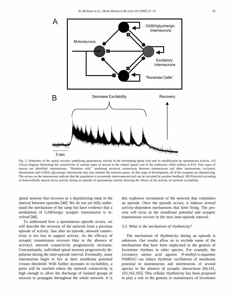
Ž .Fig. 2. Schematic of the spinal circuitry underlying spontaneous activity in the developing spinal cord and its modification by spontaneous activity. ACircuit diagram illustrating the connectivity of various types of neuron in the ventral spinal cord of the embryonic chick embryo at E10. Four types ofneuron are identified: motoneurons, ‘‘Renshaw cells’’ mediating recurrent connections between motoneurons and other interneurons, excitatoryinterneurons and GABArglycinergic interneurons that may mediate the sartorius pause. At this stage of development, all of the synapses are depolarizing.
Ž .The arrows on the interneurons indicate that the population is recurrently interconnected and can be recruited by positive feedback. B Electrical recordingof femorotibialis muscle nerve activity during an episode of spontaneous activity showing the effects of the activity on network excitability.
spinal neurons that recovers as a depolarizing ramp in thew xinterval between episodes 48 . We do not yet fully under-
stand the mechanism of the ramp but have evidence that amodulation of GABAergic synaptic transmission is in-
w xvolved 48 .To understand how a spontaneous episode occurs, we
will describe the recovery of the network from a previousepisode of activity. Just after an episode, network connec-tivity is too low to support activity. As the efficacy of
Žsynaptic transmission recovers due to the absence of.activity , network connectivity progressively increases.
Concomitantly, individual spinal neurons progressively de-polarize during the inter-episode interval. Eventually, someinterneurons begin to fire as their membrane potentialcrosses threshold. With further increases in excitability, apoint will be reached where the network connectivity ishigh enough to allow the discharge of isolated groups ofneurons to propagate throughout the whole network. It is
this explosive recruitment of the network that constitutesan episode. Once the episode occurs, it induces severalactivity-dependent mechanisms that limit firing. The pro-cess will recur as the membrane potential and synaptictransmission recover in the next inter-episode interval.
3.3. What is the mechanism of rhythmicity?
The mechanism of rhythmicity during an episode isunknown. Our results allow us to exclude some of themechanisms that have been implicated in the genesis oflocomotor rhythms in other species. For example, theexcitatory amino acid agonist N-methyl-D-aspartateŽ .NMDA can induce rhythmic oscillations of membranepotential in motoneurons and interneurons of several
wspecies in the absence of synaptic interactions 84,101,x102,162,185 . This cellular rhythmicity has been proposed
to play a role in the genesis or maintenance of locomotor

( )H. Barbeau et al.rBrain Research ReÕiews 30 1999 27–5136
w xactivity 84,164 . Spinal neurons in the chick cord alsoexpress rhythmic, voltage-dependent membrane potential
w xoscillations in the presence of bath-applied NMDA 50 .However, such oscillations do not appear to be essential,because rhythmic activity can occur in the presence ofNMDA receptor antagonists after a short period of blockw x29,49 . Nonetheless, such properties could still play animportant role — perhaps locally in dendrites where theycould serve to amplify synaptic inputs.
Another mechanism of rhythmicity originates from hy-perpolarization-activated conductances, such as the lowthreshold t-channels or the mixed cationic conductance Ihw x53,128,130 . These conductances underlie post-inhibitoryrebound and can set up oscillations when the neuronscontaining them are reciprocally connected to inhibitoryneurons. This mechanism has been proposed to be impor-tant in the genesis of swimming in the Xenopus embryow x163 . Although post-inhibitory rebound can be observed inembryonic chick motoneurons following hyperpolarizingcurrent injection, it seems unlikely that these currents playa critical role in rhythmicity. This is because all synapticpotentials we have detected in embryonic chick neurons
Ž .younger than day 12 -E12 are depolarizing. As a result,it is difficult to see how hyperpolarization activated cur-rents could be recruited in the course of synaptic activity.
Our current hypothesis is that the mechanism control-ling the cycling within an episode is similar to that regulat-ing the occurrence of episodes, albeit on a much shorterŽ .cycle to cycle time scale. The most likely candidate isactivity-dependent depletion of transmitter. Such short termsynaptic depression has been shown to generate cyclicalactivity in modeling studies of cultured spinal neuronsw x178 and has been implicated in the epileptiform bursting
w xof hippocampal slices 186 . Of course, short-term synapticdepression is not the only mechanism that could operate tolimit firing within a cycle. Other cellular processes, suchas spike frequency adaptation or summation of Ca2q acti-vated Kq conductances, could also be involved.
At present, these ideas for the rhythmicity within theepisode remain hypothetical. To establish if some form ofshort term depression is responsible for the rhythm willrequire monitoring transmitter release in the interval be-tween cycles and establishing the existence or otherwise ofother cellular processes that depress firing.
3.4. Comparison between the genesis of locomotion andembryonic motor actiÕity
w xWe have suggested elsewhere 147 that the spinalnetworks active during embryonic development may in-clude the precursors of locomotor circuits. While this maybe true, several important differences exist between themechanisms of embryonic motor activity and the produc-tion of adult locomotion.
Probably the most important distinction between thetwo types of behavior, is in the way the networks are
activated. In the adult, locomotion is initiated and con-trolled by descending commands from the brain. In theembryo, the properties of the spinal networks themselvesdetermine how, and when, they are activated. Central tothe autonomous generation of spinal activity in the em-bryo, are two properties that change during development.This is depolarizing early in development and becomes
w xhyperpolarizing later 140,143,179 . As a consequence,these classically inhibitory neurotransmitters can be func-
wtionally excitatory during development for review, seew xxRef. 47 .
A second important difference between embryonic andmature spinal networks is the existence of a ventral set ofGABAergic spinal interneurons in the embryo that disap-
w xpear later in development 15 . The fate of these neurons isunknown but we have proposed that they experience atransmitter switch, during development, from GABA to
w xglycine 35 . Although the cells could die, this seemsunlikely because developing spinal interneurons do notappear to experience a period of naturally occurring cell
w xdeath 136 . This transient population of GABAergic in-terneurons appears to be responsible, in part, for control-ling the occurrence of spontaneous activity in the embryo.The autonomous activation of embryonic spinal networksis clearly not locomotor in nature and it is presumed to be
wnecessary for normal limb and network development forw xxreviews, see Refs. 69,148 .
3.5. Conclusion
One of the important lessons to emerge from thesestudies is that spontaneous activity generated by spinalnetworks is a highly robust mechanism. By this, we meanthat the generation of activity does not depend on thedetails of circuit organization. Periodic rhythmic activitycan be produced by circuits of glutamatergic and choliner-gic neurons or alternatively, by networks of GABAergicand glycinergic neurons. At present, we do not knowwhich components and properties of the network are ro-bust and which are not. Nevertheless, the presence ofrobustness provides embryonic rhythm generating net-works with a mechanism that is resistant to major perturba-
Žtions, generated either internally or externally see Ref.w x .150 for further discussion of these ideas . It remains to bedetermined, whether locomotor networks in the adult spinalcord also exhibit robustness. If so, then it will be importantto identify which network components and properties cantolerate variations, and which are tightly controlled.
4. Triggering, modulating and adapting the spinal pat-( )tern generator for locomotion in cats Serge Rossignol
What have we learned from studies on locomotor con-trol mechanisms in cats and other animals that could beuseful to therapeutic approaches in humans with spinal-cord

( )H. Barbeau et al.rBrain Research ReÕiews 30 1999 27–51 37
injuries? Our basic assumption is that some of the studiedcontrol mechanisms have been conservatively preservedthrough evolution and that the formulation of related basicconcepts can at least help to outline a general frameworkof understanding that will guide therapeutic approaches,namely, in the field of locomotor rehabilitation. Some ofthe work reported by Barbeau below, which was initiallybased on work in animals, indeed illustrates the basicvalidity of this assumption.
ŽThis section will first define the concept of CPG its.importance and limitation and then summarize some of
the control mechanisms related to triggering, modulatingand adapting this centrally generated locomotor pattern.The topic of locomotor control has been the subject of
wseveral general reviews during the last 15 years 18,87,x154,165,189 .
4.1. The central pattern generator
Rhythmic patterns can be generated in the lumbosacralŽspinal cord in the absence of phasic paralyzed prepara-
. Ž .tions or tonic deafferented preparations afferent inputsas well as in the absence of command signals fromsupraspinal structures. The notion that neuronal circuits inthe spinal cord are capable of generating such alternatingreciprocal patterns of activity with the characteristics ofcoupling and frequency typical of locomotion, scratching,
w xor fast paw shake is well established in the cat 86,153,167Žas well as in other vertebrates i.e., lampreys, tadpoles.
chicks, neonatal rats and even in some primates such as thew w xx.marmoset reviewed in Ref. 165 . The evidence for such
a spinal CPG has been, up to now, less compelling inhumans, although rhythmic alternating activity in the lowerlimbs after complete spinal cord section in humans has
w w xbeen described Refs. 55,56,105,123 , reviewed in Ref.w xx165 and seen after epidural electrical stimulation of the
w xspinal cord 57,180 . Recent work in normal humans usingvibration of tendons also suggests the presence of a CPG
w xin humans 90 .Although the notion of CPG suggests a relative auton-
omy of function, it should be understood that this CPG is,in normal conditions, in continuous interaction with affer-ent and descending signals. The notion of CPG is impor-tant because it suggests that many of the rhythmic patternsthemselves are organized as spinal motor subroutines.These can be triggered by peripheral afferents and de-scending commands, modulated by the same signals andmost probably, modified in the long term when there arepermanent changes in these signals after central and pe-ripheral lesions. Such interactions will now be considered.
4.2. Triggering the CPG: supraspinal initiation of locomo-tion
In high decerebrate cats, locomotion on the movingtreadmill, called ‘‘fictive’’ locomotion, after paralysis, can
Žbe triggered by electrical stimulation around 100 mA at.20–60 Hz of a region of the mesencephalon located
Ž .below the inferior colliculus the cuneiformis nucleus . Asw xfirst described by Shik et al. 182 , the speed of locomotion
Žas well as the mode of coupling between limbs walking,.trotting, galloping can be adjusted by modifying the
strength of the brainstem stimulation as well as the speedof the treadmill. This mesencephalic locomotor regionŽ .MLR and its closely located structure, the pedunculo-
Ž . Ž .pontine nucleus PPN see Fig. 3 , probably exert theiraction through projections to the medial reticular forma-tion, which in turn projects to the spinal cord via tracts in
Ž .the ventral and ventro-lateral funiculus VLF in Fig. 3w x115,116,165,181 . In acute decerebrate cats, lesion ofthese pathways prevents the activation of locomotion by
w xMLR stimulation 187 .Massive chronic lesions of the ventral and ventro-lateral
pathways in cats suggest that other descending pathwayscan also be used to trigger locomotion since such cats canregain voluntary four-legged locomotion overground aftera few weeks, although some deficits such as decreasedweight support and unstable forelimb–hindlimb coupling
w xcan persist 34,38,77,78,169,203 . It is probable that path-Žways coursing through the dorsolateral funiculi DLF,
pathways from the brainstem or the telencephalon or else.propriospinal pathways are responsible for the trigger of
locomotion in such lesioned cats. Indeed, it has beensuggested that there exists a strip of cells in the brainstemŽ .pontomedullary locomotor strip whose locomotor initiat-ing capabilities are exerted through a dorsolateral systemŽ . w xDLF in Fig. 3 119,120 . Chronic lesions of the dorsalcolumns and DLF may result in foot dragging and aninability to properly negotiate obstacles placed on the
w xtreadmill belt 114 but does not prevent the ability tow xgenerate locomotion 78 .
From the above studies, we conclude that there appearto be several descending tracts capable of triggering loco-
Ž .motion in cats Fig. 3 . Similar conclusions can also bereached from study of partial spinal lesions in humansw x139 . This conclusion is important in the present contextsince it suggests the possibility that electrical stimulationof different tracts could ‘‘tap’’ the spinal cord and initiatethe locomotor pattern.
4.3. Pharmacologic triggering of locomotion
w xThe pioneering work of the Swedish school 86,111,112has laid the foundation for pharmacologic activation ofspinal locomotor circuitry. They have shown that L-DOPA,a noradrenaline precursor, especially after potentiation by amonoamine oxidase inhibitor, can activate a fictive loco-motor pattern in the paralyzed spinal cat. Similarly, it was
w xshown that clonidine, an a2-noradrenergic agonist 71 ,could trigger locomotion of the hindlimbs in acute spinalcats. We have also shown that, within the first week after

( )H. Barbeau et al.rBrain Research ReÕiews 30 1999 27–5138
spinalization, clonidine can induce locomotion and that thecharacteristics of the pattern evoked varies with time after
w xspinalization 23,25,27,28,44,45,153,168 . Fig. 4 gives a
dramatic example of locomotion being initiated by an i.t.injection of clonidine in a cat 8 days after a completespinal transection at T13. The figure represents the kine-

( )H. Barbeau et al.rBrain Research ReÕiews 30 1999 27–51 39
Ž .Fig. 4. I.t. injection of a bolus of 100 ml 3.8 mM of clonidine in a spinal cat 8 days post-lesion. See text for description. Muscle abbreviations are as inFig. 3.
matics and associated EMG activity of one flexor muscleand two extensor muscles of the knee, one on each side.Before the shaded rectangle, there is no organized EMGactivity nor any rhythmic movements of the leg facing thecamera. The leg is actually dragged from left to right bythe moving treadmill belt. The arrow points to the time ofthe end of the clonidine injection. About 1 min after theend of the injection, the cat starts to walk as evidenced bythe movements of the limb and the rhythmically organizedEMG.
We have been unable in the cat to trigger locomotion inthe first post-spinalization week with agonists of othersystems such as serotonin, dopamine or NMDA. This doesnot imply that these cannot trigger locomotion in other
species or are not important in other aspects of locomotorcontrol.
4.4. Sensory eÕoked locomotion
Sensory stimuli can trigger locomotion in high decere-brate cats. The proximity of the pontomedullary locomotor
w xstrip to the trigeminal sensory nuclei has led some 141 tohypothesize that the trigger of locomotion by stimulationof this area might be related to a stimulation of thetrigeminal nucleus. In decerebrate paralyzed cats, tonicstimulation of the dorsal roots can evoke fictive locomo-
w xtion 41 . In chronic spinal cats, simply moving the tread-w xmill belt is sufficient to initiate locomotion 26 . In less
Ž w x.Fig. 3. Summary of structures involved in the initiation and control of locomotion for more details see text and Ref. 165 . This diagram illustrates howŽ . Ž .spinal circuitry CPG is thought to produce the basic locomotor pattern by exciting and inhibiting groups of interneurons as well as flexor F and extensor
Ž . Ž .E motoneurons enclosed in the shaded area , which in turn activate muscles, each of which has its own pattern of activity or its ‘‘signature’’. This spinalŽ . Žcircuitry can be activated by supraspinal structures located in the telencephalon, diencephalon SLR, subthalamic locomotor region ; mesencephalon MLR,
.mesencephalic locomotor region; PPN, pedunculo-pontine nucleus and perhaps the cerebellum. The MLR exerts its effect through the reticular formationŽ .which projects through the VLF ventrolateral funiculus shown in transparency . Other structures may exert their effect through the DLF coursing
Ž . Ž .dorsolaterally DLF, dorsolateral funiculus and through propriospinal interneurons PS . Afferents originating from the skin and muscles, with their cellŽ . Žbodies in the DRG dorsal root ganglion can influence the CPG. Various neurotransmitter systems can also trigger or modulate the locomotor pattern NE,
Ž . .noradrenaline; 5-HT, 5-hydroxy-tryptamine serotonin ; DA, dopamine; NMDA, N-methyl-D-aspartate . Other abbreviations: co, contralateral; EMG,Ž . Ž .electromyogram; GM, gastrocnemius medialis ankle extensor ; i, ipsilateral; mrs, metersrsecond; Srt, sartorius hip flexor and knee extensorrflexor ; St,
Ž . Ž . Ž .semitendinosus knee flexor and hip extensor ; Tri, triceps elbow extensor ; VL, vastus lateralis knee extensor .

( )H. Barbeau et al.rBrain Research ReÕiews 30 1999 27–5140
active cats, especially early after spinalization, perinealstimulation is a powerful stimulus to induce locomotion.
4.5. Modulating the CPG: supraspinal stimulation
The characteristics of the locomotor pattern can bechanged by stimulation of the various descending path-
Ž .ways see Fig. 3 to accommodate for changes in speed,direction and posture. This subject has been reviewedextensively in various papers cited earlier but some generalprinciples will be recalled because these might be impor-tant in the present context where electrical stimulation maybe envisaged as a therapeutic avenue. First, responsesevoked during locomotion are quite different from thoseevoked at rest. For instance, stimulation of the medialreticular formation may evoke at rest some generalizedexcitatory responses involving several muscles, even ant-
w xagonistic muscles 60,61 . During walking, the same stimu-lus can yield well-organized reciprocal responses in sev-eral muscles that would tend to enhance the ongoinglocomotor coupling of muscles at the time of stimulationw x62 . These responses are not only state-dependent, butalso phase-dependent so that the same stimulus applied tothe reticular formation may yield an excitatory response inan extensor during one phase and an excitatory response inthe antagonist flexor in the opposite phase of the locomo-tor cycle. Altogether, this suggests that the spinal patterngenerator has some degree of autonomy and that theresponses to descending stimuli are a function of the stateof the spinal circuitry. This principle has been well estab-
w xlished now since the work of Orlovsky 151 .On the other hand, it is also becoming clear that certain
stimuli given to the reticular formation during fictivew x w xlocomotion 151 or to the MLF or Deiter’s nucleus 172 ,
as well as stimuli to the motor cortex during locomotion inw xintact cats, can also reset the locomotor cycle 16,17 and
thus have more than a modulating effect, but a ratherprofound effect on the timing of the pattern itself.
4.6. Pharmacologic modulation
While in the spinal cat the noradrenergic system ismainly implicated in triggering locomotion, all neurotrans-mitter systems exert modulating effects on some character-istics of the locomotor pattern and cutaneous reflex ex-
w xcitability 23,25,28,166,168 . For instance, in the spinalcat, noradrenergic agonists will tend to increase duration ofthe bursts as well as the step length and duration for agiven treadmill speed. Serotoninergic agonists, on the otherhand, will tend to increase markedly the amplitude ofdischarge of all muscles but especially extensor muscles.
Ž .Dopaminergic agonists Apomorphine appear to increasethe excitability of the flexor muscles. Although NMDAagonists have been shown to initiate locomotion in high
w xdecerebrate cats 59 , we have been unable to initiate
locomotion with these drugs in the very early period afterw xthe spinal lesion 46 . However, in late spinal cats, locomo-
tion can be reinstated by NMDA after previously blockingit with AP5, a specific NMDA blocker.
The effects of various agonists on locomotion can de-pend on the type of preparation. For instance, whereasclonidine can initiate locomotion in the complete spinalcat, it can be deleterious to locomotion in cats with
w xmassive ventral and ventrolateral lesions 38,39 . Althoughthis might be due to a different overall effect of the agonistin the presence of both pre- and post-synaptic receptors,there is also the possibility that the drug may act on theexcitability of reflex pathways, which could be essential tomaintain locomotion in cats with partial lesions. Theseobservations are undoubtedly important for the evaluation
w xof drugs in humans 54 .
4.7. Sensory modulation
The subject of interaction between sensory inputs andw xlocomotion is vast, has been reviewed extensively 165,171
and is covered in the section by McCrea above. Fig. 3illustrates some of the afferents originating from the skinor from muscles that may impinge on the CPG or elseterminate on neurons and interneurons which are under the
Ž .influence of the CPG activity darker shaded area . Duringfictive locomotion, the CPG can be entrained by cutaneous
w x wstimuli 170 as well as proprioceptive stimuli 165,x170,171 , indicating that they may have a powerful influ-
ence on the timing of the centrally generated pattern. Thishas been directly studied during fictive locomotion byanalyzing the effect of static limb positioning on the
w xcentrally generated pattern 154,174 . From these studies,we can conclude that retraction of the forelimb or exten-sion of the hip greatly accelerates the basic rhythm fre-quency whereas protraction of the forelimb or flexion ofthe hindlimb can altogether arrest locomotion.
Brief electrical or mechanical stimuli to the skin or skinnerves can produce various responses depending on thephase of the step cycle. Thus, a stimulus during swing canactivate flexor muscles while the same stimulus duringstance may have no effect or may excite or inhibit ongoing
w xextensor activity 165,171 . An important aspect for thefocus of this paper is that these responses are well-in-tegrated within the step cycle and therefore can be used toenhance the ongoing phase of the movement.
The importance of proprioceptive afferents during loco-motion has been assessed in various experiments. In cats,if the foot is placed in a hole instead of firmly on ground,
Žthe precontact EMG activity is the same presumably of.central origin but the subsequent normal stance activity is
turned off, suggesting that the propriospinal inputs at footcontact may be important in maintaining such activity in
w xthe ankle extensors 98 . This has also been more directlystudied in humans by evaluating the gain of the stretchreflex during the early stance phase, leading to the conclu-

( )H. Barbeau et al.rBrain Research ReÕiews 30 1999 27–51 41
sion that stretch reflexes may contribute substantially tow xthe muscle activity during stance 190,201 .
A further important consideration here is that suchproprioceptive reflexes are state-dependent. As McCreahas detailed above, afferent inputs from some propriocep-tors like the Golgi tendon organ which are inhibitory at
w xrest, become excitatory during locomotion 79,88,98 , fur-thering the importance of such proprioceptive inputs dur-ing locomotion.
4.8. Adapting the CPG
One of the key questions in therapeutic strategies forrehabilitation is the question of plasticity of locomotormechanisms. Such strategies must take into considerationthe fact that the organism has already adapted perhapsoptimally to the central or peripheral lesion of the nervoussystem. Secondly, one of the hopes in these strategies isthat external devices andror other forms of stimulationcan modify the generating and control mechanisms oflocomotion on a long-term basis. Some evidence that thereis some degree of plasticity has been described recently, asdiscussed below.
4.9. Locomotor training
After spinalization, cats progressively recuperate loco-motor functions of the hindlimbs. This progression sug-gests that the generating locomotor circuitry is being pro-gressively modified. Intensive locomotor training appearsimportant to achieve optimal locomotor recuperationw x26,33,103,152 . As mentioned above, the effect of somedrugs varies with time after spinalization, suggesting thatthe spinal circuitry changes functionally with time afterinjury. A recent study has shown that the locomotor pat-tern evoked with clonidine within the first 10 days post-
w xspinalization improves daily 44 . Furthermore, after about10 days of intensive training, with daily injection of cloni-dine, there is expression of a well-organized locomotorpattern with hindquarters weight support and proper foot
w xplacement 44 . Such results are important for the trainingstudies done in humans as will be summarized by Barbeau.
4.10. Adaptation to lesions
The mechanisms of locomotor adaptation form anemerging field in studies of locomotion. Whereas there areprobably some major hardwired components of the CPG,such as those related to the strict alternation between
w xantagonist muscles at a joint 72 , there might be otherplastic adaptation related to muscles used in compensatingstrategies in the case of peripheral lesions. In a recent
w xstudy 43 , we have shown that normal cats can adapt very
rapidly within 1–2 days to the neurectomy of the ankleflexors tibialis anterior and extensor digitorum longus byproducing a small but sufficient increase in knee androrhip flexion during the swing phase. After spinalization, thefine compensation seen in the otherwise intact cat is lostbut the spinal locomotion which is normally regular andwell organized, is now quite disorganized and dominatedby knee hyperflexions as if the spinal cord had ‘‘learned’’part of the mechanisms used to overcome the deficit beforespinalization. Other studies of this kind are now underwayto investigate the spinal capabilities to adapt locomotion tocutaneous denervation of the paws.
4.11. Conclusions
From this brief overview of animal experiments, we canextract some concepts which may be important for design-ing therapeutic strategies for humans with SCIs.
ŽØ There is most probably in humans a spinal or.brainstem–spinal component of the locomotor generating
mechanism which could be accessed through various typesof sensory or descending inputs or by pharmacologicstimulation.
Ø All types of inputs to these locomotor controlmechanisms have to be evaluated within the context oflocomotion because the effects of these stimuli are state-dependent and phase-dependent. Knowledge of responsesin static conditions only may not be adequate.
Ø These stimuli also have to be evaluated in varioustypes of spinal lesions and at various times after the onsetof the lesion because these stimuli may interact withcontrol mechanisms which have themselves been changedŽ .quantitatively or qualitatively as a result of the lesion.
Based on the knowledge obtained from animal experi-Ž .ments, a combination of 1 locomotor training with the
Žaddition of adequate pharmacotherapy to reduce spasticity.or enhance the activity of locomotor generating circuits
Ž .and 2 FNS to mimic the action of some central orperipheral pathways will probably constitute the best ap-proach to locomotor rehabilitation in patients with spinal-cord injury.
5. Can locomotor training enhance the recovery of( )walking following SCI? Hugues Barbeau
Ž .SCI with an incomplete motor function loss IMFL isassociated with multiple motor problems that lead to al-tered walking behavior, which is reflected by a reducedwalking speed, postural problems related to bearing weight,maintaining balance and hyperactive spinal reflexes. Thissection presents some of the recent developments andconcepts emerging from both animal and human studiesaimed at enhancing recovery of walking following SCI.

( )H. Barbeau et al.rBrain Research ReÕiews 30 1999 27–5142
New locomotor training and pharmacological interven-tions, used individually or together, have been identified asimportant approaches that modify the recovery processfollowing SCI in humans. We conclude that the nervoussystem continues to manifest a great deal of plasticity evenseveral years after SCI.
5.1. Locomotor training approaches
Ž .Animal findings see Rossignol’s section above andclinical observations that neurologically impaired subjectscannot adequately bear weight on the more affected lowerlimb have led to the introduction of locomotor training,
Ž .using a treadmill and body weight support BWS . Thistechnique was proposed by Finch and Barbeau in 1985w x w x70 and was introduced in SCI subjects with IMFL 22 .BWS reduces the load borne by the lower limbs and can
w xbe provided by various means: pneumatic 75 , pulleyw x w x w x22,142 , spring 95 and robotic systems 184 . Supporting
Ž .a percentage of the body weight up to 40% was associ-ated with an increase in comfortable walking speed,single-limb support time, stride length, and endurancew x195 . The kinematic pattern revealed a straighter trunkassociated with improved hip, knee and ankle joint angularexcursion. Furthermore, EMG activity throughout the cy-cle showed more normal timing.
There are many clinical implications and advantages ofsuch locomotor training strategies. The different compo-nents of gait may be retrained simultaneously under dy-namic conditions. Locomotor retraining may be initiatedearly in the rehabilitation period, with BWS provided asneeded for the subject to assume an upright position and toallow assisted or unassisted stepping by the lower limbs.As subjects walk on the treadmill with a reduced load ontheir lower extremities, equilibrium demands are reducedand gait deviations may be alleviated. The use of BWS canalso be combined with other strategies. Recent clinicalstudies suggest that such locomotor training exercise com-bined with BWS is important in optimizing walking pat-
Ž . w xterns and in achieving full weight bearing FWB 21,196 .Our laboratory has investigated the effectiveness of
such a locomotor training approach in subjects with SCI orstroke. One study examined the effect of 6 weeks ofinteractive locomotor training with BWS on the kinematicpattern in nine people with chronic SCI-IMFL. The loco-motor training was performed on a treadmill, with thesubject mechanically supported in a comfortable overheadharness at different percentages of BWS, monitored with a
w xstrain gauge transducer 22 . At the end of 6 weeks oftraining, overground walking capability improved in sub-jects who had reached FWB on the treadmill; and weightsupport capability improved in subjects who had not yet
Žreached FWB. FWB group: overground speed: 0.11 mrs"0.11; treadmill speed: 0.24 mrs"0.7 vs. non-FWBgroup: overground speed: 0.04 mrs"0.04; treadmill
.speed: 0.13 mrs"0.11 .
In a randomized clinical trial, stroke patients who re-Ž .ceived 6 weeks of gait training with BWS ns50 recov-
ered better balance and walking abilities than those whoreceived similar gait training while bearing their full weightŽ .ns50 . Fourteen sitting and standing activities were each
w x Ž .scored on a five-point scale 196 see Table 1 .A 3-month post-training follow-up revealed that those
who trained with BWS continued to have significantlyhigher scores for overground walking speed and lower
Žlimb motor recovery 0.52 mrs compared to 0.30 mrs,.ps0.006 . The effect of locomotor training using BWS is
presently being investigated in subjects with various neuro-w xlogical disorders of varying severity 21,58,97,197,197a ,
and has been compared to a conventional physiotherapyw xprogram 96,97 .
FNS was introduced more than 35 years ago by Liber-w xson et al. 127 as an orthotic system that replaced conven-
tional braces with motor responses elicited by stimulatingthe common peroneal nerve during walking. While Liber-son applied FNS to preventing footdrop in hemiplegic
w xsubjects, Kantrowitz 118 was the first to implement thenew technology in SCI subjects. FNS systems have be-come increasingly complex; some have up to 50 channelsof stimulation that operate under closed-loop-control basedon signals from foot switches or goniometers. FNS hasbeen used to restore a variety of movements including
w xwalking 202 and was first reported for SCI-IMFL sub-w xjects in 1989 19 . However, this approach has not been
widely used in rehabilitation for a number of technicalreasons. Early stimulators were bulky, unreliable, prone to
w xbreakage and expensive 188 . Recent studies of simpleŽsystems for FNS-assisted walking common peroneal nerve
.stimulator showed a limited increase of the walking speedŽ .-0.1 mrs and a reduction of oxygen consumption
w xduring gait 80,188 .In our laboratory, the combination of a simple FNS
system with locomotor training has been effective forw ximproving walking in subjects with SCI-IMFL 124a .
Within the first year, a gradual improvement of the maxi-mal walking speed could be observed, with the mean
Ž .magnitude of the increase being 0.26 mrs"0.24 Ns14 .But, more remarkably, the walking speed of people withSCI-IMFL increased even when the stimulator was tem-porarily turned off. This retained increase of walking speedwithout FNS is called the therapeutic effect and has the
Žsame magnitude as reported earlier 0.25 mrs" .20, Ns.14 . This therapeutic effect on walking has been reported
Table 1Effects of 6 weeks of gait training
BWS group Non-BWS groupU UBalance score 37.2"2.1 29.4"3.1
Ž .maximum of 70UU UUWalking speed 0.38 mrs 0.27 mrs
Ups0.001; UU ps0.02.

( )H. Barbeau et al.rBrain Research ReÕiews 30 1999 27–51 43
w xanecdotally for hemiplegic patients 127 and for incom-w xplete spinal injury patients 81,124 . Changes in their
assistive ambulatory devices from crutches to canes andchanges in the ergonomic efficiency of the walking behav-ior were also observed. From these results, we concludethat increases in maximal walking speed during trainingwith the FNS orthosis are mostly due to a therapeuticeffect, which implies that plasticity occurred during train-ing, as seen in the spinal cat. Finally, we conclude thatBWS and FNS types of locomotor training stress differentprerequisites for walking to achieve recovery of functionalwalking and, thus, their combination should be investi-gated.
5.2. Pharmacologic approaches to enhance locomotion inpeople with SCI-IMFL
w xBased on animal findings 25,166,168 , we believe thata rationale for locomotor pharmacotherapy can be devel-oped for incomplete SCI. In a placebo-controlled study ofthe effects of clonidine on the walking pattern of SCIsubjects, those with clinically complete SCIs were sup-ported and assisted in stepping and their ability did not
w xchange with clonidine 54,192 . The inability to trigger alocomotor pattern in complete SCI subjects could possiblybe explained by several factors such as the chronic state of
Ž w x.the patients see discussion in Ref. 192 .In contrast, the most severely impaired SCI-IMFL sub-
ject showed a marked improvement in walking ability, asw xrevealed by EMG and kinematic pattern 192 . More nor-
mal activation patterns of the tibialis anterior and soleusmuscles have also been observed in two cases treated with
w x w xclonidine 30 and with tizanidine 121 .
Recently, a brief report was published on the acuteeffect of an i.t. injection of a bolus of clonidine on the
w xoverground walking pattern of an SCI-IMFL subjects 160 .Within a half-hour of clonidine injection, three of the 10SCI-IMFL subjects doubled their maximal overgroundwalking speed. The speed gains were maintained or in-creased over successive evaluations during the subsequent6 h. During the same time period, the flexor reflex ampli-tude and the resistance to passive movement were reducedwhereas the H-reflex amplitude remained unchanged. Inthe same subjects, the changes in walking speed, flexionreflexes and spasticity score were minimal on days whenplacebo injections were given. The reduction of signs ofspasticity concurrent with the increase of walking speedsuggests that i.t. clonidine may be a powerful medicationfor SCI-IMFL. Furthermore, these results are consistentwith findings of changes in locomotor pattern and reducedexcitability of spinal reflexes with clonidine in chronicspinal cats. However, the relationship between change inreflex and change in locomotor behavior is still unclear.
5.3. Locomotor training combined with pharmacologicapproaches
The combination of noradrenergic drugs and locomotortraining has proven powerful in accelerating the recovery
w xof locomotion in spinal cats 23–25,166,168 . In our labo-ratory, we have conducted a similar investigation withhuman subjects. The combined effects of clonidine andcyproheptadine together with a locomotor training programusing BWS and treadmill walking exercise were investi-gated in chronic SCI-IMFL subjects. They had completedstandard rehabilitation but were unable to walk at entry,
Fig. 5. Schematic representing fundamental and functional levels of recovery of locomotion.

( )H. Barbeau et al.rBrain Research ReÕiews 30 1999 27–5144
w xinto the study 73 . Both subjects were able to initiatewalking on the treadmill while taking these drugs, and 6weeks of locomotor training with continued drug therapyfurther improved their locomotor capabilities. The subjectsachieved walking with FWB on the treadmill, and somelimited overground walking was achieved with walkingaids. On the treadmill, they showed a higher walking speedassociated with a greater stride length and greater angularexcursions at the hip, knee, and ankle. This has also beenobserved when medications have been combined with
w xlocomotor training using FNS devices 124 . These find-ings suggest that locomotor training using BWS or FNScould be powerful approaches to neurological rehabilita-tion, after the locomotor pattern has been expressed withmedication.
5.4. Conclusion
From fundamental studies in both animals and humansfollowing SCI, we can conclude that it is important tounderstand the recovery process following SCI as well asunderlying factors that can influence this recovery process.New locomotor training, pharmacological approaches andtheir combination have been identified as important factorsto modify the recovery process following SCI in bothanimal and human. These results suggest that a great dealof plasticity is possible following SCI, and that changes
Žcould take place for many years following the injury see.Fig. 5 . These new approaches are still in experimental
stages in SCI-IMFL subjects: several studies are underwayŽ .to evaluate a efficacy of locomotor training approaches
Ž .using walking exercise and BWS, b functional electricalŽ . Ž .stimulation, c pharmacological approaches and d com-Ž . Ž . Ž .binations of a , b and c at different times following
SCI. In addition, treatment strategies should address threeimportant prerequisites to performing the locomotor task
Ž .adequately and efficiently; 1 basic or stereotyped move-Ž .ment synergy, required to achieve propulsion; 2 mainte-
Ž .nance of equilibrium during propulsive movement; 3adaptation of the locomotor pattern to the behavioral goalsof the person as well as to the constraints introduced bythe environment such as obstacles, change of direction and
Ž .speed see Fig. 5 . We strongly believe that a combinedapproach can provide a comprehensive and integrated pro-gram for the enhancement of locomotion in individualswith SCI or with other neurological impairment.
6. Control issues in using spinal circuits to restore( )functional movement Michel Lemay
6.1. Cyclic Õs. static moÕement
It is very likely that the circuitry involved in the controlof posture is part of the spinal cord circuitry responsible
for cyclic motion, and it too may be activated via micros-w xtimulation. For example, Giszter et al. 76 stimulated the
paralyzed frog’s spinal cord at the lumbar–sacral level atŽ .the level where McCrea see above showed the existence
of disynaptic interneurons that had an extension functionw xduring gait. Giszter et al. 76 measured the forces pro-
duced at the ankle, and found that microstimulation pro-duced a coordinated behavior that often showed a conver-gence to a single point in the work space. He found fourtypes of force patterns that can be thought of as being parts
w xof a swimming cycle. Later, Saltiel et al. 173 usedNMDA in the frog in the same area and was able to elicittonic responses and cyclic responses. The directions of thestatic force vector measured at a single limb position werefound to be a subset of the directions of the force plateausproduced during cyclic responses. This result seems toindicate that the static force fields that could be combinedto produced novel movements may be a subset of thecircuitry used during rhythmic motion. This raises thequestion as to whether spinal circuitry for reaching existssince the upper extremities are generally not involved incyclic movements. A series of papers by Alstermark and
w x w xSasaki 3–5 and Alstermark et al. 6–11 indicate thatthere is a spinal circuit involved in the control of theforelimb in cat, although the effect of the circuit on thelimb mechanical properties has not been characterized yet.
6.2. Duration of actiÕation
Nerve and muscle activation in FNS systems is of anonroff nature: with stimulation on, muscle is activated;when stimulation is off, muscle is passive. Spinal cordcircuitry seems to be very different. Under pharmacologi-cal influence, activation of spinal circuits can be fairlyprolonged. With electrical activation the duration of theactivation varies. Some sites produced long plateaus thatoutlasted the stimulation by up to 3 s, while other sites
w xwere of an onroff nature 126 . With better mapping of thecord, we may be able to sort out which spinal circuits arebest suited to producing posture-maintaining contractions,and which circuits are best suited to producing fast motioncontractions. Using both electrical stimulation and phar-macological treatment, we may be able to selectively
Žcontrol the spinal circuits suited for movement stronger,.shorter duration and the ones better suited for the control
Ž .of posture weaker but longer duration .
6.3. Modification of the response during motion
Rossignol refers to the fact that the response obtainedduring a portion of the gait cycle may not be the responseobtained during another portion. This raises the question ofhow relevant our standard measurements of isometric forcewill be to actual muscle output during motion. Isometricmeasurements may not be sufficient. In an ongoing investi-

( )H. Barbeau et al.rBrain Research ReÕiews 30 1999 27–51 45
gation, we are taking isometric force measurements duringmicrostimulation of the spinal cord in frogs, and compar-
Žing the isometric forces measured correlated in time and.space with the trajectory to the forces obtained via an
inverse dynamic analysis of the motion produced by themicrostimulation. We are finding the forces’ magnitude tobe an order of magnitude apart, differences that are toolarge to be accounted for by unmodeled dynamic elementssuch as friction or damping.
This indicates that the measurements required to imple-ment a functional neuroprosthesis will need to be matchedto the desired functional tasks; for example, recruitmentcharacteristics for a walking system will need to be mea-sured during walking.
Finally, spinal circuits have evolved to facilitate thecontrol of multi-joint motion — walking or reaching —and they can be evoked to facilitate the functional controlof those limbs. We will have to think differently aboutcontrolling stimulation through spinal circuits than we doabout directly controlling muscle contractions to producetorques at desired times. We will probably have to useelectrical stimulation in combination with pharmacologicalagents to modify the excitability of tissue at times. We willhave to deal with the fact that responses will be state-de-pendent. However, the level of coordination provided bythe spinal cord will facilitate the control of multi-jointmovements, tasks that with traditional FNS require a so-phisticated timing of the activation pattern, timing that issensitive to the different rates of muscle fatigue.
7. Technical challenges to using spinal circuitry in( )neural prostheses Warren Grill
A central characteristic of spinal neural control circuitsis state-dependence; meaning that the output generated bya particular stimulus is dependent upon the state of thephysiological system. McCrea discusses state-dependentexcitation of extensor motoneurons by group I afferentsvia an interneuronal pathway, and also state-dependentincreases in motoneuron excitability. Rossignol reinforcesthe fact that modulatory segmental and descending inputshave state-dependent effects on ongoing motor behaviors.Since segmental reflexes are largely preserved after chronicSCI, they may influence, in a state-dependent manner,motor behaviors evoked by activation of spinal circuitry.These state-dependent effects will complicate the engineer-ing task of generating a behavior using electrical activationof the nervous system. Classical engineering control theoryis built on the premise of linear, time invariant input–out-put properties. The state dependence of spinal input–out-put systems challenges classical engineering techniques,and will require innovative solutions, perhaps includingadaptive control and fuzzy logic.
There are several other practical and technological is-sues which must be addressed. First, the location of the
spinal neurons that regulate the behavior of interest mustbe identified. In animals, there are a number of techniquesby which the locations of neurons may be identifiedincluding conventional retrograde and anterograde tracers,as well as a relatively new retrograde transneuronal viral
Ž .tracer psuedorabies virus that allows identification ofhigher order neurons after injection into an end organw x175,193 . In addition, expression of the immediate earlygene c-Fos enables identification of neurons that were
w xactive during a particular behavior 20,83,113,138 . Thus,there is a variety of powerful tools to identify neuronallocations in experimental animals, but it is not clear howthis would be accomplished in humans. If the resolution ofnon-invasive imaging techniques such as positron emission
Ž .tomography PET or functional magnetic resonance imag-Ž .ing f-MRI improves to the submillimeter range, then
these techniques will allow location in the spinal cord ofthe neurons that control specific behaviors.
Second, implementing a neural prosthesis by electricalactivation of neural circuits within the spinal cord requiresa long-term interface between the electrodes and the bio-logical tissue. High density arrays of silicon microelec-
w xtrodes are presently being developed 12,42,106 . How-ever, the mechanical properties of silicon are dramaticallydifferent from those of the spinal cord. This mismatch inmechanical impedance will create differential motion un-der acceleration and may lead to mechanically inducedtissue damage and instability. A new generation of de-vices, perhaps incorporating flexible, conducting polymersis needed to build long-term stable interfaces with the
w w xxspinal cord e.g., Ref. 137 .Third, chronic electrical activation of spinal neurons
will require knowledge of what stimulation levels can betolerated by spinal cord tissue. Although non-damaginglevels for stimulation of the cortex have been identified
w xunder certain conditions 1 , recent studies have found thatnon-damaging levels of stimulation within the brainstemŽ .cochlear nucleus are different from those in the cortexw x134 . Thus, similar differences may also exist between thenon-damaging levels for stimulation of the spinal cord andother CNS tissues. Previous data suggest that the non-damaging levels of stimulation will also depend on theelectrode material, surface area, and the pattern of theapplied stimuli. Thus, an important question is how muchstimulation can spinal cord tissue tolerate on a chronic
w xbasis 200 .Finally, if we have identified a group of neurons using
imaging or tracing, then techniques are required to localizeactivation to a small group of cells without activatingneighboring axons and cells, which may generate antago-nist functions. The spinal cord is a complex inhomoge-neous, anisotropic volume conductor, and the pattern ofneuronal excitation generated by extracellular stimulationis strongly influenced by the electrical properties of the
w xsurrounding tissue 82 . Further, spinal neurons are geo-metrically complex with extensive dendritic arbors, vary-

( )H. Barbeau et al.rBrain Research ReÕiews 30 1999 27–5146
ing somal dimensions, different electrical properties, andtortuous axons. The pattern of excitation generated by aparticular stimulus depends on the spatial relationship be-tween the electrode and the neuronal structure as well as
w xthe stimulus parameters 91,135 . Thus, new electrodegeometries and stimulus parameters are required to pro-duce controlled and selective activation of targeted neu-ronal populations.
The resolution of these scientific and technologicalquestions will make possible a new generation of neuralprostheses, which use relatively simple implanted devices,coupled to biological neural networks, to achieve controlof complex behaviors. Microstimulation of the spinal cordwith implanted electrodes in people with neurological dis-orders gives the possibility of targeted and controlledrelease of neurotransmitters and, thus, the possibility ofexploiting the mechanisms used by the nervous system toregulate complex functions.
Acknowledgements
David McCrea thanks and gratefully acknowledges theparticipation of the collaborators in the unpublished experi-ments: Drs. Michael Angel, Brent Fedirchuk, PierreGuertin, Elzbieta Jankowska, Larry Jordan, Sherry Krawitz,and Marie-Claude Perreault. All studies were supported bythe Medical Research Council of Canada. Studies reportedby Barbeau were part of a Canadian multicenter FNS studysupported by the Neuroscience Network of the CanadianCentres of Excellence. Work in Warren Grill’s laboratoryis supported by the NIH NINDS Neural Prosthesis Pro-
Ž .gram N01-NS-5-2331, N01-NS-8-2300 and by the NSFBiomedical Engineering and Research to Aid Persons with
Ž .Disabilities Program BES-9709488 . This paper resultedfrom a symposium held on May 13, 1997 in Cleveland,OH and was supported, in part, by a grant from theBuckeye Chapter of the Paralyzed Veterans of America aspart of their ongoing effort to advance research on motorprostheses. The authors thank J. Thomas Mortimer, Direc-tor of the Applied Neural Control Laboratory, Case West-ern Reserve University, for bringing them and their ideastogether; and Carole Kantor, a medical science writerredi-tor, for integrating their contributions into this reviewpaper.
References
w x1 W.F. Agnew, D.B. McCreery, Neural Prostheses: FundamentalStudies, Prentice-Hall, Englewood Cliffs, NJ, 1990.
w x2 B.B. Alconero, The nature of the earliest spontaneous activity ofŽ .the chick embryo, J. Embryol. Exp. Morphol. 13 1965 255–266.
w x3 B. Alstermark, S. Sasaki, Integration in descending motor path-ways controlling the forelimb in the cat: 13. Corticospinal effectsin shoulder, elbow, wrist, and digit motoneurones, Exp. Brain
Ž . Ž . Ž .Res. EP2 59 2 1985 353–364.
w x4 B. Alstermark, S. Sasaki, Integration in descending motor path-ways controlling the forelimb in the cat: 14. Differential projec-tion to fast and slow motoneurones from excitatory C3–C4 pro-
Ž . Ž . Ž .priospinal neurones, Exp. Brain Res. EP2 63 3 1986 530–542.w x5 B. Alstermark, S. Sasaki, Integration in descending motor path-
ways controlling the forelimb in the cat: 15. Comparison of theprojection from excitatory C3–C4 propriospinal neurones to dif-
Ž .ferent species of forelimb motoneurones, Exp. Brain Res. EP2Ž . Ž .63 3 1986 543–556.
w x6 B. Alstermark, A. Lundberg, S. Sasaki, Integration in descendingmotor pathways controlling the forelimb in the cat: 10. Inhibitorypathways to forelimb motoneurones via C3–C4 propriospinal
Ž . Ž . Ž .neurones, Exp. Brain Res. EP2 56 2 1984 279–292.w x7 B. Alstermark, A. Lundberg, U. Norrsell, E. Sybirska, Integration
in descending motor pathways controlling the forelimb in the cat:9. Differential behavioural defects after spinal cord lesions inter-rupting defined pathways from higher centres to motoneurones,
Ž .Exp. Brain Res. 42 1981 299–318.w x8 B. Alstermark, H. Kummel, M.J. Pinter, B. Tantisira, Integration
in descending motor pathways controlling the forelimb in the cat:17. Axonal projection and termination of C3–C4 propriospinal
Ž . Ž .neurones in the C6–Th1 segments, Exp. Brain Res. EP2 81 3Ž .1990 447–461.
w x9 B. Alstermark, S. Lindstrom, A. Lundberg, E. Sybirska, Integra-¨tion in descending motor pathways controlling the forelimb in thecat: 8. Ascending projection to the lateral reticular nucleus fromC3–C4 propriospinal neurones also projecting to forelimb mo-
Ž .toneurones, Exp. Brain Res. 42 1981 282–298.w x10 B. Alstermark, T. Gorska, A. Lundberg, L.G. Pettersson, Integra-
tion in descending motor pathways controlling the forelimb in thecat: 16. Visually guided switching of target- reaching, Exp. Brain
Ž . Ž . Ž .Res. EP2 80 1 1990 1–11.w x11 B. Alstermark, T. Isa, B. Tantisira, Integration in descending
motor pathways controlling the forelimb in the cat: 18. Morphol-ogy, axonal projection and termination of collaterals from C3–C4propriospinal neurones in the segment of origin, Exp. Brain Res.Ž . Ž . Ž .EP2 84 3 1991 561–568.
w x12 D.J. Anderson, K. Najafi, S.T. Tanghe, D.A. Evans, K.L. Levy,J.F. Hetke, X. Xue, J.J. Zappia, K.D. Wise, Batch fabricatedthin-film electrodes for stimulation of the central auditory system,
Ž .IEEE Trans. Biomed. Eng. 36 1989 693–698.w x13 M.J.Angel, E. Jankowska, D.A. McCrea, Candidate interneurones
mediating group I disynaptic EPSPs in extensor motoneuronesduring fictive locomotion in the cat, 1998, in preparation.
w x14 M.J. Angel, P. Guertin, I. Jimenez, D.A. McCrea, Group Iextensor afferents evoke disynaptic EPSPs in cat hindlimb exten-sor motorneurones during fictive locomotion, J. Physiol. 494Ž .1996 851–861.
w x15 M. Antal, A.C. Berki, L. Horvath, M.J. O’Donovan, Developmen-tal changes in the distribution of GABA-immunoreactive neuronsin the embryonic chick lumbosacral spinal cord, J. Comp. Neurol.
Ž .341 1994 1–9.w x16 D.M. Armstrong, T. Drew, Electromyographic responses evoked
in muscles of the forelimb by intracortical stimulation in the cat,Ž .J. Physiol. 367 1985 309–326.
w x17 D.M. Armstrong, T. Drew, Forelimb electromyographic responsesto motor cortex stimulation during locomotion in the cat, J.
Ž . Ž .Physiol. London 367 1985 327–352.w x18 D.M. Armstrong, Supraspinal contributions to the initiation and
Ž .control of locomotion in the cat, Prog. Neurobiol. 26 1986273–361.
w x19 Y. Bajd, A. Kralj, H. Turk, H. Benko, J. Sega, Use of functionalelectrical stimulation in the rehabilitation of patients with incom-
Ž .plete spinal cord injuries, J. Biomed. Eng. 11 1989 96–102.w x20 I. Barajon, J.-P. Gossard, H. Hultborn, Induction of fos expression
by activity in the spinal rhythm generator for scratching, BrainŽ .Res. 588 1992 168–172.

( )H. Barbeau et al.rBrain Research ReÕiews 30 1999 27–51 47
w x21 H. Barbeau, M. Danakas, B. Arsenault, The effects of locomotortraining in spinal cord injured subjects — a preliminary study,
Ž .Restor. Neurol. Neurosci. 5 1993 81–84.w x22 H. Barbeau, M. Wainberg, L. Finch, Description and application
of a system for locomotor rehabilitation, Med. Biol. Eng. Comput.Ž .25 1987 341–344.
w x23 H. Barbeau, S. Rossignol, Effects of noradrenergic, serotonergicand dopaminergic drugs on the initiation of locomotion in adult
Ž .spinal cat, Soc. Neurosci. Abstr. 15 1989 393, no. 160.9,Abstract.
w x24 H. Barbeau, S. Rossignol, Enhancement of locomotor recoveryŽ .following spinal cord injury, Curr. Opin. Neurol. 7 1994 517–
524.w x25 H. Barbeau, S. Rossignol, Initiation and modulation of the loco-
motor pattern in the adult chronic spinal cat by noradrenergic,Ž .serotonergic and dopaminergic drugs, Brain Res. 546 1991
250–260.w x26 H. Barbeau, S. Rossignol, Recovery of locomotion after chronic
Ž .spinalization in the adult cat, Brain Res. 412 1987 84–95.w x27 H. Barbeau, C. Chau, S. Rossignol, Noradrenergic agonists and
locomotor training affect locomotor recovery after cord transec-Ž .tion in adult cats, Brain Res. Bull. 30 1993 387–393.
w x28 H. Barbeau, C. Julien, S. Rossignol, The effects of clonidine andyohimbine on locomotion and cutaneous reflexes in the adult
Ž .chronic spinal cat, Brain Res. 437 1987 83–96.w x29 M. Barry, M.J. O’Donovan, The effects of excitatory amino acids
and their antagonists on the generation of motor activity in theŽ .isolated chick cord, Dev. Brain Res. 36 1987 271–276.
w x30 E. Bastings, G. Rapisarda, J.L. Pepin, P.J. Delwaide, Nouvellesapproches pharmacologiques du traitement des troubles de lamarche consecutifs a une lesion spinale, Rev. Med. Liege 50` ´ `Ž .1995 204–208.
w x32 A. Bekoff, Ontogeny of leg motor output in the chick embryo: aŽ .neural analysis, Brain Res. 106 1976 271–291.
w x33 M. Belanger, T. Drew, J. Provencher, S. Rossignol, A comparisonof treadmill locomotion in adult cats before and after spinal
Ž .transection, J. Neurophysiol. 76 1996 471–491.w x34 T. Bem, T. Gorska, H. Majczynski, W. Zmyslowski, Different
patterns of fore–hindlimb coordination during overground loco-motion in cats with ventral and lateral spinal lesions, Exp. Brain
Ž .Res. 104 1995 70–80.w x35 A.C. Berki, M.J. O’Donovan, M. Antal, Developmental expres-
sion of glycine-immunoreactivity and its colocalization withGABA in the embryonic chick lumbosacral spinal cord, J. Comp.
Ž .Neurol. 363 1995 1–14.w x36 R. Brownstone, L.M. Jordan, D.J. Kriellaars, B.R. Noga, S.J.
Shefchyk, On the repetitive firing in lumbar motoneurones duringŽ .fictive locomotion in the cat, Exp. Brain Res. 90 1992 441–455.
w x37 R.M. Brownstone, J.-P. Gossard, H. Hultborn, Voltage-dependentexcitation of motoneurones from spinal locomotor centres in the
Ž .cat, Exp. Brain Res. 102 1994 34–44.w x38 E. Brustein, S. Rossignol, Recovery of treadmill locomotion after
ventral and ventrolateral spinal lesions in the cat: I. Deficits andŽ .adaptive mechanisms, J. Neurophysiol. 80 1998 1245–1267.
w x39 E. Brustein, S. Rossignol, Recovery of treadmill locomotion afterventral and ventrolateral spinal lesions in the cat: II. The effects of
Ž .noradrenergic and serotininergic drugs, J. Neurophysiol. 1999 inpress.
w x41 N.N. Budakova, Stepping movements evoked in a mesencephaliccat by a rhythmic stimulation of a dorsal root in mesencephalic
Ž .cat, Sechenov Physiol. J. USSR 57 1971 1632–1640.w x42 P.K. Campbell, K.E. Jones, R.J. Huber, K.W. Horch, R.A. Nor-
man, A silicon-based three-dimensional neural interface: Manu-facturing processes for an intracortical electrode array, IEEE
Ž .Trans. Biomed. Eng. 38 1991 758–767.w x43 L. Carrier, L. Brustein, S. Rossignol, Locomotion of the hindlimbs
following a neurectomy of the ankle flexors in intact and spinal
Ž .cats: a model for study of plasticity, J. Neurophysiol. 77 19971979–1993.
w x44 C. Chau, H. Barbeau, S. Rossignol, Early locomotor training withŽ .clonidine in spinal cats, J. Neurophysiol. 79 1998 392–409.
w x45 C. Chau, H. Barbeau, S. Rossignol, The effects of intrathecal a1-and a2-noradrenergic agonists and noradrenaline on locomotion
Ž .in chronic spinal cats, J. Neurophysiol. 79 1998 2941–2963.w x46 C. Chau, J. Provencher, F. Lebel, L. Jordan, H. Barbeau, S.
Rossignol, Effects of intrathecal injection of NMDA receptoragonist and antagonist on locomotion of adult chronic spinal cats,
Ž .Soc. Neurosci. Abstr. 20 1994 573, no. 214.14, Abstract.w x47 E. Cherubini, J.L. Gaiarsa, Y. Ben-Ari, GABA: an excitatory
Ž .transmitter in early postnatal life, Trends Neurosci. 14 1991515–519.
w x48 N. Chub, M.J. O’Donovan, Multi-functional actions of GABA inthe regulation of spontaneous activity in the isolated spinal cord
Ž .of the chick embryo, Soc. Neurosci. Abstr. 21 1995 688,Abstract.
w x49 N. Chub, M.J. O’Donovan, Blockade and recovery of spontaneousrhythmic activity following application of neurotransmitter antag-onists to spinal networks of the chick embryo, J. Neurosci. 18Ž .1998 294–306.
w x50 N. Chub, L.E. Moore, M.J. O’Donovan, Comparison of NMDA-induced membrane potential oscillations and spontaneous rhyth-mic activity in the chick spinal cord, in: O. Kiehn, R.M. Harris-
Ž .Warwick, L.M. Jordan, H. Hultborn, N. Kudo Eds. , NeuronalMechanisms for Generating Locomotor Activity, Vol. 860, NewYork Academy of Science, New York, 1998, pp. 467–469.
w x51 L.J. Cleary, W.L. Lee, J.H. Byrne, Cellular correlates of long-termŽ .sensitization in Aplysia, J. Neurosci. 18 1998 5988–5998.
w x52 B.A. Conway, H. Hultborn, O. Kiehn, Proprioceptive input resetscentral locomotor rhythm in the spinal cord, Exp. Brain Res. 68Ž .1987 643–656.
w x53 A. Destexhe, T. Bal, D.A. McCormick, T.J. Sejnowski, Ionicmechanisms underlying synchronized oscillations and propagatingwaves in a model of ferret thalamic slices, J. Neurophysiol. 76Ž .1996 2049–2070.
w x54 V. Dietz, G. Colombo, L. Jensen, L. Baumgartner, Locomotorcapacity of spinal cord in paraplegic patients, Ann. Neurol. 37Ž .1995 574–582.
w x55 V. Dietz, G. Colombo, L. Jensen, Locomotor activity in spinalŽ .man, Lancet 344 1994 1260–1263.
w x56 V. Dietz, G. Colombo, L. Jensen, L. Baumgartner, Locomotorcapacity of spinal cord in paraplegic patients, Ann. Neurol. 37Ž .1995 574–582.
w x57 M.R. Dimitrijevic, J.A. Halter, Y. Gerasimenko, A.M. Sherwood,W.B. McKay, Central pattern generator in human: motor re-sponses evoked by and spinal cord evoked potentials recordedfrom epidural electrodes at different locations, Soc. Neurosci.
Ž .Abstr. 22 1996 1373, Abstract.w x58 B.H. Dobkin, V.R. Edgerton, E. Fowler, J. Hodgson, Training
induces rhythmic locomotor EMG patterns in a subject withŽ . Ž .complete spinal cord injury, Neurology 42 3 1992 207–208,
Suppl.w x59 J.R. Douglas, B.R. Noga, X. Dai, L.M. Jordan, The effects of
intrathecal administration of excitatory amino acid agonists andantagonists on the initiation of locomotion in the adult cat, J.
Ž .Neurosci. 13 1993 990–1000.w x60 T. Drew, S. Rossignol, Functional organisation within the
medullary reticular formation of the intact unanaesthetized cat: I.Movements evoked by microstimulation, J. Neurophysiol. 64Ž .1990 767–781.
w x61 T. Drew, S. Rossignol, Functional organisation within themedullary reticular formation of the intact unanaesthetized cat: II.Electromyographic activity evoked by microstimulation, J. Neuro-
Ž .physiol. 64 1990 782–795.w x62 T. Drew, Functional organization within the medullary reticular

( )H. Barbeau et al.rBrain Research ReÕiews 30 1999 27–5148
formation of the intact unanesthetized cat: III. MicrostimulationŽ .during locomotion, J. Neurophysiol. 66 1991 919–938.
w x63 J. Duysens, K.G. Pearson, Inhibition of flexor burst generator byloading ankle extensor muscles in walking cats, Brain Res. 18Ž .1980 321–332.
w x64 J.C. Eccles, R.M. Eccles, A. Lundberg, The convergence ofmonosynaptic excitatory afferents onto many different species of
Ž .alpha motoneurones, J. Physiol. 137 1957 22–50.w x65 R.M. Eccles, A. Lundberg, Synaptic action in motoneurones by
afferents which may evoke the flexion reflex, Arch. Ital. Biol. 97Ž .1959 199–221.
w x66 T. Eken, O. Kiehn, Bistable firing properties of soleus motor unitsŽ .in unrestrained rats, Acta Physiol. Scand. 136 1989 383–394.
w x67 J. Eriksson, B. Olausson, E. Jankowska, Antispastic effects ofŽ .L-DOPA, Exp. Brain Res. 111 1996 296–304.
w x68 B. Fedirchuk, M.J. O’Donovan, Evoked potentials are transientlydepressed following rhythmic activity in the embryonic chick
Ž .spinal cord, Soc. Neurosci. Abstr. 22 1996 1377.w x69 R.D. Fields, P.G. Nelson, Activity-dependent development of the
Ž .vertebrate nervous system, Int. Rev. Neurobiol. 34 1992 133–214.
w x70 L. Finch, H. Barbeau, Hemiplegic gait: new treatment strategies,Ž .Physiol. Can. 38 1996 36–41.
w x71 H. Forssberg, S. Grillner, The locomotion of the acute spinal catŽ .injected with clonidine i.v., Brain Res. 50 1973 184–186.
w x72 H. Forssberg, G. Svartengren, Hardwired locomotor network incat revealed by a retained motor pattern to gastrocnemius after
Ž .muscle transposition, Neurosci. Lett. 41 1983 283–288.w x73 J. Fung, J.E. Stewart, H. Barbeau, The combined effects of
clonidine and cyproheptadine with interactive training on themodulation of locomotion in spinal cord injured subjects, J.
Ž .Neurol. Sci. 100 1990 85–93.w x74 J. Fung, H. Barbeau, Effects of conditioning cutaneomuscular
stimulation on the soleus H-reflex in normal and spastic pareticŽ .subjects during walking and standing, J. Neurophysiol. 72 1994
2090–2104.w x75 F. Gazzani, V. Macellari, M. Torre, V. Castellano, D. Coratella,
WARD: a pneumatic system for body weight relief in gait rehabil-itation, Annual Int. Conf. IEEE Eng. Med. Biol. Soc. — Proceed-ings, Piscataway, NJ, Vol. 18, IEEE, 1996, p. 861.
w x76 S.F. Giszter, F.A. Mussa-Ivaldi, E. Bizzi, Convergent force fieldsŽ .organized in the frog’s spinal cord, J. Neurosci. 13 1993 467–
491.w x77 T. Gorska, T. Bem, H. Majczynski, Locomotion in cats with
ventral spinal lesions: support patterns and duration of supportphases during unrestrained walking, Acta Neurobiol. Exp. 50Ž .1990 191–200.
w x78 T. Gorska, T. Bem, H. Majczynski, W. Zmyslowski, Unrestrainedwalking in cats with partial spinal lesions, Brain Res. Bull. 32Ž .1993 241–249.
w x79 J.-P. Gossard, R.M. Brownstone, I. Barajon, H. Hultborn, Trans-mission in a locomotor-related group Ib pathway from hindlimb
Ž .extensor muscles in the cat, Exp. Brain Res. 98 1994 213–228.w x80 M. Granat, J.F. Keating, A.C. Smith, M. Delargy, B.J. Andrews,
The use of functional electrical stimulation to assist gait inpatients with incomplete spinal cord injury, Disabil. Rehabil. 14Ž .1992 93–97.
w x81 M.H. Granat, A.C.B. Ferguson, B.J. Andrews, M. Delargy, Therole of functional electrical stimulation in the rehabilitation ofpatients with incomplete spinal cord injury: observed benefits
Ž .during gait studies, Paraplegia 31 1993 207–215.w x82 W.M. Grill, Modelling the effects of electric fields on nerve
fibers: influence of tissue electrical properties, IEEE Trans.Biomed. Engin., in press.
w x83 W.M. Grill, B. Wang, S. Hadziefendic, M.A. Haxhiu, Identifica-
tion of the spinal neural network involved in coordination ofŽ .micturition in the male cat, Brain Res. 796 1998 150–160.
w x84 S. Grillner, T. Matsushima, The neural network underlying loco-motion in Lamprey-synaptic and cellular mechanisms, Neuron 7Ž .1991 1–15.
w x85 S. Grillner, Locomotion in vertebrates: central mechanisms andŽ .reflex interaction, Physiol. Rev. 55 1975 247–304.
w x86 S. Grillner, P. Zangger, On the central generation of locomotionŽ .in the low spinal cat, Exp. Brain Res. 34 1979 241–261.
w x87 S. Grillner, Control of locomotion in bipeds, tetrapods, and fish,Ž .in: J.M. Brookhart, V.B. Mountcastle Eds. , Handbook of Physi-
ology. The Nervous System II, Am. Physiol. Soc., Bethesda,1981, pp. 1179–1236.
w x88 P. Guertin, M.J. Angel, M.-C. Perreault, D.A. McCrea, Ankleextensor group I afferents excite extensors throughout the hindlimbduring MLR-evoked fictive locomotion in the cat, J. Physiol. 487Ž .1995 197–209.
w x89 P.A. Guertin, M.-C. Perreault, D.A. McCrea, Group I resettingduring fictive locomotion is evoked simultaneously in hindlimbextensor and flexor motoneurones in cat, 1998, in submission.
w x90 V.S. Gurfinkel, Y.S. Levik, O.V. Kazennikov, V.A. Selionov,Involuntary air-stepping induced by the increase of the level oftonic readiness: evidence for CPG in humans, Brain and Move-ment, Proceedings of St. Petersburg–Moscow Symposium, July1997, 1997, pp. 86–87, Abstract.
w x91 B. Gustafsson, E. Jankowska, Direct and indirect activation ofnerve cells by electrical pulses applied extracellularly, J. Physiol.
Ž .258 1976 33–61.w x92 V. Hamburger, The beginnings of co-ordinated movements in the
Ž .chick embryo, in: G.E.W. Wolstenholme, M. O’Connor Eds. ,J&A Churchill, Ciba Foundation Symposium on the Growth ofthe Nervous System, London, 1968, pp. 99–105.
w x93 V. Hamburger, R. Oppenheim, Prehatching motility and hatchingŽ .behavior in the chick, J. Exp. Zool. 166 1967 171–204.
w x94 S.J. Harkema, S.L. Hurley, U.K. Patel, P.S. Requejo, B.H. Dobkin,V.R. Edgerton, Human lumbosacral spinal cord interprets loading
Ž .during stepping, J. Neurophysiol. 77 1997 797–811.w x95 J.P. He, R. Kram, T.A. McMahon, Mechanics of running under
Ž .simulated low gravity, J. Appl. Physiol. 71 1991 863–870.w x96 S. Hesse, C. Bertelt, M.T. Jahnke, A. Scaffrin, P. Brake, M.
Malezic, K.H. Mauritz, Treadmill training with partial body weightsupport compared with physiotherapy in nonambulatory hemi-
Ž .paretic patients, Stroke 26 1955 976–981.w x97 S. Hesse, C. Bertelt, A. Schaffrin, M. Malezic, K.-H. Mauritz,
Restoration of gait in nonambulatory hemiparetic patients bytreadmill training with partial body weight support, Arch. Phys.
Ž .Med. Rehabil. 75 1994 1087–1093.w x98 G.W. Hiebert, M.A. Gorassini, W. Jiang, A. Prochazka, K.G.
Pearson, Corrective responses to loss of ground support duringwalking: II. Comparison of intact and chronic spinal cats, J.
Ž .Neurophysiol. 71 1994 611–622.w x99 G.W. Hiebert, P.J. Whelan, A. Prochazka, K.G. Pearson, Suppres-
sion of the corrective response to loss of ground support bystimulation of extensor group I afferents, J. Neurophysiol. 73Ž .1995 416–420.
w x101 S. Hochman, L.M. Jordan, B.J. Schmidt, TTX-resistant NMDAreceptor-mediated voltage oscillations in mammalian lumbar mo-
Ž .toneurons, J. Neurophysiol. 72 1994 2262–2559.w x102 S. Hochman, L.M. Jordan, J.F. MacDonald, N-Methyl-D-aspartate
receptor-mediated voltage oscillations in neurons surrounding thecentral canal in slices of rat spinal cord, J. Neurophysiol. 72Ž .1994 565–577.
w x103 J.A. Hodgson, R.R. Roy, R. De Leon, B. Dobkin, V.R. Edgerton,Can the mammalian lumbar spinal cord learn a motor task?, Med.
Ž .Sci. Sports Exercise 26 1994 1491–1497.

( )H. Barbeau et al.rBrain Research ReÕiews 30 1999 27–51 49
w x104 J.A. Hoffer, Techniques to study spinal-cord, peripheral nerve,and muscle activity in freely moving animals, in: A.A. Boulton,
Ž .G.B. Baker, C.H. Vanderwolf Eds. , Neuromethods: Neurophysi-ological Techniques: Applications to Neural Systems, Vol. 15,The Humana Press, Clifton, NJ, 1990, pp. 65–145.
w x Ž .105 G. Holmes, Spinal injuries of welfare, Br. Med. J. 2 1915815–821.
w x106 A.C. Hoogerwerf, K.D. Wise, A three-dimensional microelectrodearray for chronic neural recording, IEEE Trans. Biomed. Eng. 41Ž .1994 1136–1146.
w x107 J. Hounsgaard, H. Hultborn, B. Jespersen, O. Kiehn, Bistability ofalpha-motoneurones in the decerebrate cat and in the acute spinal
Ž .cat after intravenous 5-hydroxytryptophan, J. Physiol. 405 1988345–367.
w x108 R.D. Jacobson, M. Hollyday, A behavioral and electromyographicŽ .study of walking in the chick, J. Neurophysiol. 48 1982 238–256.
w x109 L. Jami, Golgi tendon organs in mammalian skeletal muscle:Ž .functional properties and central actions, Physiol. Rev. 72 1992
623–666.w x110 E. Jankowska, Interneuronal relay in spinal pathways from propri-
Ž .oceptors, Prog. Neurobiol. 38 1992 335–378.w x111 E. Jankowska, M.G. Jukes, S. Lund, A. Lundberg, The effect of
DOPA on the spinal cord: 5. Reciprocal organization of pathwaystransmitting excitatory action to alpha motoneurones of flexors
Ž .and extensors, Acta Physiol. Scand. 70 1967 369–388.w x112 E. Jankowska, M.G. Jukes, S. Lund, A. Lundberg, The effects of
DOPA on the spinal cord: 6. Half centre organization of interneu-rones transmitting effects from the flexor reflex afferents, Acta
Ž .Physiol. Scand. 70 1967 389–402.w x113 L. Jasmin, K.R. Gogas, C. Ahlgren, J.D. Levine, A.I. Basbaum,
Walking evokes a distinctive pattern of Fos-like immunoreactivityin the caudal brainstem and spinal cord of the rat, Neuroscience
Ž .58 1994 275–286.w x114 W. Jiang, T. Drew, Effects of bilateral lesions of the dorsolateral
funiculi and dorsal columns at the level of the low thoracic spinalcord on the control of locomotion in the adult cat: I. Treadmill
Ž .walking, J. Neurophysiol. 76 1996 849–866.w x115 L.M. Jordan, Brainstem and spinal cord mechanisms for the
initiation of locomotion, in: M. Shimamura, S. Grillner, V.R.Ž .Edgerton Eds. , Neurobiological Basis of Human Locomotion,
Japan Scientific Societies Press, Tokyo, 1991, pp. 3–20.w x116 L.M. Jordan, Initiation of locomotion from the mammalian brain-
stem, in: S. Grillner, P.S.G. Stein, D.G. Stuart, H. ForssbergŽ .Eds. , Neurobiology of Vertebrate Locomotion, Macmillan, Lon-don, 1986, pp. 21–37.
x Ž .117 E.R. Kandel, J.H. Schwartz, T.M. Jessel Eds. , Principles ofNeural Science, Elsevier, New York, 1991, 979 pp.
w x118 A. Kantrowitz, Electronic physiologic aids, Report of the Mai-monides Hospital, Brooklyn, NY, 1963.
w x119 O.V. Kazennikov, V.A. Selionov, M.L. Shik, On the bulbospinallocomotor column in the cat, in: V.S. Gurfinkel, M.E. Ioffe, J.
Ž .Massion, J.P. Roll Eds. , Stance and Motion. Facts and Concepts,Plenum, New York, 1987, pp. 123–131.
w x120 O.V. Kazennikov, M.L. Shik, G.V. Yakovleva, Stepping move-ments induced in cats by stimulation of the dorsolateral funiculus
Ž .of the spinal cord, Bull. Exp. Biol. Med. 96 1983 1036.w x121 E. Knutsson, Analysis of gait and isokinetic movements for
evaluation of antispastic drugs or physical therapies, in: J.E.Ž .Desmedt Ed. , Motor Control Mechanisms in Health and Disease.
Advances in Neurology, Raven, New York, 1983, pp. 1013–1034.w x122 S. Krawitz, B. Fedirchuk, Y. Dai, L.M. Jordan, Locomotion
hyperpolarizes the voltage threshold of cat lumbar motoneurones,Ž .Soc. Neurosci. Abstr. 23 1997 298.4.
w x123 R.A. Kuhn, Functional capacity of the isolated human spinal cord,Ž .Brain 78 1950 1–51.
w x124 M. Ladouceur, A. Pepin, K.E. Norman, H. Barbeau, Recovery of´Ž .walking after spinal cord injury, in: F.J. Seil Ed. , Neural Regen-
Ž .eration, Reorganization, and Repair, Adv. Neurol. 72 1997249–255.
w x124a M. Ladouceur, H. Barbeau, Effects of walking training with afunctional electrical stimulation assisted orthosis for chronic spinalchord injured persons. A longitudinal study, Scand. J. Rehabil., inpress.
w x125 L.T. Landmesser, M.J. O’Donovan, Activation patterns of embry-onic chick hindlimb muscles recorded in-ovo and in an isolated
Ž .spinal cord preparation, J. Physiol. 347 1984 189–204.w x126 M.A. Lemay, N. Hogan, E. Bizzi, Recruitment modulation of
force fields organized in the frog’s spinal cord, Proc. IEEE Eng.Med. Biol. Soc. 18th Ann. Int. Conf., CD-ROM, 1996.
w x127 W.T. Liberson, H.J. Holmquest, D. Scott, A. Dow, Functionalelectrotherapy: stimulation of the peroneal nerve synchronizedwith the swing phase of the gait in hemiplegic patients, Arch.
Ž .Phys. Med. Rehabil. 42 1961 101–105.w x128 R. Llinas, Y. Yarom, Electrophysiology of mammalian inferior
olivary neurons in vitro. Different types of voltage dependentŽ .ionic conductances, J. Physiol. 315 1981 549–567.
w x129 A. Lundberg, K. Malmgren, E.D. Schomburg, Reflex pathwaysfrom group II muscle afferents: 3. Secondary spindle afferents and
Ž .the FRAa new hypothesis, Exp. Brain Res. 65 1987 294–306.w x130 A. Luthi, D.A. McCormick, H-current: properties of a neuronal
Ž .and network pacemaker, Neuron 21 1998 9–12.w x131 D.A. McCrea, Can sense be made of spinal interneuron circuits?,
Ž .Behav. Brain Sci. 15 1992 633–643.w x132 D.A. McCrea, S. Krawitz, B. Fedirchuk, L.M. Jordan, Group
I-evoked extensor motoneurone activity is amplified by voltage-dependent depolarizations during locomotion, Soc. Neurosci. Ab-
Ž .str. 23 1997 298.3, Abstract.w x133 D.A. McCrea, S.J. Shefchyk, M.J. Stephens, K.G. Pearson, Disy-
naptic group I excitation of synergist ankle extensor motoneu-Ž .rones during fictive locomotion in the cat, J. Physiol. 487 1995
527–539.w x134 D.B. McCreery, T.G.H. Yuen, W.F. Agnew, L.A. Bullara, Stimu-
lus parameters affecting tissue injury during microstimulation inŽ .the cochlear nucleus of the cat, Hear. Res. 77 1994 105–115.
w x135 C.C. McIntyre, W.M. Grill, Excitation of central nervous systemŽ .neurons by non-uniform electric fields, Biophys. J. 16 1999
878–888.w x136 S.E. McKay, R.W. Oppenheim, Lack of evidence for cell death
among avian spinal cord interneurons during normal developmentand following removal of targets and afferents, J. Neurobiol. 22Ž .1991 721–733.
w x137 T.G. McNaughton, K.W. Horch, Metallized polymer fibers asleadwires and infrafascicular microeletrodes, J. Neurosci. Methods
Ž . Ž .70 1 1996 103–110.w x138 A.D. Miller, D.A. Ruggiero, Emetic reflex arc revealed by expres-
sion of the immediate-early gene c-fos in the cat, J. Neurosci. 14Ž .1994 871–888.
w x139 P.W. Nathan, Effects on movement of surgical incisions into theŽ .human spinal cord, Brain 117 1994 337–346.
w x140 H. Nishimaru, M. Iizuka, S. Ozaki, N. Kudo, Spontaneous mo-toneuronal activity mediated by glycine and GABA in the spinal
Ž . Ž .cord of rat fetuses in vitro, J. Physiol. London 497 1996131–143.
w x141 B.R. Noga, J. Kettler, L.M. Jordan, Locomotion produced inmesencephalic cats by injections of putative transmitter sub-stances and antagonists into the medial reticular formation and the
Ž .pontomedullary locomotor strip, J. Neurosci. 8 1988 2074–2086.w x142 K.E. Norman, A. Pepin, M. Ladouceur, H. Barbeau, A treadmill
apparatus and harness support for evaluation and rehabilitation ofŽ .gait, Arch. Phys. Med. Rehabil. 76 1995 772–778.

( )H. Barbeau et al.rBrain Research ReÕiews 30 1999 27–5150
w x143 K. Obata, M. Oide, H. Tanaka, Excitatory and inhibitory actionsof GABA and glycine on embryonic chick spinal neurons in
Ž .culture, Brain Res. 144 1978 179–184.w x144 M.J. O’Donovan, N. Chub, Population behavior and self-organiza-
tion in the genesis of spontaneous rhythmic activity by developingŽ .spinal networks, Semin. Cell Dev. Biol. 8 1997 21–28.
w x145 M.J. O’Donovan, L.T. Landmesser, The development of hindlimbmotor activity studied in an isolated preparation of the chick
Ž .spinal cord, J. Neurosci. 7 1987 3256–3264.w x146 M.J. O’Donovan, Motor activity in the isolated spinal cord of the
chick embryo: synaptic drive and firing pattern of single motoneu-Ž .rons, J. Neurosci. 9 1989 943–958.
w x147 M.J. O’Donovan, E. Sernagor, G. Sholomenko, S. Ho, M. Antal,W. Yee, Development of spinal motor networks in the chick
Ž .embryo, J. Exp. Zool. 261 1992 261–273.w x148 M. J O’Donovan, N. Chub, P. Wenner, Mechanisms of sponta-
neous activity in developing spinal networks, J. Neurobiol. 37Ž .1998 131–145.
w x149 M.J. O’Donovan, P. Wenner, N. Chub, J. Tabak, J. Rinzel,Mechanisms of spontaneous activity in the developing spinal cordand their relevance to locomotion, in: O. Kiehn, R.M. Harris-
Ž .Warwick, L.M. Jordan, H. Hultborn, N. Kudo Eds. , NeuronalMechanisms for Generating Locomotor Activity, Vol. 860, NewYork Academy of Science, New York, 1998, pp. 130–141.
w x150 M.J. O’Donovan, Robustness and self-regulation in the productionŽ .of neural activity by developing networks, Neuroscientist 5 1999
41–47.w x151 G.N. Orlovsky, The effect of different descending systems on
flexor and extensor activity during locomotion, Brain Res. 40Ž .1972 359–371.
w x152 K.G. Pearson, S. Rossignol, Fictive motor patterns in chronicŽ .spinal cats, Soc. Neurosci. Abstr. 15 1989 393, no. 160.10,
Abstract.w x153 K.G. Pearson, S. Rossignol, Fictive motor patterns in chronic
Ž .spinal cats, J. Neurophysiol. 66 1991 1874–1887.w x154 K.G. Pearson, Common principles of motor control in vertebrates
Ž .and invertebrates, Annu. Rev. Neurosci. 16 1993 265–297.w x155 K.G. Pearson, D.F. Collins, Reversal of the influence of group Ib
afferents from plantaris on activity in medial gastrocnemius mus-Ž .cle during locomotor activity, J. Neurophysiol. 70 1993 1009–
1017.w x156 K.G. Pearson, Proprioceptive regulation of locomotion, Curr.
Ž .Opin. Neurobiol. 5 1995 786–791.w x157 J.G. Pearson, J.M. Ramirez, W. Jiang, Entrainment of the locomo-
tor rhythm by group Ib afferents from ankle extensor muscles inŽ .spinal cats, Exp. Brain Res. 90 1992 557–566.
w x158 C. Perret, Centrally generated pattern of motoneuron activityŽ .during locomotion in the cat, Symp. Soc. Exp. Biol. 37 1983
405–422.w x159 A. Prochazka,. Proprioceptive feedback and movement regulation,
Ž .in: L. Rowell, J.T. Shepherd Eds. , Handbook of Physiology:Section 12. Exercise Regulation and Integration of Multiple Sys-tems, The American Physiological Society, New York, 1996, pp.89–127.
w x160 O. Remy-Neris, J.B. Thiebaut, F. Boiteau, B. Bussel, H. Barbeau,´The effects of intrathecal clonidine on spinal reflexes and onlocomotion in incomplete paraplegic subjects, Abstr. 2nd Euro-pean Mtg. Neurosci., 1996, p. 256.
w x162 M.S. Rioult-Pedotti, Intrinsic NMDA-induced oscillations in mo-toneurons of an adult vertebrate spinal cord are masked by
Ž .inhibition, J. Neurophysiol. 77 1997 717–730.w x163 A. Roberts, M.J. Tunstall, Mutual re-excitation with post-inhibi-
tory rebound: a simulation study on the mechanisms for locomo-tor rhythm generation in the spinal cord of Xenopus embryos,
Ž .Eur. J. Neurosci. 2 1990 11–23.w x164 A. Roberts, M.J. Tunstall, E. Wolf, Properties of networks con-
trolling locomotion and significance of voltage dependency ofNMDA channels: stimulation study of rhythm generation sus-
Ž .tained by positive feedback, J. Neurophysiol. 73 1995 485–495.w x165 S. Rossignol, Neural control of stereotypic limb movements, in:
Ž .L. Rowell, J.T. Shepherd Eds. , Handbook of Physiology: Sec-tion 12. Exercise Regulation and Integration of Multiple Systems,The American Physiological Society, New York, 1996, pp. 173–216.
w x166 S. Rossignol, H. Barbeau, Pharmacology of locomotion: an ac-count of studies in spinal cats and spinal cord injured subjects, J.
Ž .Am. Paraplegia Soc. 16 1993 190–196.w x167 S. Rossignol, R. Dubuc, Spinal pattern generation, Curr. Opin.
Ž .Neurobiol. 4 1994 894–902.w x168 S. Rossignol, C. Chau, H. Barbeau, Pharmacology of locomotion
in chronic spinal cats, in: A. Taylor, M.H. Gladden, R. DurbabaŽ .Eds. , Alpha and Gamma Motor Systems, Plenum, New York,1995, p. 449–455.
w x169 S. Rossignol, C. Chau, E. Brustein, M. Belanger, H. Barbeau, T.Drew, Locomotor capacities after complete and partial lesions of
Ž .the spinal cord, Acta Neurobiol. Exp. 56 1996 449–463.w x170 S. Rossignol, C. Julien, L. Gauthier, Stimulus–response relation-
Ž .ships during locomotion, Can. J. Physiol. Pharmacol. 59 1981667–674.
w x171 S. Rossignol, J.P. Lund, T. Drew, The role of sensory inputs inregulating patterns of rhythmical movements in higher vertebrates.A comparison between locomotion, respiration and mastication,
Ž .in: A. Cohen, S. Rossignol, S. Grillner Eds. , Neural Control ofRhythmic Movements in Vertebrates, Wiley, New York, 1988,pp. 201–283.
w x172 D.F. Russell, F.E. Zajac, Effects of stimulating Deiter’s nucleusand medial longitudinal fasciculus on the timing of the fictive
Ž .locomotor rhythm induced in cats by DOPA, Brain Res. 17 1979588–592.
w x173 P. Saltiel, M. Tresch, E. Bizzi, Spinal cord modulator organizationand rhythm generation: an NMDA iontophoretic study in the frog,
Ž .J. Neurophysiol. 80 1998 2323–2339.w x174 P. Saltiel, T. Reader, S. Rossignol, Restructuration of the fictive
locomotor cycle by tonic proprioceptive inputs, Soc. Neurosci.Ž .Abstr. 17 1991 1027, no. 409.4, Abstract.
w x175 J.M. Sams, A.S.P. Jansen, T.C. Mettenleiter, A.D. Loewy, Pseu-dorabies virus mutants as transneuronal markers, Brain Res. 687Ž .1995 182–190.
w x176 E.D. Schomburg, H. Steffens, Bistable characteristics of motoneu-rone activity during DOPA induced fictive locomotion in spinal
Ž .cats, Neurosci. Res. 26 1996 47–56.w x177 E.D. Schomburg, H.B. Behrends, The possibility of phase-depen-
dent monosynaptic and polysynaptic Ia excitation to homonymousŽ .motoneurons during fictive locomotion, Brain Res. 143 1978
533–537.w x178 W. Senn, K. Wyler, J. Streit, M. Larkum, H.-R. Luscher, H. Mey,
L. Muller, D. Stainhauser, K. Vogt, T. Wannier, Dynamics of arandom neural network with synaptic depression, Neural Net-
Ž .works 9 1996 575–588.w x179 E. Sernagor, N. Chub, A. Ritter, M.J. O’Donovan, Pharmacologi-
cal characterization of the rhythmic synaptic drive onto lum-bosacral motoneurons in the chick embryo spinal cord, J. Neu-
Ž .rosci. 15 1995 7452–7464.w x180 H.Y. Shapkova, T.Y. Shapkov, A.Y. Mushkin, Locomotor activity
in paraplegic children induced by spinal cord stimulation, Brainand Movement, Proceedings of St. Petersburg–Moscow Sympo-sium, July 1997, pp. 170–171, Abstract.
w x181 M.L. Shik, G.N. Orlovsky, Neurophysiology of locomotor au-Ž .tomatism, Physiol. Rev. 56 1976 465–500.
w x182 M.L. Shik, F.V. Severin, G.N. Orlovsky, Control of walking andrunning by means of electrical stimulation of the mid-brain,
Ž .Biophysics 11 1966 756–765.

( )H. Barbeau et al.rBrain Research ReÕiews 30 1999 27–51 51
w x183 G. Sholomenko, M.J. O’Donovan, Development and characteriza-tion of pathways descending to the spinal cord in the embryonic
Ž .chick, J. Neurophysiol. 73 1995 1223–1233.w x184 N. Siddiqi, F. Gazzani, M. Chen, T. Ide, K. Campbell, E.Y.S.
Chao, Effects of robot-assisted weight support on ambulation andmobility exercises, Advances in Bioeng., Am. Soc. Mech. Eng.,
Ž .Bioeng. Div. Publication 28, ASME, New York, 1994, pp.247–248
w x185 K.T. Sillar, A.J. Simmers, 5-HT induces NMDA receptor-media-ted intrinsic oscillations in embryonic amphibian spinal neurons,
Ž .Proc. R. Soc. London, Ser. B: Biol. Sci. 255 1994 139–145.w x186 K.J. Staley, M. Longacher, J.S. Bains, A. Yee, Presynaptic modu-
Ž .lation of CA3 network activity, Nat. Neurosci. 1 1998 201–209.w x187 J.D. Steeves, L.M. Jordan, Localization of a descending pathway
in the spinal cord which is necessary for controlled treadmillŽ .locomotion, Neurosci. Lett. 20 1980 283–288.
w x188 R.B. Stein, M. Belanger, G. Wheeler et al., Electrical systems for´improving locomotion after incomplete spinal cord injury: an
Ž .assessment, Arch. Phys. Med. Rehabil. 74 1993 954–959.w x189 P.S. Stein, Central pattern generators in the spinal cord, in: R.A.
Ž .Davidoff Ed. , Handbook of the Spinal Cord, Marcel Dekker,New York, 1984, pp. 647–672.
w x190 R. B. Stein, Reflex modulation during locomotion: functionalŽ .significance, in: A.E. Patla Ed. , Adaptability of Human Gait,
Elsevier, North-Holland, 1991, pp. 21–36.w x191 M.J. Stephens, J.F. Yang, Short latency, non-reciprocal group I
inhibition is reduced during walking in humans, Brain Res. 743Ž .1996 24–31.
w x192 J.E. Stewart, H. Barbeau, S. Gauthier, Modulation of locomotorpatterns and spasticity with clonidine in spinal cord injured pa-
Ž .tients, Can. J. Neurol. Sci. 18 1991 321–332.w x193 A.M. Strack, A.D. Loewy, Psuedorabies virus: a highly specific
transneuronal cell body marker in the sympathetic nervous sys-Ž .tem, J. Neurosci. 10 1990 2139–2147.
w x194 J. Tabak, W. Senn, M.J. O’Donovan, J. Rinzel, Comparison of
two models for pattern generation based on synaptic depression,Ž .Neurocomputing 1999 in press.
w x195 M. Visintin, H. Barbeau, The effects of body weight support onthe locomotor pattern of spastic paretic patients, Can. J. Neurol.
Ž .Sci. 16 1989 315–325.w x196 M. Visintin, H. Barbeau, N. Korner-Bitensky, N. Mayo, A new
approach to retrain gait in stroke patients through body weightŽ .support and treadmill stimulation, Stroke 29 1998 1122–1128.
w x197 A. Wernig, S. Muller, Laufband locomotion with body weight¨support improved walking in persons with severe spinal cord
Ž .injuries, Paraplegia 30 1992 229–238.w x197a A. Wernig, A. Nanassy, S. Muller, Maintenance of locomotor¨
Ž .abilities following Laufband treadmill therapy in para- and tetra-Ž .plegic persons: follow-up studies, Spinal Cord 36 1998 744–749.
w x198 P.J. Whelan, G.W. Hiebert, K.G. Pearson, Stimulation of thegroup I extensor afferents prolongs the stance phase in walking
Ž .cats, Exp. Brain Res. 103 1995 20–30.w x199 P.J. Whelan, K.G. Pearson, Comparison of the effects of stimulat-
ing extensor group I afferents on cycle period during walking inŽ .conscious and decerebrate cats, Exp. Brain Res. 117 1997
444–452.w x200 B.J. Woodford, R.R. Carter, D.B. McCreery, L.A. Bullara, W.F.
Agnew, Histopathologic and physiologic effects of chronic im-plantation of microelectrodes in sacral spinal cord of the cat, J.
Ž . Ž .Neuropathol. Exp. Neurol. 55 9 1996 982–991.w x201 J.F. Yang, R.B. Stein, K.B. James, Contribution of peripheral
afferents to the activation of the soleus muscle during walking inŽ .humans, Exp. Brain Res. 87 1991 679–687.
w x202 G.M. Yarkony, E.J. Roth, G. Cybulski, R.J. Jaeger, Neuromuscu-lar stimulation in spinal cord injury: I. Restoration of functionalmovement of the extremities, Arch. Phys. Med. Rehabil. 73Ž .1992 78–86.
w x203 W. Zmyslowski, T. Gorska, H. Majczynski, T. Bem, Hindlimbmuscle activity during unrestrained walking in cats with lesions of
Ž .the lateral funiculi, Acta Neurobiol. Exp. 53 1993 143–153.


