Embed Size (px)
Citation preview

Seediscussions,stats,andauthorprofilesforthispublicationat:https://www.researchgate.net/publication/23640057
BindinginteractionscontrolSNAREspecificityinvivo
ARTICLEinTHEJOURNALOFCELLBIOLOGY·JANUARY2009
ImpactFactor:9.83·DOI:10.1083/jcb.200809178·Source:PubMed
CITATIONS
8
READS
32
5AUTHORS,INCLUDING:
HidekiNakanishi
JiangnanUniversity
26PUBLICATIONS517CITATIONS
SEEPROFILE
JamesAMcNew
RiceUniversity
57PUBLICATIONS6,332CITATIONS
SEEPROFILE
Allin-textreferencesunderlinedinbluearelinkedtopublicationsonResearchGate,
lettingyouaccessandreadthemimmediately.
Availablefrom:JamesAMcNew
Retrievedon:05February2016

TH
EJ
OU
RN
AL
OF
CE
LL
BIO
LO
GY
JCB: ARTICLE
The Rockefeller University Press $30.00J. Cell Biol. Vol. 183 No. 6 1089–1100www.jcb.org/cgi/doi/10.1083/jcb.200809178 JCB 1089
Correspondence to Aaron Neiman: [email protected]
H. Nakanishi ’ s present address is Department of Cell Science, Institute of Biomedical Sciences, Fukushima Medical University School of Medicine, Fukushima 960-1295, Japan.
Abbreviation used in this paper: 5-FOA, 5-fl uoroorotic acid.
Introduction Control of membrane fusion events is critical for the mainte-
nance of an organized endomembrane system in eukaryotic cells.
Fusion must be regulated so that carrier vesicles only fuse with
the appropriate acceptor compartment. This control is exerted
on several levels by a variety of regulatory proteins including
SM proteins, Rab proteins, and tethering complexes ( McNew,
2008 ). Additionally, specifi c interactions between SNARE pro-
teins are an important factor in the specifi city of vesicle fusion
( Sollner et al., 1993 ; McNew et al., 2000 ).
SNARE proteins are the core machinery of intracellular
membrane fusion ( Weber et al., 1998 ). They are characterized
by a � 60 amino acid domain (the SNARE domain) through which
they form heterooligomers ( Sutton et al., 1998 ; Weimbs et al.,
1998 ). In addition, most SNARE proteins contain a C-terminal
transmembrane domain adjacent to the SNARE domain. Inter-
action of a SNARE protein anchored in the vesicle membrane
(a v-SNARE) with SNARE proteins in the target membrane
(t-SNAREs) leads to the assembly of the SNARE domains into
a parallel four-helix bundle ( Poirier et al., 1998 ; Sutton et al.,
1998 ). Bundle formation drives the transmembrane domains of
the SNAREs into close proximity and is proposed to provide the
potential energy necessary to allow mixing and fusion of the
lipid bilayers ( Weber et al., 1998 ; Jahn and Scheller, 2006 ).
Discrete SNARE complexes control fusion at every level
of the secretory pathway ( Pelham, 1999 ). This has led to the
suggestion that assembly of cognate SNAREs into exclusive
complexes could be a central mechanism for the control of vesi-
cle fusion in the cell ( Sollner et al., 1993 ; McNew et al., 2000 ).
Though isolated SNARE domains show little or no binding
specifi city in vitro, when full-length SNAREs are reconstituted
into synthetic liposomes, only specifi c combinations can medi-
ate fusion of the artifi cial bilayers, suggesting that this could be
the basis for in vivo control ( Yang et al., 1999 ; McNew et al.,
2000 ). However, many SNAREs have been found to participate
in more than one fusion event in vivo and in vitro ( Parlati et al.,
2000 , 2002 ; Paumet et al., 2001 , 2004 ), again raising the ques-
tion of how the participation of an individual SNARE in a par-
ticular fusion event is regulated.
The process of sporulation in the budding yeast Saccharo-myces cerevisiae provides a useful model in which to address
the question of SNARE specifi city. During sporulation, fusion
of post-Golgi vesicles with the plasma membrane stops, and in-
stead these vesicles are directed to specifi c sites in the cytoplasm
Saccharomyces cerevisiae contains two SNAP25
paralogues, Sec9 and Spo20, which mediate vesi-
cle fusion at the plasma membrane and the pro-
spore membrane, respectively. Fusion at the prospore
membrane is sensitive to perturbation of the central ionic
layer of the SNARE complex. Mutation of the central glu-
tamine of the t-SNARE Sso1 impaired sporulation, but
does not affect vegetative growth. Suppression of the
sporulation defect of an sso1 mutant requires expression
of a chimeric form of Spo20 carrying the SNARE helices
of Sec9. Mutation of two residues in one SNARE domain
of Spo20 to match those in Sec9 created a form of Spo20
that restores sporulation in the presence of the sso1 mu-
tant and can replace SEC9 in vegetative cells. This mutant
form of Spo20 displayed enhanced activity in in vitro fu-
sion assays, as well as tighter binding to Sso1 and Snc2.
These results demonstrate that differences within the
SNARE helices can discriminate between closely related
SNAREs for function in vivo.
Binding interactions control SNARE specifi city in vivo
Hui-Ju Yang , 1 Hideki Nakanishi , 1 Song Liu , 2 James A. McNew , 2 and Aaron M. Neiman 1
1 Department of Biochemistry and Cell Biology, Stony Brook University, Stony Brook, NY 11794 2 Department of Biochemistry and Cell Biology, Rice University, Houston, TX 77251
© 2008 Yang et al. This article is distributed under the terms of an Attribution–Noncommercial–Share Alike–No Mirror Sites license for the fi rst six months after the publica-tion date (see http://www.jcb.org/misc/terms.shtml). After six months it is available under a Creative Commons License (Attribution–Noncommercial–Share Alike 3.0 Unported license, as described at http://creativecommons.org/licenses/by-nc-sa/3.0/).

JCB • VOLUME 183 • NUMBER 6 • 2008 1090
gether ( Sutton et al., 1998 ). The packing interactions are primarily
hydrophobic contacts, except at the central or “ zero layer ” inter-
face ( Sutton et al., 1998 ). There, the interaction is mediated by
polar binding between conserved glutamine and arginine side
chains. Most SNARE complexes conform to a 3Q:1R rule, i.e.,
at the zero layer, three glutamine residues interact with one arginine
( Fasshauer et al., 1998 ). In the yeast plasma membrane SNARE,
Sso1/Sso2 and both helices from Sec9 contain a glutamine resi-
due, and Snc1/Snc2 provides the arginine residue. If the central
layer glutamine in Sso1 is mutated to arginine, this mutant form
of Sso1 is not functional; however, function can be restored by
coexpression of a form of Snc2 in which the arginine has been
changed to glutamine ( Katz and Brennwald, 2000 ). Such com-
pensatory Q/R mutations also work in other SNARE complexes
and have been used to demonstrate that specifi c pairs of SNARE
proteins function together in vivo ( Graf et al., 2005 ).
The interpretation of the Spo20 experiments described
above assumes that the Snc1/Snc2 proteins function as the
v-SNARE for fusion at the prospore membrane. To test this, we
sought to use compensatory Q/R mutations in the SNARE do-
mains of Sso1 and Snc2 to demonstrate a direct role of Snc2 during
sporulation. We report here that strains carrying an Sso1 Q224R mu-
tation failed to sporulate, and that compensatory mutations in
none of the S. cerevisiae R-SNAREs can rescue this sporulation
defect. Sporulation is reduced by mutation of the central layer
glutamine of Sso1 to any other residue, whereas vegetative growth
is largely unaffected by these changes. The sensitivity of sporula-
tion to changes in the Sso1 ionic layer residue, we show, is due to
the presence of Spo20 in the prospore membrane SNARE com-
plex. Co-expression of a Spo20 chimera carrying the Sec9 SNARE
helices with the Snc2 R52Q allele rescues the sporulation defect of
the sso1 Q224R mutant. Mutation of two residues located at binding
interfaces in the SNARE domain of Spo20 to the corresponding
residue in Sec9 allows Spo20 to function in concert with Sso1 Q224R
and Snc2 R52Q proteins. This mutant form of Spo20 also shows en-
hanced ability to rescue sec9-4 ts in vegetative cells. In vitro, the
mutant Spo20 forms tighter complexes with Sso1 and Snc2 and is
a more effi cient fusogen than the wild-type protein. These results
demonstrate that the intrinsic binding energy of the SNARE do-
mains can help control the specifi city of vesicle fusion in vivo.
Results Compensatory mutations in Snc2 cannot rescue the sporulation defect of a mutation in the Sso1 central ionic layer Prospore membrane formation requires the t-SNAREs Sso1p
and Spo20p ( Neiman, 1998 ; Jantti et al., 2002 ). Though the
v-SNAREs Snc1p and Snc2p localize to the prospore membrane
( Neiman et al., 2000 ), direct evidence of their involvement in
prospore membrane assembly has not been reported. Compen-
satory Q/R mutations in the central ionic layer of a t-SNARE
and a v-SNARE have been used to demonstrate specifi c SNARE
interactions in vivo ( Katz and Brennwald, 2000 ; Graf et al.,
2005 ). To examine the possible role of the Snc proteins during
sporulation, we introduced a plasmid carrying the sso1 Q224R allele into strain HI3 ( sso1 / sso1 ) alone or in combination with a
where they fuse to form new membrane compartments termed
prospore membranes ( Neiman, 1998 ). One prospore membrane
envelops each of the four nuclei produced by meiosis, packaging
the nuclei into four daughter cells or spores ( Neiman, 2005 ). In
concert with this change in the target compartment of exocytic
vesicles comes a change in one of the SNARE proteins required
for their fusion ( Neiman, 1998 ).
In vegetative cells, vesicles fusing with the plasma mem-
brane use a SNARE complex that is composed of one of two re-
dundant t-SNAREs Sso1 or Sso2, a second t-SNARE subunit,
Sec9, and one of two redundant v-SNARE proteins Snc1 or Snc2
( Aalto et al., 1993 ; Protopopov et al., 1993 ; Brennwald et al.,
1994 ). Sec9 is a member of the SNAP25 subfamily of SNARE
proteins, which differ from other SNAREs in that they lack a
transmembrane domain but contain two SNARE helices ( Oyler
et al., 1989 ; Brennwald et al., 1994 ; Weimbs et al., 1998 ). Thus, a
SNARE complex acting at the plasma membrane contains one
helix from Sso1 or Sso2, one helix from Snc1 or Snc2, and two
helices from Sec9. During sporulation, when exocytic vesicles
fuse to generate a prospore membrane, the SNARE complex used
is slightly different. Sso1 is required for this fusion, but Sso2 does
not function in this process ( Jantti et al., 2002 ). An sso1 single
mutant, though normal for vegetative secretion, is completely
blocked in fusion during sporulation. Direct evidence that Snc1
or Snc2 function at the prospore membrane has not been reported,
but a role for these v-SNAREs has been inferred by their localiza-
tion to the prospore membrane during sporulation ( Neiman et al.,
2000 ). Finally, the most notable difference is that the t-SNARE
Sec9 is not required for fusion at the prospore membrane. Rather,
it is replaced by a second sporulation-specifi c SNAP25 family
member, the Spo20 protein ( Neiman, 1998 ).
Sec9 and Spo20 are specialized for their sites of action.
Ectopic expression of SPO20 in vegetative cells cannot rescue the
growth defect of a sec9-4 ts mutant at 37 ° C, nor can overexpres-
sion of SEC9 during sporulation restore sporulation to a spo20
mutant ( Neiman, 1998 ). Chimera studies indicated that the basis
of specifi city is different for each protein. The ability of Spo20 to
work at the prospore membrane requires a lipid-binding motif in
its N-terminal domain that is not present in Sec9 ( Neiman, 1998 ;
Nakanishi et al., 2004 ). Targeting of Sec9 to the prospore mem-
brane allows it to largely compensate for loss of SPO20 ( Nakanishi
et al., 2006 ). In contrast, the ability of Sec9 to function at the
plasma membrane is a property of its SNARE domains. Forms of
Spo20 in which the SNARE domains are substituted with those
of Sec9 can replace SEC9 in vegetative cells ( Neiman et al.,
2000 ). Changes in genes involved in lipid metabolism were also
found to promote the function of Spo20 in vegetative cells
( Coluccio et al., 2004 ). Finally, in vitro experiments comparing
Sso1/Sec9 – Snc2 and Sso1/Spo20 – Snc2 complexes demonstrated
that the Spo20-containing assemblies are less potent fusogens
and that the stability of the assembled complexes is lower ( Liu
et al., 2007 ). These results suggest that the inability of Spo20 to
function at the plasma membrane may be a function of it forming
complexes that provide insuffi cient binding energy to overcome a
barrier to fusion at that compartment.
An assembled SNARE complex incorporates sixteen in-
terfaces where the side chains from all four helices pack to-

1091SNARE SPECIFICITY IN VIVO • Yang et al.
defect associated with sso1 Q224R ( Fig. 1 ). Together, these results
suggest either that no R-SNARE proteins are involved in fusion
at the prospore membrane, and therefore they cannot compen-
sate for the sso1 Q224R mutation, or that fusion at the prospore
membrane is particularly sensitive to the proper confi guration of
side chains at the central ionic layer in the SNARE complex.
Vegetative yeast cells are largely insensitive to mutation of the t-SNARE ionic layer To further explore the role of the central ionic layer in Sso1 func-
tion, codon 224 was mutated and alleles bearing all possible
amino acid replacements of the glutamine were constructed.
Each of these sso1 Q224X alleles was introduced on a plasmid into
strain HI75 ( sso1 � / sso1 � sso2 � / sso2 � p SSO1 ) and the transfor-
mants were then transferred to plates containing 5-fl uoroorotic
acid (5-FOA) to select for loss of the wild-type SSO1 -containing
plasmid. Like sso1 Q224R , sso1 Q224P failed to grow on 5-FOA, indi-
cating that a proline substitution at this position also interferes
with Sso1 function. Though arginine cannot function, lysine is
weakly tolerated at this position as cells containing sso1 Q224K as
their only source of Sso protein were viable, but slow growing at
elevated temperature ( Fig. 2 ). The phenotype of sso1 Q224K may
be due to the presence of the positively charged side chain be-
cause, as with sso1 Q224R , coexpression of snc2 R52Q suppressed the
slow growth of sso1 Q224K (unpublished data). However, other
than these three mutations, all other amino acids at position 224
were well tolerated. As judged by colony size, strains carrying
these sso1 Q224X alleles as their sole form SSO grew as well as
those carrying SSO1 even at low or high temperatures ( Fig. 2 ;
not depicted). Thus, despite the strong evolutionary conservation
of ionic layer glutamine, the yeast plasma membrane SNARE
can tolerate a wide variety of residues at this position.
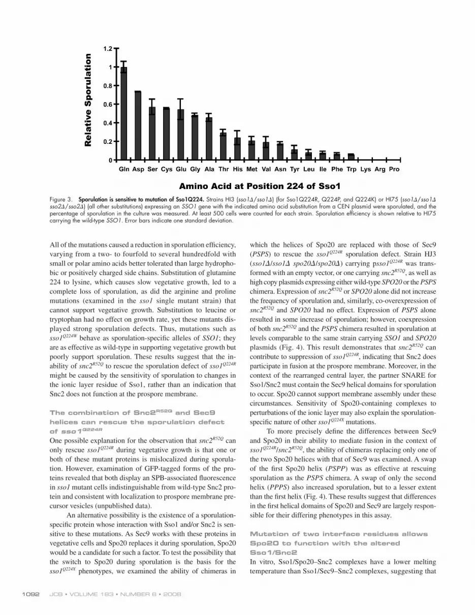
Sporulation is sensitive to perturbation of Sso1 Q224 The strains carrying the various sso1 Q224X alleles as their only SSO
were then examined for their ability to sporulate. Unlike growth
rate, sporulation was sensitive to changes at this position ( Fig. 3 ).
plasmid carrying the snc2 R52Q allele. As expected, the sso1 Q224R allele did not rescue the sporulation defect of the sso1 � ( Fig. 1 ).
Neither SNC2 nor snc2 R52Q were capable of restoring sporula-
tion in this context ( Fig. 1 ). This failure of snc2 R52Q to rescue the
sporulation defect raises the possibility that Snc2p does not par-
ticipate in vesicle fusion at the prospore membrane. We there-
fore examined whether mutation of the central layer arginine to
glutamine in any of the other S. cerevisiae R-SNAREs (Snc1,
Ykt6, Sec22, Nyv1) could restore sporulation to the sso1 Q224R strain. As with snc2 R52Q , none of these mutant SNAREs could
compensate for the sso1 Q224R mutation (unpublished data).
To ensure that snc2 R52Q was indeed capable of suppressing
sso1 Q224R in our strains, we constructed a strain homozygous for
deletion of SSO1 and SSO2 and kept alive by the SSO1 gene on
a centromeric plasmid (HI75). When the sso1 Q224R mutation was
introduced into this strain, the plasmid bearing the wild-type
gene could be lost only when the snc2 R52Q allele was also pres-
ent. The resulting strain is viable because of the compensatory
interaction between the two mutant SNAREs ( Katz and Brenn-
wald, 2000 ). However, as with the results in the sso1 � / sso1 � strain, the presence of snc2 R52Q does not rescue the sporulation
Figure 1. Compensatory mutation of SNC2 cannot rescue the sporulation defect of sso1 Q224R . Strains HI3 ( sso1 � / sso1 � ) or HI75 ( sso1 � / sso1 � sso2 � / sso2 � ) were transformed with the CEN plasmids expressing the indicated genes and sporulated in liquid culture. Sporulation was assessed by observation in the light microscope and by ether test. To determine percentage of sporulation, at least 500 cells were counted for each strain. For HI75, the plasmid carrying the wild-type SSO1 was lost by growth on 5-FOA before cells were assayed.
Figure 2. Mutation of Sso1Q224 is well tolerated in vegetative growth. Strain HI75 ( sso1 � / sso1 � sso2 � / sso2 � ) was transformed with CEN plasmids carrying all possible amino acid substitutions at position 224 of Sso1 and the plasmid carrying the wild-type SSO1 was lost by plasmid shuffl e, leaving the mutant as the sole form of Sso protein in the cell. Transformants carrying substitutions that could support growth (all except arginine and proline) were streaked out on YPD plates and incubated at 37 ° C. Letters indicate the amino acid present at position at 224 of Sso1 in each strain.
JCB • VOLUME 183 • NUMBER 6 • 2008 1092
which the helices of Spo20 are replaced with those of Sec9
( PSPS ) to rescue the sso1 Q224R sporulation defect. Strain HJ3
( sso1 � / sso1 � spo20 � / spo20 � ) carrying p sso1 Q224R was trans-
formed with an empty vector, or one carrying snc2 R52Q , as well as
high copy plasmids expressing either wild-type SPO20 or the PSPS
chimera. Expression of snc2 R52Q or SPO20 alone did not increase
the frequency of sporulation and, similarly, co-overexpression of
snc2 R52Q and SPO20 had no effect. Expression of PSPS alone
resulted in some increase of sporulation; however, coexpression
of both snc2 R52Q and the PSPS chimera resulted in sporulation at
levels comparable to the same strain carrying SSO1 and SPO20
plasmids ( Fig. 4 ). This result demonstrates that snc2 R52Q can
contribute to suppression of sso1 Q224R , indicating that Snc2 does
participate in fusion at the prospore membrane. Moreover, in the
context of the rearranged central layer, the partner SNARE for
Sso1/Snc2 must contain the Sec9 helical domains for sporulation
to occur. Spo20 cannot support membrane assembly under these
circumstances. Sensitivity of Spo20-containing complexes to
perturbations of the ionic layer may also explain the sporulation-
specifi c nature of other sso1 Q224X mutations.
To more precisely defi ne the differences between Sec9
and Spo20 in their ability to mediate fusion in the context of
sso1 Q224R / snc2 R52Q , the ability of chimeras replacing only one of
the two Spo20 helices with that of Sec9 was examined. A swap
of the fi rst Spo20 helix ( PSPP ) was as effective at rescuing
sporulation as the PSPS chimera. A swap of only the second
helix ( PPPS ) also increased sporulation, but to a lesser extent
than the fi rst helix ( Fig. 4 ). These results suggest that differences
in the fi rst helical domains of Spo20 and Sec9 are largely respon-
sible for their differing phenotypes in this assay.
Mutation of two interface residues allows Spo20 to function with the altered Sso1/Snc2 In vitro, Sso1/Spo20 – Snc2 complexes have a lower melting
temperature than Sso1/Sec9 – Snc2 complexes, suggesting that
All of the mutations caused a reduction in sporulation effi ciency,
varying from a two- to fourfold to several hundredfold with
small or polar amino acids better tolerated than large hydropho-
bic or positively charged side chains. Substitution of glutamine
224 to lysine, which causes slow vegetative growth, led to a
complete loss of sporulation, as did the arginine and proline
mutations (examined in the sso1 single mutant strain) that
cannot support vegetative growth. Substitution to leucine or
tryptophan had no effect on growth rate, yet these mutants dis-
played strong sporulation defects. Thus, mutations such as
sso1 Q224W behave as sporulation-specifi c alleles of SSO1 ; they
are as effective as wild-type in supporting vegetative growth but
poorly support sporulation. These results suggest that the in-
ability of snc2 R52Q to rescue the sporulation defect of sso1 Q224R might be caused by the sensitivity of sporulation to changes in
the ionic layer residue of Sso1, rather than an indication that
Snc2 does not function at the prospore membrane.
The combination of Snc2 R52Q and Sec9 helices can rescue the sporulation defect of sso1 Q224R One possible explanation for the observation that snc2 R52Q can
only rescue sso1 Q224R during vegetative growth is that one or
both of these mutant proteins is mislocalized during sporula-
tion. However, examination of GFP-tagged forms of the pro-
teins revealed that both display an SPB-associated fl uorescence
in sso1 mutant cells indistinguishable from wild-type Snc2 pro-
tein and consistent with localization to prospore membrane pre-
cursor vesicles (unpublished data).
An alternative possibility is the existence of a sporulation-
specifi c protein whose interaction with Sso1 and/or Snc2 is sen-
sitive to these mutations. As Sec9 works with these proteins in
vegetative cells and Spo20 replaces it during sporulation, Spo20
would be a candidate for such a factor. To test the possibility that
the switch to Spo20 during sporulation is the basis for the
sso1 Q224X phenotypes, we examined the ability of chimeras in
Figure 3. Sporulation is sensitive to mutation of Sso1Q224. Strains HI3 ( sso1 � / sso1 � ) (for Sso1Q224R, Q224P, and Q224K) or HI75 ( sso1 � / sso1 � sso2 � / sso2 � ) (all other substitutions) expressing an SSO1 gene with the indicated amino acid substitution from a CEN plasmid were sporulated, and the percentage of sporulation in the culture was measured. At least 500 cells were counted for each strain. Sporulation effi ciency is shown relative to HI75 carrying the wild-type SSO1 . Error bars indicate one standard deviation.

1093SNARE SPECIFICITY IN VIVO • Yang et al.
the relative ability of the different chimeras to promote spor-
ulation, the alterations in the second helix, SPO20 F357L,F361T and SPO20 A378L,K385N had little effect on suppression, though
SPO20 F357L,F361T did display a reproducible, slight improvement
in sporulation effi ciency. In contrast, SPO20 C224L,S231N allowed
sporulation at a level comparable to the PSPP chimera, indicat-
ing that these two residues are primarily responsible for the
ability of this chimera to function in conjunction with sso1 Q224R and snc2 R52Q .
SPO20 C224L,S231N can rescue sec9-4 ts Ectopic expression of SPO20 cannot rescue the temperature-
sensitive growth defect of a sec9-4 mutant, though a chimeric
form of Spo20 carrying the Sec9 helical regions can rescue sec9-4
( Neiman et al., 2000 ). This suggests that the inability of Spo20
to function at the plasma membrane is tied directly to its SNARE
domain. We examined whether the SPO20 C224L,S231N allele affects
the ability of Spo20 to compensate for loss of SEC9 . These ex-
periments were performed with proteins lacking the inhibitory
domain (amino acids 3 – 51) present in the N terminus of Spo20
( Neiman et al., 2000 ). As previously reported, � 3-51 SPO20
cannot rescue sec9-4 , even when present on a high copy plas-
mid, though � 3-51 PSPS was capable of rescuing growth at high
temperature. The � 3-51 SPO20 C224L,S231N allele also rescued
growth of this strain at 37 ° C when present in high copy, though
neither SPO20 C224L,S231N nor the PSPP chimera could rescue when
Spo20 binds less tightly to these other SNAREs than does Sec9
( Liu et al., 2007 ). Packing interactions between side chains of
amino acids located at interfaces on the SNARE helices are
likely to determine how tightly the SNAREs in a given complex
bind to each other. We aligned the interface residues of Spo20
and Sec9 to look for possible suboptimal residues in Spo20
( Fig. 5 ). As criteria to identify such residues, we looked for dif-
ferences in the size and/or chemical properties of the side
chains. In the fi rst helix, only two positions looked signifi cantly
different, a cysteine at the +3 layer of Spo20 that is leucine in
Sec9, and a serine at +5 that is an asparagine in Sec9. In the sec-
ond helix, four differences of note were found; phenylalanines
at the � 2 and � 1 layers that are threonine and leucine, respec-
tively, in Sec9, an alanine in the +4 layer (leucine in Sec9) and
a lysine residue at the +6 position (asparagine in Sec9). These six
residues were mutated in pairs in the context of an otherwise wild-
type SPO20 sequence. The resulting mutants, SPO20 C224L,S231N , SPO20 F357L,F361T , and SPO20 A378L,K385N were all capable of rescu-
ing the sporulation defect of a spo20 mutant, indicating that the
mutants encode functional proteins (unpublished data). They
were then tested for their ability to rescue the sporulation de-
fects of HJ3 ( sso1 � / sso1 � spo20 � / spo20 � ) expressing the
sso1 Q224R and snc2 R52Q alleles ( Fig. 5 ).
When sporulation was assessed on solid medium, the
differences between the PSPP and PPPS chimeras were more
pronounced than in liquid sporulation ( Fig. 4 ). Consistent with
Figure 4. Co-expression of snc2 R52Q and a chimeric SPO20 rescues the sporulation defect of sso1 Q224R . Strain HJ3 ( sso1 � / sso1 � spo20 � / spo20 � ) was transformed with plasmids carrying the indicated genes and sporulated in liquid culture. SSO1 alleles were expressed from CEN plasmids; SNC2 and SPO20 alleles were expressed from high copy plasmids. Sporulation was assessed by observation in the light microscope or by ether test. To determine percentage of sporulation, at least 500 cells were counted for each strain; percentages represent the average of four experiments. “ a ” , “ b ” , and “ c ” illustrate the arrangement of side chain residues at the ionic layer in the different strains.

JCB • VOLUME 183 • NUMBER 6 • 2008 1094
a direct measure of affi nity, the increased association of PSPS
and Spo20 C224L,S231N with both forms of Sso1 and Snc2 are con-
sistent with the idea that they bind more avidly to their partner
SNAREs than wild-type Spo20. Interestingly, all three forms
of Spo20 exhibited greater association with the Sso1 Q224R and
Snc2 R52Q proteins than with the wild-type SNAREs. Because the
amount of SNAREs in complex refl ects both the rates of assem-
bly and of disassembly, we suggest that the general increase in
the amount of SNARE complexes seen with Sso1 Q224R /Snc2 R52Q
might refl ect a role for the central ionic layer in complex dis-
assembly, as suggested previously ( Scales et al., 2001 ).
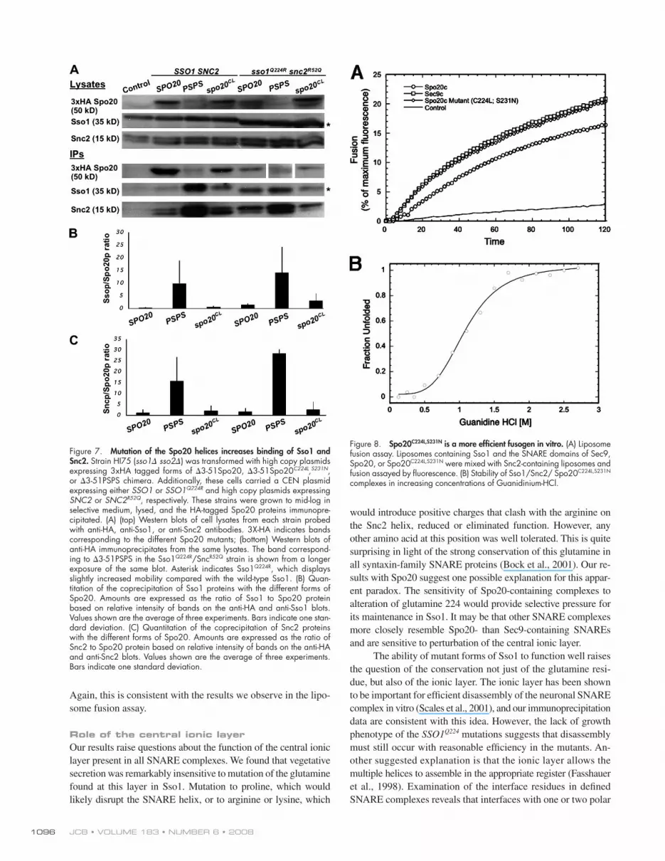
SPO20 C224L,S231N improves function of the SNARE complex in vitro Studies of Sec9- and Spo20-containing SNARE complexes in an
in vitro liposome fusion system indicate that, in a given lipid com-
position, Spo20-containing complexes are less fusogenic than
Sec9 complexes ( Liu et al., 2007 ). Moreover, this lesser activity
correlates with decreased SNARE complex stability (measured
as a lower melting temperature) of the Spo20 complexes com-
pared with Sec9 complexes. The behavior of the SPO20 C224L,S231N mutant in the genetic tests and immunoprecipitations described
above suggests that these mutations might increase the binding
energy of the Spo20-containing complexes. To test this directly,
the recombinant SNARE domain (amino acids 147 – 397) of
Spo20 C224L,S231N was purifi ed from Escherichia coli and tested
with the Sso1 and Snc2 proteins in a liposome fusion assay.
Using liposomes containing 85% POPC (palmitoyl oleoyl phos-
phatidylcholine) and 15% DOPS (di-oleoyl phosphatidylserine),
Sso1/Sec9 – Snc2 complexes promote liposome fusion at a greater
rate than the Sso1/Spo20 – Snc2 SNAREs ( Fig. 8 A ). In contrast,
Sso1/Spo20 C224L,S231N – Snc2 mediates fusion at a rate compara-
ble to the Sec9 complexes. Thus, parallel to the in vivo results,
Spo20 C224L,S231N promotes fusion more effi ciently than Spo20
in vitro.
To determine if the increased fusion activity was refl ected
in increased binding energy, the stability of the Spo20 C224L,S231N -
containing complexes was examined. We previously reported
that Sec9-containing complexes are signifi cantly more stable than
Spo20 complexes during equilibrium unfolding reactions with
chemical denaturants where the concentration of Guanidinium-
HCL required to disrupt 50% of the ternary SNARE complex
was reduced 2.1M for Sec9 vs. 0.9M for Spo20 ( Liu et al.,
2007 ). When the Spo20 C224L,S231N -containing SNAREs were ex-
amined, a concentration of 1.1M Guanidinium-HCL disrupted
50% of these ternary complexes, indicating that they were more
stable than those with Spo20, though still well below the stabil-
ity of Sec9 complexes ( Fig. 8 B ). This moderate improvement
in stability of the Spo20 C224L,S231N -containing complexes is con-
sistent with the slight increase in binding of Spo20 C224L,S231N to
Sso1 and Snc2 seen in the IP experiments ( Fig. 7 ). Together with
the liposome fusion data, these results suggest that modest changes
in affi nity can have strong effects on the fusogenic properties of
the SNAREs. For the neuronal SNARE SNAP-25 it has simi-
larly been found that mutation of interface residues can result in
large differences in function while only modestly altering sta-
bility of the SNARE complex ( Sorensen et al., 2006 ).
expressed from centromeric plasmids ( Fig. 6 ). These results
again suggest that the SPO20 C224L,S231N mutations increase the
strength of Spo20/Sso/Snc interactions, though not to the same
degree as complete replacement with the Sec9 helices. To ensure
the differences seen were not due to differential stability of the
proteins, 3xHA-tagged versions of the different SPO20 and chi-
mera genes were constructed. Western blotting with anti-HA
antibodies indicated that all the SPO20 forms were present in
comparable amounts (unpublished data). Therefore, the results
refl ect differences in the ability of the different forms to promote
vesicle fusion, not differences in protein stability.
Mutation of SPO20 increases association with Sso1 and Snc2 in vivo If alteration of the SNARE helices of Spo20 increases its affi nity
for its partner SNAREs, this should be refl ected in increased bind-
ing of the protein to Sso1 and Snc2. To address this possibility,
HA-tagged � 50-Spo20, Spo20 C224L,S231N , or PSPS were expressed
in combination with either wild-type Sso1 and Snc2 or Sso1 Q224R
and Snc2 R52Q in an sso1 sso2 strain. Lysates were made from
each strain and the Spo20 proteins immunoprecipitated using
anti-HA antibodies. Immunoprecipitates were then blotted and
probed with anti-HA, anti-Sso1, or anti-Snc2 antibodies to ex-
amine association of the three SNARE proteins ( Fig. 7 ). In the
presence of both the wild-type and mutant Sso1 and Snc2 pro-
teins the same pattern was seen; the PSPS chimera precipitated
signifi cantly more Sso1 and Snc2 than Spo20 C224L,S231N , which
in turn brought down slightly more Sso1 and Snc2 than the wild-
type Spo20. Though these immunoprecipitations do not provide
Figure 5. Mutation of two interface residues in the Spo20 SNARE helix allows it to function with sso1 Q224R . (A) Alignment of the interface residues in the SNARE domains of Spo20 and Sec9. Residues chosen for mutation are in blocks. (B) Sporulation of sso1 Q224R snc2 R52Q strains expressing dif-ferent forms of SPO20 . Strain HJ3 ( sso1 � / sso1 � spo20 � / spo20 � ) was transformed with plasmids carrying sso1 Q224R and snc2 R52Q as well as the indicated form of SPO20 . The sso1 Q224R allele was expressed from a CEN plasmid; snc2 R52Q and the SPO20 alleles were expressed from high copy plasmids. These strains were sporulated and sporulation effi ciency mea-sured in the light microscope. At least 500 cells were scored for each strain. Results are the average of three experiments. Error bars indicate one standard deviation.

1095SNARE SPECIFICITY IN VIVO • Yang et al.
Spo20 is the partner, the binding energies at other interfaces are
insuffi cient to overcome the weaker central layer interactions.
During the course of this work, a crystal structure of the
SNARE complex containing the Sso1, Snc2, and Sec9 helical
domains was published ( Strop et al., 2008 ). When this structure
is used to model in the Spo20 cysteine and serine side chains at
the +3 and +5 interface layers, the Spo20 residues result in an
apparent loss of packing interactions between the side chains
(unpublished data), consistent with our results indicating that
the Sec9 residues at these positions improve stability of the
SNARE complex. Mutational analysis of interface residues in
SNAP-25 revealed that interactions in the N-terminal half of the
SNARE domain are important for promoting priming or docking
of the vesicle, whereas interactions in the C-terminal half of the
SNARE helix are critical to drive membrane fusion ( Sorensen
et al., 2006 ). In this regard, it is noteworthy that the critical in-
terfaces differentiating the ability of Spo20 and Sec9 to promote
fusion at the plasma membrane lie in the C-terminal domain,
suggesting that fusion and not docking is the affected step.
Discussion
The use of compensatory mutations in the central ionic layer of
the SNARE domain has proven to be an effective means to dem-
onstrate the participation of different SNARE proteins in the
same complex in vivo ( Graf et al., 2005 ). Here, we attempted to
use this technique to demonstrate a role for the Snc1/2 proteins
in fusion at the prospore membrane. A compensatory mutation in
SNC2 could only rescue the sporulation defect of sso1 Q224R when
expressed in concert with forms of Spo20 carrying the Sec9
SNARE helices. Similar results were obtained using a compen-
satory mutation in SNC1 (unpublished data). These results dem-
onstrate, fi rst, that the Snc1 and Snc2 proteins indeed function as
the R-SNARE subunit of the prospore membrane SNARE com-
plex and, second, that placement of the central layer arginine in
different helices is not functionally equivalent. In this instance,
swapping the glutamine and arginine between the Sso1 and Snc2
helices creates a SNARE bundle that is more sensitive to the
composition of other interface layers in the complex. When
Figure 6. SPO20 C224L,S231N can rescue the growth defect of sec9-4 . (A) Strain AN211 ( sec9-4 ) was transformed with integrating or centromeric (for PSPP and PPPS ) plasmids expressing the indicated genes. Cells were grown to saturation in rich medium and 10-fold serial dilutions were spotted onto selective plates in-cubated at permissive (25 ° C) or restrictive (37 ° C) temperature. SPO20 CS,LN is SPO20 C224L , S231N ; SPO20 CSFF,LNTL is SPO20 C224L,S231N,F357T,F361L ; SPO20 CSFFAK,LNTLLN is SPO20 C224L,S231N,F357T,F361L,A378K,K385N . (B) A similar growth assay in strain AN211 ( sec9-4 ) using high copy plasmids to express the indicated genes.
JCB • VOLUME 183 • NUMBER 6 • 2008 1096
would introduce positive charges that clash with the arginine on
the Snc2 helix, reduced or eliminated function. However, any
other amino acid at this position was well tolerated. This is quite
surprising in light of the strong conservation of this glutamine in
all syntaxin-family SNARE proteins ( Bock et al., 2001 ). Our re-
sults with Spo20 suggest one possible explanation for this appar-
ent paradox. The sensitivity of Spo20-containing complexes to
alteration of glutamine 224 would provide selective pressure for
its maintenance in Sso1. It may be that other SNARE complexes
more closely resemble Spo20- than Sec9-containing SNAREs
and are sensitive to perturbation of the central ionic layer.
The ability of mutant forms of Sso1 to function well raises
the question of the conservation not just of the glutamine resi-
due, but also of the ionic layer. The ionic layer has been shown
to be important for effi cient disassembly of the neuronal SNARE
complex in vitro ( Scales et al., 2001 ), and our immunoprecipitation
data are consistent with this idea. However, the lack of growth
phenotype of the SSO1 Q224 mutations suggests that disassembly
must still occur with reasonable effi ciency in the mutants. An-
other suggested explanation is that the ionic layer allows the
multiple helices to assemble in the appropriate register ( Fasshauer
et al., 1998 ). Examination of the interface residues in defi ned
SNARE complexes reveals that interfaces with one or two polar
Again, this is consistent with the results we observe in the lipo-
some fusion assay.
Role of the central ionic layer Our results raise questions about the function of the central ionic
layer present in all SNARE complexes. We found that vegetative
secretion was remarkably insensitive to mutation of the glutamine
found at this layer in Sso1. Mutation to proline, which would
likely disrupt the SNARE helix, or to arginine or lysine, which
Figure 7. Mutation of the Spo20 helices increases binding of Sso1 and Snc2. Strain HI75 ( sso1 � sso2 � ) was transformed with high copy plasmids expressing 3xHA tagged forms of � 3-51Spo20, � 3-51Spo20 C224L , S231N , or � 3-51PSPS chimera. Additionally, these cells carried a CEN plasmid expressing either SSO1 or SSO1 Q224R and high copy plasmids expressing SNC2 or SNC2 R52Q , respectively. These strains were grown to mid-log in selective medium, lysed, and the HA-tagged Spo20 proteins immunopre-cipitated. (A) (top) Western blots of cell lysates from each strain probed with anti-HA, anti-Sso1, or anti-Snc2 antibodies. 3X-HA indicates bands corresponding to the different Spo20 mutants; (bottom) Western blots of anti-HA immunoprecipitates from the same lysates. The band correspond-ing to � 3-51PSPS in the Sso1 Q224R /Snc R52Q strain is shown from a longer exposure of the same blot. Asterisk indicates Sso1 Q224R , which displays slightly increased mobility compared with the wild-type Sso1. (B) Quan-titation of the coprecipitation of Sso1 proteins with the different forms of Spo20. Amounts are expressed as the ratio of Sso1 to Spo20 protein based on relative intensity of bands on the anti-HA and anti-Sso1 blots. Values shown are the average of three experiments. Bars indicate one stan-dard deviation. (C) Quantitation of the coprecipitation of Snc2 proteins with the different forms of Spo20. Amounts are expressed as the ratio of Snc2 to Spo20 protein based on relative intensity of bands on the anti-HA and anti-Snc2 blots. Values shown are the average of three experiments. Bars indicate one standard deviation.
Figure 8. Spo20 C224L,S231N is a more effi cient fusogen in vitro. (A) Liposome fusion assay. Liposomes containing Sso1 and the SNARE domains of Sec9, Spo20, or Spo20 C224L,S231N were mixed with Snc2-containing liposomes and fusion assayed by fl uorescence. (B) Stability of Sso1/Snc2/ Spo20 C224L,S231N complexes in increasing concentrations of Guanidinium-HCl.
1097SNARE SPECIFICITY IN VIVO • Yang et al.
rescue sporulation of the Sso1 Q224R mutant even in the presence
of the PSPS chimera (unpublished data), suggesting that Sec22
cannot participate in prospore membrane fusion events in vivo.
A recent study revealed that a suboptimal interface at the
+7 layer in the neuronal syx-1A gene is important for allowing
calcium-mediated regulation of secretion ( Lagow et al., 2007 ).
Mutation of the threonine residue at this position in syx-1A to
the corresponding isoleucine residue in syx-2 led to constitutive
fusion. Thus, as with Spo20 and Sec9, in the neuronal SNARE
complex tuning of the strength of binding interactions is impor-
tant for allowing proper regulation of vesicle transport.
Finally, Spo20 and Sec9 provide a useful model for the evo-
lution of novel SNARE complexes. During the evolution of Sac-charomyces , a whole genome duplication occurred that ultimately
gave rise to many related gene pairs in the S. cerevisiae genome
( Wolfe and Shields, 1997 ). Sec9 and Spo20 arose from this dupli-
cation event. In yeasts that diverged from the S. cerevisiae lineage
before the duplication, such as Schizosaccharomyces pombe , a
single Sec9/Spo20 related gene participates in fusion at both the
plasma membrane and the prospore membrane ( Nakamura et al.,
2005 ). Thus, in the S. cerevisiae lineage, the duplication event al-
lowed the two paralogues to become specialized for action at dis-
tinct compartments where the ancestral protein functioned at both
membranes. Similar patterns are likely at work in the expansions
of particular SNARE families seen in plant and mammalian ge-
nomes ( Sanderfoot et al., 2000 ; Bock et al., 2001 ).
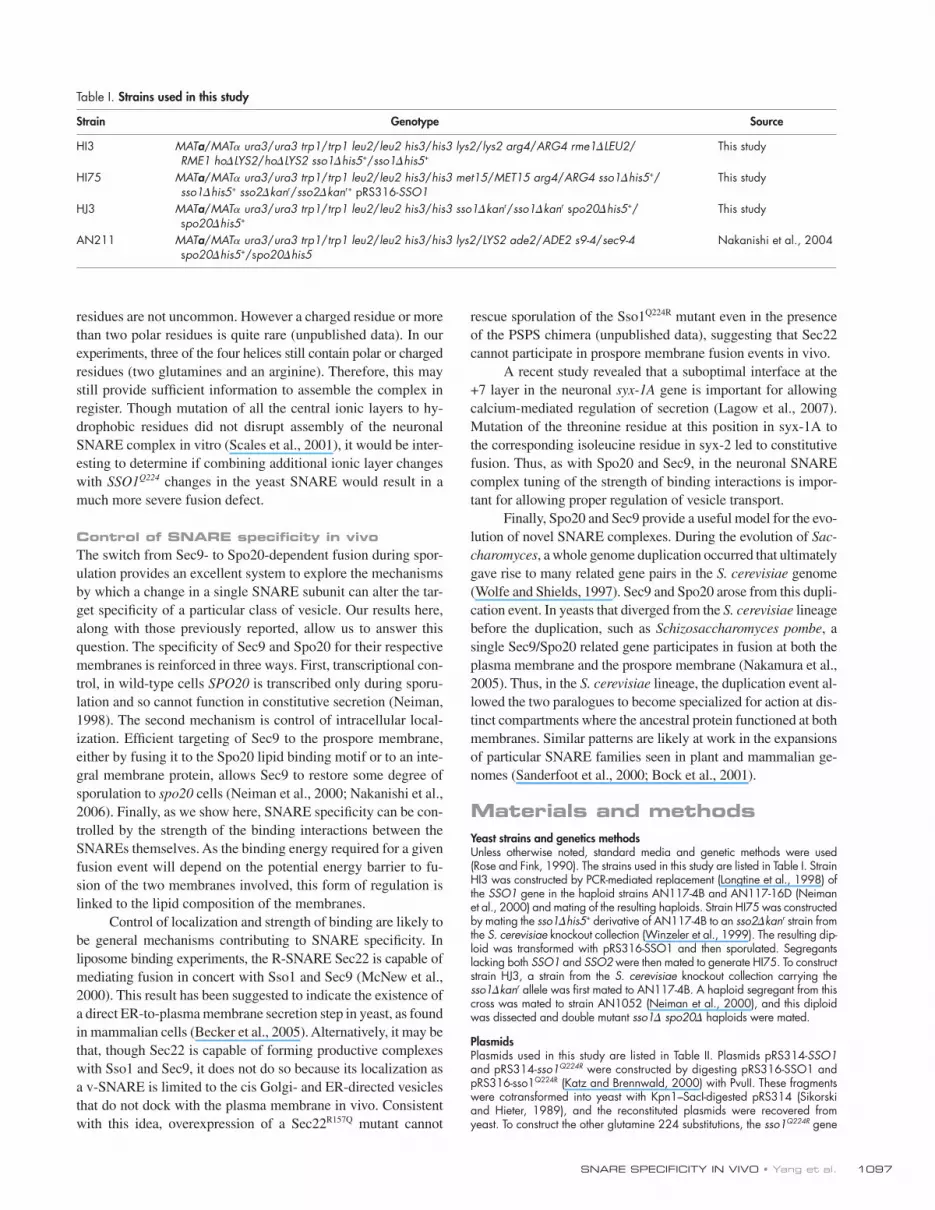
Materials and methods Yeast strains and genetics methods Unless otherwise noted, standard media and genetic methods were used ( Rose and Fink, 1990 ). The strains used in this study are listed in Table I . Strain HI3 was constructed by PCR-mediated replacement ( Longtine et al., 1998 ) of the SSO1 gene in the haploid strains AN117-4B and AN117-16D ( Neiman et al., 2000 ) and mating of the resulting haploids. Strain HI75 was constructed by mating the sso1 � his5 + derivative of AN117-4B to an sso2 � kan r strain from the S. cerevisiae knockout collection ( Winzeler et al., 1999 ). The resulting dip-loid was transformed with pRS316-SSO1 and then sporulated. Segregants lacking both SSO1 and SSO2 were then mated to generate HI75. To construct strain HJ3, a strain from the S. cerevisiae knockout collection carrying the sso1 � kan r allele was fi rst mated to AN117-4B. A haploid segregant from this cross was mated to strain AN1052 ( Neiman et al., 2000 ), and this diploid was dissected and double mutant sso1 � spo20 � haploids were mated.
Plasmids Plasmids used in this study are listed in Table II . Plasmids pRS314- SSO1 and pRS314- sso1 Q224R were constructed by digesting pRS316-SSO1 and pRS316-sso1 Q224R ( Katz and Brennwald, 2000 ) with PvuII. These fragments were cotransformed into yeast with Kpn1 – SacI-digested pRS314 ( Sikorski and Hieter, 1989 ), and the reconstituted plasmids were recovered from yeast. To construct the other glutamine 224 substitutions, the sso1 Q224R gene
residues are not uncommon. However a charged residue or more
than two polar residues is quite rare (unpublished data). In our
experiments, three of the four helices still contain polar or charged
residues (two glutamines and an arginine). Therefore, this may
still provide suffi cient information to assemble the complex in
register. Though mutation of all the central ionic layers to hy-
drophobic residues did not disrupt assembly of the neuronal
SNARE complex in vitro ( Scales et al., 2001 ), it would be inter-
esting to determine if combining additional ionic layer changes
with SSO1 Q224 changes in the yeast SNARE would result in a
much more severe fusion defect.
Control of SNARE specifi city in vivo The switch from Sec9- to Spo20-dependent fusion during spor-
ulation provides an excellent system to explore the mechanisms
by which a change in a single SNARE subunit can alter the tar-
get specifi city of a particular class of vesicle. Our results here,
along with those previously reported, allow us to answer this
question. The specifi city of Sec9 and Spo20 for their respective
membranes is reinforced in three ways. First, transcriptional con-
trol, in wild-type cells SPO20 is transcribed only during sporu-
lation and so cannot function in constitutive secretion ( Neiman,
1998 ). The second mechanism is control of intracellular local-
ization. Effi cient targeting of Sec9 to the prospore membrane,
either by fusing it to the Spo20 lipid binding motif or to an inte-
gral membrane protein, allows Sec9 to restore some degree of
sporulation to spo20 cells ( Neiman et al., 2000 ; Nakanishi et al.,
2006 ). Finally, as we show here, SNARE specifi city can be con-
trolled by the strength of the binding interactions between the
SNAREs themselves. As the binding energy required for a given
fusion event will depend on the potential energy barrier to fu-
sion of the two membranes involved, this form of regulation is
linked to the lipid composition of the membranes.
Control of localization and strength of binding are likely to
be general mechanisms contributing to SNARE specifi city. In
liposome binding experiments, the R-SNARE Sec22 is capable of
mediating fusion in concert with Sso1 and Sec9 ( McNew et al.,
2000 ). This result has been suggested to indicate the existence of
a direct ER-to-plasma membrane secretion step in yeast, as found
in mammalian cells ( Becker et al., 2005 ). Alternatively, it may be
that, though Sec22 is capable of forming productive complexes
with Sso1 and Sec9, it does not do so because its localization as
a v-SNARE is limited to the cis Golgi- and ER-directed vesicles
that do not dock with the plasma membrane in vivo. Consistent
with this idea, overexpression of a Sec22 R157Q mutant cannot
Table I. Strains used in this study
Strain Genotype Source
HI3 MAT a / MAT � ura3 / ura3 trp1 / trp1 leu2 / leu2 his3 / his3 lys2 / lys2 arg4 / ARG4 rme1 � LEU2 / RME1 ho � LYS2 / ho � LYS2 sso1 � his5 + / sso1 � his5 +
This study
HI75 MAT a / MAT � ura3 / ura3 trp1 / trp1 leu2 / leu2 his3 / his3 met15 / MET15 arg4 / ARG4 sso1 � his5 + / sso1 � his5 + sso2 � kan r / sso2 � kan r+ pRS316- SSO1
This study
HJ3 MAT a / MAT � ura3 / ura3 trp1 / trp1 leu2 / leu2 his3 / his3 sso1 � kan r / sso1 � kan r s po20 � his5 + / s po20 � his5 +
This study
AN211 MAT a / MAT � ura3 / ura3 trp1 / trp1 leu2 / leu2 his3 / his3 lys2 / LYS2 ade2 / ADE2 s9-4 / sec9-4 s po20 � his5 + /s po20 � his5
Nakanishi et al., 2004

JCB • VOLUME 183 • NUMBER 6 • 2008 1098
the genes from pRS426-SPO20pr- SPO20 and pRS426-SPO20pr- SPO20 C224L;S231N , respectively, using oligos BBO14 and ANO168. The PCR products were digested with XhoI and SacI, and cloned into similarly di-gested pRS306-SEC9pr ( Neiman et al., 2000 ). To make the sextuple mutant SPO20 C224L;S231N,F357T,F361L,A378LK385N , pRS306-SEC9pr- � 3-51 SPO20 C224L;S231N was used as template for site-directed mutagenesis using fi rst primers HJO33 and HJO34, and then HJO35 and HJO36. To construct 2 � plasmids ex-pressing the various SPO20 mutants and SPO20 / SEC9 chimeras under SEC9 promoter control, KpnI – SacI fragments containing SEC9 promoter with indicated genes were cloned from the integrating and CEN plasmids into KpnI and SacI sites of pRS426.
To construct 3xHA tagged versions of the different SPO20 mutants, two complimentary oligos (HJO72 and HJO73) were synthesized that en-code 3 HA epitopes and anneal to leave XhoI-compatible ends. The oligos were phosphorylated with T4 polynucleotide kinase (Invitrogen) at 37 ° C for 10 min, mixed, and then allowed to anneal. The annealed oligos were then ligated with XhoI-digested pRS306-SEC9pr- SEC9 ( Neiman et al., 2000 ). Site-directed mutagenesis was then performed using oligos HJO78 and HJO79 to restore an XhoI at the junction of 3xHA and SEC9 . Finally, an XhoI – SacI fragment containing SEC9 was replaced with the correspond-ing XhoI – SacI fragments from pRS306- SEC9 pr- � 3-51 SPO20 , - � 3-51 SPO20 C224L;S231N , or - � 3-51 SPO20/SEC9 chimera plasmids.
Sporulation assays Sporulation assays were performed as described previously ( Neiman et al., 2000 ). For tests on solid medium, the strains to be tested were grown overnight on selective media, and then replica plated to sporulation me-dium. After 24 h, spore formation was quantifi ed by direct observation in the light microscope.
was fi rst cloned as a BamHI – HindIII fragment from pRS314- sso1 Q224R into similarly digested pUC119. Site-directed mutagenesis was then performed using oligonucleotides ANO377 and ANO378, which contain randomized nucleotides at codon 224. Sequencing of individual clones from the muta-genesis identifi ed particular substitutions. All substitutions except lysine, glu-tamine, histidine, and aspartate were obtained in this way. For lysine, glutamine, and histidine, the randomized oligos HNO961 and HNO962 were used. For aspartate, mutagenesis was performed using oligos HNO991 and HNO992. After specifi c mutations were identifi ed by sequencing, the 3 � end of the SSO1 gene carrying the glutamine 224 substitution was swapped into pRS314- SSO1 as an NcoI – SalI fragment. To construct the pRS426 and pRS316 plasmids expressing the chimeras PSPS , PSPP , and PPPS , SacI – KpnI fragments carrying the SPO20 promoter and the indicated chimera were isolated from the corresponding integrating plasmids ( Neiman et al., 2000 ) and cloned into similarly digested pRS426 or pRS316, respec-tively ( Sikorski and Hieter, 1989 ; Christianson et al., 1992 ). To construct pRS425 – snc2 R52Q a BamHI – SalI fragment carrying the snc2 R52Q gene ( Katz and Brennwald, 2000 ) was cloned into BamHI – SalI-digested pRS426.
To make pRS316-SEC9pr- PSPP and pRS316-SEC9pr- PPPS , SpeI – SacI fragments carrying the particular chimera were excised from pRS426-SPO20pr- PSPP or - PPPS and cloned into the backbone of SpeI – SacI-digested pRS316-SEC9pr-PSPS ( Nakanishi et al., 2004 ). The plasmids pRS426-SPO20pr- SPO20 C224L;S231N , pRS426-SPO20pr- SPO20 A378L;K385N , and pRS426-SPO20pr- SPO20 F357T;F361L were created by site-directed mutagen-esis of the plasmid pRS426-SPO20pr- SPO20 ( Nakanishi et al., 2004 ) using oligos HJO31 and HJO32, HJO33 and HJO34, and HJO35 and HJO36, respectively.
Integrating plasmids expressing � 3-51 SPO20 and � 3-51 SPO20 C224L;S231N under the SEC9 promoter were assembled by amplifying
Table II. Plasmids used in this study
Name Source
pRS314 Sikorski et al., 1989
pRS314- SSO1 This study
pRS314- sso1 Q224R This study
pRS425 Christianson et al., 1992
pRS425- snc2 R52Q This study
pRS426 Christianson et al., 1992
pRS426- SPO20 Nakanishi et al., 2004
pRS426- PSPS This study
pRS426- PSPP This study
pRS426- PPPS This study
pRS426- SPO20 C224L,S231N This study
pRS426- SPO20 F357T,F361L This study
pRS426- SPO20 A378L,K385N This study
pRS306-SEC9pr- SEC9 Neiman et al., 2000
pRS306-SEC9pr- � 3-51 SPO20 This study
pRS306-SEC9pr- � 3-51 PSPS This study
pRS306-SEC9pr- � 3-51 SPO20 C224L,S231N This study
pRS306-SEC9pr- � 3-51 SPO20 C224L,S231,F357T,F361L,A378L,K385N This study
pRS316-SEC9pr- � 3-51 PSPP This study
pRS316-SEC9pr- � 3-51 PPPS This study
pRS426-SEC9pr- SEC9 This study
pRS426-SEC9pr- � 3-51 SPO20 This study
pRS426-SEC9pr- � 3-51 PSPS This study
pRS426-SEC9pr- � 3-51 SPO20 C224L,S231N This study
pRS426-SEC9pr- � 3-51 PSPP This study
pRS426-SEC9pr- � 3-51 PPPS This study
pRS426-SEC9pr-3xHA SEC9 This study
pRS426-SEC9pr-3xHA � 3-51 SPO20 This study
pRS426-SEC9pr-3xHA � 3-51 PSPS This study
pRS426-SEC9pr-3xHA � 3-51 SPO20 C224L,S231N This study
pRS426-SEC9pr-3xHA � 3-51 SPO20 F357T,F361L This study
pRS426-SEC9pr-3xHA � 3-51 PSPP This study
pRS426-SEC9pr-3xHA � 3-51 PPPS This study

1099SNARE SPECIFICITY IN VIVO • Yang et al.
The authors wish to thank Pat Brennwald and Reinhard Jahn for plasmids. This work was supported by National Institutes of Health grants
GM62184 (to A.M. Neiman) and GM071832 (to J.A. McNew).
Submitted: 24 September 2008 Accepted: 11 November 2008
References Aalto , M.K. , H. Ronne , and S. Keranen . 1993 . Yeast syntaxins Sso1p and Sso2p
belong to a family of related membrane proteins that function in vesicular transport. EMBO J. 12 : 4095 – 4104 .
Becker , T. , A. Volchuk , and J.E. Rothman . 2005 . Differential use of endoplasmic reticulum membrane for phagocytosis in J774 macrophages. Proc. Natl. Acad. Sci. USA . 102 : 4022 – 4026 .
Bock , J.B. , H.T. Matern , A.A. Peden , and R.H. Scheller . 2001 . A genomic per-spective on membrane compartment organization. Nature . 409 : 839 – 841 .
Brennwald , P. , B. Kearns , K. Champion , S. Keranen , V. Bankaitis , and P. Novick . 1994 . Sec9 is a SNAP-25-like component of a yeast SNARE complex that may be the effector of Sec4 function in exocytosis. Cell . 79 : 245 – 258 .
Carr , C.M. , E. Grote , M. Munson , F.M. Hughson , and P.J. Novick . 1999 . Sec1p binds to SNARE complexes and concentrates at sites of secretion. J. Cell Biol. 146 : 333 – 344 .
Christianson , T.W. , R.S. Sikorski , M. Dante , J.H. Shero , and P. Hieter . 1992 . Multifunctional yeast high-copy-number shuttle vectors. Gene . 110 : 119 – 122 .
Coluccio , A. , M. Malzone , and A.M. Neiman . 2004 . Genetic evidence of a role for membrane lipid composition in the regulation of soluble NEM- sensitive factor receptor function in Saccharomyces cerevisiae. Genetics . 166 : 89 – 97 .
Fasshauer , D. , R.B. Sutton , A.T. Brunger , and R. Jahn . 1998 . Conserved structural features of the synaptic fusion complex: SNARE proteins reclassifi ed as Q- and R-SNAREs. Proc. Natl. Acad. Sci. USA . 95 : 15781 – 15786 .
Graf , C.T. , D. Riedel , H.D. Schmitt , and R. Jahn . 2005 . Identifi cation of func-tionally interacting SNAREs by using complementary substitutions in the conserved ‘ 0 ’ layer. Mol. Biol. Cell . 16 : 2263 – 2274 .
Jahn , R. , and R.H. Scheller . 2006 . SNAREs – engines for membrane fusion. Nat. Rev. Mol. Cell Biol. 7 : 631 – 643 .
Jantti , J. , M.K. Aalto , M. Oyen , L. Sundqvist , S. Keranen , and H. Ronne . 2002 . Characterization of temperature-sensitive mutations in the yeast syntaxin 1 homologues Sso1p and Sso2p, and evidence of a distinct function for Sso1p in sporulation. J. Cell Sci. 115 : 409 – 420 .
Katz , L. , and P. Brennwald . 2000 . Testing the 3Q:1R “ rule ” : mutational analysis of the ionic “ zero ” layer in the yeast exocytic SNARE complex reveals no requirement for arginine. Mol. Biol. Cell . 11 : 3849 – 3858 .
Lagow , R.D. , H. Bao , E.N. Cohen , R.W. Daniels , A. Zuzek , W.H. Williams , G.T. Macleod , R.B. Sutton , and B. Zhang . 2007 . Modifi cation of a hydropho-bic layer by a point mutation in syntaxin 1A regulates the rate of synaptic vesicle fusion. PLoS Biol. 5 : e72 .
Liu , S. , K.A. Wilson , T. Rice-Stitt , A.M. Neiman , and J.A. McNew . 2007 . In vitro fusion catalyzed by the sporulation-specifi c t-SNARE light-chain Spo20p is stimulated by phosphatidic acid. Traffi c . 8 : 1630 – 1643 .
Longtine , M.S. , A. McKenzie III , D.J. Demarini , N.G. Shah , A. Wach , A. Brachat , P. Philippsen , and J.R. Pringle . 1998 . Additional modules for versatile and economical PCR-based gene deletion and modifi cation in Saccharomyces cerevisiae. Yeast . 14 : 953 – 961 .
McNew , J.A. 2008 . Regulation of SNARE-mediated membrane fusion during exocytosis. Chem. Rev. 108 : 1669 – 1686 .
McNew , J.A. , F. Parlati , R. Fukuda , R.J. Johnston , K. Paz , F. Paumet , T.H. Sollner , and J.E. Rothman . 2000 . Compartmental specifi city of cellular membrane fusion encoded in SNARE proteins. Nature . 407 : 153 – 159 .
Nakamura , T. , J. Kashiwazaki , and C. Shimoda . 2005 . A fi ssion yeast SNAP-25 homologue, SpSec9, is essential for cytokinesis and sporulation. Cell Struct. Funct. 30 : 15 – 24 .
Nakanishi , H. , P. de los Santos , and A.M. Neiman . 2004 . Positive and negative regulation of a SNARE protein by control of intracellular localization. Mol. Biol. Cell . 15 : 1802 – 1815 .
Nakanishi , H. , M. Morishita , C.L. Schwartz , A. Coluccio , J. Engebrecht , and A.M. Neiman . 2006 . Phospholipase D and the SNARE Sso1p are necessary for vesicle fusion during sporulation in yeast. J. Cell Sci. 119 : 1406 – 1415 .
Neiman , A.M. 1998 . Prospore membrane formation defi nes a developmen-tally regulated branch of the secretory pathway in yeast. J. Cell Biol. 140 : 29 – 37 .
Neiman , A.M. 2005 . Ascospore formation in the yeast Saccharomyces cerevisiae. Microbiol. Mol. Biol. Rev. 69 : 565 – 584 .
For liquid sporulation and ether tests, 1.5 ml of overnight-cultured cells were pelleted, washed once in 1 ml 2% potassium acetate, and resus-pended in 10 ml 2% potassium acetate. After 2 d of incubation at 30 ° C, the sporulation frequency was determined by observation under the light microscope; meanwhile, 5 μ l of the culture was spotted onto a YPD plate. The plate was inverted over a paper fi lter soaked with 2 ml of ethyl ether for 30 min. After 30 min the paper fi lter was removed, and the plate was incubated at 30 ° C overnight.
Growth assays To assay the growth defect of the spo20 � sec9-4 mutant, cells were fi rst cultured overnight at 25 ° C in YPD. Thereafter, 10-fold serial-diluted cell cultures were spotted onto two identical plates selective for the plasmid. One plate was placed at 25 ° C, and the other at 37 ° C to monitor the growth rate of the spo20 � sec9-4 mutant.
Immunoprecipitations The immunoprecipitation assays were modifi ed from Carr et al. (1999 ). Strain HI75 was transformed with CEN plasmids expressing the different SSO1 genes and high copy plasmids expressing the different SNC2 and SPO20 alleles. 5 ml of overnight culture was diluted into 100 ml of selective medium and grown to mid-log phase. Cells were harvested and resuspended in 1 ml of ice-cold wash buffer (20 mM Tris, pH 7.5, 20 mM NaN 3 , and 20 mM NaF). Washed cells were pelleted at 4 ° C, resuspended in 1 ml ice-cold IP buffer (50 mM Hepes, pH 7.4, 150 mM KCl, 1 mM EDTA, 1 mM DTT, and 0.5% NP-40), and treated with zymolyase (100 μ g/ml) for 10 min. Cells were pelleted and resuspended in 500 μ l ice-cold IP buffer with protease in-hibitors. Cells were lysed by shaking with glass beads (0.5 mm) at 4 ° C for 10 min. Lysed cells were pelleted for 10 min at 13,000 g and the super-natants were precleared by addition of protein G – Sepharose beads (GE Healthcare). The mixtures were rocked for 30 min at 4 ° C and then centri-fuged for 15 min at 13,000 g at 4 ° C to pellet the beads, debris, and non-specifi cally bound products. To precipitate the HA-tagged proteins, anti-HA monoclonal antibodies (clone HA-7; Sigma-Aldrich) were added to the pre-cleared supernatants for 30 min before G – Sepharose beads were added, and the mixtures were incubated at 4 ° C overnight. The beads and bound protein were pelleted for 10 s at 4,000 g , and washed fi ve times with 1 ml ice-cold IP buffer. Proteins were eluted from the beads by boiling them in 2X SDS sample buffer (60 mM Tris-HCl, pH 6.8, 10% glycerol, 2% SDS, 0.05 mg/ml bromophenol blue, and 5% � -mercaptoethanol) for 5 min.
Proteins of interest were analyzed by Western blot. 3xHA- � 3-51Spo20 species were separated by SDS-PAGE on 10% mini-gels, whereas Ssop and Sncp were resolved on 15% mini-gels. Proteins were transferred to PVDF membranes (Millipore). 3xHA- � 3-51Spo20 species were visualized with chicken anti-HA antibodies (Aves Laboratories) to minimize the cross-reactivity from the mouse HA antibodies in the IP. Sso1 and Snc2 were detected by rabbit anti-Sso1 and rabbit anti-Snc2 ( Sogaard et al., 1994 ), respectively. Peroxidase-conjugated secondary antibodies (anti – chicken or anti – rabbit) were used. The band intensities were deter-mined using ImageJ and the ratios of Sso1 or Snc2 to the precipitated 3xHA- � 3-51Spo20 protein were calculated to compare coprecipitation of Sso1 and Snc2 with the different 3xHA- � 3-51Spo20 species.
Protein expression and purifi cation Sso1, Snc1, Sec9, and Spo20 were expressed and purifi ed as previously de-scribed in detail ( Liu et al., 2007 ). Spo20 C224L,S231N was expressed and purifi ed from pET24a(+)-based plasmid pJM557 as done previously for Spo20. The Spo20 C224L,S231N (147 – 397) fragment for pJM557 was amplifi ed from pRS306SEC9pr:: Δ 50spo20 C224L;S231N using primers #303 (CCGAATTCGACTATC-CACAGTGG) and #304 (GCACGCGTCTCGAGTCACCATCTTTTCCCG).
Liposome fusion assays Liposome reconstitution and fusion assay were performed as described previously ( Liu et al., 2007 ).
Melting temperature determination The stability of Spo20 C224L,S231N ternary SNARE complex was determined by chemical denaturation using guanidine HCl as the denaturant. Changes in CD signal were performed using a spectrometer (model 62DS; Aviv) as described previously ( Liu et al., 2007 ). The [GdnHCl] 1/2 of the Spo20 C224L,S231N ternary SNARE complex was determined by KaleidaGraph (Synergy Software) using nonlinear least squares analysis.
Image acquisition and processing Images of yeast growth and Western blots were acquired on a scanner (model 2450; Epson) and fi gures were prepared using Microsoft Power-Point and Adobe Photoshop 9.0.

JCB • VOLUME 183 • NUMBER 6 • 2008 1100
Neiman , A.M. , L. Katz , and P.J. Brennwald . 2000 . Identifi cation of domains re-quired for developmentally regulated SNARE function in Saccharomyces cerevisiae. Genetics . 155 : 1643 – 1655 .
Oyler , G.A. , G.A. Higgins , R.A. Hart , E. Battenberg , M. Billingsley , F.E. Bloom , and M.C. Wilson . 1989 . The identifi cation of a novel synaptosomal- associated protein, SNAP-25, differentially expressed by neuronal subpopu-lations. J. Cell Biol. 109 : 3039 – 3052 .
Parlati , F. , J.A. McNew , R. Fukuda , R. Miller , T.H. Sollner , and J.E. Rothman . 2000 . Topological restriction of SNARE-dependent membrane fusion. Nature . 407 : 194 – 198 .
Parlati , F. , O. Varlamov , K. Paz , J.A. McNew , D. Hurtado , T.H. Sollner , and J.E. Rothman . 2002 . Distinct SNARE complexes mediating membrane fusion in Golgi transport based on combinatorial specifi city. Proc. Natl. Acad. Sci. USA . 99 : 5424 – 5429 .
Paumet , F. , B. Brugger , F. Parlati , J.A. McNew , T.H. Sollner , and J.E. Rothman . 2001 . A t-SNARE of the endocytic pathway must be activated for fusion. J. Cell Biol. 155 : 961 – 968 .
Paumet , F. , V. Rahimian , and J.E. Rothman . 2004 . The specifi city of SNARE-dependent fusion is encoded in the SNARE motif. Proc. Natl. Acad. Sci. USA . 101 : 3376 – 3380 .
Pelham , H.R. 1999 . SNAREs and the secretory pathway-lessons from yeast. Exp. Cell Res. 247 : 1 – 8 .
Poirier , M.A. , W. Xiao , J.C. Macosko , C. Chan , Y.K. Shin , and M.K. Bennett . 1998 . The synaptic SNARE complex is a parallel four-stranded helical bundle. Nat. Struct. Biol. 5 : 765 – 769 .
Protopopov , V. , B. Govindan , P. Novick , and J.E. Gerst . 1993 . Homologs of the synaptobrevin/VAMP family of synaptic vesicle proteins function on the late secretory pathway in S. cerevisiae. Cell . 74 : 855 – 861 .
Rose , M.D. , and G.R. Fink . 1990 . Methods in Yeast Genetics. Cold Spring Harbor Laboratory Press, Cold Spring Harbor, NY.
Sanderfoot , A.A. , F.F. Assaad , and N.V. Raikhel . 2000 . The Arabidopsis genome. An abundance of soluble N-ethylmaleimide-sensitive factor adaptor pro-tein receptors. Plant Physiol. 124 : 1558 – 1569 .
Scales , S.J. , B.Y. Yoo , and R.H. Scheller . 2001 . The ionic layer is required for effi cient dissociation of the SNARE complex by a-SNAP and NSF. Proc. Natl. Acad. Sci. USA . 98 : 14262 – 14267 .
Sikorski , R.S. , and P. Hieter . 1989 . A system of shuttle vectors and yeast host strains designed for effi cient manipulation of DNA in Saccharomyces cerevisiae . Genetics . 122 : 19 – 27 .
Sogaard , M. , K. Tani , R.R. Ye , S. Geromanos , P. Tempst , T. Kirchhausen , J.E. Rothman , and T. Sollner . 1994 . A rab protein is required for the assem-bly of SNARE complexes in the docking of transport vesicles. Cell . 78 : 937 – 948 .
Sollner , T. , S.W. Whiteheart , M. Brunner , H. Erdjument-Bromage , S. Geromanos , P. Tempst , and J.E. Rothman . 1993 . SNAP receptors implicated in vesicle targeting and fusion. Nature . 362 : 318 – 324 .
Sorensen , J.B. , K. Wiederhold , E.M. Muller , I. Milosevic , G. Nagy , B.L. de Groot , H. Grubmuller , and D. Fasshauer . 2006 . Sequential N- to C-terminal SNARE complex assembly drives priming and fusion of secretory vesicles. EMBO J. 25 : 955 – 966 .
Strop , P. , S.E. Kaiser , M. Vrljic , and A.T. Brunger . 2008 . The structure of the yeast plasma membrane SNARE complex reveals destabilizing water-fi lled cavities. J. Biol. Chem. 283 : 1113 – 1119 .
Sutton , R.B. , D. Fasshauer , R. Jahn , and A.T. Brunger . 1998 . Crystal structure of a SNARE complex involved in synaptic exocytosis at 2.4 A resolution. Nature . 395 : 347 – 353 .
Weber , T. , B.V. Zemelman , J.A. McNew , B. Westermann , M. Gmachl , F. Parlati , T.H. Sollner , and J.E. Rothman . 1998 . SNAREpins: minimal machinery for membrane fusion. Cell . 92 : 759 – 772 .
Weimbs , T. , K. Mostov , S.H. Low , and K. Hofmann . 1998 . A model for structural similarity between different SNARE complexes based on sequence rela-tionships. Trends Cell Biol. 8 : 260 – 262 .
Winzeler , E.A. , D.D. Shoemaker , A. Astromoff , H. Liang , K. Anderson , B. Andre , R. Bangham , R. Benito , J.D. Boeke , H. Bussey , et al . 1999 . Functional characterization of the S. cerevisiae genome by gene deletion and parallel analysis. Science . 285 : 901 – 906 .
Wolfe , K.H. , and D.C. Shields . 1997 . Molecular evidence for an ancient duplica-tion of the entire yeast genome. Nature . 387 : 708 – 713 .
Yang , B. , L. Gonzalez Jr ., R. Prekeris , M. Steegmaier , R.J. Advani , and R.H. Scheller . 1999 . SNARE interactions are not selective. Implications for membrane fusion specifi city. J. Biol. Chem. 274 : 5649 – 5653 .


