Embed Size (px)
Citation preview

B
JFa
b
a
ARRAA
KANDPA
1
f8pzat(tstsagt
cc
0h
Journal of Pharmaceutical and Biomedical Analysis 70 (2012) 13– 25
Contents lists available at SciVerse ScienceDirect
Journal of Pharmaceutical and Biomedical Analysis
jou rn al h om epage: www.elsev ier .com/ locate / jpba
ioactive alkaloid extracts from Narcissus broussonetii: Mass spectral studies
ean Paulo de Andradea, Natalia Belén Pignia, Laura Torras-Claveriaa, Strahil Berkova,b, Carles Codinaa,rancesc Viladomata, Jaume Bastidaa,∗
Department of Natural Products, Plant Biology and Soil Science, Faculty of Pharmacy, Av. Diagonal 643, 08028 Barcelona, SpainAgroBioInstitute, 8 Dragan Tzankov Blvd., Sofia 1164, Bulgaria
r t i c l e i n f o
rticle history:eceived 15 February 2012eceived in revised form 21 April 2012ccepted 4 May 2012vailable online 14 May 2012
eywords:maryllidaceaearcissusinitrogenous alkaloidsretazettine
a b s t r a c t
Plants of the Amaryllidaceae family are a well-known source of tetrahydroisoquinoline alkaloids with awide range of biological activities, including antiviral, antitumoral, antiparasitic, psychopharmacological,and acetylcholinesterase inhibitory, among others. Recent advances in the use of GC or LC coupled to MShave allowed a chemically guided isolation of uncommon and bioactive alkaloids. In the present work,analytical methods were applied to study the alkaloid profile of Narcissus broussonetii, a plant endemicto North Africa. Using the GC–MS technique and an in-home mass fragmentation database, twenty-three alkaloids were identified, including the very rare dinitrogenous alkaloids obliquine, plicamine, andsecoplicamine. Applying LC–ESI-LTQ-Orbitrap-MS, fragmentation profiles were found to be similar forobliquine and plicamine but different for secoplicamine. Pretazettine, a potent cytotoxic alkaloid, wasalso isolated from N. broussonetii, although its identification by GC–MS was only possible after a BSTFA-
ntiprotozoal studies derivatization. The silylated crude methanolic extract only showed the presence of pretazettine–TMS,confirming that tazettine was formed after the alkaloid extraction. The same observation was made inNarcissus cultivars in which tazettine had been detected as the major alkaloid. As part of an ongoing projecton MS of Amaryllidaceae alkaloids, the silylated tazettine and pretazettine were studied by GC–MS/MS,and found to differ in their fragmentation routes. Finally, the EtOAc extract of N. broussonetii showed
gains
notable in vitro activity a. Introduction
Narcissus is the most common genus of the Amaryllidaceaeamily in the Iberian Peninsula and North Africa, comprising0–100 wild species. The alkaloids found in Amaryllidaceae speciesossess putative pharmacological properties such as antiproto-oal, antiviral, antitumoral, and acetylcholinesterase inhibitoryctivity [1–3]. The well-known Amaryllidaceae alkaloid, galan-hamine, is a marketed drug for Alzheimer’s disease therapyRazadyne®, formerly Reminyl®). Alkaloids like narciclasine, pre-azettine and others bearing haemanthamine- and lycorine-typekeletons have demonstrated interesting antitumoral and/or apop-otic effects [4–6]. Some compounds of the lycorine series havehown interactions with the human cytochrome P450 3A4 [7]. From
biosynthetic point of view, the Amaryllidaceae alkaloids in theenus Narcissus are grouped in eight skeleton types formed fromhe common precursor O-methylnorbelladine [2].
The development of metabolite profiling methods usingapillary electrophoresis, gas chromatography (GC), gashromatography coupled to mass spectrometry (GC–MS) or
∗ Corresponding author. Tel.: +34 934020268; fax: +34 934029043.E-mail address: [email protected] (J. Bastida).
731-7085/$ – see front matter © 2012 Elsevier B.V. All rights reserved.ttp://dx.doi.org/10.1016/j.jpba.2012.05.009
t Trypanosoma cruzi, with an IC50 value of 1.77 �g/ml.© 2012 Elsevier B.V. All rights reserved.
high-performance liquid chromatography (HPLC) has allowed theidentification and quantification of many Amaryllidaceae alkaloids[8–11]. In biological samples, liquid chromatography–electrosprayionization–tandem mass spectrometry (LC–ESI-MS/MS) has beensuccessfully used to quantify alkaloids bearing the galanthamine-type skeleton [12]. It has been found that Amaryllidaceae alkaloids,with a few exceptions, can be analyzed by GC–MS without anyprevious derivatization, and they show a mass spectral fragmen-tation pattern very similar to those recorded by direct insertionprobe [13]. Furthermore, the high resolution ability of the capillarycolumn allows the separation of more than 60–70 compounds fromcomplex mixtures, while the identification of known alkaloidsis achieved through the specific mass fragmentation by electronimpact mass spectrometry (EI-MS) and retention indices usingdeconvolution software [14]. These features facilitate the devel-opment of an in-home spectral database of known compounds,allowing their rapid identification and the isolation of compoundsshowing unusual EI-MS fragmentations. Nevertheless, a drawbackof GC–MS is the lack of information about some unstable com-pounds (or those in N-oxide form) together with the low stability
of several molecular ions, as found in homolycorine-type alka-loids. Compounds such as haemanthamine may undergo thermaldegradation and hence show different mass fragmentation underGC conditions [13].
1 utical
atwaasa
t(cdt
2
2
aafSgaaanft((S
2
atctScacD
2
bi5f
2
wbufircr
Ion Trap detector (Thermo Scientific, Hemel Hempstead, UnitedKingdom). An HP-5 MS column (30 m × 0.25 mm × 0.25 �m, Agi-
4 J.P. de Andrade et al. / Journal of Pharmace
The growing interest in Amaryllidaceae alkaloids after the ther-peutic success of galanthamine has also prompted the study ofhese compounds using metabolomic approaches. GC–MS analysisas performed to investigate the alkaloid profile of N. broussonetii,
plant species with remarkable in vitro anti-Trypanosoma cruzictivity. Since the dinitrogenous alkaloids obliquine, plicamine, andecoplicamine were not very well detected by GC–MS, they werelso analyzed by LC–ESI-MS/MS.
The sole presence of pretazettine rather than tazettine inhe methanolic macerates was confirmed by previous N,O-bis-trimethylsilyl)trifluoroacetamide (BSTFA) derivatization and alsoorroborated in Narcissus cultivars in which tazettine had beenetected as the major alkaloid. The GC–MS/MS spectra of silylatedazettine and pretazettine are also discussed.
. Experimental
.1. Used chemicals and standards
Methanol (MeOH), diethyl ether (Et2O), n-hexane (n-Hex), ethylcetate (EtOAc), chloroform (CHCl3), acetone (Me)2CO, sulfuriccid (H2SO4) and ammonia (NH3) of analytical grade were usedor the extraction and isolation procedure, being purchased fromDS (France). MeOH, CHCl3, and acetic acid (MeCOOH) of HPLCrade were used in GC–MS (/MS) and HPLC procedures, beinglso purchased from SDS (France). Deuterated methanol (CD3OD)nd deuterated chloroform (CDCl3) with trimethylsilane (TMS)s the internal standard were used for recording nuclear mag-etic resonance (NMR) spectra. Tazettine and pretazettine isolated
rom N. broussonetii were used as positive standards for deriva-ization and/or GC–MS (/MS) study. The hydrocarbon mixtureC9–C36, Restek, Cat no. 31614) was supplied by TeknokromaSpain). The solutions of BSTFA and pyridine were purchased fromigma–Aldrich (St. Louis, MO, USA).
.2. General experimental procedures
Nuclear magnetic resonance (NMR) spectra were recorded in Varian Gemini 300 MHz or Varian VNMRS 400 MHz spectrome-er. Chemical shifts were reported in ı units (ppm) and couplingonstants (J) were expressed in Hz. A Jasco-J-810 Spectrophotome-er was used to run CD spectra, all recorded in MeOH. Silica gelDS chromagel 60 A CC (6–35 �m) was used for vacuum liquidhromatography (VLC), and silica gel 60 F254 SDS for analyticalnd semi-preparative thin layer chromatography (TLC). Spots onhromatograms were detected under UV light (254 nm) and withragendorff’s reagent.
.3. Plant material
N. broussonetii Lag. was collected on Wad Mouzeg dar Bouazaeach, near Casablanca (Morocco), in October 2008. A voucher spec-
men was deposited at the University of Barcelona Herbarium (BCN8745). Narcissus cultivars ‘Toto’ and ‘Pencrebar’ were obtainedrom Ludwig Ltd. (The Netherlands).
.4. Extraction procedure
Fresh bulbs (2 kg) of N. broussonetii were macerated thoroughlyith MeOH at room temperature for 48 h (4× 2.0 l), then the com-
ined macerate was filtered and the solvent evaporated to drynessnder reduced pressure. The bulb crude extract (70.7 g) was acidi-
ed to pH 3 with dilute H2SO4 (2%, v/v) and the neutral material wasemoved using Et2O (4× 500 ml). EtOAc (4× 500 ml) was used toarry out a first alkaloid extraction in acid media but with negativeesults. The aqueous solution was basified up to pH 9–10 with NH3and Biomedical Analysis 70 (2012) 13– 25
(25%, v/v) and extracted with n-Hex (5× 500 ml) to give the n-Hexextract (274 mg), followed by extraction with EtOAc (7× 500 ml)to provide the EtOAc extract (1.2 g). Finally, the basic solution wasextracted with an EtOAc–MeOH mixture (3:1) but no alkaloids weredetected.
A rapid alkaloid extraction was performed using 50 mg of driedbulbs from Narcissus cultivars ‘Toto’ and ‘Pencrebar’ in screw-top1.5 ml Eppendorf tubes (6 tubes for each cultivar). The macerationprocedure was carried out with 1 ml of MeOH adjusted to pH 8 withNH3 (25%, v/v). After 2 h of extraction at room temperature assistedby 15 min ultrasonic baths every 30 min, the samples were cen-trifuged at 10,000 rpm for 2 min. Three tubes of each cultivar wereused for alkaloid extraction as follows: 500 �l aliquots of methano-lic macerate were acidified with 750 �l of H2SO4 (2%, v/v) and theneutral material was removed with CHCl3 (3× 700 �l). The aque-ous fraction was then basified with 250 �l of NH3 (25%, v/v) and thealkaloids were extracted with CHCl3 (3× 700 �l). Finally, the puri-fied alkaloid extract was dried under N2 and re-dissolved in 300 �lof CHCl3 for GC–MS analysis. The alkaloid extraction of Narcissuscultivars was carried out to confirm the presence of tazettine byGC–MS.
The other three glass tubes for each cultivar were reserved forthe derivatization process, transferring aliquots of 500 �l to vials tobe dried under N2. The derivatization method is explained belowin Section 2.6.
2.5. GC–MS
The EI-MS spectra were obtained on an Agilent 6890NGC 5975 inert MSD operating in EI mode at 70 eV (Agi-lent Technologies, Santa Clara, CA, USA). A DB-5 MS column(30 m × 0.25 mm × 0.25 �m, Agilent Technologies) was used. Thetemperature program was: 100–180 ◦C at 15 ◦C min−1, 1 min holdat 180 ◦C and 180–300 ◦C at 5 ◦C min−1 and 40 min hold at 300 ◦C.The injector temperature was 280 ◦C. The flow rate of carriergas (helium) was 0.8 ml min−1. The split ratio was 1:20 for theanalysis of the N. broussonetii extracts, 1:10 for extracts fromthe Narcissus cultivars ‘Toto’ and ‘Pencrebar’, and 1:5 for isolatedcompounds.
2.6. Derivatization method
Five mg of the EtOAc extract from N. broussonetii was dissolvedin 150 �l of pyridine and derivatized with 150 �l of BSTFA for 2 hat 70 ◦C. After cooling, 300 �l of CHCl3 was added and the sam-ples were analyzed by GC–MS. Derivatization of the crude extractsfrom Narcissus ‘Toto’ and ‘Pencrebar’ was also carried out using thesame method and quantities. For the GC–MS/MS study, one mg ofpretazettine and tazettine were each dissolved in 200 �l of pyridineand derivatized at 70 ◦C with 200 �l of BSTFA for 2 h. The derivatizedsolution was diluted up to 1 ml with CHCl3 and 100 �l of dilutedsolution was evaporated to dryness with N2 and dissolved in 300 �lof CHCl3 before GC–MS/MS injection.
2.7. GC–MS/MS
GC–MS/MS spectra were obtained on a Thermo Scientific TraceGC Ultra operating in EI mode at 70 eV coupled with an ITQ 900
lent Technologies) was used. The temperature program was:180–300 ◦C at 5 ◦C min−1, 1 min hold at 180 ◦C and 5 min hold at300 ◦C. The flow rate of carrier gas (helium) was 0.8 ml min−1. Theanalyses were carried out in splitless mode.

utical and Biomedical Analysis 70 (2012) 13– 25 15
2
pLsg(CMA%os
Hicrsu2oaetMvecs
2
tTlsTfiGtatlTl1
rsw(
ttpcmqa
2
l
for
Nar
ciss
us
brou
sson
etii
alka
loid
s.
Val
ues
are
exp
ress
ed
as
a
rela
tive
per
cen
tage
of
TIC
.
R.T
.
[M]+
MS
dat
a
%
in
n-H
ex
%
in
EtO
Ac
)a,**
19.2
8
257
(35)
238(
100)
, 211
(6),
196(
8), 1
68(6
),
154(
3), 1
06(4
),
77(3
)
2.75
2.55
ine
(18)
a,**
20.1
6
223
(100
)22
2(38
),
167(
8), 1
65(9
),
164(
14),
138(
20),
137(
9), 1
11(1
3)0.
190.
27co
ren
ine
(7)a,
**
22.2
0
331
(–)
300(
3), 1
91(8
),
110(
9), 1
09(1
00),
108(
15),
94(3
),
82(2
),
42(2
)
Trac
es
Trac
esor
ine
(3)b
22.4
9
251
(43)
250(
100)
, 192
(13)
, 191
(11)
, 165
(4),
164(
3), 1
39(2
),
124(
7)Tr
aces
Trac
ese
(9)/
6-ep
i-p
apyr
amin
e
(10)
a,**
22.7
7
317
(–)
302(
20),
301(
100)
, 286
(33)
, 270
(34)
, 246
(23)
, 231
(73)
, 217
(19)
, 123
(22)
0.92
Trac
esol
e
(20)
a,**
22.8
3
281
(100
)28
0(7)
, 264
(13)
, 263
(17)
, 262
(20)
, 252
(15)
, 238
(0.5
),
204(
7), 1
91(1
4), 1
32(8
),
107(
6)
8.32
6.13
(13)
a23
.03
287
(97)
268(
15),
244(
32),
215(
100)
, 203
(56)
, 189
(22)
, 128
(23)
, 115
(26)
, 71(
11),
56(2
0)–
0.15
etti
ne
(17)
b23
.96
315
(21)
300(
15),
260(
5), 2
31(1
00),
227(
10),
211(
15),
197(
10),
152(
8), 1
15(9
),
141(
8)
0.10
Trac
es
(6)a,
**
24.1
7
317
(–)
299(
3), 1
91(1
),
179(
1), 1
10(9
),
109(
100)
, 108
(17)
, 94
(2),
82(2
),
44(4
)Tr
aces
Trac
es14
)a,**
25.2
2
331
(31)
316(
15),
298(
23),
247(
100)
, 230
(12)
, 201
(15)
, 181
(11)
, 152
(7)
20.3
0
66.4
0d
ine
(11)
/6-e
pi-h
aem
anth
idin
e
(12)
a,**
25.7
9
317
(59)
284(
52),
233(
48),
211(
45),
201(
80),
199(
70),
181(
69),
173(
71),
115(
100)
, 56(
71)
Trac
es
Trac
esin
e
(4)a,
**
26.5
2
315
(–)
206(
<1),
178(
2), 1
09(1
00),
150(
1), 1
08(2
2), 9
4(3)
, 82(
3)55
.61
2.53
)a,**
26.5
3
287
(31)
286(
19),
268(
24),
250(
15),
227(
79),
226(
100)
, 211
(7),
147(
15)
1.15
2.35
hyl
hom
olyc
orin
e
(5)a,
**
27.3
8
301
(–)
192(
<1),
164(
2), 1
10(8
),
109(
100)
, 108
(23)
, 94(
3), 8
2(3)
9.01
14.4
9on
ine
(16)
a,**
27.4
2
329
(27)
314(
23),
245(
100)
, 225
(14)
, 201
(83)
, 139
(16)
, 70(
18)
1.18
3.50
lyco
rin
e
(2)a
28.3
1
329
(16)
328(
20),
270(
34),
269(
57),
268(
83),
252(
35),
251(
33),
250(
100)
, 226
(55)
, 43(
36)
Trac
es
Trac
es21
)*47
.25
448
(–)
342(
22),
341(
100)
, 327
(4),
270(
6), 2
58(7
),
242(
4), 2
12(2
),
121(
3), 1
07(4
),
77(4
)–
0.77
ine
(23)
*
49.4
3
464
(–)
432(
100)
, 379
(38)
, 348
(23)
, 272
(29)
, 253
(22)
, 228
(48)
, 216
(39)
, 121
(13)
, 107
(13)
, 77(
12)
–
Trac
es22
)*
54.9
2
462
(–)
355(
34),
344(
19),
343(
100)
, 258
(5),
254(
4), 2
26(5
),
139(
2), 1
20(5
),
107(
5), 7
7(3)
–
Trac
esap
.100
ap. 1
00
:aco
mp
oun
ds
iden
tifi
ed
usi
ng
in-h
ome
MS
dat
abas
e;bN
IST
05
dat
abas
e;
recu
rsiv
e
pro
ced
ure
, HR
-MS
and
lite
ratu
re
dat
a.
The
com
pou
nd
s
mar
ked
wit
h
*
toge
ther
wit
h
pre
taze
ttin
e
(15)
and
hom
olyc
orin
e-N
-oxi
de
tifi
ed
afte
r
isol
atio
n
and
NM
R
exp
erim
ent.
The
com
pou
nd
s
mar
ked
wit
h
**
wer
e
also
isol
ated
in
the
cou
rse
of
ph
ytoc
hem
ical
pro
ced
ure
. Val
ues
less
than
0.10
are
des
crib
ed
as
“tra
ces”
. R.T
.:
rete
nti
on
tim
e.
J.P. de Andrade et al. / Journal of Pharmace
.8. LC–ESI-LTQ-Orbitrap-MS
The LC–MS/MS analysis of the dinitrogenous alkaloids obliquine,licamine, and secoplicamine were performed on an AccelaC (Thermo Scientific, Hemel Hempstead, United Kingdom)ystem coupled with ESI-LTQ-Orbitrap–MS. The chromato-raphic method was optimized using a Luna C18(2)-HST column100 mm × 2.00 mm, 2.5 �m particle size; Phenomenex®, Torrance,A, USA) at a constant solvent flow rate of 150 �l/min with aqueouseCOOH (0.05%, v/v) as solvent A and MeOH (100%) as solvent B.
n increasing linear gradient (v/v) of solvent B was applied (min,B): (0, 55), (30, 65), (31, 100), (36, 55) and (46, 55). The compoundbliquine and a mixture of plicamine and secoplicamine were dis-olved in 100 �l CHCl3 and a volume of 5 �l was injected for each.
An LTQ Orbitrap Velos mass spectrometer (Thermo Scientific,emel Hempstead, United Kingdom) equipped with an ESI source
n positive mode was used to acquire mass spectra of obliquine, pli-amine, and secoplicamine in profile mode with a setting of 15,000esolution at m/z 400. Operating parameters were as follows:ource voltage, 3.5 kV; sheath gas, 40; auxiliary gas, 10 (arbitrarynits); sweep gas, 10 (arbitrary units); and capillary temperature,75 ◦C. The collision energy for MSn experiments (expressed as a %f 5 V) varied between 20 and 80. The isolation width (m/z) was 2nd the Activation Q was 0.25 for all MSn experiment. All the MSn
xperiments were done using CID activation with the exception ofhe MS2 spectra, which required a higher energy CID (HCD). The
S2 spectra of secoplicamine were performed using both CID acti-ation and HCD. The mass range was from m/z 100 to 600 in FTMSxperiments. Calibration was done using LTQ Velos ESI positive ionalibration solution. Data analyses were performed using XCaliburoftware.
.9. Identification of alkaloids by GC–MS
The alkaloids were identified by comparing their GC–MS spec-ra and Kovats retention indices (RI) with our own library database.his library has been continually updated and reviewed with alka-oids repeatedly isolated by our group and identified using otherpectroscopic techniques such as NMR, UV, CD and MS [2,15–21].he alkaloids pretazettine and homolycorine-N-oxide were identi-ed after NMR experiments since they could not be identified byC–MS analysis. Their NMR spectral data were in agreement with
hose previously reported [16,22]. Additionally, the dinitrogenouslkaloids obliquine, plicamine, and secoplicamine were isolated forhe first time in a Narcissus species, being therefore added to ouribrary database after their identification by 2D NMR experiments.heir spectral data were in agreement with those previously pub-ished [17,23]. Galanthindole was identified by comparison of itsH NMR data and EI mass fragmentation with those previouslyeported [24]. Mass spectra were deconvoluted using AMDIS 2.64oftware (NIST). Kovats retention indices (RI) of the compoundsere recorded with a standard calibration n-hydrocarbon mixture
C9–C36) using AMDIS 2.64 software.The proportion of each individual compound in the alkaloid frac-
ions analyzed by GC–MS (Table 1) is expressed as a percentage ofhe total alkaloids (TIC – total ion current). The area of the GC–MSeaks depends not only on the concentration of the correspondingompound but also on the intensity of their mass spectral frag-entation. Although data given in Table 1 do not express a real
uantification, they can be used for a relative comparison of thelkaloids, which is the aim of this work.
.10. Isolation of alkaloids
Nineteen alkaloids were obtained during the phytochemical iso-ation procedure. Homolycorine (4, 25 mg) and lycorine (1, 124 mg) Ta
ble
1G
C–M
S
dat
a
Com
pou
nd
Ism
ine
(19
Tris
ph
aeri
dO
-Met
hyl
lyA
nh
ydro
lyc
Pap
yram
inG
alan
thin
dM
arit
idin
e6-
Deo
xyta
zLy
core
nin
eTa
zett
ine
(H
aem
anth
iH
omol
ycor
Lyco
rin
e
(18-
O-D
emet
3-ep
i-M
acr
2-O
-Ace
tyl
Obl
iqu
ine
(Se
cop
lica
mPl
icam
ine
(
Iden
tifi
cati
on(8
)
wer
e
iden

16 J.P. de Andrade et al. / Journal of Pharmaceutical and Biomedical Analysis 70 (2012) 13– 25
F solatec
prsEiaDicTnot
ii(ift(wm(ca1t(2n6r
ig. 1. Biogenetic pathway of the identified alkaloids in Narcissus broussonetii. * ilassified as “Miscellaneous” due to still uncertain biogenesis.
recipitated spontaneously from the n-Hex and EtOAc extracts,espectively, after re-suspension in MeOH. The n-Hex extract wasubjected to VLC (3 cm × 4.5 cm) over silica gel, eluting with n-Hex,tOAc and EtOAc–MeOH (1:1, v/v) in increasing order of polar-ty. Fractions of 100 ml were collected (100 in total) and combinedccording to their TLC profiles monitored by UV light 254 nm andragendorff’s reagent. From the first fractions (17–34) the alkaloids
smine (19, 4 mg), trisphaeridine (18, 2.5 mg) and again homoly-orine (4, 9 mg) were isolated after purification by semi-preparativeLC using n-Hex–EtOAc (7:2, v/v). All remaining fractions from the-Hex extract showed a similar alkaloid profile in GC–MS to thosebserved in the EtOAc extract and so were not purified and quan-ified.
The EtOAc extract was subjected to VLC (5 cm × 5 cm) over sil-ca gel, eluting with n-Hex, EtOAc and EtOAc–MeOH (1:1, v/v) inncreasing order of polarity. Fractions of 200 ml were collected150 in total) and combined according to their TLC profiles mon-tored by UV light 254 nm and Dragendorff’s reagent. Combinedractions were submitted to GC–MS and separately processed inhree groups. From the first group (fractions 27–31), tazettine14, 230 mg) precipitated spontaneously and obliquine (21, 7.2 mg)as isolated along with 3-epi-macronine (16, 5.4 mg) after opti-ization of a semi-preparative TLC using n-Hex–acetone–EtOAc
7:3:0.5, v/v/v). From the second group (fractions 32–43), pli-amine (22) and secoplicamine (23), which were isolated as
mixture (7 mg), lycorenine (6, 2 mg), O-methyllycorenine (7,.5 mg) and tazettine (14, 40 mg) were isolated after purifica-ion using a semi-preparative TLC run with n-Hex–acetone–EtOAc6:2:0.5, v/v/v). Lycorenine (6, 2.5 mg) and O-methyllycorenine (7,
mg) were purified once more by semi-preparative TLC using-Hex–acetone–EtOAc (4:2:0.5, v/v/v). The last group (fractions0–110, 350 mg) was submitted to a new VLC (3 cm × 4.5 cm)esulting in 100 fractions of 100 ml each. After purification by
d alkaloids. The alkaloids are listed based in the skeleton-type [2]. alkaloids
semi-preparative TLC using EtOAc–acetone–MeOH (3:2:1, v/v/v),pretazettine (15, 7 mg), papyramine and 6-epi-papyramine (9and 10, 5 mg), haemanthidine and 6-epi-haemanthidine (11and 12, 3 mg) together with homolycorine-N-oxide (8, 1.5 mg)were isolated. Finally, using semi-preparative TLC optimizedwith an EtOAc–acetone–MeOH (3:0.5:1, v/v/v) mixture, 8-O-demethylhomolycorine (5, 5 mg) was isolated along with a traceof the alkaloid galanthindole (20, around 1 mg).
2.11. Antiprotozoal in vitro assay
T. cruzi. Rat skeletal myoblasts (L-6 cells) were seeded in 96-well microtiter plates at 2000 cells/well/100 ml in RPMI 1640medium with 10% FBS and 2 mM l-glutamine. After 24 h, 5000trypomastigotes of T. cruzi (Tulahuen strain C2C4 containing the �-galactosidase (Lac Z) gene) were added in 100 ml per well with 2× ofa serial drug dilution. The plates were incubated at 37 ◦C in 5% CO2for 4 days. For measurement of the IC50, the substrate CPRG/Nonidetwas added to the wells. The color reaction that developed duringthe following 2–4 h was read photometrically at 540 nm. IC50 valueswere calculated from the sigmoidal inhibition curve.
Trypanosoma brucei rhodesiense. Serial drug dilutions in sup-plemented Minimum Essential Medium were added to microtiterplates. Trypomastigotes of T. brucei rhodesiense STIB 900 wereadded to each well and the plates were incubated for 72 h. Via-bility was assessed by Alamar Blue and read in a fluorescencescanner (Millipore Cytofluor 2300). Fluorescence development wasexpressed as a percentage of the control, and IC50 values deter-mined.
Leishmania donovani. Mouse peritoneal macrophages wereseeded in RPMI 1640 medium with 10% heat-inactivated FBS intoLab-tek 16-chamber slides. After 24 h, L. donovani amastigote wereadded and the medium containing free amastigotes was replaced
J.P. de Andrade et al. / Journal of Pharmaceutical and Biomedical Analysis 70 (2012) 13– 25 17
entat
brtfdt
ucidmti
tOasi
3
3
fTs(
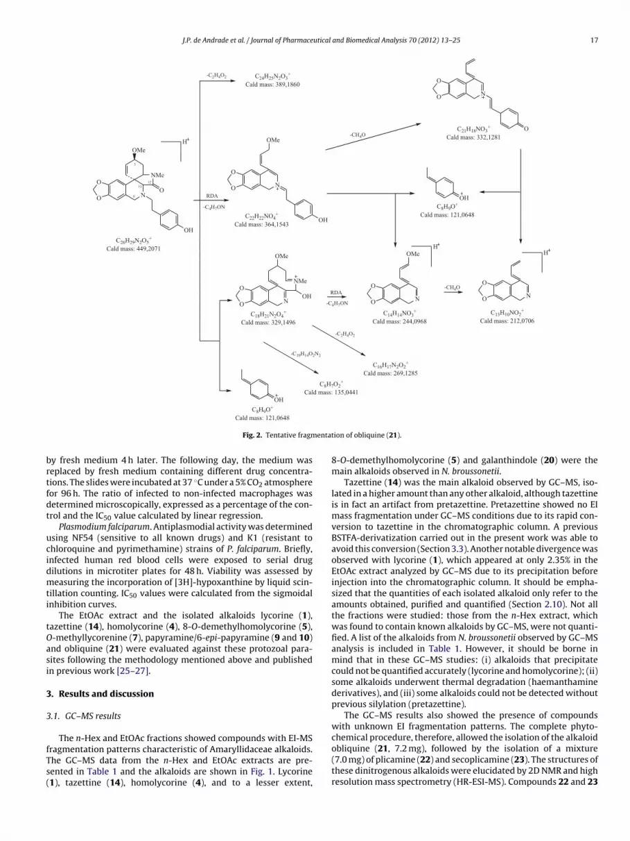
Fig. 2. Tentative fragm
y fresh medium 4 h later. The following day, the medium waseplaced by fresh medium containing different drug concentra-ions. The slides were incubated at 37 ◦C under a 5% CO2 atmosphereor 96 h. The ratio of infected to non-infected macrophages wasetermined microscopically, expressed as a percentage of the con-rol and the IC50 value calculated by linear regression.
Plasmodium falciparum. Antiplasmodial activity was determinedsing NF54 (sensitive to all known drugs) and K1 (resistant tohloroquine and pyrimethamine) strains of P. falciparum. Briefly,nfected human red blood cells were exposed to serial drugilutions in microtiter plates for 48 h. Viability was assessed byeasuring the incorporation of [3H]-hypoxanthine by liquid scin-
illation counting. IC50 values were calculated from the sigmoidalnhibition curves.
The EtOAc extract and the isolated alkaloids lycorine (1),azettine (14), homolycorine (4), 8-O-demethylhomolycorine (5),-methyllycorenine (7), papyramine/6-epi-papyramine (9 and 10)nd obliquine (21) were evaluated against these protozoal para-ites following the methodology mentioned above and publishedn previous work [25–27].
. Results and discussion
.1. GC–MS results
The n-Hex and EtOAc fractions showed compounds with EI-MS
ragmentation patterns characteristic of Amaryllidaceae alkaloids.he GC–MS data from the n-Hex and EtOAc extracts are pre-ented in Table 1 and the alkaloids are shown in Fig. 1. Lycorine1), tazettine (14), homolycorine (4), and to a lesser extent,ion of obliquine (21).
8-O-demethylhomolycorine (5) and galanthindole (20) were themain alkaloids observed in N. broussonetii.
Tazettine (14) was the main alkaloid observed by GC–MS, iso-lated in a higher amount than any other alkaloid, although tazettineis in fact an artifact from pretazettine. Pretazettine showed no EImass fragmentation under GC–MS conditions due to its rapid con-version to tazettine in the chromatographic column. A previousBSTFA-derivatization carried out in the present work was able toavoid this conversion (Section 3.3). Another notable divergence wasobserved with lycorine (1), which appeared at only 2.35% in theEtOAc extract analyzed by GC–MS due to its precipitation beforeinjection into the chromatographic column. It should be empha-sized that the quantities of each isolated alkaloid only refer to theamounts obtained, purified and quantified (Section 2.10). Not allthe fractions were studied: those from the n-Hex extract, whichwas found to contain known alkaloids by GC–MS, were not quanti-fied. A list of the alkaloids from N. broussonetii observed by GC–MSanalysis is included in Table 1. However, it should be borne inmind that in these GC–MS studies: (i) alkaloids that precipitatecould not be quantified accurately (lycorine and homolycorine); (ii)some alkaloids underwent thermal degradation (haemanthaminederivatives), and (iii) some alkaloids could not be detected withoutprevious silylation (pretazettine).
The GC–MS results also showed the presence of compoundswith unknown EI fragmentation patterns. The complete phyto-chemical procedure, therefore, allowed the isolation of the alkaloid
obliquine (21, 7.2 mg), followed by the isolation of a mixture(7.0 mg) of plicamine (22) and secoplicamine (23). The structures ofthese dinitrogenous alkaloids were elucidated by 2D NMR and highresolution mass spectrometry (HR-ESI-MS). Compounds 22 and 23
18 J.P. de Andrade et al. / Journal of Pharmaceutical and Biomedical Analysis 70 (2012) 13– 25
[M+H
h[psnaamcas
3
miata
a
Fig. 3. ESI-MS/MS of the
ave previously been found in Galanthus plicatus subsp. byzantinus23] and, together with 21, in Cyrthanthus obliquus [17]. These com-ounds have an unusual modification in the basic tazettine-typekeleton, in which the oxygen atom at position 5 is replaced by aitrogen atom, which has a pendant 4-hydroxyphenethyl moiety as
substituent. Biogenetically, this replacement probably occurs viaminoaldehyde followed by Schiff base formation with a tyramineolecule [23]. Galanthindole (20) is another example of an unusual
ompound that has been classified as a new skeleton-type [24],lthough the possibility that it is an artifact from the homolycorineeries should be considered.
.2. LC–ESI-LTQ-Orbitrap-MS
Under GC–MS conditions, dinitrogenous compounds eluteduch later than the other alkaloids and their [M]+ ions appeared
n very low abundance (<1%). Due to the interesting skeletalrrangement presented by these alkaloids and the difficulties for
heir study by GC–MS, we performed an LC–ESI-LTQ-Orbitrap-MSnalysis.Compounds 21 and 22 have a closed ring D like tazettinend pretazettine, even though they have a carbonyl group at
]+ ions of obliquine (21).
C-12 forming a cyclic amide function. In contrast, compound23 shows an open ring D with an N-methyl-N-formylaminomoiety attached to C-4a and a carbonyl group at C-11. Consid-ering the MSn of the main fragments from compound 21, theMS2 [449.20→] showed the two most abundant ion peaks atm/z 329.1504 [C18H21N2O4]+ and m/z 121.0652 [C8H9O]+, whichsuggests that compound 21 initially tends toward losing thesubstituent 4-hydroxyphenethyl (see Figs. 2 and 3). All other frag-ments displayed less than 50% of relative abundance. In somefragments, the ion peaks at m/z 389.1871 [C24H25N2O3]+ andm/z 364.1553 [C21H18NO3]+ are in agreement with the loss of60 amu (C2H4O2) and 85 amu (C4H7NO), respectively. The lossof 85 amu after a Retro-Diels Alder (RDA) process in ring Cis in agreement with related compounds Latifaliumin A and B[29].
The MS3 [449.20 → 364.15] showed a very abundant fragmentat m/z 332.1279 [C21H18NO3]+ along with less abundant peaksat m/z 244.0966 [C14H14NO3]+, m/z 212.0703 [C13H10NO2]+ and
m/z 121.0648 [C8H9O]+. Interestingly, the only abundant ion peakshown by MS4 [449.20 → 364.15 → 332.13] was at m/z 212.0707. Anion peak at m/z 121.0650 was also observed, although in low abun-dance and these results suggest a straightforward fragmentation of
J.P. de Andrade et al. / Journal of Pharmaceutical and Biomedical Analysis 70 (2012) 13– 25 19
Fig. 4. Tentative fragmentation of plicamine (22).
Fig. 5. ESI-MS/MS of the [M+H]+ ions of plicamine (22).

20 J.P. de Andrade et al. / Journal of Pharmaceutical and Biomedical Analysis 70 (2012) 13– 25
ntatio
m4
2aw3
Fig. 6. Tentative fragme
/z 332.1283 to m/z 212.0707 through the loss of the substituent-hydroxyphenethyl.
The MS3 [449.20 → 329.15] showed an ion peak at m/z
69.1287 [C16H17N2O2]+, followed by ion peaks at m/z 244.0970nd m/z 212.0707. The fragment at m/z 269.1287 agreesith the loss of 60 amu (C2H4O2) from the ion peak at m/z29.15. The most interesting result was observed with the MS4
Fig. 7. ESI-MS/MS of the [M+H]+
n of secoplicamine (23).
spectra. The MS4 [449.20 → 329.15 → 244.10] along with MS4
[449.20 → 364.15 → 244.10] spectra showed the ion peak at m/z212.0703, which means that the fragment [C13H10NO2]+ (calcu-
lated mass of 212.0706) arose from three different routes, sinceit was also observed in MS4 [449.20 → 364.15 → 332.13] spectra.The very minor fragment at m/z 135.0443 [C8H7O2]+ coming fromthe ion peak at m/z 329.15 was also observed, as in other relatedions of secoplicamine (23).

utical and Biomedical Analysis 70 (2012) 13– 25 21
ca
�ps3ptsaOT32p2[s2ywIaepm3irMma
imFiw4tfl[vTa
Fig. 8. (A) GC–MS chromatogram of EtOAc extract with main alkaloids identified.
FB
J.P. de Andrade et al. / Journal of Pharmace
ompounds [29]. All the proposed fragments showed an error ofround ±1 mDa (Fig. 2).
A carbonyl group at C-6 and the methoxy substituent at the-position at C-3 are the only differences presented by com-ounds 21 and 22. Again, the loss of the 4-hydroxyphenethylubstituent gave ion peaks at m/z 121.0653 [C8H9O]+ and m/z43.1299 [C18H19N2O5]+ as was observed in the MS2 of com-ound 22 (see Figs. 4 and 5). This first fragmentation seemedo be more predominant for compound 22 than compound 21ince the remaining fragments showed no more than 20% of rel-tive abundance using the same collision energy in both cases.ther similarities were observed in their fragmentation mode.he MS2 [463.20→] spectra showed minor fragments at m/z78.1345 [C22H20NO5]+, m/z 258.0767 [C14H12NO4]+ and m/z26.0504 [C13H8NO3]+. The MS3 [463.20 → 378.13] showed ioneaks at m/z 346.1078 [C21H16NO4]+, m/z 258.0762 and m/z26.0502. The MS4 [463.20 → 378.13 → 346.11] along with MS4
463.20 → 378.13 → 258.08] and MS4 [463.20 → 343.13 → 258.08]pectra only showed an ion peak at m/z 226.05 (m/z 226.0497, m/z26.0498 and m/z 226.0500, respectively), confirming that theyielded the same fragment [C13H8NO3]+ through different routes,hich was in agreement with a calculated mass of 226.0499 amu.
n the case of the fragmentation of the ion peaks at m/z 378.13nd m/z 346.11, the substituent 4-hydroxyphenethyl should beliminated as neutral loss. It was also difficult to observe this ioneak in the MS3 and MS4 of compound 21. Other very small frag-ents were observed in the MS2, such as the ion peak at m/z
69.1454 [C20H21N2O5]+ and m/z 311.1032 [C17H15N2O4]+. Theon peak at m/z 369.1454 is in agreement with the loss of C6H6Oesidue and the ion peak at m/z 311.1026 was observed in theS3 [463.20 → 343.13], which is in agreement with the loss of aethanol residue. All the proposed fragments showed an error of
round ± 1 mDa (Fig. 4).The MS2 [465.20→] for compound 23 showed an abundant
on peak at m/z 433.1761 [C25H25N2O5]+ and minor fragments at/z 348.1232 [C21H18NO4]+ and m/z 211.0755 [C14H11O2]+ (see
igs. 6 and 7). The first loss of the 4-hydroxyphenethyl observedn compounds 21 and 22 was not observed in compound 23, even
hen applying stronger collision energy. The ion peak at m/z33.1761 was formed after the loss of a methanol residue fromhe methoxy group at C-3. The ion peak at m/z 348.1232 aroserom m/z 433.18 after an RDA process in ring C, which led to theoss of [C4H7NO] residue. This sequence is suggested by the MS3
465.20 → 433.18]. A careful look at this fragmentation revealedery small ion peaks at m/z 211.0754 and m/z 181.0649 [C13H9O]+.he MS3 [465.20 → 211.07] spectra confirmed that the ion peakt m/z 181.0649 arose from m/z 211.07. Both these ion peaks are
ig. 9. Chromatogram of Narcissus cultivar ‘Toto’ with selected ions at m/z 256 (continuo of pretazettine–TMS. No signal from tazettine–TMS was observed.
(B) Section of chromatogram of TMS-derivatized EtOAc extract with identificationof silylated tazettine and pretazettine along with 6-epimers of papyramine andhaemanthidine.
well-known fragments from tazettine, crinine andhaemanthamine-type alkaloids [28,29]. Regarding the ion peak atm/z 348.1232, the MS3 [465.20 → 348.12] showed the presence ofboth fragments at m/z 228.0656 [C13H10NO3]+ and m/z 121.0650[C8H9O]+, which was indicative of the loss of 4-hydroxyphenethylresidue. The fragments proposed for 23 are shown in Fig. 6 with anerror of around ±1 mDa.
3.3. BSTFA-derivatization
Tazettine (14), one of the most widely distributed Amarylli-
daceae alkaloids, is considered to be an artifact from pretazettinedue to a rearrangement during the routine acid–base alkaloidextraction [30,31]. In the course of the phytochemical proceduretazettine was isolated as a major alkaloid although a small quantityus line) and m/z 319 (discontinuous line). In detail, identification of epimers A and

22 J.P. de Andrade et al. / Journal of Pharmaceutical and Biomedical Analysis 70 (2012) 13– 25
ne–TM
oPic
brawtctataptwtda
mafes[awttrtm
Fig. 10. Tentative fragmentation of tazetti
f pretazettine (15, 7 mg) was also isolated and identified by NMR.retazettine shows an EI mass fragmentation and retention indexdentical to those observed in tazettine, which indicates that it alsoonverts to tazettine in GC–MS analysis.
The well-known instability of pretazettine, especially in strongasic conditions, is due to its trans B–D ring fusion resulting in aelatively strained molecule, while the cis B–D fusion of tazettinellows more flexibility. The driving force for the B-ring openingould appear to be the relief of this internal strain. The comple-
ion of the rearrangement may be considered an intramolecularrossed-Cannizzaro reaction with subsequent hemiaketal forma-ion [31,32]. The BSTFA-derivatization introduces a bulky group as
substituent at position 6, thus blocking the reaction. It is acceptedhat pretazettine is converted into tazettine in extraction processes,lthough the presence of small quantities of tazettine in the initiallant extract cannot be ruled out. Aiming to clarify their detec-ion, we submitted the isolated tazettine and pretazettine togetherith both alkaloid and crude methanolic extracts to a derivatiza-
ion process. Fig. 8 compares the EtOAc extract before and after theerivatization process and pretazettine–TMS was only identifiedfter derivatization.
To check the presence of tazettine in the crude extract, a rapidaceration in MeOH was carried out with bulbs of N. broussonetii
nd two Narcissus cultivars “Toto” and “Pencrebar” previouslyound to contain important quantities of tazettine by GC–MS. Thisxtraction procedure, with a slightly basic pH (pH 8) and ultra-onic bath-assisted, is frequently used in metabolomic studies11]. In the silylated crude extracts of both Narcissus cultivarsnd N. broussonetii, no peak for silylated tazettine was observed,hich strongly suggests that 100% of the tazettine arose from pre-
azettine (Fig. 9). Previously published work [30,31] has raised
his hypothesis, but the unique presence of pretazettine as aeal metabolite in the crude plant extract (without any traces ofazettine) has been confirmed here for the first time using analyticalethods.
S assigned after GC–MS/MS experiments.
3.4. GC–MS/MS
Silylated tazettine and pretazettine demonstrated differentfragmentation patterns by GC–MS/MS analysis. Tazettine–TMS dis-played a molecular ion peak at m/z 403, a base peak at m/z 319and an abundant peak at m/z 298. Its MS fragmentation, rational-ized following Duffield et al. [28], is proposed in Fig. 10. GC–MS/MSexperiments showed that the ion fragment at m/z 388 is formedfrom the molecular ion as a result of the loss of a methyl group,most probably from the O-TMS substituent at position 11, whilethe base peak at m/z 319 appeared after an RDA process in ring C.Further loss of Si(CH3)2OH from the ion fragment at m/z 388 pro-duced an ion at m/z 313, from which the ion at m/z 298 arose afterthe loss of a methyl radical. The loss of 28 amu from the m/z 298corresponded to a CO group, which led to a fragment at m/z 270.An MS/MS experiment with the ion at m/z 319 displayed peaks atm/z 304 and m/z 230 with low abundance, which was in agreementwith the loss of a methyl radical from O-TMS together with thesubsequent expulsion of dimethylsilane residue.
Although the 1H NMR of pretazettine confirmed the purityof the sample, two peaks with similar fragmentation patternswere observed by GC–MS, differing only in the relative abun-dance of fragments. This suggests the co-existence of C-6 epimersof pretazettine, as reported in alkaloids bearing crinine- andhaemanthamine-type skeletons [16,31]. In the GC–MS spectrumof epimer A (Fig. 11) of silylated pretazettine, a molecular ion peakwas observed at m/z 403 along with a base and abundant peak atm/z 319 and m/z 388, respectively. The GC–MS/MS data for pre-tazettine showed that, similarly to tazettine, the ions at m/z 388and m/z 319 arose from the molecular ion (at m/z 403). The ionpeak at m/z 388 may also be formed after the loss of a methyl rad-
ical from the methoxyl group at C-3. Further elimination of the(CH3)3SiO residue resulted in the formation of an ion at m/z 298from which an ion fragment at m/z 256 was formed after the lossof C3H7N. The MS spectrum of pretazettine showed a characteristic
J.P. de Andrade et al. / Journal of Pharmaceutical and Biomedical Analysis 70 (2012) 13– 25 23
(B) fr
iihtt[G
Fig. 11. Tentative fragmentation of the epimer (A) and epimer
on at m/z 372 [M−31]+ arising from the ion at m/z 388 [M−15]+,ndicating a cyclization process [33]. This rearrangement processas been frequently observed in silylated hydroxypyrene deriva-
ives showing an aromatic ring at the alpha position of the carbonhat bears the hydroxyl group undergoing the silylation reaction34,35]. A tentative fragmentation of pretazettine assigned afterC–MS/MS analysis is presented in Fig. 11.om pretazettine–TMS assigned after GC–MS/MS experiments.
3.5. In vitro assay
The EtOAc extract and some isolated compounds were tested
against the parasitic protozoa T. cruzi, T. brucei rhodesiense, L. dono-vani and P. falciparum. This assay was performed as describedby Labrana et al. [27]. Only the EtOAc extract showed signifi-cant in vitro activity against T. cruzi, with an IC50 of 1.77 �g/ml
2 utical
(rttma
4
idrapls
wttacofwrarmoOhf
itepfoNtp
Asntpa3
atw
A
–tALB
[
[
[
[
[
[
[
[
[
[
[
[
[
[
[
4 J.P. de Andrade et al. / Journal of Pharmace
ref. value of 0.349 �g/ml for Benznidazole). Although the alkaloid-ich extract demonstrated notable activity, the main alkaloidsazettine (14), lycorine (1) and homolycorine (4), and those foundo a lesser extent, such as 8-O-demethylhomolycorine (5), O-
ethyllycorenine (7), papyramine/6-epi-papyramine (9 and 10)nd obliquine (21), showed no significant activity against T. cruzi.
. Conclusion
The alkaloids identified in N. broussonetii are commonly foundn Narcissus species, with the exception of galanthindole and theinitrogenous obliquine, plicamine, and secoplicamine, which areeported here for the first time in the Narcissus genus. The mainlkaloids were lycorine, homolycorine and tazettine (arising fromretazettine). Also noteworthy is the absence of crinine-type alka-
oids in this plant species, as occurs in all the Narcissus speciestudied to date.
The alkaloids obliquine, plicamine, and secoplicamine, alongith some related compounds, have been the subject of several
otal syntheses, prompted by the particular interest of their dis-inctive dinitrogenous skeletal arrangement [36,37]. GC–MS/MSnalysis of dinitrogenous alkaloids using BSTFA-derivatization wasarried out to improve their elution and detection, but with-ut any success. Using an LC–ESI-LTQ-Orbitrap-MS a very similarragmentation mode was observed for obliquine and plicamine,hich yielded the fragments [C13H10NO2]+ and [C13H8NO3]+,
espectively, through different routes. The compounds obliquinend plicamine showed a very similar structure, particularly withespect to the closed ring D, which seemed to influence their frag-entation pattern, since the fragments corresponding to the loss
f the substituent 4-hydroxyphenethyl appeared as base peaks.therwise, the compound secoplicamine showed a pendant 4-ydroxyphenethyl moiety on the core structure and the remaining
ragments presented very low abundance.It is accepted that the conversion of pretazettine to tazettine
n a routine alkaloid extraction is near total, without ruling outhe presence of small quantities of tazettine in the initial plantxtract. A BSTFA-derivatization step confirmed that all the tazettineresent in the plant extract arose from pretazettine, and was there-ore absent as a natural alkaloid. The crude methanolic extractbtained by a fast extraction in a slightly basic environment from. broussonetii and two Narcissus cultivars previously found to con-
ain tazettine as a major alkaloid revealed the sole presence ofretazettine.
As part of an ongoing project on the chemical aspects ofmaryllidaceae alkaloids, silylated pretazettine and tazettine wereubmitted to an MS/MS study. In contrast with the dinitroge-ous alkaloids, the GC–MS/MS analysis proved to be an importantool for the study of their fragmentation. Silylated tazettine andretazettine showed distinct fragmentation routes in GC–MS/MS,lthough a Retro-Diels Alder process formed the base peak at m/z19 in both.
Finally, N. broussonetii exhibited important in vitro activitygainst T. cruzi, which could be due to a synergic action of some ofhe identified alkaloids, since they showed no significant activityhen tested individually.
cknowledgements
The authors are grateful to the Generalitat de Catalunya (2009 SGR1060) for financial support of this work. The authors are also
hankful to the SCT-UB technicians, mainly Dr. Asunción Marín, Dr.lberto Adeva and Dr. Olga Jáuregui for performing GC/MS andC–ESI-MS spectra, as well for Dr. Marcel Kaiser and Prof. Retorun for antiprotozoal studies. Special thanks to Prof. Mohammed[
and Biomedical Analysis 70 (2012) 13– 25
Benaissa and Dr. Abdelaziz Elamrani for their helpful collaborationin the collection of N. broussonetii and Dr. Nehir Ünver for provid-ing us with the 1H NMR and EI-MS spectra of galanthindole. J.P.A.is thankful to the Agencia Espanola de Cooperación Internacionalpara el Desarollo (BECAS-MAEC-AECID) for a doctoral fellowship.
References
[1] S. López, J. Bastida, F. Viladomat, C. Codina, Acetylcholinesterase inhibitoryactivity of some Amaryllidaceae alkaloids and Narcissus extracts, Life Sci. 71(2002) 2521–2529.
[2] J. Bastida, R. Lavilla, F. Viladomat, Chemical and biological aspects of Narcissusalkaloids, in: G. Cordell (Ed.), The Alkaloids: Chemistry and Biology, ElsevierInc., Amsterdam, 2006, pp. 87–179.
[3] E.J. Osorio, S.M. Robledo, J. Bastida, Alkaloids with antiprotozoal activity, in: G.Cordell (Ed.), The Alkaloids: Chemistry and Biology, Elsevier Inc., Amsterdam,2008, pp. 113–190.
[4] J. McNulty, J.J. Nair, C. Codina, J. Bastida, S. Pandey, J. Gerasimoff, C. Griffin,Selective apoptosis-inducing activity of crinum-type Amaryllidaceae alkaloids,Phytochemistry 68 (2007) 1068–1074.
[5] J. McNulty, J.J. Nair, J. Bastida, S. Pandey, C. Griffin, Structure–activity studies onthe lycorine pharmacophore: a potent inducer of apoptosis in human leukemiacells, Phytochemistry 70 (2009) 913–919.
[6] I. Zupkó, B. Réthy, J. Hohmann, J. Molnár, I. Ocsovszki, G. Falkay, Antitumoractivity of alkaloids derived from Amaryllidaceae species, In Vivo 23 (2009)41–48.
[7] J. McNulty, J.J. Nair, M. Singh, D.J. Crankshaw, A.C. Holloway, J. Bastida, Selectivecytochrome P450 3A4 inhibitory activity of Amaryllidaceae alkaloids, Bioorg.Med. Chem. Lett. 19 (2009) 3233–3237.
[8] R. Gotti, J. Fiori, M. Bartolini, V. Cavrini, Analysis of Amaryllidaceae alkaloidsfrom Narcissus by GC–MS and capillary electrophoresis, J. Pharm. Biomed. Anal.42 (2006) 17–24.
[9] S. Berkov, J. Bastida, F. Viladomat, C. Codina, Analysis of galanthamine-typealkaloids by capillary gas chromatography–mass spectrometry in plants, Phy-tochem. Anal. 19 (2008) 285–293.
10] L. Torras-Claveria, S. Berkov, O. Jáuregui, J. Caujapé, F. Viladomat, C. Codina,J. Bastida, Metabolic profiling of bioactive Pancratium canariense extracts byGC–MS, Phytochem. Anal. 21 (2010) 80–88.
11] S. Berkov, J. Bastida, F. Viladomat, C. Codina, Development and validationof a GC–MS method for a rapid determination of galanthamine in Leuco-jum aestivum and Narcissus ssp.: a metabolomic approach, Talanta 83 (2011)1455–1465.
12] T. Verhaeghe, L. Diels, R. de Vries, M. De Meulder, J. de Jong, Development andvalidation of a liquid chromatographic–tandem mass spectrometric methodfor the determination of galanthamine in human heparinised plasma, J. Chro-matogr. B: Anal. Technol. Biomed. Life Sci. 789 (2003) 337–346.
13] M. Kreh, R. Matusch, L. Witte, Capillary gas chromatography–mass spectrom-etry of Amaryllidaceae alkaloids, Phytochemistry 38 (1995) 773–776.
14] C. Wagner, M. Sefkow, J. Kopka, Construction and application of a mass spec-tral and retention time index database generated from plant GC/EI-TOF-MSmetabolite profiles, Phytochemistry 62 (2003) 887–900.
15] F. Viladomat, J. Bastida, G. Tribó, C. Codina, M. Rubiralta, Alkaloids from Narcis-sus bicolor, Phytochemistry 29 (1990) 1307–1310.
16] J. Bastida, C. Codina, F. Viladomat, M. Rubiralta, J.C. Quirion, H.P. Husson, G. Ma,Narcissus alkaloids XIII. Complete assignment of the NMR spectra of papyra-mine and 6-epi-papyramine by two-dimensional NMR spectroscopy, J. Nat.Prod. 53 (1990) 1456–1462.
17] N.D. Brine, W.E. Campbell, J. Bastida, M.R. Herrera, F. Viladomat, C. Codina, P.J.Smith, A dinitrogenous alkaloid from Cyrtanthus obliquus, Phytochemistry 61(2002) 443–447.
18] A.K. Machocho, J. Bastida, C. Codina, F. Viladomat, R. Brun, S.C. Chhabra,Augustamine type alkaloids from Crinum kirkii, Phytochemistry 65 (2004)3143–3149.
19] S. Berkov, C. Codina, F. Viladomat, J. Bastida, Alkaloids from Galanthus nivalis,Phytochemistry 68 (2007) 1791–1798.
20] S. Berkov, R. Reyes-Chilpa, C. Codina, F. Viladomat, J. Bastida, Revised NMRdata for incartine: an alkaloid from Galanthus elwesii, Molecules 12 (2007)1430–1435.
21] S. Berkov, J. Bastida, B. Sidjimova, F. Viladomat, C. Codina, Phytochemical dif-ferentiation of Galanthus nivalis and Galanthus elwesii (Amaryllidaceae): a casestudy, Biochem. Syst. Ecol. 36 (2008) 638–645.
22] R. Suau, A.I. Gómez, R. Rico, M.P. Vázquez Tato, L. Castedo, R. Riguera, AlkaloidN-oxides of Amaryllidaceae, Phytochemistry 27 (1988) 3285–3287.
23] N. Ünver, T. Gözler, N. Walch, B. Gözler, M. Hesse, Two novel dinitrogenousalkaloids from Galanthus plicatus subsp. byzantinus (Amaryllidaceae), Phyto-chemistry 50 (1999) 1255–1261.
24] N. Ünver, G.I. Kaya, C. Werner, R. Verpoorte, B. Gözler, Galanthindole: a newindole alkaloid from Galanthus plicatus ssp. byzantinus, Planta Med. 69 (2003)869–871.
25] R.G. Ridley, W. Hofheinz, H. Matile, C. Jacquet, A. Dorn, R. Masciadri, S. Jolidon,W.F. Richter, A. Guenzi, M.A. Girometta, H. Urwyler, W. Huber, S. Thaitong, W.Peters, 4-Aminoquinolone analogs of chloroquine with shortened side chainsretain activity against chloroquine-resistant Plasmodium falciparum, Antimi-crob. Agents Chemother. 40 (1996) 1846–1854.

utical
[
[
[
[
[[
[
[
[
[
[
J.P. de Andrade et al. / Journal of Pharmace
26] B. Räz, M. Iten, Y. Grether-Bühler, R. Kaminsky, R. Brun, The Alamar Blue assayto determine drug sensitivity of African trypanosomes (T. b. rhodesiense and T.b. gambiense) in vitro, Acta Trop. 68 (1997) 139–147.
27] J. Labrana, A.K. Machocho, V. Kricsfalusy, R. Brun, C. Codina, F. Viladomat, J.Bastida, Alkaloids from Narcissus angustifolius subsp. transcarpathicus (Amaryl-lidaceae), Phytochemistry 60 (2002) 847–852.
28] A.M. Duffield, R.T. Aplin, H. Budzikiewicz, C. Djerassi, C.F. Murphy, W.C. Wild-man, Mass spectrometry in structural and stereochemical problems. LXXXII. Astudy of the fragmentation of some Amaryllidaceae alkaloids, J. Am. Chem. Soc.87 (1965) 4902–4912.
29] X. Zhang, H. Huang, X. Liang, H. Huang, W. Dai, Y. Shen, S. Yan, W. Zhang, Analysisof Amaryllidaceae alkaloids from Crinum by high-performance liquid chro-
matography coupled with electrospray ionization tandem mass spectrometry,Rapid Commun. Mass Spectrom. 23 (2009) 2903–2916.30] W.C. Wildman, D.T. Bailey, Pretazettine, J. Am. Chem. Soc. 89 (1967) 5514–5515.31] W.C. Wildman, D.T. Bailey, Amaryllidaceae interconversions. Partial synthesis
of [2]benzopyrano[3,4-c]indoles, J. Am. Chem. Soc. 91 (1969) 150–157.
[
and Biomedical Analysis 70 (2012) 13– 25 25
32] W.C. Wildman, D.T. Bailey, Novel alkaloids containing the [2]benzopyrano[3,4-c]indole nucleus, J. Org. Chem. 33 (1968) 3749–3753.
33] J. Jacob, A. Schmoldt, G. Grimmer, Influence of monooxygenase inducers onthe metabolic profile of phenanthrene in rat liver microsomes, Toxicology 25(1982) 333–343.
34] G. Gmeiner, P. Gärtner, C. Krassnig, H. Tausch, Identification of various uri-nary metabolites of fluorene using derivatization solid-phase microextraction,J. Chromatogr. B: Anal. Technol. Biomed. Life Sci. 766 (2002) 209–218.
35] C. Schummer, O. Delhomme, B.M.R. Appenzeller, R. Wennig, M. Millet, Com-parison of MTBSTFA and BSTFA in derivatization reactions of polar compoundsprior to GC/MS analysis, Talanta 77 (2009) 1473–1482.
36] I.R. Baxendale, S.V. Ley, M. Nessi, C. Piutti, Total synthesis of the Amaryllidaceae
alkaloid (+)-plicamine using solid-supported reagents, Tetrahedron 58 (2002)6285–6304.37] I.R. Baxendale, S.V. Ley, Synthesis of the alkaloid natural products (+)-plicaneand (−)-obliquine, using polymer-supported reagents and scavengers, Ind. Eng.Chem. Res. 44 (2005) 8588–8592.