Embed Size (px)
Citation preview

VETERINARY RESEARCHBello-Ortí et al. Veterinary Research 2014, 45:104http://www.veterinaryresearch.org/content/45/1/104
RESEARCH Open Access
Biofilm formation by virulent and non-virulentstrains of Haemophilus parasuisBernardo Bello-Ortí1†, Vincent Deslandes2†, Yannick DN Tremblay2, Josée Labrie2, Kate J Howell3,Alexander W Tucker3, Duncan J Maskell3, Virginia Aragon1,4 and Mario Jacques2*
Abstract
Haemophilus parasuis is a commensal bacterium of the upper respiratory tract of healthy pigs. It is also theetiological agent of Glässer’s disease, a systemic disease characterized by polyarthritis, fibrinous polyserositis andmeningitis, which causes high morbidity and mortality in piglets. The aim of this study was to evaluate biofilmformation by well-characterized virulent and non-virulent strains of H. parasuis. We observed that non-virulentstrains isolated from the nasal cavities of healthy pigs formed significantly (p < 0.05) more biofilms than virulentstrains isolated from lesions of pigs with Glässer’s disease. These differences were observed when biofilms wereformed in microtiter plates under static conditions or formed in the presence of shear force in a drip-flow apparatusor a microfluidic system. Confocal laser scanning microscopy using different fluorescent probes on a representativesubset of strains indicated that the biofilm matrix contains poly-N-acetylglucosamine, proteins and eDNA. Thebiofilm matrix was highly sensitive to degradation by proteinase K. Comparison of transcriptional profiles of biofilmand planktonic cells of the non-virulent H. parasuis F9 strain revealed a significant number of up-regulatedmembrane-related genes in biofilms, and genes previously identified in Actinobacillus pleuropneumoniae biofilms.Our data indicate that non-virulent strains of H. parasuis have the ability to form robust biofilms in contrast tovirulent, systemic strains. Biofilm formation might therefore allow the non-virulent strains to colonize and persist inthe upper respiratory tract of pigs. Conversely, the planktonic state of the virulent strains might allow them todisseminate within the host.
IntroductionHaemophilus parasuis is a Gram-negative bacterium anda commensal organism of the upper respiratory tract ofhealthy pigs. It is also the etiological agent of Glässer’sdisease, a systemic disease characterized by polyarthritis,fibrinous polyserositis and meningitis, which causes highmorbidity and mortality in piglets [1]. Glasser’s diseaseis recognized as one of the main causes of economic lossin the pig industry.The heterogeneity among H. parasuis strains has been
demonstrated by multiple methods, including multilocussequence typing (MLST) which has shown a geneticlineage associated with polyserositis (cluster A) and an-other lineage associated with nasal colonization (cluster C)
* Correspondence: [email protected]†Equal contributors2Groupe de recherche sur les maladies infectieuses du porc, Faculté de médecinevétérinaire, Université de Montréal, St-Hyacinthe, Québec J2S 7C6, CanadaFull list of author information is available at the end of the article
© 2014 Bello-Orti et al.; licensee BioMed CentrCommons Attribution License (http://creativecreproduction in any medium, provided the orDedication waiver (http://creativecommons.orunless otherwise stated.
[2]. Serum resistance [3], phagocytosis resistance [4], andinvasion of endothelial cells [5,6] have been associatedwith virulence of H. parasuis. Some putative virulence fac-tors have been reported [7,8]. Those include the capsule[4], lipooligosaccharide (LOS) [9-11] and other genes in-volved in polysaccharide production, such as galE andgalU, which have been associated with serum resistanceand biofilm production [12]. Genes involved in sialic acidutilization were identified as potential virulence factors[13]. The sialytransferase gene lsgB was predominantlypresent in systemic isolates and not in nasal isolates, andsialylation of the LOS was observed in the virulent strainH. parasuis Nagasaki. In addition, a family of trimericautotransporters, designated virulence associated trimericautotransporters (VtaA) has been described [14,15] andthese can be divided into three groups based on theirtranslocation domains [14]. Group 3 vtaA gene is highlyconserved among invasive and non-invasive strains, whilst
al Ltd. This is an Open Access article distributed under the terms of the Creativeommons.org/licenses/by/4.0), which permits unrestricted use, distribution, andiginal work is properly credited. The Creative Commons Public Domaing/publicdomain/zero/1.0/) applies to the data made available in this article,

Table 1 Haemophilus parasuis strains used in the presentstudy
Strains Susceptibility1 to lsgB2 group1vtaA2
MLST3 Serovar
Serum Phagocytosis
Nasal strains
F9 S S - - C 6
ND14-1 S S - - C 7
SC14-1 S S - - C 15
MU21-2 S S - - C 7
SL3-2 S S - + B 10
Strains from lesions
Nagasaki R R + + A 5
P015/96 R R + + A 5
ER-6P R R + + A 15
264/99 R R + + A 10
IT29205 R R + + A 4
2725 S R + + A 10
373/03A I R - + A 7
PV1-12 S R - + B 15
9904108 S R - - C 41(S) sensitive; (I) intermediate; (R) resistant.2(+) gene present; (−) gene absent.3Cluster associated with isolates from systemic lesions (A); with nasal isolation(C); or cluster with no statistical association with clinical origin (B) [2].
Bello-Ortí et al. Veterinary Research 2014, 45:104 Page 2 of 14http://www.veterinaryresearch.org/content/45/1/104
groups 1 and 2 vtaA genes were detected only in virulentstrains.Bacterial biofilms are structured clusters of bacterial
cells enclosed in a self-produced polymer matrix that areattached to a surface [16-18]. Bacteria can adhere to bi-otic surfaces (e.g. cells at the mucous layer) as well asabiotic surfaces (e.g. floor or equipment found at afarm). The polymer matrix is often composed of exopo-lysaccharides, proteins and nucleic acids. The biofilmprotects bacteria from hostile environmental conditions.Bacteria within a biofilm can resist attack from the hostimmune response, and are less sensitive than planktoniccells to desiccation and to the action of biocides. Biofilmformation by nasal strains of H. parasuis has been previ-ously reported [19]. Although the role of biofilm in H.parasuis pathogenesis is not clear, expression of geneswith putative function in biofilm formation was detectedduring pulmonary infection [20]. The aims of this studywere to compare biofilm formation by well-characterizedvirulent and non-virulent strains of H. parasuis and toanalyse the gene expression during biofilm growth. Suchanalyses would help explore the possible role of biofilmsin the pathogenesis of H. parasuis.
Materials and methodsBacterial strainsThe H. parasuis strains used in this study and their mainphenotypic and genotypic characteristics are listed inTable 1. Bacteria were grown on Brain Heart Infusion(BHI; Oxoid Ltd, Basingstoke, Hampshire, UK) agar con-taining 10 μg/mL NAD or on chocolate agar (Biomerieux,Madrid, Spain). Plates were incubated overnight at 37 °Cwith 5% CO2.
Biofilm assaysStatic conditionsBiofilms were cultured in 96-well microtiter plates as de-scribed by Wu et al. [21], with some modifications. Briefly,colonies from overnight agar cultures were resuspended inBHI-NAD containing 5 μg/mL NAD, and the suspensionwas aliquoted (100 μL) in triplicate in a flat-bottom 96-well polystyrene plate (Costar® 3599, Corning, NY, USA)and incubated for 40 h at 37 °C. Wells containing sterilebroth were used as negative control. Following incubation,biofilms were treated as described before [22] with somemodifications. Briefly, the liquid medium was removedusing a vacuum and unattached cells were removed by im-mersing the plate once in MilliQ water. The water was re-moved with a vacuum and excess water was removed byinverting plates onto a paper towel. Biofilms were thenstained with 0.1% (w/v) crystal violet for 2 min. Biofilmswere washed once with distilled water and then dried at37 °C for 15 min. The stain was then released with 100 μLof 70% (v/v) ethanol and the amount of released stain was
quantified by measuring the absorbance at 590 nm witha microplate reader (Powerwave; BioTek Instruments,Winooski, VT, USA).
Shear force conditionsBiofilms were cultured in a drip-flow apparatus (DFR 110Biofilm Reactor, BioSurface Technologies Corp. Bozeman,MT, USA) as described by Goeres et al. [23] with somemodifications [24]. Briefly, colonies of H. parasuis strainsF9, MU21-2, ER-6P or Nagasaki on BHI-NAD agar wereresuspended into 13 mL of fresh BHI-NAD to an OD600 of0.1 and 12.5 mL of this inoculum was transferred into achannel containing a glass slide (Catalogue #48300-025,VWR, Ville Mont-Royal, QC, Canada). The apparatus wasincubated for 24 h at 37 °C with 5% CO2 to allow the bio-film to form under static conditions. The apparatus legswere then attached to create a 10° downward slope. The ap-paratus was then connected to the nutrient system contain-ing pre-warmed (37 °C) BHI-NAD. The flow (~25 mL perhour per channel) of the medium was then initiated andmaintained for 24 h at 37 °C. After 24 h, the glass slide wasremoved and gently washed once with sterile MiliQ water.The biofilms were resuspended in 1.5 mL of MiliQ water,centrifuged and dried with a DNA 120 Speed Vac® (ThermoScientific, Ottawa, ON, Canada). The weight of dry biofilmswas then measured.

Bello-Ortí et al. Veterinary Research 2014, 45:104 Page 3 of 14http://www.veterinaryresearch.org/content/45/1/104
Biofilms were also cultured in a microfluidic system.Growth of biofilms in the BioFlux 200 device (FluxionBiosciences, South San Francisco, CA, USA) was adaptedfrom Benoit et al. [25] and the manufacturer’s recommen-dations. Briefly, colonies of H. parasuis strains F9, MU21-2, ER-6P or Nagasaki on BHI-NAD agar were resuspendedinto 2.5 mL of fresh BHI-NAD to an OD600 of 1.0 and thissuspension was serially diluted to OD600 of 0.5 and 0.25.The microfluidic channels were wetted with BHI-NADand were then inoculated with different bacterial suspen-sions (OD600 1.0, 0.5 or 0.25). The microfluidic plate wasincubated for 2 h at 37 °C to allow bacteria to bind to thesurface. The flow of fresh medium was then initiated andwas set from 0.5 dyne/cm2 to 1.0 dyne/cm2. Growth of thebiofilms was monitored for up to 24 h and, in some cases,fresh medium was added and the “waste” outlet was emp-tied to ensure that wells would not dry or spill. Images ofBioFlux biofilms were obtained using an inverted fluores-cence microscope (Olympus CKX41, Markham, ON,Canada), a digital camera (Retiga EX; Q Imaging, Surrey,BC, Canada), and the software provided with the BioFlux200 device.
Confocal laser scanning microscopy (CLSM)Biofilms were prepared under static conditions as de-scribed above and were stained with FilmTracer™ FM®1-43Green biofilm cell stain (Molecular Probes, Eugene, OR,USA) according to manufacturer’s instructions. To deter-mine the composition of the biofilm matrix, biofilms werestained with Wheat Germ Agglutinin (WGA-OregonGreen 488, Molecular Probes; binds to N-acetyl-D-glucosamine and N-acetylneuraminic acid residues), Film-Tracer™ SYPRO® Ruby biofilm matrix stain (MolecularProbes; labels most classes of proteins) or BOBO™-3 iodide(Molecular Probes; stains extracellular DNA) according tomanufacturer’s instructions. After a 30 min incubation atroom temperature, the fluorescent marker solution was re-moved, biofilms were washed with water and the wellswere then filled with 100 μL of water or PBS for WGA-stained biofilms. Stained biofilms were visualized by CLSM(Olympus FV1000 IX81, Markham, ON, Canada).
Dispersion of biofilm by enzymatic treatmentsA biofilm dispersion assay was performed as describedpreviously [26]. Briefly, biofilms were grown under staticconditions as described above, and after the 40 h incuba-tion, 50 μL of DNase I (500 μg/mL in 150 mM NaCl,1 mM CaCl2), 50 μL of dispersin B (100 μg/mL in PBS;Kane Biotech Inc., Winnipeg, MB, Canada), or 50 μL ofproteinase K (500 μg/mL in 50 mM Tris–HCl pH 7.5,1 mM CaCl2) were added directly to the biofilms. Con-trol wells were treated with 50 μL of the buffer withoutthe enzyme. Wells treated with dispersin B were incubatedfor 5 min at 37 °C, and those treated with proteinase K or
DNase I were incubated for 1 h at 37 °C. After the treat-ments, the biofilms were stained with crystal violet asdescribed above.
Effect of fibrinogen and fibronectin on biofilm formationBiofilms were prepared under static conditions as de-scribed above with some modifications. Prior to inocula-tion, various concentrations of fibrinogen (bovine, porcineor human) (0.5, 1, 2 and 5 mg/mL) or fibronectin (human)(0.5, 1 and 2 mg/mL) were added to the biofilm medium.Plates were prepared in duplicate for each experiment: oneplate was used to measure biofilm formation and the otherplate was used to measure growth of the bacteria in thepresence of proteins. Both plates were incubated and oneplate was stained as described before. The unstained repli-cate plate was used to evaluate growth by measuring theabsorbance at 600 nm.
Genome sequencing and assemblyGenomic DNA of the high biofilm producer strain F9was prepared using the Blood and Tissue DNeasy kit(Qiagen) according to the manufacturer’s instructions.Illumina libraries were prepared using 500 ng of gen-omic DNA and modified Illumina protocols [27,28].Paired-end sequencing was performed on an IlluminaHiSeq 2000 analyzer for 75 cycles at the Wellcome TrustSanger Institute (Hinxton, Cambridge, UK). The fastqreads were mapped to the complete SH0165 genomeusing Stampy to check for quality and purity of the se-quence before any analyses [29,30].A custom-made bioinformatics pipeline was used to as-
semble the draft genome sequence. Cutadapt was used toremove the adaptor sequences from the sequence readsthat were previously introduced during the library prepar-ation [31]. Any undetermined nucleotides (N’s) were re-moved from reads and the program sickle [32] was used totrim low-quality sequences found at the ends of paired-endsequence reads resulting in a minimum length of 31 bp,using the program’s default quality thresholds for the reads.Next, a custom Perl script was used to screen out low qual-ity fastq reads and produce a single fastq file containingthe good paired reads, and a separate file containing goodquality single reads. Finally, we used Velvet [33] and Velve-tOptimiser 2.2.0 [34] to assemble the fastq files into thede-novo genome assembly, made up of contiguous se-quences (contigs). Assembly parameters were optimised toproduce the highest quality assembly (i.e. highest n50value) using VelvetOptimiser, which runs through all pos-sible k-mer values from 19 to 71 in increments of 2. Thedraft genome sequence was annotated using the automaticannotation software Prokka [35], including the rfam op-tion. This Whole Genome Shogun project has been depos-ited [DDBJ/EMBL/GenBank: JHQI00000000]. The versiondescribed in this paper is version JHQI01000000.

Bello-Ortí et al. Veterinary Research 2014, 45:104 Page 4 of 14http://www.veterinaryresearch.org/content/45/1/104
Transcriptomics analysisThe high biofilm producer strain F9 was chosen fortranscriptomic analysis. Planktonic and biofilm sampleswere obtained under static conditions as described abovewith some modifications. Bacteria were cultured in 6-well microtiter plates and after 36 h, planktonic cultureswere transferred to sterile tubes, whereas biofilm bac-teria were collected by scraping the surface of the wellswith lysis buffer (2% SDS in PBS). Bacteria from bothsamples, biofilm or planktonic, were recovered by centri-fugation and pellets were used for RNA extraction. Forcomparison, F9 was grown with shaking (220 rpm) untilthe culture reached stationary phase and this culturewas then processed for RNA extraction. This is consid-ered to be the “stationary culture” sample.To extract RNA, bacterial pellets were resuspended in
2% SDS, 16 mM EDTA, 10 mM Tris (pH 8.0) and incu-bated for 5 min at 100 °C. Afterwards, samples were proc-essed by two hot acid phenol-chloroform extractions,followed by two chloroform/isoamyl alcohol extractions.RNA was then precipitated with 0.6 volumes of isopropa-nol, 0.1 volumes of 5 M ammonium acetate and 1 μL ofglycogen. After centrifugation, the pellet was washed with70% ethanol, dried and resuspended in warmed RNase-freewater. To ensure that contaminating bacterial DNA waseliminated from the samples, treatment with RNase-freeDNase (Qiagen) was performed. In addition, ribosomalRNA was eliminated with the Ribo-Zero rRNA removal kit(Epicentre Biotechnologies, Madison, WI, USA) followingmanufacturer instructions. PCR reactions using primersspecific for H. parasuis 16S rRNA gene [36] were carriedout to ensure that no bacterial DNA was left in the sample.Final RNA quality was verified with a Nanodrop spectro-photometer and the integrity was analyzed using AgilentBioanalyzer 2100 (Agilent technologies). Libraries weregenerated using an Ion Torrent RNA-Seq v2 kit (LifeTechnologies) and sequenced using an Ion Torrent PGM(Life Technologies) with an Ion 318 chip (Life technolo-gies) at the Centre for Research in Agricultural Genomics(CRAG, Campus de Bellaterra-UAB, Spain).Bioinformatic analysis was performed following the
count-based differential expression method. Briefly, readsquality control was performed using FastQC [37] andFASTX-Toolkit [38], and reads were then mapped to H.parasuis F9 genome using the recommended TorrentMapping Alignment Program (TMAP) v3.4.1 with map2setting [39]. Alignments were inspected using SAM-tools [40] and Integrative Genomics Viewer (IGV) [41].HTSeq v0.5.4p3 [42] was used for feature counting withintersection-nonempty setting and discarding non-protein coding CDS. Differentially expressed genes(DEGs) were identified with edgeR R package [43] witha 5% P-value cut-off, using an assigned dispersion valueof 0.04.
To perform gene set enrichments, a custom GeneOntology (GO) database was built. Protein coding geneswere BLASTed using BLASTp (version 2.2.28) against thenon-redundant NCBI database (April 2014), e value of10−3 and keeping first 20 hits. GO terms were mapped toBlast hits using Blast2GO [44]. The most significantly up-regulated genes (P < 0.05) were identified as candidates forclassic Fisher’s exact testing through Blast2GO. Tests wereperformed for biological process (BP), cellular component(CC) and molecular function (MF) with P < 0.05. Conser-vation of membrane-related genes among all 14H. para-suis sequences available in GenBank [GenBank: APCA00000000.1, APBW00000000.1, ABKM00000000.2, APBX00000000.1, AZQU00000000.1, APBZ00000000.1, AOSU00000000.1, APBY00000000.1, APBV00000000.1, CP001321.1, APBT00000000.1, APBU00000000.1, APCB00000000.1,CP005384.1] was achieved as follows: whole F9 proteomewas analyzed using Phobius [45] via Blast2GO, positivelypredicted membrane-related genes were blasted againstthe 14H. parasuis genomic sequences using tBLASTnwith the following settings: e value of 10−5, alignmentlength > 70% and match identity > 60%. Whole-genomeBLASTp comparisons were performed between H. parasuisand Actinobacillus pleuroneumoniae serovar 5b strain L20[GenBank: NC_009053.1], using the following settings:e value of 10−5, alignment length > 90% and match iden-tity > 40%. All transcriptomic data were deposited in theGene Expression Omnibus database [GEO: GSE56428].
ResultsBiofilm formation under static conditionsThe phenotypic and genotypic characteristics of the H.parasuis strains used in the present study are given inTable 1. Five non-virulent strains were recovered from thenasal cavities of healthy pigs while nine virulent strainswere recovered from lesions of pigs with Glässer’s disease.The ability of H. parasuis to form biofilms at the solid–liquid interface in polystyrene microtiter plates was deter-mined for each of the virulent and non-virulent strains(Figure 1A). Interestingly, the nasal, non-virulent strainsformed significantly (p < 0.05) more biofilms than the viru-lent strains isolated from lesions of pigs with Glässer’s dis-ease (Figure 1B). We also noticed that biofilm productionwas stronger in strains sensitive to serum compared to re-sistant strains (Additional file 1A, p = 0.059); in strainsnegative for vtaA group 1 genes compared to strains posi-tive for these genes (Additional file 1B, p = 0.189); in strainsbelonging to MLST cluster C compared to strains belong-ing to MLST cluster A (Additional file 1C, p = 0.202), andin strains negative for the sialyltransferase gene lsgB com-pared to strains positive for lsgB (Additional file 1D, p =0.228). These differences however were not statistically sig-nificant probably due to the small number of strains insome of the groups that were compared.

Figure 1 Biofilm formation by H. parasuis isolates. (A) Biofilm formation under static conditions in microtiter plates for Haemophilus parasuisnasal strains (n = 5) and strains isolated from lesions of pigs with Glässer’s disease (n = 9). (B) Medians of biofilm formation for H. parasuis strainsisolated from the nasal cavities of healthy pigs (n = 5) or for strains isolated from the lesions of pigs with Glasser’s disease (n = 9). Differencebetween the median of the two groups of strains was statistically significant (p < 0.05).
Bello-Ortí et al. Veterinary Research 2014, 45:104 Page 5 of 14http://www.veterinaryresearch.org/content/45/1/104
Confocal laser scanning microscopy using differentfluorescent probes was performed with the nasal, high-biofilm producer strains F9 and MU21-2 and the weak-biofilm producer virulent strains Nagasaki and ER-6P.The biofilm cells were stained with FilmTracer™FM®1-43, which becomes fluorescent once it is inserted in thecell membrane. The biofilms were also stained withfluorescent probes to label poly-N-acetylglucosamine(PGA), proteins, or extracellular DNA that could bepresent in the biofilm matrix. The data indicated thatthe biofilm matrix of these strains contains PGA, pro-teins, and eDNA; strain MU21-2 seemed however toproduce less PGA than the other three strains (Figure 2).To further characterize the biofilms, 15 images of bio-film layers were recorded and stacked, and 3D-images of
the biofilms were generated (Figure 3). Based on thesereconstructions, the thickness as well as the biomass ofthe biofilm produced by each strain was evaluated. Thethickness of the weak-biofilm producer strains Nagasakiand ER-6P was approximately 40 μm while the thicknessof the high-biofilm producer strains MU21-2 and F9 was50 μm and 70 μm, respectively. This was in agreementwith the biomass (μm3/μm2) which was 17 for the weak-biofilm producer strain Nagasaki while the biomass ofthe high-biofilm producer strain F9 was 34.Biofilms of strains F9, MU21-2, Nagasaki and ER-6P
were digested with enzymes to further characterize thecomposition of the matrix. All strains were resistant todispersin B (more than 70% of the biofilm remained aftertreatment) (Figure 4A). All strains were however sensitive

Figure 2 Images of H. parasuis biofilms obtained by CLSM. Confocal laser scanning microscopy of Haemophilus parasuis strains F9, MU21-2,Nagasaki and ER-6P biofilms formed under static conditions in wells of microtiter plates. Biofilms were stained with FilmTracer™ FM 1–43, wheat-germagglutinin (WGA)-Oregon green 488 (for poly-N-acetyl glucosamine), SYPRO Ruby (for proteins) and BOBO-3 (for eDNA).
Bello-Ortí et al. Veterinary Research 2014, 45:104 Page 6 of 14http://www.veterinaryresearch.org/content/45/1/104
to the proteinase K treatment (Figure 4B), with strainNagasaki showing the lowest sensitivity (~80% of thebiofilm remaining after treatment) and strain ER-6Pshowing the highest sensitivity (less than 20% of thebiofilm remaining after treatment). Most strains were
Figure 3 3D images of H. parasuis strain F9 obtained by CLSM. Confocparasuis strain F9 biofilm formed under static conditions in wells of a microtitegreen 488. Stack of sections of the X-Z plane of the biofilm.
not affected by DNase I treatment (more than 70% ofthe biofilm remained after treatment) except strain F9which was highly sensitive (with less than 20% of thebiofilm remaining after treatment) (Figure 4C). This sug-gested that proteins, and, in one strain, extracellular DNA
al laser scanning microscopy three-dimensional images of Haemophilusr plate. Biofilm was stained with wheat-germ agglutinin (WGA)-Oregon

Figure 4 Effect of enzymatic treatments on H. parasuis biofilms.Dispersion of Haemophilus parasuis biofilms formed under static conditionsin microtiter plates by (A) dispersin B, (B) proteinase K, and (C) DNase I.
Bello-Ortí et al. Veterinary Research 2014, 45:104 Page 7 of 14http://www.veterinaryresearch.org/content/45/1/104
play a larger role than PGA in H. parasuis biofilmformation.
Biofilm formation under shear force conditionsPrevious experiments with A. pleuropneumoniae and thedrip-flow apparatus indicated that this biofilm reactorcould be used to grow biofilm of fastidious Pasteurella-ceae isolated from the upper respiratory tract [24]. The
conditions (50 mL/channel/hour; 50% BHI-NAD) usedto grow A. pleuropneumoniae biofilms in the drip-flowapparatus were used for the first test. However, these didnot support the growth of H. parasuis biofilms. Giventhat H. parasuis is more fastidious than A. pleuropneu-moniae, a full strength BHI-NAD was used and thespeed of the flow was reduced by half. These conditionsallowed the two strains with a strong-biofilm phenotype,MU21-2 and F9, to produce visible biofilms (Figure 5).Although a thin film was observable for Nagasaki andER-6P, which are strains with a weak-biofilm phenotypeunder static conditions, the conditions of the drip-flowapparatus did not enhance the biofilm formation bythese strains (Figure 5). As indicated by the biofilm dryweight, strain MU21-2 produced a larger biomass thanstrain F9 under these flow conditions (Table 2).The BioFlux 200 flowthrough device is a high through-
put microfluidic system that has been recently tested forthe growth of bacterial biofilms [25]. This system has yetto be tested with Pasteurellaceae and could provide cer-tain advantages over the drip-flow apparatus. For example,the system requires smaller volumes, which range in μL tomL, and can be used for high throughput screens. Initialdensity of the inoculum (OD600 of 1.0, 0.5 and 0.25) andthe incubation time (2 h and 4 h) for the initial attachmentwere the first parameters tested with a relatively low shearforce (0.5 dyne/cm2). An OD600 of 0.25 and an incubationtime of 2 h for the initial attachment were sufficient forstrains MU21-2 and F9 to form biofilms that block themicrofluidic channel after 6 h (Figure 6); however, strainsNagasaki and ER-6P did not form biofilms (Figure 6). Toprevent blocking of the channel, a range of shear forcewas tested with an inoculum at OD600 of 0.25 and an incu-bation time of 2 h for the initial attachment. Both strainsMU21-2 and F9 were not able to form a biofilm when theshear force was equal or above 0.7 dyne/cm2. Between 0.5and 0.7 dyne/cm2, both strains always blocked the channelwithin 12 h but it took longer as the shear force was in-creased. Therefore, in our hands, this microfluidic systemcan only be used to study biofilm formation of H. parasuisduring short incubation periods.
Effect of fibrinogen and fibronectin on biofilm formationIt has been shown that supplementing the culture mediumwith fibrinogen induces biofilm formation of Streptococcussuis, another important swine pathogen [46]. Thus, weevaluated the effect of supplemental fibrinogen on H.parasuis biofilm formation. As shown in Figure 7, fibrino-gen (at a concentration of 1 mg/mL) inhibited biofilm for-mation by all four strains. Fibronectin had no effect onbioflm formation (data not shown). Inhibition of biofilmformation by fibrinogen was not related to an inhibition ofgrowth since fibrinogen did not affect growth of H. para-suis (data not shown).

Figure 5 Biofilm formation by H. parasuis in a drip-flow apparatus. Biofilm formation under low shear force in a drip-flow apparatus. Images oftypical biofilms for Haemophilus parasuis strains MU21-2, F9, ER-6P, and Nagasaki visible after 24 h of incubation with continuous flow (25 mL/h).
Bello-Ortí et al. Veterinary Research 2014, 45:104 Page 8 of 14http://www.veterinaryresearch.org/content/45/1/104
Transcriptional profile of H. parasuis F9 grown in biofilmGiven that the genome of strain F9 was not previously se-quenced, the genome of this high-biofilm producer wassequenced to facilitate transcriptional analysis. The assem-bly size of the F9 genome was 2.49 Mb, with an n50 of 44023 and 644 contigs, with a G + C content of 39%, whichis comparable to that of the draft and complete genomesof H. parasuis [30,47]. Transcriptomic analysis was per-formed with an average of 2 M sequence reads per mRNAsample. More than 75% of the reads for each sample weremapped. The majority of reads mapped with a mappingquality (MAPQ) ≥ 20, and only reads mapping with aMAPQ ≥ 10 were further processed for differential geneexpression analysis (Additional file 2A). Some readsmapped in non-protein coding sequences, mainly in tRNAgene sequences, and were particularly high in the station-ary culture sample (Additional file 2B). Differential expres-sion analysis found 425 DEGs in biofilm (B) whencompared to planktonic growth (P) (Table 3). When B orP condition was compared to the stationary-phase culture(S), a notable increase in the number of up-regulatedgenes was observed (Figure 8 and Table 3). Filtered lists ofDEGs for B vs P, P vs S and B vs S are shown in Additionalfiles 3, 4 and 5, respectively. A large number of up- anddown-regulated genes were shared between B vs Sand P vs S comparisons (Figure 9A and B), althougha considerable amount was unique to each condition.Fifty-five genes were up-regulated in all three comparisons,
Table 2 Average dry weight (in mg) for drip-flow biofilmsformed after 24 h of continuous flow by 4 differentstrains of Haemophilus parasuis
Strain Biofilm dry weight (±Standard error of the mean)
MU21-2 8.33 (±3.23)
F9 3.37 (±0.38)
Nagasaki ND1
ER-6P ND1The amount of biofilm was below the detection limit.
which included 8 ribosomal proteins. On the other hand,56 up-regulated genes were unique to the biofilm andincluded, among others, six transcriptional regulators,possibly involved in biofilm formation (Additional file 6).Blast2GO allowed 76% of GO term assignment to H.
parasuis proteome, with a good GO level distribution(mean level = 6.8; SD = 2.7) and more than 8 K annota-tions. Enrichment analysis of the DEGs (P < 0.05) identi-fied a large number of up- or down-regulated pathways
Figure 6 Biofilm formation by H. parasuis in a microfluidicsystem. Biofilm formation under controlled shear force in a BioFlux200 microfluidic system. Phase-contrast images of typical biofilms ofHaemophilus parasuis non-virulent strains MU21-2 and F9, and virulentstrains ER-6P and Nagasaki obtained after 24 h of incubation with aninoculum of OD600 of 0.25 and a shear force of 0.5 dyne/cm2.
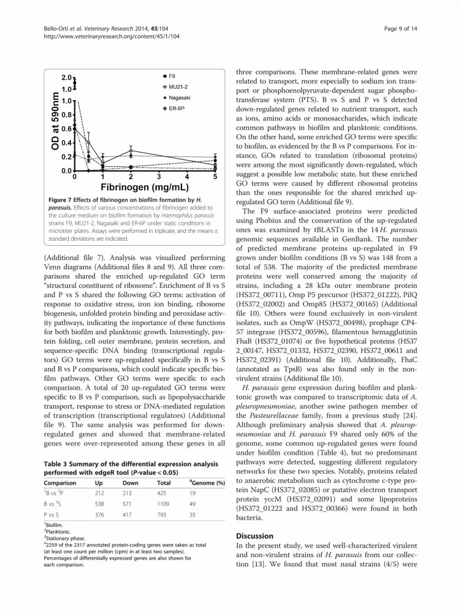
Figure 7 Effects of fibrinogen on biofilm formation by H.parasuis. Effects of various concentrations of fibrinogen added tothe culture medium on biofilm formation by Haemophilus parasuisstrains F9, MU21-2, Nagasaki and ER-6P under static conditions inmicrotiter plates. Assays were performed in triplicate, and the means ±standard deviations are indicated.
Bello-Ortí et al. Veterinary Research 2014, 45:104 Page 9 of 14http://www.veterinaryresearch.org/content/45/1/104
(Additional file 7). Analysis was visualized performingVenn diagrams (Additional files 8 and 9). All three com-parisons shared the enriched up-regulated GO term“structural constituent of ribosome”. Enrichment of B vs Sand P vs S shared the following GO terms: activation ofresponse to oxidative stress, iron ion binding, ribosomebiogenesis, unfolded protein binding and peroxidase activ-ity pathways, indicating the importance of these functionsfor both biofilm and planktonic growth. Interestingly, pro-tein folding, cell outer membrane, protein secretion, andsequence-specific DNA binding (transcriptional regula-tors) GO terms were up-regulated specifically in B vs Sand B vs P comparisons, which could indicate specific bio-film pathways. Other GO terms were specific to eachcomparison. A total of 20 up-regulated GO terms werespecific to B vs P comparison, such as lipopolysaccharidetransport, response to stress or DNA-mediated regulationof transcription (transcriptional regulators) (Additionalfile 9). The same analysis was performed for down-regulated genes and showed that membrane-relatedgenes were over-represented among these genes in all
Table 3 Summary of the differential expression analysisperformed with edgeR tool (P-value < 0.05)
Comparison Up Down Total 4Genome (%)1B vs 2P 212 213 425 19
B vs 3S 538 571 1109 49
P vs S 376 417 793 351Biofilm.2Planktonic.3Stationary phase.42259 of the 2317 annotated protein-coding genes were taken as total(at least one count per million (cpm) in at least two samples).Percentages of differentially expressed genes are also shown foreach comparison.
three comparisons. These membrane-related genes wererelated to transport, more especially to sodium ion trans-port or phosphoenolpyruvate-dependent sugar phospho-transferase system (PTS). B vs S and P vs S detecteddown-regulated genes related to nutrient transport, suchas ions, amino acids or monosaccharides, which indicatecommon pathways in biofilm and planktonic conditions.On the other hand, some enriched GO terms were specificto biofilm, as evidenced by the B vs P comparisons. For in-stance, GOs related to translation (ribosomal proteins)were among the most significantly down-regulated, whichsuggest a possible low metabolic state, but these enrichedGO terms were caused by different ribosomal proteinsthan the ones responsible for the shared enriched up-regulated GO term (Additional file 9).The F9 surface-associated proteins were predicted
using Phobius and the conservation of the up-regulatedones was examined by tBLASTn in the 14H. parasuisgenomic sequences available in GenBank. The numberof predicted membrane proteins up-regulated in F9grown under biofilm conditions (B vs S) was 148 from atotal of 538. The majority of the predicted membraneproteins were well conserved among the majority ofstrains, including a 28 kDa outer membrane protein(HS372_00711), Omp P5 precursor (HS372_01222), PilQ(HS372_02002) and Omp85 (HS372_00165) (Additionalfile 10). Others were found exclusively in non-virulentisolates, such as OmpW (HS372_00498), prophage CP4-57 integrase (HS372_00596), filamentous hemagglutininFhaB (HS372_01074) or five hypothetical proteins (HS372_00147, HS372_01332, HS372_02390, HS372_00611 andHS372_02391) (Additional file 10). Additionally, FhaC(annotated as TpsB) was also found only in the non-virulent strains (Additional file 10).H. parasuis gene expression during biofilm and plank-
tonic growth was compared to transcriptomic data of A.pleuropneumoniae, another swine pathogen member ofthe Pasteurellaceae family, from a previous study [24].Although preliminary analysis showed that A. pleurop-neumoniae and H. parasuis F9 shared only 60% of thegenome, some common up-regulated genes were foundunder biofilm condition (Table 4), but no predominantpathways were detected, suggesting different regulatorynetworks for these two species. Notably, proteins relatedto anaerobic metabolism such as cytochrome c-type pro-tein NapC (HS372_02085) or putative electron transportprotein yccM (HS372_02091) and some lipoproteins(HS372_01222 and HS372_00366) were found in bothbacteria.
DiscussionIn the present study, we used well-characterized virulentand non-virulent strains of H. parasuis from our collec-tion [13]. We found that most nasal strains (4/5) were

Figure 8 Identification of H. parasuis genes differentially expressed. MA plots generated by EdgeR showing transcript expression profiles inthe three comparisons performed: biofilm vs planktonic (A), biofilm vs stationary phase (B) and planktonic vs stationary phase (C). For each gene,log2(fold change) between the two conditions is plotted (M, y axis) against the gene’s log2(average expression) in the two samples (A, x axis). Theblue lines indicate 2-fold changes. Red dots highlight the genes at 5% P-value.
Bello-Ortí et al. Veterinary Research 2014, 45:104 Page 10 of 14http://www.veterinaryresearch.org/content/45/1/104
strong-biofilm producers whereas most systemic strains(8/9) formed no biofilm or were very weak-biofilm pro-ducers. Similarly, a strong association was found betweenMLST clusters [2] and biofilm formation. Strains of MLSTcluster C (which are associated with nasal isolation)formed more biofilms than strains of MLST cluster A(which are isolated from systemic lesions). Similarly, thepresence of autotransporter vtaA group 1 genes and ofsialyltransferase lsgB gene associated with virulent strains[13,48], and was not associated with strong biofilm forma-tion. Overall, our results are in agreement with Jin et al.[19] who observed that, generally, serovars considerednon-virulent showed a higher degree of biofilm formationthan virulent serovars. Although it is important to notethat a strict relationship between serovars and virulence inH. parasuis has not been demonstrated [1].
Figure 9 Comparison of H. parasuis genes that were up- or down-regup- (A) and down-regulated (B) under different growth states.
The use of CLSM and fluorescent probes showed thepresence of PGA, proteins, and eDNA in the biofilmmatrix of H. parasuis nasal, high-biofilm producer strainsF9 and MU21-2 and of the weak-biofilm producer strainsNagasaki and ER-6P recovered from lesions of pigs withGlässer’s disease. This is, to the best of our knowledge, thefirst report of the presence of PGA and proteins in thebiofilm matrix of H. parasuis. PGA is the major compo-nent of the biofilm matrix of several bacteria includingother Pasteurellaceae members such as A. pleuropneumo-niae and Aggregatibacter actinomycetemcomitans [18,49].Nevertheless all 4 strains tested were resistant to dispersinB. All strains were however sensitive to a proteinase Ktreatment suggesting that proteins play a larger rolethan PGA in H. parasuis biofilm formation. Tang et al.[50] have shown that treatment with the staphylococcal
ulated. Venn diagrams of Haemophilus parasuis genes identified as

Table 4 Common up-regulated genes betweenHaemophilus parasuis F9 and Actinobacilluspleuropneumoniae when grown in biofilm condition
H. parasuis (this study) vs A. pleuropneumoniae [22]
H. parasuisF9
App Product
HS372_02083 APL_1425 Cytochrome c-type protein NapC
HS372_02085 APL_1427 Putative electron transport protein yccM
HS372_02091 APL_1821 50S ribosomal protein L31 type B
HS372_00945 APL_1440 High-affinity zinc uptake system protein znuAprecursor
HS372_00147 APL_1894 hypothetical protein
HS372_02009 APL_1423 Putative esterase
HS372_02012 APL_0433 Peptide methionine sulfoxide reductase MsrB
HS372_01222 APL_0460 Outer membrane protein P5 precursor
HS372_00060 APL_1206 putative ribonuclease FitB
HS372_01900 APL_1173 Nicotinamide riboside transporter pnuC
HS372_00666 APL_0442 vancomycin high temperature exclusionprotein
HS372_01587 APL_0484 Alpha-aminoadipate–lysW ligase lysX
HS372_02062 APL_0038 hypothetical protein
HS372_00364 APL_0222 Putative lipoprotein/NMB1162 precursor
HS372_01892 APL_0133 Cys regulon transcriptional activator
HS372_00988 APL_1295 Arginine repressor
HS372_02252 APL_1873 Succinyl-diaminopimelate desuccinylase
HS372_02064 APL_0036 hypothetical protein
HS372_01893 APL_0134 hypothetical protein
HS372_01521 APL_1320 Thiamine import ATP-binding protein ThiQ
HS372_02387 APL_1059 Integrase core domain protein
HS372_00061 APL_1207 prevent-host-death family protein
HS372_00916 APL_0423 Ribonuclease HI
HS372_01200 APL_0593 Inosine-5’-monophosphate dehydrogenase
HS372_01950 APL_1574 Putative hydrolase ydeN
HS372_02244 APL_0254 Cytosol non-specific dipeptidase
HS372_01281 APL_1230 Phosphoserine phosphatase
HS372_01385 APL_0967 Glutamate permease
HS372_01220 APL_0463 Putative phosphinothricin acetyltransferaseYwnH
HS372_01490 APL_0928 hypothetical protein
HS372_01179 APL_1499 Threonine synthase
HS372_02366 APL_0395 Sigma-E factor negative regulatory protein
HS372_01208 APL_0895 Formate dehydrogenase-N subunit gamma
HS372_01221 APL_0461 putative phosphatase YwpJ
HS372_02265 APL_0687 D-lactate dehydrogenase
HS372_01342 APL_1448 Spermidine/putrescine import ATP-bindingprotein PotA
HS372_00366 APL_0220 Putative lipoprotein/NMB1164 precursor
HS372_00099 APL_1853 Ketol-acid reductoisomerase
Bello-Ortí et al. Veterinary Research 2014, 45:104 Page 11 of 14http://www.veterinaryresearch.org/content/45/1/104
nuclease NUC1 decreased slightly the biofilm formationof H. parasuis strain 0322. This is in agreement withresults of this study showing the presence of eDNA inthe biofilm matrix of H. parasuis and the high sensitiv-ity of strain F9 biofilm to DNase I digestion.This is also the first description of H. parasuis biofilm
formation under shear force in a drip-flow reactor and amicrofluidic system. Interestingly, similar biofilm pheno-types (i.e. high-producer vs weak-producer) were observedwhen static or controlled flow conditions were used. Thedrip-flow apparatus is a system that it is thought to createan environment with an air-liquid interface which closelyresembles the lung environment [23]. Under the condi-tions we selected, the two strains with a strong-biofilmphenotype, MU21-2 and F9, produced visible biofilmswhile a thin film was observable for Nagasaki and ER-6P,which are strains with a weak-biofilm phenotype understatic conditions. We also used the BioFlux flowthroughdevice, a high throughput microfluidic system that hasbeen recently tested for the growth of dental plaque bac-teria biofilms [51] and Pseudomonas aeruginosas biofilms[25]. Again, strains Nagasaki and ER-6P did not form bio-films under the conditions tested but strains MU21-2 andF9 formed biofilms that rapidly blocked the microfluidicchannel. This microfluidic system can therefore be used tostudy biofilm formation of H. parasuis but for short incu-bation periods. This potentially can be used to investigategenes involved in the early steps of biofilm formation and,if the appropriate tools are combined, to study real-timegene expression during the early steps as demonstrated inStaphylococcus aureus [52].The genes involved in H. parasuis biofilm formation are
currently not known. Recently, it was reported that bothgalU and galE genes seemed to play a role in biofilm for-mation of H. parasuis [12]. A galU mutant was unable toform biofilm in a glass tube while a galE mutant producedmore biofilm than the parent wild-type strain. Surprisinglyhowever, the galU mutant also showed an increased ten-dency to autoagglutinate which is usually associated with agreater ability to form biofilm. Here, the transcriptome ofH. parasuis F9 strain showed that static biofilm and plank-tonic cultures are in similar biological states but not identi-cal, whereas greater differences in gene expression wereevident when compared to stationary-phase culture. Subtledifferences in gene expression between biofilm and plank-tonic cells have been also reported for other bacterial spe-cies [53,54]. In addition to identifying DEGs related tometabolism, gene enrichment allowed the identification ofa large proportion of membrane-related genes among theup-regulated genes in biofilm, including some that havebeen reported for other bacterial species [53,54]. The con-servation of some highly expressed membrane-proteingenes in biofilms among the 14H. parasuis genomes avail-able indicates that those may not specifically associate

Bello-Ortí et al. Veterinary Research 2014, 45:104 Page 12 of 14http://www.veterinaryresearch.org/content/45/1/104
with biofilm formation. A sub-set of these up-regulatedmembrane-protein genes, such as the type IV pilus biogen-esis protein pilQ, may be involved in adhesion to differentabiotic and biotic surfaces. Interestingly, some up-regulatedgenes, such as fhaB and fhaC (tpsB) or ompW, were onlyfound in the genome of non-virulent strains. This finding,together with the fact that non-virulent strains formedstronger biofilms, suggests a possible role of these genes inbiofilm formation. In fact, Fha and OmpW, together withtype IV pilus, have been shown to play a role in biofilmformation in other bacteria [55,56]. Additionally, some li-poproteins, such as lipoprotein Plp4, as well as signs of an-aerobic metabolism were evidenced in the biofilms of H.parasuis and A. pleuropneumoniae, which may indicate apotential role in biofilm formation of both bacterial spe-cies. The function of specific genes in biofilm formationwill need further confirmation.Interestingly, experimental infections in snatch-farrowed,
colostrum-deprived piglets showed that the H. parasuisstrains that proceeded to invade the host were not main-tained well in the nasal cavities of the piglets [57]. Thissuggests changes in the bacteria from a “colonizing state”to an “invasive state”, which could be modulated by theability of each animal to control the infection. In the lightof the results of the present study, these changes from a“colonizing state” to an “invasive state” could also be dueto the ability of a given bacterial strain to form or not a ro-bust biofilm. We propose that biofilm formation mightallow the non-virulent strains to colonize and persist in theupper respiratory tract of pigs. Conversely, the predomin-ant planktonic state of the virulent strains might allowthem to disseminate within the host. This latter state-ment is supported by the inhibition of biofilm forma-tion by fibrinogen. It is worth noting that a recentstudy on the human pathogen Streptococcus pneumo-niae showed that biofilm formation in vivo is associatedwith reduced invasiveness and a dampened cytokine re-sponse [58]. High-biofilm production phenotype mighttherefore not always be linked to virulence.
Additional files
Additional file 1: Haemophilus parasuis biofilm formation understatic conditions in microtiter plates. (A) Medians of biofilm formationfor strains that are sensitive or show intermediate resistance to serum(n = 9) or for strains that are resistant to serum (n = 5). (B) Medians ofbiofilm formation for strains negative (n = 5) or positive (n = 9) for vtaAgroup 1 genes. (C) Medians of biofilm formation for strains belonging toMLST cluster C (n = 5) or strains belonging to MLST cluster A (n = 7).(D) Medians of biofilm formation for strains negative (n = 8) or positive(n = 6) for the sialyltransferase gene lsgB. Differences between the medianof the two groups of strains were not statistically significant (A: p = 0.059;B: p = 0.189; C: p = 0.202; D: p = 0.228).
Additional file 2: Mapping of RNA sequencing reads to theH. parasuis strain F9 genome. Overview of the mapping (A) and readcounts (B) results. Mapping quality (MAPQ) shows that most of the reads
were aligned with MAPQ ≥ 20 but a considerable percentage of readswere not taken into account for differential expression because mappingto non-protein coding regions, particularly for stationary culture sample.
Additional file 3: List of H. parasuis strain F9 genes differentiallyexpressed in biofilm vs planktonic cells. Differentially expressed genesin biofilm versus planktonic H. parasuis cells (FDR < 0.05). Sorted by logFC.Actinobacillus pleuropneumoniae (App) first BLASTp hits are also shown.
Additional file 4: List of H. parasuis strain F9 genes differentiallyexpressed in planktonic vs stationary phase cells. Differentiallyexpressed genes in planktonic versus stationary H. parasuis cells (FDR < 0.05).Sorted by logFC. Actinobacillus pleuropneumoniae (App) first BLASTp hits arealso shown.
Additional file 5: List of H. parasuis strain F9 genes differentiallyexpressed in biofilm vs stationary phase cells. Differentially expressedgenes in biofilm versus stationary H. parasuis cells (FDR < 0.05). Sorted bylogFC. Actinobacillus pleuropneumoniae (App) first BLASTp hits are also shown.
Additional file 6: List of H. parasuis strain F9 up-regulated genes orunique to biofilm. Haemophilus parasuis F9 genes up-regulated in allthree comparisons (biofilm vs planktonic; biofilm vs stationary phase;planktonic vs stationary phase) or unique to biofilm.
Additional file 7: List of gene ontology (GO) terms followingenrichment analysis of H. parasuis strain F9 differentially expressedgenes. Enriched Gene Ontology biological process and cellularcomponent nodes among up-regulated genes (FDR < 0.001). No enrichedCC GO terms were found for biofilm vs planktonic comparison.
Additional file 8: Visualization of gene ontology (GO) termsfollowing enrichment analysis of H. parasuis strain F9 differentiallyexpressed genes. Venn diagrams of Haemophilus parasuis enrichedGene Ontology (GO) terms among the subsets of differentially expressedgenes identified as up- (A) and down-regulated (B) under differentgrowth states. Only most specific GO terms are shown.
Additional file 9: List of H. parasuis strain F9 GO terms shown inAdditional file 8 diagrams. H. parasuis F9 GO terms results fromAdditional file 8 Venn diagrams.
Additional file 10: List of H. parasuis strain F9 membrane-relatedgenes differentially expressed in biofilm compared to planktonic orstationary phase cells. Conservation of Haemophilus parasuis F9membrane-related genes differentially expressed (P < 0.05) in biofilms vsstationary culture or biofilms vs planktonic culture among 14 H. parasuisisolates. Genes are sorted by logFC. *Truncated.
Competing interestsThe authors declare that they have no competing interests.
Authors’ contributionsBBO carried the transcriptomics experiments, and participated in the writingof the manuscript. VD carried the static biofilm formation assays, andparticipated in the writing of the manuscript. YDNT carried the biofilmformation assays under shear force, and participated in the writing of themanuscript. JL carried the confocal microscopy, the enzymatic treatmentsand the statistical analysis, and participated in the writing of the manuscript.KJH, AWT and DJM sequenced and assembled the genome of strain F9 andparticipated in the writing of the manuscript. VA conceived the study,participated in its design, and participated in the writing of the manuscript.MJ conceived the study, participated in its design, and drafted themanuscript. All authors read and approved the final manuscript.
AcknowledgementsThis work was supported in part by a discovery grant from the Natural Sciencesand Engineering Research Council of Canada to MJ and by a grant AGL2010-15232 from the Ministerio de Economía y Competitividad of Spain to VA. VDwas a recipient of a doctoral scholarship and of an international training awardfrom Fonds de recherche du Québec – Nature et technologies. BBO is arecipient of a FPI fellowship from the Spanish Ministerio de Economía yCompetitividad. The authors would also like to acknowledge the support of theGouvernement du Québec, Ministère des Relations internationales, de laFrancophonie et du Commerce extérieur (VIIe rencontre du Comité mixteQuébec-Catalogne, projet 07.304).

Bello-Ortí et al. Veterinary Research 2014, 45:104 Page 13 of 14http://www.veterinaryresearch.org/content/45/1/104
Author details1Centre de Recerca en Sanitat Animal (CReSA), UAB-IRTA, Campus de laUniversitat Autònoma de Barcelona, 08193 Bellaterra, Cerdanyola del Vallès,Spain. 2Groupe de recherche sur les maladies infectieuses du porc, Facultéde médecine vétérinaire, Université de Montréal, St-Hyacinthe, Québec J2S7C6, Canada. 3Department of Veterinary Medicine, University of Cambridge,Cambridge, UK. 4Institut de Recerca i Tecnologia Agroalimentàries (IRTA),Barcelona, Spain.
Received: 30 May 2014 Accepted: 26 September 2014
References1. Aragon V, Segalés J, Oliveira S: Glässer’s disease. In Diseases of Swine. 10th
edition. Edited by Zimmerman JJ, Karriker LA, Ramirez A, Schwartz KJ,Stevenson GW. Chichester, UK: Wiley-Blackwell; 2012:760–769.
2. Olvera A, Cerdà-Cuéllar M, Aragon V: Study of the population structure ofHaemophilus parasuis by multilocus sequence typing. Microbiology 2006,152:3683–3690.
3. Cerdà-Cuéllar M, Aragon V: Serum-resistance in Haemophilus parasuis isassociated with systemic disease in swine. Vet J 2008, 175:384–389.
4. Olvera A, Ballester M, Nofrarias M, Sibila M, Aragon V: Differences inphagocytosis susceptibility in Haemophilus parasuis strains. Vet Res 2009, 40:24.
5. Aragon V, Bouchet B, Gottschalk M: Invasion of endothelial cells by systemicand nasal strains of Haemophilus parasuis. Vet J 2010, 186:264–267.
6. Vanier G, Szczotka A, Friedl P, Lacouture S, Jacques M, Gottschalk M:Haemophilus parasuis invades porcine brain microvascular endothelialcells. Microbiology 2006, 152:135–142.
7. Zhang B, Tang C, Liao M, Yue H: Update on the pathogenesis ofHaemophilus parasuis infection and virulence factors. Vet Microbiol 2014,168:1–7.
8. Costa-Hurtado M, Aragon V: Advances in the quest for virulence factors ofHaemophilus parasuis. Vet J 2013, 198:571–576.
9. Bouchet B, Vanier G, Jacques M, Gottschalk M: Interactions of Haemophilusparasuis and its LOS with porcine brain microvascular endothelial cells.Vet Res 2008, 39:42.
10. Bouchet B, Vanicer G, Jacques M, Auger E, Gottschalk M: Studies on theinteractions of Haemophilus parasuis with porcine epithelial trachealcells: limited role of LOS in apoptosis and pro-inflammatory cytokinerelease. Microb Pathog 2009, 46:108–113.
11. Xu C, Zhang L, Zhang B, Feng S, Zhou S, Li J, Zou Y, Liao M: Involvement oflipooligosaccharide heptose residues of Haemophilus parasuis SC096 strainin serum resistance, adhesion and invasion. Vet J 2013, 195:200–204.
12. Zou Y, Feng S, Xu C, Zhang B, Zhou S, Zhang L, He X, Li J, Yang Z, Liao M:The role of galU and galE of Haemophilus parasuis SC096 in serumresistance and biofilm formation. Vet Microbiol 2013, 162:278–284.
13. Martínez-Moliner V, Soler-Llorens P, Moleres J, Garmendia J, Aragon V:Distribution of genes involved in sialic acid utilization in strains ofHaemophilus parasuis. Microbiology 2012, 158:2117–2124.
14. Pina S, Olvera A, Barcelo A, Bensaid A: Trimeric autotransporters ofHaemophilus parasuis: generation of an extensive passenger domainrepertoire specific for pathogenic strains. J Bacteriol 2009, 191:576–587.
15. Costa-Hurtado M, Ballester M, Galofré-Milà N, Darji A, Aragon V: VtaA8 andVtaA9 from Haemophilus parasuis delay phagocytosis by alveolarmacrophages. Vet Res 2012, 43:57.
16. Costerton JW, Stewart PS, Greenberg EP: Bacterial biofilms: a commoncause of persistent infections. Science 1999, 284:1318–1322.
17. Hall-Stoodley L, Costerton JW, Stoodley P: Bacterial biofilms: from the naturalenvironment to infectious diseases. Nat Rev Microbiol 2004, 2:95–108.
18. Jacques M, Aragon V, Tremblay YDN: Biofilm formation in bacterialpathogens of veterinary importance. Anim Health Res Rev 2010, 11:97–121.
19. Jin H, Zhou R, Kang M, Luo R, Cai X, Chen H: Biofilm formation by fieldisolates and reference strains of Haemophilus parasuis. Vet Microbiol 2006,118:117–123.
20. Jin H, Wan Y, Zhou R, Li L, Luo R, Zhang S, Hu J, Langford PR, Chen H:Identification of gene transcribed by Haemophilus parasuis in necroticporcine lung through the selective capture of transcribed sequences(SCOTS). Environ Microbiol 2008, 10:3326–3336.
21. Wu C, Labrie J, Tremblay YDN, Haine D, Mourez M, Jacques M: Zinc as anagent for the prevention of biofilm formation by pathogenic bacteria.J Appl Microbiol 2013, 115:30–40.
22. Labrie J, Pelletier-Jacques G, Deslandes V, Ramjeet M, Auger E, Nash JH,Jacques M: Effects of growth conditions on biofilm formation byActinobacillus pleuropneumoniae. Vet Res 2010, 41:03.
23. Goeres DM, Hamilton MA, Beck NA, Buckingham-Meyer K, Hilyard JD,Loetterle LR, Lorenz LA, Walker DK, Stewart PS: A method for growing abiofilm under low shear at the air-liquid interface using the drip flowbiofilm reactor. Nat Protoc 2009, 4:783–788.
24. Tremblay YDN, Deslandes V, Jacques M: Actinobacillus pleuropneumoniaegenes expression in biofilms cultured under static conditions and in adrip-flow apparatus. BMC Genomics 2013, 14:364.
25. Benoit MR, Conant CG, Ionescu-Zanetti C, Schwartz M, Matin A: New devicefor high-throughput viability screening of flow biofilms. Appl EnvironMicrobiol 2010, 76:4136–4142.
26. Tremblay YDN, Lamarche D, Chever P, Haine D, Messier S, Jacques M:Characterization of the ability of coagulase-negative staphylococci isolatedfrom the milk of Canadian farms to form biofilms. J Dairy Sci 2013, 96:234–246.
27. Quail MA, Kozarewa I, Smith F, Scally A, Stephens PJ, Durbin R, Swerdlow H,Turner DJ: A large genome centre’s improvements to the Illuminasequencing system. Nat Methods 2008, 5:1005–1010.
28. Quail M, Swerdlow H, Turner DJ: Improved protocols for the Illuminagenome analyzer sequencing system. Curr Protoc Hum Genet 2009, 18:1–27.
29. Lunter G, Goodson M: Stampy : A statistical algorithm for sensitive andfast mapping of Illumina sequence reads. Genome Res 2011, 21:936–939.
30. Xu Z, Yue M, Zhou R, Jin Q, Fan Y, Bei W, Chen H: Genomiccharacterization of Haemophilus parasuis SH0165, a highly virulent strainof serovar 5 prevalent in China. PLoS One 2011, 6:e19631.
31. Martin M: Cutadapt removes adapter sequences from high-throughputsequencing reads. EMBnet J 2011, 17:10–12.
32. Joshi N: UC Davis Bioinformatics Core. https://github.com/ucdavis-bioinformatics/sickle.
33. Zerbino DR, Birney E: Velvet: algorithms for de novo short read assemblyusing de Bruijn graphs. Genome Res 2008, 18:821–829.
34. Gladman S: Velvet Optimiser. http://www.vicbioinformatics.com/velvetoptimiser.manual.txt.
35. Seemann T: Prokka User Manual. http://bioinformatics.net.au/prokka-manual.html.36. Oliveira S, Galina L, Pijoan C: Development of a PCR test to diagnose
Haemophilus parasuis infections. J Vet Diagn Invest 2001, 13:495–501.37. FastQC: http://www.bioinformatics.babraham.ac.uk/projects/fastqc/38. FASTX-Toolkit: [http://hannonlab.cshl.edu/fastx_toolkit/]39. Torrent Mapping Alignment Program: [https://github.com/iontorrent/TMAP]40. Li H, Handsaker B, Wysoker A, Fennell T, Ruan J, Homer N, Marth G, Abecasis
G, Durbin R and 1000 Genome Project Data Processing Subgroup:The sequence alignment/map (SAM) format and SAMtools. Bioinformatics2009, 25:2078–2079.
41. Thorvaldsdóttir H, Robinson JT, Mesirov JP: Integrative Genomics Viewer(IGV): high-performance genomics data visualization and exploration.Brief Bioinform 2013, 14:178–192.
42. HTSeq v0.5.4p3: [https://pypi.python.org/pypi/HTSeq]43. Robinson MD, McCarthy DJ, Smyth GK: edgeR: a Bioconductor package for
differential expression analysis of digital gene expression data.Bioinformatics 2010, 26:139–140.
44. Conesa A, Götz S, Garcia-Gomez JM, Terol J, Talon M, Robles M: Blast2GO:a universal tool for annotation, visualization and analysis in functionalgenomics research. Bioinformatics 2005, 21:3674–3676.
45. Käll L, Krogh A, Sonnhammer EL: A combined transmembrane topologyand signal peptide prediction method. J Mol Biol 2004, 338:1027–1036.
46. Bonifait L, Grignon L, Grenier D: Fibrinogen induces biofilm formation byStreptococcus suis and enhances its antibiotic resistance. Appl EnvironMicrobiol 2008, 74:4969–4972.
47. Mullins MA, Register KB, Bayles DO, Dyer DW, Kuehn JS, Phillips GJ: Genomesequence of Haemophilus parasuis strain 29755. Stand Genomic Sci 2011,5:61–68.
48. Olvera A, Pina S, Macedo N, Oliveira S, Aragon V, Bensaid A: Identification ofpotentially virulent strains of Haemophilus parasuis using a multiplex PCRfor virulence-associated autotransporters (vtaA). Vet J 2012, 191:213–218.
49. Kaplan JB, Velliyagounder K, Ragunath C, Rohde H, Mack D, Knobloch JK,Ramasubbu N: Genes involved in the synthesis and degradation ofmatrix polysaccharide in Actinobacillus actinomycetemcomitans andActinobacillus pleuropneumoniae biofilms. J Bacteriol 2004, 186:8213–8220.
50. Tang JN, Kang MS, Chen HC, Shi XM, Zhou R, Chen J, Du YW: Thestaphylococcal nuclease prevents biofilm formation in Staphylococcus

Bello-Ortí et al. Veterinary Research 2014, 45:104 Page 14 of 14http://www.veterinaryresearch.org/content/45/1/104
aureus and other biofilm-forming bacteria. Sci China Life Sci 2011,54:863–869.
51. Nance WC, Dowd SE, Samarian D, Chludzinski J, Delli J, Battista J, RickardAH: A high-throughput microfluidic dental plaque biofilm system tovisualize and quantify the effect of antimicrobials. J Antimicrob Chemother2013, 68:2550–2560.
52. Moormeier DE, Endres JL, Mann EE, Sadykov MR, Horswill AR, Rice KC, FeyPD, Bayles KW: Use of microfluidic technology to analyze geneexpression during Staphylococcus aureus biofilm formation revealsdistinct physiological niches. Appl Environ Microbiol 2013, 79:3413–3424.
53. Yadav MK, Kwon SK, Cho CG, Park S-W, Chae S-W, Song J-J: Geneexpression profile of early in vitro biofilms of Streptococcus pneumoniae.Microbiol Immunol 2012, 56:621–629.
54. Dötsch A, Eckweiler D, Schniederjans M, Zimmerman A, Jensen V, ScharfeM, Geffers R, Häussler S: The Pseudomonas aeruginosa transcriptome inplanktonic cultures and static biofilms using RNA sequencing. PLoS One2012, 7:e31092.
55. Ritter A, Com E, Bazire A, Goncalves Mdos S, Delage L, Le Pennec G, Pineau C,Dreanno C, Compère C, Durour A: Proteomic studies highlight outer-membrane proteins related to biofilm development in the marinebacterium Pseudoalteromonas sp. D41. Proteomics 2012, 12:3180–3192.
56. Irie Y, Mattoo S, Yuk MH: The Bvg virulence control system regulates biofilmformation in Bordetella bronchiseptica. J Bacteriol 2004, 186:5692–5698.
57. Aragon V, Cerdà-Cuéllar M, Fraile L, Monbarg M, Nofrarias M, Olvera A, SibilaM, Solanes D, Segalés J: Correlation between clinico-pathologicaloutcome and typing of Haemophilus parasuis field strains. Vet Microbiol2010, 142:387–393.
58. Blanchette-Cain K, Hinojosa CA, Suresh Babu RA, Lizcano A, Gonzalez-JuarbeN, Munoz-Almagro C, Sanchez CJ, Bergman MA, Orihuela CJ: Streptococcuspneumoniae biofilm formation is strain dependent, multifactorial, andassociated with reduced invasiveness and immunoreactivity duringcolonization. MBio 2013, 4:e00745–13.
doi:10.1186/s13567-014-0104-9Cite this article as: Bello-Ortí et al.: Biofilm formation by virulent andnon-virulent strains of Haemophilus parasuis. Veterinary Research2014 45:104.
Submit your next manuscript to BioMed Centraland take full advantage of:
• Convenient online submission
• Thorough peer review
• No space constraints or color figure charges
• Immediate publication on acceptance
• Inclusion in PubMed, CAS, Scopus and Google Scholar
• Research which is freely available for redistribution
Submit your manuscript at www.biomedcentral.com/submit


