Embed Size (px)
Citation preview

Citation: Vincent, J.; Lau, K.S.;
Evyan, Y.C.-Y.; Chin, S.X.;
Sillanpää, M.; Chia, C.H. Biogenic
Synthesis of Copper-Based
Nanomaterials Using Plant Extracts
and Their Applications: Current and
Future Directions. Nanomaterials 2022,
12, 3312. https://doi.org/10.3390/
nano12193312
Academic Editor: Sotirios Baskoutas
Received: 6 September 2022
Accepted: 19 September 2022
Published: 23 September 2022
Publisher’s Note: MDPI stays neutral
with regard to jurisdictional claims in
published maps and institutional affil-
iations.
Copyright: © 2022 by the authors.
Licensee MDPI, Basel, Switzerland.
This article is an open access article
distributed under the terms and
conditions of the Creative Commons
Attribution (CC BY) license (https://
creativecommons.org/licenses/by/
4.0/).
nanomaterials
Review
Biogenic Synthesis of Copper-Based Nanomaterials Using PlantExtracts and Their Applications: Current and Future DirectionsJei Vincent 1 , Kam Sheng Lau 1,*, Yang Chia-Yan Evyan 2 , Siew Xian Chin 3 , Mika Sillanpää 1,4,5,6
and Chin Hua Chia 1,*
1 Materials Science Program, Department of Applied Physics, Faculty of Science and Technology,Universiti Kebangsaan Malaysia, Bangi 43600, Selangor, Malaysia
2 Faculty of Engineering, Science and Technology, Nilai University, Nilai 71800, Negeri Sembilan, Malaysia3 ASASIpintar Program, Pusat GENIUS@Pintar Negara, Universiti Kebangsaan Malaysia,
Bangi 43600, Selangor, Malaysia4 Department of Chemical Engineering, School of Mining, Metallurgy and Chemical Engineering,
University of Johannesburg, P.O. Box 17011, Doornfontein 2028, South Africa5 Sustainable Membrane Technology Research Group (SMTRG), Chemical Engineering Department,
Persian Gulf University, Bushehr P.O. Box 75169-13817, Iran6 Zhejiang Rongsheng Environmental Protection Paper Co., Ltd., NO. 588 East Zhennan Road,
Pinghu Economic Development Zone, Zhejiang 314213, China* Correspondence: [email protected] (K.S.L.); [email protected] (C.H.C.)
Abstract: Plants have been used for multiple purposes over thousands of years in various applicationssuch as traditional Chinese medicine and Ayurveda. More recently, the special properties of phyto-chemicals within plant extracts have spurred researchers to pursue interdisciplinary studies unitingnanotechnology and biotechnology. Plant-mediated green synthesis of nanomaterials utilises thephytochemicals in plant extracts to produce nanomaterials. Previous publications have demonstratedthat diverse types of nanomaterials can be produced from extracts of numerous plant components.This review aims to cover in detail the use of plant extracts to produce copper (Cu)-based nanoma-terials, along with their robust applications. The working principles of plant-mediated Cu-basednanomaterials in biomedical and environmental applications are also addressed. In addition, itdiscusses potential biotechnological solutions and new applications and research directions concern-ing plant-mediated Cu-based nanomaterials that are yet to be discovered so as to realise the fullpotential of the plant-mediated green synthesis of nanomaterials in industrial-scale production andwider applications. This review provides readers with comprehensive information, guidance, andfuture research directions concerning: (1) plant extraction, (2) plant-mediated synthesis of Cu-basednanomaterials, (3) the applications of plant-mediated Cu-based nanomaterials in biomedical andenvironmental remediation, and (4) future research directions in this area.
Keywords: biogenic synthesis; copper-based nanomaterials; extraction method; plant extract
1. Introduction
Apart from food, plants have traditionally been used intensively in textile, cosmetics,and medicine. Beyond traditional Chinese medicine and Ayurveda, which have devel-oped over thousands of years of interest in the prevention and treatment of diseases, thebiomedical applications of plants have broadened even further due to the advancementof technology and of time [1–4]. These biomedical applications are mainly due to thephytochemicals within plants [5–7], which are among the most fascinating aspects of plantsdue to their having activities such as antimicrobial, antitumour, antiaging, and others [8].Awareness of such properties has driven researchers to discover still more applications ofphytochemicals. In 1959, Richard Feynman illustrated the controlling of single atoms andmolecules under the topic of “There’s plenty of room at the bottom”, which first shed light
Nanomaterials 2022, 12, 3312. https://doi.org/10.3390/nano12193312 https://www.mdpi.com/journal/nanomaterials

Nanomaterials 2022, 12, 3312 2 of 44
on the novel nanotechnology research field. More recently, an innovative interdisciplinarystudy pioneered nanomaterial synthesis from the phytochemicals within plant extracts, aprocess which is more eco-friendly than conventional methods and avoids the usage ofhazardous chemicals [9,10].
Plant-mediated nanomaterials synthesis is a branch of green synthesis in whichthe phytochemical compounds in plant extracts are utilised as stabilizing and reducingagents [9–13]. In addition to the pros and cons inherent in the synthesis method, thechoice of method and parameters in the nanomaterial production process also affect thegeometry of the obtained nanomaterials [14]. Typically, plant-mediated synthesis usesa bottom-up approach for material synthesis from plant extracts with the assistance ofdifferent biotechnological methods [11,13,15,16]. Conventional synthesis techniques havetheir disadvantages, such as use of hazardous chemicals, biological risks, and high energyconsumption [11,13,17–20]. Relative to conventional approaches like physical and chemicalsynthesis routes, plant synthesis is considered more eco-friendly and less toxic [13].
Many researchers have successfully synthesised, via plant-mediated synthesis, vari-ous types of nanomaterials that were previously produced by conventional synthesis ap-proaches, such as alloys, pure metals, metal oxides, and core shells [21–28]. The producednanomaterials have been used for numerous applications, including as antibacterials, anti-cancer agents, antifungals, antiparasitics, antioxidants, catalytic reduction agents, catalysts,biosensors, drug delivery vehicles, fuel cells, photocatalysts, and theranostics [24,29–35].However, there remain some limitations to the plant-mediated synthesis method that needto be addressed, such as the complexity and diversity of phytochemicals in plant systems,bio-reduction reactions, homogeneity, scaling-up, reproducibility, material accessibility,and product stability [31,36].
Cu is an element that has drawn significant attention from researchers in nanotechnology,specifically in the nanomaterial sector [37]. This is owing to the low cost, good abundance, andconductivity exhibited by Cu as compared to silver (Ag) and gold (Au) [38–43]. Accordingly,not only have various Cu nanomaterials (Cu-NMs) been developed, but there is a good body ofliterature on the plant-mediated synthesis of Cu nanomaterials with various applications [11].
Therefore, this review will focus on the synthesis of Cu-NMs from several perspectives,including their conventional, green, and especially plant-mediated synthesis, and, relatedly,plant extraction methods, parameters of plant-mediated nanomaterials, applications ofplant-mediated Cu-NMs, limitations of plant-based synthesis and proposed solutions, andpotential new applications and new research directions that are yet to be explored regardingplant-mediated Cu-NMs.
2. Synthesis of Nanomaterials: Conventional and Green Approaches
Approaches for the production of nanomaterials can be categorised according to twopredominant aspects: top-down and bottom-up [44]. Examples of the subdivisions withineach sector will be discussed. Firstly, top-down nanomaterial synthesis methods composedof ball milling and laser ablation, then bottom-up methods including hydrothermal, vapordeposition, microwave, chemical reduction, and green synthesis [45,46].
2.1. Disadvantages of Conventional Nanomateiral Synthesis Method
The typical demerits of conventional methods can be observed clearly in the case of ballmilling, as it is both energy-intensive and time-consuming to produce nanomaterials by thismethod, and, hence, ball milling is neither economical nor industry-friendly [45–47]. Simi-larly, the other top-down approach, laser ablation, requires high energy input to producea sufficiently intense laser for the continuous ablation process [46,48]. On the other hand,among bottom-up approaches, hydrothermal and microwave methods require an expensiveautoclave and complex equipment; thus, they are not applicable economically [45,46,48,49],while vapor deposition also necessitates high energy consumption [50]. Meanwhile, chemi-cal reduction utilises many substances that exhibit high toxicity toward living organismsand the environment, such as hydrazine, N, N-dimethylformamide, and sodium borohy-

Nanomaterials 2022, 12, 3312 3 of 44
dride; this results in additional treatment processes also being required [51,52]. Given allof the above drawbacks, many researchers have investigated green synthesis methods inorder to discover more biologically friendly alternatives for producing nanomaterials.
2.2. Green Synthesis Method of Nanomaterials
Green synthesis methods that utilise natural or biological compounds to producenanomaterials, such as bacterial-, fungal-, algae-, and plant-based methods, have beenfound to be non-toxic, non-harmful, and eco-friendly [14,37,47]. The utilization of naturaland biologically friendly compounds as reducing or capping agents also offers otheradvantages such as reducing energy requirements, avoiding usage of toxic/hazardouschemicals, and being simple and cheap [11,13,53]. The synthesis of nanomaterials usingbacteria has particular advantages as bacteria is abundant, easily cultured with a shortgeneration time, inexpensive to cultivate, stable, and easy to manipulate at the geneticlevel [46,54]. On top of that, previous reports have described the adaptability of bacteriato environments with a high concentration of heavy metals via transforming the toxicmetal ions to non-toxic metal oxide nanomaterials, which provides another rationale forthe utilization of bacteria in nanomaterial production, as precursors could be introduced inhigher concentrations [55,56].
In the fungal-mediated synthesis of nanomaterials, fungi show outstanding heavy-metaltolerance, internalization, and bioaccumulation capability, making them good candidates asreducing and stabilizing agents in the synthesis of metal nanomaterials [57]. Moreover, fungican be reproduced in large quantities, and by the parity of reasoning nanomaterials can besynthesised in quantity [58]. Relative to bacteria, fungi produce higher amounts of proteins andenzymes; thus, they can provide higher productivity of synthesis [46,59].
Algal-mediated synthesis of nanomaterials involves the utilization of carbohydrates,proteins, minerals, lipids, and bioactive compounds within algae as reducing agents toreduce metal precursor ions into nanomaterials [60]. Given their heavy-metal hyperac-cumulation capability, algae are excellent candidates for nanomaterials synthesis [61,62].The algae-mediated production of nanomaterials can occur either via extracellular or in-tracellular processes and affords good control over production parameters [60]. However,although the various microorganism-mediated synthesis processes offer many benefits, thepathogenic properties of organisms, underlying safety concerns, and deficit of knowledgeregarding synthesis mechanisms are drawbacks that yet hinder the use of these processesin industrial nanomaterial production and applications [46,54,63].
Plants are particularly good candidates for nanomaterial synthesis since they have nopathogenic effects as microorganisms do, plus the nanomaterials produced via plant biogenicsynthesis are more homogenous in comparison to the products of other methods [46,54]. Inaddition, unlike other synthesis methods, the mechanism of plant-mediated metal nanoparticlesynthesis is limited to the reduction of a precursor salt via agents within the plant extract in thepresence of a metal ion precursor. Moreover, stabilizing agents within a plant extract can alsoattach to the surface of the produced nanoparticles, improving the surface reaction kinetics aswell as particle stability and, hence, reducing the deformation and agglomeration of particles [64].The reducing and stabilizing agents that participate in the formation of nanomaterials consist ofphytochemicals such as amino acids, proteins, vitamins, terpenes, flavones, ketones, amides,saponins, phenolics, terpenoids, aldehydes, alkaloids, carboxylic acids, and polysaccharidesnaturally found within the plant [11,13].
3. Plant-Mediated Nanomaterial Synthesis
The most essential element in plant-mediated nanomaterial synthesis is the plantextract. While a number of approaches have been developed for obtaining extracts, theoverall technique can be generalised into the few steps illustrated in Figure 1.
Notably, extracts can be obtained from multiple different parts of plants, includingleaves, fruits, peelings, flowers, rhizomes, roots, and seeds; see Table 1.

Nanomaterials 2022, 12, 3312 4 of 44
Nanomaterials 2022, 12, x FOR PEER REVIEW 5 of 49
Figure 1. General steps in plant extraction.
Cleaning
• No drying• Air drying• Shade drying• Vacuum drying• Oven drying • Sun drying• Filter paper• Acquiring in
dry form
Drying
• No downsizing• Grinding• Crushing• Pulverizing• Cutting• Milling• Crumpling• Chopping• Obtain in
powder form
Downsizing
• Boiling• Heating• Decoction• Maceration• Microwave
extraction• Reflux
extraction• Soxhlet
extraction• Ultrasonication
Extraction
• Decantation• Whatman
paper• Membrane
filter• Buchner
system• Vacuum rotary
evaporator• Centrifugation• Muslin cloth• Combination
of methods
Filtration
Extract acquirement
& preservation
Plant extraction process for nanomaterials synthesis
Figure 1. General steps in plant extraction.
Nanomaterials 2022, 12, 3312 5 of 44
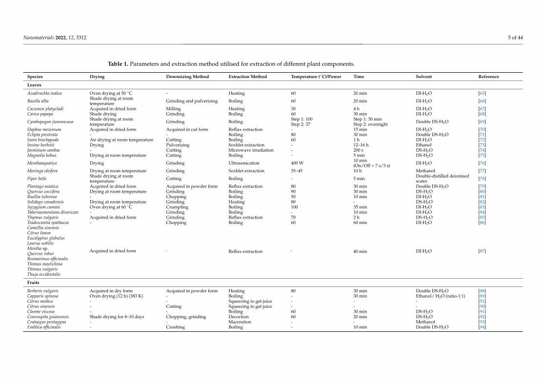
Table 1. Parameters and extraction method utilised for extraction of different plant components.
Species Drying Downsizing Method Extraction Method Temperature (◦C)/Power Time Solvent Reference
Leaves
Azadirachta indica Oven drying at 50 ◦C - Heating 60 20 min DI-H2O [65]
Basella alba Shade drying at roomtemperature Grinding and pulverizing Boiling 60 20 min DI-H2O [66]
Cacumen platycladi Acquired in dried form Milling Heating 30 4 h DI-H2O [67]Carica papaya Shade drying Grinding Boiling 60 30 min DI-H2O [68]
Cymbopogon jwarancusa Shade drying at roomtemperature Grinding Boiling Step 1: 100
Step 2: 37Step 1: 30 minStep 2: overnight Double DS-H2O [69]
Daphne mezereum Acquired in dried form Acquired in cut form Reflux extraction - 15 min DI-H2O [70]Eclipta prostrata - - Boiling 80 30 min Double DS-H2O [71]Ixora brachypoda Air drying at room temperature Cutting Boiling 60 1 h DI-H2O [72]Iresine herbstii Drying Pulverizing Soxhlet extraction - 12–16 h Ethanol [73]Jasminum sambac - Cutting Microwave irradiation - 200 s DS-H2O [74]Magnolia kobus Drying at room temperature Cutting Boiling - 5 min DS-H2O [75]
Menthaaquatica Drying Grinding Ultrasonication 400 W 10 min(On/Off = 7 s/3 s) DI-H2O [76]
Moringa oleifera Drying at room temperature Grinding Soxhlet extraction 35–45 10 h Methanol [77]
Piper betle Shade drying at roomtemperature Cutting Boiling - 5 min Double-distilled deionised
water [78]
Plantago asiatica Acquired in dried form Acquired in powder form Reflux extraction 80 30 min Double DS-H2O [79]Quercus coccifera Drying at room temperature Grinding Boiling 90 30 min DS-H2O [80]Ruellia tuberosa - Chopping Boiling 50 10 min DI-H2O [81]Solidago canadensis Drying at room temperature Grinding Heating 80 - DS-H2O [82]Syzygium cumini Oven drying at 60 ◦C Crumpling Boiling 100 35 min DI-H2O [83]Tabernaemontana divaricate - Grinding Boiling - 10 min DI-H2O [84]Thymus vulgaris Acquired in dried form Grinding Reflux extraction 70 2 h DS-H2O [85]Tradescantia spathacea - Chopping Boiling 60 60 min DI-H2O [86]Camellia sinensis
Acquired in dried form - Reflux extraction - 40 min DI-H2O [87]
Citrus limonEucalyptus globulusLaurus nobilisMentha sp.Quercus roburRosmarinus officinalisThimus mastichinaThimus vulgarisThuja occidentalis
Fruits
Berberis vulgaris Acquired in dry form Acquired in powder form Heating 80 30 min Double DS-H2O [88]Capparis spinosa Oven drying (12 h) (383 K) - Boiling - 30 min Ethanol/ H2O (ratio-1:1) [89]Citrus medica - - Squeezing to get juice - - - [51]Citrus sinensis - Cutting Squeezing to get juice - - - [90]Cleome viscosa - - Boiling 60 30 min DS-H2O [91]Couroupita guianensis Shade drying for 8–10 days Chopping, grinding Decoction 60 20 min DS-H2O [92]Crataegus pentagyna - - Maceration - - Methanol [93]Emblica officinalis - Crushing Boiling - 10 min Double DS-H2O [94]
Nanomaterials 2022, 12, 3312 6 of 44
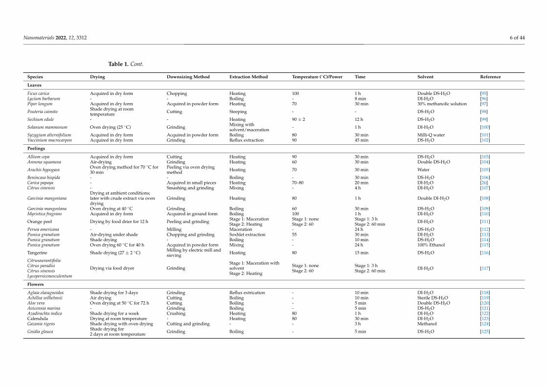
Table 1. Cont.
Species Drying Downsizing Method Extraction Method Temperature (◦C)/Power Time Solvent Reference
Leaves
Ficus carica Acquired in dry form Chopping Heating 100 1 h Double DS-H2O [95]Lycium barbarum - - Boiling - 8 min DI-H2O [96]Piper longum Acquired in dry form Acquired in powder form Heating 70 30 min 30% methanolic solution [97]
Pouteria caimito Shade drying at roomtemperature Cutting Steeping - - DS-H2O [98]
Sechium edule - - Heating 90 ± 2 12 h DS-H2O [99]
Solanum mammosum Oven drying (25 ◦C) Grinding Mixing withsolvent/maceration - 1 h DI-H2O [100]
Syzygium alternifolium Acquired in dry form Acquired in powder form Boiling 80 30 min Milli-Q water [101]Vaccinium macrocarpon Acquired in dry form Grinding Reflux extraction 90 45 min DS-H2O [102]
Peelings
Allium cepa Acquired in dry form Cutting Heating 90 30 min DS-H2O [103]Annona squamosa Air-drying Grinding Heating 60 30 min Double DS-H2O [104]
Arachis hypogaea Oven drying method for 70 ◦C for30 min
Peeling via oven dryingmethod Heating 70 30 min Water [105]
Benincasa hispida - - Boiling - 30 min DS-H2O [106]Carica papaya - Acquired in small pieces Heating 70–80 20 min DI-H2O [26]Citrus sinensis - Smashing and grinding Mixing - 4 h DI-H2O [107]
Garcinia mangostanaDrying at ambient conditions;later with crude extract via ovendrying
Grinding Heating 80 1 h Double DI-H2O [108]
Garcinia mangostana Oven drying at 40 ◦C Grinding Boiling 60 30 min DS-H2O [109]Myristica fragrans Acquired in dry form Acquired in ground form Boiling 100 1 h DI-H2O [110]
Orange peel Drying by food drier for 12 h Peeling and grinding Stage 1: MacerationStage 2: Heating
Stage 1: noneStage 2: 60
Stage 1: 3 hStage 2: 60 min DI-H2O [111]
Persea americana - Milling Maceration - 24 h DS-H2O [112]Punica granatum Air-drying under shade Chopping and grinding Soxhlet extraction 55 30 min DI-H2O [113]Punica granatum Shade drying - Boiling - 10 min DS-H2O [114]Punica granatum Oven drying 60 ◦C for 40 h Acquired in powder form Mixing - 24 h 100% Ethanol [115]
Tangerine Shade drying (27 ± 2 ◦C) Milling by electric mill andsieving Heating 80 15 min DS-H2O [116]
Citrusaurantifolia
Drying via food dryer GrindingStage 1: Maceration withsolventStage 2: Heating
Stage 1: noneStage 2: 60
Stage 1: 3 hStage 2: 60 min DI-H2O [117]Citrus paradisi
Citrus sinensisLycopersiconesculentum
Flowers
Aglaia elaeagnoidea Shade drying for 3 days Grinding Reflux extrication - 10 min DI-H2O [118]Achillea wilhelmsii Air drying Cutting Boiling - 10 min Sterile DS-H2O [119]Aloe vera Oven drying at 50 ◦C for 72 h Cutting Boiling - 5 min Double DS-H2O [120]Avicennia marina - Grinding Boiling - 5 min DS-H2O [121]Azadirachta indica Shade drying for a week Crushing Heating 80 1 h DI-H2O [122]Calendula Drying at room temperature - Heating 80 30 min DI-H2O [123]Gazania rigens Shade drying with oven drying Cutting and grinding - - 3 h Methanol [124]
Gnidia glauca Shade drying for2 days at room temperature Grinding Boiling - 5 min DS-H2O [125]

Nanomaterials 2022, 12, 3312 7 of 44
Table 1. Cont.
Species Drying Downsizing Method Extraction Method Temperature (◦C)/Power Time Solvent Reference
Leaves
Hibiscus sabdariffa Air drying under shadeat room temperature Soaking Room temperature 2 h DS-H2O [126]
Muntingia calabura - - Boiling via microwaveoven -
Boiling: 1 min* Process repeated at 1 hintervals forup to 6 h
DS-H2O [127]
Tagetes erecta - Cutting Boiling - 10 min Ultra-pure water [128]
Trifolium pratense Air drying for 5 days at roomtemperature - Heating 80 45 min Double DS-H2O [129]
Roots and Rhizomes
Berberis vulgaris Drying at ambient temperaturefor 2 days Grinding - Room temperature 2 days Sterile
DS-H2O [130]
Bergenia ciliata Air drying at 25 ◦C Acquired in powder form Boiling 60 30 min Milli-Q water [131]
Chromolaena odorata Sun dryingAt 22 ◦C ± 2 ◦C for 14 days Crushing Heating 85 2 h DI-H2O [132]
Cibotium barometz Drying Cutting and pulverizing Boiling 100 30 min DS-H2O [133]Diospyros paniculata Air drying Grinding Soxhlet extraction - - Methanol [134]
Licorice - - Heating - - Ethanol anddouble-ionised water [135]
Morinda citrifolia Shade drying at roomtemperature Grinding Boiling - 15 min DS-H2O [136]
Nepeta leucophylla Shade drying for 30 days at roomtemperature (24–32 ◦C) Grinding Soxhlet extraction Boiling point of methanol 8 h Methanol [137]
Panax ginseng - Cutting and grinding Boiling - 30 min Sterile water [138]Rheum palmatum Acquired in dry form Acquired in powder form Reflux extraction 80 45 min Ethanol [139]Rheum palmatum - Acquired in powder form Incubating/heating 40 24 h Milli-Q DI-H2O [140]
Rhodiola rosea - Grinding and screening viasieve Boiling 100 30 min DI-H2O [141]
Scutellaria baicalensis Acquired in dry form Grinding Autoclave heating 100 30 min DS-H2O [142]Zingiber officinale - Grinding Microwave 1 min DI-H2O [143]Zingiber officinale - Cutting and pulverizing Squeezing - - - [144]
Seeds
Bixa orellana Vacuum drying at 60 ◦C Crushing Steeping In dark environment 24 h Ethanol [145]Caesalpinia bonducella - Grinding Sonication - 30 min DI-H2O [146]Coffea arabica - Grinding Heating 85 25 min DS-H2O [147]Cucurbita pepo Shade air drying for 2 days - Heating 90 2 h DS-H2O [148]
Eriobotrya japonica Oven dryingat 50 ◦C for 24 h Grinding Heating 40 60 min DI-H2O [149]
Persea americana Drying in dryer for 12 h Grinding -Stage 1—roomtemperatureStage 2—65 ± 1
Stage 1: 60 minStage 2: 60 min DI-H2O [150]
Phoenix dactylifera - Milling Boiling 80 20 min Sterile DS-H2O [151]Phoenix sylvestris - - Steeping 45 12 h Sterile double DI-H2O [152]Pomegranate - - Crushing to get juice - - DI-H2O [153]Punica granatum Drying by pressing in filter paper Grinding Heating 80–85 10 min Ultra-pure water [154]Punica granatum - Grinding Mixing - 2 h Water [155]

Nanomaterials 2022, 12, 3312 8 of 44
Table 1. Cont.
Species Drying Downsizing Method Extraction Method Temperature (◦C)/Power Time Solvent Reference
Leaves
Quince - - Heating 60 4 h DS-H2O [156]Salvia hispanica Drying - Heating 60 120 min DS-H2O [157]
Tectona grandis Drying at room temperature for3–4 days Crushing Boiling 80 15–20 min Double DS-H2O [158]
Theobroma cacao Drying at room temperature for aweek Grinding Maceration - A week Methanol [159]

Nanomaterials 2022, 12, 3312 9 of 44
3.1. Plant Extraction Method
The first step of plant extraction is the cleaning process, which mainly aims to remove debrisor dust with water so as to avoid any form of contamination that might affect the subsequentsynthesis process. The second step consists of drying and downsizing. Drying is necessary toavoid the deterioration of phytochemicals that results from enzymatic and microbial activities dueto the presence of water moisture [160].
3.1.1. Drying
Typically, drying is performed via air drying, shade drying, oven drying, drying in adehydrator, vacuum drying, sun drying, or on filter paper; plant materials can also be acquired inthe dry form (Table 1 and Figure 1).
Each of the abovementioned drying methods is able to successfully yield plant extracts withphytochemicals. Shade and air drying are considered among the best methods as they allow thegreatest preservation of nutrients, such as proximate and ascorbic acid, and do so with lowerfinancial cost as compared to mechanical drying methods such as oven drying, vacuum drying,or using a food dryer [160]. As a case in point, tangerine peel was shade dried at 27± 2 ◦C forthe synthesis of iron oxide nanoparticles [116]. However, due to being carried out at a lowertemperature, shade and air drying require a longer period of time than other drying methods,which might reduce their applicability in the industrial plant-mediated synthesis of Cu-NMs [160].For instance, in preparation for Au nanoparticle synthesis, Nepeta leucophylla root was shade driedat room temperature (24–32 ◦C) for 30 days [137]. Sun drying was also used in drying Chromolaenaodorata for the synthesis of Fe3O4 nanoparticles from phenolic components of the extract [132].While sun drying can reduce the cost of drying just as can shade drying, it is not recommendedfor industrial synthesis due to high labour demand, low efficiency, hygiene issues, and moreprecautions being required to avoid contamination of samples [160].
The temperature of the drying process also plays a major role in preserving the phytochem-icals within a plant. Specifically, drying temperatures in the range of 40–60 ◦C are reported tosupport the minimal loss of phytochemicals in plant components [160]. In prior studies, neemleaves (Azadirachta indica) were oven dried for 15 min at 50 ◦C [65], and Garcinia mangostanapeelings for 10 min at 40 ◦C [109]. Although the range of 40–60 ◦C is recommended, the fi-nal decision on which temperature is most suitable for the drying process should be basedon the characteristics of the plant material being dried. For example, a study oven driedArachis hypogaea at 70 ◦C for 30 min due to its anthocyanin content, which is highly preservedunder those drying parameters [105]. Nonetheless, drying at room/ambient temperature remainsthe most used method owing to the low cost requirement being beneficial to industrialization. Forinstance, Irum et al. [69] shade dried C. jwarancusa at room temperature while Elgorban et al. [123]dried calendula flowers at room temperature to acquire phytochemicals. This is despite the timerequirement being much higher; for instance, when drying at room temperature, Yulizar et al. [159]took a week to dry Theobroma cacao seeds and Rautela et al. [158] 3–4 days for Tectona grandis seedsin preparation for nanoparticle synthesis. On the other hand, Pan et al. [105] only need 30 min todry Arachis hypogaea with an oven at 70 ◦C in plant-mediated iron nanoparticle synthesis, whileDoan Thi et al. [111] took 12 h to dry orange peels for ZnO nanoparticle production.
In addition to the abovementioned drying methods, some plant components simply arenot subjected to any drying process, mainly in the interest of cost saving and because certaincomponents have high water contents that will increase the cost if a drying process is applied. Suchplant components can include fruits, flowers, seeds, roots, and rhizomes. In one example, Jahanet al. [90] squeezed the juice from Citrus sinensis fruits to acquire reducing sugars, amino acids,proteins, and metabolites such as flavanones and terpenoids for the synthesis of Cu nanoparticles.The same squeezing method was also applied to Zingiber officinale root by Velmurugan et al. [144] toacquire alkaloids and flavonoids for the synthesis of Au and Au nanoparticle. Crushing is anothertechnique for acquiring plant extracts; for example, Kumari et al. [153] crushed pomegranateseeds to obtain flavonoids and terpenoids for the synthesis of Au-Ag bimetallic nanoparticles.Moreover, some methods forgo any drying treatment, such as when Patra et al. [127] directlyextracted Muntingia calabura flowers to acquire phytochemicals for nanoparticle synthesis and
Nanomaterials 2022, 12, 3312 10 of 44
Al-Radadi [135] used licorice root without drying to obtain glycosides, organic acids, phenoliccompounds, and flavonoids for the synthesis of Au nanoparticles. Ultimately, the characteristicsof the plant component being used and the potential cost are important factors informing thebest drying method and parameters by which to obtain the most phytochemicals from plantcomponents for Cu-NM synthesis for either research or industrial purposes.
3.1.2. Downsizing
Regarding the downsizing step, its primary purpose is to reduce the size of the plantcomponents and increase their surface area, leading to better diffusivity and mass transfer inorder to extract the greatest yields of phytochemicals such as polyphenolic compounds, phenolicacids, and tannins [161]. There are various routes for achieving this objective, presented inTable 1 and Figure 1. Interestingly, miniscule deviations of plant component size can causesignificant alterations in overall phytochemical yield [161]. Therefore, it is necessary to considercarefully the most suitable methods and cost requirements so as to acquire the smallest plantcomponents with the highest phytochemical yields for Cu-NM synthesis. Just as with thedrying process, there are some plant components that do not undergo any downsizing, suchas those with high water content; for example, Crataegus pentagyna fruits were extracted byEbrahimzadeh et al. [93] without any downsizing.
3.1.3. Plant Extraction Methods
Plant extraction methods are mainly based on boiling and heating (Table 1).Mani et al. [66] conducted an extraction from dried, ground, and pulverised Basella alba leavesby mixing them with DI-H2O and boiling them in a water bath at 60 ◦C for 20 min. Nnadozieand Ajibade [132] similarly heated crushed Chromolaena odorata at 85 ◦C for 2 h in DI-H2O, andAbisharani et al. [148] heated Cucurbita pepo seeds with DS-H2O at 90 ◦C for 2 h.
Interestingly, some alternative methods have been introduced and successfully usedto extract phytochemical products from plants (Table 1). For example, Siddiqui et al. [113]boiled powdered Punica granatum peels in sterile DI-H2O at 55 ◦C for 30 min on a Soxhletapparatus, and Singh and Dhaliwal similarly performed Soxhlet extraction on powdered Nepetaleucophylla roots with methanol held at boiling for 8 h [137]. Sonication has also been usedin plant extractions; for instance, one study removed the coats of Caesalpina bonducella seedsand then sonicated the ground kernels for 30 min [146]. Likewise, reflux extraction has beenused with various plant components. Beheshtkhoo et al. [70] extracted Daphne mezereum leavesby refluxing the dried leaves with a 5% (w/v) mixture in DI-H2O for 15 min. Microwaveirradiation has also been used in extraction, such as in a study that irradiated cut Jasminumsambac leaves in DS-H2O for 200 s to extract phytochemicals for the synthesis of Au, Ag, andAu−Ag alloy nanoparticles [74]. Maceration has also been used by many researchers, mainlydue to its low cost and eco-friendliness; for instance, ground Solanum mammosum fruits weremacerated with DI-H2O at room temperature and constant agitation for 1 h [100] and Crataeguspentagyna fruits with methanol at room temperature for the synthesis of Fe3O4-SiO2-Cu2O–Agnanocomposites [93]. In addition to the above, autoclaving was carried out on dried and groundroots of Scutellaria baicalensis with DS-H2O for 30 min at 100 ◦C in preparation for the synthesisof ZnO nanoparticles [142].
Aside from single extraction methods, combinations of methods have also been appliedto acquire extracts from various plants. For example, Nava et al. [117] macerated the peels ofCitrus aurantifolia, Citrus paradisi, Citrus sinensis and Lycopersicon esculentum for 3 h with stirring,then heated the mixture at 60 ◦C for 60 min. For plant components with high water content, asqueezing method may be introduced. For example, in the preparation of Zingiber officinale rootextract by Velmurugan et al. [144], the downsized roots were squeezed via muslin cloth.
Every extraction method has its pros and cons, summarised in Table 2 andFigure 1 [4,161,162]. The most suitable method for any given use case depends on thetypes of plant components as well as the requirements and restriction posed by the actualenvironment, such as a need to reduce financial and labour costs for industrial purposes aswell as a requirement for eco-friendliness.

Nanomaterials 2022, 12, 3312 11 of 44
Table 2. Pros and cons of various plant extraction methods.
Extraction Methods
Pros Cons
Boiling/heating/decoction • Water-soluble constituents can be extracted • Inefficient for light-/heat-sensitive compounds
Maceration• Simple• Low cost and little experimental set-up• Eco-friendly
• Batch-to-batch variation potential• Long extraction time
Microwave extraction
• Fast extraction• Less solvent needed• Produce extract with high purity and phenolic yield• Cost effective
• High heat and energy loss during the extraction
Reflux extraction• Less solvent and extraction time required• Good contact efficiency and mass transfer• Simple and easy operation
• Not suitable for thermolabile compounds
Soxhlet extraction
• Displacement of transfer equilibrium between plant components and thesolvent could be acquired
• High extraction temperature could be provided• No filtration requirement after leaching
• Large sample, extraction time, solvent requirements• Excessive loss of heat energy
Ultrasonication• Less residence time of plant particles in the solvent• Lower material and solvent requirements• Fast extraction process
• Energy intensive

Nanomaterials 2022, 12, 3312 12 of 44
Solvents in Plant Extraction
In addition to the extraction method used, solvent, energy consumption, time required,and other parameters are also critical to the extraction of phytochemicals [161,163,164].Extraction solvents can be divided into two types. i.e., water (distilled, double distilled,Milli-Q, ultra-pure, and deionised) and alcoholic solvents (ethanol and methanol), as pre-sented in Table 1. Water (DS-H2O) was used to extract dried and ground Quercus cocciferaleaves with boiling for 30 min at 90 ◦C [80]. Conversely, Boruah et al. [77] producedMoringa oleifera leaf extract by Soxhlet extraction with methanol as the solvent, incubat-ing the dried and powdered leaves at 35–45 ◦C for 10 h. Some phytochemicals, suchas polyphenolic compounds, anthocyanins, and polyphenols, can be obtained at higheryields when an alcoholic solvent is involved. Conversely, Do et al. [165] found that thephytochemical extraction yield from Limnophila aromatica improves as the solvent polarityincreases; in particular, methanol could extract more phytochemicals than ethanol. Thereare also some cases that benefit from extraction solvents combining both water and analcoholic solvent. For example, Piper longum fruits were dried, powdered and extractedwith 30% methanolic solution at 70 ◦C for 30 min [97], and Zarei et al. [89]used ethanol andwater at a 1:1 ratio with boiling for 30 min. Remarkably, such combinations of alcoholicsolvents with water can achieve the highest yields due to allowing for greater solubility ofplant components [161,165,166]. Therefore, it could be concluded that for plant phytochem-ical extraction, an alcoholic aqueous solvent is generally the most suitable. Nonetheless,the specific characteristics of the plant and phytochemicals should be considered beforeapplying a particular type of solvent. As a case in point, Maurya et al. [145] producedBixa orellana seed extract using ethanol mainly due to the primary phytochemical cis-bixinbeing water insoluble.
Temperature in Plant Extraction
The temperature applied is also a crucial factor in the plant extraction process as it cangreatly impact the yield and quality of phytochemicals and, thus, affect the nanoparticlesynthesised. As listed in Table 1, the temperature for extraction may range from room tem-perature to 100 ◦C. As an example of room-temperature extraction, Pilaquinga et al. [100]subjected pre-washed, oven-dried, and ground Solanum mammosum fruits to macerationwith DI-H2O at room temperature with constant agitation for an hour, while, as an exampleof the highest temperature, Hu et al. [141] extracted Rhodiola rosea rhizome powder by heat-ing with DI-H2O at 100 ◦C for 30 min. The temperature applied has a directly proportionalrelationship with solubility and diffusion. Nevertheless, when the temperature surpasses aparticular threshold, it might lead to several problems such as solvent loss, introductionof impurities in the produced extract, and decomposition of thermolabile phytochemicals.For instance, when synthesizing Ag and Au nanoparticles from Impinella anisum seedsextracted at temperatures ranging from 25 to 60 ◦C, high surface plasmon resonance (SPR)peak intensities accompanied the raising of temperature due to the increased diffusion rateof the solvent, which destroyed the plant cell structure. However, when temperatures in therange of 60 to 85 ◦C were used, reduction in SPR was observed due to the decompositionof some thermolabile phytochemicals [167].
Extraction Time in Plant Extraction
Extraction time is another synergic factor that can greatly affect the phytochemicalsextracted. Durations reported in the literature range from 200 s to a week; in addition,it can also be observed that the higher the temperature applied, the lower the extractionduration, and vice versa (Table 1). At the short end, Yallappa et al. [74] conducted anextraction of Jasminum sambac leaves in DS-H2O assisted by microwave irradiation for 200 s.Meanwhile, for the longest duration, Yulizar et al. [159] macerated Theobroma cacao seedbark powder in methanol with stirring for a week. Extending the extraction duration canimprove extraction efficiency as the mass transfer coefficient between plant componentsand solvent increases; accordingly, longer extractions can boost the quantities of extracted
Nanomaterials 2022, 12, 3312 13 of 44
phytochemicals and so enhance the formation of subsequently synthesised nanoparticles.However, such phenomena are restricted to within a certain time range, as when equilib-rium has been reached inside and outside of the plant components, the extraction efficiencywill not be further improved and could even worsen if the extraction period is excessivelyprolonged [4,167]. For example, extraction of Impinella anisum seeds for 60 min results inthe greatest band intensity for subsequently produced nanoparticles, and band intensitythen declines as the extraction duration increases due to the oxidation and thermal de-composition of phytochemicals [167]. Therefore, attentive consideration should be maderegarding the duration of, and temperature during, phytochemical extraction.
Filtration and Preservation
After extraction, the next step is filtration, in which solid components are removedfrom the plant extract. There are many filtration techniques in use, as illustrated in Figure 1.
Following filtration, the obtained extracts are preserved for nanomaterial-synthesisresearch. Preservation is mainly achieved via refrigeration, directly using the extract fornanoparticle synthesis, or storing the extract in a container/environment with or withoutspecial conditions such as airtightness and light exclusion so as to avoid any manner of theoxidation or photodegradation of the phytochemicals. The temperature of refrigerationis mainly 4 ◦C as it was found that this temperature can best preserve the quality ofAnanas comosus juice; moreover, increasing storage duration and temperature can greatlyreduce the phytochemicals within the obtained plant extract [168]. Therefore, in the greensynthesis of Cu-NMs, the freshness of the plant extract is very significant. Once a plantextract is produced, it should be utilised for nanoparticle synthesis as soon as possible and,in the interim, stored at low temperature.
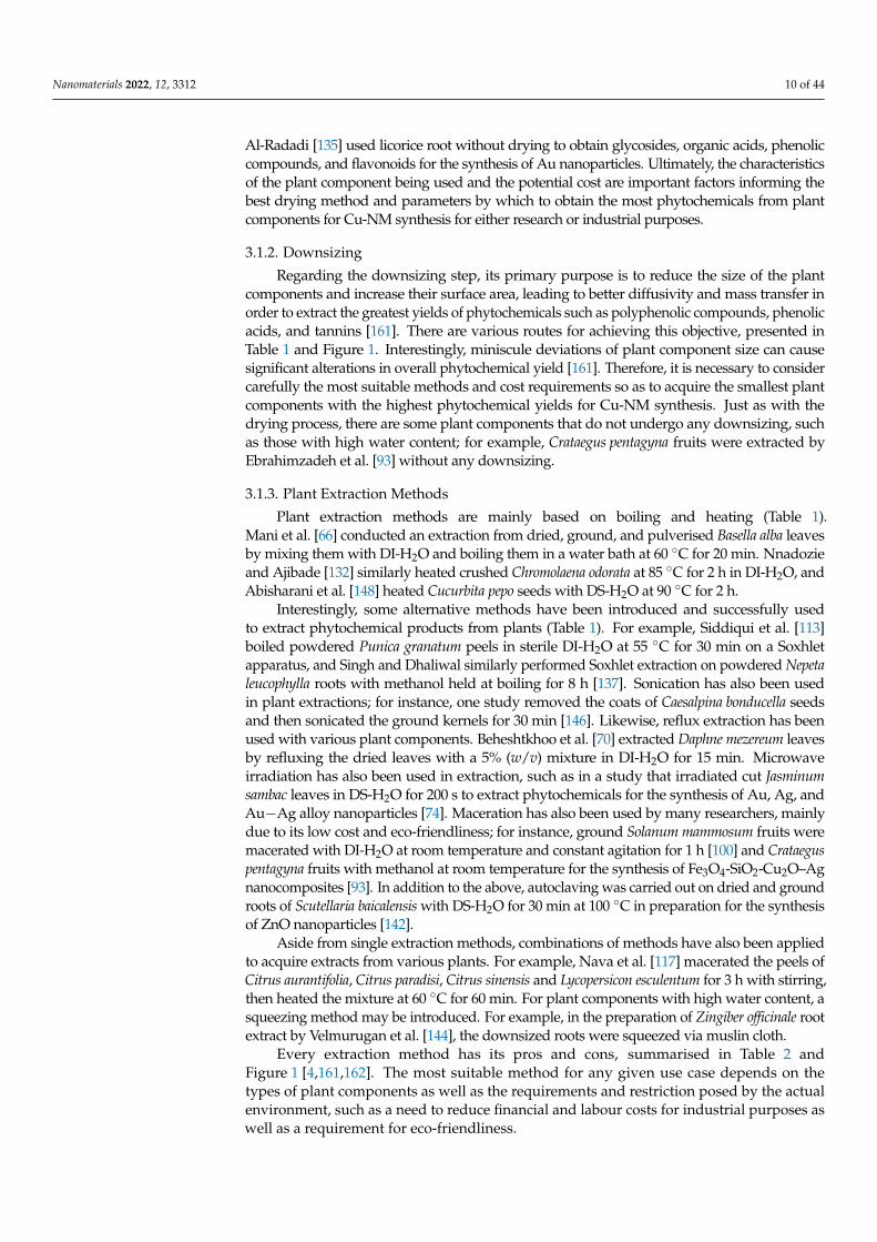
Finally, the obtained plant extract is prepared for the synthesis of nanomaterials; forexample, Nasrollahzadeh et al. [85,169] produced Thymus vulgaris leaf extract and used itto synthesise CuO and Cu nanoparticles, as shown in Figure 2.
Nanomaterials 2022, 12, x FOR PEER REVIEW 17 of 49
improve extraction efficiency as the mass transfer coefficient between plant components and solvent increases; accordingly, longer extractions can boost the quantities of extracted phytochemicals and so enhance the formation of subsequently synthesised nanoparticles. However, such phenomena are restricted to within a certain time range, as when equilib-rium has been reached inside and outside of the plant components, the extraction effi-ciency will not be further improved and could even worsen if the extraction period is ex-cessively prolonged [4,167]. For example, extraction of Impinella anisum seeds for 60 min results in the greatest band intensity for subsequently produced nanoparticles, and band intensity then declines as the extraction duration increases due to the oxidation and ther-mal decomposition of phytochemicals [167]. Therefore, attentive consideration should be made regarding the duration of, and temperature during, phytochemical extraction.
Filtration and Preservation After extraction, the next step is filtration, in which solid components are removed
from the plant extract. There are many filtration techniques in use, as illustrated in Figure 1.
Following filtration, the obtained extracts are preserved for nanomaterial-synthesis research. Preservation is mainly achieved via refrigeration, directly using the extract for nanoparticle synthesis, or storing the extract in a container/environment with or without special conditions such as airtightness and light exclusion so as to avoid any manner of the oxidation or photodegradation of the phytochemicals. The temperature of refrigera-tion is mainly 4 °C as it was found that this temperature can best preserve the quality of Ananas comosus juice; moreover, increasing storage duration and temperature can greatly reduce the phytochemicals within the obtained plant extract [168]. Therefore, in the green synthesis of Cu-NMs, the freshness of the plant extract is very significant. Once a plant extract is produced, it should be utilised for nanoparticle synthesis as soon as possible and, in the interim, stored at low temperature.
Finally, the obtained plant extract is prepared for the synthesis of nanomaterials; for example, Nasrollahzadeh et al. [85,169] produced Thymus vulgaris leaf extract and used it to synthesise CuO and Cu nanoparticles, as shown in Figure 2.
Figure 2. (a) Thymus vulgaris leaf extract and (b) solution after green synthesis of CuO nanoparticles.Adapted with permission from Ref. [85]. 2016, Elsevier.

Nanomaterials 2022, 12, 3312 14 of 44
It is worth knowing that, although the production of other type of nanomaterialsvia green synthesis methods are referenced in this review, the plant extraction methodsmentioned above are compatible in Cu-NMs synthesis.
Although the above paragraphs generalised the parameters and methods for plantextraction, there is no one best universal extraction method and parameter set for extractingall phytochemicals from all plant components. The final selections should depend on thetype of plant, the plant component, and any industrial requirements.
Next, this review covers the synthesis of Cu-NMs using plant extracts. There are severalfactors that need to be taken into account to ensure the successful production of nanomaterials,including reaction time, temperature, pH, and the extract/precursor used; these will all influencethe size and geometry of the nanomaterial produced. Table 3 summarises previously reportedworks on the synthesis of Cu nanomaterials using plant extracts.

Nanomaterials 2022, 12, 3312 15 of 44
Table 3. Summary of plant-mediated Cu nanomaterial synthesis: plant extract type, key compounds, Cu precursors, synthesis time and temperature, reactioncompletion colour, and the Cu nanomaterial product, geometry, and size.
Plant Cu Precursor Synthesis Time SynthesisTemperature (◦C) Key Compounds Colour of the Product Nanomaterials Size (nm) Geometry Reference
Leaves
Agrimoniae herba• K2PtCl6• CuSO4
• 4 h• 8 h• 16 h• 24 h
65 • Flavonoids -Core-shellCu-corePt-shell
30 Spherical [170]
Azadirachta indica
• Cu(NO3)2• AgNO3• ammonium
molybdenate
Stage 1—26 hStage 2—1 h
Stage 1—noneStage 2—500(calcination)
- -
CuO nanoparticles -
Nanoflake [171]Ag-CuO nanoparticles -Mo-CuO nanoparticles -Ag-Mo-CuOnanoparticles 12
Carica papaya • CuSO4.5H2O 24 h 50–60• Flavonoids• Phenolics Green to blackish brown CuO nanoparticles <50 Spherical [68]
Carica papaya• AgNO3• Cu(NO3)2
2 h 90 - Light yellow green toolive green precipitate Bimetallic Ag-Cu alloy TEM-90-150
DLS-420.7 Tentacle-like [172]
Cyclea peltata• FeSO4.7H2O• CuSO4.5H2O 4 h Room temperature
•Carbohydrates
• Amino acids• Alkaloids• Flavonoids• Saponins• Gallotannins
Light yellow to greenCore-shellCu-coreFe-shell
45–50 Spherical [173]
Eclipta prostrata• Cu
(CH3COO)224 h Room temperature - - Cu nanoparticles 28–45 Spherical, hexagonal,
cubical [71]
Magnolia kobus • CuSO4·5H2O -95 • Terpenoids
• Reducingsugars
- Cu nanoparticles37–91
Spherical [75]25 11060 90
Ocimum tenuiflorum• Cu (NO3)2• Ag (NO3)2
6 h 80 - Brownish blueCore-shellCuO-shellAg-core
Ag core:28–30CuO shells: 6–10
Spherical [174]

Nanomaterials 2022, 12, 3312 16 of 44
Table 3. Cont.
Plant Cu Precursor Synthesis Time SynthesisTemperature (◦C) Key Compounds Colour of the
Product Nanomaterials Size (nm) Geometry Reference
Leaves
Opuntiaficus-indica • AgNO3• Cu (NO3)2
1 h 55 • Ascorbic acid Slight green shade Core-shellAg-coreCu-shell 10–20
Ellipsoidal [175]
Slight blue shade Bimetallic Ag-Cualloy -
Origanum vulgare
•Cu(NO3)2·3H2O
•Ni(NO3)2·6H2O
•Co(NO3)2·6H2O
Until alteration ofcolour 40
• Phenoliccompounds
• Water-solubleglycosides
• Rosmarinicacid
• Water-solubleglycosides
• Caffeic acid• Protocatechuic
acid• Glycoside
protocatechuicacid
• Derivatives ofrosmarinicacid
• 2-caffeoyloxy-3-[2-(4-hydroxybenzyl)-4,5-dihydroxy]phenylpropionicacid
• Flavonoids
Darkgreenish-brown
TrimetallicCu-Co-Nialloy 28.25 Nanoflake [176]
Pisonia grandis
•Zn(NO3)2.6H2O
•Mg(NO3)2.6H2O
•Cu(NO3)2.9H2O
Stage 1—4 hStage 1—80Stage 2—450(calcination)
• Flavonoids Green to brownishblack
Zn-Mg-Cu oxidenanocomposites 50 Cubic [177]
Plantago asiatica • CuCl2·2H2O 5 min 80 • Polyphenolics Dark Cu nanoparticles 7–35 Spherical [79]
Tabernaemontanadivaricate• CuSO4 7–8 h 100• Enzymes• Proteins Brownish black CuO nanoparticles 46 ± 4 Spherical [84]
Thymus vulgaris • CuCl2.2H2O 5 min 60 • Polyphenolics Change from yellowto dark brown CuO nanoparticles <30 - [85]
Vitex negundo• AgNO3• CuSO4
24 h - - Green toBrown
Bimetallic Ag-Cunanoparticles 60 Spherical [178]

Nanomaterials 2022, 12, 3312 17 of 44
Table 3. Cont.
Plant Cu Precursor Synthesis Time SynthesisTemperature (◦C) Key Compounds Colour of the
Product Nanomaterials Size (nm) Geometry Reference
Leaves
Fruits
Crataegus pentagyna
• FeCl3·6H2O• FeCl2·4H2O• Ag(NO3)•
Cu(NO3)2·3H2O• tetra ethyl
orthosilicate
- Room temperature - -Fe3O-SiO2-Cu2O-Agnanocomposites
55–75 Spherical [93]
Piper retrofractum • CuSO4·5H2O 60 min 60
• Flavonoids• Phenolic
compounds• Piperidine
alkaloids•
Phenylpropanoids• Amides
Dark green Cu nanoparticles 2–10 Spherical [179]
Prunus nepalensis • CuSO4 Overnight Room temperature - Light green to brownand then to pink Cu nanoparticles 35–50 Centred cubic [180]
Rosa canina• Cu
(CH3COO)21 h 100 - Dark brown CuO nanoparticles 15–25 Spherical [181]
Rubus glaucus•
Cu(NO3)2·3H2O 6 h 75–80• Flavonoids• Phenolic
compounds- CuO nanoparticles 45 Spherical [182]
Syzygiumalternifolium
• CuSO4·5H2O 2 h 50 - - CuO nanoparticles 2–21 Spherical [101]
Ziziphus spina-christi • CuSO4 - 80• Polyphenolic
compoundsGreen to reddishbrown Cu nanoparticles 5–20 Elongated spherical [183]
Peelings
Carica papaya•
Cu(NO3)2·3H2OStage 1— noneStage 2—2 h
Stage 1—70–80Stage 2—450(calcination)
• Phenoliccompounds
• Flavonoids• Catechins
Greenish-blue togreen to dark greento black powder
CuO nanoparticles 85–140 Agglomeratedspherical [26]
Cavendish banana•
Cu(NO3)2·3H2OStage 1— noneStage 2—2 h
Stage 1—BoilingStage 2—400 - Brown paste to black
powder CuO nanoparticles 50–85 Agglomeratedspherical [184]

Nanomaterials 2022, 12, 3312 18 of 44
Table 3. Cont.
Plant Cu Precursor Synthesis Time SynthesisTemperature (◦C) Key Compounds Colour of the
Product Nanomaterials Size (nm) Geometry Reference
Leaves
Citrusparadisi(grapefruit)
• AnhydrousCuSO4
Stage 1—20 minStage 2—72 h
Stage 1—70Stage 2—roomtemperature
- Brown precipitate Cu nanoparticles 56–59 Spherical [185]
Citrus reticulata • CuSO4·5H2O 10 min
25 ± 2
- Brown Cu nanoparticles 54–72 Spherical [186]304050
Punica granatum • CuSO4Stage 1—10 minStage 2—4 h
Stage 1—80Stage 2—40 - - Cu nanoparticles 15–20 Spherical [187]
Flowers
Acacia caesia
• AgNO3• CuNO3• ZnO
nanoparticles- Stage 1—none
Stage 2—400(calcination)
- -Ag-Cu-ZnOnanocomposite
Ag -7Cu-12 ZnO-none Spherical [188]
• CuNO3• ZnO
nanoparticlesCu-ZnOnanocomposite 14
Aglaia elaeagnoidea•
Cu(NO3)2.3H2O 5 min Room temperature• Phenolic
compounds• Proteins
Light brownishred to brick red CuO nanoparticles 3–54 Spherical [118]
Aloe vera• Cu
(CH3COO)2Stage 1—30 minStage 2— overnight
Stage 1—50Stage 2—roomtemperature
- Lightgreen to dark green Cu nanoparticles 40 Spherical [120]
Azadirachta indica • CuSO4·5H2O 1 h 80
• Terpenoids• Terpenes• Flavonoids• Alkaloids• Carotenoids
Light blue to lightgreen to dark yellowto brown precipitate
Cu nanoparticles 5 Spherical [122]
Bougainvillea sp.• Cu
(CH3COO)2- - - Blue to black-blue
colour CuO nanoparticles 12–20 Spherical [188]
Calendula sp.
• Fe3O4nanoparticles
• Cu(NO3)2.3H2O
Stage 1—1 hStage2—6 h Room temperature - - Cu-Fe3O4
nanocomposite 20–40 Globular [123]

Nanomaterials 2022, 12, 3312 19 of 44
Table 3. Cont.
Plant Cu Precursor Synthesis Time SynthesisTemperature (◦C) Key Compounds Colour of the
Product Nanomaterials Size (nm) Geometry Reference
Leaves
Eichhornia crassipes • CuSO4 48 h Room temperature
• Aromaticcompoundslike lawsoneand phenol
Colourless to lightred Cu nanoparticles 12–15 Spherical [189]
Lantana camara• Cu
(CH3COO)2Stage 1—10 minStage 2—2 h 65 - - CuO nanoparticles 13–28 Spherical [190]
Roots and Rhizomes
Asparagus adscendens • CuSO4•5H2O 1 h Room temperature - Pale yellow to skyblue Cu nanoparticles 10–15 Spherical [191]
Asparagus racemosus•
Cu(NO3)2.3H2O 8 h 60• Phenolic
compounds - CuO nanoparticles Diameter: 50–100Length: 400–500 Rod-like [192]
Corallocarbus epigaeus • CuSO4 12 h 80–100 -
Deep blue tocolourless and thento brick red and darkred
Cu nanoparticles 65–80 Spherical [193]
Polyalthia longifolia • CuSO430 min with stirringand 24 h storage -
• Phenoliccompounds
• FlavonoidsDark green colour
Cu, CuO2, Cu2O,and CuOnanoparticles
30 Spherical [194]
Rheum emodi
• AgNO3• Cu
(CH3COO)23 h 90 • Physcion
• Chrysophanol• Aloe-emodin• Emodin• Chrysophanol
glycoside
Light brownto black
Bimetallic Ag-Cunanoparticles 40–50 Pseudo-spherical [195]
• Cu(CH3COO)2
4 h 90 Blue to brown Cunanoparticles - -
Senna didymobotrya • CuSO4·5H2O -40 • Alizarin
• Quercetin- Cu nanoparticles 5.55–63.60 Spherical [196]60
80Zingiber officinalis • Copper
sulphate- Room temperature - Straw yellow to sea
greenCunanoparticles Around 20–100 Spherical [197]Curcuma longa
Seeds
Caesalpinia bonducella•
Cu(NO3)2·3H2OStage 1—5 hStage2—2 h - -
Stage1—blue-colouredsolution turnedgreenStage 2—dark brownprecipitate
CuO nanoparticles - Rice-grain-shaped [146]

Nanomaterials 2022, 12, 3312 20 of 44
Table 3. Cont.
Plant Cu Precursor Synthesis Time SynthesisTemperature (◦C) Key Compounds Colour of the
Product Nanomaterials Size (nm) Geometry Reference
Leaves
Carum carvi - - - - - Cu nanoparticles 37 Spherical [198]Fe3O4-Cunanocomposite 62 Spherical
Koelreuteria apiculata • CuCl2.2H2O 24 h - - Precipitateformation Cu nanoparticles 20 Spherical [199]
Persea americana • CuSO4 6–7 h 45–50• Flavonoids• Phenolic
compoundsBrownish black Cu nanoparticles 42–90 Spherical [200]
Punica granatum • CuCl2·2H2OStage 1—10 minStage 2—1–2 hStage 3—4–6 h
Stage 1—60–70Stage 2—60Stage 3—roomtemperature
• Alkaloids• Flavonoids• Polyphenols
Stage 2—dull bluishbrown colourChanged to darkgreen
Cu nanoparticles 40–80 Spherical [154]
Silybum marianum• FeCl3·6H2O• CuCl2.2H2O 5 h 60
• Flavonoids• Phenolic
compounds
Dark solution andforming ofprecipitate
Cu-Fe3O4nanoparticles 8.5–60 Spherical [201]
Theobroma cacao• PdCl2• CuCl2·2H2O 2 h 50
• Flavonolantioxidantssuch asepicatechin,catechin
- Pd-CuOnanoparticles 40 - [202]
Triticum aestivum • CuSO4·5H2OStage 1—1 hStage 2—10 minStage 3—20 min
Stage 1—roomtemperature (25)Stage 2—sonicationStage 3—70
• Starch Dark blue to darkbrown CuO nanoparticles 21–42 Spherical [203]

Nanomaterials 2022, 12, 3312 21 of 44
Nanomaterials 2022, 12, x FOR PEER REVIEW 25 of 49
Figure 3. Representative TEM images of (a) spherical CuO nanoparticles synthesised using Annona squamosa seed extract. Reproduced from [204]. 2021with permission from the Royal Society of Chemistry, (b) tentacle-like bimetallic Ag-Cu nanoparticles synthesised using Carica papaya extract. Adapted with permission from Ref. [172]. 2017, Elsevier, (c) cubical Cu nanoparticles synthesised from Azadirachta indica leaf extract Adapted with permission from Ref. [65]. 2018, Elsevier; (d) SEM image of spherical Cu-Pt core shell nanoparticles synthesised using Agrimoniae herba extract. Adapted with permission from Ref. [170]. 2018, Elsevier; and (e) TEM image of Cu-Co-Ni trimetallic nanoalloy nanoflakes synthesised using Origanum vulgare leaf extract. Adapted with permission from Ref. [176]. 2020, MDPI.
3.2. Cu-NMs Synthesis Method 3.2.1. Production of High Tunable Cu-NMs
All extracts from plant components are composed of various types of phytochemicals such as flavonoids, phenolics, alizarin, quercetin, terpenoids, terpenes, alkaloids, carote-noids, and others. These phytochemicals can potentially be used for the synthesis of vari-ous types of Cu-based nanomaterials such as pure Cu nanoparticles, CuO nanoparticles, Cu-based nanocomposites, core-shell nanoparticles, and nanoalloys. For example, Ituen et al. [186] used Citrus reticulata peel extract to produce spherical Cu nanoparticles with sizes of 54 and 72 nm. Similarly, Azadirachta indica flower extract has been used to produce pure spherical 5 nm Cu nanoparticles [122], and Thymus vulgaris leaf extract to produce CuO nanoparticles with spherical morphology and a particle size less than 30 nm [85]. In addition, Dobrucka and Dlugaszewska synthesised 30 nm spherical bimetallic Pt-Cu core-shell particles with Cu as the core and Pt as the shell from the ethanolic extract of Agrimo-niae herba leaves [170]. As an example of root-extract-mediated nanoparticle synthesis, Pallela et al. [192] successfully produced rod-shaped CuO nanoparticles with diameters of 50–100 nm and lengths of 400–500 nm from Asparagus racemosus root extract. Regarding nanoparticle synthesis from fruits, Ebrahimzadeh et al. [93] produced spherical
Figure 3. Representative TEM images of (a) spherical CuO nanoparticles synthesised using Annonasquamosa seed extract. Reproduced from [204]. 2021with permission from the Royal Society ofChemistry, (b) tentacle-like bimetallic Ag-Cu nanoparticles synthesised using Carica papaya extract.Adapted with permission from Ref. [172]. 2017, Elsevier, (c) cubical Cu nanoparticles synthesisedfrom Azadirachta indica leaf extract Adapted with permission from Ref. [65]. 2018, Elsevier; (d) SEMimage of spherical Cu-Pt core shell nanoparticles synthesised using Agrimoniae herba extract. Adaptedwith permission from Ref. [170]. 2018, Elsevier; and (e) TEM image of Cu-Co-Ni trimetallic nanoalloynanoflakes synthesised using Origanum vulgare leaf extract. Adapted with permission from Ref. [176].2020, MDPI.
3.2. Cu-NMs Synthesis Method3.2.1. Production of High Tunable Cu-NMs
All extracts from plant components are composed of various types of phytochem-icals such as flavonoids, phenolics, alizarin, quercetin, terpenoids, terpenes, alkaloids,carotenoids, and others. These phytochemicals can potentially be used for the synthesisof various types of Cu-based nanomaterials such as pure Cu nanoparticles, CuO nanopar-ticles, Cu-based nanocomposites, core-shell nanoparticles, and nanoalloys. For example,Ituen et al. [186] used Citrus reticulata peel extract to produce spherical Cu nanoparticleswith sizes of 54 and 72 nm. Similarly, Azadirachta indica flower extract has been usedto produce pure spherical 5 nm Cu nanoparticles [122], and Thymus vulgaris leaf extractto produce CuO nanoparticles with spherical morphology and a particle size less than30 nm [85]. In addition, Dobrucka and Dlugaszewska synthesised 30 nm spherical bimetal-lic Pt-Cu core-shell particles with Cu as the core and Pt as the shell from the ethanolicextract of Agrimoniae herba leaves [170]. As an example of root-extract-mediated nanopar-ticle synthesis, Pallela et al. [192] successfully produced rod-shaped CuO nanoparticleswith diameters of 50–100 nm and lengths of 400–500 nm from Asparagus racemosus rootextract. Regarding nanoparticle synthesis from fruits, Ebrahimzadeh et al. [93] produced

Nanomaterials 2022, 12, 3312 22 of 44
spherical Fe3O4/SiO2/Cu2O–Ag nanocomposites with diameters of 55 and 75 nm fromCrataegus pentagyna fruit extract. Meanwhile, Sajadi et al. [201] used Silybum marianumseed extract to produce agglomerated Cu/Fe3O4 nanoparticles with sizes of 8.5–60 nm andmagnetic properties.
As indicated in Table 3, the sizes of nanoparticles produced using plant-mediatedsynthesis ranges from 2 to 150 nm. On the other hand, the morphology of the producednanoparticles is predominantly spherical [172,205,206] with some having other shapes suchas hexagonal, cubical [65,71], ellipsoidal [175], tentacle-like [172], or nanoflake [171,176](Table 3 and Figure 3). Several characterization methods are used in determining the sizeand morphology of nanoparticles, with the preeminent being scanning electron microscopy(SEM), transmission electron microscopy (TEM), and dynamic light scattering (DLS). Itshould be noted that the findings elucidated by each characterization method might havesome discrepancies. For instance, Rosbero and Camacho observed synthesised Ag/Cunanoparticles to have a size of 420.70 nm according to DLS, but a size range of 90–150 nmby TEM [172]. This discrepancy is attributable to the presence of solvent molecules on thenanoparticle surface and DLS only determining the hydrodynamic size of the particlesrather than the core diameter. That is to say, when there is a hydration layer surroundingthe nanoparticles, only the solvated particle size is indicated by a particle’s diffusionalcharacteristics [172,186]. Hence, to obtain the most accurate results and perform effectivequality control of nanomaterials, multiple characterization methods should be employed.
Overall, it can be observed that Cu-NMs produced via plant-mediated synthesis fea-ture size and morphology tunability comparable to those obtained with chemical synthesismethods. Specifically, tunability can be achieved via altering parameters such as the pre-cursor concentration, plant extract, reaction time, and the temperature applied duringnanoparticle synthesis.
3.2.2. Precursor
As listed in Table 3, most studies to date have utilised CuSO4 [188,202,205],Cu(NO3)2 [120,186,194], and CuCl2 [79,199] as the Cu precursor, while some used copperacetate (Cu(OAc)2) [71,181]. Interestingly, for some multi-metallic nanoparticles (nanoalloys,core-shell particles, and nanocomposites), multiple precursors have been utilised and the type ofnanoparticle formed depends on the methodology. For example, Cu-Co-Ni trimetallic nanoalloywas synthesised using Origanum vulgare leaf extract and the precursors of Cu(NO3)2·3H2O,Ni(NO3)2·6H2O, and Co(NO3)2·6H2O [176], while bimetallic Pt-Cu core-shell structures withCu as core and Pt as shell were synthesised from Agrimoniae herba leaf ethanolic extract withK2PtCl6 and CuSO4 as the precursors [170]. Basically, multiple metallic precursors are mixedwith a plant extract and stirring and heat applied to yield multi-metallic nanoparticles. Generally,a combination of two metals will lead to the synthesis of alloy or core-shell nanoparticles. Whichform of Cu-NM is produced can be determined based on SPR from UV–visible analysis: if asingle SPR is found, an alloy was formed, while if two independent and continuous peaks areevident, a core-shell-type structure resulted [172]. For instance, Ag/Cu nanoparticles producedvia bio-reduction with Carica papaya extract exhibited a single peak at 776 nm as the maximumabsorption, suggesting an alloyed structure [172]. Cu-based nanocomposites are also ableto be synthesised via plant-mediated methods; Ebrahimzadeh et al. [93] produced sphericalFe3O4/SiO2/Cu2O–Ag nanocomposites of 55 and 75 nm in size using Crataegus pentagyna fruitextract with FeCl3·6H2O, FeCl2·4H2O, Ag(NO3), Cu(NO3)2·3H2O, and tetra ethyl orthosilicate.
In addition to precursor choice, the concentration of precursor applied in the reac-tion also plays an important role in determining the Cu-NMs synthesised. Lee et al. [75]used Magnolia kobus leaf extract to produce spherical Cu nanoparticles, mixing it withCuSO4·5H2O at 0.5, 1, and 2 mmol/L and reducing the Cu ions to atoms. Given constanttemperature and plant extract concentration, the time required to achieve a conversionrate of more than 90% was 1600, 1400, and 200 min for the concentrations of 0.5, 1, and2 mmol/L, respectively. Therefore, it could be concluded that a higher precursor concentra-tion can accelerate nanoparticle formation. In addition, a study that performed precursor

Nanomaterials 2022, 12, 3312 23 of 44
optimization for the green synthesis of Cu nanoparticles from Senna didymobotrya root ex-tract utilised CuSO4·5H2O at concentrations of 0.0125, 0.03125, and 0.05 M [196]. This studyrevealed that the higher the precursor concentration, the higher the nanoparticle size. Theauthors noted this could be due to a low concentration of Cu ions reducing the chance ofCu-Cu interactions and, hence, reducing agglomeration [196]. Thus, the formation rate andsize of synthesised nanoparticles can be controlled via altering the precursor concentration.However, the balance between conversion rate (nanoparticle formation) and nanoparticlesize should be taken into account when carrying out green-synthesis research to ensure thedesired nanoparticle is produced while also achieving a highly productive and efficientsynthesis process.
3.2.3. Plant Extract
The plant extract utilised is also another major factor that should be considered inplant-mediated Cu-NM synthesis. A variety of plant extracts have demonstrated greatimpact on the synthesis of Cu-NMs (Table 3). In a study examining the effect of extractconcentration on nanoparticle synthesis rate and characteristics, Magnolia kobus leaf extractat a range of concentrations (5–20%) was used to produce spherical Cu nanoparticles [75].The highest synthesis rate was obtained with an extract concentration of 20%, while highaverage particle sizes were obtained for both the lowest (5%) and highest (20%) extractconcentrations, with diameters of 91 and 82 nm, respectively. Meanwhile, an extract con-centration of 15% produced nanoparticles with diameter 37 nm, which was the smallestamong all the results [75]. The reason for the production of large nanoparticles from highextract concentrations is due to the excessive abundance of capping materials promotingthe aggregation of Cu particles owing to the interaction between nanoparticles that aresurrounded with proteins and metabolites (reducing sugar, terpenoid, and other metabo-lites) [75]. Therefore, it can be concluded that if a high yield of a small Cu nanoparticle isrequired, the leaf extract concentration should be optimised before conducting the plant-mediated synthesis process at scale, whereas if the most rapid production is required, ahigh concentration of plant extract should be applied.
3.2.4. Temperature
Aside from material inputs, the temperature applied during plant-mediated Cu-NM synthesis is also an essential parameter to be investigated. According to Table 3,the temperatures utilised in existing reports range between 25 and 100 ◦C. For example,temperatures of 25 ± 2, 30, 40, and 50 ◦C have been used for Cu nanoparticle productionfrom Citrus reticulata peel extract [186], and 100 ◦C for CuO nanoparticle synthesis from Rosacanina fruit extract [181]. Notably, the use of different temperatures can greatly impact theCu-NM synthesis process. For example, in the abovementioned study using Citrus reticulatapeel extract combined with CuSO4.5H2O [186], successful bio-reduction and nanoparticleproduction was indicated by a colour change to brown with absorption at 442 nm (Figure 4).At reaction temperatures of 25, 30, 40, and 50 ◦C with a constant pH, achieving this endpointrequired 72 h, 60 h, 10 h, and 105 min, respectively. Therefore, plant-mediated synthesis ofCu-NMs is a temperature-dependent process with a positive proportional relationship: thehigher the temperature, the higher the rate of conversion from Cu ion to Cu metal.

Nanomaterials 2022, 12, 3312 24 of 44Nanomaterials 2022, 12, x FOR PEER REVIEW 28 of 49
Figure 4. Colour change over time during the reaction between Citrus reticulata peel extract and CuSO4.5H2O at (a) 0 min, (b) 60 min and (c) 105 min. Adapted with permission from Ref. [186]. 2020, Elsevier.
Interestingly, nanoparticle synthesis rate and size behave differently under a given reaction temperature increment, as the conversion rate increases whereas nanoparticle size decreases with increasing reaction temperatures. For instance, as mentioned above, Lee et al. [75] synthesised Cu nanoparticles using Magnolia kobus leaf extract and observed a size reduction from 110 nm at low temperature (25 °C, conversion rate 70%) to 37 nm at high temperature (95 °C, conversion rate ~80–100%). The rationale behind such phenom-ena is that the increasing temperature improves the reaction rate. When the reaction rate is increased, Cu ions in the reaction solution are only able to be consumed for the for-mation of nuclei; the secondary reduction process on the nuclei is avoided. Thus, larger nanoparticles cannot be produced at higher temperatures [75,196]. Consequently, it can be concluded that nanomaterials synthesis is temperature-dependent, but the size of na-nomaterials has a negative proportional relationship with temperature such that produc-ing nanoparticles with a larger size necessitates utilising a lower temperature.
There have been studies conducted on further calcination of metal oxides at temper-atures ranging from 400 to 500 °C after the synthesis process (Table 3) [27,179,190]. One of the purposes of calcination is to produce stable metal oxides or metal oxide nanocompo-sites through oxidation [177]. For example, Suresh et al. [177] used Pisonia grandis leaf ex-tract to synthesise Zn-Mg-Cu oxide nanocomposites, then calcinated them at 450 °C to obtain mixed metal oxide nanocomposites. In addition to producing stable oxides, in-creasing calcination temperature can boost the size of Cu-NMs and produce black precip-itates of agglomerated cubical nanomaterials, where uncalcinated nanomaterials have elongated morphology [205].
3.2.5. pH Solution pH is also a very significant factor in plant-mediated nanomaterial synthesis
as it can affect the synthesis rate and products. Mechanistically, the importance of pH is due to the reducing and stabilizing agents being greatly dependent on the phytochemicals within the plant extract, which might be readily affected by pH. Generally, the best pH values for plant-mediated nanomaterial synthesis are in the range of pH 7–9, and varying the pH will alter the nanoparticle synthesised [14]. Nagar and Devra utilised Azadirachta indica leaves for Cu nanoparticle synthesis at various pH values [65]. They found that na-noparticle synthesis is more effective at higher pH and abolished in an extreme acidic environment, such as pH 4.7. A solution with pH 6 produced small-sized nanoparticles of 56 nm, while in an alkaline environment of pH 9.3, the nanoparticles produced were of size 73 nm. In an acidic environment, the phytochemicals in the plant extract might be inactivated [65]. In addition, lower pH can cause nanoparticles to experience high electro-static repulsion which reduces the chances of agglomeration and, thus, yields nanomateri-als of smaller size [196]. Conversely, in a more alkaline condition such as pH 10, the low electrostatic forces of the nanoparticles allow further particle growth and agglomeration, which produces larger nanomaterials [65,196]. It is worth noting that during the plant-mediated synthesis process, the pH of the medium will drop as the Cu2+ ions cause
Figure 4. Colour change over time during the reaction between Citrus reticulata peel extract and CuSO4.5H2Oat (a) 0 min, (b) 60 min and (c) 105 min. Adapted with permission from Ref. [186]. 2020, Elsevier.
Interestingly, nanoparticle synthesis rate and size behave differently under a givenreaction temperature increment, as the conversion rate increases whereas nanoparticlesize decreases with increasing reaction temperatures. For instance, as mentioned above,Lee et al. [75] synthesised Cu nanoparticles using Magnolia kobus leaf extract and observeda size reduction from 110 nm at low temperature (25 ◦C, conversion rate 70%) to 37 nm athigh temperature (95 ◦C, conversion rate ~80–100%). The rationale behind such phenomenais that the increasing temperature improves the reaction rate. When the reaction rate isincreased, Cu ions in the reaction solution are only able to be consumed for the formation ofnuclei; the secondary reduction process on the nuclei is avoided. Thus, larger nanoparticlescannot be produced at higher temperatures [75,196]. Consequently, it can be concludedthat nanomaterials synthesis is temperature-dependent, but the size of nanomaterials has anegative proportional relationship with temperature such that producing nanoparticleswith a larger size necessitates utilising a lower temperature.
There have been studies conducted on further calcination of metal oxides at tempera-tures ranging from 400 to 500 ◦C after the synthesis process (Table 3) [27,179,190]. One ofthe purposes of calcination is to produce stable metal oxides or metal oxide nanocompositesthrough oxidation [177]. For example, Suresh et al. [177] used Pisonia grandis leaf extractto synthesise Zn-Mg-Cu oxide nanocomposites, then calcinated them at 450 ◦C to obtainmixed metal oxide nanocomposites. In addition to producing stable oxides, increasingcalcination temperature can boost the size of Cu-NMs and produce black precipitates ofagglomerated cubical nanomaterials, where uncalcinated nanomaterials have elongatedmorphology [205].
3.2.5. pH
Solution pH is also a very significant factor in plant-mediated nanomaterial synthesisas it can affect the synthesis rate and products. Mechanistically, the importance of pH isdue to the reducing and stabilizing agents being greatly dependent on the phytochemicalswithin the plant extract, which might be readily affected by pH. Generally, the best pH val-ues for plant-mediated nanomaterial synthesis are in the range of pH 7–9, and varying thepH will alter the nanoparticle synthesised [14]. Nagar and Devra utilised Azadirachta indicaleaves for Cu nanoparticle synthesis at various pH values [65]. They found that nanoparti-cle synthesis is more effective at higher pH and abolished in an extreme acidic environment,such as pH 4.7. A solution with pH 6 produced small-sized nanoparticles of 56 nm, whilein an alkaline environment of pH 9.3, the nanoparticles produced were of size 73 nm. In anacidic environment, the phytochemicals in the plant extract might be inactivated [65]. Inaddition, lower pH can cause nanoparticles to experience high electrostatic repulsion whichreduces the chances of agglomeration and, thus, yields nanomaterials of smaller size [196].Conversely, in a more alkaline condition such as pH 10, the low electrostatic forces of thenanoparticles allow further particle growth and agglomeration, which produces largernanomaterials [65,196]. It is worth noting that during the plant-mediated synthesis process,the pH of the medium will drop as the Cu2+ ions cause oxidation of the plant extract, lead-ing to the release of H+ ions and, hence, acidification; this is also another important aspectto be considered by researchers carrying out plant-mediated nanomaterial synthesis [65].

Nanomaterials 2022, 12, 3312 25 of 44
Therefore, it is necessary to achieve a balance in producing nanoparticles with a desiredsize while maintaining high nanomaterial productivity.
3.2.6. Reaction Time
In terms of duration, it can be seen that the range of reaction times in the literatureis relatively large, ranging from as low as 5 min to as long as 72 h. The duration is notas impactful compared to the other parameters mentioned above [205]. Although a longsynthesis duration allows improvement in the nanomaterial nucleation rate, the reactionrate will not continue to increase after the optimum time has been reached. In some cases,prolonging the incubation might even cause nanomaterial aggregation [206]. In fact, otherparameters such as temperature, precursor, and type of plant extract can greatly impactthe time needed to achieve complete conversion from metallic ions (Cu and other ionsdepending on the type of nanomaterial being produced) to metallic atoms and, finally,nanomaterials, as was mentioned previously in relation to other parameters. However,compared with other materials such as Au, Ag, and Pt, Cu forms nanoparticles relativelyslowly as the initiation of Cu nucleus formation is much more difficult [75]. Hence, thegreen synthesis of Cu nanoparticles necessitates longer reaction times in order to achieve100% conversion.
3.2.7. Indication of Cu-NMs Production
During the production of Cu-NMs, a successful reaction is indicated by the colouralteration of the reaction solution. For example, Thymus vulgaris leaf extract mixed withCuCl2.2H2O with constant stirring at 60 ◦C undergoes a change in colour from yellow todark brown, as shown in Figure 2. In addition, calcination will also change the colour ofnanomaterials produced. Carica papaya peel extract combined with Cu(NO3)2.3H2O andheated at 70–80 ◦C changes colour from greenish-blue to green and finally produces a darkgreen paste. Upon calcination, a fine black-coloured powder was obtained and harvestedas CuO nanoparticles [26].
4. Applications of Cu-NMs from Plant-Mediated Synthesis
After the synthesis reaction is completed, the obtained nanoparticles are washed,dried, and employed in applications. Cu-NMs synthesised using plant extracts have beenutilised in two major areas, namely, biomedical and environmental remediation [31].
4.1. Biomedical
Plant-mediated Cu-NMs have demonstrated antimicrobial, antioxidant, and anticanceractivities, and have potential as nano-sensors and in various medical applications. In thissection, details and mechanisms pertaining to this area will be discussed.
4.1.1. Antimicrobial
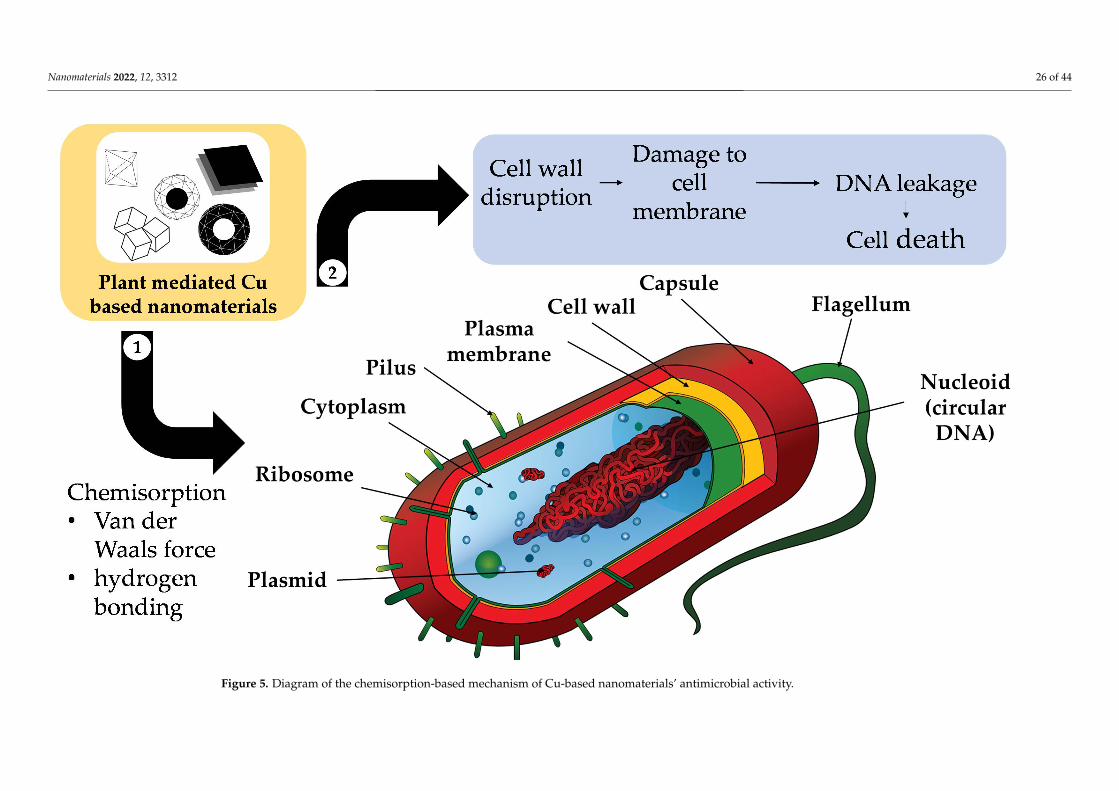
Firstly, antibacterial activity has been observed for plant-mediated Cu-NMs [205] andcan be attributed to several putative pathways. Bhavyasree and Xavier suggested thatCu-NMs, including both Cu and CuO nanoparticles produced via plant-mediated synthesis,can carry out antibacterial activity through a chemisorption-based mechanism [206]. Thismechanism involves microbial adsorption to the nanoparticle surface, which has beenbio-functionalised by phytochemicals during the plant-mediated synthesis process. Theadsorption is mainly due to chemisorption via non-electrostatic forces (Van der Waalsforce and hydrogen bonding), which causes the destruction of the microbial cell wall andsubsequent cell membrane damage, DNA breakage, and eventually cell death, as illustratedin Figure 5.
Nanomaterials 2022, 12, 3312 26 of 44Nanomaterials 2022, 12, x FOR PEER REVIEW 30 of 49
Figure 5. Diagram of the chemisorption-based mechanism of Cu-based nanomaterials’ antimicrobial activity.Figure 5. Diagram of the chemisorption-based mechanism of Cu-based nanomaterials’ antimicrobial activity.

Nanomaterials 2022, 12, 3312 27 of 44
Another antibacterial mechanism is mediated by reactive oxygen species (ROS) andthe release of Cu2+ ions [207]. First, the CuO nanoparticles are much smaller (being of ananometre scale) than the micrometre-scale pores of bacterial cells, which allows them toeasily penetrate the cells. In addition, Cu2+ ions are attracted toward bacterial cells due tothe abundance of carboxyl and amine groups on the cell surface; this is another factor inantibacterial ability. However, the antibacterial interactions are different for Gram-positiveand Gram-negative bacteria, as described in Figure 6a.

Nanomaterials 2022, 12, 3312 28 of 44
Nanomaterials 2022, 12, x FOR PEER REVIEW 32 of 49
Figure 6. Cont.

Nanomaterials 2022, 12, 3312 29 of 44Nanomaterials 2022, 12, x FOR PEER REVIEW 33 of 49
Figure 6. (a) Diagram of the respective mechanisms of CuO nanoparticle antibacterial activity in Gram-positive and Gram-negative bacteria and (b) diagram of the mechanism of CuO nanoparticle antifungal activity. Adapted with permission from Ref. [205]. 2020, MDPI.
Figure 6. (a) Diagram of the respective mechanisms of CuO nanoparticle antibacterial activity in Gram-positive and Gram-negative bacteria and (b) diagram of themechanism of CuO nanoparticle antifungal activity. Adapted with permission from Ref. [205]. 2020, MDPI.

Nanomaterials 2022, 12, 3312 30 of 44
After bypassing the cell wall, Cu2+ ions relocate intracellularly to the cytosol dueto the internalization of CuO nanoparticles and Cu2+ ions, where they cause ROS toaccumulate [205]. Consequently, DNA and mitochondria damage occur. Cu2+ ions withina bacterium may also stimulate cellular responses that lead to bactericidal activity. Forexample, radicals produced by CuO nanoparticles, such as superoxide and hydroxylradicals, can have synergic effects in causing bacterial membrane destruction, DNA damage,attachment to ribosomes, oxidative injury, and protein and proton efflux pump damage;they can also prevent biofilm production [205,207].
Cu-NMs exert antibacterial activity through mechanisms similar to those of Cu andCuO nanoparticles. Generally, the antibacterial activity of nanomaterials is mainly owedto the induction of oxidative stress, such as through the production of free radicals andROS. Notably, nano-sized particles will feature a smaller surface-to-volume ratio, harbourmore surface defects due to oxygen vacancies, and feature greater electrostatic attrac-tion and release of Cu2+ ions and generate more oxidative stress within the bacterialcells [178]. In addition, bimetallic nanoparticles can demonstrate a synergic effect with im-proved antibacterial ability. For example, bimetallic Ag and Cu nanoparticles produced byVitex negundo-mediated synthesis demonstrate antibacterial activity when applied in a cel-lulose matrix via the disc method against both Gram-positive (Escherichia coli, Pseudomonas,Klebsiella) and Gram-negative (Staphylococcus, Bacillus) species [178]. Particles having anequal ratio of Ag and Cu (2.5 mM each) exhibited the greatest antibacterial ability, with a9 mm zone of inhibition for all the tested species.
Secondly, Cu-NMs have also been demonstrated to possess antifungal activity. A num-ber of fungi can cause infections in humans with severe symptoms, such asCandida albicans which can cause mucosal infections (oropharyngeal or vulvovaginal can-didiasis), or Trichophyton mentagrophytes which can cause dermatophytosis [208,209]. Anti-fungal activity is more challenging to realise than antibacterial as a fungus cell has severallayers of lipids within its cell wall which impede the penetration and internalization of Cunanomaterials [205]. Although fewer publications exist regarding the antifungal testing ofCu nanomaterials synthesised by green methods, their hypothesised antifungal mechanismis based on altering the structure and function of fungal cell components [210]. That is, thenanoparticles first distort the cell wall and become internalised by the fungus (Figure 6b).After internalization, the same process of ROS generation and subsequent process disrup-tion ensues as in bacteria, impacting DNA, mitochondria, replication, protein synthesis andother essential elements, eventually leading to cell death [205,210].
In one report of Cu-NM antifungal activity, Mali et al. [210] tested the efficacy of Cunanoparticles derived from Celastrus paniculatus leaf extract against Fusarium oxysporum.Concentrations of 0.12%, 0.18% and 0.24% (w/v) Cu nanoparticles were found able toinhibit mycelial growth by 76.29 ± 1.52%, 73.70 ± 1.52%, and 59.25 ± 0.57%, respectively,calculated via the following formula:
(% inhibition rate) =(Mc−Mt)
Mc× 100 (1)
where Mc represents mycelial growth in the control (with water) while Mt is mycelialgrowth under the Cu nanoparticle treatment. The inhibition rate was found to be dosage-dependent: the higher the Cu nanoparticle dosage, the higher the degree of inhibition.
Dobrucka and Dlugaszewska similarly studied the antibacterial and antifungal activi-ties of Cu-Pt core-shell nanoparticles synthesised using Agrimoniae herba extract [170]. Thenanoparticles were applied via the well-diffusion method to three species of bacteria, includingStaphylococcus aureus, Escherichia coli, and Pseudomonas aeruginosa, and three of fungi: Candidaalbicans, Trichophyton mentagrophytes, and Aspergillus fumigatus; the authors then determined theminimal inhibitory concentration (MIC), minimal bactericidal concentration (MBC), and mini-mal fungicidal concentration (MFC) [170]. The Cu-Pt nanoparticles exhibited good inhibitoryfunction on all tested bacteria and Trichophyton Mentagrophytes. The overall best antibacterial

Nanomaterials 2022, 12, 3312 31 of 44
and antifungal performances were obtained on Staphylococcus aureus (MIC of 16.7 and MBC of33.3) and Trichophyton mentagrophytes (MIC and MFC of 26.7).
From the above reports, it can be concluded that plant-mediated Cu-NMs are suitableas antibacterial (for both Gram-positive and -negative) and antifungal agents. Such charac-teristics are useful in further broadening the application of Cu-NMs in the pharmaceuticaland medical sectors.
In addition to direct antimicrobial effects, many plant-mediated Cu-NMs have alsodemonstrated antioxidant properties which also contribute to antibacterial and antifungalactivities as a synergic factor [205]. Multiple mechanisms contribute to antioxidant ability,which are: (1) binding of transition metal ion catalysts, (2) reductive capacity, (3) radicalscavenging activity, (4) decomposition of peroxides, (5) prevention of continued hydrogenabstraction, and (6) prevention of chain initiation.
Interestingly, plant selection has been shown to impact the antioxidant ability ofCu-NMs. For example, Rehana et al. [211] synthesised nanoparticles using extracts ofAzadirachta indica, Hibiscus rosa-sinensis, Murraya koenigii, Moringa oleifera, andTamarindus indica, then tested their antioxidant capabilities with ABTS, DPPH, and hy-drogen peroxide assays. Tamarindus indica-mediated nanoparticles were found to have thehighest antioxidant activity, and Moringa oleifera the lowest, though still superior to CuOnanoparticles produced via a chemical method. Therefore, plant-mediated nanomaterialshave much higher antioxidant ability as compared to chemical-mediated materials, and theplant used is an essential consideration for antioxidant purposes.
4.1.2. Nano-Sensor
Plant-extract-mediated Cu-NMs have also been utilised in the preparation of nano-sensors.Cu nanomaterials, such as CuO nanoparticles, are suitable for nano-sensor production owingto their characteristic high electron-transfer rate, superior catalytic activity, large surface area,high glucose selectivity in heterogenous samples (such as blood or urine), chlorine poisoningresistance, and corrosion resistance. For example, Ag-CuO core-shell nanoparticles producedusing Ocimum tenuiflorum extract have been used for non-enzymatic glucose sensing witha screen-printed electrode [174]. The synthesised electrode provided good glucose-sensingperformance with a sensitivity of 3763.44 µAmM−1cm−2, linear range of 1 to 9.2 mM, detectionlimit of 0.006 mM (S/N = 3), and response time of less than 1 s. Moreover, the CuO-Ag core-shell-modified bio-nano-sensors demonstrated exceptional adhesion and structural strengthalong with great long-term stability for up to 60 days, exhibiting 99.2% of the initial value afterone month with excellent repeatability and reproducibility.
The mechanism by which these nanoparticles sense glucose is based on electrontransfer from the screen-printed electrode to the CuO nanoparticle core via the conductionband electrons of the Ag shell. This electron transfer occurs because the work function ofCuO is bigger than that of Ag, and equalization of Fermi levels ensues after the materialscome into electrical contact and the mobility of electrons is improved. The progressionof current-induced charge carriers can boost electrocatalytic efficiency through a chargetransfer mechanism; therefore, the Ag-CuO core-shell nanoparticles are electro-catalyticallyactive and can induce electron-transfer reactions. The energy of a nanoparticle is dependenton the charge distribution within the energy levels of its component metal. Ultimately, theadditional electrons can be discharged when glucose is introduced into the system as anelectron acceptor.
This glucose oxidation mechanism can be summarised as: (1) deprotonation of glucosethat causes oxidation, (2) isomerization and enediol formation, and, finally, (3) adsorptionto the electrode surface, which leads to the oxidation of Cu(II)/Cu(III):
CuO + OH− → CuOOH + e− (2)
CuOOH + glucose + e− → CuO + OH− + gluconic acid (3)

Nanomaterials 2022, 12, 3312 32 of 44
In the core-shell nanoparticle, Cu(II) was oxidised to Cu(III) and this catalysed glucoseoxidation to produce gluconolactone, which was further oxidised to gluconic acid aspresented in Equations (2) and (3).
4.1.3. Anticancer
Lastly, plant extract-mediated Cu-NMs have been studied for their anticancer prop-erties. Generally, this activity can be realised through multiple routes including ROSgeneration, antioxidant activity, cell cycle arrest, apoptosis, and autophagy [205,207]. Onestudy produced CuO nanoparticles using extracts of Azadirachta indica, Hibiscus rosa-sinensis,Murraya koenigii, Moringa oleifera, and Tamarindus indica and used MTT assays to test theiractivity against four cancer cell lines, i.e., human breast, cervical, epithelioma, and lungcancer cells, along with one normal human dermal fibroblast (NHDF) cell line [211]. AllCuO nanoparticles exhibited anticancer ability towards all cancer cell types in a dose-dependent manner: higher concentrations of CuO nanoparticles resulted in lower cancercell viability. Interestingly, the type of plant utilised also affected anti-cancer ability, withTamarindus indica-mediated CuO nanoparticles exhibiting greater cytotoxicity over theothers; this indicates that the phytochemicals in the plant extract used for nanoparticlesynthesis impact the resulting particles’ anti-cancer activity.
The toxicity of Cu-NMs is one of the limitations that hinder their application biomed-ically. However, it has been reported that plant-mediated Cu nanomaterials have lesstoxicity to normal human cell lines [205]. Therefore, such Cu nanomaterials may be moresafely applied in biomedical applications. For example, CuO nanoparticles synthesisedusing extracts of Azadirachta indica, Hibiscus rosa-sinensis, Murraya koenigii, Moringa oleifera,and Tamarindus indica exhibited lower toxicity in NHDF cells, which suggests these to bepromising anticancer agents for use in the pharmaceutical industry [211].
4.2. Environmental Remediation
The usage of Cu-NMs in environmental applications is mainly focused on the remediationof dyes and toxic compounds, with mechanisms primarily based on photocatalysis or catalysis.
The mechanism of photocatalysis by nanomaterials is as follows: when the nano-materials are deposited into an aqueous sample containing compounds that are desiredto be degraded, such as dye, and exposed to light, an interaction occurs in which a pho-togenerated electron is converted from the valence band (VB) to the conduction band(CB) in the nanomaterial. A hole in the VB then results, producing an electron (e−)-hole(h+) pair. The holes react with OH ions in the water molecules to yield OH radicals viaoxidation, while the electrons react with dissolved O2 to generate O2 radicals via reduction.Those radicals are then responsible for the degradation of the dye into non-toxic degradedproducts [171,176]. Alshehri and Malik investigated the ability of Origanum vulgare extract-mediated Cu-Co-Ni trimetallic nanoparticles to photocatalyse the degradation of methyleneblue [176]. They observed degradation efficiency of more than 50% and 92.67% after 50 and100 min, respectively. The rate of degradation could be increased via increasing nanoparti-cle concentration, but after a certain threshold was surpassed, the photocatalytic efficiencycould be enhanced no further due to the aggregation of the nanomaterials.
With regard to catalysis mechanisms, nanoparticles can catalyse reactions by borylation,clock reactions, oxidative coupling, A3 coupling, click chemistry, tandem and multicomponentreactions, C–H functionalization, cross-coupling, reduction and oxidation reactions, and othermixed reactions [212]. Successful catalysis via plant-mediated Cu-NMs has been achieved, suchas when Suvarna et al. [173] studied the degradation of methyl green dye using bimetallicspherical Fe-Cu nanoparticles produced using Cyclea peltata extract, and achieved a degradationefficacy of 82% within 105 min. The Fe-Cu nanoparticles promoted hydrolysis and deproto-nation reactions on the dye molecules, resulting in the demineralization of the dye moleculesinto simpler structures. In another example, Rosbero and Camacho utilised bimetallic (Agand Cu) alloy nanoparticles produced via Carica papaya leaf extract to degrade the pesticidechlorpyrifos in water [172]. The degradation was observed for 24 h, and yielded the products

Nanomaterials 2022, 12, 3312 33 of 44
3,5,6-trichloropyridinol (TCP) and diethylthiophosphate (DETP), of which the former is lesstoxic than chlorpyrifos and not mutagenic.
5. Future Research Directions
Although plant-mediated Cu-NMs have numerous benefits and applications, theyalso have considerable potential yet to be discovered along with disadvantages that areunavoidable and need to be addressed to ensure realization of the applicability of thesenanoparticles toward industrial production with wider applications. This section suggestsareas of future research to increase the potential of Cu-NMs and propel the applicability oftheir production at a larger scale via eradicating current limitations; specifically, it discusses:(i) solutions by which to overcome limitations, (ii) potential new applications, and (iii) newresearch directions regarding Cu-NM synthesis.
5.1. Limitations and Solutions
This section illustrates the limitations of green Cu nanomaterial production andassociated solutions. There are several that need attention in this respect, mainly withregard to biomass obtainability, the complexity of plant systems, the underlying synthesisprocess, nanomaterial quality, and low productivity.
Concerning bioresource accessibility, most research to date has focused on the use oflocal plant species that are not widely available throughout the globe; notably, variationin plant species and also geographical cultivation areas affect the phytochemicals withinthe plant extract produced [36]. Moreover, the inherent complexity of plants is anotherhindrance to the industrial production of plant-mediated Cu-NMs. That is, the phytochem-icals within a plant are greatly affected by external factors such as abiotic environmentalfactors, cultivar, and mutagenesis [213–215]. These will cause batch-to-batch variationamong raw materials, which might adversely affect the homogeneity and reproducibility ofnanomaterial synthesis. On top of those considerations, another drawback to this methodis the diversity of phytochemicals in a plant system. This can be addressed by applyingmolecular science techniques such as genetic engineering to maximize the most relevantphytochemicals in the target plant. Combining these techniques with plant-tissue culturemethods such as cloning can allow the quality of a plant (target phytochemical compositionand content) to be preserved and controlled and, thus, avoid batch-to-batch variationand mutagenic factors that might affect the phytochemical profile. Plant-tissue culturetechniques can also minimize the time, cost, and labour force needed for the planting ofbioresources and overcome geographical limitations [216].
At present, most research into plant-mediated nanoparticles is carried out in low quan-tities, and, hence, with low productivity. Although the quantity of nanoparticles requiredfor characterization or application research purposes is not high, mass production via thegreen synthesis method is little-studied and needs to be researched in order to producenanoparticles in a large quantity. Bioprocess methods can be used to produce and maxi-mize specific phytochemicals for nanomaterial production and allow large-scale industrialproduction. With these solutions and more research, the global industrial production ofuniform plant-mediated Cu nanomaterial products could be realised.
However, given the limited determinations of phytochemical profiles, it is not fea-sible to elucidate the mechanism of nanomaterial synthesis. This will affect the possi-bility of producing nanomaterials with good homogeneity in terms of size, shape, andcrystal structure [71,75,79,84,217]. When it comes to investigating those phytochemicalsthat are responsible for stabilizing and reducing the ions within a precursor material,Fourier-transform infrared spectroscopy (FTIR) is the current technique of choice. Thischaracterisation method mainly examines the functional groups that are deposited on thenanoparticle surface [71,79,84,177]. Although FTIR can identify the functional groups thatact as stabilizing and reducing agents, it has difficulty determining which specific phyto-constituents of a complex plant extract they originated with. Further research employingother characterization methods such as liquid chromatography–mass spectrometry and

Nanomaterials 2022, 12, 3312 34 of 44
nuclear magnetic resonance can be carried out to identified the chemical structures of thecontributing phytochemicals [218,219].
5.2. Potential New Applications
At present, most Cu-NMs produced via plant-mediated methods are synthesised usingleaf extracts. There is plenty of room for future research into the exploitation of other plantcomponents (peelings, roots and rhizomes, fruits, flowers, and seeds) for the synthesis of Cualloy, core shell, and nanoparticles. In addition, there remains a knowledge gap regardingthe effect of method parameters on the morphology of the synthesised particles. From anapplication perspective, most uses of plant-mediated Cu nanomaterials are focused onbiomedical and environmental remediation. However, there are more applications thathave yet to be discovered. For example, in the biological sector, research into the use ofplant-mediated Cu-NMs mainly concerns their antibacterial ability and lesser antifungalability. The relatively lower antifungal performance is owed to fungal cells less readilyadsorbing nanoparticles at low concentrations as compared to bacterial cells. In addition,the binding of nanoparticles to the bacterial surface blocks bacterial respiration, whereasfor fungal or eukaryotic cells, respiration occurs in the mitochondrial membrane and sois less susceptible to direct inhibition by nanoparticles [31]. There has also been limitedresearch on the antiviral and antiparasitic abilities of plant-mediated Cu nanoparticles;further investigation in this area may expand their antimicrobial capabilities. Finally,other noble-metal (Au and Ag) nanomaterials produced via plant-mediated synthesishave been employed in other applications such as electrochemistry, detection, surface-enhanced Raman scattering, phase transfer, transmetallation, and modified glassy carbonelectrodes; therefore, research can be carried out to expand the usage of Cu-NMs to theseapplications [31].
5.3. New Research Directions for Synthesis
One area of future research for the application of Cu-NMs and their industrial-scalegreen production is leveraging machine learning. Specifically, machine-learning algorithmscan be used in two respects, synthetic outcome prediction and experiment planning [220].For the first, an algorithm mathematically learns the relationship between nanomaterialproperties and experimental conditions, then predicts from an example synthetic param-eters dataset and the results of past experiments the characteristics of the nanoparticlesthat will be produced. Meanwhile, experiment-planning algorithms aim to suggest the bestreaction conditions for achieving desired nanomaterial properties [220]. This can aid inreducing the time and research effort required to obtain a desired outcome, such as the uni-formity of the produced particles. Most machine-learning studies to date have concentratedon chemical-based nanomaterial synthesis; only a limited number of publications have con-cerned green synthesis; hence, there remains a large gap in this area [221–224]. Addressingthis gap can help in making the process of Cu nanomaterial synthesis become less labourintensive, more cost effective, less time consuming, more productive, and able to yieldhigher quality nanoparticles, all of which are important from the industrial perspective.
6. Conclusions
This literature review focused on the green synthesis of Cu nanomaterials. Compared tochemical or physical synthesis methods, green synthesis and especially plant-mediated synthesisis more environment-friendly, less toxic, and safe throughout the whole production process.The production methodology was discussed with further focus on plant-mediated nanomaterialsynthesis, including the plant extraction method and Cu-NM (pure metal, metal oxide, alloy,core shell, and nanoparticles) synthesis. Leaf-extract-mediated Cu nanomaterials comprisethe majority produced to date, with few synthesised using other types of plant components.The review also considered the biological and environmental applications of plant-mediatedCu-NMs. With regard to biological applications, antiviral and antiparasitic activities havereceived less focus than antibacterial. There also remain many research gaps regarding the

Nanomaterials 2022, 12, 3312 35 of 44
application of green synthesis Cu-NMs in other sectors. Finally, current limitations and solutionswith potential future research targets were described. Biomass obtainability, complexity ofplant systems, underlying synthesis process, nano-material quality, and low productivity arethe future challenges that need to be addressed in order to further broaden the application ofplant-mediated nanomaterial synthesis.
In short, plant-mediated nanomaterial synthesis is eco-friendly, has low toxicity, andavoids using hazardous chemicals. The process can be separated into two parts, the plantextraction and the nanomaterial production. Different plant extracts with different pa-rameters can produce nanomaterials of different sizes and geometries. As such, plantsource accessibility, diversity of phytochemicals in extracts, knowledge of the synthesismechanism, and nanomaterial quality are the limitations that presently hinder the futureindustrial production and application of plant-mediated nanomaterials. More research isneeded in areas of the biotechnological sector such as phytochemical profiling, molecularscience, tissue culture, and bioprocesses to overcome these issues. Separately, machinelearning can also be adopted as a new research topic to further improve the green synthesisof Cu-NMs with better industrial applicability. Once these problems and research directionsare resolved and fulfilled, respectively, the potential of plant-mediated nanomaterial syn-thesis could be fully unleashed in myriad applications, providing processes and materialswith better sustainability and friendliness toward the environment.
Author Contributions: Investigation, writing—original draft, J.V.; Conceptualization, writing—review and editing, K.S.L.; Supervision, visualization, Y.C.-Y.E.; Funding acquisition, validation,S.X.C.; Supervision, validation, M.S.; Conceptualization, supervision, writing—review and editing,C.H.C. All authors have read and agreed to the published version of the manuscript.
Funding: This research was funded by the Center for Research and Instrumentation Management(CRIM), Universiti Kebangsaan Malaysia, grant number FRGS/1/2019/STG01/UKM/02/11.
Data Availability Statement: Not applicable.
Acknowledgments: Not applicable.
Conflicts of Interest: The authors declare no conflict of interest. The funders had no role in the designof the study; in the collection, analyses, or interpretation of data; in the writing of the manuscript; orin the decision to publish the results.
References1. Coffin, J.D.; Rao, R.; Lurie, D.I. Translational Potential of Ayurveda Prakriti: Concepts in the Area of Personalized Medicine. In
Translational Ayurveda; Springer: Singapore, 2019; pp. 21–32.2. Liu, W.; Lu, L.; Ma, C.; Yan, C.; Zhao, Z.; Mohammadtursun, N.; Hu, L.; Tulake, W.; Jiang, S.; Gao, Z.; et al. The Evolution of
Traditional Chinese Medicine as a Disciplinary Concept and Its Essence throughout History. Tradit. Med. Mod. Med. 2018, 01,171–180. [CrossRef]
3. Wang, J.; Wong, Y.-K.; Liao, F. What Has Traditional Chinese Medicine Delivered for Modern Medicine? Expert Rev. Mol. Med.2018, 20, e4. [CrossRef] [PubMed]
4. Zhang, Q.-W.; Lin, L.-G.; Ye, W.-C. Techniques for Extraction and Isolation of Natural Products: A Comprehensive Review. Chin.Med. 2018, 13, 20. [CrossRef] [PubMed]
5. Jiang, S.; Wang, M.; Jiang, L.; Xie, Q.; Yuan, H.; Yang, Y.; Zafar, S.; Liu, Y.; Jian, Y.; Li, B.; et al. The Medicinal Uses of the GenusBletilla in Traditional Chinese Medicine: A Phytochemical and Pharmacological Review. J. Ethnopharmacol. 2021, 280, 114263.[CrossRef]
6. Nille, G.C.; Chaudhary, A.K. Potential Implications of Ayurveda in Psoriasis: A Clinical Case Study. J. Ayurveda Integr. Med. 2021,12, 172–177. [CrossRef] [PubMed]
7. Hu, Y.; Zhai, W.; Chen, H.; Li, L.; Gao, W.; Wei, Y.; Wu, J. Current Understanding of Phytochemicals from Chinese HerbalMedicines for Ferroptosis-Mediated Cancer Prevention and Treatment. Pharmacol. Res. Mod. Chin. Med. 2022, 3, 100100.[CrossRef]
8. Ekiert, H.; Pajor, J.; Klin, P.; Rzepiela, A.; Slesak, H.; Szopa, A. Significance of Artemisia vulgaris L. (Common Mugwort) in theHistory of Medicine and Its Possible Contemporary Applications Substantiated by Phytochemical and Pharmacological Studies.Molecules 2020, 25, 4415. [CrossRef]
9. Dubey, S.P.; Lahtinen, M.; Sillanpää, M. Green Synthesis and Characterizations of Silver and Gold Nanoparticles Using LeafExtract of Rosa rugosa. Colloids Surf. A Physicochem. Eng. Asp. 2010, 364, 34–41. [CrossRef]

Nanomaterials 2022, 12, 3312 36 of 44
10. Dubey, S.P.; Lahtinen, M.; Sillanpää, M. Tansy Fruit Mediated Greener Synthesis of Silver and Gold Nanoparticles. Process Biochem.2010, 45, 1065–1071. [CrossRef]
11. Jadoun, S.; Arif, R.; Jangid, N.K.; Meena, R.K. Green Synthesis of Nanoparticles Using Plant Extracts: A Review. Environ. Chem.Lett. 2021, 19, 355–374. [CrossRef]
12. Azri, F.A.; Selamat, J.; Sukor, R.; Yusof, N.A.; Raston, N.H.A.; Nordin, N.; Jambari, N.N. Etlingera Elatior-Mediated Synthesis ofGold Nanoparticles and Their Application as Electrochemical Current Enhancer. Molecules 2019, 24, 3141. [CrossRef]
13. Pal, G.; Rai, P.; Pandey, A. Green Synthesis of Nanoparticles: A Greener Approach for a Cleaner Future. In Green Synthesis,Characterization and Applications of Nanoparticles; Elsevier: Amsterdam, The Netherlands, 2019; pp. 1–26.
14. Waris, A.; Din, M.; Ali, A.; Ali, M.; Afridi, S.; Baset, A.; Ullah Khan, A. A Comprehensive Review of Green Synthesis of CopperOxide Nanoparticles and Their Diverse Biomedical Applications. Inorg. Chem. Commun. 2021, 123, 108369. [CrossRef]
15. Jusoh, R.; Kamarudin, N.H.N.; Kamarudin, N.S.; Sukor, N.F. Green Synthesis of Spherical Shaped Silver Nanoparticles UsingAllium cepa Leaves Extract and Its Photocatalytic Activity. Mater. Sci. Forum 2019, 962, 57–62. [CrossRef]
16. Md Ishak, N.A.I.; Kamarudin, S.K.; Timmiati, S.N. Green Synthesis of Metal and Metal Oxide Nanoparticles via Plant Extracts:An Overview. Mater. Res. Express 2019, 6, 112004. [CrossRef]
17. Chin, P.P.; Ding, J.; Yi, J.B.; Liu, B.H. Synthesis of FeS2 and FeS Nanoparticles by High-Energy Mechanical Milling andMechanochemical Processing. J. Alloys Compd. 2005, 390, 255–260. [CrossRef]
18. De Carvalho, J.F.; De Medeiros, S.N.; Morales, M.A.; Dantas, A.L.; Carriço, A.S. Synthesis of Magnetite Nanoparticles by HighEnergy Ball Milling. Appl. Surf. Sci. 2013, 275, 84–87. [CrossRef]
19. Prabhu, S.; Poulose, E.K. Silver Nanoparticles: Mechanism of Antimicrobial Action, Synthesis, Medical Applications, and ToxicityEffects. Int. Nano-Lett. 2012, 2, 32. [CrossRef]
20. Seino, S.; Kinoshita, T.; Nakagawa, T.; Kojima, T.; Taniguci, R.; Okuda, S.; Yamamoto, T.A. Radiation Induced Synthesis ofGold/Iron-Oxide Composite Nanoparticles Using High-Energy Electron Beam. J. Nanopart. Res. 2008, 10, 1071–1076. [CrossRef]
21. Agarwal, H.; Nakara, A.; Shanmugam, V.K. Anti-Inflammatory Mechanism of Various Metal and Metal Oxide NanoparticlesSynthesized Using Plant Extracts: A Review. Biomed. Pharmacother. 2019, 109, 2561–2572. [CrossRef]
22. Iravani, S. Green Synthesis of Metal Nanoparticles Using Plants. Green Chem. 2011, 13, 2638–2650. [CrossRef]23. Kumaran, A.; Joel Karunakaran, R. In Vitro Antioxidant Activities of Methanol Extracts of Five Phyllanthus Species from India.
LWT Food Sci. Technol. 2007, 40, 344–352. [CrossRef]24. Lu, F.; Sun, D.; Huang, J.; Du, M.; Yang, F.; Chen, H.; Hong, Y.; Li, Q. Plant-Mediated Synthesis of Ag-Pd Alloy Nanoparticles and
Their Application as Catalyst toward Selective Hydrogenation. ACS Sustain. Chem. Eng. 2014, 2, 1212–1218. [CrossRef]25. Madhubala, V.; Kalaivani, T. Phyto and Hydrothermal Synthesis of Fe3O4 @ZnO Core-Shell Nanoparticles Using Azadirachta
indica and Its Cytotoxicity Studies. Appl. Surf. Sci. 2018, 449, 584–590. [CrossRef]26. Phang, Y.K.; Aminuzzaman, M.; Akhtaruzzaman, M.; Muhammad, G.; Ogawa, S.; Watanabe, A.; Tey, L.H. Green Synthesis and
Characterization of CuO Nanoparticles Derived from Papaya Peel Extract for the Photocatalytic Degradation of Palm Oil MillEffluent (POME). Sustainability 2021, 13, 796. [CrossRef]
27. Selvanathan, V.; Aminuzzaman, M.; Tey, L.H.; Razali, S.A.; Althubeiti, K.; Alkhammash, H.I.; Guha, S.K.; Ogawa, S.; Watanabe, A.;Shahiduzzaman, M.; et al. Muntingia Calabura Leaves Mediated Green Synthesis of Cuo Nanorods: Exploiting Phytochemicalsfor Unique Morphology. Materials 2021, 14, 6379. [CrossRef]
28. Karim, N.A.; Rubinsin, N.J.; Burukan, M.A.A.; Kamarudin, S.K. Sustainable Route of Synthesis Platinum Nanoparticles UsingOrange Peel Extract. Int. J. Green Energy 2019, 16, 1518–1526. [CrossRef]
29. Ali, A.; Sattar, M.; Hussain, F.; Tareen, M.H.K.; Militky, J.; Noman, M.T. Single-step Green Synthesis of Highly Concentratedand Stable Colloidal Dispersion of Core-shell Silver Nanoparticles and Their Antimicrobial and Ultra-high Catalytic Properties.Nanomaterials 2021, 11, 1007. [CrossRef]
30. Villalobos-Noriega, J.M.A.; Rodríguez-León, E.; Rodríguez-Beas, C.; Larios-Rodríguez, E.; Plascencia-Jatomea, M.; Martínez-Higuera, A.; Acuña-Campa, H.; García-Galaz, A.; Mora-Monroy, R.; Alvarez-Cirerol, F.J.; et al. Au@Ag Core@Shell NanoparticlesSynthesized with Rumex Hymenosepalus as Antimicrobial Agent. Nanoscale Res. Lett. 2021, 16, 118. [CrossRef]
31. Dauthal, P.; Mukhopadhyay, M. Noble Metal Nanoparticles: Plant-Mediated Synthesis, Mechanistic Aspects of Synthesis, andApplications. Ind. Eng. Chem. Res. 2016, 55, 9557–9577. [CrossRef]
32. Andra, S.; Balu, S.K.; Jeevanandham, J.; Muthalagu, M.; Vidyavathy, M.; Chan, Y.S.; Danquah, M.K. Phytosynthesized MetalOxide Nanoparticles for Pharmaceutical Applications. Naunyn Schmiedebergs Arch. Pharmacol. 2019, 392, 755–771. [CrossRef]
33. Annu, A.A.; Ahmed, S. Green Synthesis of Metal, Metal Oxide Nanoparticles, and Their Various Applications. In Handbook ofEcomaterials; Springer International Publishing: Cham, Switzerland, 2018; pp. 1–45.
34. Song, X.R.; Yu, S.X.; Jin, G.X.; Wang, X.; Chen, J.; Li, J.; Liu, G.; Yang, H.H. Plant Polyphenol-Assisted Green Synthesis of HollowCoPt Alloy Nanoparticles for Dual-Modality Imaging Guided Photothermal Therapy. Small 2016, 12, 1506–1513. [CrossRef][PubMed]
35. Chowdhury, R.; Mollick, M.M.R.; Biswas, Y.; Chattopadhyay, D.; Rashid, M.H. Biogenic Synthesis of Shape-Tunable Au-Pd AlloyNanoparticles with Enhanced Catalytic Activities. J. Alloys Compd. 2018, 763, 399–408. [CrossRef]
36. Ying, S.; Guan, Z.; Ofoegbu, P.C.; Clubb, P.; Rico, C.; He, F.; Hong, J. Green Synthesis of Nanoparticles: Current Developmentsand Limitations. Environ. Technol. Innov. 2022, 26, 102336. [CrossRef]

Nanomaterials 2022, 12, 3312 37 of 44
37. Mutalik, C.; Okoro, G.; Krisnawati, D.I.; Jazidie, A.; Rahmawati, E.Q.; Rahayu, D.; Hsu, W.-T.; Kuo, T.-R. Copper Sulfide withMorphology-Dependent Photodynamic and Photothermal Antibacterial Activities. J. Colloid Interface Sci. 2022, 607, 1825–1835.[CrossRef] [PubMed]
38. Guo, H.; Lin, N.; Chen, Y.; Wang, Z.; Xie, Q.; Zheng, T.; Gao, N.; Li, S.; Kang, J.; Cai, D.; et al. Copper Nanowires as FullyTransparent Conductive Electrodes. Sci. Rep. 2013, 3, 2323. [CrossRef]
39. Hashimi, A.S.; Ginting, R.T.; Chin, S.X.; Lau, K.S.; Nazhif Mohd Nohan, M.A.; Zakaria, S.; Yap, C.C.; Chia, C.H. Fast Microwave-Assisted Synthesis of Copper Nanowires as Reusable High-Performance Transparent Conductive Electrode. Curr. Appl. Phys.2020, 20, 205–211. [CrossRef]
40. Jin, M.; He, G.; Zhang, H.; Zeng, J.; Xie, Z.; Xia, Y. Shape-Controlled Synthesis of Copper Nanocrystals in an Aqueous Solutionwith Glucose as a Reducing Agent and Hexadecylamine as a Capping Agent. Angew. Chem. Int. Ed. 2011, 50, 10560–10564.[CrossRef]
41. Rathmell, A.R.; Wiley, B.J. The Synthesis and Coating of Long, Thin Copper Nanowires to Make Flexible, Transparent ConductingFilms on Plastic Substrates. Adv. Mater. 2011, 23, 4798–4803. [CrossRef]
42. Ingle, A.P.; Duran, N.; Rai, M. Bioactivity, Mechanism of Action, and Cytotoxicity of Copper-Based Nanoparticles: A Review.Appl. Microbiol. Biotechnol. 2014, 98, 1001–1009. [CrossRef]
43. Jeong, S.; Woo, K.; Kim, D.; Lim, S.; Kim, J.S.; Shin, H.; Xia, Y.; Moon, J. Controlling the Thickness of the Surface Oxide Layer onCu Nanoparticles for the Fabrication of Conductive Structures by Ink-Jet Printing. Adv. Funct. Mater. 2008, 18, 679–686. [CrossRef]
44. Christian, P.; Von Der Kammer, F.; Baalousha, M.; Hofmann, T. Nanoparticles: Structure, Properties, Preparation and Behaviourin Environmental Media. Ecotoxicology 2008, 17, 326–343. [CrossRef] [PubMed]
45. Abid, N.; Khan, A.M.; Shujait, S.; Chaudhary, K.; Ikram, M.; Imran, M.; Haider, J.; Khan, M.; Khan, Q.; Maqbool, M. Synthesis ofNanomaterials Using Various Top-down and Bottom-up Approaches, Influencing Factors, Advantages, and Disadvantages: AReview. Adv. Colloid Interface Sci. 2022, 300, 102597. [CrossRef] [PubMed]
46. Jamkhande, P.G.; Ghule, N.W.; Bamer, A.H.; Kalaskar, M.G. Metal Nanoparticles Synthesis: An Overview on Methods ofPreparation, Advantages and Disadvantages, and Applications. J. Drug Deliv. Sci. Technol. 2019, 53, 101174. [CrossRef]
47. Sopicka-Lizer, M. (Ed.) High Energy Ball Milling; Woodhead Publishing Ltd.: Cambridge, UK, 2010; ISBN 9781845692704.48. Rane, A.V.; Kanny, K.; Abitha, V.K.; Thomas, S. Methods for Synthesis of Nanoparticles and Fabrication of Nanocomposites. In
Synthesis of Inorganic Nanomaterials; Woodhead Publishing Ltd.: Cambridge, UK, 2018; pp. 121–139. [CrossRef]49. Hasanpoor, M.; Aliofkhazraei, M.; Delavari, H. Microwave-Assisted Synthesis of Zinc Oxide Nanoparticles. Procedia Mater. Sci.
2015, 11, 320–325. [CrossRef]50. Horwat, D.; Zakharov, D.I.; Endrino, J.L.; Soldera, F.; Anders, A.; Migot, S.; Karoum, R.; Vernoux, P.; Pierson, J.F. Chemistry, Phase
Formation, and Catalytic Activity of Thin Palladium-Containing Oxide Films Synthesized by Plasma-Assisted Physical VaporDeposition. Surf. Coat. Technol. 2011, 205, S171–S177. [CrossRef]
51. Shende, S.; Ingle, A.P.; Gade, A.; Rai, M. Green Synthesis of Copper Nanoparticles by Citrus medica Linn. (Idilimbu) Juice and ItsAntimicrobial Activity. World J. Microbiol. Biotechnol. 2015, 31, 865–873. [CrossRef] [PubMed]
52. Machado, S.; Pacheco, J.G.; Nouws, H.P.A.; Albergaria, J.T.; Delerue-Matos, C. Characterization of Green Zero-Valent IronNanoparticles Produced with Tree Leaf Extracts. Sci. Total Environ. 2015, 533, 76–81. [CrossRef] [PubMed]
53. Kataria, N.; Garg, V.K. Green Synthesis of Fe3O4 Nanoparticles Loaded Sawdust Carbon for Cadmium (II) Removal from Water:Regeneration and Mechanism; Elsevier: Amsterdam, The Netherlands, 2018; Volume 208, ISBN 9198120581.
54. Pantidos, N. Biological Synthesis of Metallic Nanoparticles by Bacteria, Fungi and Plants. J. Nanomed. Nanotechnol. 2014, 5, 233.[CrossRef]
55. Nies, D.H. Microbial Heavy-Metal Resistance. Appl. Microbiol. Biotechnol. 1999, 51, 730–750. [CrossRef]56. Silver, S. Bacterial Heavy Metal Resistance Systems and Possibility of Bioremediation. In Biotechnology: Bridging Research and
Applications; Springer: Dordrecht, The Netherlands, 1991; pp. 265–287.57. Guilger-Casagrande, M.; Lima, R. de Synthesis of Silver Nanoparticles Mediated by Fungi: A Review. Front. Bioeng. Biotechnol.
2019, 7, 287. [CrossRef]58. El-Sayed, E.S.R.; Mousa, S.A.; Abdou, D.A.M.; Abo El-Seoud, M.A.; Elmehlawy, A.A.; Mohamed, S.S. Exploiting the Exceptional
Biosynthetic Potency of the Endophytic Aspergillus terreus in Enhancing Production of Co3O4, CuO, Fe3O4, NiO, and ZnONanoparticles Using Bioprocess Optimization and Gamma Irradiation. Saudi J. Biol. Sci. 2021, 29, 2463–2474. [CrossRef] [PubMed]
59. Alghuthaymi, M.A.; Almoammar, H.; Rai, M.; Said-Galiev, E.; Abd-Elsalam, K.A. Myconanoparticles: Synthesis and Their Role inPhytopathogens Management. Biotechnol. Biotechnol. Equip. 2015, 29, 221–236. [CrossRef] [PubMed]
60. Khanna, P.; Kaur, A.; Goyal, D. Algae-Based Metallic Nanoparticles: Synthesis, Characterization and Applications. J. Microbiol.Methods 2019, 163, 105656. [CrossRef] [PubMed]
61. Mahmood, Q.; Mirza, N.; Shaheen, S. Phytoremediation Using Algae and Macrophytes: I. In Phytoremediation: Managementof Environmental Contaminants, Volume 2; Springer International Publishing: Cham, Switzerland, 2015; pp. 265–289. ISBN9783319109695.
62. Khan, F.; Shahid, A.; Zhu, H.; Wang, N.; Javed, M.R.; Ahmad, N.; Xu, J.; Alam, M.A.; Mehmood, M.A. Prospects of Algae-BasedGreen Synthesis of Nanoparticles for Environmental Applications. Chemosphere 2022, 293, 133571. [CrossRef]
63. Sharma, D.; Kanchi, S.; Bisetty, K. Biogenic Synthesis of Nanoparticles: A Review. Arab. J. Chem. 2019, 12, 3576–3600. [CrossRef]
Nanomaterials 2022, 12, 3312 38 of 44
64. Verma, N.; Kumar, N. Synthesis and Biomedical Applications of Copper Oxide Nanoparticles: An Expanding Horizon. ACSBiomater. Sci. Eng. 2019, 5, 1170–1188. [CrossRef]
65. Nagar, N.; Devra, V. Green Synthesis and Characterization of Copper Nanoparticles Using Azadirachta indica Leaves. Mater. Chem.Phys. 2018, 213, 44–51. [CrossRef]
66. Mani, M.; Pavithra, S.; Mohanraj, K.; Kumaresan, S.; Alotaibi, S.S.; Eraqi, M.M.; Gandhi, A.D.; Babujanarthanam, R.; Maaza, M.;Kaviyarasu, K. Studies on the Spectrometric Analysis of Metallic Silver Nanoparticles (Ag NPs) Using Basella alba Leaf for theAntibacterial Activities. Environ. Res. 2021, 199, 111274. [CrossRef]
67. Zhang, G.; Du, M.; Li, Q.; Li, X.; Huang, J.; Jiang, X.; Sun, D. Green Synthesis of Au-Ag Alloy Nanoparticles Using Cacumenplatycladi Extract. RSC Adv. 2013, 3, 1878–1884. [CrossRef]
68. Dulta, K.; Agçeli, G.K.; Chauhan, P.; Chauhan, P.K. Biogenic Production and Characterization of CuO Nanoparticles by CaricaPapaya Leaves and Its Biocompatibility Applications. J. Inorg. Organomet. Polym. Mater. 2021, 31, 1846–1857. [CrossRef]
69. Irum, S.; Jabeen, N.; Ahmad, K.S.; Shafique, S.; Khan, T.F.; Gul, H.; Anwaar, S.; Shah, N.I.; Mehmood, A.; Hussain, S.Z. BiogenicIron Oxide Nanoparticles Enhance Callogenesis and Regeneration Pattern of Recalcitrant Cicer arietinum L. PLoS ONE 2020, 15,e0242829. [CrossRef] [PubMed]
70. Beheshtkhoo, N.; Kouhbanani, M.A.J.; Savardashtaki, A.; Amani, A.M.; Taghizadeh, S. Green Synthesis of Iron Oxide Nanoparti-cles by Aqueous Leaf Extract of Daphne Mezereum as a Novel Dye Removing Material. Appl. Phys. A Mater. Sci. Process. 2018,124, 363. [CrossRef]
71. Chung, I.; Rahuman, A.A.; Marimuthu, S.; Kirthi, A.V.; Anbarasan, K.; Padmini, P.; Rajakumar, G. Green Synthesis of CopperNanoparticles Using Eclipta prostrata Leaves Extract and Their Antioxidant and Cytotoxic Activities. Exp. Ther. Med. 2017, 14,18–24. [CrossRef] [PubMed]
72. Bhat, M.; Chakraborty, B.; Kumar, R.S.; Almansour, A.I.; Arumugam, N.; Kotresha, D.; Pallavi, S.S.; Dhanyakumara, S.B.; Shashiraj,K.N.; Nayaka, S. Biogenic Synthesis, Characterization and Antimicrobial Activity of Ixora Brachypoda (DC) Leaf Extract MediatedSilver Nanoparticles. J. King Saud Univ. Sci. 2021, 33, 101296. [CrossRef]
73. Dipankar, C.; Murugan, S. The Green Synthesis, Characterization and Evaluation of the Biological Activities of Silver NanoparticlesSynthesized from Iresine Herbstii Leaf Aqueous Extracts. Colloids Surf. B Biointerfaces 2012, 98, 112–119. [CrossRef] [PubMed]
74. Yallappa, S.; Manjanna, J.; Dhananjaya, B.L. Phytosynthesis of Stable Au, Ag and Au-Ag Alloy Nanoparticles Using J. SambacLeaves Extract, and Their Enhanced Antimicrobial Activity in Presence of Organic Antimicrobials. Spectrochim. Acta Part A Mol.Biomol. Spectrosc. 2015, 137, 236–243. [CrossRef]
75. Lee, H.J.; Song, J.Y.; Kim, B.S. Biological Synthesis of Copper Nanoparticles Using Magnolia kobus Leaf Extract and TheirAntibacterial Activity. J. Chem. Technol. Biotechnol. 2013, 88, 1971–1977. [CrossRef]
76. Nouri, A.; Tavakkoli Yaraki, M.; Lajevardi, A.; Rezaei, Z.; Ghorbanpour, M.; Tanzifi, M. Ultrasonic-Assisted Green Synthesis ofSilver Nanoparticles Using Mentha aquatica Leaf Extract for Enhanced Antibacterial Properties and Catalytic Activity. ColloidsInterface Sci. Commun. 2020, 35, 100252. [CrossRef]
77. Boruah, J.S.; Devi, C.; Hazarika, U.; Bhaskar Reddy, P.V.; Chowdhury, D.; Barthakur, M.; Kalita, P. Green Synthesis of GoldNanoparticles Using an Antiepileptic Plant Extract: In Vitrobiological and Photo-Catalytic Activities. RSC Adv. 2021, 11,28029–28041. [CrossRef]
78. Usha Rani, P.; Rajasekharreddy, P. Green Synthesis of Silver-Protein (Core-Shell) Nanoparticles Using Piper betle L. Leaf Extractand Its Ecotoxicological Studies on Daphnia Magna. Colloids Surf. A Physicochem. Eng. Asp. 2011, 389, 188–194. [CrossRef]
79. Nasrollahzadeh, M.; Momeni, S.S.; Sajadi, S.M. Green Synthesis of Copper Nanoparticles Using Plantago asiatica Leaf Extractand Their Application for the Cyanation of Aldehydes Using K 4 Fe(CN) 6. J. Colloid Interface Sci. 2017, 506, 471–477. [CrossRef][PubMed]
80. Kocadag Kocazorbaz, E.; Moulahoum, H.; Tut, E.; Sarac, A.; Tok, K.; Yalcin, H.T.; Zihnioglu, F. Kermes Oak (Quercus cocciferaL.) Extract for a Biogenic and Eco-Benign Synthesis of Silver Nanoparticles with Efficient Biological Activities. Environ. Technol.Innov. 2021, 24, 102067. [CrossRef]
81. Vasantharaj, S.; Sathiyavimal, S.; Senthilkumar, P.; LewisOscar, F.; Pugazhendhi, A. Biosynthesis of Iron Oxide NanoparticlesUsing Leaf Extract of Ruellia tuberosa: Antimicrobial Properties and Their Applications in Photocatalytic Degradation. J. Photochem.Photobiol. B Biol. 2019, 192, 74–82. [CrossRef] [PubMed]
82. Elemike, E.E.; Onwudiwe, D.C.; Fayemi, O.E.; Botha, T.L. Green Synthesis and Electrochemistry of Ag, Au, and Ag–Au BimetallicNanoparticles Using Golden Rod (Solidago canadensis) Leaf Extract. Appl. Phys. A Mater. Sci. Process. 2019, 125, 42. [CrossRef]
83. Sadiq, H.; Sher, F.; Sehar, S.; Lima, E.C.; Zhang, S.; Iqbal, H.M.N.; Zafar, F.; Nuhanovic, M. Green Synthesis of ZnO Nanoparticlesfrom Syzygium Cumini Leaves Extract with Robust Photocatalysis Applications. J. Mol. Liq. 2021, 335, 116567. [CrossRef]
84. Sivaraj, R.; Rahman, P.K.S.M.; Rajiv, P.; Salam, H.A.; Venckatesh, R. Biogenic Copper Oxide Nanoparticles Synthesis UsingTabernaemontana divaricate Leaf Extract and Its Antibacterial Activity against Urinary Tract Pathogen. Spectrochim. Acta Part A Mol.Biomol. Spectrosc. 2014, 133, 178–181. [CrossRef]
85. Nasrollahzadeh, M.; Sajadi, S.M.; Rostami-Vartooni, A.; Hussin, S.M. Green Synthesis of CuO Nanoparticles Using AqueousExtract of Thymus vulgaris L. Leaves and Their Catalytic Performance for N-Arylation of Indoles and Amines. J. Colloid InterfaceSci. 2016, 466, 113–119. [CrossRef]

Nanomaterials 2022, 12, 3312 39 of 44
86. Le, N.T.T.; Thi, T.T.H.; Ching, Y.C.; Nguyen, N.H.; Nguyen, D.Y.P.; Truong, Q.M.; Nguyen, D.H. Garcinia Mangostana Shell andTradescantia Spathacea Leaf Extract- Mediated One-Pot Synthesis of Silver Nanoparticles with Effective Antifungal Properties.Curr. Nanosci. 2020, 17, 762–771. [CrossRef]
87. Martínez-cabanas, M.; López-garcía, M.; Rodríguez-barro, P.; Vilariño, T.; Lodeiro, P.; Herrero, R.; Barriada, J.L.; de Vicente, M.E.S.Antioxidant Capacity Assessment of Plant Extracts for Green Synthesis of Nanoparticles. Nanomaterials 2021, 11, 1679. [CrossRef]
88. Nasrollahzadeh, M.; Maham, M.; Rostami-Vartooni, A.; Bagherzadeh, M.; Sajadi, S.M. Barberry Fruit Extract Assisted in Situ GreenSynthesis of Cu Nanoparticles Supported on a Reduced Graphene Oxide-Fe3O4 Nanocomposite as a Magnetically Separable andReusable Catalyst for the O-Arylation of Phenols with Aryl Halides under Ligand-Free Cond. RSC Adv. 2015, 5, 64769–64780.[CrossRef]
89. Zarei, M.; Seyedi, N.; Maghsoudi, S.; Nejad, M.S.; Sheibani, H. Green Synthesis of Ag Nanoparticles on the Modified GrapheneOxide Using Capparis spinosa Fruit Extract for Catalytic Reduction of Organic Dyes. Inorg. Chem. Commun. 2021, 123, 108327.[CrossRef]
90. Jahan, I.; Erci, F.; Isildak, I. Facile Microwave-Mediated Green Synthesis of Non-Toxic Copper Nanoparticles Using Citrus sinensisAqueous Fruit Extract and Their Antibacterial Potentials. J. Drug Deliv. Sci. Technol. 2021, 61, 102172. [CrossRef]
91. Lakshmanan, G.; Sathiyaseelan, A.; Kalaichelvan, P.T.; Murugesan, K. Plant-Mediated Synthesis of Silver Nanoparticles UsingFruit Extract of Cleome viscosa L.: Assessment of Their Antibacterial and Anticancer Activity. Karbala Int. J. Mod. Sci. 2018, 4, 61–68.[CrossRef]
92. Sathishkumar, G.; Logeshwaran, V.; Sarathbabu, S.; Jha, P.K.; Jeyaraj, M.; Rajkuberan, C.; Senthilkumar, N.; Sivaramakrishnan, S.Green Synthesis of Magnetic Fe3O4 Nanoparticles Using Couroupita guianensis Aubl. Fruit Extract for Their Antibacterial andCytotoxicity Activities. Artif. Cells Nanomed. Biotechnol. 2018, 46, 589–598. [CrossRef] [PubMed]
93. Ebrahimzadeh, M.A.; Mortazavi-Derazkola, S.; Zazouli, M.A. Eco-Friendly Green Synthesis and Characterization of NovelFe3O4/SiO2/Cu2O–Ag Nanocomposites Using Crataegus pentagyna Fruit Extract for Photocatalytic Degradation of OrganicContaminants. J. Mater. Sci. Mater. Electron. 2019, 30, 10994–11004. [CrossRef]
94. Ramesh, P.S.; Kokila, T.; Geetha, D. Plant Mediated Green Synthesis and Antibacterial Activity of Silver Nanoparticles UsingEmblica officinalis Fruit Extract. Spectrochim. Acta Part A Mol. Biomol. Spectrosc. 2015, 142, 339–343. [CrossRef]
95. Aksu Demirezen, D.; Yıldız, Y.S.; Yılmaz, S.; Demirezen Yılmaz, D. Green Synthesis and Characterization of Iron OxideNanoparticles Using Ficus carica (Common Fig) Dried Fruit Extract. J. Biosci. Bioeng. 2019, 127, 241–245. [CrossRef] [PubMed]
96. Sun, L.; Yin, Y.; Lv, P.; Su, W.; Zhang, L. Green Controllable Synthesis of Au-Ag Alloy Nanoparticles Using Chinese WolfberryFruit Extract and Their Tunable Photocatalytic Activity. RSC Adv. 2018, 8, 3964–3973. [CrossRef]
97. Nasrollahzadeh, M.; Mohammad Sajadi, S.; Maham, M.; Ehsani, A. Facile and Surfactant-Free Synthesis of Pd Nanoparticles bythe Extract of the Fruits of Piper Longum and Their Catalytic Performance for the Sonogashira Coupling Reaction in Water underLigand- and Copper-Free Conditions. RSC Adv. 2015, 5, 2562–2567. [CrossRef]
98. Veeramani, C.; Newehy, A.S.E.; Alsaif, M.A.; Al-Numair, K.S. Pouteria Caimito Nutritional Fruit Derived Silver Nanoparticlesand Core-Shell Nanospheres Synthesis, Characterization, and Their Oral Cancer Preventive Efficiency. J. Mol. Struct. 2021, 1245,131227. [CrossRef]
99. Chelli, V.R.; Golder, A.K. One Pot Green Synthesis of Pt, Co and Pt@Co Core-Shell Nanoparticles Using Sechium edule. J. Chem.Technol. Biotechnol. 2019, 94, 911–918. [CrossRef]
100. Pilaquinga, F.; Morejón, B.; Ganchala, D.; Morey, J.; Piña, N.; Debut, A.; Neira, M. Green Synthesis of Silver Nanoparticles UsingSolanum mammosum L. (Solanaceae) Fruit Extract and Their Larvicidal Activity against Aedes aegypti L. (Diptera: Culicidae). PLoSONE 2019, 14, e0224109. [CrossRef] [PubMed]
101. Yugandhar, P.; Vasavi, T.; Jayavardhana Rao, Y.; Uma Maheswari Devi, P.; Narasimha, G.; Savithramma, N. Cost Effective,Green Synthesis of Copper Oxide Nanoparticles Using Fruit Extract of Syzygium alternifolium (Wt.) Walp., Characterization andEvaluation of Antiviral Activity. J. Clust. Sci. 2018, 29, 743–755. [CrossRef]
102. Khodadadi, B.; Bordbar, M.; Yeganeh-Faal, A.; Nasrollahzadeh, M. Green Synthesis of Ag Nanoparticles/Clinoptilolite UsingVaccinium macrocarpon Fruit Extract and Its Excellent Catalytic Activity for Reduction of Organic Dyes. J. Alloys Compd. 2017, 719,82–88. [CrossRef]
103. Yap, Y.H.; Azmi, A.A.; Mohd, N.K.; Yong, F.S.J.; Kan, S.Y.; Thirmizir, M.Z.A.; Chia, P.W. Green Synthesis of Silver NanoparticleUsing Water Extract of Onion Peel and Application in the Acetylation Reaction. Arab. J. Sci. Eng. 2020, 45, 4797–4807. [CrossRef]
104. Gangapuram, B.R.; Bandi, R.; Alle, M.; Dadigala, R.; Kotu, G.M.; Guttena, V. Microwave Assisted Rapid Green Synthesis of GoldNanoparticles Using Annona squamosa L. Peel Extract for the Efficient Catalytic Reduction of Organic Pollutants. J. Mol. Struct.2018, 1167, 305–315. [CrossRef]
105. Pan, Z.; Lin, Y.; Sarkar, B.; Owens, G.; Chen, Z. Green Synthesis of Iron Nanoparticles Using Red Peanut Skin Extract: SynthesisMechanism, Characterization and Effect of Conditions on Chromium Removal. J. Colloid Interface Sci. 2019, 558, 106–114.[CrossRef] [PubMed]
106. Babita Devi, T.; Ahmaruzzaman, M. Bio-Inspired Facile and Green Synthesis of Au@Ag@AgCl Nanoparticles Using Benincasahispida Peel Extract and Their Photocatalytic Activity for the Removal of Toxic Dye Under Solar Irradiation. In Advances in WasteManagement; Kalamdhad, A.S., Singh, J., Dhamodharan, K., Eds.; Springer: Singapore, 2019; pp. 525–534. ISBN 978-981-13-0214-5.

Nanomaterials 2022, 12, 3312 40 of 44
107. Wicaksono, W.P.; Kadja, G.T.M.; Amalia, D.; Uyun, L.; Rini, W.P.; Hidayat, A.; Fahmi, R.L.; Nasriyanti, D.; Leun, S.G.V.;Ariyanta, H.A.; et al. A Green Synthesis of Gold–Palladium Core–Shell Nanoparticles Using Orange Peel Extract throughTwo-Step Reduction Method and Its Formaldehyde Colorimetric Sensing Performance. Nano-Struct. Nano-Objects 2020, 24, 100535.[CrossRef]
108. Yusefi, M.; Shameli, K.; Yee, O.S.; Teow, S.Y.; Hedayatnasab, Z.; Jahangirian, H.; Webster, T.J.; Kuca, K. Green Synthesis of Fe3O4Nanoparticles Stabilized by a Garcinia Mangostana Fruit Peel Extract for Hyperthermia and Anticancer Activities. Int. J. Nanomed.2021, 16, 2515–2532. [CrossRef]
109. Xin Lee, K.; Shameli, K.; Miyake, M.; Kuwano, N.; Bt Ahmad Khairudin, N.B.; Bt Mohamad, S.E.; Yew, Y.P. Green Synthesis ofGold Nanoparticles Using Aqueous Extract of Garcinia mangostana Fruit Peels. J. Nanomater. 2016, 2016, 8489094. [CrossRef]
110. Sasidharan, D.; Namitha, T.R.; Johnson, S.P.; Jose, V.; Mathew, P. Synthesis of Silver and Copper Oxide Nanoparticles UsingMyristica fragrans Fruit Extract: Antimicrobial and Catalytic Applications. Sustain. Chem. Pharm. 2020, 16, 100255. [CrossRef]
111. Doan Thi, T.U.; Nguyen, T.T.; Thi, Y.D.; Ta Thi, K.H.; Phan, B.T.; Pham, K.N. Green Synthesis of ZnO Nanoparticles Using OrangeFruit Peel Extract for Antibacterial Activities. RSC Adv. 2020, 10, 23899–23907. [CrossRef]
112. Adebayo, A.E.; Oke, A.M.; Lateef, A.; Oyatokun, A.A.; Abisoye, O.D.; Adiji, I.P.; Fagbenro, D.O.; Amusan, T.V.; Badmus, J.A.;Asafa, T.B.; et al. Biosynthesis of Silver, Gold and Silver–Gold Alloy Nanoparticles Using Persea americana Fruit Peel AqueousExtract for Their Biomedical Properties. Nanotechnol. Environ. Eng. 2019, 4, 13. [CrossRef]
113. Siddiqui, V.U.; Ansari, A.; Chauhan, R.; Siddiqi, W.A. Green Synthesis of Copper Oxide (CuO) Nanoparticles by Punica GranatumPeel Extract. Mater. Today Proc. 2019, 36, 751–755. [CrossRef]
114. Ghidan, A.Y.; Al-Antary, T.M.; Awwad, A.M. Green Synthesis of Copper Oxide Nanoparticles Using Punica granatum PeelsExtract: Effect on Green Peach Aphid. Environ. Nanotechnol. Monit. Manag. 2016, 6, 95–98. [CrossRef]
115. Ravikumar, K.V.G.; Sudakaran, S.V.; Ravichandran, K.; Pulimi, M.; Natarajan, C.; Mukherjee, A. Green Synthesis of NiFe NanoParticles Using Punica granatum Peel Extract for Tetracycline Removal. J. Clean. Prod. 2019, 210, 767–776. [CrossRef]
116. Ehrampoush, M.H.; Miria, M.; Salmani, M.H.; Mahvi, A.H. Cadmium Removal from Aqueous Solution by Green Synthesis IronOxide Nanoparticles with Tangerine Peel Extract. J. Environ. Health Sci. Eng. 2015, 13, 84. [CrossRef]
117. Nava, O.J.; Soto-Robles, C.A.; Gómez-Gutiérrez, C.M.; Vilchis-Nestor, A.R.; Castro-Beltrán, A.; Olivas, A.; Luque, P.A. Fruit PeelExtract Mediated Green Synthesis of Zinc Oxide Nanoparticles. J. Mol. Struct. 2017, 1147, 43–54. [CrossRef]
118. Manjari, G.; Saran, S.; Arun, T.; Vijaya Bhaskara Rao, A.; Devipriya, S.P. Catalytic and Recyclability Properties of PhytogenicCopper Oxide Nanoparticles Derived from Aglaia Elaeagnoidea Flower Extract. J. Saudi Chem. Soc. 2017, 21, 610–618. [CrossRef]
119. Karimi Andeani, J.; Mohsenzadeh, S. Phytosynthesis of Cadmium Oxide Nanoparticles from Achillea Wilhelmsii Flowers. J. Chem.2013, 2013, 147613. [CrossRef]
120. Karimi, J.; Mohsenzadeh, S. Rapid, Green, and Eco-Friendly Biosynthesis of Copper Nanoparticles Using Flower Extract of Aloevera. Synth. React. Inorg. Met. Nano-Met. Chem. 2015, 45, 895–898. [CrossRef]
121. Karpagavinayagam, P.; Vedhi, C. Green Synthesis of Iron Oxide Nanoparticles Using Avicennia marina Flower Extract. Vacuum2019, 160, 286–292. [CrossRef]
122. Gopalakrishnan, V.; Muniraj, S. Neem Flower Extract Assisted Green Synthesis of Copper Nanoparticles—Optimisation, Charac-terisation and Anti-Bacterial Study. Mater. Today Proc. 2019, 36, 832–836. [CrossRef]
123. Elgorban, A.M.; Marraiki, N.; Ansari, S.A.; Syed, A. Green Synthesis of Cu/Fe3O4 nanocomposite Using Calendula Extract andEvaluation of Its Catalytic Activity for Chemoselective Oxidation of Sulfides to Sulfoxides with Aqueous Hydrogen Peroxide. J.Organomet. Chem. 2021, 954–955, 122077. [CrossRef]
124. Younas, U.; Gulzar, A.; Ali, F.; Pervaiz, M.; Ali, Z.; Khan, S.; Saeed, Z.; Ahmed, M.; Alothman, A.A. Antioxidant and Organic DyeRemoval Potential of Cu-Ni Bimetallic Nanoparticles Synthesized Using Gazania rigens Extract. Water 2021, 13, 2653. [CrossRef]
125. Ghosh, S.; Patil, S.; Ahire, M.; Kitture, R.; Gurav, D.D.; Jabgunde, A.M.; Kale, S.; Pardesi, K.; Shinde, V.; Bellare, J.; et al. GnidiaGlauca Flower Extract Mediated Synthesis of Gold Nanoparticles and Evaluation of Its Chemocatalytic Potential. J. Nanobiotechnol.2012, 10, 17. [CrossRef]
126. Thovhogi, N.; Diallo, A.; Gurib-Fakim, A.; Maaza, M. Nanoparticles Green Synthesis by Hibiscus Sabdariffa Flower Extract: MainPhysical Properties. J. Alloys Compd. 2015, 647, 392–396. [CrossRef]
127. Patra, N.; Sahoo, A.; Behera, A. Synthesis and Differential Antibacterial Activity of Bioconjugated Bimetallic Nanoparticles.Pharm. Chem. J. 2020, 54, 865–869. [CrossRef]
128. Padalia, H.; Moteriya, P.; Chanda, S. Green Synthesis of Silver Nanoparticles from Marigold Flower and Its Synergistic Antimicro-bial Potential. Arab. J. Chem. 2015, 8, 732–741. [CrossRef]
129. Dobrucka, R.; Długaszewska, J. Biosynthesis and Antibacterial Activity of ZnO Nanoparticles Using Trifolium pratense FlowerExtract. Saudi J. Biol. Sci. 2016, 23, 517–523. [CrossRef]
130. Behravan, M.; Hossein Panahi, A.; Naghizadeh, A.; Ziaee, M.; Mahdavi, R.; Mirzapour, A. Facile Green Synthesis of SilverNanoparticles Using Berberis vulgaris Leaf and Root Aqueous Extract and Its Antibacterial Activity. Int. J. Biol. Macromol. 2019,124, 148–154. [CrossRef] [PubMed]
131. Dulta, K.; Kosarsoy Agçeli, G.; Chauhan, P.; Jasrotia, R.; Chauhan, P.K. A Novel Approach of Synthesis Zinc Oxide Nanoparticlesby Bergenia Ciliata Rhizome Extract: Antibacterial and Anticancer Potential. J. Inorg. Organomet. Polym. Mater. 2021, 31, 180–190.[CrossRef]

Nanomaterials 2022, 12, 3312 41 of 44
132. Nnadozie, E.C.; Ajibade, P.A. Green Synthesis and Characterization of Magnetite (Fe3O4) Nanoparticles Using Chromolaenaodorata Root Extract for Smart Nanocomposite. Mater. Lett. 2020, 263, 127145. [CrossRef]
133. Wang, D.; Markus, J.; Wang, C.; Kim, Y.J.; Mathiyalagan, R.; Aceituno, V.C.; Ahn, S.; Yang, D.C. Green Synthesis of Gold and SilverNanoparticles Using Aqueous Extract of Cibotium barometz Root. Artif. Cells Nanomed. Biotechnol. 2017, 45, 1548–1555. [CrossRef]
134. Rao, N.H.; Lakshmidevi, N.; Pammi, S.V.N.; Kollu, P.; Ganapaty, S.; Lakshmi, P. Green Synthesis of Silver Nanoparticles UsingMethanolic Root Extracts of Diospyros paniculata and Their Antimicrobial Activities. Mater. Sci. Eng. C 2016, 62, 553–557.[CrossRef]
135. Al-Radadi, N.S. Facile One-Step Green Synthesis of Gold Nanoparticles (AuNp) Using Licorice Root Extract: Antimicrobial andAnticancer Study against HepG2 Cell Line. Arab. J. Chem. 2021, 14, 102956. [CrossRef]
136. Suman, T.Y.; Radhika Rajasree, S.R.; Ramkumar, R.; Rajthilak, C.; Perumal, P. The Green Synthesis of Gold Nanoparticles Usingan Aqueous Root Extract of Morinda citrifolia L. Spectrochim. Acta Part A Mol. Biomol. Spectrosc. 2014, 118, 11–16. [CrossRef]
137. Singh, J.; Dhaliwal, A.S. Novel Green Synthesis and Characterization of the Antioxidant Activity of Silver Nanoparticles Preparedfrom Nepeta leucophylla Root Extract. Anal. Lett. 2019, 52, 213–230. [CrossRef]
138. Singh, P.; Kim, Y.J.; Wang, C.; Mathiyalagan, R.; Yang, D.C. The Development of a Green Approach for the Biosynthesis of Silverand Gold Nanoparticles by Using Panax ginseng Root Extract, and Their Biological Applications. Artif. Cells Nanomed. Biotechnol.2016, 44, 1150–1157. [CrossRef]
139. Bordbar, M.; Sharifi-Zarchi, Z.; Khodadadi, B. Green Synthesis of Copper Oxide Nanoparticles/Clinoptilolite Using Rheumpalmatum L. Root Extract: High Catalytic Activity for Reduction of 4-Nitro Phenol, Rhodamine B, and Methylene Blue. J. Sol-GelSci. Technol. 2017, 81, 724–733. [CrossRef]
140. Arokiyaraj, S.; Vincent, S.; Saravanan, M.; Lee, Y.; Oh, Y.K.; Kim, K.H. Green Synthesis of Silver Nanoparticles Using Rheumpalmatum Root Extract and Their Antibacterial Activity against Staphylococcus Aureus and Pseudomonas Aeruginosa. Artif. CellsNanomed. Biotechnol. 2017, 45, 372–379. [CrossRef]
141. Hu, D.; Yang, X.; Chen, W.; Feng, Z.; Hu, C.; Yan, F.; Chen, X.; Qu, D.; Chen, Z. Rhodiola RoseaRhizome Extract-Mediated GreenSynthesis of Silver Nanoparticles and Evaluation of Their Potential Antioxidant and Catalytic Reduction Activities. ACS Omega2021, 6, 24450–24461. [CrossRef] [PubMed]
142. Chen, L.; Batjikh, I.; Hurh, J.; Han, Y.; Huo, Y.; Ali, H.; Li, J.F.; Rupa, E.J.; Ahn, J.C.; Mathiyalagan, R.; et al. Green Synthesis ofZinc Oxide Nanoparticles from Root Extract of Scutellaria baicalensis and Its Photocatalytic Degradation Activity Using MethyleneBlue. Optik 2019, 184, 324–329. [CrossRef]
143. Judith Vijaya, J.; Jayaprakash, N.; Kombaiah, K.; Kaviyarasu, K.; John Kennedy, L.; Jothi Ramalingam, R.; Al-Lohedan, H.A.;Mansoor-Ali, V.M.; Maaza, M. Bioreduction Potentials of Dried Root of Zingiber Officinale for a Simple Green Synthesis of SilverNanoparticles: Antibacterial Studies. J. Photochem. Photobiol. B Biol. 2017, 177, 62–68. [CrossRef] [PubMed]
144. Velmurugan, P.; Anbalagan, K.; Manosathyadevan, M.; Lee, K.J.; Cho, M.; Lee, S.M.; Park, J.H.; Oh, S.G.; Bang, K.S.; Oh, B.T. GreenSynthesis of Silver and Gold Nanoparticles Using Zingiber officinale Root Extract and Antibacterial Activity of Silver Nanoparticlesagainst Food Pathogens. Bioprocess Biosyst. Eng. 2014, 37, 1935–1943. [CrossRef]
145. Maurya, I.C.; Singh, S.; Senapati, S.; Srivastava, P.; Bahadur, L. Green Synthesis of TiO2 Nanoparticles Using Bixa orellana SeedExtract and Its Application for Solar Cells. Sol. Energy 2019, 194, 952–958. [CrossRef]
146. Sukumar, S.; Rudrasenan, A.; Padmanabhan Nambiar, D. Green-Synthesized Rice-Shaped Copper Oxide Nanoparticles UsingCaesalpinia bonducella Seed Extract and Their Applications. ACS Omega 2020, 5, 1040–1051. [CrossRef]
147. Bogireddy, N.K.R.; Pal, U.; Gomez, L.M.; Agarwal, V. Size Controlled Green Synthesis of Gold Nanoparticles Using Coffea arabicaSeed Extract and Their Catalytic Performance in 4-Nitrophenol Reduction. RSC Adv. 2018, 8, 24819–24826. [CrossRef]
148. Abisharani, J.M.; Devikala, S.; Dinesh Kumar, R.; Arthanareeswari, M.; Kamaraj, P. Green Synthesis of TiO2 Nanoparticles UsingCucurbita pepo Seeds Extract. Mater. Today Proc. 2019, 14, 302–307. [CrossRef]
149. Shabaani, M.; Rahaiee, S.; Zare, M.; Jafari, S.M. Green Synthesis of ZnO Nanoparticles Using Loquat Seed Extract; BiologicalFunctions and Photocatalytic Degradation Properties. LWT 2020, 134, 110133. [CrossRef]
150. Girón-Vázquez, N.G.; Gómez-Gutiérrez, C.M.; Soto-Robles, C.A.; Nava, O.; Lugo-Medina, E.; Castrejón-Sánchez, V.H.; Vilchis-Nestor, A.R.; Luque, P.A. Study of the Effect of Persea Americana Seed in the Green Synthesis of Silver Nanoparticles and TheirAntimicrobial Properties. Results Phys. 2019, 13, 102142. [CrossRef]
151. Ansari, M.A.; Alzohairy, M.A. One-Pot Facile Green Synthesis of Silver Nanoparticles Using Seed Extract of Phoenix dactyliferaand Their Bactericidal Potential against MRSA. Evid. Based Complement. Altern. Med. 2018, 2018, 4923062. [CrossRef] [PubMed]
152. Qidwai, A.; Kumar, R.; Dikshit, A. Green Synthesis of Silver Nanoparticles by Seed of Phoenix sylvestris L. and Their Role in theManagement of Cosmetics Embarrassment. Green Chem. Lett. Rev. 2018, 11, 176–188. [CrossRef]
153. Meena Kumari, M.; Jacob, J.; Philip, D. Green Synthesis and Applications of Au-Ag Bimetallic Nanoparticles. Spectrochim. ActaPart A Mol. Biomol. Spectrosc. 2015, 137, 185–192. [CrossRef] [PubMed]
154. Nazar, N.; Bibi, I.; Kamal, S.; Iqbal, M.; Nouren, S.; Jilani, K.; Umair, M.; Ata, S. Cu Nanoparticles Synthesis Using BiologicalMolecule of P. granatum Seeds Extract as Reducing and Capping Agent: Growth Mechanism and Photo-Catalytic Activity. Int. J.Biol. Macromol. 2018, 106, 1203–1210. [CrossRef]
155. Bibi, I.; Nazar, N.; Ata, S.; Sultan, M.; Ali, A.; Abbas, A.; Jilani, K.; Kamal, S.; Sarim, F.M.; Khan, M.I.; et al. Green Synthesis of IronOxide Nanoparticles Using Pomegranate Seeds Extract and Photocatalytic Activity Evaluation for the Degradation of Textile Dye.J. Mater. Res. Technol. 2019, 8, 6115–6124. [CrossRef]

Nanomaterials 2022, 12, 3312 42 of 44
156. Tabrizi Hafez Moghaddas, S.M.; Elahi, B.; Javanbakht, V. Biosynthesis of Pure Zinc Oxide Nanoparticles Using Quince SeedMucilage for Photocatalytic Dye Degradation. J. Alloys Compd. 2020, 821, 153519. [CrossRef]
157. Sabouri, Z.; Rangrazi, A.; Amiri, M.S.; Khatami, M.; Darroudi, M. Green Synthesis of Nickel Oxide Nanoparticles Using Salviahispanica L. (Chia) Seeds Extract and Studies of Their Photocatalytic Activity and Cytotoxicity Effects. Bioprocess Biosyst. Eng.2021, 44, 2407–2415. [CrossRef]
158. Rautela, A.; Rani, J.; Debnath (Das), M. Green Synthesis of Silver Nanoparticles from Tectona Grandis Seeds Extract: Characteri-zation and Mechanism of Antimicrobial Action on Different Microorganisms. J. Anal. Sci. Technol. 2019, 10, 5. [CrossRef]
159. Yulizar, Y.; Bakri, R.; Apriandanu, D.O.B.; Hidayat, T. ZnO/CuO Nanocomposite Prepared in One-Pot Green Synthesis UsingSeed Bark Extract of Theobroma cacao. Nano-Struct. Nano-Objects 2018, 16, 300–305. [CrossRef]
160. Babu, A.K.; Kumaresan, G.; Raj, V.A.A.; Velraj, R. Review of Leaf Drying: Mechanism and Influencing Parameters, DryingMethods, Nutrient Preservation, and Mathematical Models. Renew. Sustain. Energy Rev. 2018, 90, 536–556. [CrossRef]
161. Sridhar, A.; Ponnuchamy, M.; Kumar, P.S.; Kapoor, A.; Vo, D.-V.N.; Prabhakar, S. Techniques and Modeling of PolyphenolExtraction from Food: A Review. Environ. Chem. Lett. 2021, 19, 3409–3443. [CrossRef] [PubMed]
162. Wang, L.; Weller, C.L. Recent Advances in Extraction of Nutraceuticals from Plants. Trends Food Sci. Technol. 2006, 17, 300–312.[CrossRef]
163. Bozinou, E.; Karageorgou, I.; Batra, G.; Dourtoglou, V.G.; Lalas, S.I. Pulsed Electric Field Extraction and Antioxidant ActivityDetermination of Moringa oleifera Dry Leaves: A Comparative Study with Other Extraction Techniques. Beverages 2019, 5, 8.[CrossRef]
164. Che Sulaiman, I.S.; Basri, M.; Fard Masoumi, H.R.; Chee, W.J.; Ashari, S.E.; Ismail, M. Effects of Temperature, Time, and SolventRatio on the Extraction of Phenolic Compounds and the Anti-Radical Activity of Clinacanthus Nutans Lindau Leaves by ResponseSurface Methodology. Chem. Cent. J. 2017, 11, 54. [CrossRef] [PubMed]
165. Do, Q.D.; Angkawijaya, A.E.; Tran-Nguyen, P.L.; Huynh, L.H.; Soetaredjo, F.E.; Ismadji, S.; Ju, Y.-H. Effect of Extraction Solventon Total Phenol Content, Total Flavonoid Content, and Antioxidant Activity of Limnophila aromatica. J. Food Drug Anal. 2014, 22,296–302. [CrossRef]
166. Lapornik, B.; Prošek, M.; Golc Wondra, A. Comparison of Extracts Prepared from Plant By-Products Using Different Solvents andExtraction Time. J. Food Eng. 2005, 71, 214–222. [CrossRef]
167. Vanlalveni, C.; Lallianrawna, S.; Biswas, A.; Selvaraj, M.; Changmai, B.; Rokhum, S.L. Green Synthesis of Silver NanoparticlesUsing Plant Extracts and Their Antimicrobial Activities: A Review of Recent Literature. RSC Adv. 2021, 11, 2804–2837. [CrossRef]
168. Laorko, A.; Tongchitpakdee, S.; Youravong, W. Storage Quality of Pineapple Juice Non-Thermally Pasteurized and Clarified byMicrofiltration. J. Food Eng. 2013, 116, 554–561. [CrossRef]
169. Issaabadi, Z.; Nasrollahzadeh, M.; Sajadi, S.M. Green Synthesis of the Copper Nanoparticles Supported on Bentonite andInvestigation of Its Catalytic Activity. J. Clean. Prod. 2017, 142, 3584–3591. [CrossRef]
170. Dobrucka, R.; Dlugaszewska, J. Antimicrobial Activity of the Biogenically Synthesized Core-Shell Cu@Pt Nanoparticles. SaudiPharm. J. 2018, 26, 643–650. [CrossRef] [PubMed]
171. Rajendaran, K.; Muthuramalingam, R.; Ayyadurai, S. Green Synthesis of Ag-Mo/CuO Nanoparticles Using Azadirachta indica LeafExtracts to Study Its Solar Photocatalytic and Antimicrobial Activities. Mater. Sci. Semicond. Process. 2019, 91, 230–238. [CrossRef]
172. Rosbero, T.M.S.; Camacho, D.H. Green Preparation and Characterization of Tentacle-like Silver/Copper Nanoparticles forCatalytic Degradation of Toxic Chlorpyrifos in Water. J. Environ. Chem. Eng. 2017, 5, 2524–2532. [CrossRef]
173. Suvarna, A.R.; Shetty, A.; Anchan, S.; Kabeer, N.; Nayak, S. Cyclea Peltata Leaf Mediated Green Synthesized BimetallicNanoparticles Exhibits Methyl Green Dye Degradation Capability. Bionanoscience 2020, 10, 606–617. [CrossRef]
174. Dayakar, T.; Venkateswara Rao, K.; Park, J.; Krishna, P.; Swaroopa, P.; Ji, Y. Biosynthesis of Ag@CuO Core–Shell Nanostructuresfor Non-Enzymatic Glucose Sensing Using Screen-Printed Electrode. J. Mater. Sci. Mater. Electron. 2019, 30, 9725–9734. [CrossRef]
175. Rocha-Rocha, O.; Cortez-Valadez, M.; Hernández-Martínez, A.R.; Gámez-Corrales, R.; Alvarez, R.A.B.; Britto-Hurtado, R.;Delgado-Beleño, Y.; Martinez-Nuñez, C.E.; Pérez-Rodríguez, A.; Arizpe-Chávez, H.; et al. Green Synthesis of Ag-Cu NanoalloysUsing Opuntia ficus-indica. J. Electron. Mater. 2017, 46, 802–807. [CrossRef]
176. Alshehri, A.A.; Malik, M.A. Facile One-Pot Biogenic Synthesis of Cu-Co-Ni Trimetallic Nanoparticles for Enhanced PhotocatalyticDye Degradation. Catalysts 2020, 10, 1138. [CrossRef]
177. Suresh, J.; Ragunath, L.; Hong, S.I. Biosynthesis of Mixed Nanocrystalline Zn-Mg-Cu Oxide Nanocomposites and Their Antimi-crobial Behavior. Adv. Nat. Sci. Nanosci. Nanotechnol. 2019, 10, 045014. [CrossRef]
178. Mamatha, G.; Sowmya, P.; Madhuri, D.; Mohan Babu, N.; Suresh Kumar, D.; Vijaya Charan, G.; Varaprasad, K.; Madhukar, K.Antimicrobial Cellulose Nanocomposite Films with In Situ Generations of Bimetallic (Ag and Cu) Nanoparticles Using Vitexnegundo Leaves Extract. J. Inorg. Organomet. Polym. Mater. 2021, 31, 802–815. [CrossRef]
179. Amaliyah, S.; Pangesti, D.P.; Masruri, M.; Sabarudin, A.; Sumitro, S.B. Green Synthesis and Characterization of CopperNanoparticles Using Piper retrofractum Vahl Extract as Bioreductor and Capping Agent. Heliyon 2020, 6, e04636. [CrossRef]
180. Biresaw, S.S.; Taneja, P. Copper Nanoparticles Green Synthesis and Characterization as Anticancer Potential in Breast CancerCells (MCF7) Derived from Prunus Nepalensis Phytochemicals. Mater. Today Proc. 2022, 49, 3501–3509. [CrossRef]
181. Hemmati, S.; Mehrazin, L.; Hekmati, M.; Izadi, M.; Veisi, H. Biosynthesis of CuO Nanoparticles Using Rosa canina Fruit Extractas a Recyclable and Heterogeneous Nanocatalyst for C-N Ullmann Coupling Reactions. Mater. Chem. Phys. 2018, 214, 527–532.[CrossRef]

Nanomaterials 2022, 12, 3312 43 of 44
182. Kumar, B.; Smita, K.; Cumbal, L.; Debut, A.; Angulo, Y. Biofabrication of Copper Oxide Nanoparticles Using Andean Blackberry(Rubus glaucus Benth.) Fruit and Leaf. J. Saudi Chem. Soc. 2017, 21, S475–S480. [CrossRef]
183. Khani, R.; Roostaei, B.; Bagherzade, G.; Moudi, M. Green Synthesis of Copper Nanoparticles by Fruit Extract of Ziziphus spina-christi (L.) Willd.: Application for Adsorption of Triphenylmethane Dye and Antibacterial Assay. J. Mol. Liq. 2018, 255, 541–549.[CrossRef]
184. Aminuzzaman, M.; Kei, L.M.; Liang, W.H. Green Synthesis of Copper Oxide (CuO) Nanoparticles Using Banana Peel Extractand Their Photocatalytic Activities. In AIP Conference Proceedings; AIP Publishing LLC: New York, NY, USA, 2017; Volume 1828.[CrossRef]
185. Ghaffar, A.; Kiran, S.; Rafique, M.A.; Iqbal, S.; Nosheen, S.; Hou, Y.; Afzal, G.; Bashir, M.; Aimun, U. Citrus Paradisi Fruit PeelExtract Mediated Green Synthesis of Copper Nanoparticles for Remediation of Disperse Yellow 125 Dye. Desalin. Water Treat.2021, 212, 368–375. [CrossRef]
186. Ituen, E.; Ekemini, E.; Yuanhua, L.; Li, R.; Singh, A. Mitigation of Microbial Biodeterioration and Acid Corrosion of PipeworkSteel Using Citrus reticulata Peels Extract Mediated Copper Nanoparticles Composite. Int. Biodeterior. Biodegrad. 2020, 149, 104935.[CrossRef]
187. Kaur, P.; Thakur, R.; Chaudhury, A. Biogenesis of Copper Nanoparticles Using Peel Extract of Punica granatum and TheirAntimicrobial Activity against Opportunistic Pathogens. Green Chem. Lett. Rev. 2016, 9, 33–38. [CrossRef]
188. Manjari, G.; Saran, S.; Radhakrishanan, S.; Rameshkumar, P.; Pandikumar, A.; Devipriya, S.P. Facile Green Synthesis of Ag–CuDecorated ZnO Nanocomposite for Effective Removal of Toxic Organic Compounds and an Efficient Detection of Nitrite Ions. J.Environ. Manag. 2020, 262, 110282. [CrossRef]
189. Roy, K.; Ghosh, C.K.; Sarkar, C.K. Rapid Detection of Hazardous H2O2 by Biogenic Copper Nanoparticles Synthesized UsingEichhornia crassipes Extract. Microsyst. Technol. 2019, 25, 1699–1703. [CrossRef]
190. Chowdhury, R.; Khan, A.; Rashid, M.H. Green Synthesis of CuO Nanoparticles Using Lantana camara Flower Extract and TheirPotential Catalytic Activity towards the Aza-Michael Reaction. RSC Adv. 2020, 10, 14374–14385. [CrossRef]
191. Thakur, S.; Sharma, S.; Thakur, S.; Rai, R. Green Synthesis of Copper Nano-Particles Using Asparagus adscendens Roxb. Root andLeaf Extract and Their Antimicrobial Activities. Int. J. Curr. Microbiol. Appl. Sci. 2018, 7, 683–694. [CrossRef]
192. Pallela, P.N.V.K.; Ummey, S.; Ruddaraju, L.K.; Kollu, P.; Khan, S.; Pammi, S.V.N. Antibacterial Activity Assessment andCharacterization of Green Synthesized CuO Nano Rods Using Asparagus racemosus Roots Extract. SN Appl. Sci. 2019, 1, 421.[CrossRef]
193. Selvam, K.; Sudhakar, C.; Selvankumar, T.; Senthilkumar, B.; Selva Kumar, R.; Kannan, N. Biomimetic Synthesis of CopperNanoparticles Using Rhizome Extract of Corallocarbus epigaeus and Their Bactericidal with Photocatalytic Activity. SN Appl. Sci.2020, 2, 1028. [CrossRef]
194. Maulana, I.; Fasya, D.; Ginting, B. Biosynthesis of Cu Nanoparticles Using Polyalthia longifolia Roots Extracts for Antibacterial,Antioxidant and Cytotoxicity Applications. Mater. Technol. 2022, 1–5. [CrossRef]
195. Sharma, D.; Ledwani, L.; Kumar, N.; Mehrotra, T.; Pervaiz, N.; Kumar, R. An Investigation of Physicochemical and BiologicalProperties of Rheum Emodi-Mediated Bimetallic Ag–Cu Nanoparticles. Arab. J. Sci. Eng. 2021, 46, 275–285. [CrossRef]
196. Sadia, B.O.; Cherutoi, J.K.; Achisa, C.M. Optimization, Characterization, and Antibacterial Activity of Copper NanoparticlesSynthesized Using Senna didymobotrya Root Extract. J. Nanotechnol. 2021, 2021, 1–15. [CrossRef]
197. Varghese, B.; Kurian, M.; Krishna, S.; Athira, T.S. Biochemical Synthesis of Copper Nanoparticles Using Zingiber officinalis andCurcuma Longa: Characterization and Antibacterial Activity Study. Mater. Today Proc. 2019, 25, 302–306. [CrossRef]
198. Heydari, R.; Koudehi, M.F.; Pourmortazavi, S.M. Antibacterial Activity of Fe 3 O 4 /Cu Nanocomposite: Green Synthesis UsingCarum carvi L. Seeds Aqueous Extract. ChemistrySelect 2019, 4, 531–535. [CrossRef]
199. Jasrotia, T.; Chaudhary, S.; Kaushik, A.; Kumar, R.; Chaudhary, G.R. Green Chemistry-Assisted Synthesis of Biocompatible Ag,Cu, and Fe2O3 Nanoparticles. Mater. Today Chem. 2020, 15, 100214. [CrossRef]
200. Rajeshkumar, S.; Rinitha, G. Nanostructural Characterization of Antimicrobial and Antioxidant Copper Nanoparticles SynthesizedUsing Novel persea Americana Seeds. OpenNano 2018, 3, 18–27. [CrossRef]
201. Sajadi, S.M.; Nasrollahzadeh, M.; Maham, M. Aqueous Extract from Seeds of Silybum marianum L. as a Green Material forPreparation of the Cu/Fe3O4 Nanoparticles: A Magnetically Recoverable and Reusable Catalyst for the Reduction of Nitroarenes.J. Colloid Interface Sci. 2016, 469, 93–98. [CrossRef] [PubMed]
202. Nasrollahzadeh, M.; Sajadi, S.M.; Rostami-Vartooni, A.; Bagherzadeh, M. Green Synthesis of Pd/CuO Nanoparticles by Theobromacacao L. Seeds Extract and Their Catalytic Performance for the Reduction of 4-Nitrophenol and Phosphine-Free Heck CouplingReaction under Aerobic Conditions. J. Colloid Interface Sci. 2015, 448, 106–113. [CrossRef] [PubMed]
203. Buazar, F.; Sweidi, S.; Badri, M.; Kroushawi, F. Biofabrication of Highly Pure Copper Oxide Nanoparticles Using Wheat SeedExtract and Their Catalytic Activity: A Mechanistic Approach. Green Process. Synth. 2019, 8, 691–702. [CrossRef]
204. Singh, P.; Singh, K.R.; Singh, J.; Das, S.N.; Singh, R.P. Tunable Electrochemistry and Efficient Antibacterial Activity of Plant-Mediated Copper Oxide Nanoparticles Synthesized by Annona squamosa Seed Extract for Agricultural Utility. RSC Adv. 2021, 11,18050–18060. [CrossRef] [PubMed]
205. Letchumanan, D.; Sok, S.P.M.; Ibrahim, S.; Nagoor, N.H.; Arshad, N.M. Plant-Based Biosynthesis of Copper/Copper OxideNanoparticles: An Update on Their Applications in Biomedicine, Mechanisms, and Toxicity. Biomolecules 2021, 11, 564. [CrossRef]

Nanomaterials 2022, 12, 3312 44 of 44
206. Bhavyasree, P.G.; Xavier, T.S. Green Synthesised Copper and Copper Oxide Based Nanomaterials Using Plant Extracts and TheirApplication in Antimicrobial Activity: Review. Curr. Res. Green Sustain. Chem. 2022, 5, 100249. [CrossRef]
207. Akintelu, S.A.; Folorunso, A.S.; Folorunso, F.A.; Oyebamiji, A.K. Green Synthesis of Copper Oxide Nanoparticles for BiomedicalApplication and Environmental Remediation. Heliyon 2020, 6, e04508. [CrossRef]
208. Klinger, M.; Theiler, M.; Bosshard, P.P. Epidemiological and Clinical Aspects of Trichophyton Mentagrophytes/TrichophytonInterdigitale Infections in the Zurich Area: A Retrospective Study Using Genotyping. J. Eur. Acad. Dermatol. Venereol. 2021, 35,1017–1025. [CrossRef]
209. D’Enfert, C.; Kaune, A.-K.; Alaban, L.-R.; Chakraborty, S.; Cole, N.; Delavy, M.; Kosmala, D.; Marsaux, B.; Fróis-Martins, R.;Morelli, M.; et al. The Impact of the Fungus-Host-Microbiota Interplay upon Candida albicans Infections: Current Knowledge andNew Perspectives. FEMS Microbiol. Rev. 2021, 45, fuaa060. [CrossRef]
210. Mali, S.C.; Dhaka, A.; Githala, C.K.; Trivedi, R. Green Synthesis of Copper Nanoparticles Using Celastrus paniculatus Willd. LeafExtract and Their Photocatalytic and Antifungal Properties. Biotechnol. Rep. 2020, 27, e00518. [CrossRef] [PubMed]
211. Rehana, D.; Mahendiran, D.; Kumar, R.S.; Rahiman, A.K. Evaluation of Antioxidant and Anticancer Activity of Copper OxideNanoparticles Synthesized Using Medicinally Important Plant Extracts. Biomed. Pharmacother. 2017, 89, 1067–1077. [CrossRef][PubMed]
212. Gawande, M.B.; Goswami, A.; Felpin, F.-X.; Asefa, T.; Huang, X.; Silva, R.; Zou, X.; Zboril, R.; Varma, R.S. Cu and Cu-BasedNanoparticles: Synthesis and Applications in Catalysis. Chem. Rev. 2016, 116, 3722–3811. [CrossRef] [PubMed]
213. Wu, S.; Ni, Z.; Wang, R.; Zhao, B.; Han, Y.; Zheng, Y.; Liu, F.; Gong, Y.; Tang, F.; Liu, Y. The Effects of Cultivar and Climate Zoneon Phytochemical Components of Walnut (Juglans regia L.). Food Energy Secur. 2020, 9, e196. [CrossRef]
214. Szakiel, A.; Paczkowski, C.; Henry, M. Influence of Environmental Abiotic Factors on the Content of Saponins in Plants. Phytochem.Rev. 2011, 10, 471–491. [CrossRef]
215. Park, Y.D.; Lee, Y.M.; Kang, M.A.; Lee, H.J.; Jin, C.H.; Choi, D.S.; Kim, D.S.; Kang, S.Y.; Kim, W.G.; Jeong, I.Y. PhytochemicalProfiles and in Vitro Anti-Inflammatory Properties of Perilla Frutescens Cv. Chookyoupjaso Mutants Induced by Mutagenesiswith γ-Ray. Food Sci. Biotechnol. 2010, 19, 305–311. [CrossRef]
216. Nielsen, E.; Temporiti, M.E.E.; Cella, R. Improvement of Phytochemical Production by Plant Cells and Organ Culture and byGenetic Engineering. Plant Cell Rep. 2019, 38, 1199–1215. [CrossRef]
217. Nagajyothi, P.C.; Muthuraman, P.; Sreekanth, T.V.M.; Kim, D.H.; Shim, J. Green Synthesis: In-Vitro Anticancer Activity of CopperOxide Nanoparticles against Human Cervical Carcinoma Cells. Arab. J. Chem. 2017, 10, 215–225. [CrossRef]
218. Panneerselvam, C.; Murugan, K.; Roni, M.; Aziz, A.T.; Suresh, U.; Rajaganesh, R.; Madhiyazhagan, P.; Subramaniam, J.; Dinesh,D.; Nicoletti, M.; et al. Fern-Synthesized Nanoparticles in the Fight against Malaria: LC/MS Analysis of Pteridium aquilinum LeafExtract and Biosynthesis of Silver Nanoparticles with High Mosquitocidal and Antiplasmodial Activity. Parasitol. Res. 2016, 115,997–1013. [CrossRef]
219. De Araujo, A.R.; Ramos-Jesus, J.; de Oliveira, T.M.; de Carvalho, A.M.A.; Nunes, P.H.M.; Daboit, T.C.; Carvalho, A.P.; Barroso,M.F.; de Almeida, M.P.; Plácido, A.; et al. Identification of Eschweilenol C in Derivative of Terminalia fagifolia Mart. and GreenSynthesis of Bioactive and Biocompatible Silver Nanoparticles. Ind. Crops Prod. 2019, 137, 52–65. [CrossRef]
220. Tao, H.; Wu, T.; Aldeghi, M.; Wu, T.C.; Aspuru-Guzik, A.; Kumacheva, E. Nanoparticle Synthesis Assisted by Machine Learning.Nat. Rev. Mater. 2021, 6, 701–716. [CrossRef]
221. Lv, H.; Chen, X. Intelligent Control of Nanoparticle Synthesis through Machine Learning. Nanoscale 2022, 14, 6688–6708. [CrossRef][PubMed]
222. Mekki-Berrada, F.; Ren, Z.; Huang, T.; Wong, W.K.; Zheng, F.; Xie, J.; Tian, I.P.S.; Jayavelu, S.; Mahfoud, Z.; Bash, D.; et al.Two-Step Machine Learning Enables Optimized Nanoparticle Synthesis. NPJ Comput. Mater. 2021, 7, 55. [CrossRef]
223. Devaraj, T.; Aathika, S.; Mani, Y.; Jagadiswary, D.; Evangeline, S.J.; Dhanasekaran, A.; Palaniyandi, S.; Subramanian, S. Applicationof Artificial Neural Network as a Nonhazardous Alternative on Kinetic Analysis and Modeling for Green Synthesis of CobaltNanocatalyst from Ocimum tenuiflorum. J. Hazard. Mater. 2021, 416, 125720. [CrossRef] [PubMed]
224. Pellegrino, F.; Isopescu, R.; Pellutiè, L.; Sordello, F.; Rossi, A.M.; Ortel, E.; Martra, G.; Hodoroaba, V.-D.; Maurino, V. MachineLearning Approach for Elucidating and Predicting the Role of Synthesis Parameters on the Shape and Size of TiO2 Nanoparticles.Sci. Rep. 2020, 10, 18910. [CrossRef]


