Embed Size (px)
Citation preview
1203| Biolife | 2014 | Vol 2 | Issue 4
B I O L I F E R E S E A R C H A R T I C L E
EVALUATION OF PHARMACOLOGICAL ACTIVITIES OF PECTIN EXTRACTED
FROM APPLE AND CITRUS POMACE
Niharika Sood1*
and Abhishek Mathur2
1Shri Venkateshwara University, Gajraula, India;
2Institute of Transgene Life Sciences, Dehradun (U.K), India
E-mail: [email protected]
ABSTRACT
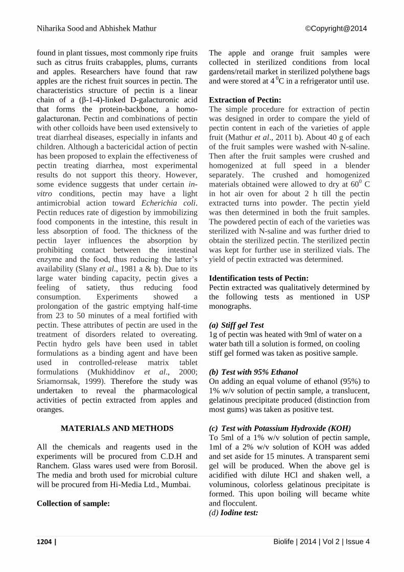
In the present study, the pectin, a natural gelling plant polysaccharide was extracted from pomace portion of apples and
oranges. Further the antimicrobial, antioxidant and anti-inflammatory activities of pectin extracts were investigated. It was
found that the yield of pectin content extracted was predominant in apples (20.60 %) in comparison to that of oranges
(10.56%). The pectin samples extracted from apples and oranges were qualitatively evaluated for characteristic properties.
Further the phytochemical constituents were qualitatively determined by conventional procedures. The results confirmed
the presence of alkaloids, flavanoids, reducing sugars, steroids and cardiac glycosides in orange pectin while alkaloids,
reducing sugars, steroids, cardiac glycosides and anthraquinones were present in apple pectin. Tannins, saponin and
anthraquinones were absent in orange pectin while tannins, flavanoids and saponin were absent in apple pectin. In
separation of pectin extracts by TLC, the Rf values of the pectin samples were found to be similar viz. Apple pectin (0.62),
Orange pectin (0.62) to that of standard pectin (procured from Ranchem), 0.63. The apple pectin and orange pectin showed
almost similar retention time viz. 1.821 minutes and 1.757 minutes specifically on HPLC chromatogram. The FT-IR
spectra of extracted pectin samples were determined using a computerized FTIR spectrometer in the range of 4400–400 cm-
1 by the KBr pellet technique. Two outstanding features of the infrared spectra of pectinate are bands at 1745-1743 and at
1607-1604 cm-1
. One other feature which should be mentioned is a band, beyond the afore mentioned range, appearing at
1418-1417 cm-l. In the spectra of polygalacturonate, the absence of carbonyl stretching bands above 1680 cm-l
demonstrates that the polygalacturonate is de-esterified to a degree of <2%. The extracts were also found to have
antimicrobial potential against the Methicillin resistant Staphylococcus aureus. It was found that apple pectin had potent
antimicrobial activity against E.coli, Methicillin resistant strain of S. aureus (isolated from blood) and Aspergillus niger
while orange pectin was found to have antimicrobial potential against E.coli and Aspergillus niger. Apple pectin extracts
were found to have potent antimicrobial activity in comparison to that of orange pectin. Apple pectin extracts showed least
MIC values viz. 10-4
µg/ml against Aspergillus niger, 10-5
µg/ml against E.coli and Methicilin resistant S. aureus (isolated
from blood). Orange pectin extracts showed similar MIC values viz. 10-5
µg/ml against E.coli and Aspergllus niger.
Amongst both the pectin extracts, Total phenolic content of orange pectin was found to be much more (38 µg/ml) in
comparison to that of apple pectin (20 µg/ml). The results of TPC correlate the findings of Total antioxidant activity
determination assay (based on absorbance) which also illustrates that orange pectin had potent antioxidant activity in
comparison to apple pectin. The extracts were also screened for ant-inflammatory activities and the results were found to be
satisfactory as the extracts showed promising anti-inflammatory activities too.
Key words: Pectin, pharmacological activities, active constituents, antimicrobial, antioxidant, anti-inflammatory activity.
INTRODUCTION
Pectin is a complex mixture of polysaccharides
that makes up about one third of the cell wall dry
substances of higher plants. Much smaller
proportions of these substances are found in the
cell walls of grasses. The highest concentration
of pectin is found in the middle lamella of cell
wall, with a gradual decrease as one passes
through the primary wall toward the plasma
membrane. Apple is particularly rich in pectin,
the name applied to any one of a group of white,
amorphous complex carbohydrates with a high
molecular weight. These water-soluble fibers are
AN INTERNATIONAL QUARTERLY JOURNAL OF BIOLOGY & LIFE SCIENCES
2(4):1203-1217 ISSN (online): 2320-4257
www.biolifejournal.com
Niharika Sood and Abhishek Mathur ©Copyright@2014
1204 | Biolife | 2014 | Vol 2 | Issue 4
found in plant tissues, most commonly ripe fruits
such as citrus fruits crabapples, plums, currants
and apples. Researchers have found that raw
apples are the richest fruit sources in pectin. The
characteristics structure of pectin is a linear
chain of a (β-1-4)-linked D-galacturonic acid
that forms the protein-backbone, a homo-
galacturonan. Pectin and combinations of pectin
with other colloids have been used extensively to
treat diarrheal diseases, especially in infants and
children. Although a bactericidal action of pectin
has been proposed to explain the effectiveness of
pectin treating diarrhea, most experimental
results do not support this theory. However,
some evidence suggests that under certain in-
vitro conditions, pectin may have a light
antimicrobial action toward Echerichia coli.
Pectin reduces rate of digestion by immobilizing
food components in the intestine, this result in
less absorption of food. The thickness of the
pectin layer influences the absorption by
prohibiting contact between the intestinal
enzyme and the food, thus reducing the latter’s
availability (Slany et al., 1981 a & b). Due to its
large water binding capacity, pectin gives a
feeling of satiety, thus reducing food
consumption. Experiments showed a
prolongation of the gastric emptying half-time
from 23 to 50 minutes of a meal fortified with
pectin. These attributes of pectin are used in the
treatment of disorders related to overeating.
Pectin hydro gels have been used in tablet
formulations as a binding agent and have been
used in controlled-release matrix tablet
formulations (Mukhiddinov et al., 2000;
Sriamornsak, 1999). Therefore the study was
undertaken to reveal the pharmacological
activities of pectin extracted from apples and
oranges.
MATERIALS AND METHODS
All the chemicals and reagents used in the
experiments will be procured from C.D.H and
Ranchem. Glass wares used were from Borosil.
The media and broth used for microbial culture
will be procured from Hi-Media Ltd., Mumbai.
Collection of sample:
The apple and orange fruit samples were
collected in sterilized conditions from local
gardens/retail market in sterilized polythene bags
and were stored at 4 0
C in a refrigerator until use.
Extraction of Pectin:
The simple procedure for extraction of pectin
was designed in order to compare the yield of
pectin content in each of the varieties of apple
fruit (Mathur et al., 2011 b). About 40 g of each
of the fruit samples were washed with N-saline.
Then after the fruit samples were crushed and
homogenized at full speed in a blender
separately. The crushed and homogenized
materials obtained were allowed to dry at 600 C
in hot air oven for about 2 h till the pectin
extracted turns into powder. The pectin yield
was then determined in both the fruit samples.
The powdered pectin of each of the varieties was
sterilized with N-saline and was further dried to
obtain the sterilized pectin. The sterilized pectin
was kept for further use in sterilized vials. The
yield of pectin extracted was determined.
Identification tests of Pectin:
Pectin extracted was qualitatively determined by
the following tests as mentioned in USP
monographs.
(a) Stiff gel Test
1g of pectin was heated with 9ml of water on a
water bath till a solution is formed, on cooling
stiff gel formed was taken as positive sample.
(b) Test with 95% Ethanol
On adding an equal volume of ethanol (95%) to
1% w/v solution of pectin sample, a translucent,
gelatinous precipitate produced (distinction from
most gums) was taken as positive test.
(c) Test with Potassium Hydroxide (KOH)
To 5ml of a 1% w/v solution of pectin sample,
1ml of a 2% w/v solution of KOH was added
and set aside for 15 minutes. A transparent semi
gel will be produced. When the above gel is
acidified with dilute HCl and shaken well, a
voluminous, colorless gelatinous precipitate is
formed. This upon boiling will became white
and flocculent.
(d) Iodine test:
Niharika Sood and Abhishek Mathur ©Copyright@2014
1205 | Biolife | 2014 | Vol 2 | Issue 4
To 5ml of recently boiled and cooled 2% w/v
solution of sample, 0.15 ml of Iodine solution
was added. No blue color presence was taken as
an indicator of positive test.
(d) Test for Acidity:
An aqueous solution of pectin sample was acidic
to blue litmus paper.
Phytochemical Screening for pectin extracted: The portion of the dry extracts was subjected to
the phytochemical screening using the method
adopted by Trease and Evans (1983) and
Harborne (1983). Phytochemical screening was
performed to test for alkaloids, saponin, tannins,
flavanoids, steroids, sugars, cardiac glycosides
and anthraquinones (Sofowora, 1993).
Test for alkaloids: The 0.5 g of the extract was dissolved in 5 ml of
1% HCl and kept in water bath for about 2
minutes. 1ml of the filtrate was treated with
Dragendroff’s reagent. Turbidity or precipitation
was taken as indicator for the presence of
alkaloids.
Test for Tannins: About 0.5 g of the sample was dissolved in 10
ml of boiling water and was filtered. Few ml of
6% FeCl3 was added to the filtrate. Deep green
colour appeared confirmed the presence of
Tannins (Trease and Evans, 1983).
Test for Flavanoids: About 0.2 gm of the extract was dissolved in
methanol and heated for some time. A chip of
Mg metal was introduced followed by the
addition of few drops of conc. HCl. Appearance
of red or orange color was taken as indicator of
the flavanoids.
Test for Saponin: About 0.5 g of the extract was stirred with water
in the test tube. Frothing persists on warming
was taken as a evidence for the presence of
saponin.
Test for Steroids: Salkowski’s method was adopted for the
detection of steroids. About 0.5 g of extract was
dissolved in 3 ml of chloroform and filtered. To
the filtrate, conc. H2 SO4 was added to form a
lower layer. Reddish brown color was taken as
positive for the presence of steroids ring
(Agarwal et al., 2011).
Test for Cardiac glycosides
About 0.5 g of the extract was dissolved in 2ml
of glacial acetic acid containing 1 drop of 1%
FeCl3. This was under laid with conc. H2SO4. A
brown ring obtained at the interphase indicates
the presence of deoxy sugar. A violet ring
appeared below the ring while in the acetic acid
layer a greenish ring appeared just above ring
and gradually spread throughout this layer.
Test for reducing Sugars: 1ml each of Fehling’s solutions, I and II was
added to 2 ml of the aqueous solution of the
extract. The mixture was heated in a boiling
water bath for about 2-5 minutes. The production
of a brick red precipitate indicated the presence
of reducing sugars.
Test for Anthraquinones:
5ml of the extract solution was hydrolyzed with
dil/conc. H2SO4. 1 ml of dilute ammonia was
then after be added to it. Rose pink colour
confirmed the presence of anthraquinones.
Estimation of Total Phenolic Content (TPC) of
Pectin:
The Total phenolic content of each pectin
sample was determined by the method of
Singleton and Rossi (1965). The phenolic
content was expressed as mg/g gallic acid
equivalents. In brief 100 µl aliquots of the
sample were added to 2 ml of 0.2 % (w/v)
Na2CO3 solution. After 2 minutes of the
incubation. 100 µl of 500 ml/1 Follin-Ciocalteu
reagent was added and the mixture was allowed
to stand for 30 minutes at 25 0C. The absorbance
was measured at 750 nm using a UV-VIS
Systronics spectrophotometer. The blank consist
of all reagents and solvents but no sample. The
Total Phenolic Content (TPC) was determined
using the standard gallic acid calibration curve.
Purification and Characterization of pectin
extracted of both apples and oranges via
Niharika Sood and Abhishek Mathur ©Copyright@2014
1206 | Biolife | 2014 | Vol 2 | Issue 4
chromatographic and spectroscopic
techniques:
Separation of Pectin extracted by TLC:
TLC Separation was performed for the pectin
extracted from apples and oranges. Standard
pectin was also taken as reference. In TLC,
detection system used was Butanol: Water:
Acetic acid (5:4:1). Iodine chamber was also
used for detection. Rf values were determined
after the appearance of spots.
High-performance liquid chromatography
(HPLC):
HPLC analysis of extracted pectin samples were
performed in Roorkee Research and Analytical
Laboratory Pvt. Ltd., Roorkee (Uttarakhand),
India using a Shimadzo LC- 2010 HPLC system
(Kyoto, Japan), equipped with a Shimadzo LC
2010 UV-VIS detector with a thermostated flow
cell and a selectable two wavelengths of 190 -
370 nm or 371–600nm. The detector signal was
recorded on a Shimadzo LC2010 integrator. The
column used was a Chiral Column block
heating-type Shim-pack VP-ODS (4.6 mm
interior diameter × 150 mm long) with a particle
size of 5 μm. Mobile phase was used containing
50 % acetonitrile along with 50 % Phosphate
buffer was used at a flow rate of 3.0 ml/min,
column temperature 25°C. Injection volume was
40 µl and detection was carried out at specific
wavelength having maximum absorbance as
calculated by UV absorption spectra at
maximum wavelength.
Fourier Transform Infrared (FTIR) studies:
The IR spectrum of purified pectin extracted
from apples and oranges was recorded in
Roorkee Research and Analytical Laboratory
Pvt. Ltd., Roorkee (Uttarakhand), India using a
computerized FTIR spectrometer (Perkin Co.,
Germany) in the range of 4400–400 cm-1
by the
KBr pellet technique.
Determination of in vitro antimicrobial
activity of pectin extracted from apples and
oranges:
The pectin extracted from apples and oranges
was dissolved in N-saline (1 mg/ml) and
evaluated for in vitro antimicrobial activity.
Culture Media:
The media used for antibacterial test is Nutrient
agar/broth and Sabouraud’s dextrose agar/broth
of Hi media Pvt. Bombay, India.
Inoculum:
The bacterial pathogen was inoculated into
nutrient broth and incubated at 37 0
C for 4 h and
the suspension will be checked to provide
approximately 105
CFU/ml. Similar procedure
was done for fungal strains by inoculating in
Sabouraud’s dextrose broth for 6 h.
Culture medium:
The pathogenic bacterial cultures were
inoculated into Nutrient broth and incubated at
37 0
C for 18 h and suspension was checked to
provide approximately, 105 CFU/ml. The same
procedure was done for fungal pathogens and
there strains will be inoculated into Sabouraud’s
dextrose broth but the fungal broth cultures were
incubated at 48-72 h.
Microorganisms used :
Pure cultures of various pathogenic bacterial and
fungal strains, E. coli NCIM 2065, Lactobacillus
plantarum NCIM 2083, Micrococcus luteus
ATCC 9341, Salmonella abony NCIM 2257,
Candida albicans NCIM 3471, Aspergillus niger
NCIM 1196 and Methicillin resistant strains of
Staphylococcus aureus (MRSA) isolated from
clinical specimens viz. pus and blood of infected
patients were procured with authentication for
the study. The standard bacterial and fungal
cultures used for the study were procured from
Roorkee Research & Analytical Labs Pvt. Ltd.,
Roorkee (U.K), India and MRSA strains were
procured from Shooloni University, H.P., India.
Determination of diameter of zone of inhibition
by well diffusion method: The agar well diffusion method (Perez et al.,
1993) was modified. Nutrient agar medium
(NAM) was used for growth of pathogenic
bacteria cultures. The culture medium was
inoculated with the bacterial pathogen separately
suspended in nutrient broth. Sabouraud’s
dextrose agar/broth was used for growth of
pathogenic fungal cultures. The culture medium
was inoculated with the fungus separately
Niharika Sood and Abhishek Mathur ©Copyright@2014
1207 | Biolife | 2014 | Vol 2 | Issue 4
suspended in Sabouraud’s dextrose broth. A total
of 8 mm diameter wells were punched into the
agar and filled with separate endophytic
fractions and solvent blanks. Standard antibiotic
(Erythromycin, 1 mg/ml) was simultaneously
used as the positive control. The plates were
incubated at 37 0
C for 18 h. The antibacterial
activity was evaluated by measuring the
diameter of zone of inhibition observed. For
assaying, antifungal activity of endophytic
fractions, Sabouraud’s dextrose agar/ broth
medium plates was used. The same procedure as
that for determination of antibacterial property
was adopted and then after the diameter of zone
of inhibition was observed after 48-72 h.
Fucanazole (1mg/ml) was used as standard for
determination of antifungal activity. The
procedure for assaying antibacterial and
antifungal activity was performed in triplicates
to confirm the average readings of diameter of
zone of inhibition observed for each of the test
organism.
Determination of MIC and MLC :
The broth dilution method (Vollekova et al.,
2001 and Usman et al., 2007) was adopted for
determination of MIC and MLC values against
the pathogens. The pectin extracts (1 mg/ml)
were serially diluted in different aliquots and the
final volumes of the aliquots were made up to 1
ml with N-saline (0.85 % NaCl). Equal amount
of the specific pathogen was added in different
aliquots and the test tubes were kept for 48 h at
30 0
C. The minimum dilution of the pectin
extract that kills the bacterial and fungal growth
was taken as MLC (Minimum lethal count)
while the minimum dilution that inhibits the
growth of the organism was taken as MIC.
Determination of in vitro antioxidant activity
of pectin extracted and enzyme purified:
Determination of Total Antioxidant Activity:
Total antioxidant activities of pectin fractions
and ascorbic acid were determined by the
method of Pan et al., 2008. An aliquot (0.1M) of
these fractions was combined with 1ml of
reagent solution (0.6 M sulphuric acid, 28 mM
sodium phosphate and 4 mM ammonium
molybdate). The tubes were then capped and
incubated at 95 o
C for 90 minutes. After that the
samples were cooled at 25oC, the absorbance
was measured at 695 nm against blank. The
blank contained 1ml of reagent solution without
sample. The total antioxidant activity was
expressed as an absorbance value at 695 nm.
Higher absorbance value indicates the maximum
antioxidant activity.
Determination of in vitro anti-inflammatory
potential of pectin extracted of both apples
and oranges:
(A) The human red blood cell (HRBC)
membrane stabilization method:
The method as prescribed (Gopalkrishnan et al.,
2009; Sakat et al., 2010) was adopted with some
modifications. The blood was collected from
healthy human volunteer who had not taken any
NSAIDS for 2 weeks prior to the experiment and
mixed with equal volume of Alsever solution (2
% dextrose, 0.8 % sodium citrate, 0.5 % citric
acid and 0.42 % NaCl) and centrifuged at 3,000
rpm. The packed cells were washed with
isosaline and a 10 % suspension will be made.
Pectin extracts were prepared (in appropriate
concentrations) using distilled water and to each
concentrations, 1 ml of phosphate buffer, 2 ml
hypo saline and 0.5 ml of HRBC suspension
were added. It was then incubated at 370C for 30
minutes and centrifuged at 3,000 rpm for 20
minutes and the hemoglobin content of the
supernatant solution was estimated
spectrophotometrically at 560 nm. Diclofenac
(100 µg/ml) was used as reference standard and
a control was prepared by omitting the extracts.
The experiments were performed in triplicates
and mean values of the three will be considered.
The percentage (%) of HRBC membrane
stabilization or protection was calculated using
the following formula:
Percent Protection (%) = (100- OD of drug
treated sample/OD of Control) X 100
(B) Inhibition of Albumen Denaturation:
Method as prescribed (Sakat et al., 2010) was
followed with minor modifications. The reaction
mixture will be consisting of test extracts and
1% aqueous solution of bovine albumin fraction,
Niharika Sood and Abhishek Mathur ©Copyright@2014
1208 | Biolife | 2014 | Vol 2 | Issue 4
pH of the reaction mixture was adjusted using
small amount at 37°C HCl. The sample extracts
were incubated at 37°C for 20 minutes and then
heated to 51°C for 20 minutes, after cooling the
samples the turbidity was measured
spectrophotometrically at 660 nm. Diclofenac
sodium was taken as a standard drug. The
experiment was performed in triplicates. Percent
inhibition of protein denaturation was calculated
as follows:
Percent inhibition (%) = (OD of Control-OD of
Sample/OD of Control) X100
(C) Heat induced hemolysis:
The reaction mixture (2 ml) consisted of 1 ml of
test sample solution and 1 ml of 10 % RBCs
suspension, instead of test sample only saline
was added to the control test tube. Diclofenac
sodium was taken as a standard drug. All the
centrifuge tubes containing reaction mixture
were incubated in water bath at 56°C for 30
minutes. At the end of the incubation the tubes
were cooled under running tap water. The
reaction mixtures were then centrifuged at 2500
rpm for 5 minutes and the absorbance of the
supernatants were taken at 560 nm. The
experiment was performed in triplicates for all
the test samples. Percent hemolysis was
calculated by the formula mentioned in the
above procedure.
RESULTS
In the present investigation the pectin extracted
from apples and oranges procured from local
market were used for evaluation of
antimicrobial, antioxidant and anti-inflammatory
properties.
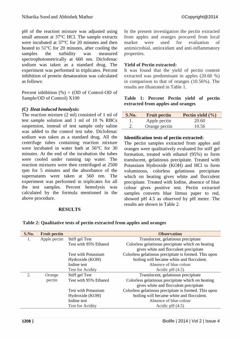
Yield of Pectin extracted:
It was found that the yield of pectin content
extracted was predominant in apples (20.60 %)
in comparison to that of oranges (10.56%). The
results are illustrated in Table 1.
Table 1: Percent Pectin yield of pectin
extracted from apples and oranges
S.No. Fruit pectin Pectin yield (%)
1. Apple pectin 20.60
2. Orange pectin 10.56
Identification tests of pectin extracted:
The pectin samples extracted from apples and
oranges were qualitatively evaluated for stiff gel
formation, treated with ethanol (95%) to form
translucent, gelatinous precipitate. Treated with
Potassium Hydroxide (KOH) and HCl to form
voluminous, colorless gelatinous precipitate
which on heating gives white and flocculent
precipitate. Treated with Iodine, absence of blue
colour gives positive test. Pectin extracted
samples converts blue litmus paper to red,
showed pH 4.5 as observed by pH meter. The
results are shown in Table 2.
Table 2: Qualitative tests of pectin extracted from apples and oranges
S.No. Fruit pectin Observation
1. Apple pectin Stiff gel Test Translucent, gelatinous precipitate
Test with 95% Ethanol Colorless gelatinous precipitate which on heating
gives white and flocculent precipitate
Test with Potassium
Hydroxide (KOH)
Colorless gelatinous precipitate is formed. This upon
boiling will became white and flocculent.
Iodine test Absence of blue colour
Test for Acidity Acidic pH (4.5)
2. Orange
pectin
Stiff gel Test Translucent, gelatinous precipitate
Test with 95% Ethanol Colorless gelatinous precipitate which on heating
gives white and flocculent precipitate
Test with Potassium
Hydroxide (KOH)
Colorless gelatinous precipitate is formed. This upon
boiling will became white and flocculent.
Iodine test Absence of blue colour
Test for Acidity Acidic pH (4.5)
Niharika Sood and Abhishek Mathur ©Copyright@2014
1209 | Biolife | 2014 | Vol 2 | Issue 4
Phytochemical Screening for pectin extracted: The portion of the dry extract of pectin obtained
was subjected to phytochemical screening for
qualitative examination of alkaloids, tannins,
flavanoids, steroids, saponin, cardiac glycosides,
reducing sugar and anthraquinones. The results
confirmed that alkaloids, flavanoids, reducing
sugars, steroids and cardiac glycosides were
present in orange pectin while alkaloids,
reducing sugars, steroids, cardiac glycosides and
anthraquinones were present in apple pectin.
Tannins, saponin and anthraquinones were
absent in orange pectin while tannins, flavanoids
and saponin were absent in apple pectin (Table-
3).
Purification and Characterization of pectin
extracted of both apples and oranges via
chromatographic and spectroscopic
techniques:
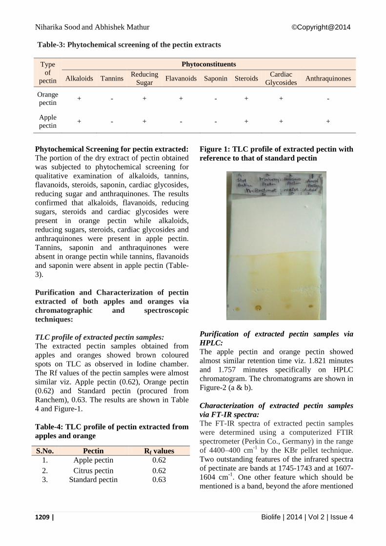
TLC profile of extracted pectin samples:
The extracted pectin samples obtained from
apples and oranges showed brown coloured
spots on TLC as observed in Iodine chamber.
The Rf values of the pectin samples were almost
similar viz. Apple pectin (0.62), Orange pectin
(0.62) and Standard pectin (procured from
Ranchem), 0.63. The results are shown in Table
4 and Figure-1.
Table-4: TLC profile of pectin extracted from
apples and orange
Figure 1: TLC profile of extracted pectin with
reference to that of standard pectin
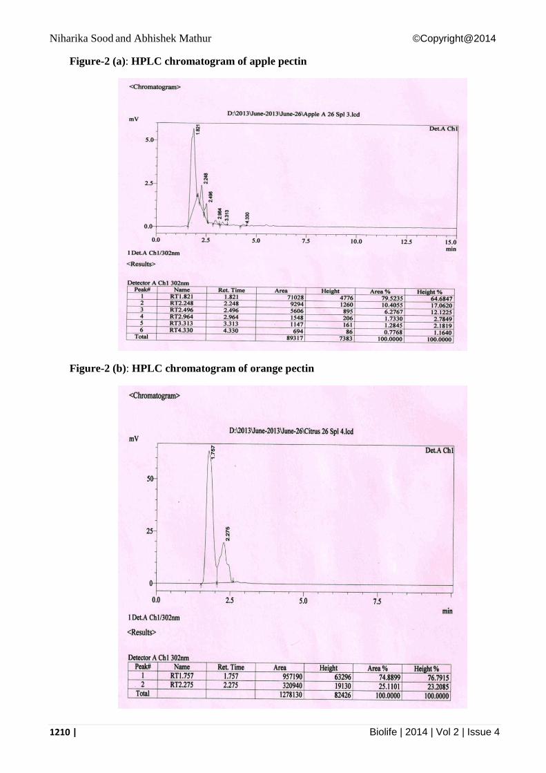
Purification of extracted pectin samples via
HPLC:
The apple pectin and orange pectin showed
almost similar retention time viz. 1.821 minutes
and 1.757 minutes specifically on HPLC
chromatogram. The chromatograms are shown in
Figure-2 (a & b).
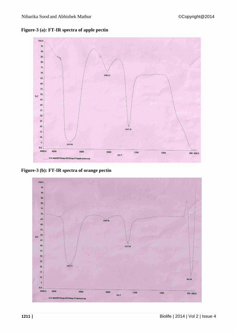
Characterization of extracted pectin samples
via FT-IR spectra:
The FT-IR spectra of extracted pectin samples
were determined using a computerized FTIR
spectrometer (Perkin Co., Germany) in the range
of 4400–400 cm-1
by the KBr pellet technique.
Two outstanding features of the infrared spectra
of pectinate are bands at 1745-1743 and at 1607-
1604 cm-1
. One other feature which should be
mentioned is a band, beyond the afore mentioned
S.No. Pectin Rf values
1. Apple pectin 0.62
2. Citrus pectin 0.62
3. Standard pectin 0.63
Table-3: Phytochemical screening of the pectin extracts
Type
of
pectin
Phytoconstituents
Alkaloids Tannins Reducing
Sugar Flavanoids Saponin Steroids
Cardiac
Glycosides Anthraquinones
Orange
pectin + - + + - + + -
Apple
pectin + - + - - + + +
Niharika Sood and Abhishek Mathur ©Copyright@2014
1210 | Biolife | 2014 | Vol 2 | Issue 4
Figure-2 (a): HPLC chromatogram of apple pectin
Figure-2 (b): HPLC chromatogram of orange pectin
Niharika Sood and Abhishek Mathur ©Copyright@2014
1211 | Biolife | 2014 | Vol 2 | Issue 4
Figure-3 (a): FT-IR spectra of apple pectin
Figure-3 (b): FT-IR spectra of orange pectin
Niharika Sood and Abhishek Mathur ©Copyright@2014
1212 | Biolife | 2014 | Vol 2 | Issue 4
range, appearing at 1418-1417 cm-l. In the
spectra of polygalacturonate, the absence of
carbonyl stretching bands above 1680 cm-l
demonstrates that the polygalacturonate is de-
esterified to a degree of <2%. The bands at X07-
1604 and 1417 cm-l correspond to the
antisymmetric and symmetric stretching
vibrations of the carboxylate groups. The spectra
are shown in Figures 3 (a & b).
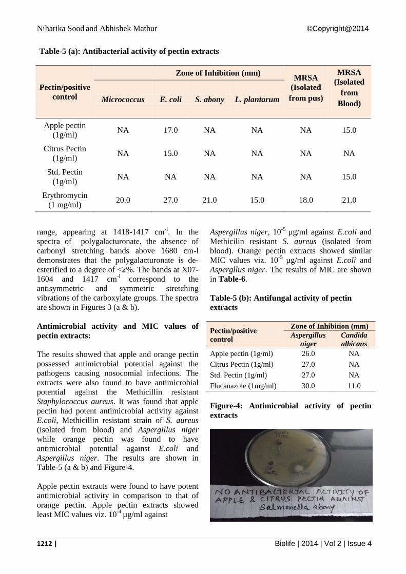
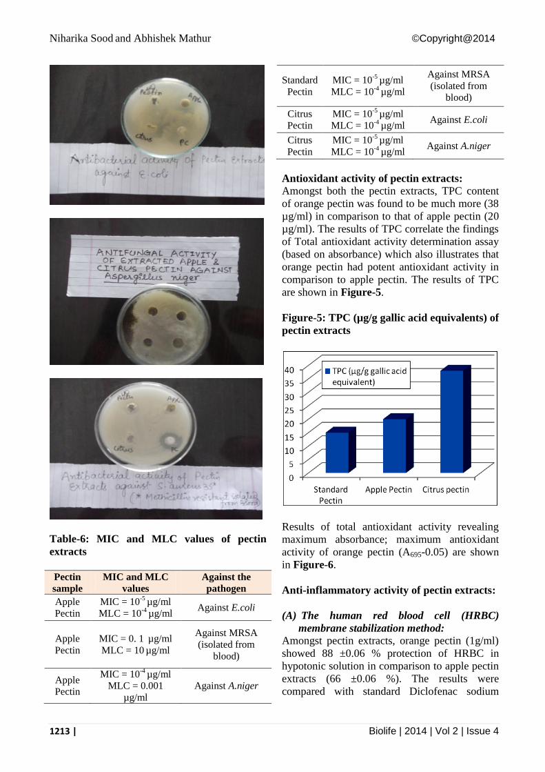
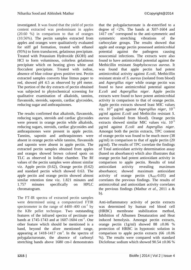
Antimicrobial activity and MIC values of
pectin extracts:
The results showed that apple and orange pectin
possessed antimicrobial potential against the
pathogens causing nosocomial infections. The
extracts were also found to have antimicrobial
potential against the Methicillin resistant
Staphylococcus aureus. It was found that apple
pectin had potent antimicrobial activity against
E.coli, Methicillin resistant strain of S. aureus
(isolated from blood) and Aspergillus niger
while orange pectin was found to have
antimicrobial potential against E.coli and
Aspergillus niger. The results are shown in
Table-5 (a & b) and Figure-4.
Apple pectin extracts were found to have potent
antimicrobial activity in comparison to that of
orange pectin. Apple pectin extracts showed
least MIC values viz. 10-4
µg/ml against
Aspergillus niger, 10-5
µg/ml against E.coli and
Methicilin resistant S. aureus (isolated from
blood). Orange pectin extracts showed similar
MIC values viz. 10-5
µg/ml against E.coli and
Aspergllus niger. The results of MIC are shown
in Table-6.
Table-5 (b): Antifungal activity of pectin
extracts
Pectin/positive
control
Zone of Inhibition (mm)
Aspergillus
niger
Candida
albicans
Apple pectin (1g/ml) 26.0 NA
Citrus Pectin (1g/ml) 27.0 NA
Std. Pectin (1g/ml) 27.0 NA
Flucanazole (1mg/ml) 30.0 11.0
Figure-4: Antimicrobial activity of pectin
extracts
Table-5 (a): Antibacterial activity of pectin extracts
Pectin/positive
control
Zone of Inhibition (mm) MRSA
(Isolated
from pus)
MRSA
(Isolated
from
Blood) Micrococcus E. coli S. abony L. plantarum
Apple pectin
(1g/ml) NA 17.0 NA NA NA 15.0
Citrus Pectin
(1g/ml) NA 15.0 NA NA NA NA
Std. Pectin
(1g/ml) NA NA NA NA NA 15.0
Erythromycin
(1 mg/ml) 20.0 27.0 21.0 15.0 18.0 21.0
Niharika Sood and Abhishek Mathur ©Copyright@2014
1213 | Biolife | 2014 | Vol 2 | Issue 4
Table-6: MIC and MLC values of pectin
extracts
Pectin
sample
MIC and MLC
values
Against the
pathogen
Apple
Pectin
MIC = 10-5
µg/ml
MLC = 10-4
µg/ml Against E.coli
Apple
Pectin
MIC = 0. 1 µg/ml
MLC = 10 µg/ml
Against MRSA
(isolated from
blood)
Apple
Pectin
MIC = 10-4
µg/ml
MLC = 0.001
µg/ml
Against A.niger
Standard
Pectin
MIC = 10-5
µg/ml
MLC = 10-4
µg/ml
Against MRSA
(isolated from
blood)
Citrus
Pectin
MIC = 10-5
µg/ml
MLC = 10-4
µg/ml Against E.coli
Citrus
Pectin
MIC = 10-5
µg/ml
MLC = 10-4
µg/ml Against A.niger
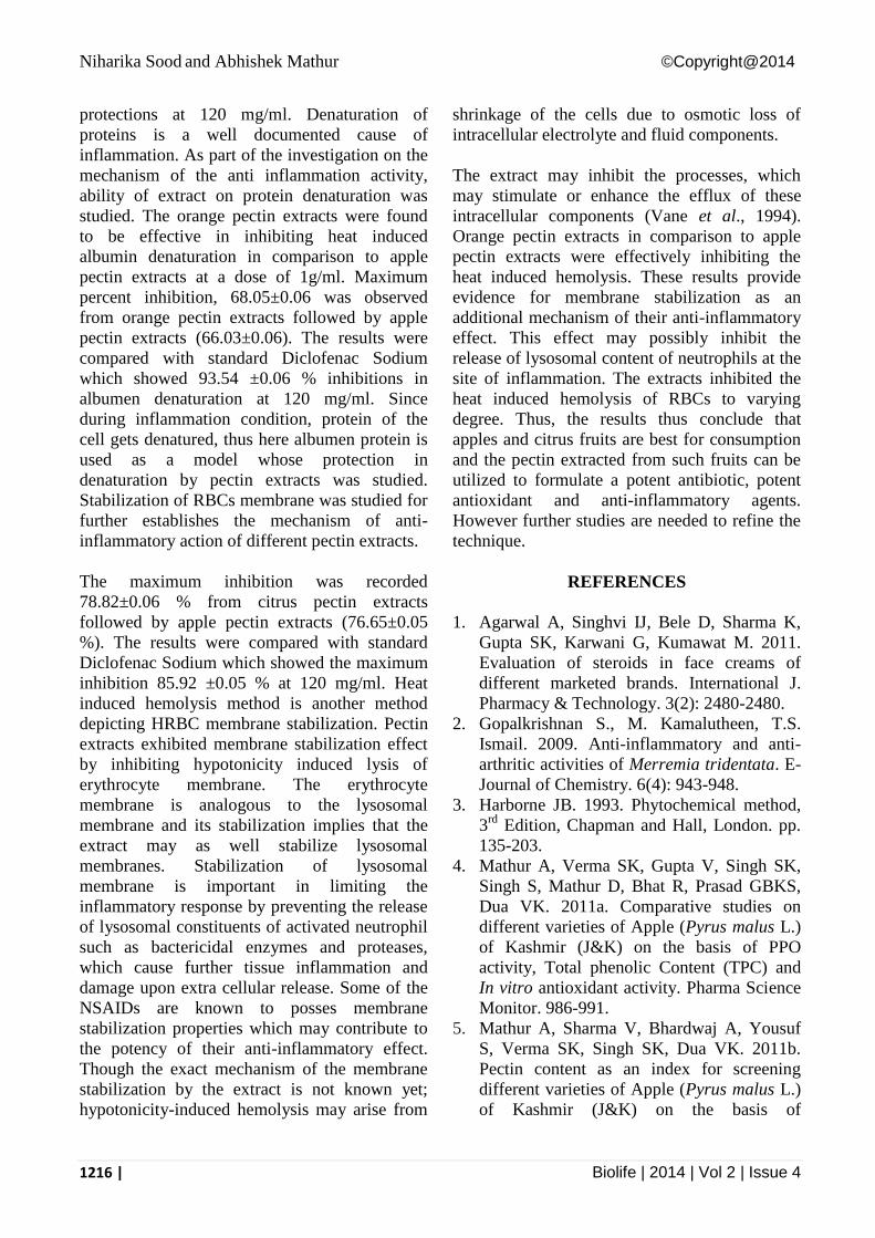
Antioxidant activity of pectin extracts:
Amongst both the pectin extracts, TPC content
of orange pectin was found to be much more (38
µg/ml) in comparison to that of apple pectin (20
µg/ml). The results of TPC correlate the findings
of Total antioxidant activity determination assay
(based on absorbance) which also illustrates that
orange pectin had potent antioxidant activity in
comparison to apple pectin. The results of TPC
are shown in Figure-5.
Figure-5: TPC (µg/g gallic acid equivalents) of
pectin extracts
Results of total antioxidant activity revealing
maximum absorbance; maximum antioxidant
activity of orange pectin (A695-0.05) are shown
in Figure-6.
Anti-inflammatory activity of pectin extracts:
(A) The human red blood cell (HRBC)
membrane stabilization method:
Amongst pectin extracts, orange pectin (1g/ml)
showed 88 ±0.06 % protection of HRBC in
hypotonic solution in comparison to apple pectin
extracts (66 ±0.06 %). The results were
compared with standard Diclofenac sodium
Niharika Sood and Abhishek Mathur ©Copyright@2014
1214 | Biolife | 2014 | Vol 2 | Issue 4
which showed 90.54 ±0.06 % protections at 120
mg/ml. The results are shown in Table-7.
Figure-6: Total antioxidant activity (A695) of
pectin extracts
Table-7: Percent protection of HRBC
membrane/membrane stabilization of pectin
extracts and positive control
(B) Inhibition of Albumen Denaturation:
Denaturation of proteins is a well documented
cause of inflammation. As part of the
investigation on the mechanism of the anti
inflammation activity, ability of extract on
protein denaturation was studied. The orange
pectin extracts were found to be effective in
inhibiting heat induced albumin denaturation in
comparison to apple pectin extracts at a dose of
1g/ml. Maximum percent inhibition, 68.05±0.06
was observed from orange pectin extracts
followed by apple pectin extracts (66.03±0.06).
The results were compared with standard
Diclofenac Sodium which showed 93.54 ±0.06
% inhibitions in albumen denaturation at 120
mg/ml. The results are reported in Table-8. Since
during inflammation condition, protein of the
cell gets denatured, thus here albumen protein is
used as a model whose protection in
denaturation by pectin extracts was studied.
Table-8: Percent protection/Inhibition of
Albumen Denaturation of pectin extracts and
positive control
(C) Heat induced hemolysis: Stabilization of RBCs membrane was studied for
further establishes the mechanism of anti-
inflammatory action of different pectin extracts.
The maximum inhibition was recorded
78.82±0.06 % from citrus pectin extracts
followed by apple pectin extracts (76.65±0.05
%). The results were compared with standard
Diclofenac Sodium which showed the maximum
inhibition 85.92 ±0.05 % at 120 mg/ml. The
results are reported in Table-9. Heat induced
hemolysis method is another method depicting
HRBC membrane stabilization.
Table-9. Percent protection in heat induced
hemolysis of HRBC membrane by pectin
extracts and positive control
DISCUSSION AND CONCLUSION
Apples and citrus fruits rich in Vitamin C are
rich sources of pectin. The pectin contains
various active constituents. In the present study,
the antimicrobial, antioxidant activity and anti-
inflammatory activity of pectin has been
Pectin sample Percent protection of
HRBC membrane
Standard Pectin 56 ±0.06
Apple Pectin 66 ±0.06
Citrus pectin 88 ±0.06
Diclofenac sodium
(positive control) 90.54 ±0.06
Pectin sample Percent Inhibition of
Albumen Denaturation
Standard Pectin 50 ±0.06
Apple Pectin 66.03±0.06
Citrus pectin 68.05±0.06
Diclofenac sodium
(positive control) 93.54 ±0.06
Pectin sample
Percent protection in
heat induced hemolysis
of HRBC membrane
Standard Pectin 56 ±0.06
Apple Pectin 76.65±0.05
Citrus pectin 78.82±0.06
Diclofenac
sodium (positive
control)
85.92 ±0.06
Niharika Sood and Abhishek Mathur ©Copyright@2014
1215 | Biolife | 2014 | Vol 2 | Issue 4
investigated. It was found that the yield of pectin
content extracted was predominant in apples
(20.60 %) in comparison to that of oranges
(10.56%). The pectin samples extracted from
apples and oranges were qualitatively evaluated
for stiff gel formation, treated with ethanol
(95%) to form translucent, gelatinous precipitate.
Treated with Potassium Hydroxide (KOH) and
HCl to form voluminous, colorless gelatinous
precipitate which on heating gives white and
flocculent precipitate. Treated with iodine,
absence of blue colour gives positive test. Pectin
extracted samples converts blue litmus paper to
red, showed pH 4.5 as observed by pH meter.
The portion of the dry extracts of pectin obtained
was subjected to phytochemical screening for
qualitative examination of alkaloids, tannins,
flavanoids, steroids, saponin, cardiac glycosides,
reducing sugar and anthraquinones.
The results confirmed that alkaloids, flavanoids,
reducing sugars, steroids and cardiac glycosides
were present in orange pectin while alkaloids,
reducing sugars, steroids, cardiac glycosides and
anthraquinones were present in apple pectin.
Tannins, saponin and anthraquinones were
absent in orange pectin while tannins, flavanoids
and saponin were absent in apple pectin. The
extracted pectin samples obtained from apples
and oranges showed brown colored spots on
TLC as observed in Iodine chamber. The Rf
values of the pectin samples were almost similar
viz. Apple pectin (0.62), Orange pectin (0.62)
and standard pectin which showed 0.63. The
apple pectin and orange pectin showed almost
similar retention time viz. 1.821 minutes and
1.757 minutes specifically on HPLC
chromatogram.
The FT-IR spectra of extracted pectin samples
were determined using a computerized FTIR
spectrometer in the range of 4400–400 cm-1
by
the KBr pellet technique. Two outstanding
features of the infrared spectra of pectinate are
bands at 1745-1743 and at 1607-1604 cm-1
. One
other feature which should be mentioned is a
band, beyond the afore mentioned range,
appearing at 1418-1417 cm-l. In the spectra of
polygalacturonate, the absence of carbonyl
stretching bands above 1680 cm-l demonstrates
that the polygalacturonate is de-esterified to a
degree of <2%. The bands at X07-1604 and
1417 cm-l correspond to the anti-symmetric and
symmetric stretching vibrations of the
carboxylate groups. The results showed that
apple and orange pectin possessed antimicrobial
potential against the pathogens causing
nosocomial infections. The extracts were also
found to have antimicrobial potential against the
Methicillin resistant Staphylococcus aureus. It
was found that apple pectin had potent
antimicrobial activity against E.coli, Methicillin
resistant strain of S. aureus (isolated from blood)
and Aspergillus niger while orange pectin was
found to have antimicrobial potential against
E.coli and Aspergillus niger. Apple pectin
extracts were found to have potent antimicrobial
activity in comparison to that of orange pectin.
Apple pectin extracts showed least MIC values
viz. 10-4
µg/ml against Aspergillus niger, 10-5
µg/ml against E.coli and Methicilin resistant S.
aureus (isolated from blood). Orange pectin
extracts showed similar MIC values viz. 10-5
µg/ml against E.coli and Aspergllus niger.
Amongst both the pectin extracts, TPC content
of orange pectin was found to be much more (38
µg/ml) in comparison to that of apple pectin (20
µg/ml). The results of TPC correlate the findings
of Total antioxidant activity determination assay
(based on absorbance) which also illustrates that
orange pectin had potent antioxidant activity in
comparison to apple pectin. Results of total
antioxidant activity revealing maximum
absorbance; showed maximum antioxidant
activity of orange pectin (A695-0.05) and
correlates the previous findings. The results of
antimicrobial and antioxidant activity correlates
the previous findings (Mathur et al., 2011 a &
b).
Anti-inflammatory activity of pectin extracts
was determined by human red blood cell
(HRBC) membrane stabilization method,
Inhibition of Albumen Denaturation and Heat
induced hemolysis. Amongst pectin extracts,
orange pectin (1g/ml) showed 88 ±0.06 %
protection of HRBC in hypotonic solution in
comparison to apple pectin extracts (66 ±0.06
%). The results were compared with standard
Diclofenac sodium which showed 90.54 ±0.06 %
Niharika Sood and Abhishek Mathur ©Copyright@2014
1216 | Biolife | 2014 | Vol 2 | Issue 4
protections at 120 mg/ml. Denaturation of
proteins is a well documented cause of
inflammation. As part of the investigation on the
mechanism of the anti inflammation activity,
ability of extract on protein denaturation was
studied. The orange pectin extracts were found
to be effective in inhibiting heat induced
albumin denaturation in comparison to apple
pectin extracts at a dose of 1g/ml. Maximum
percent inhibition, 68.05±0.06 was observed
from orange pectin extracts followed by apple
pectin extracts (66.03±0.06). The results were
compared with standard Diclofenac Sodium
which showed 93.54 ±0.06 % inhibitions in
albumen denaturation at 120 mg/ml. Since
during inflammation condition, protein of the
cell gets denatured, thus here albumen protein is
used as a model whose protection in
denaturation by pectin extracts was studied.
Stabilization of RBCs membrane was studied for
further establishes the mechanism of anti-
inflammatory action of different pectin extracts.
The maximum inhibition was recorded
78.82±0.06 % from citrus pectin extracts
followed by apple pectin extracts (76.65±0.05
%). The results were compared with standard
Diclofenac Sodium which showed the maximum
inhibition 85.92 ±0.05 % at 120 mg/ml. Heat
induced hemolysis method is another method
depicting HRBC membrane stabilization. Pectin
extracts exhibited membrane stabilization effect
by inhibiting hypotonicity induced lysis of
erythrocyte membrane. The erythrocyte
membrane is analogous to the lysosomal
membrane and its stabilization implies that the
extract may as well stabilize lysosomal
membranes. Stabilization of lysosomal
membrane is important in limiting the
inflammatory response by preventing the release
of lysosomal constituents of activated neutrophil
such as bactericidal enzymes and proteases,
which cause further tissue inflammation and
damage upon extra cellular release. Some of the
NSAIDs are known to posses membrane
stabilization properties which may contribute to
the potency of their anti-inflammatory effect.
Though the exact mechanism of the membrane
stabilization by the extract is not known yet;
hypotonicity-induced hemolysis may arise from
shrinkage of the cells due to osmotic loss of
intracellular electrolyte and fluid components.
The extract may inhibit the processes, which
may stimulate or enhance the efflux of these
intracellular components (Vane et al., 1994).
Orange pectin extracts in comparison to apple
pectin extracts were effectively inhibiting the
heat induced hemolysis. These results provide
evidence for membrane stabilization as an
additional mechanism of their anti-inflammatory
effect. This effect may possibly inhibit the
release of lysosomal content of neutrophils at the
site of inflammation. The extracts inhibited the
heat induced hemolysis of RBCs to varying
degree. Thus, the results thus conclude that
apples and citrus fruits are best for consumption
and the pectin extracted from such fruits can be
utilized to formulate a potent antibiotic, potent
antioxidant and anti-inflammatory agents.
However further studies are needed to refine the
technique.
REFERENCES
1. Agarwal A, Singhvi IJ, Bele D, Sharma K,
Gupta SK, Karwani G, Kumawat M. 2011.
Evaluation of steroids in face creams of
different marketed brands. International J.
Pharmacy & Technology. 3(2): 2480-2480.
2. Gopalkrishnan S., M. Kamalutheen, T.S.
Ismail. 2009. Anti-inflammatory and anti-
arthritic activities of Merremia tridentata. E-
Journal of Chemistry. 6(4): 943-948.
3. Harborne JB. 1993. Phytochemical method,
3rd
Edition, Chapman and Hall, London. pp.
135-203.
4. Mathur A, Verma SK, Gupta V, Singh SK,
Singh S, Mathur D, Bhat R, Prasad GBKS,
Dua VK. 2011a. Comparative studies on
different varieties of Apple (Pyrus malus L.)
of Kashmir (J&K) on the basis of PPO
activity, Total phenolic Content (TPC) and
In vitro antioxidant activity. Pharma Science
Monitor. 986-991.
5. Mathur A, Sharma V, Bhardwaj A, Yousuf
S, Verma SK, Singh SK, Dua VK. 2011b.
Pectin content as an index for screening
different varieties of Apple (Pyrus malus L.)
of Kashmir (J&K) on the basis of
Niharika Sood and Abhishek Mathur ©Copyright@2014
1217 | Biolife | 2014 | Vol 2 | Issue 4
antimicrobial activity. Journal of Chemical &
Pharmaceutical Research. 3 (2): 886-891.
6. Mukhiddinov, Z.K., et al. 2000. Isolation and
structural characterization of a pectin homo
and ramnogalacturonan. Talanta. 53: 171-
176.
7. Pan Y, Wang K, Huang S, Wang H, Mu X,
He C. 2008. Antioxidant activity of
microwave assisted extract of longan. Food
Chemistry. 106(3): 1264-1270.
8. Perez C, Anesini C. 1993. In vitro
antimicrobial activity of Argentine folk
medicinal plants against Salmonella typhii.
Journal of Ethnopharmacology. 44: 41-46.
9. Sakat S., A.R. Juvekar, M.N. Gambhire.
2010. In vitro antioxidant and anti-
inflammatory activity of methanol extract of
Oxalis corniculata Linn. Int. J. Pharm.
Pharmacol. Sci. 2(1): 146-155.
10. Singleton VL and Rossi JA. 1965.
Colorimetry of total phenolics with
phosphomolybdic and phosphotungstic acid
reagents. American Journal of Ecology and
Viticulture. 16: 144-158.
11. Slany, J., et al. 1981a. Evaluation of tablets
with pectin as a binding agent.
Farmaceuticky Obzor. 50: 491-498.
12. Slany, J., et al. 1981b. Study of functional
action of citrus pectins in tablets. Ceska a
Slovenska Farmacie. 30: 195-200.
13. Sofowora AO. 1993. Traditional Medicine
and Medicinal Plants in Africa. 2nd
Ed.,
University of Life Press. 320.
14. Sriamornsak, P. 1999. Effect of calcium
concentration, hardening agent and drying
condition on release characteristics of oral
proteins from calcium pectinate gel beads.
European Journal of Pharmaceutical
Sciences. 8: 221-227.
15. Trease GE, Evans WC. 1989.
Pharmacogonasy, 14th
Edition, Brown
Publication.
16. Usman H, Abdulrahman FI and Ladan AH.
2007. Phytochemical and antimicrobial
evaluation of Tribulus terrestris L.
(Zygophylaceae). Growing in Nigeria. Res.
J. Bio. Sci. Medwell Journals. 2(3): 244-247.
17. Vollekova AD, Kostalova, Sochorova R.
2001. Isoquinoline Alkaloids from Mahonia
aquifolium stem bark is active against
Malassezia sp. Folia Microbiol. 46: 107-111.
*****