Embed Size (px)
Citation preview

Kuo et al. BMC Musculoskeletal Disorders 2010, 11:151http://www.biomedcentral.com/1471-2474/11/151
Open AccessR E S E A R C H A R T I C L E
Research articleBiomechanical analysis of the lumbar spine on facet joint force and intradiscal pressure - a finite element studyChing-Sung Kuo*1,2, Hsuan-Teh Hu1, Ruey-Mo Lin3,4, Kuo-Yuan Huang3,5, Po-Chun Lin1, Zheng-Cheng Zhong6 and Mu-Lin Hseih1
AbstractBackground: Finite element analysis results will show significant differences if the model used is performed under various material properties, geometries, loading modes or other conditions. This study adopted an FE model, taking into account the possible asymmetry inherently existing in the spine with respect to the sagittal plane, with a more geometrically realistic outline to analyze and compare the biomechanical behaviour of the lumbar spine with regard to the facet force and intradiscal pressure, which are associated with low back pain symptoms and other spinal disorders. Dealing carefully with the contact surfaces of the facet joints at various levels of the lumbar spine can potentially help us further ascertain physiological behaviour concerning the frictional effects of facet joints under separate loadings or the responses to the compressive loads in the discs.
Methods: A lumbar spine model was constructed from processes including smoothing the bony outline of each scan image, stacking the boundary lines into a smooth surface model, and subsequent further processing in order to conform with the purpose of effective finite element analysis performance. For simplicity, most spinal components were modelled as isotropic and linear materials with the exception of spinal ligaments (bilinear). The contact behaviour of the facet joints and changes of the intradiscal pressure with different postures were analyzed.
Results: The results revealed that asymmetric responses of the facet joint forces exist in various postures and that such effect is amplified with larger loadings. In axial rotation, the facet joint forces were relatively larger in the contralateral facet joints than in the ipsilateral ones at the same level. Although the effect of the preloads on facet joint forces was not apparent, intradiscal pressure did increase with preload, and its magnitude increased more markedly in flexion than in extension and axial rotation.
Conclusions: Disc pressures showed a significant increase with preload and changed more noticeably in flexion than in extension or in axial rotation. Compared with the applied preloads, the postures played a more important role, especially in axial rotation; the facet joint forces were increased in the contralateral facet joints as compared to the ipsilateral ones at the same level of the lumbar spine.
BackgroundThe lumbar spine is a part of the human body that is fre-quently activated during daily life. This consequentlyleads to a high incidence of disc problems, such as herni-ated disc, sciatica, and low back pain. Such disorders mayarise from the wide range of motion in the lumbar spine,
improper posture during the lifting of heavy objects, ormaintaining an irregular posture for a long period oftime. Up to now, many finite element (FE) simulations [1-9] as well as in vivo or in vitro studies [10-15] have beenconducted for biomechanical analyses of the lumbarspine. However, most of the previous FE studies haveused simplified models such as a quarter of the vertebraeand discs [1], a half of the vertebrae [2,4], or a lumbarspine model with a regular shape [3,5-8]. Additionally,many of the models have some asymmetry in the geome-
* Correspondence: [email protected] Department of Civil Engineering, National Cheng Kung University, Tainan, TaiwanFull list of author information is available at the end of the article
© 2010 Kuo et al; licensee BioMed Central Ltd. This is an Open Access article distributed under the terms of the Creative Commons At-tribution License (http://creativecommons.org/licenses/by/2.0), which permits unrestricted use, distribution, and reproduction in anymedium, provided the original work is properly cited.

Kuo et al. BMC Musculoskeletal Disorders 2010, 11:151http://www.biomedcentral.com/1471-2474/11/151
Page 2 of 13
try with respect to the sagittal plane, it will undoubtedlyreflect the asymmetric responses more or less to the leftand right joints unless the model has been set up for sym-metric simulation purposes. Because there is a lack ofinformation regarding spine geometry, little processing totake into account irregular spine shapes, and no standardprocedure to create a high quality biomechanical FEmodel, the simplifications made in the aforementionedmodels would adversely affect the results of the FE simu-lation.
In a real human spine, the geometry is different at eachspinal level, such as the curvature of the facet joint, thedimensions of the vertebrae, and the height of the verte-bral discs. Even for individuals of a similar stature, vari-ability exists in vertebral responses in the human spine.Therefore, investigators are interested to know how thedetailed geometry of the lumbar spine, which is com-posed of highly irregular posterior parts, affects its bio-mechanical behaviour. In addition, the discs at levels L4/L5 and L5/S1 are the sites that appear to be most associ-ated with clinical problems and the development of spinaldiseases. Furthermore, elucidating how the posture orloading mode influences the biomechanical behaviour atsuch levels is also of interest to researchers.
This study mainly takes into account the facet force andintradiscal pressure, which have a significant influence onhuman spine health, and have been considered by manyresearchers. For example, Shirazi-Adl et al. [1] used athree-dimensional nonlinear finite element study basedon in vitro measurements to study the intradiscal pres-sure when under compressive loads. Lee et al. [2] indi-cated that nucleus pressure depends on the magnitude ofcompressive force rather than the loading rate (i.e. theimpact force). In addition, Wang et al. [3] partly exploredthe effect of loading rates on intradiscal pressure using aviscoelastic finite element model of the L2/L3 motionsegment, and found that the peak intradiscal pressureincreased by 5.3% and 12.4% at the medium and fast load-ing rates, respectively, over the slow rate. They also indi-cated that the effect of posture on facet joint forces ismore significant than that of loading rate. Rohlmann et al.[16] created a three-dimensional finite element model ofthe lumbar spine and reported that bone fusion affectsintradiscal pressure in the adjacent intervertebral discsfor extension. Zander et al. [17] found that an additionaldynamic fixator below a rigid implant does not exertmuch influence on intradiscal pressure, but that it doesreduce facet joint forces for axial rotation at its insertionlevel, and the hypothesis that intradiscal pressure isreduced by a dynamic implant could not be corroboratedby their results. Rohlmann et al. [18] indicated that, com-pared to an intact spine, a dynamic implant reduces intra-discal pressure in a healthy disc for the purpose ofextension and standing, and decreases facet joint forces at
the implant level. They also found that some calculatedparameters mostly represent trends, and due to the sim-plifications and assumptions necessary to create a finiteelement model of the lumbar spine, the absolute valuesare not always very precise. Moreover, Shirazi-Adl andParnianpour [19] noted that the facet joint forces exhib-ited asymmetric behaviour in the left and right facetjoints. In the Shirazi-Adl's study [20], a wrapping-elementmodel to deal with large compression loads was built, inwhich it was emphasized that the ligamentous lumbarspine devoid of musculature could barely resist largecompressive forces. The present study developed an FEmodel of the lumbar spine with a realistic geometricshape, particularly in the posterior bony parts of thespine, to simulate the lumbar spine subjected to severalloading conditions and approached the above mentionedclaim, in order to investigate to what extent the realgeometry of the lumbar spine is affected by asymmetry.We also compared the effects of symmetric postures,such as left and right axial rotations, on the facet jointforces at various levels of the lumbar spine to explore theextent to which they were affected. In addition, we inves-tigated the effect of various postures on intradiscal pres-sures in the nuclei pulposi.
MethodsCT scanning, image processing, and bony outline smoothingComputed tomography (CT) images with a slice distanceof 1 mm (512 × 512 resolution, 16-bit, and a pixel size of0.3516 mm × 0.3516 mm) were acquired from scanning aspecimen of a lumbar spine model. Bony boundary out-lines were depicted from each DICOM image filteredusing a gray value threshold. These contour lines couldnot be stacked efficiently into a better surface modelowing to their sawtooth shapes (Figure 1(a)). Throughfurther processing of the bony outlines (Figure 1(b)) andthe use of 3D-DOCTOR software, a smooth surfacemodel was created.
Preprocessing of the FE modelThe stereolithography (STL) format surface model thusobtained was, however, still not suitable for FE analysis; itrequired further preprocessing in order to detect whetherhigh aspect ratio elements and gaps were left in themodel, and also to adjust the element side between 1 mmand 3 mm, so as to retain the accurate geometry of thespine. The relative changes in vertebral volume beforeand after the smoothing process using PATRAN are listedin Table 1 for comparison. There was on average only a1.95% volume change after the smoothing process. As aconsequence of this improvement not only was the realis-tic geometry of lumbar spine retained but the perfor-mance efficiency of this model on computers was also

Kuo et al. BMC Musculoskeletal Disorders 2010, 11:151http://www.biomedcentral.com/1471-2474/11/151
Page 3 of 13
maintained. In this study, the contact behaviour of facetjoints was simulated, with the coefficient of friction set to0.1-similar to that used by Polikeit et al. [21] - and theeffect of the capsular ligaments was incorporated withthat of the facet joints modelled by CONTACT PAIRSURFACE elements.
Materials and element typesA vertebra consists of a cancellous bone, cortical shell(thickness, 0.35 mm), posterior bone, and endplates(thickness, 0.5 mm). A disc is composed of a nucleus pul-posus, annulus fibrosus, and annulus ground substance.This study adopted linear and isotropic material proper-ties for most spinal components such as the cancellousbone, cortical shell, posterior bone, endplate, annulusfiber layer, annulus ground substance, and nucleus pulpo-sus (Table 2), whereas spinal ligaments were modelled asbilinear materials (Table 3).
In order to preserve the original geometry of the lum-bar spine, the current model used solid tetrahedral linearelements (C3D4, ABAQUS) instead of hexahedral ones to
simulate the irregular posterior bone, cancellous bone,and annulus ground substance. For the nucleus pulposus,near incompressible tetrahedral elements were employed.Cortical shell (bone), endplate, and annulus fiber layerswere modelled by triangular shell elements (M3D3) withelement side in the range of 1 mm to 3 mm, and ligamentswere modelled as narrow strip-shaped membrane ele-ments (M3D3) under the control of no resistance in com-pression by the user-subroutine in ABAQUS (Ver. 6.5-1).The element types and number of elements used in thecomponents of the spine are listed in Table 4.
Loading and boundary conditionsThe loading conditions consisted of an evenly distributedload of 300 N, 460 N, or 600 N as the upper body weightfor the case of standing, as well as combinations of a pre-load of 300 N, 460 N, or 600 N; forward/backward bend-ing moments of 5 Nm, 10 Nm, 15 Nm, and 20 Nm forflexion and extension; and left/right rotation moments of5 Nm, 10 Nm, 15 Nm, and 20 Nm for axial rotation. Allthese moments were applied on the superior surface ofthe L1 vertebral body. The boundary conditions imposedwere set with the nodes on the endplate of S1 constrainedin all directions.
Convergence test and validationAlthough Ramos et al. [22] indicated that hexahedralquadratic elements appeared to be more stable and lessinfluenced by the degree of refinement of the mesh whenmodelling a simplified proximal femur, their results fromsimulating a realistic proximal femur with first and sec-ond order tetrahedral and hexahedral elements did notdemonstrate significant differences. We used the L1 ver-tebra for convergence test due to similar considerationand formulation for the other vertebrae, and measured
Figure 1 The (a) jagged and (b) smooth contour lines of a DICOM image.
Table 1: Relative changes in vertebral volume, before and after smoothing with PATRAN
Vertebra 3D-Doctor PATRAN Relative
(mm3) (mm3) Error (%)
L1 55967.03 54740.16 2.19
L2 62952.83 61648.31 2.07
L3 62931.89 61842.08 1.73
L4 70881.32 69461.11 2.01
L5 65987.41 64817.10 1.77
Total 318720.48 312508.76 1.95

Kuo et al. BMC Musculoskeletal Disorders 2010, 11:151http://www.biomedcentral.com/1471-2474/11/151
Page 4 of 13
the displacement of a reference point on the top surfaceof L1 vertebral body under a uniformly distributed load of0.5 MPa. Five different amounts - 33797, 24190, 19012,14939, and 12044 elements - were compared for theircorresponding displacements. By setting the displace-ment of the L1 vertebra to 33797 elements as a referencevalue, the errors with the total number of elements werereduced - all were within 1.2%. In this model, we selecteda total of 17719 elements for the L1 vertebra based on thesmall relative displacement error of 0.33%.
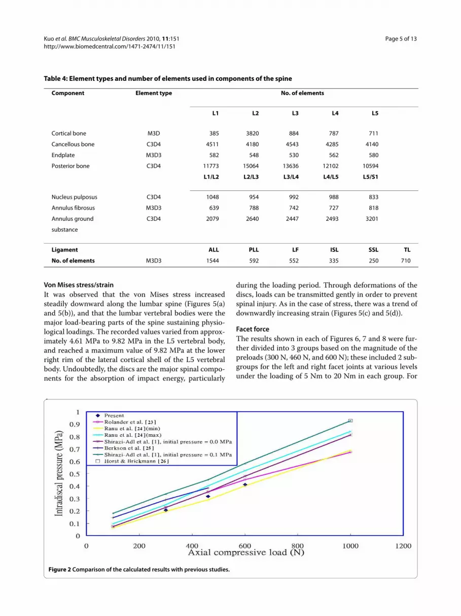
To validate the constructed model, we compared thecalculated intradiscal pressures of the L2/L3 disc, underthe preload of 300N, 460 N and 600N respectively overthe superior surface of the L1 vertebral body in a standingposture, with those reported in the literature [1,23-26](Figure 2), and found that the linearity with compressiveload is in agreement with their studies. Although the cal-culated data in this study appears to be relative lower
than most of the previous results, it could be interpretedreasonably from the fact that the stronger homogeneousannulus fiber elements were used in this study, and thesensitivity analysis for the intradiscal pressure versus theinner fiber strength at the level L2/L3, under preload460N, is shown in Figure 3. The changes in intradiscalpressures at various levels in different postures alsoexhibit the similarity when compared to the research byRohlmann et al. [9] as shown in Figure 4.
ResultsThe numerical results were principally concerned withfacet joint forces at various levels of the lumbar spine andthe intradiscal pressures in the discs under different pre-loads. In addition, the von Mises stresses/strains in thelumbar spine in the standing position were also studiedfor analysis.
Table 2: Properties of the materials used in this study
Material Young's modulus Poisson ratio Reference
E (MPa) ν
Vertebra
Cortical bone 12000 0.3 [21,27-32]
Cancellous bone 100 0.2 [28-32]
Endplate 12000 0.3 [12,27,29]
Posterior elements 3500 0.25 [18,21,30,33,34]
Disc
Nucleus pulposus 1 0.4999 [16,30,35,36]
Annulus ground substance 4.2 0.45 [27-30,36-38]
Fiber
(inner) 360 0.3 [29,39] 360~550(E)
(outer) 550 0.3 [27,40] 450(E)
Table 3: Properties of the ligaments used in this study
Ligament ALL PLL LF ISL SSL TL
Elastic modulus (small strain) (MPa) 7.8 10 15 10 8 10
Transition strain (%) 12 11 6.2 14 20 18
Elastic modulus (large strain) (MPa) 20 50 19.5 11.6 15 59
Cross-sectional area (mm2) 53 16 67 26 23 1.8
Length (mm) 13 11 19 13 11 22
Max. failure load (N) 510 384 340 130 200 70
ALL, Anterior Longitudinal LigamentTL, Transverse LigamentSSL, SupraSpinous LigamentPLL, Posterior Longitudinal LigamentLF, Ligamentum FlavumISL, InterSpinous Ligament
Kuo et al. BMC Musculoskeletal Disorders 2010, 11:151http://www.biomedcentral.com/1471-2474/11/151
Page 5 of 13
Von Mises stress/strainIt was observed that the von Mises stress increasedsteadily downward along the lumbar spine (Figures 5(a)and 5(b)), and that the lumbar vertebral bodies were themajor load-bearing parts of the spine sustaining physio-logical loadings. The recorded values varied from approx-imately 4.61 MPa to 9.82 MPa in the L5 vertebral body,and reached a maximum value of 9.82 MPa at the lowerright rim of the lateral cortical shell of the L5 vertebralbody. Undoubtedly, the discs are the major spinal compo-nents for the absorption of impact energy, particularly
during the loading period. Through deformations of thediscs, loads can be transmitted gently in order to preventspinal injury. As in the case of stress, there was a trend ofdownwardly increasing strain (Figures 5(c) and 5(d)).
Facet forceThe results shown in each of Figures 6, 7 and 8 were fur-ther divided into 3 groups based on the magnitude of thepreloads (300 N, 460 N, and 600 N); these included 2 sub-groups for the left and right facet joints at various levelsunder the loading of 5 Nm to 20 Nm in each group. For
Table 4: Element types and number of elements used in components of the spine
Component Element type No. of elements
L1 L2 L3 L4 L5
Cortical bone M3D 385 3820 884 787 711
Cancellous bone C3D4 4511 4180 4543 4285 4140
Endplate M3D3 582 548 530 562 580
Posterior bone C3D4 11773 15064 13636 12102 10594
L1/L2 L2/L3 L3/L4 L4/L5 L5/S1
Nucleus pulposus C3D4 1048 954 992 988 833
Annulus fibrosus M3D3 639 788 742 727 818
Annulus ground C3D4 2079 2640 2447 2493 3201
substance
Ligament ALL PLL LF ISL SSL TL
No. of elements M3D3 1544 592 552 335 250 710
Figure 2 Comparison of the calculated results with previous studies.

Kuo et al. BMC Musculoskeletal Disorders 2010, 11:151http://www.biomedcentral.com/1471-2474/11/151
Page 6 of 13
example, in the case of extension postures (Figure 6),under preloads of 300 N, 460 N, and 600 N and differentloadings from 5 Nm to 20 Nm by an increment of 5 Nm,Figures 6(a) and 6(b), Figures 6(c) and 6(d), and Figures6(e) and 6(f ) respectively, denote the facet joint forcesbetween the left and right facet joints.
The data shown in Figures 6, 7 and 8 indicate that thefacet joint forces at various levels of the lumbar spine
under different loadings, in extension, and with left/rightaxial rotations, were affected slightly by the preloads inthe range of 300 N to 600 N, particularly in right rotation,and that they increased comparatively little (contrast Fig-ure 6(a) with Figures 6(c) and 6(e); contrast Figure 6(b)with Figures 6(d) and 6(f ); similarly for Figures 7 and 8).However, the differences of facet joint forces between theleft and right facet joints varied with the preload in exten-
Figure 3 Sensitivity analysis for the intradiscal pressure versus the inner fiber strength, under preload 460N, at level L2/L3.
Figure 4 The changes in intradiscal pressure in the (a) L1/L2 and (b) L4/L5 disc of present study and Rohlmann et al. [9]for the intact lumbar spine.

Kuo et al. BMC Musculoskeletal Disorders 2010, 11:151http://www.biomedcentral.com/1471-2474/11/151
Page 7 of 13
sion and left rotation, particularly in the right joints. Italso appeared that there was an asymmetric behaviourbetween left and right rotations for both the left and rightfacet joints at each level, irrespective of the type of load-ing that was applied. This could be observed from thatthe magnitudes of facet joint forces (75.95 N and 63.42Nin left joint at levels L2/L3 and L3/L4); however, at thecorresponding levels, the values were 112.08 N and123.31 N in the right joints under an extension momentof 5 Nm and a preload of 300 N. When the extensionmoments was increased from 5 Nm to 20 Nm, the resultsindicated that the facet joint forces had larger values atlevels L2/L3 and L3/L4 than at levels L1/L2 and L4/L5(Figures 6(a) and 6(b)), and increased with an increase inthe applied moment. At the same level and under thesame extension moment, facet joint forces in the rightjoints were larger than those in left joints (contrast Fig-ures 6(b), (d) and 6(f ) with Figures 6(a), (c) and 6(e),respectively; similarly for Figure 7). Furthermore, if thelumbar spine was loaded by a left rotation moment, the
right (opposite) joint had a larger facet force than the ipsi-lateral (left) joint at the corresponding level (contrast Fig-ure 7(a) with Figure 7(b); contrast Figure 7(c) with Figure7(d), etc.), and vice versa for the case of right rotation(contrast Figure 8(a) with Figure 8(b); contrast Figure 8(c)with Figure 8(d), etc.). We therefore observed that apply-ing bending or rotation moments to the lumbar spineplayed a more important role than the applied preload onthe facet force.
Intradiscal pressureFigures 9(a), (b) and 9(c) show the intradiscal pressures ofthe nuclei pulposi at various levels under preloads of 300N, 460 N, and 600N and at different loadings. From thesefigures, the calculated data for intradiscal pressuresincreased with preload. In an upright standing posture,the average pressure at all levels (L1/L2 to L4/L5) wasapproximately 0.2 MPa under a preload of 300 N, and was0.324 MPa and 0.42275 MPa under preloads of 460 N and600 N, respectively. In flexion, the pressure increased
Figure 5 (a) Front view/(b) Back view of von Mises stress distribution and (c) front view/(d) back view of von Mises strain distribution in the lumbar spine (without ligaments) under an evenly distributed load of 460 N over the superior surface of the L1 vertebral body in a standing posture.

Kuo et al. BMC Musculoskeletal Disorders 2010, 11:151http://www.biomedcentral.com/1471-2474/11/151
Page 8 of 13
noticeably compared with other postures and had a valueof approximately 0.9 MPa at level L1/L2 under a forwardbending (flexion) moment of 20 Nm. In the case of leftand right rotations, the intradiscal pressures were rela-tively larger at level L1/L2 than those at other levelsunder the same loading and different preloads. For theleft rotation, levels L1/L2 and L4/L5 had values higherthan levels L2/L3 and L3/L4. Level L2/L3 did not appearto be affected by the left/right rotation postures andmaintained a value of approximately 0.2 MPa when sub-jected to a rotation moment of less than 15 Nm. Similarlyresults were obtained for level L3/L4 under a rotationmoment of less than 10 Nm. As in the case of extension,
initially the intradiscal pressures at levels L2/L3 and L3/L4 were reduced temporarily, then after the backwardbending moment exceeded 10 Nm or 15 Nm, the pres-sures gradually increased.
DiscussionIn this study, we developed a realistic model preservingthe complex geometry of the posterior parts of the lum-bar spine in order to investigate the relevant biomechani-cal behaviour and to examine whether the asymmetricresponses increase with loading, and if even larger loadsamplify the effect of asymmetry. The facet geometry ofour study model was obtained through stacking the bony
Figure 6 Facet joint forces at various levels under different combinations of preloads and loadings in extension.
Figure 7 Facet joint forces at various levels under different combinations of preloads and loadings in left rotation.

Kuo et al. BMC Musculoskeletal Disorders 2010, 11:151http://www.biomedcentral.com/1471-2474/11/151
Page 9 of 13
outline of each DICOM image file from scanning a lum-bar spine specimen (1 mm space apart in the CT seriesimages), and there existed tiny asymmetries about thesagittal plane of the spine if we inspected the specimencarefully. Even when we examined the shapes of some ofthe investigated models, asymmetry also appeared tosome extent. In addition, most of the related studies havehypotheses about symmetric behaviour. The spinal bonesdid not seem to remain absolutely symmetric during thegrowth period due to the frequent variations in the spinaldevelopment environment, and, as the vertebral bone ofthe spine is alive, it adapts itself to such changes. Because
of the lack of the geometric dimensions used in therelated experimental data, different loading and bound-ary conditions, various curvatures of the facet joints, andso on in the related research, it is difficult to make exactcomparisons between our results and those of other stud-ies. Therefore, in most instances, we present only thetrends in the responses of the lumbar spine observed inthe present analysis.
From Figures 5(a), (b), (c) and 5(d), it can be seen thatthe lower lumbar spine has a larger stress or strain distri-bution. From the data obtained in our simulation, theresults show that the asymmetry is gradually more obvi-
Figure 8 Facet joint forces at various levels under different combinations of preloads and loadings in right rotation.
Figure 9 Intradiscal pressures at various levels of the lumbar spine under preloads of (a) 300 N, (b) 460 N, and (c) 600 N and different load-ings, including forward/backward bending moments and left/right rotation moments of 5 Nm, 10 Nm, 15 Nm, and 20 Nm.

Kuo et al. BMC Musculoskeletal Disorders 2010, 11:151http://www.biomedcentral.com/1471-2474/11/151
Page 10 of 13
ous with preloading in the right joints under extensionposture if we compare Figures 6(b), (d) and 6(f ), whilethere are few variations with preloading in the left jointsseen in Figures 6(a), (c) and 6(e). Similarly for the case ofleft rotation, if we examine Figures 7(b), (d) and 7(f ) andanother group of Figures 7(a), (c) and 7(e), the sameresponse modes appear. However, in Figures 8(a), (c) and8(e) or Figures 8(b), (d) and 8(f ), there are few changes inleft or right facets, i.e., the asymmetry is not evident withpreloading, but it increases with the applied moment.These results made us associate this asymmetry with thegeometric defects in the right facet joint, no matter whatthe cause originated from the specimen geometry or themanual work in depicting the bony outlines. The asym-metry diminished if the lumbar spine rotated to the rightand decreased the contact area between the right facetsurfaces.
In order to validate the constructed model, we rear-ranged the results shown in Figure 9(b) by separating lev-els L1/L2 to L4/L5 for the intradiscal pressures of thelumbar spine under a preload of 460 N and different load-ings into two parts: levels L1/L2 and L4/L5 in Figure10(a), and levels L2/L3 and L3/L4 in Figure 10(b). Thecalculated data shown in Figures 10(a) and 10(b) appearto exhibit a trend similar to that for the intact lumbarspine reported in the literature [9,18], in which a totalload of 460 N resulting from an applied load of 260 N rep-resenting the weight of upper body was adopted, togetherwith a compressive follower load of 200 N representingthe stabilizing effect of the local muscle forces. In addi-tion to the above loadings, Rohlmann et al. [9] employedthe following physiological loadings: (1) standing, 30°flexion (forward bending), 15° extension of the lumbarspine, and 6° torsion (axial rotation); and (2) standing, 30°
flexion, 20° extension, and 10° torsion [18]. In the case ofstanding, the intradiscal pressures at levels L1/L2 and L4/L5 calculated in the present study were 0.351 MPa and0.349 MPa, respectively. However, Rohlmann et al.obtained higher values of approximately 0.61 MPa and0.58 MPa at the corresponding levels. This disparity canbe attributed to an additional force in the erector spinaeor the rectus abdominis, and/or the different manner ofapplying loads used in the previous studies, as well as thestronger homogeneous annulus fiber elements that wereused in our study. An additional sensitivity analysis wasconducted to validate our assumption, as shown in Figure3. In the cases of other postures, intradiscal pressures hadlarger values in flexion rather than in extension and left/right rotations, particularly at level L1/L2. There was asimilar trend of changes with loading in flexion, exten-sion, and left/right rotations between the present analysisand that reported in the literature [9,18]. A further trendcan be observed in Figure 10(b); namely, that the pres-sures at levels L2/L3 and L3/L4 were clearly reduced inthe case of extension under moments of 5 Nm to 10 Nmor 15 Nm. This phenomenon might be interpreted as avariation in the curvature of the lumbar spine at differentlevels. The magnitude of the moments appeared toslightly alter the curvature of the spine at level L2/L3 orL3/L4 during the period of extension, whereas the pres-sure had a large value at level L1/L2. The latter observa-tion could be related to the backward bending momentthat was applied over the superior surface of the L1 verte-bral body nearing the disc most closely at level L1/L2. Inaddition, the intradiscal pressures in the nuclei pulposiincreased with increasing magnitude of preloads (Figures9(a), (b) and 9(c)), and this appeared to be a reasonableoutcome for the general physiological loading cases.
Figure 10 Intradiscal pressures at (a) levels L1/L2 and L4/L5, and (b) levels L2/L3 and L3/L4 under a preload of 460 N.

Kuo et al. BMC Musculoskeletal Disorders 2010, 11:151http://www.biomedcentral.com/1471-2474/11/151
Page 11 of 13
For the facet joint forces at various levels of the lumbarspine under a preload of 460 N and left/right rotationmoments of 5 Nm and 10 Nm, Figure 11 indicates thatforces increased with increasing axial rotation momentand were higher in the contralateral facet joints than inthe ipsilateral joints, as reported by Shirazi-Adl's study[20], particularly for levels L2/L3 and L3/L4 in the case ofleft rotation. It was also observed that the facet jointforces in the left and right facet joints exhibited asymmet-ric behaviour in left/right rotation, as also reported byShirazi-Adl and Parnianpour [19]. For example, the facetjoint forces at levels L2/L3 to L4/L5 in the right jointsunder a left rotation moment of 5 Nm differed consider-ably from those at the corresponding levels in the samejoints under right rotation. Similar patterns wereobserved for level L2/L3 in the left joint under a rotationmoment of 10 Nm, and at levels L2/L3 to L4/L5 in theright joint under a rotation moment of 10 Nm.
In addition to the above arguments concerning theasymmetric response of facet joint forces, there were dif-ferences between left and right facet joints at each level ofthe lumbar spine for extension and left/right rotation.The facet force at level L3/L4 did not have a higher valuethan that at level L2/L3 until the backward (extension)moment reached 20 Nm (Figure 6(a)); however, in theright facet joints, the facet force at level L3/L4 alwaysslightly exceeded that at level L2/L3.
As to the asymmetric behaviours in left and right rota-tion in our model, they could be due to the small inherentasymmetry in the specimen geometry or some manualerrors in construction process of the bony outlines. Theasymmetric behaviour due to the geometrical factor inthe present model did not appear to be expressed as some
mathematical relation, such as a linear relation, squaredrelation, or some combination of these. And the forma-tion of the asymmetry was not attributed to a single fac-tor, as geometrical factors, vertebral body dimensions,facet joints, endplates, pedicles, alignment of the verte-bral bodies, or loading condition factors, like appliedforce, moment, torque, even the constraint (support)condition, for example, the lumbar spine with one sidefacet resection, would all affect the global asymmetricbehaviour. How to quantify the asymmetric behaviourwas thus really a complicated process that deserves fur-ther attention in future work. The effect of asymmetricloading of the facet joints with respect to the saggitalplane in left and right rotation would be amplified withlarger applied loads, because the magnitude of themoment (or torque) is the product of force and length ofarm of force. In addition, the real physiological responseor mechanical behaviour of the spine with activatingcomponents like soft tissues, ligaments, tendons, or mus-cles could differ to some extents from that of the FEmodel purely derived from the bony outline, without tak-ing into account muscular tissues. Thus, how to improvethe asymmetry might be another issue for futureresearchers to investigate, as the image processing of thebony outlines of the lumbar spine was a tedious manualtask. Even if the specimen had inherent defects or theoutlines of facet joints, which were depicted and modifiedby hand, were somewhat incorrect, the correction workcannot be finished without further FE preprocessing andexecution by the FE program. From the anatomical pointof view, our model was obtained through stacking thebony outlines of vertebral bones which were molded froma human cadaver which, to the best of the authors' knowl-
Figure 11 Facet joint forces at various levels under a preload of 460 N and left/right rotation moments of 5 Nm and 10 Nm, (L) and (R) de-note left facet joint and right facet joint, respectively.
Kuo et al. BMC Musculoskeletal Disorders 2010, 11:151http://www.biomedcentral.com/1471-2474/11/151
Page 12 of 13
edge, was without bony defects prior to the process ofproducing the lumbar specimen. In addition, unless thereis sufficient spinal dimensions data available to make astatistical analysis and conclude that the real situation ofmost of the human spine is symmetric with respect to thesagittal plane of the spine, the tiny asymmetries about thesagittal plane of the spine can not be avoided in the modelconstruction. When only the simulation data has beencorrected and validated, it is hard to conclude whetherthe asymmetry is due to only the manual errors or theinherent defects. This issue deserves further attention infuture research. As long as the model has few shifts fromthe symmetric geometry, and there is a larger appliedload, without considering the effect of muscles balancingthe left and right facet joints, the asymmetry effect doesnot seem to be easily removed.
To the best of the authors' knowledge, whether theasymmetry would alter the coupled motion or not isrelated to factors such as the speed of movement of thespine, the strength of muscles attaching to the spinalbones in individuals, forces in the erector spinae or rectusabdominis, the relative sliding smoothness of spinal jointsand so on. So to some extent the asymmetry in the pres-ent model would make a difference to the action behav-iour with regard to the sagittal plane and to the responsesin left and right joints, and thus would also affect the cou-pled motion, which associates lateral bending in left andright directions with horizontal (axial) rotation.
ConclusionsThe results suggest that von Mises stresses/strainsresponded to a preload of 460 N with higher values in thelower part of the lumbar spine. Intradiscal pressures inthe nuclei pulposi increased with preload and increasedmore noticeably with flexion than with extension or axialrotation. In extension postures, pressures were reduced atlevels L2/L3, L3/L4, and L4/L5 under different preloads.With regard to the facet joint forces, forward/backwardbending and left/right axial rotations produced asymmet-ric responses in the facet joints. Left axial rotationresulted in a larger facet force in the contralateral (right)facet joint than that in the ipsilateral (left) joint at thesame level, and vice versa. Moreover, it also appeared thatthe influence of the magnitude of preloads on the facetforce was less important than that due to the various pos-tures. In addition, the inherent geometric asymmetry thatexists in the model or coupled motion in the spine is apossible influencing factor with regard to the results, andthis should be considered carefully in future studies.
Competing interestsThe authors declare that they have no competing interests.
Authors' contributionsCSK participated in the study design, in collecting the data and drafting of themanuscript. HTH, RML, and KYH participated in the study design. PCL and ZCZ
participated in revising the manuscript. MLH advised and assisted drafting ofthe manuscript. All authors read and approved the final manuscript.
AcknowledgementsThe CT image scans for the FE model were acquired with the support of the faculty of the Department of Diagnostic Radiology, National Cheng Kung Uni-versity Medical Center. The authors would like to express their sincere gratitude for this assistance.
Author Details1Department of Civil Engineering, National Cheng Kung University, Tainan, Taiwan, 2Center for General Education, Nan Jeon Institute of Technology, Yenshui, Taiwan, 3Department of Orthopaedics, National Cheng Kung University Hospital, Tainan, Taiwan, 4National Cheng Kung University Hospital Dou-Liou Branch, Douliou, Taiwan, 5Institute of Clinical Medicine, College of Medicine, National Cheng Kung University, Tainan, Taiwan and 6Department of Physical Therapy and Assistive Technology, National Yang-Ming University, Taipei, Taiwan
References1. Shirazi-Adl A, Shrivastava SC, Ahmed AM: Stress analysis of the lumbar
disc-body unit in compression, a three-dimensional nonlinear finite element study. Spine 1984, 9(2):120-134.
2. Lee CK, Kim YE, Lee CS, Hong YM, Jung J, Goel VK: Impact response of the intervertebral disc in a finite element model. Spine 2000, 25(19):2431-2439.
3. Wang JL, Parnianpour M, Shirazi-Adl A, Engin AE: Viscoelastic finite ele-ment analysis of a lumbar motion segment in combined compression and sagittal flexion. Spine 2000, 25(3):310-318.
4. Cao KD, Grimm MJ, Yang KH: Load sharing within a human lumbar vertebral body using the finite element method. Spine 2001, 26(12):E253-E260.
5. Zhong ZC, Wei SH, Wang JP, Feng CK, Chen CS, Yu CH: Finite element analysis of the lumbar spine with a new cage using a topology optimization method. Med Engng Phys 2006, 28:90-98.
6. Cheung JT-M, Zhang M, Chow DH-K: Biomechanical responses of the intervertebral joints to static and vibrational loading: a finite element study. Clin Biomech 2003, 18:790-799.
7. Zander T, Rohlmann A, Bergmann G: Influence of ligament stiffness on the mechanical behaviour of a functional spinal unit. J Biomech 2004, 37:1107-1111.
8. Chen CS, Feng CK, Cheng CK, Tzeng MJ, Liu CL, Chen WJ: Biomechanical analysis of the disc adjacent to posterolateral fusion with laminectomy in lumbar spine. J Spinal Disord 2005, 18(1):58-65.
9. Rohlmann A, Zander T, Bergmann G: Comparison of the biomechanical effects of posterior and anterior spine stablizing implants. Eur Spine J 2005, 14(5):445-453.
10. Nachemson A, Morris JM: In vivo measurements of intradiscal pressure. J Bone Joint Surg Am 1964, 46:1077-1092.
11. Nachemson AL: Disc pressure measurements. Spine 1981, 6:93-97.12. Wilke HJ, Neef P, Caimi M, Hoogland T, Claes L: New intradiscal pressure
measurements in vivo during daily activities. Spine 1999, 24:755-762.13. Brinckman P: Injury of the anulus fibrosus and disc protrusions: An in
vitro investigation on human lumbar discs. Spine 1986, 11:149-153.14. Pintar FA, Yoganandan N, Myers T, Elhagediab A, Sances A: Biomechanical
properties of human lumbar spine ligaments. J Biomech 1992, 25(11):1351-1356.
15. McNally DS, Adams MA: Internal intervertebral disc mechanics as revealed by stress profilometry. Spine 1992, 17(1):66-73.
16. Rohlmann A, Zander T, Schmidt H, Wilke HJ, Bergmann G: Analysis of the influence of disc degeneration on the mechanical behaviour of a lumbar motion segment using the finite element method. J Biomech 2006, 39:2484-2490.
17. Zander T, Rohlmann A, Burra NK, Bergmann G: Effect of a posterior dynamic implant adjacent to a rigid spinal fixator. Clin Biomech 2006, 21:767-774.
18. Rohlmann A, Burra NK, Zander T, Bergmann G: Comparison of the effects of bilateral posterior dynamic and rigid fixation devices on the loads in the lumbar spine. Eur Spine J 2007, 16(8):1223-1231.
Received: 10 December 2009 Accepted: 5 July 2010 Published: 5 July 2010This article is available from: http://www.biomedcentral.com/1471-2474/11/151© 2010 Kuo et al; licensee BioMed Central Ltd. This is an Open Access article distributed under the terms of the Creative Commons Attribution License (http://creativecommons.org/licenses/by/2.0), which permits unrestricted use, distribution, and reproduction in any medium, provided the original work is properly cited.BMC Musculoskeletal Disorders 2010, 11:151

Kuo et al. BMC Musculoskeletal Disorders 2010, 11:151http://www.biomedcentral.com/1471-2474/11/151
Page 13 of 13
19. Shirazi-Adl A, Parnianpour M: Load-bearing and stress analysis of the human spine under a novel wrapping compression loading. Clin Biomech 2000, 15:718-725.
20. Shirazi-Adl A: Analysis of large compression loads on lumbar spine in flexion and in torsion using a novel wrapping element. J Biomech 2006, 39:267-275.
21. Polikeit A, Ferguson SJ, Nolte LP, Orr TE: Factors influencing stresses in the lumbar spine after the insertion of intervertebral cages: finite element analysis. Eur Spine J 2003, 12:413-420.
22. Ramos A, Simoes JA: Tetrahedral versus hexahedral finite elements in numerical modelling of the proximal femur. Med Engng Phys 2006, 28:916-924.
23. Rolander SD: Motion of the lumbar spine with special reference to the stablizing effect of posterior fusion. Acta Orthop Scand 1966, 90(Suppl):1-144.
24. Ranu HS, Denton RA, King AI: Pressure distribution under an inter - vertebral disc - an experimental study. J Biomech 1979, 12:807-812.
25. Berkson MH, Nachemson AL, Schultz AB: Mechanical properties of human lumbar spine motion segments. Part II. Response in compression and shear, influence of gross morphology. J Biomech Eng 1979, 101:53-57.
26. Horst M, Brinckmann P: Measurement of the distribution of axial stress on the endplate of the vertebral body. Spine 1981, 6:217-232.
27. Pitzen T, Geisler FH, Matthis D, Storz HM, Pedersen K, Steudel WI: The influence of cancellous bone density on load sharing in human lumbar spine: a comparison between an intact and a surgically altered motion segment. Eur Spine J 2001, 10:23-29.
28. Natarajan RN, Andersson GBJ: Modeling the annular incision in a herni-ated lumbar intervertebral disc to study its effect on disc stability. Compu Struc 1997, 64(5-6):1291-1297.
29. Denozi'ere G: Numerical modeling of a ligamentous lumber motion segment. M.S. thesis, Department of Mechanical Engineering, Georgia Institute of Technology, Georgia, USA; 2004.
30. Sairyo K, Goel VK, Masuda A, Vishnubhotla S, Faizan A, Biyani A, Ebraheim N, Yonekura D, Murakami RI, Terai T: Three-dimensional finite element analysis of the pediatric lumbar spine. Eur Spine J 2006, 15(6):923-929.
31. Goel VK, Ramirez SA, Kong WZ, Gilbertson LG: Cancellous bone Young's modulus variation within the vertebral body of a ligamentous lumbar spine-application of bone adaptive remodeling concepts. J Biomech Eng 1995, 117:266-271.
32. Goto K, Tajima N, Chosa E, Totoribe K, Kuroki H, Arizumi Y, Takashi A: Mechanical analysis of the lumbar vertebrae in a three-dimensional finite element method model in which intradiscal pressure in the nucleus pulposus was used to establish the model. J Orthop Sci 2002, 7:243-246.
33. Rohlmann A, Zander T, Bergmann G: Effect of total disc replacement with ProDisc on the biomechanical behaviour of the lumbar spine. Spine 2005, 30(7):738-743.
34. Shirazi-Adl A, Ahmed AM, Shrivastava SC: Mechanical response of a lum-bar motion segment in axial torque alone and combined with compression. Spine 1986, 11(9):914-927.
35. Ng HW, Teo EC: Nonlinear finite-element analysis of the lower cervical spine (C4-C6) under axial loading. J Spinal Disord 2001, 14(3):201-210.
36. Smit T, Odgaard A, Schneider E: Structure and function of vertebral trabecular bone. Spine 1997, 22(24):2823-2833.
37. Sharma M, Langrana NA, Rodriguez J: Role of ligaments and facets in lumbar spinal stability. Spine 1995, 20(8):887-900.
38. Lee KK, Teo EC: Effects of laminectomy and facetectomy on the stability of the lumbar motion segment. Med Engng Phys 2004, 26:183-192.
39. Polikeit A: Finite element analyses of the lumbar spine:clinical application. Inaugural-Dissertation, University of Bern; 2002.
40. Shin G: Viscoelastic responses of the lumbar spine during prolonged stooping. Ph.D. dissertation, NCSU, USA; 2005.
41. White AA, Panjabi MM: Clinical biomechanics of the spine. Philadelphia: J.B. Lippincott; 1990.
42. Kim YE, Goel VK, Weinstein JN, Lim TH: Effect of disc degeneration at one level on the adjacent level in axial mode. Spine 1991, 16(3):331-335.
Pre-publication historyThe pre-publication history for this paper can be accessed here:http://www.biomedcentral.com/1471-2474/11/151/prepub
doi: 10.1186/1471-2474-11-151Cite this article as: Kuo et al., Biomechanical analysis of the lumbar spine on facet joint force and intradiscal pressure - a finite element study BMC Muscu-loskeletal Disorders 2010, 11:151


