Embed Size (px)
Citation preview
University of Vermont University of Vermont
UVM ScholarWorks UVM ScholarWorks
Graduate College Dissertations and Theses Dissertations and Theses
2008
Biophysical Characterization of the Sequsingle-Stranded DNA-Biophysical Characterization of the Sequsingle-Stranded DNA-
Binding Properties of Mouse Pur : a Repressor of Smooth Muscle Binding Properties of Mouse Pur : a Repressor of Smooth Muscle
-Actin Gene Expression -Actin Gene Expression
Jon Ramsey University of Vermont
Follow this and additional works at: https://scholarworks.uvm.edu/graddis
Recommended Citation Recommended Citation Ramsey, Jon, "Biophysical Characterization of the Sequsingle-Stranded DNA-Binding Properties of Mouse Pur : a Repressor of Smooth Muscle -Actin Gene Expression" (2008). Graduate College Dissertations and Theses. 189. https://scholarworks.uvm.edu/graddis/189
This Dissertation is brought to you for free and open access by the Dissertations and Theses at UVM ScholarWorks. It has been accepted for inclusion in Graduate College Dissertations and Theses by an authorized administrator of UVM ScholarWorks. For more information, please contact [email protected].
BIOPHYSICAL CHARACTERIZATION OF THE SEQUENCE-SPECIF IC SINGLE-STRANDED DNA-BINDING PROPERTIES OF MOUSE PURββββ: A REPRESSOR OF SMOOTH MUSCLE αααα-ACTIN GENE EXPRESSION
A Dissertation Presented
by
Jon Eric Ramsey
to
The Faculty of the Graduate College
of
The University of Vermont
In Partial Fulfillment of the Requirements for the Degree of Doctor of Philosophy
Specializing in Biochemistry
October, 2008

Accepted by the Faculty of the Graduate College, The University of Vermont, in partial fulfillment of the requirements for the degree of Doctor of Philosophy specializing in Biochemistry.
Thesis Examination Committee:
Advisor ~ b b & J. Kelm ~ r - 3 ~ ~ .
an- Anne B. Mason, Ph.D.
/mPP@&, Chairperson Robert B. Low, Ph. D.
5 , ,. ,L j . , Vice President for Research C Frd~ced ~ . d and Dean of Graduate Studies
Date: August 29th, 2008
ABSTRACT
Regulation of gene transcription by structural interconversions of genomic DNA is an emerging biochemical and genetic paradigm that adds to the already diverse repertoire of eukaryotic gene regulatory mechanisms. The appearance of paranemic structures coincident with changes in gene activity, as well as participation of transcription factors that recognize and bind single-stranded DNA at numerous gene promoters in vivo illustrates the authenticity of this concept and its importance in cellular homeostasis. Despite its acceptance, this concept has been minimally described at the biochemical and biophysical levels, as the means by which sequence-specific single-stranded DNA-binding proteins exert transcriptional influence in double-stranded genomes remains largely undefined.
Purβ is a sequence-specific single-stranded DNA/RNA-binding protein that acts as a repressor of smooth muscle α-actin (SMαA) gene transcription, and mRNA translation. SMαA is an important cytoskeletal protein that contributes contractile, antimigratory, and nonproliferative functions in smooth muscle. In concert with Pur protein family member Purα, and Y-box protein MSY1, Purβ enacts repression of SMαA gene expression by interacting with a cryptic cis-regulatory element in the 5’ region of the SMαA promoter that has been shown to transiently adopt single-stranded conformations in vivo, and to confer transcriptional activation when trans-activator occupied while in a double-stranded conformation. Downregulation of SMαA gene expression has been identified to be a contributing factor to cardiovascular disease progression; therefore a thorough understanding of SMαA repression mechanisms is critical for clinical management of these conditions.
Although highly homologous at the primary sequence level, Purβ and Purα display significant conserved regions of sequence divergence that suggest these paralogs exert distinct cellular functions in various vertebrate classes. A goal of the studies presented herein was to delineate exhibited functional differences with respect to SMαA repression in pertinent mouse cell lines. Loss-of-function and chromatin immunoprecipitation studies verified that Purβ differs from Purα in that Purβ is the dominant Pur protein repressor of SMαA expression in embryonic fibroblasts and vascular smooth muscle cells, although by different, cell type-specific mechanisms.
Biophysical assessment of Purβ single-stranded DNA binding properties showed that despite the ability of Purβ to self-dimerize in the absence of nucleic acid, Purβ binds to the cryptic SMαA enhancer by a sequential and cooperative mechanism, with remarkable affinity and a terminal stoichiometry of 2 to 1. Footprinting and in vitro binding site characterization confirms two Purβ binding sites exist within this element and display slight degeneracy from a proposed Pur protein-binding consensus motif. These findings delineate binding mechanisms adopted by Purβ and provide a means to identify putative Purβ binding sites throughout the genome.

DEDICATION
For Kendra and Cooper……
I owe you both so much for your unconditional love, patience, and enthusiasm. Everyday
you remind me how much I need you. Your support has made all of this possible, without
it I would not have been able to pursue this goal. Your contributions to my success are
beyond words, yet plentiful and essential are certainly ones that come to mind.

iii
ACKNOWLEDGMENTS
First and foremost I wish to thank my advisor Dr. Robert Kelm, Jr. for providing me
with opportunity, support, advice, and freedom to explore new and exciting avenues of
scientific investigation. I would also like to thank other members of the Kelm laboratory
both past and present that helped me during my graduate career: Shu-Xia Wang, Anna
Knapp, and numerous rotation students who helped advance my various projects. I
would like to thank the American Heart Association and Dr. Kenneth G. Mann for
financial support. I would also like to express gratitude to the members of my
committee, Drs. Stephen J. Everse, Anne B. Mason, Scott W. Morrical, Mercedes
Rincon, and Robert B. Low for guiding me through my thesis research. I would like to
thank all members of the Department of Biochemistry, especially Stephen J. Everse,
Anne B. Mason, and Scott W. Morrical for providing me with technical resources, Paula
Tracy for generous advisement, and Margaret Daugherty for employment,
encouragement, and plentiful advice before and during my graduate career. I am also
grateful to members of the Vascular Biology group at the Colchester Research Facility,
Drs. David Schneider, Burton Sobel, Chris Holmes, and Jeffrey Spees for insightful
comments regarding my research. I am deeply indebted to Ewa Folta-Stogniew of Yale
University for her help with light-scattering studies, and to Keith Connaghan and David
Bain of the University of Colorado for extensive and courteous help with the nuts and
bolts of DNase I footprinting and analysis.

iv
None of this would have been possible without the loving support of my wife,
Kendra, who has encouraged me at every step of my pre-doctoral training, my son
Cooper, who is my inspiration, and my parents Jacqueline and Richard and sister Jan for
telling me I could accomplish anything and everything I put my mind to.
Thank You.
v
TABLE OF CONTENTS
DEDICATION ............................................................................................................... ii
ACKNOWLEDGMENTS ............................................................................................. iii
LIST OF TABLES ....................................................................................................... vii
LIST OF FIGURES..................................................................................................... viii
LIST OF USED ABBREVIATIONS..............................................................................xi
CHAPTER I. INTRODUCTION .....................................................................................1
BACKGROUND TO THE PRESENT WORK............................................................1 PHENOTYPIC PLASTICITY OF VASCULAR SMOOTH MUSCLE CELLS ...........2 FUNCTIONAL ASPECTS OF SMOOTH MUSCLE α-ACTIN IN SMOOTH MUSCLE CELL BIOLOGY......................................................................................13 REGULATION OF SMOOTH MUSCLE α-ACTIN GENE EXPRESSION..............15 PUR PROTEIN STRUCTURE AND FUNCTION ....................................................32
THE BREADTH OF PUR PROTEIN FUNCTION................................................32 STRUCTURAL CONSIDERATIONS...................................................................39 NUCLEIC ACID-BINDING PROPERTIES..........................................................48
CHAPTER II. SCOPE OF THE PRESENT WORK .....................................................67
CHAPTER III. PUR PROTEIN LOSS-OF-FUNCTION STUDIES IDENTIFY PURβ
AS A DOMINANT REPRESSOR OF SMαA EXPRESSION.......................................70
INTRODUCTION.....................................................................................................70 MATERIALS AND METHODS ...............................................................................75 RESULTS .................................................................................................................85 DISCUSSION ...........................................................................................................96
CHAPTER IV. SOLUTION CHARACTERIZATION REVEALS THE
QUATERNARY STRUCTURE OF RECOMBINANT MOUSE PURβ ......................102
INTRODUCTION...................................................................................................102 MATERIALS AND METHODS .............................................................................105
vi
RESULTS ...............................................................................................................114 DISCUSSION .........................................................................................................130
CHAPTER V. THERMODYNAMIC ANALYSIS OF SEQUENCE-SPECIFIC
SINGLE-STRANDED DNA-BINDING BY RECOMBINANT PURβ REVEALS A
COOPERATIVE MECHANISM OF NUCLEOPROTEIN ASSEMBLY.....................136
INTRODUCTION...................................................................................................136 MATERIALS AND METHODS .............................................................................143 RESULTS ...............................................................................................................159 DISCUSSION .........................................................................................................190
CHAPTER VI. TOWARDS THE CHARACTERIZATION OF THE MINIMAL
SINGLE-STRANDED DNA BINDING SITE OF PURβ WITHIN THE SMαA
PROXIMAL MCAT ENHANCER ELEMENT...........................................................203
INTRODUCTION...................................................................................................203 MATERIALS AND METHODS .............................................................................206 RESULTS ...............................................................................................................210 DISCUSSION .........................................................................................................213
CHAPTER VII. CONCLUDING REMARKS ............................................................220
COMPREHENSIVE BIBLIOGRAPHY ......................................................................222
vii
LIST OF TABLES
Table 1.1. Oligonucleotides with established interactions with Pur proteins ..................51
Table 1.2. Purα binding properties of mutant c-myc-associated PUR-element
oligonucleotides.............................................................................................................55
Table 4.1 Sedimentation equilibrium data: Parameters from global analysis...............129
Table 5.1. Microscopic configurations and corresponding interaction free energies for a
two site system. ...........................................................................................................154
Table 5.2. Microscopic configurations and corresponding interaction free energies for
multiple models of two site-binding. ............................................................................160
Table 6.1. Oligonucleotides used in this study as fluid-phase competitors in Purβ
ssDNA-binding functional ELISA. ..............................................................................209
viii
LIST OF FIGURES
Figure 1.1. Regulation of SMαA gene expression involves numerous trans-activating
and trans-repressing factor interactions at multiple cis-regulatory elements. ..................17
Figure 1.2. Multiple primary sequence alignments of Purα orthologs versus Purβ
orthologs. ......................................................................................................................41
Figure 1.3. Purα nucleotide preferences within putative origin-associated PUR-elements.
......................................................................................................................................57
Figure 1.4 Nucleotides flanking the core MCAT enhancer are critical for Pur protein
association. ....................................................................................................................60
Figure 3.1. Target positions of Purα- and Purβ-specific shRNA....................................84
Figure 3.2. Specific knockdown of Purα or Purβ by shRNA-mediated RNAi. ...............87
Figure 3.3. De-repression of the SMαA promoter by Purβ shRNA................................91
Figure 3.4. De-repression of the SMαA promoter in response to knockdown of Purα
and/or Purβ and the requirement for Pur/Pyr element integrity.......................................94
Figure 3.5. Relative Levels of Purα, Purβ, and MSY1 occupancy at the SMαA promoter
by ChIP. ........................................................................................................................97
Figure 4.1. Expression and purification of N-HisPurβ..................................................115
ix
Figure 4.2. Molecular size measurements of N-HisPurβ in solution by light scattering
techniques....................................................................................................................117
Figure 4.3. Sedimentation velocity analysis of N-HisPurβ. ..........................................121
Figure 4.4. Analysis of N-HisPurβ sedimentation velocity by direct fitting of time-
resolved concentration difference curves......................................................................123
Figure 4.5. Analysis of concentration-dependant N-HisPurβ solution non-ideality by
sedimentation equilibrium. ..........................................................................................126
Figure 4.6. Sedimentation equilibrium analysis of N-HisPurβ......................................128
Figure 4.7. Species plot of N-HisPurβ..........................................................................132
Figure 5.1. Schematic of cis-regulatory elements of the SMαA promoter....................142
Figure 5.2. Extent of recombinant and nuclear extract-derived Purβ binding to PE32-F.
....................................................................................................................................162
Figure 5.3 Serial dilution EMSA determination of the N-HisPurβ:PE32-F* nucleoprotein
complex stoichiometry.................................................................................................169
Figure 5.4. Quantitative electrophoretic mobility shift assay of N-HisPurβ binding to
PE32-F*. .....................................................................................................................171
Figure 5.5. Quantitative DNase I footprinting of N-HisPurβ:SMP382-F* interactions.176
x
Figure 5.6. N-HisPurβ binds to the SMαA MCAT enhancer element via a cooperative 3’
to 5’ mechanism. .........................................................................................................181
Figure 5.7. Monte Carlo error simulations to assess model confidence. .......................184
Figure 5.8. Monte Carlo error simulations to assess converged parameter confidence. 187
Figure 5.9. N-HisPurα and N-HisPurβ bind supercoiled and linearized plasmids VSMP8
and pBLCAT3. ............................................................................................................189
Figure 5.10. Comparison of quantitative footprinting-resolved parameters to individual
quantitative electrophoretic mobility shift species isotherms. .......................................194
Figure 6.1. Analysis of N-HisPurβ:PE32-F nucleotide interactions by truncated
oligonucleotide competition.........................................................................................212
Figure 6.2. Analysis of N-HisPurβ:PE32-F nucleotide interactions by scanning
oligonucleotide competition.........................................................................................214
Figure 6.3. Recognition and binding of N-HisPurβ to the purine-rich strand of the SMαA
cryptic MCAT enhancer element. ................................................................................217
xi
LIST OF USED ABBREVIATIONS
αMHC, α-myosin heavy chain; ANF, atrial natriuretic factor; ATP, adenosine
triphospate; bHLH, basic helix-loop-helix; bp, basepair; BPV, bovine papilloma virus;
BSA, bovine serum albumin; CArG, 5’- CC(A/T)6GG-3’ element; AT, chloramphenicol
acetyl transferase; CE, coding element; dhfr, dihydrofolate reductase; DLS, dynamic
light-scattering; DNA, deoxyribonucleic acid; dsDNA, double-stranded deoxyribonucleic
acid; dsRNA, double-stranded ribonucleic acid; DTT, dithiothreitol; EDTA,
ethylendiamine tetraacetic acid; ELISA, enzyme-linked immunosorbent assay; EMSA,
electrophoretic mobility shift assay; HeLa, Henrietta Lacks cervical cancer cell line; HIV,
human immunodeficiency virus; HJCV, human John Cunningham virus; hnRNP,
heterogeneous nuclear ribonucleoprotein; LCE, lytic control element; LLS, laser light
scattering; MBP, myelin basic protein; MCAT, muscle-specific 5’-CATTCCT-3’ box;
MEF, mouse embryonic fibroblast; mRNP, messenger ribonucleoprotein; MSY1, mouse
Y-box protein 1; NRE, negative regulatory element; nt, nucleotides; OB fold,
oligonucleotide/oligosaccharide-binding fold; PCR, polymerase chain reaction; PDGF,
platelet-derived growth factor; PE, promoter element; PNR, purine-rich negative
regulatory element; pRb, retinoblastoma protein; PrM, purine-rich motif; PUR, purine-
rich Pur protein-binding element; RI, refractive index; RNA, ribonucleic acid; SEC, size-
exclusion chromatography; SMαA, smooth muscle α-actin; SMC, smooth muscle cell;
snRNA, small nuclear ribonucleic acid; SRF, serum response factor; SRP, signal
recognition particle; SSB, single-stranded deoxyribonucleic acid-binding protein;
ssDNA, single-stranded deoxyribonucleic acid; ssRNA, single-stranded ribonucleic acid;
xii
TCE, transforming growth factor β1-control element; TEF-1, transcriptional enhancer
factor 1; TGF-β1, transforming growth factor β1; THR, transforming growth factor β1-
hypersensitive region; USF, upstream stimulatory factor; UTR, untranslated region; UV,
ultraviolet; VACssBF, vascular actin single-stranded deoxyribonucleic acid-binding
factor; VSMC, vascular smooth muscle cell; YB-1, Y-box protein 1.
1
CHAPTER I. INTRODUCTION
BACKGROUND TO THE PRESENT WORK
Investigations into cellular processes that contribute to tissue remodeling associated
with development, response to injury, and disease progression have determined that these
courses are the result of phenotypic modulation of cells resident to remodeled areas.
Sequence-specific single-stranded DNA/RNA-binding transcription/translation factor
Purβ (Pur protein isoform β), has been implicated in the phenotypic modulation of
vascular smooth muscle cells that become activated and phenotypically reprogrammed
during vessel wall remodeling associated with arteriosclerotic disease progression. A
cause and effect relationship between vascular smooth muscle cell phenotype-switching
and cytoskeletal protein smooth muscle α-actin expression has been experimentally
established. Repression of smooth muscle α-actin, accomplished in part by Purβ, has
been linked to activation, migration, proliferation, and hypersynthetic properties of
resident smooth muscle cells at sites of vessel wall remodeling. Contrastingly,
derepression, or activation of smooth muscle α-actin expression is coincident to injury-
induced myofibroblast activation and adoption of contractile properties necessary for
wound closure and resolution by these cells. The potential involvement of Purβ in these
pathophysiological processes have made this protein an important target of investigation
for understanding dynamic smooth muscle α-actin expression in phenotypically
reprogrammed cells, as well as a model for understanding sequence-specific single-
stranded DNA-protein interactions.
2
The following is a review of the existing literature regarding the various physiological
and pathological processes that rely heavily on vascular smooth muscle cell phenotype
plasticity, molecular mechanisms governing dynamic smooth muscle α-actin expression,
and Pur protein structural/functional properties as they relate to these important aspects of
vascular smooth muscle biology.
PHENOTYPIC PLASTICITY OF VASCULAR SMOOTH MUSCLE CEL LS
The primary recognized role of vascular smooth muscle cells (vascular SMCs,
VSMCs) is that of generating contractile force within blood vessels, thus providing a
means to regulate vessel tone, blood pressure, and the appropriate distribution of blood to
the periphery. The contractile phenotype of VSMCs in adults represents the full extent of
differentiation for this cell type, and in addition to contractility, is generally regarded as
being quiescent or slow to proliferate, nonmigratory, and nonsynthetic. The contractile
phenotype is routinely characterized at the molecular level by positive expression of a
repertoire of contractile proteins, cell-surface receptors, and ion channels that have been
identified as necessary for contractility (reviewed in (210, 315)). However, unlike their
skeletal and cardiac muscle cell counterparts, in whom contractile phenotypes represent
terminal differentiation, VSMCs can undergo reversal of differentiation, or
dedifferentiation, and revert to a broad and continuous spectrum of cell phenotypes, or
various levels of differentiation that defy categorization, ranging from contractile to those
reminiscent of fibroblasts. Consistent with this spectrum of cell phenotypes,
combinations and levels of differentiation marker expression, which in all likelihood
3
dictate phenotype and by which these cell types are characterized, are vastly
heterogeneous, making practical and absolute categorical determination of cell
phenotypes very difficult. Nevertheless, VSMC-to-fibroblast phenotype conversion is
marked by loss of contractility, hyperproliferation, increased migratory capacity, and
elevated synthesis of extracellular matrix proteins and proteases (209).
The value of vascular SMC phenotypic plasticity has long been debated. This
discussion arises primarily from the fact that phenotypic modulations of VSMCs and
other contractile cell types are observed in various physiological and pathological
processes including tissue development, wound repair, and disease progression. The
roles and duties carried out by SMCs in these pathophysiological scenarios will be
described here to divulge the importance of phenotypic plasticity by examining our
current understanding of this phenomenon, as well as point out gaps in the existing
knowledge.
The full breadth and complexity of vasculature development including the spatial and
temporal participation by VSMCs in this elaborate process is beyond the scope of this
review, except to say that phenotypic plasticity of cells that participate in the construction
of blood vessels is critical. A common belief in the field of developmental biology is that
phenotypic plasticity possessed by VSMCs provides multifunctionality and hyper-
responsiveness to environmental cues that coordinate developmental events (210)
Examples of this utility include the ability of VSMCs to exhibit synthetic phenotypes
during investment in vessel wall construction, manufacturing significant levels of
collagen, elastin, paracrine factors, and adhesion molecules while expression of
4
contractile apparatus components is either downregulated or not yet activated (122).
Evidence supports that unifying contractile phenotypes are derived from diverse lineages
during embryonic development, however the precursor origins of differentiated VSMCs
and SMC-like cells are not completely known. Cells comprising the ectodermal neural
crest and mesodermal proepicardium have been shown to differentially commit to the
construction of distinct vessels (great vessels and epicardial vessels, respectively) and
assume SMC-like properties, despite originating from different transient embryonic
entities (12, 53, 193). It appears partitioning of these cells of differing origins during
development is coincident with required morphological alterations characteristic of their
fully-developed vessel destinations. A wide variety of environmental cues regulate cell
commitment during embryogenesis and development, however direction of VSMC
commitment appears to be dominated by signaling of transforming growth factor β1
(TGFβ1, (38, 254, 261)), and requires activation of several downstream coordinators,
including the transcription factors Msx2 and Necdin (25), and none more important than
serum response factor (SRF)(147). Transient dedifferentiation of committed VSMCs
appears to also occur during vessel remodeling that transpires in later developmental
stages (122, 193). Collectively, these findings suggest that forward (differentiation) and
reverse (dedifferentiation) phenotypic plasticity of VSMCs is tightly regulated and
crucial for proper vasculogenesis.
Wound repair represents another aspect of smooth muscle biology that relies heavily
on phenotypic plasticity of contractile cell types beyond differentiated VSMCs, and
similar to tissue development, is regarded as being beneficial to vertebrates. Similar to
5
developmental mechanisms, non-vessel wound repair requires phenotypic modulation of
resident fibroblasts to gain contractile capacity, similar to SMCs, necessary for closure
and resolution of the wound, while maintaining synthetic, migratory and proliferative
properties necessary for populating the wound area and secretion of growth factors that
aid the healing course (63). Clearly, this modulated cell type is neither VSMC-like nor
fibroblast-like, but shares properties of both. Accordingly, this cell type has been termed
the myofibroblast to reflect the contractile fibroblast trait (89). Biochemically,
myofibroblasts within granulation tissue are characterized by positive expression of
protein components of contractile stress fibers, in particular smooth muscle α-actin
(SMαA), vimentin, desmin, lamin, and tubulins (127, 241) as well as non-muscle myosin
and collagen type I (60). Resident fibroblasts of diverse tissues throughout the human
body exhibit transdifferentiation capacities to adopt myofibroblast phenotypes, and
accordingly roles beyond wound closure have been assigned to myofibroblasts, but
typically involve contraction and secretion of extracellular matrix proteins and cytokines
necessary for development, repair, and maintenance of anatomical structures (reviewed in
(219, 253)). Worth noting, however, is the possibility that myofibroblasts originate from
discrete progenitor stem cells early in development, not necessarily resident fibroblasts,
and reside in tissues as quiescent proto-myofibroblasts (20). Controversy in the literature
surrounds this issue as propagation of cultured fibroblasts in media containing TGF-β1
has certainly shown the ability of these cells to assume myofibroblast phenotypes ex vivo
(64), and the detection of fibroblastic cells in animal injury models that stain positive for
SMαA further supports the notion that transdifferentiation of resident fibroblasts gives
6
rise to myofibroblasts (60, 240). This disparity may be a simple reflection of differences
in the developmental stages of the organisms in which these observations were made, and
both observations may be accurate. The detection of circulating myofibroblasts
progenitors, termed fibrocytes (26), clouds this issue further, but describes the complexity
of cellular reprogramming and recruitment in wound healing, as well as the importance of
phenotypically-flexible cells in supporting this process. It should also be noted that
epithelial cells have also shown the capacity to transdifferentiate to myofibroblast-like
phenotypes during epithelial-to-mesenchymal transitions observed in metastatic
processes (83).
As noted above, an additional similarity between the transdifferentiation of
fibroblasts in the formation of myofibroblasts and the phenotypic commitment of VSMCs
in tissue development is the involvement of platelet-derived TGF-β1 in signaling this
progression (64), suggesting that this factor signals similar downstream events that
coordinate programmed expression of smooth muscle associated genes. Constitutive
overexpression of this factor in rats causes systemic fibrosis characterized by high levels
of collagen deposition (45). Platelet-derived growth factor-BB (PDGF-BB) (283),
interleukin 4 (IL-4) (72), heparinoids (67), thrombin (119), and the ED-A domain of
fibronectin (252) have also shown pro-transdifferentiation properties on fibroblasts. Other
cytokines have displayed potential to subtly modulate myofibroblast phenotypes, with
particular respect to the way these cells respond and adjust to extracellular matrix
dynamics (reviewed in (253)). Whereas fibronectin ED-A epitopes are generated by
traumatic sheer force (252), the source(s) of cytokines for propagation of the injury
7
response is (are) less clear. Studies indicate that infiltrate white blood cells and local
endothelial cells secrete these factors (45) or that myofibroblasts secrete factors
themselves in an autocrine fashion (13). Post-healing withdrawal of myofibroblast
proliferation, migration and synthetic character, as well as SMαA expression, can be
initiated by exposure to interferons α and γ (66) secreted by natural killer lymphocytes
that infiltrate the wound area shortly after injury (124). In most instances, myofibroblasts
undergo apoptosis after resolution of the wound (60), however under circumstances in
which myofibroblasts forgo programmed cell death, for reasons that are not clear,
pathological wound healing and scarring (fibrosis) is the result (65).
The arena in which VSMC phenotype plasticity has gained the most attention is that
of vascular disease progression, with the most prominent human condition being
arteriosclerosis. Interest in this area has been fueled by the alarmingly high mortality rate
in humans afflicted with this disease. The American Heart Association reports that in
2004, cardiovascular diseases accounted for nearly 37% of all deaths in the United States
of America, whereas more than 27% of all U.S. citizens exhibit symptoms of the disease,
including high blood pressure, coronary artery disease, peripheral vessel disease, stroke,
and heart failure. In the period 1994 to 2004, deaths related to cardiovascular disease
decreased by nearly 25%, suggesting that efforts aimed at understanding the pathological
progression of this disease are beneficial (statistics obtained from
www.americanheart.org). Despite this positive progress, deaths related to coronary artery
disease continue to be the leading cause of death among cardiovascular disease-
associated conditions.
8
Atherosclerosis is a complex disease, as indicated by the historical shift in the
mechanistic view of atherosclerotic progression, or atherogenesis. Early perceptions of
atherogenesis were that it was precipitated by continual deposition of circulating low-
density lipoproteins, particularly those rich in cholesterol, on the vessel wall. This view
has changed substantially to one in which atherogenesis is now considered to be a
response and overcompensation to vascular injury, and as such, is primarily an
inflammatory disease (174, 232, 233). The nature of causative agents is also hotly
debated and may be a variety/combination of factors including hypercholesterolemia,
dyslipidemia, hyperhomocysteinemia, hypertension, diabetes, oxidative stress, infection,
genetic predisposition, and/or trauma, all resulting in either denudation or dysfunction of
the endothelium (232). Maturation of atheroma is the result of a vast combination of
contributing events and factors including initial insult on resident endothelial and
VSMCs, plasma proteins, cellular blood components, oxidized lipoproteins, and cellular
inflammatory mediators such as lymphocytes and monocytes. Contrary to early belief,
mature atheroma are highly cellular structures, consisting of a fatty core composed of
lipidated macrophage or foam cells, layers of smooth muscle, and if progressed, a fibrous
cap. The response of resident medial VSMCs and adventitial (myo)fibroblasts to
promote morphological changes at sites of atherogenesis is collectively referred to as
vessel remodeling, and employs phenotypic modulation capabilities of these cells. It
should be noted that vascular wall remodeling exists in other pathophysiological
conditions, namely post-angioplasty restenosis, and venous graft transplant vasculopathy,
where endothelial disruption occurs and injury responses ensue.
9
To understand the role of VSMC phenotype plasticity in atherogenesis, it must first
be put into the context of the sequence of events that proceed after primary insult to the
vessel (the following description has been extensively reviewed in (174, 232)). In
response to vascular injury, for example, disruption of vessel lumen endothelium,
endothelial cell activation and resultant exposure of collagenous extracellular matrix
surfaces recruits a host of circulating cells and cell particles (platelets) to the site of
injury. The most important of these are monocytes which attach to the endothelium via
interaction with adhesion molecules (vascular cell adhesion molecule-1, intercellular
adhesion molecule-1) presented by the endothelium upon disruption and initiation of
early inflammatory events, or are prone to localization in areas of turbulent and reduced
blood flow. Proinflammatory, chemoattractant cytokines (monocyte chemoattractant
protein-1) expressed in the subendothelium stimulate the migration of monocytes through
the endothelial layer and occupation of the intima. Upon intimal residency, monocytes
assume macrophage phenotypes, scavenge lipids and aid in the propagation of the
inflammatory process by further secretion of inflammatory cytokines that recruit T-
lymphocytes to the atheroma and activate endothelial cells and VSMCs. T-lymphocytes
and activated vascular cells then amplify the response by presentation of cytokines and
growth factors that cause phenotypic modulation of medial VSMCs. It is this
dedifferentiation that permits vascular wall remodeling: migration through the elastic
laminae to the intima, proliferation, and population of “newly formed” vessel wall, or
what is termed the neointima. However, due to the popular view that neointimal vessel
occusion is a significant cardiovascular complication, phenotypic plasticity of VSMCs in
10
the context of atherosclerosis is often considered a contributor to disease progression;
however more recent findings have implicated the phenomenon as beneficial in
stabilizing vulnerable atheroma from rupture and subsequent thrombosis (2, 61).
A closer look at the phenotypic modulation or activation of VSMCs during the course
of atherogenesis shows vast reprogramming of gene expression which we will now
consider. Comparative analyses looking at medial VSMCs from normal vessels and
neointimal VSMC or VSMC-like cells have highlighted some of the prominent
molecular, morphological, and functional differences between differentiated and
phenotypically modified VSMCs in vivo that are the result of reprogramming of over 140
genes (96).
As stated before, differentiated VSMCs have been traditionally characterized as
contractile and quiescent (nonproliferative and nonsynthetic), and as expected, VSMCs
undergoing dedifferentiation exhibit alternate phenotypes. By-and-large, the most widely
used markers of contractile phenotypes are the expression levels of SMαA and SM-
MHC, and as such reduced levels of these proteins are observed in migratory and
proliferative VSMCs compared to controls (3, 150, 165, 199). It is the feeling of some
researchers, however, that SM-MHC is a more appropriate marker of VSMC activation,
as a more drastic reduction in SM-MHC expression is observed in neointimal VSMCs
compared to that observed for SMαA (3). Loss of contractile protein expression has also
been shown to correlate with loss of contractile function in vivo (71), validating the use of
the molecular approaches to gauge the extent of VSMC activation.
11
Closely related to contractile function at the molecular level is migratory capacity of
activated VSMCS, and cell morphology. Increased migratory (chemotactic) capacity of
dedifferentiated VSMCs has been a hallmark of VSMC differentiation status both in vivo
(248), and as a diagnostic in vitro (5, 171, 320). A distinctive morphological
characteristic of differentiated VSMCs is their elongated spindle shape dictated by
cytoskeletal filament arrangement. Not surprising is the finding that loss of cytoskeletal
protein expression accompanying phenotypic modulation coincides with rounding of the
cell ultrastructure (150, 199).
Extensive genetic reprogramming is also observed, and has been inferred as
necessary, for proliferative properties in activated VSMCs. Specifically, increases in
cyclin and proliferative cell nuclear antigen (PCNA) expression (100), and DNA
synthesis has been observed in coronary atherosclerotic plaques ex vivo (100), and in
cultured cell models in vitro (137, 299).
The array of cytokines, cell mediators, reactive oxygen species, lipids and lipid
products that act as effectors of VSMC dedifferentitation are vast, however a few
standout that have profound effects on this process. PDGF isoforms, namely the BB
homodimer, as noted above promotes transdifferentiation of fibroblasts toward a
myofibroblasts phenotype (283), causes downregulation of numerous VSMC
differentiation markers including SMαA (16, 17, 52), smooth muscle myosin heavy-
chain (SM-MHC) and smooth muscle α-tropomyosin (SM-αTM)(117). Thus, a disparity
on the effects of PDGF-BB on contractile apparatus proteins in different cell types has
been pointed out. However, exposure of cultured VSMCs and myofibroblasts to PDGF-
12
BB has shown this factor to be a chemoattractant, promoting migration of both cell types
in vitro (5, 170, 320). Whereas TGF-β1 promotes SMαA and SM-MHC expression in
fibroblasts (64), this potent growth factor displays prosynthetic effects when over
expressed in medial VSMCs in vivo, causing extensive collagen deposition and
pronounced neointimal growth (247). A protective role for TGF-β1 in fibrous and stable
cap formation has since been verified in mice treated with TGF-β1-neutralizing
antibodies (185). Interestingly, both PDGF (42) and TGF-β1 (181) cause upregulation of
matrix metalloproteases (MMPs) and downregulation of tissue inhibitors of MMPs
(TIMPs) in cultured SMCs. In normal tissues, matrix remodeling is held in check by a
MMP:TIMP ratio less than one. A positive shift in the MMP:TIMP ratio has been linked
to increases in neotintimal hyperplasia and plaque vulnerability in late stages of disease
progression, but is generally regarded as necessary for initial stages of vessel remodeling
(78, 168). The notion of matrix remodeling by MMPs propagating further phenotypic
modulation of VSMCs in lesion areas has been hypothesized (210) but not demonstrated.
The large numbers of signaling molecules present within atherosclerotic lesions have
profound and varied effects on expression of genes necessary for maintenance and
modulation of VSMC phenotypes. It is in this manner that the continuous spectrum of
VSMC-like and myofibroblasts-like phenotypes are generated in vivo. Difficulties in
assessing the extent of differentiation of myofibroblasts and VSMCs (and endothelial
cells and fibroblasts, for that matter) and the origin of these cells in injured or diseased
tissue stems from the continuous, diverse, and combinatorial spectra of marker
expression. This difficulty has prevented the establishment of an index of differentiation
13
for VSMCs and myofibroblasts. The fact that marker expression is not exclusive to these
cells compounds the difficulties encountered by researchers, however, measurement of
marker expression remains the best suited tool for studying phenotypic modulation. An
accepted caveat in the field is that the profile of marker expression ultimately dictates the
phenotypic state of VSMCs and myofibroblasts, and probably holds true for all cell types.
Thus, an understanding of the mechanisms controlling marker expression is critical.
FUNCTIONAL ASPECTS OF SMOOTH MUSCLE αααα-ACTIN IN SMOOTH
MUSCLE CELL BIOLOGY
Absolute verification of VSMC and myofibroblast lineage by detection of a single
marker is both inappropriate and impossible (210). As mentioned above, detection of
SMC-like phenotypes has been routinely carried out by analysis of marker expression
fingerprints. Batteries of marker-specific antibodies and marker mRNA-specific primers
have facilitated this pursuit, and have been employed literally hundreds of times in the
literature (for examples see (25, 55, 76, 120, 126, 186, 189, 218, 242, 302), reviewed
extensively in (210)). Among the mentioned markers for detection of SMC lineage,
SMαA is one of six actin isoforms in mammals, and is the most abundant protein in
VSMCs and is a major component of the cytoskeletal foundation on which contractile
force is generated (257). SMαA has been noted to account for ~40% of total cellular
protein in differentiated VSMCs (79), and resultant ease of detection has made it the most
prominent marker for SMC and myofibroblasts differentiation despite the fact that
14
expression of SMαA is not restricted to VSMCs and myofibroblasts (83, 309). SMαA
has been determined to be the principle contractile protein expressed by activated
myofibroblasts at sites of wound closure, pathophysiologial fibrosis, and neoplastic
stromal response (226, 231, 271).
Gene knockout studies have substantiated the claim that SMαA is absolutely required
for proper vessel dilation for blood pressure homeostastis, as null mice display defects of
vascular contractility (243). Additionally, SMαA expression and contractile function is
necessary in wound closure by myofibroblasts (219, 287), and neural tube formation in
early embryogenesis (12, 53, 122). In addition to contractile functions, SMαA
expression has been shown to foster migration-restrictive properties in myofibroblasts.
Electroporation of monoclonal antibodies specific for the amino-terminal epitope (Ac-
EEED), which has been shown to be necessary for polymerization of SMαA (35, 43),
decreases migratory capacity of cultured myofibroblasts in in vitro migration assays
(231). These findings are in line with the nonmigratory characteristics of differentiated
VSMCs. SMαA expression has also been linked to inhibition of cell proliferation
capacity as drastic downregulation of SMαA expression coincides with transformation to
tumorigenicity in mouse and rat fibroblasts (166).
SMαA has itself been shown to modulate the phenotypic properties of VSMCs and
myofibroblasts (281), and related cardiomyocytes (44, 271). Although unequivocal
verification of SMC lineage by detection of SMαA is not possible, the phenotypic
altering properties of this protein have made the regulation of SMαA expression a topic
15
of intense study, as well as a focus of this dissertation, and shall constitute a considerable
portion of this review.
REGULATION OF SMOOTH MUSCLE αααα-ACTIN GENE EXPRESSION
Since the initial characterization of the SMαA promoter in the pursuit of
understanding pathophysiologically specific scenarios and mechanism of gene
expression, a vast array of cis- and trans regulatory elements have been identified that
permit highly plastic and responsive modes of transcription. The following is a summary
of the genomic and protein components of the SMαA regulatory network that is partially
limited, by design, to those components which feature Pur protein participation or
involvement. Extensive literature reviews have been published elsewhere that cover the
broad scope of SMαA gene regulation (140, 158, 315). For a diagrammatic summary of
composition, location, and designation of SMαA gene regulatory elements, as well as a
pictorial synopsis of trans-acting factors and their reported regulatory functions, refer to
Figure 1.1.
Initial investigations aimed at identifying regulatory elements of the SMαA gene
promoter were geared towards surveying the 5’ upstream region of the gene for
sequences necessary for activation of expression. The first steps made in this effort were
the isolations of genomic promoter sequences from chicken (29), mouse (194), and
humans (225). These studies were quick to point out extensive homology of at least two
(depending on species) cis-regulatory elements bearing high resemblance to CArG
16
(CC(A/T)6GG) boxes, that had been noted previously in the α-cardiac actin gene 5’
flanking region (195, 197). Demonstrable interaction of α-cardiac actin promoter CArG
boxes with trans-activating factors (196) underpinned the need to study the role of CArG
box cis-regulatory elements in the SMαA promoter. Similar to what is observed for α-
cardiac actin, SMαA promoter CArG box elements provide for activatable transcription
of both chicken and mouse promoter-reporter constructs transfected in cultured SMCs
(15, 194), however this effect has been determined to be cell type-dependent (30). Serum
requirements for CArG-box mediated activation of SMαA expression in fibroblastic cell
lines suggest that these elements are downstream targets of serum growth factors (147,
270). Furthermore, interruption of serum stimulation by c-fos overexpression implies
that serum response factor (SRF) is responsible for trans-activation of SMαA expression
by interaction with CArG cis-elements (147), a finding that has been verified by
immunological techniques (258). It has now been established that interaction of SRF
with muscle-specific (as opposed to cell growth-specific) SREs is directed by modulating
factors such as Mhox (111), Nkx3.1 (31), Barx2b (116), SSRP1 (264), and/or the master
smooth muscle regulator, myocardin (172, 300), as well as the position, number and
precise sequence of the CArG boxes present (reviewed in (158)). The tandem nature of
the proximal CArG boxes in the SMαA promoter has been deemed a signature promoter
motif of smooth muscle-specific genes, and drives timely, tissue-specific expression in
smooth muscle tissue by virtue of myocardins capacity to modulate SRF sequence
specificity (300).
17
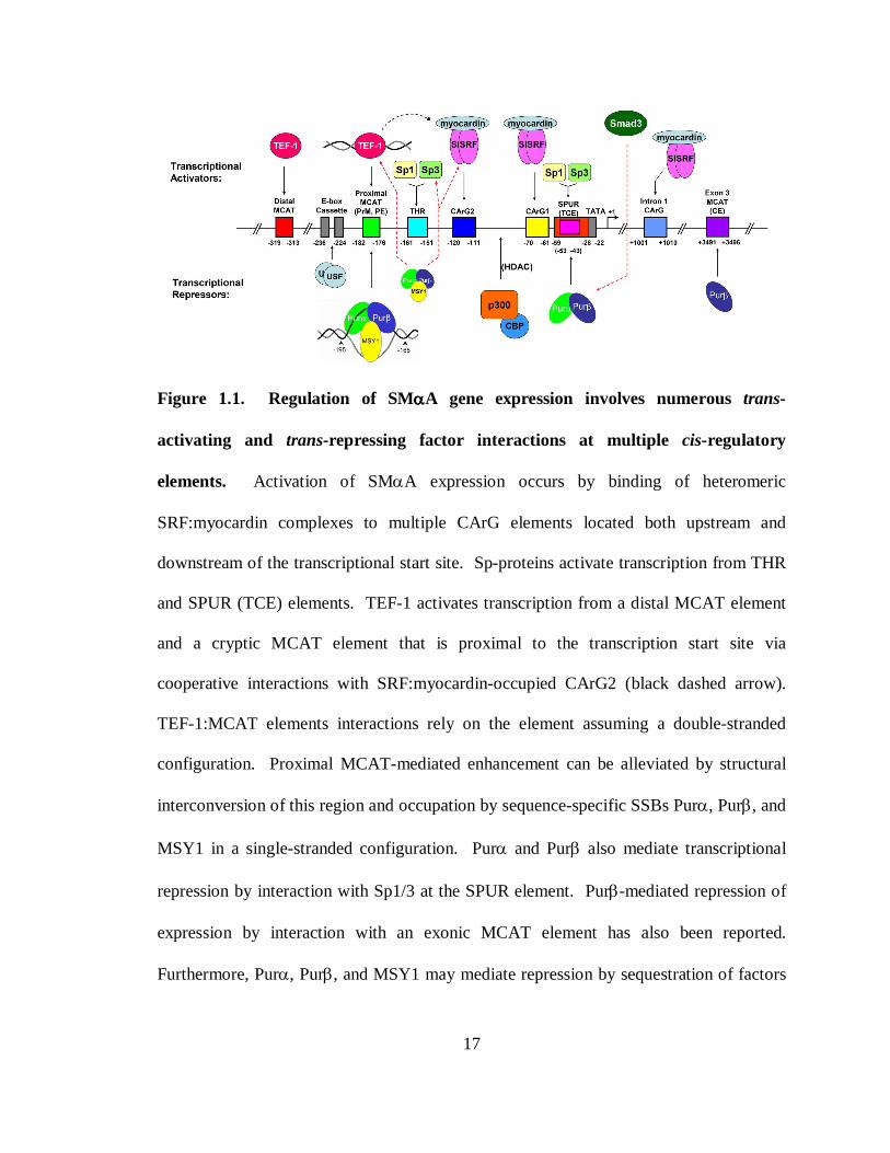
Figure 1.1. Regulation of SMααααA gene expression involves numerous trans-
activating and trans-repressing factor interactions at multiple cis-regulatory
elements. Activation of SMαA expression occurs by binding of heteromeric
SRF:myocardin complexes to multiple CArG elements located both upstream and
downstream of the transcriptional start site. Sp-proteins activate transcription from THR
and SPUR (TCE) elements. TEF-1 activates transcription from a distal MCAT element
and a cryptic MCAT element that is proximal to the transcription start site via
cooperative interactions with SRF:myocardin-occupied CArG2 (black dashed arrow).
TEF-1:MCAT elements interactions rely on the element assuming a double-stranded
configuration. Proximal MCAT-mediated enhancement can be alleviated by structural
interconversion of this region and occupation by sequence-specific SSBs Purα, Purβ, and
MSY1 in a single-stranded configuration. Purα and Purβ also mediate transcriptional
repression by interaction with Sp1/3 at the SPUR element. Purβ-mediated repression of
expression by interaction with an exonic MCAT element has also been reported.
Furthermore, Purα, Purβ, and MSY1 may mediate repression by sequestration of factors
18
TEF-1, SRF:myocardin, and Sp1/3 (red dashed arrows). Purα and Purβ, themselves can
be sequestered by Smad3 to preserve expression (red dashed arrow). Epigenetic control
of SMαA expression is accomplished by chromatin rearrangement created by p300/CBP
histone deacetylase (HDAC) activity within the immediate upstream CArG region.
Tissue specific and cell density-responsive repression can be enforced by bHLH factors
(USF) binding to upstream tandem E-box elements. Numbers represent nucleotide
positions relative to the transcriptional start site.
19
The causative extracellular signaling component for SRF upregulation in SMαA
expressing cell-types has been determined to be TGF-β1, however CArG-box
involvement only accounts for partial activation. This finding led to the discovery of an
additional positive cis-regulatory element, the TGF-β1-control element, or TCE, located
at position -53 to -43 relative to the transcriptional start site (110). Interestingly,
occupation of TCE is not accomplished by SRF in response to TGF-β1 exposure, but
instead by Kruppel-like factors, Sp1 and Sp3, and Smad signaling proteins during
activation of SMαA expression (47, 110). Examination of the 5’ flanking region of the
mouse SMαA promoter also identified six putative E-box sequences (CANNTG) that
have been shown to bind trans-activating basic helix-loop-helix (bHLH) proteins
involved in differentiation of cells of myogenic lineage (131, 135, 194, 304). The
function for these elements appears cell type and cell density restrictive (82, 131), as
combinatorial control by bHLH protein USF (upstream stimulatory factor) at a
tandemized E-box cassette located between positions -236 and -224 confers repression of
SMαA expression in rat aortic SMCs (131, 135).
When it was noted that full activation of SMαA expression in mouse embryo
fibroblastic cell line AKR-2B required the deletion of a 33 base-pair (bp) sequence
corresponding to bases –224 to -191, the realization came that this region contains a
negative regulatory element (82, 270). However, direct sequence examination also
concluded that the region directly 3’ to this newly recognized repressive element (bases -
181 to -176) contains a potent muscle-specific inverted CATTCCT (MCAT) activation
motif which is able to bind transcriptional enhancer factor 1 (TEF-1) in a double-stranded
20
configuration (48, 275) . Indeed, binding of the proximal MCAT enhancer element by
TEF-1 provides synergistic activation of SMαA expression in collaboration with the
distal CArG 2 element (48), which has been shown in previous studies to be essential for
high level activation of expression by SRF (270). Deletion of the core sequence of the
proximal MCAT element results in diminished expression (48). Further investigative
elaboration of the function of the proximal MCAT and repressive elements, by Cogan,
Getz, Strauch and coworkers, verified overlapping sequences between bases -195 and -
164 that constitute both a transcriptional enhancing-, and repressive element (48).
Thorough analysis of the rat and mouse SMαA promoters detected two MCAT elements
(proximal, -182 to -176; distal -320 to -314), both of which have been shown to be
important for transcriptional activation of the gene in fibroblasts, myoblasts, and aortic
endothelial cells (278), however cryptic enhancer activity, as possessing both activator
and repressor character, has only been established for the proximal site in the SMαA
promoter. The work performed by Cogan et al (48), and Sun, et al (275), not only
verified binding of TEF-1 to the double-stranded proximal MCAT element, but also
detected binding of several trans-repressors that exhibit affinity for sequences adjacent to
the core MCAT sequence only when in single-stranded conformations. The identities of
the trans-acting repressors were perceived unique at the time of their discovery, and were
referred to as vascular actin single-stranded binding factors 1 and 2 (VACssBF1 and
VACssBF2).
A closer examination of the region encompassing the proximal MCAT enhancer also
showed an interesting characteristic; this stretch of sequence possesses extensive
21
polypurine/polypyrimidine asymmetry. The coding (forward, non-transcribed) strand of
this region shows greater than 80% purine content in the mouse and rat promoters,
whereas human and chicken are slightly lower (275). This is an important finding, as
was the fact that VACssBF1 and VACssBF2 display specificity for binding of the
pyrimidine-rich and purine-rich strand, respectively (48, 275). The significance and
repercussions of polypurine/polypyrimide tracts within gene promoters will be discussed
in a following section.
MCAT enhancer sequences and their corresponding trans-binding factors are
common in numerous promoter regions of cardiac, skeletal, and smooth muscle genes
(314). These elements generally drive enhanced expression by binding of TEF-family
enhancer factors, although as noted before, cell type restrictions have been pointed out
(278). A mechanistic basis for differential MCAT function was examined by Gan, and
colleagues (94), in which they noted that MCAT-driven SMαA expression follows
distinct mechanisms in activated myofibroblasts compared to differentiated SMCs.
Briefly, MCAT elements are essential for de novo expression of SMαA in myofibroblasts
and developing embryonic SMCs, but appear dispensable for expression in adult
differentiated SMCs. Furthermore, this effect also coincides with TEF-family binding
activity of the MCAT elements, as knockdown of RTEF-1 (TEF-3) decreases expression
of SMαA promoter-driven reporter expression in myofibroblasts and embryonic SMCs,
whereas knockdown of all TEF-family members has no effect on reporter expression in
adult SMCs. These findings speak clearly to the mechanism of SMαA expression during
vascular development and myofibroblast transdifferentiation, and provide a means for
22
distinguishing myofibroblasts from dedifferentiating adult SMCs, both of which express
high levels of SMαA. These studies fail, however, to describe the role of cryptic MCAT
enhancer-mediated repression in cell types with known pathophysiological functions;
explicitly activated SMCs. The location of nucleotide mutations within the proximal
MCAT element used by Gan, et al. were engineered to preclude (R)TEF-1 binding, but
have been shown previously to not substantially effect VACssBF binding (48, 275). The
implications of SMαA downregulation in pathophysiological situations have been
outlined above; hence factors affecting SMαA repression are an important area of
investigation.
Delineation of molecular mechanism fostering bifacial character of cis-regulatory
elements, as observed for the proximal MCAT enhancer element of the SMαA promoter,
represents a challenge for genetic researchers. As stated above, mutational analysis of
the coding strand of the -195 to -164 region, referred to at the time as the purine-rich
motif, showed that mutations abolishing TEF-1 binding do not affect VACssBF1 or
VACssBF2 binding, and mutations reducing VACssBF2 binding do not affect
transcriptional activation (48, 275). These results suggest that binding of these putative
activation and repression factors to particular nucleotides is not mutually exclusive per
se, however the possibility of disparate double-stranded versus single-stranded entities
could not be discounted at the time, and a mechanism by which binding of VACssBF1
and VACssBF2 to their respective strands causes disruption of the double-stranded
MCAT sequence was proposed (275). This latter caveat was the focus of subsequent
investigations by Kelm, Strauch and Getz and colleagues, whose primary aim was to
23
delineate the repressive mechanism employed by the cryptic MCAT enhancer element
and associated trans-repressor proteins, as well as the biochemical characterization of
these novel proteins (28, 47, 129, 141-145, 148, 149, 222, 272, 297, 319).
Initial experiments aimed at delineating VACssBF-mediated SMαA repression
illustrated ssDNA-binding specificity of both VACssBFs and TEF-1 towards the
proximal promoter MCAT element (promoter element, PE) versus a newly identified
MCAT element positioned within exon 3 (coding element, CE) (144). These studies
showed that VACssBF1 and TEF-1 shows no affinity for the CE sequence (single, and/or
double-stranded) despite 100% conservation of the GGAATG TEF-1 recognition motif;
however the purine-rich strand of the CE is tightly bound by VACssBF2. Additionally,
replacement of the PE sequence with CE does not permit transcriptional activity in
reporter based assays, consistent with lack of TEF-1 binding capacity for CE. However,
introduction of mutations nullifying VACssBF2 binding rescues transcription suggesting
that VACssBF2 possesses enhancer disruption activity and that this activity is
independent of enhancer recognition by TEF-1. These studies also highlighted the
importance of downstream regulatory elements that govern SMαA expression. The
discovery of CArG elements within the first intron fortifies this claim (184), and the
importance of downstream promoter elements for SMC-dependent expression in vivo has
been further verified (296, 321).
Preliminary electrophoretic mobility shift studies gauging purine-rich strand binding
by VACssBF2 illustrated that either this factor assembles on purine-rich ssDNA
sequences to varying stoichiometric degrees, forms heterogeneous nucleoprotein
24
complexes, or is in-fact not a single polypeptide (48, 144, 275). The indicated
possibilities stem from the observation that multiple electophoretically shifted species are
generated when cell extracts are allowed to bind single-stranded purine-rich probes
derived from the SMαA promoter. Biochemical characterization of these complexes
confirmed that VACssBF2 is in fact multiple polypeptides able to form homo- and
hetero-mulitimers (141). Cloning of these factors led to the identification of VACssBF2
as Purα and Purβ (143), which was augmented by later work identifying VACssBF1 as
MSY1 (141). The identification of these proteins was of critical importance as it showed
that their respective activites characterized therein were consistent with other biological
systems/scenarios in which their involvement had been detected. For example, Purα
(HeLa Pur Factor) had been implicated in recognizing and binding stretches of single-
stranded DNA generated at origins of DNA replication proximal to the c-myc promoter
(10). In this and subsequent studies it was observed that Purα recognizes purine-rich
ssDNA, especially those rich in guanine nucleotides, and was assigned a consensus
(PUR) binding sequence GGNNGAGGAGARRRR (N = any nucleotide, R = A/G) based
on other known origin sequences, although Purα binding activity was not substantiated at
these sites (10, 11). Cloning of Purα from human cDNA libraries showed the presence of
a distinct isoforms, Purβ, however no function was assigned at this time (11). In this
regard, the human ortholog of MSY1, YB-1 for Y-box binding protein 1, was shown to
facilitate, and bind a pyrimidine-rich ssDNA sequence important for regulation of the
human DRA promoter (major histocompatiblity complex II gene), called the Y-box (69,
183).
25
Consistent with early models of cryptic MCAT enhancer regulation of SMαA
expression, Purα and YB-1 were shown to cooperatively regulate expression from the
human JC virus (HJCV) lytic control element (36). This finding, along with studies by
Sun, et al (275), suggested that Purα, and YB-1/MSY1, and possibly Purβ, constituted an
ensemble of cooperative transcriptional regulators, which by interaction was able to
modulate and augment individual ssDNA-binding properties (36). As stated above,
studies by Kelm and colleagues (141) investigating molecular interactions between
recombinant Purα, Purβ, and MSY1, established that Pur proteins are able to bind the
forward strand of PE (PE-F) as homo- and/or heteromultimers, suggesting that Pur
protein self-association and Purα/Purβ association may either facilitate binding to
ssDNA, represent a regulatory step in ssDNA-binding, or determine repressive activities
of the PE-nucleoprotein complex. Furthermore, these studies also showed direct
interaction between MSY1 with Purα and Purβ.
To test the hypothesis that sequence-specific ssDNA-binding proteins (sequence-
specific SSBs) are responsible for preventing MCAT occupation by TEF-1, Carlini, et al,
systematically analyzed the ability of SMαA promoter constructs harboring mutations
that selectively inhibit binding of Purα, Purβ, and MSY1, but not TEF-1, to drive
expression of a reporter gene (28). These studies showed that loss of sequence-specific
SSB-binding releases repressive effects of the cryptic MCAT enhancer, and that drastic
reduction of Pur protein binding can be induced by deletion of nucleotides -194 to -192,
highlighting the importance of these nucleotides in Pur protein-ssDNA nucleoprotein
complex formation. Additionally, these studies also showed that Purα, Purβ, and MSY1
26
possess the capacity to interact with TEF-1, SRF, and Sp3, as well as with double-
stranded PE, albeit weakly, by virtue of low-stringency DNA pull-down techniques. This
data supports the concept that sequence-specific SSB-mediated repression of the MCAT
enhancer element occurs by protein-protein interaction masking effects, although
observable indirect association (DNA or ternary complex-mediated) could not be
discounted by this approach. Nonetheless, opposing competitive and masking models for
cryptic MCAT enhancer regulation by Purα, Purβ, and MSY1 were proposed (28),
however, a thorough experimental attempt to discredit either of these models has not yet
been performed. The implications of detectable interaction of Purα, Purβ, and MSY1
with SRF and Sp3 are that sequence-specific SSB-mediated repression of SMαA
expression may be accomplished by disruption of SRF and Sp3 trans-activation
properties by virtue of protein-protein interactions. On a similar note, Purβ has been
shown to competitively disrupt muscle-specific CArG box binding, and not c-fos CArG
box binding, by SRF and gene activation in cardiac muscle gene expression (103).
A consistency in the literature surrounding Purα, Purβ, and YB-1/MSY1 function in
the mechanism of cryptic MCAT enhancer element regulation of SMαA is that these
proteins are able to function as sequence-specific SSB transcription factors in a
predominantly dsDNA genome despite exhibiting low affinity for dsDNA. A general
supposition for sequence-specific SSB activity at gene promoter sequences is the
coincident existence of structural perturbations within the DNA duplex structure that are
either created by virtue of SSB binding or facilitated by auxiliary factors to provide
binding sites for SSBs. Widespread dynamic structural rearrangements have been noted
27
in promoter regions of numerous protein encoding genes, notably c-Myc (8, 75, 153, 192,
288), platelet-derived growth factor A-chain (298, 301), vascular endothelial growth
factor (274), tyrosine kinase pp60c-src (229), high mobility group protein A (235), insulin
receptor (285), androgen receptor (39), and epidermal growth factor receptor (130). The
distribution of non-B-DNA structures in vivo appears to be non-random, as it is limited to
specific genes, although transcriptional activity is not universally coincident with
structural alterations (33, 161). Observed non-B-DNA, or paranemic structures within
gene promoters include ssDNA, slippage structures, cruciforms, (left handed) Z-DNA,
(triple helix) H-DNA, quadruplexes, and protein stabilized paranemic structures (313).
The observation of non-B-DNA structures is most common in sequences of DNA
possessing tracts of asymmetrically distributed nucleotides (A/T or G/C rich),
polypurine/polypyrimidine, alternating purine/pyrimidine, or dinucleotide repeats (204,
227, 235). Often intervening or non-B-DNA/B-DNA transition nucleotides exhibit stable
ssDNA character (105) detectable by use of ssDNA-specific reagents (9, 130, 192, 229,
235, 274, 285, 298, 301). Physical analysis has shown that topological stress further
facilitates B-DNA to non-B-DNA structural conversions (204, 260), as well as localized
duplex melting in vitro (153) and in vivo (154), and that non-B-DNA structures typically
possess lower melting temperatures (227, 249). The nature of topological stress in vivo is
believed to be negative supercoiling which is generated upstream of transcriptional
machinery by associated helicase-mediated unwinding occurring downstream of
transcription (154, 179, 260). The reality of localized melting of promoter sequences is
exemplified not only by ssDNA-specific reagent sensitivity, as mentioned above, but also
28
by the involvement of sequence-specific SSB transcription factors that regulate
expression at these and other promoters. Collectively, these findings support the
importance of non-B-DNA structures in mechanisms of gene regulation.
The existence of the proximal MCAT enhancer of the SMαA promoter embedded
within a region of extensive purine/pyrimidine asymmetry coupled with the involvement
of sequence-specific SSBs in the regulation of this element fueled speculation of DNA
structural interconversion as being a possible component of SMαA transcriptional
regulation. As suspected, detectable single-stranded character is observed within the
vicinity of the proximal MCAT enhancer element of the SMαA promoter (9). Treatment
of cultured AKR-2B mouse embryonic fibroblasts with TGF-β1, a serum factor well
established to induce SMαA expression in fibroblasts (64), causes transient changes in
the sensitivity of genomic SMαA promoter DNA to reagents that preferentially react with
unpaired or unstacked nucleotides, including choroacetaldehyde and potassium
permanganate, as assessed by ligation-mediated polymerase chain reaction (PCR)
techniques. Specifically, hypereactivity in bases immediately upstream of the core
MCAT sequence is observed upon activation of SMαA expression, signifying induced
vacancy of this region by dissociation of sequence-specific SSBs at nucleotides deemed
to be necessary for binding of these factors (28, 48, 275). As a result of TGF-β1-induced
hyper-reactivity, the region encompassing the core MCAT element and adjacent
sequences was referred to as the TGF-β1 hypereactive region, or THR (9). The
importance of this region proximal to the cryptic MCAT enhancer element was verified
by responsiveness to TGF-β1 treatment in AKR-2B and trans-activation by Sp1/3,
29
similar to what was observed for TCE (47). It has been noted that upon TGF-β1
treatment of cultured human pulmonary fibroblasts, YB-1 dissociates from the
pyrimidine-rich strand of the SMαA MCAT enhancer element and shuttles to the cytosol
as SMαA messenger ribonucleoprotein complexes (mRNPs) via a mitogen activated
kinase pathway (319) and possible C-terminal processing mechanism (269). A similar
mechanism for Pur protein shuttling has been proposed, but not published (A.R. Strauch,
personal communication), as has been mRNP involvement by Pur proteins. This latter
point has been hypothesized based on observations of Pur proteins binding to the CE in
the 5’ untranslated region (UTR) of reporter mRNAs causing reduced levels of
translation (142). Thrombin treatment of human pulmonary fibroblasts induces
dissociation of Purα, Purβ, and YB-1 from mRNPs and subsequent shuttling of these
proteins back to the nucleus, thus permitting fast translation of SMαA transcripts and thin
filament assembly (319). These findings have direct implications in the role of TGF-β1,
thrombin, and sequence-specific SSB/RNA-binding proteins Purα, Purβ, and MSY1 in
excessive myofibroblast differentiation and subsequent destructive tissue remodeling, and
may represent a permissive mechanism for SMαA derepression.
Pur protein involvement in regulation of SMαA gene transcription and translation has
been detected outside of cryptic MCAT enhancer repression and mRNP sequestration.
As noted before, serum-responsive cis-elements beyond the non-canonical CArG
elements contribute to SMαA gene regulation. Namely, trans-activation of gene
expression by binding of Sp1/3 to the TCE and THR elements has been demonstrated
(47, 110). Examination of sequences flanking the TCE identified an overlapping purine-
30
rich subdomain similar to what is observed in the THR, suggesting cryptic character of
this cis-element as well. Examination of the ability of this sequence to interact with Pur
proteins verified not only binding capacity, but also that occupation of this element by
sequence-specific SSBs can occur in a double-stranded configuration in cell extracts.
Furthermore, overexpression of Purα or Purβ reduces reporter expression from a SMαA
promoter construct in which the proximal MCAT element had been deleted, suggesting
that this element, designated as the SPUR element (Sp1/3 – Pur protein), confers both
positive and negative regulatory functions in vivo (272). Additionally, TGF-β1
exposure is coincident with dissociation of Pur proteins from SPUR, as a detectable Pur
protein:Smad2/3 complex, thus demonstrating physical interaction of Purα and Purβ with
Smad proteins, and elucidating a regulatory mechanism for sequestration of repressors in
SMαA activation during myofibroblast differentiation and tissue remodeling (272).
What's more, SRF has exhibited potential to circumvent Purα-mediated repression at
SPUR in stressed adult cardiac myocytes undergoing SMαA reprogramming (271, 318).
This capacity of SRF to neutralize Purα repression comes from its ability to form a
SRF:Purα heteromeric complex. Similarly SRF-overexpression has been shown to
circumvent Purβ-mediated repression of SMαA (145), thus underlying the importance of
protein-protein interactions and the dynamic interplay of trans-regulatory factors in
regulating SMαA expression during phenotypic modulation by a variety of cell types.
The complex nature of SMαA promoter regulation by the involvement of numerous
cis-regulatory elements, and diverse interactions with a multitude of interacting trans-
31
acting factors epitomizes eukaryotic gene expression. However, additional layers of
epigenetic regulation have been identified at the level of chromatin modification as well.
Extensive chromatin histone acetylation and concomitant SRF occupation in the -150 to -
50 (CArG1 and CArG2) region of the genomic SMαA promoter is observed in SMC-
lineage restricted cell types in vivo (189). Interaction of SRF with myocardin augments
association of SRF to acetylated histones during gene activation, and deacetylation of
histone H4 is coupled to SMαA repression in response to vascular injury. Adenoviral
E1A cotransfection experiments in AKR-2B mouse embryonic fibroblasts and rat smooth
muscle cells confirmed that specific targeting of the CBP/p300 family of histone
acetyltransferases and pRb pocket proteins causes SMαA promoter inactivation in a cis-
element and cell cycle-dependent fashion, thus implicating these proteins in epigenetic
and cell cycle-dependent regulation of SMαA expression (297).
In conclusion, the regulated expression of SMαA expression in various cell types is
the consequence of a diverse and extensive protein-protein and protein-DNA interaction
ensemble that, in turn, permits highly plastic expression of this important functional
filamentous protein. Adaptability of SMαA expression is a crucial component for
cellular responses to physiological and pathological stimuli that impart either beneficial
or malevolent phenotypic consequences. The documented involvement of Pur proteins in
pathophysiological SMαA repression makes them suitable targets for intense biochemical
study.
32
PUR PROTEIN STRUCTURE AND FUNCTION
The Pur family of proteins is comprised of a group of functionally-related, highly
homologous DNA/RNA-binding proteins, and consists of four members in mammals,
Purα, Purβ, and the two isoforms, Purγa and Purγb, however in lower eukaryotes, namely
Drosophila melanogaster, multiple Purα isoforms have been detected (118). Despite the
fact that a relatively high amount of knowledge regarding Purα and Purβ structure and
function has accumulated in recent years, very little is known about corresponding
properties in Purγa,b. The focus of the following section is a review of Purα and Purβ
structure/function characteristics, as this pertains to the scope of the work presented here.
THE BREADTH OF PUR PROTEIN FUNCTION
Much of what we know about structure and function of Pur proteins comes from their
involvement in diverse cellular events, with binding of nucleic acids being a common
aspect in these processes, whether direct or indirect. Discovery of Purα was the result of
a survey of proteins that were believed to be responsible for enforcement of structural
perturbations that are observed in origins of DNA replication in HeLa cells (10).
Investigators examining a zone of DNA replication neighboring the c-myc promoter in
humans identified a protein that binds a purine-rich element (so called, PUR-element) at a
site of DNA bending that, presumably, causes disruption of helix conformation and
localized melting of strands. It was also noted that this protein displays specificity for
purine-rich ssDNA sequences, endorsing the claim that bending induces unpairing of
33
complementary strands in this region of genomic DNA, or permits helix disruption by
occupancy of SSBs. Methylation interference patterns showed that this protein forms
specific contacts with guanines in ssDNA sequences representative of the PUR-element,
and similar purine-rich motifs were identified within other known zones of DNA
replication, suggesting the importance of these sequences and the PUR-binding factor in
the initiation of cellular DNA replication (10, 259) as well as in the replication of viral
genomes (36, 136, 155, 238). Cloning and sequencing of the so-called PUR-factor led to
the identification of Purα (11).
Numerous roles for Purα have been elucidated in regards to regulation of cell cycle
progression. Aside from cellular replication origin interactions described above, clues to
further involvement in cell cycle control came from examination of the replication of
viral genomes. Regulation of lysogeny in the human JC polyoma virus (HJCV) has been
shown to require the differential and reciprocal activities of Purα and YB-1, modulated
by interactions with the JC tumor antigen (36). The competition between opposing
activities of Purα and YB-1 dictates entry into the lytic cycle, with Purα maintaining
lysogeny by governing expression of early genes via displacement of YB-1. Modulation
of YB-1 ssDNA-binding affinity by HJCV tumor antigen association causes disruption of
Purα:promoter interactions, expression of late genes, and commitment to the lytic cycle
(238), whereas association of Purα and HJCV tumor antigen results in attenuation of T-
antigen-mediated transcriptional activation of viral genes necessary for lytic entry (93).
Overexpression of Purα suppresses replication of HJCV genomic DNA in infected glial
cells (34), but has the opposite effect on HIV-1 genome replication (41). Interestingly,
34
the capacity of coinfected HIV Tat protein to bind, sequester and modulate Purα
sequence specificity to activate late gene expression and impose lytic entry by HJCV
supports the notion that partitioning of Purα activity is important for regulation of
transcription of viral genes and viral DNA replication (155).
The repertoire of Purα-viral protein interactions utilized in the regulation of viral
genome replication suggested similar mechanisms may be employed by cells during
regulation of cell cycle progression. Examination of Purα primary structure identified a
signature motif utilized by several cell cycle regulators for protein-protein interaction (11,
182). Namely, the presence of a C-terminal “psycho” motif in Purα suggested this
protein might interact with the tumor suppressor retinoblastoma protein, pRb, a factor
known to cause G0 cell-arrest when normally expressed or loss of growth control when
mutated or deleted. Indeed, direct interaction of Purα with hypophosphorylated pRb was
established in a psycho motif-dependent manner, and this interaction modulates the
binding affinity of Purα for its ssDNA recognition element in the c-myc origin of
replication. The implications of Purα and pRb association were not realized until studies
were performed that correlated Purα:pRb complex level alterations with growth phase
entry suggesting that this complex may aid in preventing assembly or processivity of
replication machinery from origins of replication. Levels of Purα significantly drop just
prior to the onset of S-phase, and return prior to mitosis in CV-1 fibroblast cell lines
(125). This is consistent with findings that show microinjection of NIH3T3 mouse
fibroblasts with Purα causes cell cycle arrest in populations in early S phase and G2
35
(265), and that overexpression of Purα inhibits Ras-induced colony formation in NIH3T3
cells (7) similar to its effects on HJCV (34). Furthermore, the subcellular localization of
Purα is governed by either pRb or cyclin A. Detectable interaction of Purα with cyclin
A, via interaction with a cyclin A/Cdk2 ternary complex, has been shown to stimulate the
phosphorylation of histone H1 by cyclin A/Cdk2 in vitro (177), suggesting that cyclin A-
sequestration of Purα may result in permitting S-phase entry and facilitation of chromatin
decondensation necessary for replication. Purα may also play dominant-negative roles
on other regulatory proteins, as association of Purα with transcription factor E2F-1
suppresses E2F-1-induced activation of S phase-specific genes necessary for cell cycle
progression (58). Collectively, these studies point out that Purα exerts negative
regulation of cellular and viral DNA replication, although mechanistic aspects of Purα
protein-protein and protein:ssDNA interactions remain uncertain, as do possible parallel
roles for Purβ.
Since its discovery, Purα has been implicated in numerous aspects of nucleic acid
processing aside from DNA replication, none more important perhaps than its
involvement in gene regulation. The verification of Purα involvement in transcriptional
regulation is the direct result of identification of gene regulatory cis-elements that bear
resemblance to purine-rich PUR-elements. The first identification of Purα as a
transcriptional regulator was in the regulation of the clusterin gene in quail infected with
the Rous sarcoma virus, showing that this protein not only was important for mammalian
DNA replication (182), but is also conserved for transcription regulation in numerous
36
vertebrate classes (115). Involvement of PUR-elements and Purα interaction is
especially widespread in the expression of genes important for neuron development and
function. For example PUR-elements and Purα binding has been identified in promoter
regions of protein FE65 (316), neuronal nicotinic acetylcholine receptor (74), and myelin
basic protein (107), where Purα activates transcription. Contrastingly, Purα has also
displayed negative regulatory functions as a neuronal transcriptional repressor. For
example, Purα represses expression of the neuronal and hematopoietic transcription
factor Gata2 in the central nervous system, which is opposed by the trans-activator Sp8
(214). Similarly, cAMP-response elements of the somatostatin and tyrosine hydroxylase
gene promoters in opiate-exposed neuronal cell line extracts are suppressed by Purα in a
PUR-element-dependent manner (70, 207, 237). Autoregulation of Purα expression in
glial cells has also been reported, as PUR-element-dependent repression of Purα
promoter activity by ectopic expression of Purα has been observed (200).
Tightly controlled expression of Purα during neuronal development suggests that
gene activation afforded by Purα:promoter interactions is crucial to development of
functioning neurons (107). Exemplification of this trend is provided by mouse models
harboring nullifying homozygous mutations in Purα alleles (PURA-/-) which exhibit
severe postnatal neurological defects and eventual fatality due to lack of neuron
population, neuron myelination, and neurofilament assembly (146), moreover
heterozygous deletions of both Purα and Purβ have been noted in human patients with
acute myeloid leukemia (169). In addition to temporal expression and promoter
37
interaction of Purα, protein-protein and protein-RNA interactions appear to be a crucial
component of Purα-mediated gene expression. For example, temporal expression, and
interaction of Sp1 with Purα augments Purα:promoter interactions and expression of
myelin basic protein (MBP) in neuronal cells (291). Association of HJCV tumor antigen
with Purα causes a downregulation of MBP expression, and subsequent hypomyelination
of infected, non-lytic brain cells in vivo (290). Translocation of Purα from the nucleus
to the cytoplasm during neuronal development by virtue of interactions with three
distinct, yet uncharacterized, Pur-binding proteins are key to developmental timing in
mouse brains (317), suggesting that compartmentalization of Pur proteins is a mechanism
utilized by cells to partition transcriptional/translational regulatory activities of these
proteins in accordance to what is observed for DNA replication-governing by Purα (125).
As discussed extensively in a previous section, Pur proteins have been implicated in
the transcriptional regulation of numerous genes, outside the realm of neuron-specific
proteins. For example, Purα and YB-1, in conjunction with Purβ and AP-1 (c-Fos and c-
Jun), regulate expression of the cell death-associated surface protein, Fas, in a complex
and dynamic manner. Specifically, Purα and YB-1 overexpression results in repression,
which can be antagonized by Purβ, suggesting that either co-association of these proteins
modulates DNA-binding properties of Purα:YB-1 complexes or that Purβ competes for
promoter sites resulting in derepression (162). These studies also highlight functional
differences between Purα and Purβ in mechanisms of gene regulation. Purα has also
been shown to repress expression of CD43 in activated leukocyctes in conjunction with
38
heterogeneous nuclear ribonucleoporotein K (hnRNP-K) (54), and the expression of the
nicotinic acetylcholine receptor β4 subunit in neurons (74), also in conjunction with
hnRNP-K, Sp1, Sp3, and Sox10 (190). Similarly, Purα-Tat heteromeric complexes
repress TNFα expression in HIV-1-infected glial cells (59). The implication of Purα and
Purβ in repression of SMαA expression, as discussed extensively in a previous section,
has also created a focus on these proteins in regulating other muscle-specific genes.
Similar to SMαA expression, Purα and Purβ negatively regulate expression of cardiac α-
myosin heavy chain (α-MHC) via interactions with a purine-rich regulatory element in
the first intron which is essential for cardiac-restricted expression, and by direct
interaction with the transcript thus regulating translation of the mRNA in the cytoplasm
(103, 104). Contrary to repressive roles exhibited by Pur proteins in cellular gene
expression, Purα has shown the capacity to activate transcription of the PDGF-A gene
via interactions with a purine-rich, and S1 nuclease-sensitive region of the promoter
(322).
As indicated, several studies have implicated Pur proteins in transcriptional and
translational regulatory mechanisms. In addition to SMαA and cardiac α-MHC
translational regulation, in which binding of mRNA by Purα and Purβ (and MSY1 in the
case of SMαA mRNA) attenuates translation of the mature transcripts (104, 142), Pur
protein involvement in the translational regulation of numerous mRNAs has been
identified. Purα and Purβ were identified as two of 42 proteins involved in kinesin-
associated large mRNA transport granules (138, 206). A multitude of interactions
39
between Purα and other proteins with extensive mRNP involvement, namely YB-
1/MSY1 (50, 77, 87, 112, 188) and hnRNP-K (190, 286), have fueled speculation as to
whether Pur proteins are intimately involved with widespread translational regulation. It
has been speculated that YB-1 at low concentrations destabilizes mRNA tertiary
structures to permit ribosomal access and assembly on the template, whereas higher
concentrations may restrict access (188). It is unknown whether similar mechanisms are
employed by Pur proteins. Copurification of Purα with 18S ribosomal RNA prompted
the discovery of Purα's ability to limit translation in reconstituted assays in a dose-
dependent manner (90). Validation of this theme has been noted in neuronal cells in
which association of Purα with ribosomes in mouse brain homogenates increases during
postnatal brain development (173). RNA-mediated activity modulation of Purα has also
been noted. For example, association of HIV-1 Tat protein with Purα is dependent upon
ligand-induced reconfiguration of Purα upon binding to RNA derived from the HJCV
PUR-element (311). Similarly, association of RNA homologous to the signal recognition
particle (SRP) 7 SL RNA (so called Purα-associated, or PU-RNA) has been shown to
negatively alter Purα binding affinity for the MBP promoter (289), suggesting that
additional layers of regulatory activity exist within this multifunctional protein.
STRUCTURAL CONSIDERATIONS
High primary sequence homology between Purα and Purβ suggests that these
proteins are paralogs within given organisms and have arisen as a result of gene
40
duplication and divergence. This is exemplified by primary sequence alignment, as
shown in Figure 1.2 (alignments performed with T-COFFEE web-based software,
http://tcoffee.vital-it.ch, (205), using amino acid sequences deposited to the National
Center for Biotechnological Information). Various regions of high sequence homology
and identity can be observed between paralogs, and these similarities are present within
orthologs from diverse species spanning the vertebrate branches of the phylogenetic tree,
suggesting that these regions correspond to modular structural elements critical for
function. Despite extensive sequence identity between paralogs (~71% in mouse),
distinct regional differences can be observed, especially in the terminal regions, as well
as intervening sequences between putative domains. The level of sequence conservation
within regions of paralog divergence among orthologs indicates that these regions are
critical for distinct paralog functions. As described above, examination of protein
components that regulate and recognize origins of DNA replication led to the discovery
of Purα, a HeLa cell nuclear component that binds to purine-rich ssDNA sequences
derived from c-myc and dhfr origins (10). Initial cloning and analysis of Purα alluded to
modularity and repetition of tertiary structure within the central region and led to putative
domain assignments for the detected domains (11). The proposed domain architecture
for Purα was described, as indicated in Figure 1.2, inset. As of the date in which this
review was composed, three-dimensional structure data (crystallographic, NMR, high-
resolution microscopy) has not been reported for any Pur protein, nor has any low
resolution analysis been presented to argue against the hypothetical domain assignments.
The modular structure proposal stems from the observation of two repeating sequence
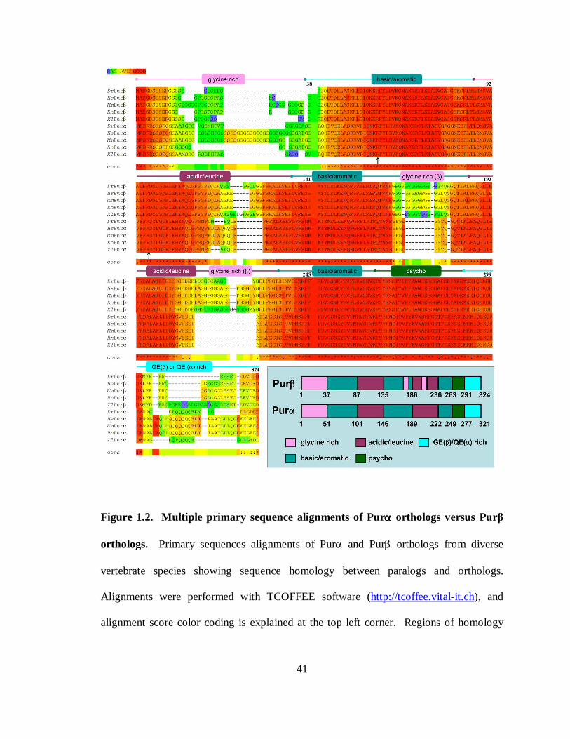
41
Figure 1.2. Multiple primary sequence alignments of Purαααα orthologs versus Purβ
orthologs. Primary sequences alignments of Purα and Purβ orthologs from diverse
vertebrate species showing sequence homology between paralogs and orthologs.
Alignments were performed with TCOFFEE software (http://tcoffee.vital-it.ch), and
alignment score color coding is explained at the top left corner. Regions of homology
42
follow a modular protein structural arrangement as indicated above the alignments and in
the lower right panel. Positions of residues implicated in ssDNA-binding are denoted by
arrows (Wortman, et al (2005) Biochem Biophys Acta 1743:64-78). Primary amino acid
sequences were obtained from the National Center for Biotechnological Information
protein database. Dr, Danio rerio (Purα NP_00101846, Purβ Q6PHK6); Hs, Homo
sapiens (Purα Q00577, Purβ AAK72642); Mm, Mus musculus (Purα NP_033015, Purβ
NP_035351); Rn, Rattus norvegicus (Purα XP_001063244, Purβ NP_001017503); Xl,
Xenopus laevis (Purα NP_001086909, Purβ NP_001079178).
43
motifs. The first class, referred to as class I, consists of three 23 amino acid repeats
separated by the other repeating motifs, two class II repeats of 26 amino acids. Both
class I and class II repeats exhibit high degrees of homology. Class I repeats are greater
than 65% homologous with 17% amino acid identity, whereas class I repeats are 69%
homologous with 34% of residues completely conserved. The names of these repeats
have been changed in order to more completely reflect sequence character, as indicated in
Figure 1.2, inset. Class I repeats are abundant in basic and aromatic residues, while class
II shows high levels of leucine and acidic residues (92, 145).
The primary function that has been proposed for both Purα and Purβ is preferential
binding to ssDNA that is rich in purine nucleotides, particularly guanines, and as such,
has been referred to as sequence specific. This character is based on compiled findings of
many groups that have examined DNA-binding properties of these proteins. Assignment
of putative domain functions was carried out using Pur-deletion mutants in ssDNA/RNA-
binding functional assays. In human Purα, it was shown that a core domain consisting of
the first two class I and class II repeats (amino acids 65-191 of the human ortholog) is
necessary and sufficient for binding to short purine-rich ssDNA sequences derived from
the human JC virus Mad1 control element pentanucleotide repeat (36), the c-myc
replication origin (133, 310), MBP promoter regulatory sequence MB1 (289) and cAMP
response elements (CREs) of somatostatin and tyrosine hydroxylase promoters (70).
Additionally, this core region was identified to be necessary for helix-destabilization
properties of Purα, as assessed by the protein’s ability to displace short strands of DNA
complementary to regions of single-stranded M13mp18 plasmid DNA (56). Equivalent
44
regions in mouse Purβ also constitute binding to ssDNA and RNA corresponding to the
purine-rich strands of CE and PE (PE-F), albeit with lower affinity than the full-length
protein (145). However, a difference between Purα and Purβ arises from the requirement
of the third class I repeat for binding of Purβ to ssDNA, whereas this repeat appears
dispensable in Purα (145). Point mutations of Arg residues within class I repeats one and
two of human Purα (R71E and R110E, respectively) appear to have drastic inhibitory
consequences in ssDNA binding, as well as the ability of the protein to displace shorter
complementary strands of linear partial duplex DNAs, suggesting that these residues may
participate in ssDNA-binding via sidechain guanidinium electrostatic interactions with
either the phosphate backbone or hydrogen bonding face(s) of nucleotides (presumably
guanines), or by cation-π interactions (310). Experiments evaluating equivalent point
mutations in mouse Purβ suggested, however, that these mutations may result in
destabilization of tertiary and quaternary structure of the protein and do not necessarily
reflect that these arginines make atomic contacts with ssDNA (149). Unequivocal
identification of ssDNA-ligating amino acids in Pur proteins has not been reported.
Reasons for this are clearly that the degree of sequence identity between Pur protein
paralogs and orthologs has made the identification of critical residues non-obvious.
Additionally, modularity may serve as a means of compensating for deleterious amino
acid substitutions in regard to nucleic acid-binding functions.
The repeat region of Purα has also been implicated in contributing to diverse protein-
protein interactions. For example, interaction of Purα to viral protein HIV-1 Tat protein,
and HJCV and SV-40 large T-antigens is confined to amino acids 85-215 (155), and 72-
45
123 (93), respectively, while association of YB-1 has been localized to amino-acids 174-
215 (238). The overlapping nucleic acid-binding and protein-protein interaction
properties of Tat and YB-1 association regions may help explain, in part, how Tat and
YB-1 enhance ssDNA/RNA-binding affinities of Purα for specific sequences of the
HJCV Tat-responsive element (155) and lytic control element (238), respectively.
Furthermore, RNA-mediated high-affinity self-association of Purα has been localized to
the second class II acidic/leucine-rich repeat (91), which is interruptible by Tat
association (155), suggesting that this region contributes to heterogenous protein-protein
interaction and self-association in a mutually exclusive manner, and that self-association
may enhance nucleic-acid binding in a way similar to Tat and YB-1 heteromeric
association.
Speculation regarding the tertiary structure of Pur proteins has been limited in large-
part by the fact that Pur proteins share very little sequence homology to other proteins of
known structure. However close examination of the primary sequences of mouse and
human Purα led to the discovery that a structural motif located near the C-terminal end
bears striking similarities to other cellular and viral proteins known to be involved in
initiation of DNA replication. Initial analysis of this region suggested that it might adopt
an amphipathic helix, based on predicition rules (11). A core motif conserved among
several viral transforming T-antigen peptides is Pro-Ser-Tyr (PSY), followed downstream
by a Cys (C) residue, while Purα exhibits slight degeneration of this motif with PTY.
Based on this character, this region was termed the psycho motif (182). Interestingly, the
corresponding motif in the simian virus large T-antigen 40 protein is known to interact
46
with pRb. As suspected, and described above, the psycho domain of Purα constitutes a
pRb interaction face (133). Thus far, in vivo interaction between Purβ and pRb has not
been reported, despite ~92% motif homology between murine paralogs. Perhaps
positions of non-conservation represent residues that contribute important atomic
contacts, although this matter has not been investigated.
As mentioned above, striking differences in primary sequence are observed near the
N-, and C-termini of Purα and Purβ, especially in regard to the positioning of
pronounced polyglycine tracts. Purβ contains two stretches of polyglycine of eight and
nine residues separated by FQPAPR, whereas Purα has a seventeen residue polyglycine
stretch interrupted by a single serine. The lack of N-terminal conservation among Purα
and Purβ orthologs suggests that the functions contributed by these regions are species-
specific, and has been the subject of study (discussed below). Internal polyglycine
stretches in Purβ are also observed that disrupt the second basic/aromatic, and the second
acidic/leucine rich repeat, in addition to a tract found near the C-terminus, all of which
are absent in Purα (Figure 1.2). All told, glycine content reaches 22.2% and 15.0% in
murine Purβ and Purα, respectively, perhaps signifying a reason for the lack of highly
ordered crystal structures for these proteins. High glycine content and polyglycine
stretches have been observed in several proteins possessing nucleic acid helix-
destabilization properties, including the UP1 subunit of heterogeneous ribonucleoprotein
A1 (46, 86), nucleolin (99), and the c-myc promoter far-upstream element binding protein
(FBP)(62), although the exact function attributable to polyglycine tracts is unknown.
Peptide backbone flexibility imparted by these regions may aid in adaptive structural
47
rearrangements necessary for concerted modular domain interaction and function.
Additionally, removal of the N-terminus (residues 1-84) in Purα has created aggregation
problems making its study difficult (133).
Multiple studies have shown that the N-terminal regions of Pur proteins are
dispensable for single-stranded nucleic-acid binding (145, 310), however ,appear critical
for binding to long dsDNA molecules (310). Therefore, N-termini are generally regarded
as regulatory motifs, necessary for seemingly distinct functions among the paralogs based
on sequence divergence, although specific interaction partners remain unidentified.
Despite sequence differences, similar repressive activities of Purα and Purβ have been
localized to the N-terminus. Removal of amino acids 1-86 of Purα results in loss of
autorepression of Purα promoter:reporter constructs in multiple cell types (200), whereas
equivalent deletion in Purβ results in loss of repression of a full-length SMαA
promoter:reporter construct in rat smooth muscle cells that is otherwise repressed when
cotransfected with constructs that overexpress wild-type Purβ (145). It remains to be
seen if these N-terminal-mediated mechanisms of repression are equivalent in terms of
protein-protein interaction profiles.
Other structural distinctions between Purα and Purβ made obvious by sequence
alignment are the divergence observed in the C-termini. While both paralogs possess
high Glu (E) content, mouse Purα displays a high number of Gln (Q) residues including a
seven polyglutamine stretch. The corresponding motif in mouse Purβ is replaced by a
polyglycine stretch (Figure 1.2). Glutamine-rich domains have historically been ascribed
transcriptional activation functions (97), however polyglutamine tracts have also been
48
associated with DNA helix distortion and unwinding properties in Drosophila GAGA
factor (307). Removal of the C-terminal portion of Purα, (215-322) has deleterious
effects on the ability of the protein to bind linear dsDNA (310). Similar to Purα N-
terminal deletion, removal of amino acids 264-324 of Purβ also results in a loss of
repression of SMαA expression in an MCAT enhancer element-dependent manner (145)
and loss of interaction capabilities with MSY1, and Purα (142). Divergent means of
MSY1/YB-1 association between Purα and Purβ suggests that varied stoichiometric
combinations of these corepressors at the SMαA MCAT enhancer may serve different
regulatory functions. Loss of function studies addressing this issue is the focus of
Chapter III. Interestingly, despite different reported interaction interfaces utilized in YB-
1/MSY1 association, Purα (238) and Purβ (145) both display modulated ssDNA-binding
properties in the presence of these Y-box proteins, however a mechanism attributable to
this feature has not been described.
NUCLEIC ACID-BINDING PROPERTIES
Discovery of the prototypic Pur protein, Purα, was the result of investigation into
cellular protein components that bind and stabilize purine-rich regions of the c-myc
associated origin of DNA replication that displays single-stranded character (10, 11).
From the very beginnings of Pur protein investigation, a paradigm has emerged regarding
Pur protein function, as well as those of other known sequence-specific SSBs. The
challenge has been defining the mechanism by which sequence-specific SSBs recognize
49
and stably bind target sequences in the presence of complementary strands that possess
high degrees of affinity for the same target sequence. Speculative hypothoses to this
problem have predicted that inherent capabilities of sequence-SSBs or associated SSBs to
destabilize double-helix conformations of DNA permit stable interaction, either by
ATPase (helicase) activity or by direct thermodynamic competition with annealing. Of
course, speculation has also proposed that coupling of helix destabilization by topological
stress generated by negative supercoiling may aid in thermodynamic competition by
reducing melting free energies of DNA duplexes. To delineate these issues, and to
further identify genomic targets of sequence-specific SSBs, researchers have examined
many aspects of ssDNA-binding of sequence-specific SSBs, including sequence-
specificity, affinities, stoichiometries, and strand-displacement capabilities. In this
regard, work that has been performed on Pur protein DNA-binding properties has
suffered from inabilities to designate “consensus” sequences for Pur proteins, and if they
differ between paralogs and/or orthologs. As stated above, high degrees of conservation
within nucleic acid-binding domains of Purα and Purβ among diverse organisms suggests
a preservation of nucleic acid-binding properties; however this has not been
substantiated, and the lack of designation of ligating amino acids within these domains
leaves this claim open to scrutiny. The following is a description of the nucleic acid-
binding properties of Purα and Purβ as reported in the literature thus far.
Towards the goal of understanding shared and distinct nucleic acid-binding properties of
Pur proteins, numerous and diverse ssDNA and RNA oligonucleotides have been used to
gauge Pur protein involvement in promoter, replication origin, and ribonucleoprotein
50
interactions. Comprehensive examination of the historically utilized nucleic acid probes,
as they are reported in the literature, unfortunately fails to provide speculative insight into
the nature of Pur protein-nucleic acid interactions due to the vast heterogeneity of these
sequences. Table 1.1 offers a wide-ranging summary, albeit non-all-inclusive, of various
sequence identities, nucleotide lengths, and Pur protein interaction affinities for these
molecular probes, compiled from numerous reports in the early and contemporary
literature (6, 7, 10, 11, 28, 34, 48, 91, 103, 104, 106, 107, 129, 133, 136, 141-145, 149,
155, 238, 259, 272, 275, 289, 291, 322).
What can be ascertained from Table 1.1 is that Pura and Purb possess diverse targets
throughout numerous genomes and transcriptomes, with most target sequences exhibiting
a high degree of purine content, and a high level of affinity where it has been reported.
Sequences with a relatively low purine content show reduced affinity, as demonstrated by
comparison of affinities of Pur proteins for short complementary oligonucleotides.
Comparing affinities of BPV1 to BPV2, for example, shows a difference in affinity close
to an order of magnitude. This also, however, shows that Pur proteins exhibit low levels
of affinity for pyrimidine-rich ssDNA. For this reason Pur protein ssDNA/RNA-binding
is generally regarded as promiscuous based on the fact that these sequences exhibit
extensive heterogeneity, and a clearly defined binding site can not be established by this
simple approach. A consistency in the literature surrounding Pur protein target sequences
is that these proteins bind to defined (GGN)n repeat motifs. This trend stems from early
surveys of demonstrated targets, and can be seen in Table 1.1. However, issues arise
from this definition of a Pur-binding site due to the fact that demonstrated
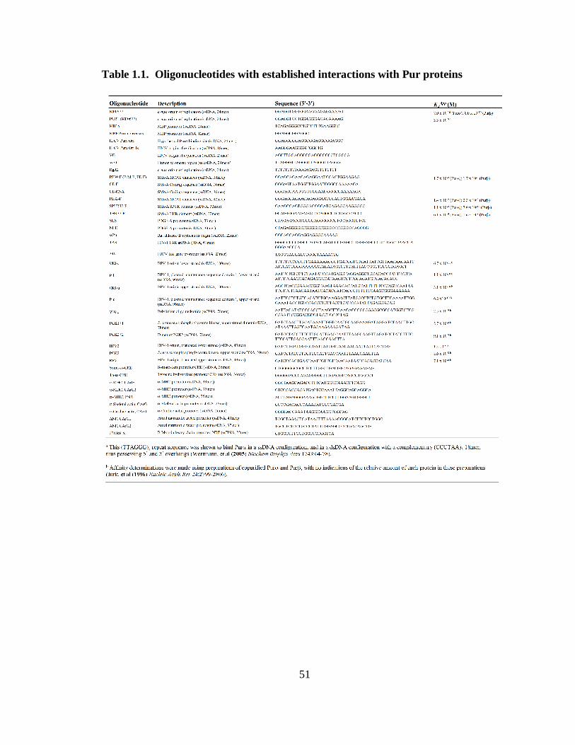
51
Table 1.1. Oligonucleotides with established interactions with Pur proteins
52
targets display extensive heterogeneity regarding the number, position, spacing and exact
sequence of these GGN repeats. For example, It has been stipulated that high affinity
binding requires multiple GGN repeats, and that N not be G (92, 148), however binding
of Purα to probe RgR2 (310) which harbors a single purine-rich motif, (GGGAGAG) and
only one GGN repeat, refutes this claim, although no indication of the relative affinity of
Purα for RgR2 compared to other, more elaborate purine-rich probes was given. The
heterogeneity of GGN repeat spacing among established probes also confounds attempts
to unambiguously define a Pur protein binding site. An interesting trend pointed out by
Jurk, et al (136) is that apparent binding affinities increase with increasing probe length.
This effect could be due to increases in binding site concentrations that accompany the
use of probes with multiple binding sites, or that Pur proteins bind to multi-site lattices
via cooperative mechanisms. This possibility has not been thoroughly examined through
dissection of intrinsic interaction energetics between Pur proteins and individual binding
sites or intersite communications. Furthermore, the stoichiometric species of Purα, Purβ,
or heteromeric complexes thereof that are competent to bind nucleic acids has not been
clearly established. Many groups have shown that particular Pur protein nucleoprotein
complexes are heterogeneous with respect to Pur protein content (136, 141), whereas
others have shown that homomultimers of Purα only form in the presence of RNA (91),
although the exact stoichiometry of self-association in these cases is unknown.
Most of what we knoe today regarding Pur protein nucleic acid binding properties is
the result of early work performed by Bergemann, et al (10, 11). This work represents
the initial discovery of Purα which was preceded by identification of a purine-rich motif
53
flanking the c-myc promoter shown to adopt bent conformations and serves as an origin
of DNA replication in HeLa cells. Competition-based analyses identified a candidate
24mer ssDNA oligonucleotide sequence for Purα binding (termed MF0677): 5’-
GGAGGTGGTGGAGGGAGAGAAAAG-3’. The length of this sequence in this study
was largely arbitrary, as it represents a portion of the entire bend-region of the replication
origin under investigation, and thus it was unknown at the time whether this sequence
represented a partial binding site, or an array of multiple binding sites. Competition
experiments resulted in the disappearance of a single electrophoretic species suggesting
that this sequence represented an entirety of binding sites in the context of the c-myc-
associated origin of replication. These studies also demonstrated a clear preference of
Purα for ssDNA versus dsDNA sequences. Furthermore, methylation interference
footprinting of the Purα:MF0677 complex detected several guanine-specific contact
points, suggesting that Purα has a guanine preference over adenines, and substitution of
several guanines with adenines results in loss of affinity. The ability of ssDNA
oligonucleotides derived from other replication origin purine-rich elements to compete
for binding to the c-myc-associated purine-rich motif suggested that this motif may be
common to replication origins, thus implicating Purα in cell cycle regulation. This
discovery also prompted an evaluation of sequence similarities among putative Purα
binding sites throughout known origins of replication, using the candidate MF0677
24mer sequence as a reference (10). In doing so, a core 16mer Purα consensus sequence
was developed from 11 different PUR-elements identified in six different organisms: 5’-
GGNNGAGGGAGARRRR-3’ (N = any nucleotide, R = A/G).
54
A quasi-systematic validation of this consensus sequence by direct demonstration of
Purα binding capacity to parent PUR-elements was performed by measuring the ability
of Purα, in the context of a library-candidate λ phage-infected E.coli cell extract, to alter
the electrophoretic mobility of oligonucleotides harboring mutations of parent MF0677
(10, 11). A compiled view of these analyses is displayed in Table 1.2. The results of this
analysis suggest that substitution of consensus-defined guanines within the core-
consensus element disrupt binding (MM0677), consistent with methylation interference
data suggesting these residues represent essential contact points within the nucleoprotein
complex. Transversional mutation (G to T) of these nucleotides also shows the
importance of these conserved guanines in high affinity nucleoprotein assembly
(ME0677 and MG0677), but also highlights the dispensability of nucleotides at the 3’ end
of the core consensus sequence (MC0677). Disruption of GNGG or GGNGG motifs in
MF0677 within the core consensus sequence (MA0677 and MB0677) also diminishes
binding of Purα, validating the notion of the core consensus sequence. This is further
supported by results of competitions with oligonucleotides in which the G at position 1 of
the core consensues sequence is restored (compare MI0677 to MH0677). However,
guanine nucleotides flanking this putative core sequence were identified to be important
for nucleoprotein stability (MJ0677), suggesting that the core consensus sequence notion
is not resolute. Interestingly, an oligonucleotide representation of the hamster
dihydrofolate reductase (dhfr)-associated replication origin PUR-element (DR3529),
which completely obeys consensus rules, binds poorly to Purα, again suggesting that the
core consensus sequence generated by sequence data mining and mutagenesis may be
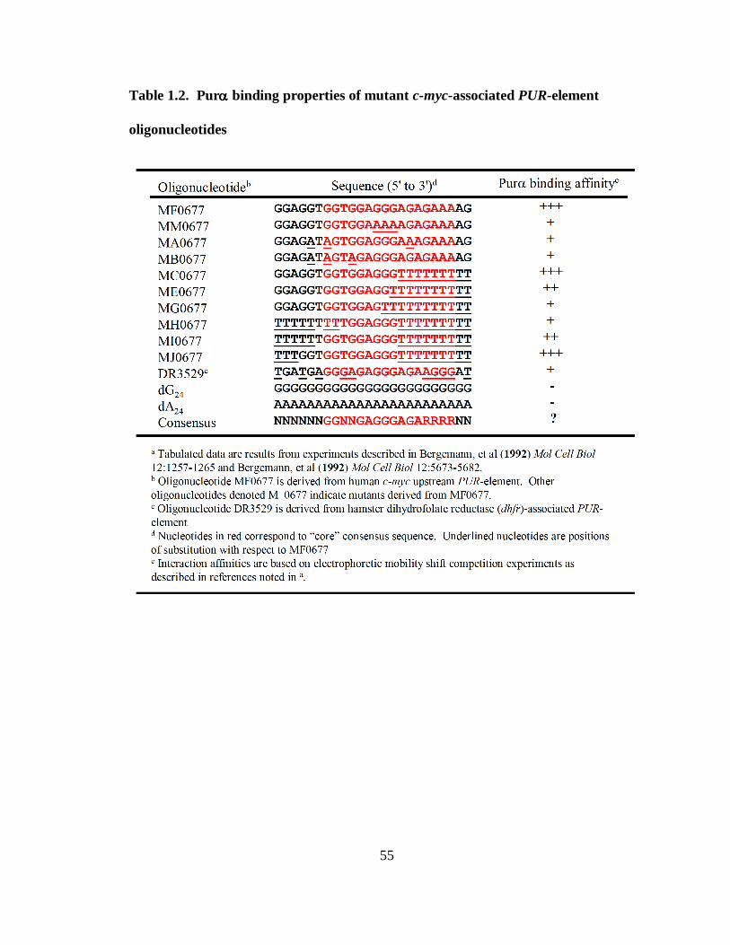
55
Table 1.2. Purαααα binding properties of mutant c-myc-associated PUR-element
oligonucleotides
56
inaccurate. The inability of Purα to bind dG24 shows that guanine content alone cannot
establish a high affinity interaction. A sequence Logo of the core Purα binding site, based
on compilation of putative PUR-elements flanking origins of DNA replication as
determined by Bergemann, et al (10), is shown in Figure 1.3 (performed using web-based
software: http://weblogo.berkeley.edu/ (246)). This shows that regions of high
conservation are both essential and dispensable for binding to Purα, as indicated by
arrows and circles, respectively. The Logo does not however indicate the positions of
critical nucleotides outside this core region, as detected with MJ0677. It should also be
noted that conserved residues in oligonucleotide MJ0677, as compared to MF0677, may
represent a more accurate core sequence based on similar binding affinities between these
sequences.
The appropriateness of consensus sequence usage has been questioned by several
researchers that point out potential genomic binding sites for proteins with defined
consensus sequences are often missed due to the relatively low amount of information
portrayed in consensus sequence representations and the inherent sequence promiscuity
of DNA-binding proteins (245). Usage of sequence Logos has sidestepped this limitation
to some degree; however their construction relies on accurate footprints of protein
binding sites and that they be constructed from numerous sequences to accurately depict
nucleotide preference patterns. This problem is likely enhanced when defining consensus
sequences for sequence-specific SSBs, especially those which have no defined footprint.
Specifically, errors in consensus sequence and sequence logo accuracy may arise from
the fact that ssDNA is generally more flexible than its dsDNA counterparts. Binding site
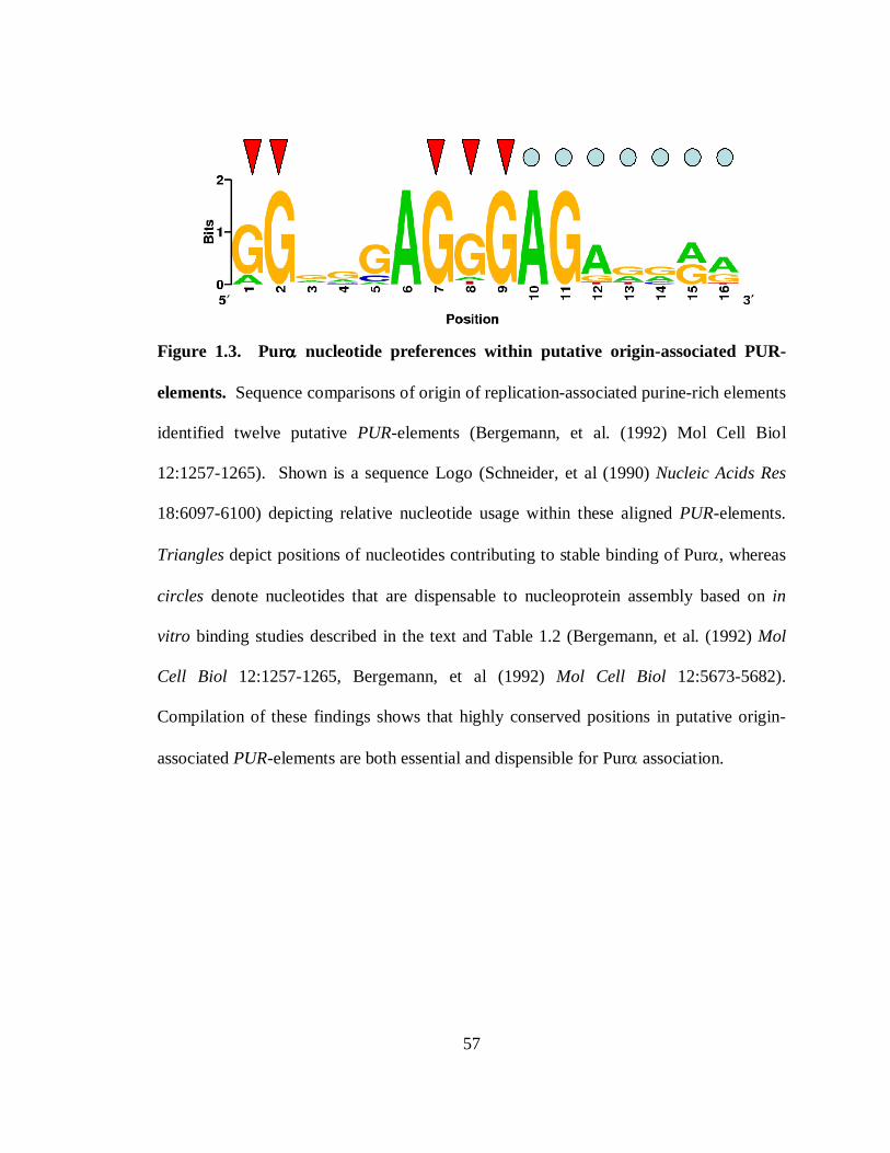
57
Figure 1.3. Purαααα nucleotide preferences within putative origin-associated PUR-
elements. Sequence comparisons of origin of replication-associated purine-rich elements
identified twelve putative PUR-elements (Bergemann, et al. (1992) Mol Cell Biol
12:1257-1265). Shown is a sequence Logo (Schneider, et al (1990) Nucleic Acids Res
18:6097-6100) depicting relative nucleotide usage within these aligned PUR-elements.
Triangles depict positions of nucleotides contributing to stable binding of Purα, whereas
circles denote nucleotides that are dispensable to nucleoprotein assembly based on in
vitro binding studies described in the text and Table 1.2 (Bergemann, et al. (1992) Mol
Cell Biol 12:1257-1265, Bergemann, et al (1992) Mol Cell Biol 12:5673-5682).
Compilation of these findings shows that highly conserved positions in putative origin-
associated PUR-elements are both essential and dispensible for Purα association.
58
recognition in dsDNA usually proceeds via scanning of the major or minor groove of
dsDNA until a chemical group signature is encountered. The orientation of hydrogen-
bond donors and acceptors, as well as methyl groups is recognized by complementary
surfaces on the protein, which permits reversible interaction. Sequence promiscuity
arises from slight variations in the chemical landscapes of the major or minor groove that
are tolerated by the surface of the protein, and this degree of tolerability, in the eyes of
this author, likely dictates binding affinity. In ssDNA, however, the major and minor
groove faces of nucleotides are supplemented with the available Watson-Crick base-
pairing face to provide additional ligating chemical groups. Seemingly this characteristic
would make ssDNA less permissive to binding promiscuity; however near-limitless
rotational freedom of the glycosidic bond (syn- versus anti- rotamers) in single-stranded
configurations permits a vast array of chemical-group orientations, presenting chemical
groups of all three faces to the binding surface of the protein. Hypothetically, reverse
directional binding (5’ to 3’ versus 3’ to 5’) should be possible as well if ligating
nucleotides are rotated 180° around their glycosidic bonds, and assuming that specific
sugar contacts are not crucial for interaction. Additional backbone flexibility may
compound this promiscuity further by permitting bulges, loops, and other three-
dimensional structures.
To circumvent issues of binding promiscuity, several researchers have used
oligonucleotide MF0677 as a “standard” for Purα ssDNA-binding (10, 11, 133, 136,
310), despite the lack of extensive and rigorous evaluation of the Purα:MF0677
nucleoprotein complex stoichiometry or interaction energetics. Recent studies by
59
Wortmann, et al (310) have investigated this issue using a Scatchard analysis approach,
and found that truncated recombinant FLAG epitope (DYKDDDK)-fusion forms of both
Purα and Purβ (amino acids 1-314 and 1-316 of Purα and Purβ, respectively) bind
MF0677 with 1:1 (Pur:MF0677) nucleoprotein complex stoichiometries exhibiting
similar subnanomolar affinities (listed in Table 1.1). However, the graphical linearization
procedure used by these researchers may have caused a misestimation of complex
stoichiometry, as multiple binding sites (low and high affinity) were observed, as was
considerable curvature of the linear Scatchard plots. Also, the use of truncated
recombinant proteins may alter binding properties, although this contention has not been
experimentally tested. Nonetheless, the results of this study suggest that the ssDNA-
binding-competent species of Pur proteins is a monomer.
Similar mutation analyses have been performed in regards to Pur protein binding to
the proximal MCAT enhancer element of SMαA, as described previously. Figure 1.4
depicts a summary of nucleotides identified to be critical for Pur protein interactions
within the SMαA MCAT enhancer region based on results from various studies
investigating Pur-responsiveness (repression) of promoter constructs used in transfection
assays (48, 275) as well as direct binding assays (28, 148). As stated previously,
transient transfection analysis of nucleotide sequences flanking the core MCAT sequence
suggested that repressor binding occurs at nucleotides 5’ to the TEF-1 binding site, as
transversional mutation of nucleotides in this reigon results in loss of repression of
SMαA promoter:reporter constructs TV191, TV189, and TV187. The prevalence of
guanines in this area also supports this hypothesis. Similarly, constructs containing
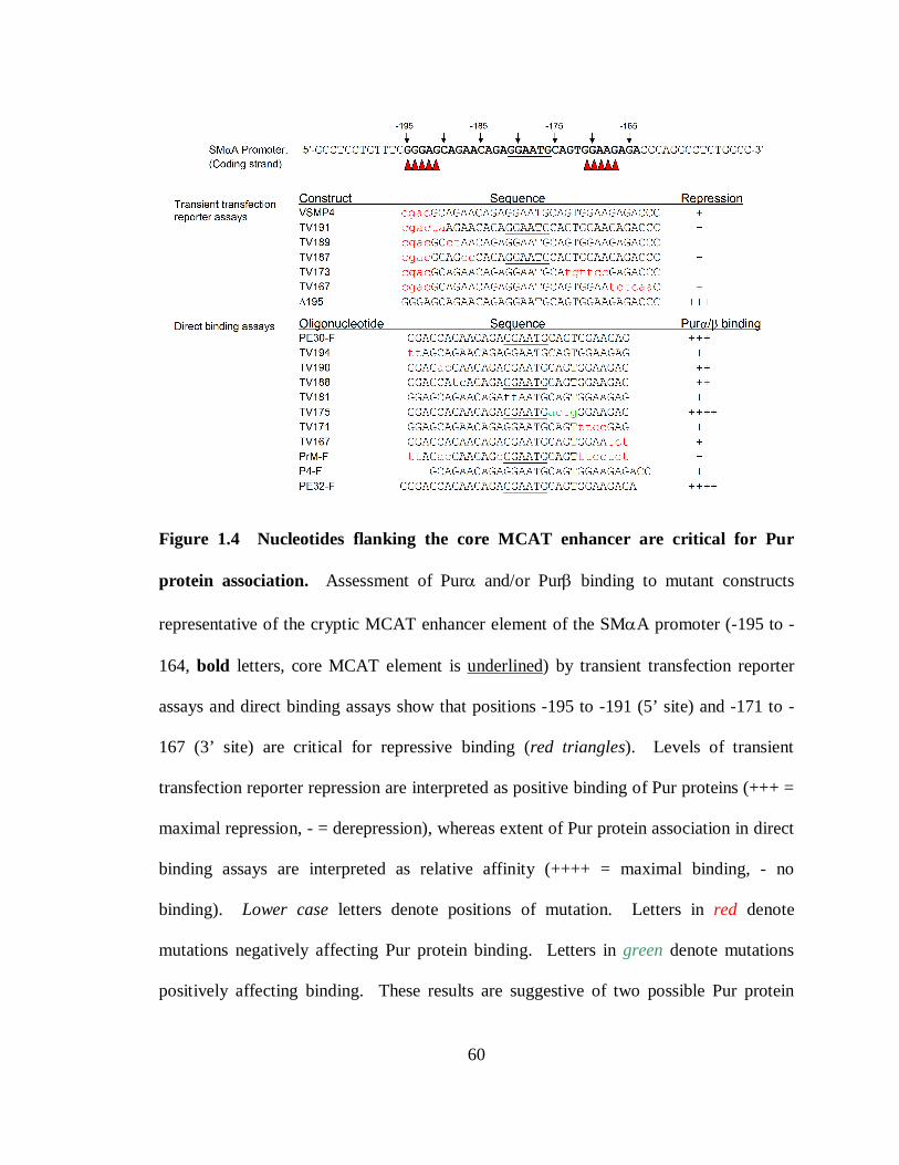
60
Figure 1.4 Nucleotides flanking the core MCAT enhancer are critical for Pur
protein association. Assessment of Purα and/or Purβ binding to mutant constructs
representative of the cryptic MCAT enhancer element of the SMαA promoter (-195 to -
164, bold letters, core MCAT element is underlined) by transient transfection reporter
assays and direct binding assays show that positions -195 to -191 (5’ site) and -171 to -
167 (3’ site) are critical for repressive binding (red triangles). Levels of transient
transfection reporter repression are interpreted as positive binding of Pur proteins (+++ =
maximal repression, - = derepression), whereas extent of Pur protein association in direct
binding assays are interpreted as relative affinity (++++ = maximal binding, - no
binding). Lower case letters denote positions of mutation. Letters in red denote
mutations negatively affecting Pur protein binding. Letters in green denote mutations
positively affecting binding. These results are suggestive of two possible Pur protein

61
binding sites with slight degeneracy of a core GGGAGA element. Data represent a
compilation of findings published previously (Cogan, et al (1995) J Biol Chem
270:11310-11321; Sun, et al (1995) Mol Cell Biol 15:2429-2436; Carlini, et al (2002) J
Biol Chem 277:8682-8692; Knapp, et al, (2006) J Biol Chem 281:7907-7918).
62
transversions of nucleotides 3’ to the core MCAT motif (TV173, TV167) also display
loss of repression, suggesting that these nucleotides are also responsible for binding Pur
proteins (48). Furthermore, addition of nucleotides spanning -195 to -192 augments
repression (∆195), also suggesting that this construct contributes an additional Pur protein
binding site, or that it represents a more complete binding site with higher repressor
affinity than the truncated construct VSMP4 that possesses nucleotides -191 to +46 of the
SMαA promoter (28). Direct ssDNA-binding experiments showed a similar trend, that
being the importance of nucleotides flanking the core MCAT sequence (28, 275).
Oligonucleotides that accurately represent the region encompassing nucleotides -196 to -
164 (PE32-F) exhibit the highest affinity for Pur proteins (148), where as deletion or
mutation of these nucleotides results in loss of affinity. Interesting is the observation that
transversion of nucleotides -175 to -172 (TV175) results in enhanced binding affinity
(275). Reasons for this are unclear but may be due to addition of a guanine at position -
172 which is proximal to other recognizably important nucleotides. Collectively, the data
represented by Figure 1.4 show the importance of two regions of high guanine content
(red arrows), consistent with the possibility that this region contains two Pur protein
binding sites, as suggested previously by experiments detecting multimeric Pur:PE
complexes (141). These putative Pur protein binding sites also show resemblance to core
PUR-element sequences as identified by researchers investigating the c-myc-associated
PUR-element (10, 11). These studies proposed a core consensus sequence of 5’-
GGGAGA-3’, for which oligonucleotide MF0677 has only one, and has been shown to
bind only one mole of Purα or Purβ (310). The two sites in PE32-F show slight
63
degeneracy; the site on the 5’-flank of the core MCAT element is GGGAGC, whereas the
3’ site is GGAAGA (degenerate nucleotides are shown in italics). Experimental evidence
testing this hypothesis has not been reported, but its acquisition is a goal described in
Chapter VI of this dissertation. Interestingly, mutations in either putative site cause
drastic loss of repression or observed ssDNA-binding consistent with the notion that
cooperative interactions between sites might dictate affinity.
A remaining aspect of Pur protein nucleic acid-binding that needs consideration is
that involving dsDNA. Numerous reports have surfaced reporting binding of both Purα
and Purβ to dsDNA probes in vitro, including the c-myc-associated replication of origin
(10, 11, 310), the rat aldolase B-associated origin of replication (259), the mouse α-
skeletal actin promoter CArG element (103), and mouse SMαA promoter elements PE
(148) and SPUR (148, 272). In each case however, a clear preference has been
established of Pur proteins for single-stranded configurations of PUR-elements. So
questions remain as to whether observed binding of Pur proteins to dsDNA represents a
distinct binding mode, that it is equivalent to ssDNA-binding in the sense that major
contact points between Pur proteins and dsDNA exist on the purine-rich strand while the
complementary pyrimidine rich strand is completely displaced (dissociates) or partially
displaced (incomplete helix destabilization, or “bubble” formation), or is mediated by an
unknown dsDNA-binding protein via protein-protein interactions. Work with purified
proteins certainly refutes this latter claim, as investigations have shown the ability of
recombinant Purα to completely displace short pyrimidine-rich strands from duplexes
containing protruding purine-rich strands in the absence of ATP (56, 310). The ability of
64
Pur proteins to displace strands from blunt-ended dsDNA probes has not been
established. Both Purα and Purβ have exhibited the ability to bind dsDNA in the context
of plasmid DNA, with both showing higher affinities for supercoiled configurations
(310). This finding supports the notion that dsDNA binding is not a distinct functional
mode of Pur proteins. This claim is based on the fact that both supercoiled and relaxed
topoisomers of plasmid DNA are double-stranded per se, but torsional stress existing in
supercoiled plasmids imparts destabilization of the forces maintaining helical structure,
namely hydrogen-bonds as indicated by differences in melting temperatures (249, 293)
and the increased prevalence of regions assuming non-B-DNA conformations (204, 220,
227, 260, 294). Disruption of annealing free energy associated with supercoiling may
permit localized strand displacement and binding of sequence-specific SSBs in a manner
that is observed as dsDNA-binding by conventional methods, like electrophoretic
mobility shift assays (310). Interestingly, observed binding of Purα to supercoiled
plasmids pUC19, and a pUC19 construct harboring the c-myc-associated replication
origin produces discrete, quantized bands of DNA as judged by agarose gel
electrophoretic mobility shift assays (310) in a manner similar to banding patterns of
topoisomers. This characteristic could suggest that binding of Purα to these supercoiled
plasmids increases the twisting number or writhe in neighboring regions. However,
similar electrophoretic patterns observed with linearized plasmid suggests that these
bands represent successive stoichiometric complexes of Purα:plasmid DNA (310).
A veritable aspect of dsDNA binding by Pur proteins illustrated by studies reported
by Wortman, et al (310) is that the length of the DNA molecule (number of base-pairs)
65
plays a significant role. For example, binding of Purα to a 343 bp PCR-product
containing the same PUR-element as MF0677 is negatively influenced by competition
with MF0677 but not double-stranded MF0677 (24 bp). This shows that longer
molecules of dsDNA bind to Purα with greater affinity than shorter ones. Reasons for
this are unclear. Nearest-neighbor algorithms for estimating two state annealing free
energies of short oligonucleotides have proven to be extraordinarily accurate, comparable
to experimental determinations. However, their usage with long DNA molecules breaks
down considerably as they predict annealing free energies approaching infinity and
extremely high melting temperatures (239). Thermal denaturation of long, linear DNA
molecules has shown that melting occurs at temperatures as low as 64°C, lower than
predicted by nearest-neighbor methods, and lower than that of purine-rich
oligonucleotides approximately 30 nt in length. Furthermore, melting is likely not a two-
step process as indicated by the presence of locally melted subdomains (234, 262).
Evidently, long dsDNA molecules have an added destabilizing component which is
currently unidentified; although this component may be diffusion-related as relaxed, long
dsDNA molecules display lower rotational and translational diffusion coefficients than
supercoiled molecules of similar molecular weight (230). Whether or not entropic
limitations on rotational and translational diffusion associated with long dsDNA
molecules potentiate enhancement of intramolecular vibration and localized
melting/unstacking is unknown, and its involvement in a possible mechanism for helix
destabilization and SSB-binding is purely speculative.

66
In conclusion, mechanisms describing Pur protein single stranded nucleic-acid
recognition and binding in vivo remain poorly defined. Unsuccessful attempt to
accurately determine Pur protein target sequences and to quantitatively detail binding
events have undermined this effort. The detectable multi-faceted involvement of Pur
proteins in gene regulation coupled with their clear preference for binding single-stranded
targets in vitro indicates that single-stranded nucleic acid binding is also accomplished in
vivo. To understand how this process occurs, details of Pur protein structure, function,
and chemistry must be methodically and experimentally uncovered.
67
CHAPTER II. SCOPE OF THE PRESENT WORK
As details of Pur protein function have emerged from various laboratories
investigating aspects of transcriptional, translational, and DNA replication regulation
associated with these proteins, so have numerous questions. A major goal of the
laboratory of Dr. Robert J. Kelm, Jr. is the delineation of functional distinctions between
mouse Pur-paralogs Purα and Purβ. As described previously, subtle yet orthologously
conserved differences in Purα and Purβ primary sequences suggest that functional
differences exist between these proteins in vivo. Towards the testing of the hypothesis
that states regions of sequence heterogeneity confer distinct functions between Purα and
Purβ, I and members of the Kelm laboratory undertook a parallel gain and loss-of-
function approach to delineate functions of Purα and Purβ in regards to regulation of
SMαA expression in appropriate cell culture models. Chapter III is an excerpt from two
papers published by our laboratory (148, 149) which collectively describes the
contributions I made to this body of work, and focuses on the loss-of-function angle.
This work details the differing contributions Purα and Purβ make to SMαA repression in
both cell-type and ortholog-dependent fashions, and establishes the dominant repressor
activity of Purβ in this context.
Identification of Purβ as a dominant repressor of SMαA expression in mouse embryo
fibroblasts and vascular smooth muscle cells fueled a rigorous thermodynamic analysis of
the ssDNA-binding properties of this protein which I performed. Extensive studies
68
focusing on the solution behavior and quaternary structure of recombinant Purβ are
described in detail in Chapter IV, with the results of this study showing the capability of
recombinant Purβ to self-dimerize in the absence of nucleic acid and that monomeric and
dimeric species adopt elongated, asymmetric geometries. These findings were also
published (222). Establishment of Purβ dimer-formation at protein concentrations that
are thought to be attained in cells that exhibit SMαA repression led to the hypothesis
which states Purβ self-association represents a prerequisite step for binding to the
MCAT-associated Pur protein binding sites. Testing of this hypothesis involved a
rigorous thermodynamic assessment of the mechanism used by recombinant Purβ in
binding to ssDNA sequences representative of the proximal MCAT enhancer element of
the SMαA promoter, and is described in Chapter V. These studies corroborate findings
outlined in the background that suggest that two Purβ binding sites exist in this element
and that successive binding of two Purβ monomers proceeds with high affinity stabilized
by cooperative interactions, thus refuting the original hypothesis of obligate dimer
formation. To substantiate the nucleotide sequence identities of putative Purβ binding
sites within the SMαA MCAT enhancer region, I performed a systematic analysis of the
stabilities of nucleoprotein complexes composed of Purβ and truncated mutant PE
oligonucleotides. The employed methodologies and results of this approach are
described in Chapter VI.
Collectively this body of work describes the ssDNA-recognition and binding
mechanisms used by Purβ in the repression of SMαA expression, as it pertains to

69
molecular mechanism governing phenotypic plasticity of cell types implicated in
physiological responses to injury and pathological progressions of numerous disease
states.
70
CHAPTER III. PUR PROTEIN LOSS-OF-FUNCTION STUDIES
IDENTIFY PUR ββββ AS A DOMINANT REPRESSOR OF SMααααA
EXPRESSION
The work described herein has been published as part of greater bodies of work authored
by the Kelm laboratory and can be found in its orginial form in:
Knapp AM, Ramsey JE, Wang SX, Godburn KE, Strauch AR, Kelm RJ, Jr. (2006)
Nucleoprotein interactions governing cell type-dependent repression of the mouse
smooth muscle alpha-actin promoter by single-stranded DNA-binding proteins Pur alpha
and Pur beta. J Biol Chem.281(12):7907-18.
and
Knapp AM, Ramsey JE, Wang SX, Strauch AR, Kelm RJ, Jr. (2007) Structure-function
analysis of mouse Pur beta II. Conformation altering mutations disrupt single-stranded
DNA and protein interactions crucial to smooth muscle alpha-actin gene repression. J
Biol Chem.282(49):35899-909.
INTRODUCTION
Phenotypic plasticity of VSMCs and adventitial fibroblasts is a feature critical to
adaptive pathophysiological tissue functions supported by these cell types in vivo,
including vessel wall remodeling associated with atherogenesis, and restenosis (120, 174,
71
175, 232, 242). Dynamic phenotypic modulation is the result of widespread genetic
reprogramming of genes essential to cell function, and requires extensive cooperative
networking of factors that regulate gene expression. Indices of differentiation status are
difficult to establish due to the continuous spectrum of biomarker expression profiles that
are exhibited by cell types undergoing transdifferentiation, however profiles of marker
expression remain the best way to categorically measure phenotypes (209, 210). Absence
or reduction of staining for contractile proteins is a characteristic feature of cells
comprising the neointima of vulnerable atheroma (210). Among the battery of
contractility markers commonly used for assessing phenotypic status of VSMCs and
adventitial fibroblasts, SMαA is the most widely utilized marker due to its dynamic range
of expression in the aforementioned cell types (210). Furthermore, SMαA is the
principle contractile protein upregulated in myofibroblasts responding to signals
promoting normal and pathological wound healing (226, 231, 271).
SMαA is the most abundant protein expressed in VSMCs, accounting for
approximately 10-40% of the total protein (79, 257). Genetic inactivation of SMαA in
mice germ cells indicate that SMαA plays an essential role in regulating vascular
contractility and blood pressure homeostasis (98). Down-regulation of SMαA has been
implicated in microfilament rearrangement and changes in cell shape consistent with a
transformed fibroblast phenotype, suggesting a role in non-myogenic cell types (166).
Loss-of-function studies conducted in cultured cells also suggest that another essential
function of SMαA is to inhibit cell motility (231). It follows that repression of SMαA
expression in activated VSMCs may be a key molecular event leading to enhanced cell
72
migration and proliferation, as well as contributing to the vulnerability of atheroma in
vivo (88, 151). Therefore, elucidation of repressive mechanisms governing SMαA
expression is paramount to understanding and managing conditions associated with
pathophysiological vessel remodeling.
Spatial and temporal regulation of SMαA expression is mediated by an array of
trans- and cis-acting components that rely on dynamic functional interplay to provide
highly plastic expression (158, 315). Potent activation and repression of full length
SMαA promoter constructs has been confined to a cryptic MCAT enhancer element
located -195 to -164 nucleotides relative to the start of transcription (48, 275) that has
been shown to possess extensive purine/pyrimidine asymmetry and to transiently adopt
single-stranded conformations in vivo (9). Bifacial enhancer activity has been shown to
be regulated by structure-specific occupation by dsDNA-dependent transcriptional
activator TEF-1, or sequence-specific SSB repressors Purα, Purβ, and MSY1 (28, 48,
141, 143-145, 275). Repression has been shown to require binding of Purα and/or Purβ
to the purine-rich strand of the MCAT enhancer and occupation of the pyrimidine-rich
strand by MSY1. It has not been established whether structural interconversions are
cause or consequence of differential transcription factor occupancy.
Purα and Purβ are members of the Pur family of proteins and were so named based
on their preference for binding purine-rich ssDNA and RNA sequences (92). Purα and
Purβ are ~70% identical at the amino acid level (143), with each protein possessing a
minimal ssDNA/RNA-binding domain composed of highly homologous sequence repeats
unique among other known nucleic acid-binding proteins (132). However, significant
73
sequence differences between Purα and Purβ exist near the N- and C-termini suggesting
each protein may have divergently evolved to perform distinct functions. A report has
suggested that Purα and Purβ bind to ssDNA as either homo- or heterodimers and can
each interact with MSY1 (28). Despite these biochemical similarities, gain-of-function
(over-expression) studies clearly illustrate that Purα and Purβ are not redundant in terms
of their transcriptional repressor activity toward the full-length mouse SMαA promoter in
transfected VSMCs (145).
The potential capacity of Purα, Purβ, and MSY1 to collaboratively regulate
expression of SMαA has been extended to the post-transcriptional level as well. The
sequence of the MCAT enhancer in the 5’-flanking region bears striking resemblance to a
region of exon 3 in the SMαA open reading frame. It has been reported that Purα, Purβ,
and MSY1 can form a stable messenger ribonucleoprotein complex (mRNP) with the
exon 3 RNA sequence in a manner which blocks the translational efficiency of a reporter
mRNA (142). These results raise the possibility that these proteins may also participate in
post-transcriptional control mechanisms for SMαA protein levels by either directly
regulating SMαA translation or by reducing nuclear repressor concentrations that
normally restrict transcription. Purα- and Purβ-dependent repression of a-myosin heavy
chain gene expression in cardiomyocytes has been shown to occur at both the
transcriptional and translational levels, thus supporting this notion (104).
It has been reported that profiles of interactions of Purα and Purβ with certain
transcriptional activators are altered during transforming growth factor β1 (TGFβ1)-
74
induced myofibroblast differentiation in vitro (272). These activators include Sp1, Sp3,
and Smad proteins (Smad2, Smad3). DNA-binding studies have revealed distinct cis-
element binding sites for these factors that are downstream of the MCAT enhancer
suggesting that Purα and Purβ may facilitate cell type-specific repression by mechanisms
involving protein-protein interactions in addition to competitive DNA-binding. The
importance of functional interplay between activators and Pur-repressors was also
revealed in over-expression experiments in which serum response factor (SRF) was
found to reverse Purβ-mediated repression of the SMαA promoter (145). Given that SRF
and Smad proteins are essential downstream targets of signaling pathways that promote
smooth muscle differentiation (28, 47, 145, 157, 210), it is likely that the gene regulatory
effects of Pur repressors are not limited to SMαA but likely include other markers of
VSMC differentiation. Furthermore, in light of the fact that Purα has also been
implicated in regulation of cell cycling due to its interaction with retinoblastoma tumor
suppressor protein (pRB) (133), E2F-1 (58), and cyclin A (125), the possibility exists that
Pur proteins may be directly involved in mechanisms modulating cell cycle progression
in vivo (146).
In the present study, we have employed a RNA-interference (RNAi)-mediated loss-
of-function approach to study functional similarities and differences between Purα and
Purβ with respect to regulation of SMαA expression. While stable and transient
epigenetic knockdown of both Purα and Purβ results in derepression of endogenous
SMαA expression in AKR-2B MEFs, Purβ appears to be the dominant Pur protein
repressor in this cell type. This finding is corroborated by chromatin
75
immunoprecipitation experiments showing elevated levels of Purβ promoter occupancy.
This effect is restricted to serum-deprived AKR-2B MEFs, as transient transfection in
C57BL/6J mouse aortic VSMC outgrowth cultures showed Purα to be similarly
repressive. Knockdown of Purα and Purβ results in synergistic derepression of SMαA
promoter-driven reporter expression in both cell types in a MCAT enhancer element-
dependent manner. These studies, as previously published (148) point to cell type-
restricted collaborative and distinct functions for Purα and Purβ in regards to repression
of SMαA expression, and complement parallel gain-of-function studies that show similar
results.
MATERIALS AND METHODS
Cell culture, transient transfection, and reporter gene assay − Aortic segments from
C57BL/6J mice were obtained following protocols approved by the University of
Vermont Institutional Animal Care and Use Committee. VSMCs were isolated by cell
outgrowth from aortic tissue explants and characterized as previously described (40,
244). Primary VSMCs were cultivated in a 90% air/10% CO2 incubator at 37°C in
growth medium consisting of Dulbecco’s modified Eagle’s medium, 1× insulin-
transferrin-selenium supplement (Invitrogen), and 20% v/v heat-inactivated fetal bovine
serum (FBS) (Hyclone). For transient transfection studies, primary VSMCs were seeded
in six-well plates at a density of 10,000 cells/well and transfected with the use of jetPEI
reagent at a ratio of 2 µl/µg plasmid DNA as directed by the manufacturer (Qbiogene).
76
AKR-2B mouse embryonic fibroblasts (MEFs) or rat A7r5 VSMCs were cultured and
transiently transfected as previously described (297). Briefly, subconfluent AKR-2B or
A7r5 cells seeded in 60 mm dishes were transfected with the use of GenePORTER
reagent (Gene Therapy Systems) at a ratio of 3 µl/µg plasmid DNA. After 48 h
incubation in growth medium, cells were washed with phosphate-buffered saline and
extracted using 1× reporter lysis buffer (Roche Applied Science) supplemented with
protease inhibitors. Total protein in transfected cell lysates was determined by
bicinchoninic acid assay (Sigma) using BSA as a protein standard. Commercial
immunoassays were used to measure chloramphenicol acteyltransferase (CAT) or β-
galactosidase (β-gal) reporter proteins as directed by the manufacturer (Roche Applied
Science). Reporter values were corrected for total protein content. Transfections were
typically performed in triplicate and repeated two to three times to ensure reproducibility.
Data sets were subjected to one-way analysis of variance to identify differences among
group means at the p < 0.05 significance level. Post-hoc comparisons were performed
using the Bonferroni adjustment for multiple comparisons.
Construction of shRNA expression plasmids targeting mouse Purαααα or Purβ − The
design of mouse Purα- and Purβ-specific shRNA sequences was facilitated by free
software available through Invitrogen Corporation
(https://rnaidesigner.invitrogen.com/rnaiexpress/). Nucleotide sequences encoding full-
length mouse Purα and Purβ were previously reported (143). Parameters set to the mouse
genome database for BLAST, sense-loop-antisense configuration for shRNAs, and a loop
sequence of CGAA yielded two shRNA-encoding sequences predicted to be Purβ-
77
specific and four predicted to be Purα-specific: Purβ-I) 5’-
GTCGGTATGCAGATGAAATGACGAATCATTTCATCTGCATACCGAC-3’ (888),
Purβ-II) 5’-
GATGAAATGAAAGAGATCCAGCGAACTGGATCTCTTTCATTTCATC-3’ (899),
Purα-I) 5'-GCAAGTACTACATGGATCTCACGAATGAGATCCATGTAGTACTTGC
-3’ (973), Purα-II) 5’-
GTGGACAACAAGCGCTTCTTCCGAAGAAGAAGCGCTTGTTGTCCAC-3’ (1191),
Purα-III) 5’-
GACAACAAGCGCTTCTTCTTCCGAAGAAGAAGAAGCGCTTGTTGTC-3’ (1194),
Purα-IV): 5’-
GCGTGTTTATGCGAGTGAGTGCGAACACTCACTCGCATAAACACGC-3’ (1237),
Purβ-Scm) 5’-
GATCCTAAGTCTGACTTGCAACGAAGGTCAATCCTATAGTGCTAAG-3’, Purα-
Scm1) 5’-
GTCATCGAATGCCATGTCAGTCGAAACTGACATGGCATTCGATGAC-3’, Purα-
Scm2) 5’-GGTATGCGTTAGTGCTGAGTGCGAACACTCAGCACTAACGCATACC-
3’. Numbers in parentheses to the right of each sequence indicate the first nucleotide in
the open reading frame of either Purα or Purβ mRNA predicted to be targeted by that
particular shRNA. Bold type indicates the position of the loop sequence. Sequence 5′ of
the loop corresponds to the sense strand of the transcribed shRNA. Scrambled sequences
(Scm) that contained similar base content as the Purα- or Purβ-specific shRNAs were
78
designed for use as negative controls in knockdown experiments. Complementary
oligonucleotides sequences encoding the shRNAs were generated by chemical synthesis
and obtained from a commercial vendor (Sigma-Genosys). To facilitate unidirectional
cloning into pENTR™/U6 shRNA expression vector (Invitrogen), CACC was included
on the 5’ end of sense strand oligonucleotides while AAAA was included on the 5’ end of
antisense strand oligonucleotides. A double-stranded DNA insert encoding a LacZ-
specific shRNA was supplied by the manufacturer. Annealing of complementary strands,
ligation into pENTR™/U6, and subsequent transformation into E. coli strain TOP10 were
performed as recommended by the manufacturer. Colonies were selected in the presence
of 25 µg/ml kanamycin. E. coli were propagated in 5 ml cultures in the presence of
antibiotic. Plasmids were isolated with the use of a mini-preparation kit (Qiagen).
Insertional and sequence integrity was verified by automated DNA sequencing performed
in the DNA Analysis Core Facility of the Vermont Cancer Center.
Construction of lentiviral Pur shRNA expression constructs and transduction of
AKR-2B cells − Selected U6 RNAi expression cassettes (Purβ-I or Purβ-Scm) were
transferred from pENTR™/U6 vector into pLenti6/BLOCK-iT™-DEST vector via a site-
specific LR recombination reaction as directed by the manufacturer (Invitrogen).
Recombination reactions were used to transform One Shot Stbl3™ competent E. coli.
Recombinant subclones were selected based on resistance to blasticidin (50 µg/ml) and
ampicillin (100 µg/ml) and sensitivity to chloramphenicol (30 µg/ml). Successful transfer
of each U6 RNAi cassette into pLenti6/BLOCK-iT™-DEST vector was confirmed by
restriction enzyme digestion of purified plasmids with NdeI. Cloned expression plasmids
79
were maintained in One Shot ccdB Survival™ E. coli cells and purified by double
cesium chloride gradient ultracentrifugation. To generate lentiviral stocks,
pLenti6/BLOCK-iT™-DEST/U6 RNAi expression constructs (3 µg) were co-transfected
along with the ViraPower™ packaging mix (9 µg) into the 293FT producer cell line (6 ×
106 cells per transfection) using Lipofectamine™ 2000 reagent in serum-free Opti-
MEM I medium as directed by the manufacturer (Invitrogen). Cells were maintained
for 16 h at 37° C in a humidified 5% CO2 incubator. Transfection medium was replaced
by complete growth medium supplemented with 1 mM sodium pyruvate and cells were
cultured for an additional 72 h. Cell supernatants containing lentivirus were collected and
titers of each lentiviral construct were determined based on transduction efficiency of
cultured AKR-2B MEFs under blasticidin-resistant selection conditions.
To isolate stable lentiviral-transduced AKR-2B MEF clones, subconfluent AKR-2B
cells (4 × 10⁵ in a 25 cm2 flask) were infected with 250 transducing units of lentivirus in
McCoys 5A medium (Gibco BRL/Invitrogen) with 5% v/v FBS for 24 h. After an
additional 24 h incubation in growth medium without virus, cells were switched to
selection medium (McCoys 5A, 5% v/v FBS with 50 µg/ml blasticidin) and cultured for
ten days with periodic replacement of selection medium in order to remove dead (non-
transduced) cells. Once a suitable number of blasticidin-resistant cells were obtained
(~25-50% coverage of the flask with densely colonized cells observed by microscopy),
diluted cell suspensions were prepared by trypsinization and resuspension in selection
medium. Cells were counted and then seeded by serial 2-fold dilution in a 96-well plate
starting at 500 cells/ml (50 cells/well) to allow for colony growth and expansion from
80
single cell clones. Cell colonies arising in wells seeded with the lowest cell density (≤ 1)
and exhibiting circular morphology (suggestive of originating from a single cell) were
once again subcloned by limiting dilution in selection medium (20 days post-infection) to
ensure clonality. Lentiviral-transduced MEF clones were then expanded to prepare
freezer stocks. Clonal cell lines were maintained in growth medium consisting of
McCoys 5A, 5% v/v FBS with 10 µg/ml blasticidin at 37°C in a humidified 5% CO2
incubator. Confluent cultures of lentiviral-transduced MEF cell clones were washed with
cold PBS then extracted with 1× CAT lysis buffer (Roche Applied Science) plus protease
inhibitors. After centrifugation of whole cell extracts for 10 min at 14,000 rpm, protein
content in cleared lysates was measured by BCA assay (Sigma). Relative expression of
Purα, Purβ, SMαA, and GAPDH was assessed by Western blotting.
Construction of promoter-reporter plasmids and expression vectors − Mouse SMαA
promoter-reporters and mammalian expression plasmids encoding His-epitope tagged
versions of mouse Purα and Purβ were described previously (28, 296). All plasmids used
for transfection were purified from E. coli cultures by double cesium chloride gradient
centrifugation.
Western blotting of transgene-expressed and endogenous proteins − Ectopic
expression of His-tagged Purα or Purβ was monitored by Western blotting of lysed cell
protein with an RGS(H)4 monoclonal antibody (Qiagen) as previously described (145).
Expression of endogenous Pur proteins was similarly assessed with a rabbit polyclonal
antibody that recognizes a conserved sequence present in both mouse Purα and Purβ
(141). Commercial monoclonal antibodies were used for detection of SMαA (clone 1A4,
81
Sigma) and glyceraldehyde-3-phosphate dehydrogenase (GAPDH) (clone 6C5, Research
Diagnostics Inc.).
Chromatin immunoprecipitation (ChIP) assay − AKR-2B MEFs were seeded in 10
cm dishes at a density of 5 × 10⁵ cells in growth medium consisting of McCoys 5A with
5% heat-inactivated FBS. After reaching 70-80% confluency (~36 h), cells were washed
twice and incubated in 10 ml serum-free MCDB-402 medium (JRH Biosciences) for 48 h
in a humidified 37°C, 5% CO2 incubator. A separate group of cells was left untreated and
allowed to grow to near confluence in complete growth medium prior to fixation.
Formaldehyde was added to culture medium of growth-arrested (serum-free) or
asynchronously growing cells to a final concentration of 1% v/v. After 10 min at 37˚C,
medium was aspirated and the cells were washed with phosphate-buffered saline and
harvested by scraping. Cells were counted and collected by centrifugation at 1500 rpm
for 10 min. Cell pellets were extracted using a ratio of 0.2 ml lysis buffer (1% w/v SDS,
10 mM EDTA, 50 mM Tris-HCl pH 8.1) per 106 cells. Cell lysates were sonicated (4 ×
10 s bursts) using a Branson model 150 sonifier at maximum power and then centrifuged
at 14,000 rpm for 10 min at 4˚C. Supernatant was removed and diluted 10-fold in ChIP
dilution buffer consisting of 0.01% w/v SDS, 1.1% v/v Triton X-100, 1.2 mM EDTA,
167 mM NaCl, 16.7 mM Tris-HCl pH 8.1 plus 1 mM PMSF and 1 µg/ml each aprotinin,
pepstatin A and leupeptin. A sample of the diluted cell lysate was set aside at this step for
later reverse crosslinking and isolation of input DNA for use as a positive control in PCR
amplification reactions.
82
For immunoprecipitation, each 2 ml sample of diluted cell lysate was pre-incubated at
4˚C for 1 h with 75 µl protein A agarose blocked with salmon sperm DNA (Upstate Cell
Signaling Solutions). After centrifugation, the pre-cleared lysate was combined with 4 µg
of primary rabbit polyclonal antibody against Purα (A291-313), Purβ (B302-324), or
MSY1 (M242-267) (141). No primary antibody and non-immune rabbit IgG controls
were included as well. After overnight incubation at 4˚C, 60 µl blocked protein A agarose
was added to each sample and incubated for 1 h at 4˚C. Samples were centrifuged at 1000
rpm at room temperature for 1 min. After removing the supernatant, agarose pellets were
washed sequentially with 1 ml of the following buffers: low salt wash buffer (0.1% w/v
SDS, 1% v/v Triton X-100, 2 mM EDTA, 150 mM NaCl, 20 mM Tris-HCl pH 8.1), high
salt wash buffer (0.1% w/v SDS, 1% v/v Triton X-100, 2 mM EDTA, 500 mM NaCl, 20
mM Tris-HCl pH 8.1), LiCl wash buffer (0.25 M LiCl, 1% v/v IGEPAL CA-630, 1% w/v
sodium deoxycholate, 1 mM EDTA, 10 mM Tris-HCl pH 8.1). Pellets were then washed
twice with 1 ml TE buffer (10 mM Tris-HCl pH 8.0, 1 mM EDTA) and immune
complexes were eluted by resuspending the agarose pellets twice in 250 µl elution buffer
(1% w/v SDS, 0.1 M NaHCO3). Eluates were combined, supplemented with 20 µl 5 M
NaCl, and incubated at 65˚C overnight to reverse the formaldehyde crosslinks. Samples
set aside for isolation of input DNA were processed similarly here and at subsequent
steps. Following reverse crosslinking, proteins were digested by addition of 10 µl 0.5 M
EDTA, 20 µl 1 M Tris-HCl pH 6.5, and 1 µl 20 mg/ml proteinase K and incubating at
45˚C for 1 h. Samples were then sequentially extracted with an equal volume of buffered
phenol, phenol:chloroform:isoamyl alcohol (25:24:1), and chloroform. Genomic DNA
83
fragments in the aqueous phase were precipitated by addition of 1/10 volume 3 M sodium
acetate pH 5.2 plus 20 µg each of inert carriers glycogen (Sigma) and yeast tRNA
(Sigma) and two volumes of ice cold 100% ethanol. After an overnight incubation at –
20˚C, precipitated DNA was collected by centrifugation at 14,000 rpm for 20 min at 4˚C.
Pellets were washed with 500 µl 70% (v/v) ethanol and centrifuged again. Each pellet
was dissolved in 50 µl 10 mM Tris pH 8.0 for use in subsequent PCR reactions.
The 5′ flanking region of the mouse SMαA gene (382 bp product) was amplified by
PCR using the following primers (5′-TTCTGAGGAATGTGCAAACCGTG-3′ and 5′-
GGCTACTTACCCTGACAGCGACT-3′). PCR cycling conditions were optimized based
on the calculated melting temperatures of each primer duplex and by assessing the
efficiency of product formation using “input” DNA samples equivalent to 1/50 or 1/100
of diluted cell lysate. For immunoprecipitated DNA samples, PCR mixtures contained 8.5
µl nuclease-free water, 1 µl 5 µM forward primer, 1 µl 5 µM reverse primer, 2 µl template
DNA, and 12.5 µl AccuPrime™ SuperMix I (Invitrogen). No template control reactions
included 2 µl of water in place of the DNA. Initial denaturation was conducted at 95°C
for 5 min followed by 36 cycles of amplification. Each cycle entailed denaturation for 1
min at 95°C, annealing for 1 min at 63°C, and extension for 1.5 min at 72°C. PCR
products were resolved by electrophoresis on 2% w/v SeaKem LE agarose gels and
illuminated by exposure to UV light. Images of ethidium bromide-stained bands were
digitally captured with the Gel Doc XR system (Bio-Rad).
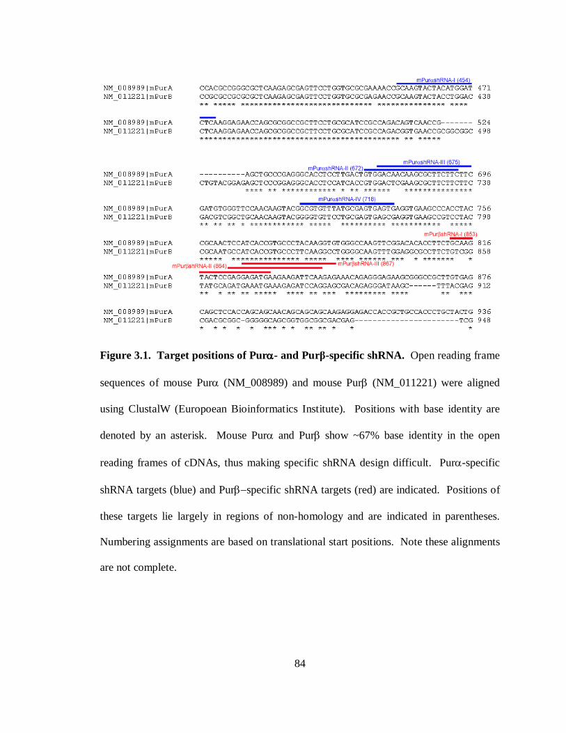
84
Figure 3.1. Target positions of Purαααα- and Purβ-specific shRNA. Open reading frame
sequences of mouse Purα (NM_008989) and mouse Purβ (NM_011221) were aligned
using ClustalW (Europoean Bioinformatics Institute). Positions with base identity are
denoted by an asterisk. Mouse Purα and Purβ show ~67% base identity in the open
reading frames of cDNAs, thus making specific shRNA design difficult. Purα-specific
shRNA targets (blue) and Purβ−specific shRNA targets (red) are indicated. Positions of
these targets lie largely in regions of non-homology and are indicated in parentheses.
Numbering assignments are based on translational start positions. Note these alignments
are not complete.
85
RESULTS
Validation of shRNA-mediated knockdown of mouse Purαααα or Purββββ in transfected
fibroblasts − To investigate the functional properties of endogenous Purα and Purβ in
cultured fibroblasts and VSMCs, a loss-of-function approach utilizing shRNA-mediated
RNA interference (RNAi) was undertaken. Mouse Purα and Purβ show ~67% base
identity in the open reading frames of cDNAs, thus making specific shRNA design
somewhat challenging. Nonetheless, taking advantage of several regions of non-
homology, multiple shRNA sequences were identified by computer analysis with the
theoretical potential to render the transcript encoding either mouse Purα or Purβ
susceptible to destruction by the RNAi pathway (134). The sites of Purα-, and Purβ-
specific transcript targeting by shRNA-mediated RNAi are shown in Figure 3.1. To test
whether constructed U6 promoter-driven shRNA expression plasmids would generate
functional shRNAs for knockdown of Purα or Purβ in cultured cells, immunoblotting of
whole cell extracts from transiently transfected AKR-2B MEFs co-expressing His-tagged
Purα or Purβ along with selected shRNAs was performed. His-tagged Pur transgenes
were used because of low shRNA plasmid transfection efficiency and since endogenous
Purα and Purβ migrate as a closely spaced doublet on Western blots making
interpretation of specific knockdown somewhat ambiguous. Results shown in Figure 3.2
verify that transfection of AKR-2B cells with wild type Purα or Purβ shRNA constructs
dramatically reduced the expression of either His-Purα (Figure 3.2, panel B) or His-Purβ
(Figure 3.2, panel A) in a highly specific manner. Importantly, the Purβ-specific shRNAs
86
had no discernable effect on His-Purα expression while Purα-specific shRNAs had no
effect on His-Purβ expression. LacZ shRNA or constructs encoding scrambled Pur
shRNA sequences (Scm) served as negative controls for Purα or Purβ-specific
knockdown (Figure 3.2). Similar results were obtained in transfected A7r5 cells
indicating that the RNAi pathway is functional in VSMCs as well. These findings also
reveal the necessity of empirically testing multiple shRNA sequences in cell types of
interest as the efficiency of RNAi (i.e. relative level of knockdown) can vary significantly
(Figure 3.2, panel B). To ensure that selected shRNAs would reduce endogenous Purα or
Purβ expression in fibroblasts or VSMCs, several additional transfection/infection and
immunoblotting experiments were performed. As shown in panel C of Figure 3.2, shRNA
sequences identified as promoting specific knockdown of His-tagged Pur proteins, also
facilitated knockdown of endogenous Purα or Purβ relative to scrambled control
sequences when assayed in plasmid-transfected primary mouse VSMCs. Moreover,
evaluation of multiple lentiviral-transduced AKR-2B MEF clones stably expressing Purβ
shRNA indicated definitive Purβ knockdown relative to a control clone expressing
scrambled sequence (Figure 3.2, panel D). As anticipated, there was some variability in
the absolute level of Purβ deficiency among different clones. One serendipitous clone
was isolated which was also lacking in Purα suggesting perhaps the ability of Purβ to
affect Purα gene expression (200). Importantly, deficiency of one or both Pur proteins in
these MEF clones correlated with an increase in smooth muscle α-actin (SMαA)
expression consistent with loss of Pur repressor activity. Relative differences in the levels
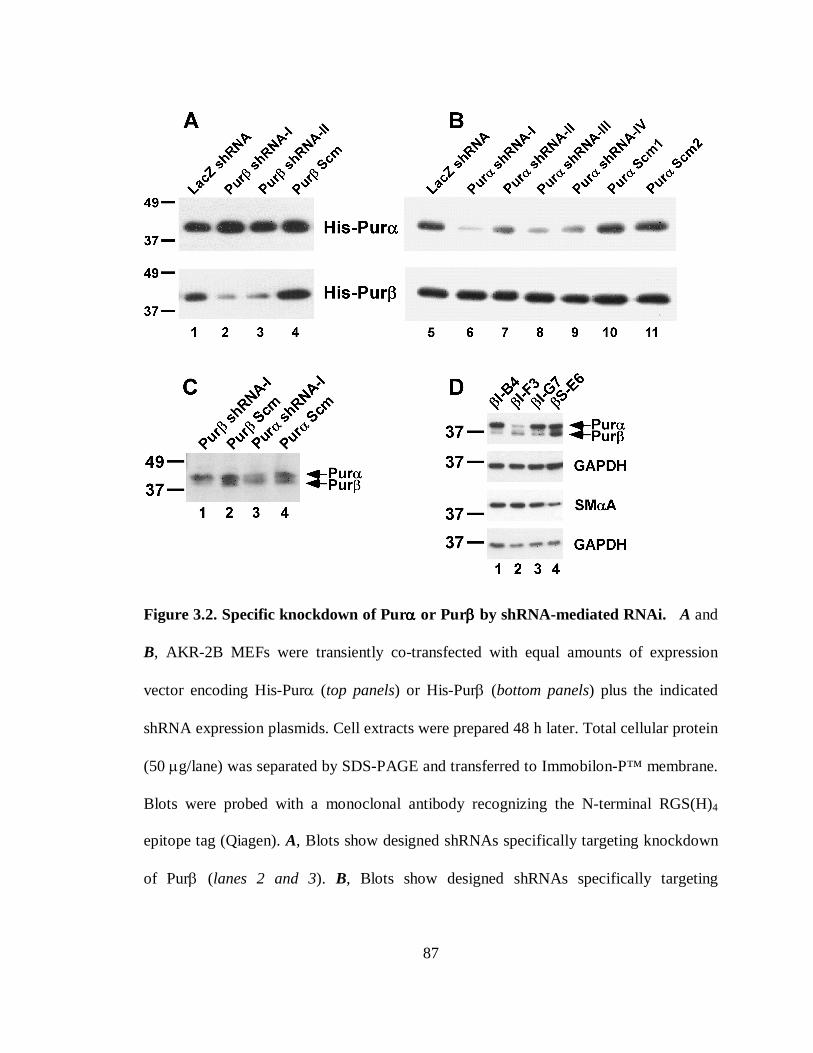
87
Figure 3.2. Specific knockdown of Purαααα or Purββββ by shRNA-mediated RNAi. A and
B, AKR-2B MEFs were transiently co-transfected with equal amounts of expression
vector encoding His-Purα (top panels) or His-Purβ (bottom panels) plus the indicated
shRNA expression plasmids. Cell extracts were prepared 48 h later. Total cellular protein
(50 µg/lane) was separated by SDS-PAGE and transferred to Immobilon-P™ membrane.
Blots were probed with a monoclonal antibody recognizing the N-terminal RGS(H)4
epitope tag (Qiagen). A, Blots show designed shRNAs specifically targeting knockdown
of Purβ (lanes 2 and 3). B, Blots show designed shRNAs specifically targeting
88
knockdown of Purα (lanes 6−9). Scm designates control constructs with scrambled
sequences (lanes 4, 10, and 11). LacZ designates cells transfected with a LacZ shRNA
construct (lanes 1 and 5). C, Primary C57BL/6J VSMCs were transiently transfected with
2 µg of the indicated shRNA expression plasmids. After 48 h incubation in growth
medium, whole cell extracts were prepared for analysis by immunoblotting (10 µg
protein/lane) with a rabbit polyclonal pan Pur antibody. D, Whole cell extracts of the
lentiviral-transduced AKR-2B MEFs clones expressing Purβ shRNA (lanes 1−3) or
scrambled control sequence (lane 4) were assayed by immunoblotting to detect Purα and
Purβ (10 µg/lane) or SMαA (1 µg /lane). Each blot was reprobed with a GAPDH
antibody to confirm equivalent protein loading. Numbers on the left denote the size of
prestained protein markers in kilodaltons.
89
of Pur and SMαA expression detected in lentiviral-transduced clones were validated by
reprobing immunoblots with anti-GAPDH as a loading control.
Functional consequences of Purαααα or Purββββ knockdown in fibroblasts and VSMCs −
Given previous studies indicating that Purα and Purβ function as co-repressors of the
SMαA promoter (28), we hypothesized that knockdown of Purα and/or Purβ in non-
differentiated fibroblasts would result in promoter activation. A series of co-transfection
experiments were conducted in AKR-2B MEFs with the use of a full-length SMαA
promoter-CAT reporter known as VSMP8 (Figure 3.3, panel A) and selected Purα- or
Purβ-specific shRNA expression plasmids. The VSMP8 promoter construct was chosen
because it contains mouse SMαA sequence (−1074 through the first intron) required for
smooth muscle-specific transgene expression in vivo (296) and, as such, exhibits very
weak transcriptional activity in transfected AKR-2B cells due, in part, to negative
regulation by endogenous Pur repressor proteins. As shown in Figure 3.3, panel B, co-
transfection of Purβ shRNA-I resulted in a dose-dependent increase in VSMP8 promoter
activity while a scrambled control construct (Purβ Scm) had no effect on this reporter.
Corroborative results were obtained in lentiviral-transduced AKR-2B MEFs where stable
shRNA-mediated knockdown of Purβ augmented SMαA protein expression (Figure 3.3,
panel C and Figure 3.2 panel D).
To evaluate whether the effect of Purβ knockdown was promoter context-dependent,
a truncated SMαA reporter known as VSMP4 was also tested for responsiveness to Purβ
deficiency. As previously documented (28), this mutant reporter exhibits unrestricted
90
MCAT enhancer activity due to the absence of 5′-nucleotides required for strand-specific
Pur/Pyr element recognition by endogenous Pur repressors (see Figure 3.3, panel A for
schematic). Hence it was not surprising to find that expression of Purβ shRNA-I had no
discernable effect on the transcriptional activity of VSMP4 in transfected MEFs (Figure
3.3, panel D). To assess whether knockdown of Purα would yield analogous results,
shRNA constructs demonstrating the maximum efficiency of Purα or Purβ knockdown
(Figure 3.2, Purα shRNA-I and Purβ shRNA-I) were transfected either individually or in
combination into AKR-2B MEFs together with VSMP8 or VSMP4 reporters (Figure 3.4,
panel A). Knockdown of Purα alone resulted in a modest ~2-fold enhancement in
VSMP8 activity while knockdown of Purβ alone induced VSMP8 by ~4-fold.
Importantly, a synergistic response (~12-fold activation over reporter only control) was
observed in AKR-2B MEFs expressing both Purα and Purβ shRNAs implying that
endogenous Pur repressors likely function in a collaborative manner to regulate the
transcriptional activity of the full-length SMαA promoter in this cell type. Transient
transfection of the same Purα or Purβ shRNA constructs in primary mouse VSMCs
yielded similar results although the extent of de-repression was less pronounced than seen
in AKR-2B MEFs (Figure 3.4, panel B). Relative to VSMP8, VSMP4 exhibited little or
no responsiveness to combined Purα and Purβ knockdown in both cell types. Evaluation
of other SMαA reporter constructs containing differing lengths of 5′-flanking region
indicated that significant promoter induction in response to co-knockdown of Purα and
Purβ in AKR-2B cells minimally required 5′ sequence extending to −195 (Figure 3.4,
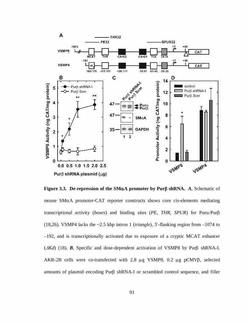
91
Figure 3.3. De-repression of the SMααααA promoter by Purβ shRNA. A, Schematic of
mouse SMαA promoter-CAT reporter constructs shows core cis-elements mediating
transcriptional activity (boxes) and binding sites (PE, THR, SPUR) for Purα/Purβ
(18,26). VSMP4 lacks the ~2.5 kbp intron 1 (triangle), 5′-flanking region from –1074 to
–192, and is transcriptionally activated due to exposure of a cryptic MCAT enhancer
(∆Kd) (18). B, Specific and dose-dependent activation of VSMP8 by Purβ shRNA-I.
AKR-2B cells were co-transfected with 2.8 µg VSMP8, 0.2 µg pCMVβ, selected
amounts of plasmid encoding Purβ shRNA-I or scrambled control sequence, and filler
92
DNA to 5.0 µg/dish. Whole cell extracts were prepared 48 h after transfection and
assayed for total protein and CAT reporter. C, Extracts of AKR-2B MEFs transduced
with lentiviral vectors encoding Purβ shRNA-I (lane 1) or scrambled sequence (lane 2)
were analyzed by Western blotting for detection of Purα and Purβ (10 µg protein/lane) or
SMαA (1 µg protein/lane). Note that the faster migrating Purβ band is specifically
reduced while SMαA is increased in shRNA-expressing cells. The SMαA blot was
reprobed for GAPDH as a loading control. Numbers on the left denote the size of
prestained protein markers in kilodaltons. D, De-repression of the SMαA promoter by
Purβ shRNA-I is promoter context-dependent. AKR-2B cells were co-transfected with
VSMP8 or VSMP4 ± 1 µg of the indicated shRNA plasmids then assayed as described
above. B and D, Promoter activity is expressed as CAT reporter divided by total cell
protein (mean ± SD). *, p < 0.025; **, p < 0.01 compared to reporter only control.
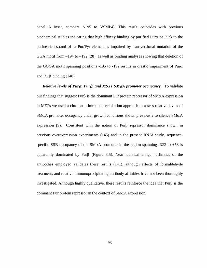
93
panel A inset, compare ∆195 to VSMP4). This result coincides with previous
biochemical studies indicating that high affinity binding by purified Purα or Purβ to the
purine-rich strand of a Pur/Pyr element is impaired by transversional mutation of the
GGA motif from −194 to −192 (28), as well as binding analyses showing that deletion of
the GGGA motif spanning positions -195 to -192 results in drastic impairment of Purα
and Purβ binding (148).
Relative levels of Purαααα, Purββββ, and MSY1 SMααααA promoter occupancy. To validate
our findings that suggest Purβ is the dominant Pur protein repressor of SMαA expression
in MEFs we used a chromatin immunoprecipitation approach to assess relative levels of
SMαA promoter occupancy under growth conditions shown previously to silence SMαA
expression (9). Consistent with the notion of Purβ repressor dominance shown in
previous overexpression experiments (145) and in the present RNAi study, sequence-
specific SSB occupancy of the SMαA promoter in the region spanning -322 to +58 is
apparently dominated by Purβ (Figure 3.5). Near identical antigen affinities of the
antibodies employed validates these results (141), although effects of formaldehyde
treatment, and relative immunoprecipitating antibody affinities have not been thoroughly
investigated. Although highly qualitative, these results reinforce the idea that Purβ is the
dominant Pur protein repressor in the context of SMαA expression.
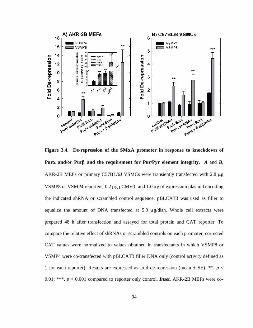
94
Figure 3.4. De-repression of the SMααααA promoter in response to knockdown of
Purαααα and/or Purββββ and the requirement for Pur/Pyr element integrity. A and B,
AKR-2B MEFs or primary C57BL/6J VSMCs were transiently transfected with 2.8 µg
VSMP8 or VSMP4 reporters, 0.2 µg pCMVβ, and 1.0 µg of expression plasmid encoding
the indicated shRNA or scrambled control sequence. pBLCAT3 was used as filler to
equalize the amount of DNA transfected at 5.0 µg/dish. Whole cell extracts were
prepared 48 h after transfection and assayed for total protein and CAT reporter. To
compare the relative effect of shRNAs or scrambled controls on each promoter, corrected
CAT values were normalized to values obtained in transfectants in which VSMP8 or
VSMP4 were co-transfected with pBLCAT3 filler DNA only (control activity defined as
1 for each reporter). Results are expressed as fold de-repression (mean ± SE). **, p <
0.01; ***, p < 0.001 compared to reporter only control. Inset, AKR-2B MEFs were co-
95
transfected with the indicated SMαA promoter-reporter constructs and a combination of
Purα plus Purβ shRNA-I plasmids or scrambled control plasmids then assayed as
described above. To assess the relative level of shRNA-mediated induction of each
reporter, CAT values obtained in Purα plus Purβ shRNA co-transfectants were divided
by CAT values measured in scrambled control co-transfectants. Results are expressed as
relative promoter induction (mean ± SE). **, p < 0.01; ***, p < 0.001 compared to
VSMP4.
96
DISCUSSION
Purα and Purβ are highly homologous proteins exhibiting ~70% primary sequence
identity (143). The highest levels of conservation are restricted to the central modular
repeat region of each molecule (10, 11) where the minimal nucleic acid-binding domain
resides (11, 143, 212, 310). Not surprising then is the finding that these proteins display
near-identical affinity for ssDNA oligonucleotides harboring PUR-elements (310).
Regions of primary sequence divergence within the amino- and carboxy-termini have
been implicated in directing specific interactions of Purα or Purβ with auxiliary protein
and/or nucleic acid-binding partners (92, 145). This finding has fueled speculation that
these regions of non-homology also confer paralog-specific function, as indicated by the
distinct transcriptional properties ascribed to Purα and/or Purβ in different cell types and
promoter contexts (54, 74, 162, 176, 214, 237, 238, 255, 256, 291, 303, 322). To test this
speculative view, we have utilized parallel loss-of-function approach to assess Pur-
paralog functional differences in the context of SMαA repression. These studies were
published in a report describing parallel gain-of-function analyses, as well as biochemical
dissection of protein/DNA and protein/protein interaction profiles comparing Purα and
Purβ (148). Collectively these results confirm that paralog and cell type-specific
functional differences exist between Purα and Purβ in the context of SMαA repression,
cis-element recognition, and transcription factor interaction profiles.
Mechanisms of ssDNA generation/binding by Pur proteins have consistently shown
that Purα and Purβ can associate with purine-rich ssDNA sequences in a sequence-
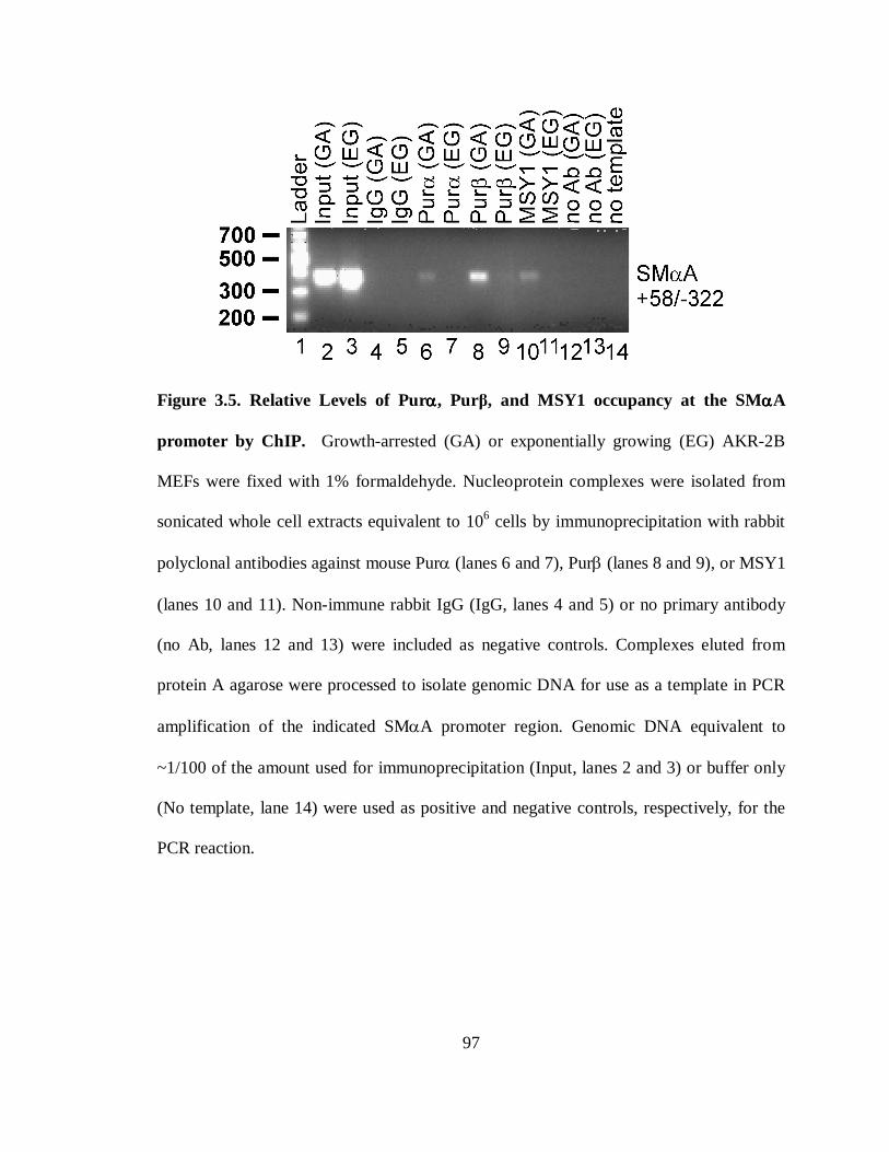
97
Figure 3.5. Relative Levels of Purαααα, Purβ, and MSY1 occupancy at the SMααααA
promoter by ChIP. Growth-arrested (GA) or exponentially growing (EG) AKR-2B
MEFs were fixed with 1% formaldehyde. Nucleoprotein complexes were isolated from
sonicated whole cell extracts equivalent to 106 cells by immunoprecipitation with rabbit
polyclonal antibodies against mouse Purα (lanes 6 and 7), Purβ (lanes 8 and 9), or MSY1
(lanes 10 and 11). Non-immune rabbit IgG (IgG, lanes 4 and 5) or no primary antibody
(no Ab, lanes 12 and 13) were included as negative controls. Complexes eluted from
protein A agarose were processed to isolate genomic DNA for use as a template in PCR
amplification of the indicated SMαA promoter region. Genomic DNA equivalent to
~1/100 of the amount used for immunoprecipitation (Input, lanes 2 and 3) or buffer only
(No template, lane 14) were used as positive and negative controls, respectively, for the
PCR reaction.
98
specific fashion as either homo- or heteromultimers of varying degrees depending upon
sequence and length of ssDNA (28, 141, 143, 144, 201, 310). RNA-mediated self-
association of Purα (91) and interaction between Purα and Purβ in the absence of nucleic
acid has also been reported (141, 142) although the stoichiometric extent of Pur
oligomerization and impact on nucleic acid binding remains unknown. The observation
of these seemingly heterogeneous complexes from nuclear extracts suggests that
collaborative activities of Purα and Purβ are necessary for in vivo repression of SMαA
expression. Results from chromatin immunoprecipitation (Figure 3.5) and transient
knockdown experiments in MEFs indicate that while Purβ appears to be the dominant
Pur protein repressor, Purα also contributes in a synergistic and cis-element-dependent
manner, as suggested by simultaneous knockdown in the presence of the full length
promoter (Figure 3.3 and Figure 3.4). These findings substantiate the idea that Purα and
Purβ are distinct and authentic repressors of the SMαA promoter, whereas the observed
derepression of SMαA expression in MEFs exhibiting stable Purα and/or Purβ
knockdown also validates their authenticity (Figure 3.2, panel D).
Consistent with previous models of repression, our data support a functional
cooperation between endogenous Purα and Purβ for cryptic MCAT enhancer regulation
(28). However marked synergistic activation of the full-length SMαA promoter in
response to simultaneous knockdown of both Purα and Purβ MEFs suggests that
transcriptional activation in AKR-2B cells is not only the result of cryptic MCAT
enhancer vacancy, but also a result of derepression at additional cis-elements and
99
liberation of other trans-activators influenced by Pur proteins in fibroblasts (47, 272).
This explanation may explain the less significant response observed during simultaneous
knockdown of Pur proteins in primary VSMCs (Figure 3.4, panel B). An alternate
explanation is that the existing levels of endogenous Purα and Purβ in VSMCs are
sufficiently low such that SMαA expression is constitutively active and knockdown
effects are difficult to detect.
Delineation of this issue was goal of gain-of-function studies that complemented the
loss-of-function studies presented here (148). The results of these investigations showed
that forced overexpression of Purβ, but not Purα, confers repression in cultured clonal
and primary VSMCs from both full-length promoter constructs (VSMP8) and truncated
constructs lacking Pur-binding sites flanking the core MCAT enhancer (VSMP4). These
findings contrast Purα overexpression studies in AKR-2B MEFs which showed Purα-
mediated repression is restricted to the core enhancer element. These studies confirm that
Pur proteins repress expression of SMαA by distinct mechanisms, dependent upon cell
type. The preference for Purβ, but not Purα, to repress expression of SMαA at cis-
elements proximal to the MCAT enhancer element also suggests that differences exist
either in the transcription factor interaction profiles between Purα and Purβ or their
inherent binding affinites to these elements.
Interaction profiles of Purα and Purβ versus transcription factors known to interact
and activate transcription of SMαA from other numerous cis-elements were generated by
an ELISA-based method (elements and factors detailed in Figure 1.1)(28, 47, 145, 272,
319). The results of this analysis showed that indeed Purα and Purβ display differential
100
affinity for other known transcriptional activators. In both AKR-2B and A7r5 clonal rat
VSMCs, Purβ shows greater interaction potential with MSY1, TEF-1, and Sp1/3 than
does Purα, although slight cell type and subcellular compartmental differences were
noted. The elevated interaction of Purβ with Sp1/3 is particularly interesting due to
recent findings which showed the capacity of Pur proteins to restrict enhancer activity by
displacing Sp1/3 and Smad proteins from THR and SPUR elements (47, 272). In vitro
binding studies examining the apparent affinities of recombinant Purα and Purβ for
ss/dsDNA oligonucleotides representative of the MCAT enhancer element (PE32), THR,
and SPUR show that both Purα and Purβ display high, near identical affinities for the
purine-rich strands of the PE and SPUR elements. Both proteins show reduced affinity
for either strand of THR, a predictable finding based on observed purine content. The
results of ss/dsDNA-binding studies indicate that differences in repressive capacities
observed between Purα and Purβ are not due to differences in the abilities of these
proteins to recognize and bind cis-elements, but likely due to distinct capacities to
interact with or displace transcriptional activators.
Collectively these findings support a model that suggests that reprogramming of
constitutive SMαA in activated SMCs towards a repressive state likely involves
deactivation of Sp1/3:SPUR enhancer complex by displacement caused by increased
nuclear concentrations and binding of Purβ to this element. Furthermore,
compartmentalization of Purα to the nucleus may permit this mechanism. Maintenance
of the repressed state, as it exists in fibroblast-like cell types, likely proceeds through
restriction of the MCAT enhancer element by both Purα and Purβ. This model also

101
suggests that distinct functions of Purα and Purβ aid in the plasticity of SMαA
expression in response to environmental cues. The identity of environmental factors that
upregulate, compartmentalize, or ‘activate’ Pur proteins for genetic and phenotypic
reprogramming of VSMCs remains to be determined.
102
CHAPTER IV. SOLUTION CHARACTERIZATION REVEALS THE
QUATERNARY STRUCTURE OF RECOMBINANT MOUSE PUR ββββ
The experimental methods, results and interpretations described herein have been
published previously in a revised form:
Ramsey JE, Daugherty MA, Kelm RJ, Jr. (2007) Hydrodynamic studies on the quaternary
structure of recombinant mouse Purbeta. J Biol Chem.282(3):1552-60.
INTRODUCTION
Purα and Purβ are members of a highly conserved family of nucleic acid-binding
proteins related by primary structure and a propensity to interact with single-stranded
DNA (ssDNA)1 or RNA sequences rich in purine nucleotides (reviewed in (92)). Diverse
regulatory functions attributed to these proteins include gene transcription, mRNA
translation, cell growth, and cell cycle progression (7, 57, 58, 125, 133, 146, 155, 177,
265). Purα and Purβ are ~70% identical at the amino acid level (143). Biochemical
investigation of deletion mutants has shown that each protein possesses a minimal
ssDNA/RNA-binding domain composed of a unique set of highly homologous sequence
repeats (70, 133, 145). However, significant sequence differences between Purα and Purβ
existing near the N- and C-termini suggest that each protein may have evolved to perform
distinct functions (143). Purα and Purβ have been reported to bind to a PUR-element in a
highly asymmetric polypurine/polypyrimidine tract located in the 5′ flanking region of
103
the mouse SMαA gene (28, 141). It has been hypothesized that strand-specific binding by
Purα/Purβ to this element disrupts a core MCAT enhancer motif thereby repressing
SMαA promoter activity in cultured fibroblasts and vascular smooth muscle cells (28).
Despite biochemical similarities, gain-of-function studies suggest that Purα and Purβ are
not redundant in terms of their transcriptional repressor activity toward the full-length
mouse SMαA promoter in transfected vascular smooth muscle cells (145).
In light of the specific protein-DNA, protein-RNA, and protein-protein interactions
attributed to Purα and Purβ, and their potential relevance in modulating cell growth and
differentiation, a need has emerged for the elucidation of the biophysical factors
governing nucleic acid recognition. The mechanism of DNA-binding is of particular
interest since DNA is thought to exist primarily in a double-stranded B-form helical
configuration in vivo. Since Purα and Purβ preferentially bind to ssDNA or, in some
cases, non-B-form structures (6), this aspect of Pur protein function has been investigated
to a greater degree. Several reports have indicated that Purα is capable of helix
destabilization despite showing no intrinsic helicase or ATPase activity (56, 310). This
has led to speculation that Pur proteins may promote strand displacement by
opportunistic binding to transiently-formed ssDNA structures. This notion is supported
by the finding that the 5′-MCAT enhancer region of the SMαA promoter has the
propensity to adopt a partially unpaired configuration in response to transforming growth
factor β1 signaling (9). Another potentially important aspect of DNA-binding that was
hinted at by previous studies involves the oligomeric state of Pur proteins in the presence
and absence of ssDNA. Experiments reported by Kelm and coworkers (104), suggested
104
that Purα and Purβ bound to a SMαA promoter-derived PUR element as either hetero- or
homodimeric nucleoprotein complexes. However, the conditions used in those
experiments did not permit determination of whether or not dimerization was a
prerequisite for, or a consequence of, ssDNA-binding. In this regard, a report by Gallia
and colleagues has pointed to a critical role for RNA in mediating self-association of
Purα (91).
In the present study, we elucidate the quaternary structure of purified recombinant
Purβ by employing hydrodynamic and thermodynamic approaches to examine the
macromolecular character of nucleic-acid free Purβ in solution. Size exclusion
chromatography coupled to static and dynamic light scattering-based detection systems
revealed Purβ to be an asymmetric protein capable of homodimeric self-association. This
principal finding was confirmed by analytical ultracentrifugation which established that
mouse Purβ does indeed exist in a reversible monomer-dimer equilibrium characterized
by a dissociation constant of ~1 µM in the absence of nucleic acid. Hydrodynamic
analyses further suggested that homodimeric Purβ assumes a non-spherical conformation
in solution. We propose a model in which dimerization may affect ssDNA-binding in a
manner regulated by a mass-action governed self-association of Purβ. This type of a
mechanism may be particularly relevant to pathophysiological states of the heart and
vasculature where elevated Purβ levels have been noted (104, 271).
105
MATERIALS AND METHODS
Expression and purification of nucleic acid-free N-HisPurββββ − E. coli strain JM109
was transformed with the plasmid pQE30-N-HisPurβ which contains cDNA encoding an
amino-terminal hexahistidine tagged mouse Purβ fusion protein (N-HisPurβ) (141).
Transformants were selected after growth on LB-agar medium containing 40 µg/ml
carbenicillin (Sigma) for 16 h at 37°C. A single colony was used to inoculate 50 ml LB
medium containing 40 µg/ml carbenicillin. These starter cultures were allowed to grow
for 8 h at 37°C to an optical density of >1.2 at 600 nm, at which time 10 ml of starter was
used to inoculate 1 liter of pre-warmed Terrific Broth II (QBiogene) with 40 µg/ml
carbenicillin. Cultures were allowed to grow at 37°C until an optical density of 0.6 at 600
nm was reached. Cultures were supplemented with ampicillin (Sigma) to 0.1 mg/ml and
isopropylthiogalactopyranoside (Sigma) was then added to a final concentration of 1 mM
to induce N-HisPurβ expression. After 4 h of additional growth, cells were harvested by
centrifugation and cell pellets were stored at -80°C. Cell pellets equivalent to 4 liters
were allowed to thaw in 20 ml of buffer A (50 mM sodium phosphate pH 8.0, 300 mM
NaCl, 10 mM imidazole, 10 mM β-mercaptoethanol) and completely resuspended on ice.
Protease inhibitors leupeptin, aprotinin, and pepstatin A were each added to a final
concentration of 1 µg/ml and phenylmethysulfonyl fluoride was added to 0.1 mM. Lysis
was facilitated by the addition of egg white lysozyme (Sigma) to a final concentration of
1 mg/ml and incubated on ice for 20 min with occasional stirring. The cell suspension
106
was sonicated with a Branson Sonifier model 150 (setting 10) for a total of six 10 s bursts
with 1 min incubations on ice between bursts to avoid overheating. Lysates were cleared
by centrifugation at 14,000 × g for 30 min at 4°C. A total of 5 ml of 50% Ni-NTA
agarose slurry (Qiagen) was added to the cleared lysate followed by 5M NaCl to a final
concentration of 1 M. Bovine pancreatic DNase I and RNase A (Sigma) were each added
to 59 and 92 units/ml, respectively (based on manufacturer specified activities) and the
lysate-Ni-NTA agarose mixture was slowly rocked for 2 h at room temperature. The
lysate-resin mixture was then gently centrifuged at 1000 × g for 2 min at 4°C to pellet the
Ni-NTA agarose. Supernatant was removed and 25 ml of buffer B (50 mM sodium
phosphate pH 8.0, 2 M NaCl, 10 mM imidazole, 10 mM β-mercaptoethanol plus protease
inhibitors) was added to the resin. The mixture was then incubated approximately 14 h at
4°C with slow rocking. Gentle centrifugation at 1000 × g for 2 min at 4°C was used to
pellet the resin which was subsequently resuspended in buffer A and loaded into a 1.5 cm
diameter column. The rest of the purification procedure was carried out at room
temperature. The resin was washed with buffer A until the absorbance of the flow
through at 280 nm reached a baseline level (A280 ≤ 0.02 absorbance units). N-HisPurβ
was eluted by application of buffer C (50 mM sodium phosphate pH 8.0, 300 mM NaCl,
500 mM imidazole, 10 mM β-mercaptoethanol). Eluted protein was concentrated using a
centrifugal device (Centriprep YM-10, Millipore). Size exclusion chromatography was
carried out on a 1.5 cm × 98 cm column packed with Sephacryl 200 HR resin (Sigma)
equilibrated in buffer E (50 mM sodium phosphate pH 7.5, 200 mM NaCl, 0.5 mM
EDTA, 2 mM dithiothreitol) and run at a flow rate of 0.5 ml/min. The optical density of
107
the eluate was monitored with a Pharmacia model UV-10 UV/Vis detector. The column
was calibrated using bovine serum albumin, ovalbumin, carbonic anhydrase, and
cytochrome c (low molecular weight size-exclusion protein standards, Sigma). Fractions
corresponding to the major dimeric peak of Purβ were pooled and concentrated as
described above. Protein purity was assessed by Coomassie Blue staining of N-HisPurβ
reduced with 300 mM β-mercaptoethanol and resolved by SDS-PAGE on 12% (40:1
acrylamide:bisacrylamide) mini-gels. Preparations used for hydrodynamic studies were
judged to be >95% homogenous N-HisPurβ under reducing conditions. To assess the
level of nucleic acid-contamination, baseline-corrected absorbance spectra of purified N-
HisPurβ were obtained using a Cary Bio100 dual beam spectrophotometer (Varian). A
theoretical molar extinction spectrum of N-HisPurβ was calculated using SEDNTERP
software (164) based on the method of Pace and coworkers (211). Protein concentration
was determined spectrophotometrically assuming an extinction coefficient of 18,610 M-
1cm-1 at a wavelength of 280 nm and a monomeric relative molecular weight of 35,168.6
(calculated using SEDNTERP). To further test for the presence of nucleic acid, purified
N-HisPurβ (1.4 mg in 500 µl) was extracted twice with an equal volume of buffered-
phenol, followed by extraction with an equal volume of phenol:chloroform:isoamyl
alcohol (25:24:1), and an equal volume of chloroform. The aqueous phase of the extract
was lyophilized and redissolved in ultrapure water three times and then dissolved one
final time in 200 µl of ultra pure water. Baseline corrected absorbance spectrum of the
lyophilized extract was obtained to ensure the absence of a peak at 260 nm.
108
Hydrodynamic analysis of N-HisPurββββ by size-exclusion chromatography-laser light
scattering and dynamic light scattering − Molecular mass and hydrodynamic radius
measurements of N-HisPurβ in solution were made by size-exclusion chromatography
(SEC) coupled light scattering techniques. These experiments were performed at the W.
M. Keck Foundation Biotechnology Resource Laboratory, Biophysics Facility, Yale
University, New Haven, CT as described (81, 306). The analytical system consisted of a
Superose 6 (Pharmacia-Amersham) size-exclusion column coupled to four detection
systems configured in series including a UV absorbance detector (Kratos model 773
variable wavelength, Applied Biosystems), a laser (static) light scattering (LLS) detector
(DAWN DSP, Wyatt Technologies), a refractive index detector (OPTILAB, Wyatt
Technologies), and a dynamic light scattering (DLS) detection system (DYNAPRO
TITAN, Wyatt Technologies). The DLS light source used was a DAWN EOS 633 nm
laser (Wyatt Technologies). Solvent delivery was carried out by a Waters 510 HPLC
pump (Waters Corp.) equipped with pulse-dampening transducers and a Rheodyne 7125
sample injection valve. Buffer E was used as the solvent in this analysis. Multiple
volumes of pre-filtered (0.22 µm Durapore, Millipore) N-HisPurβ (2.0 mg/ml) were
injected and eluted at controlled flow rate of 0.3 ml/min.
LLS data was collected at an angle of 90° (the highest scattering signal) and analyzed
by ASTRA software package (Wyatt Technologies) fitting implementations of the Zimm
formalism of the Rayleigh-Debye-Gans light scattering model for dilute solutions of
polymers, which relates the amount of scattered light to the concentration and weight
average molecular weight of solute (81, 279, 306):
109
K*c/R(θ) = 1/MwP(θ) + 2A2c (Equation 4.1)
In this relationship R(θ) is the intensity of excess scattered light at angle θ, c is the
concentration of the solute, Mw is the weight average molecular weight of the solute, A2 is
the second virial coefficient, K* is an optical parameter equal to 4π2n2(dn/dc)2/(λ4NA), n
is the refractive index, NA is Avogadro’s number, and λ is the wavelength of the scattered
light. The angular dependence of the scattered light is described by the function 1/P(θ),
whose first order expansion gives:
1/P(θ) = 1 + (16π2/3λ2)<r g2>sin2(θ/2) + … (Equation 4.2)
where <r g2> is the root mean square radius of gyration. The Zimm fitting formalism (81)
was used for the determination of Mw of N-HisPurβ.
DLS measurements were made at an angle of 111° with a 2 second collection
interval. Time resolved homodyne scatter intensity fluctuations were analyzed using
Dynamics Software (Protein Solutions) which implements the cumulants method (152) to
determine the time dependence of diffusive motion also referred to as the intensity
autocorrelation function, G(τ) (18, 213, 308):
G(τ) = B[1+αexp(-2DTq2τ)] (Equation 4.3)
110
where B is the average baseline intensity, α is an instrument-specific correction factor, DT
is the concentration-dependent translational diffusion constant of the solute, τ is a delay
time, and q is the scattering vector equal to (4πn/λ)sin(θ/2), in which n is the refractive
index of the solvent, λ is the wavelength of the scattered light and θ is the scattering
angle. Equation 4.3 describes the relationship between the time dependency of fluctuation
in scatter intensity and the translational diffusion coefficient. Generally, large molecules
diffuse slowly and generate scatter signals that fluctuate slowly. The opposite is true for
small molecules. The value of DT was used to estimate the apparent hydrodynamic radius
of an equivalent sphere by way of the Stokes-Einstein relationship:
Rh= kT/6πηDT (Equation 4.4)
where k is the Boltzmann constant, T is the absolute temperature, and η is the temperature
corrected viscosity of the solvent.
Sedimentation velocity analytical ultracentrifugation of N-HisPurββββ −
Sedimentation velocity experiments were carried out in a Beckman/Coulter Optima XL-
I/XL-A equipped with an An50Ti rotor. Radial concentration distributions were
measured at 50,000 rpm and 4°C using Rayleigh interference optics. Protein samples
were gel filtered over a 1.5 cm × 98 cm Sephacryl 200 HR size-exclusion column pre-
equilibrated and eluted with buffer E (described above) as a final step prior to
sedimentation. The fraction corresponding to the absorbance maximum of the presumed
dimeric peak was used to prepare a dilution series of N-HisPurβ over a ten-fold
111
concentration range starting at 43.5 µM. The reference buffer consisted of a buffer E
eluate from the size exclusion column. Blank subtracted Rayleigh interference scans were
recorded at 1 min intervals. Sedimentation velocity data were analyzed by the dc/dt
method to generate apparent sedimentation coefficient distributions, g(s*) (266), with the
use of DCDT+ software (217). Direct fitting of time-resolved concentration difference
(∆c versus radius) curves to numerical solutions to the Lamm equation describing
multiple, interacting species models and kinetic models was performed using SEDANAL
v4.3 software (268). Goodness of fits were judged by visualization of residuals and fitting
statistics. Temperature corrected values for the partial specific volume of N-HisPurβ (ῡ),
as well as density (ρ), and viscosity (η), of buffer E were calculated using the program
SEDNTERP. Resulting values are as follows: ῡ = 0.7109 ml g-1, ρ = 1.0149 g ml-1, and η
= 0.1635 g cm-1 s-1. Molecular shape modeling was also carried out with SEDNTERP.
Protein integrity was assessed after sedimentation by SDS-PAGE to ensure that the
samples were intact.
Sedimentation equilibrium analytical ultracentrifugation of N-HisPurββββ −
Sedimentation equilibrium experiments were carried out in cells fitted with six-sector
charcoal-Epon centerpieces (1.2-cm path). Protein and reference buffer samples were
prepared as described above. Sample dilution series were made from the dimeric peak
fraction over a ten-fold concentration range. Sedimentation was carried out at rotor
speeds of 22,000, 28,000, and 35,000 rpm at 4°C. Five scans were averaged to remove
noise. Equilibrium was judged to be achieved by the superposition of scans taken 6 h
112
apart and by analysis with the MATCH package implemented by HETEROANALYSIS
software (49).
Blank corrected sedimentation equilibrium data were fit using HETEROANALYSIS
software to numerous mathematical models describing radial concentration distributions
including single ideal species (equation 4.5), single non-ideal species (equation 4.6),
monomer-Nmer equilibria (equation 4.7), monomer-dimer with incompetent monomer
(equation 4.8), monomer-dimer with incompetent dimer (equation 4.9) and monomer-
Nmer-Qmer equilibria (equation 4.10):
A(r,λ) = δλ + ελl c0exp[M*φ(r2-r02)] (Equation 4.5)
A(r,λ) = δλ + ελl c0exp[M*φ(r2-r02) – 2A2M{w(r)-w(r0)}] (Equation 4.6)
A(r,λ) = δλ + ελl c0exp[M*φ(r2-r02)] + Nελl c0
Nexp[lnKa + NM*φ(r2-r02)]
(Equation 4.7)
A(r,λ) = δλ + ελl c0exp[M*φ(r2-r02)] + ελl c’0exp[M*φ(r2-r0
2)]
+ 2ελl c02exp[lnKa + 2M*φ(r2-r0
2)]
(Equation 4.8)
113
A(r,λ) = δλ + ελl c0exp[M*φ(r2-r02)] + ελl d’0exp[2M*φ(r2-r0
2)]
+ 2ελl c02exp[lnKa + 2M*φ(r2-r0
2)]
(Equation 4.9)
A(r,λ) = δλ + ελl c0exp[M*φ(r2-r02)] + Nελl c0
Nexp[lnKa1 + NM*φ(r2-r02)] +
Qελl c0Qexp[lnKa2 + QM*φ(r2-r0
2)]
(Equation 4.10)
where A(r,λ) is the radially-dependent absorbance at radial position, r, and wavelength,
λ, δλ is the baseline offset, ελ is the molar extinction coefficient, l is the path length, c0 is
the molar concentration of the monomer at the arbitrary reference radial position r0, c’0 is
the molar concentration of the incompetent monomer at the arbitrary reference radial
position r0, d’0 is the molar concentration of the incompetent dimer at the arbitrary
reference radial position r0, A2 is the second virial coefficient, M is the monomer
molecular weight, w(r) refers to the concentration of the monomer on a weight/volume
scale at distance r, w(r0) is the weight/volume concentration at reference position r0, N is
the stoichiometry of association, Ka is the association constant, Q is the stoichiometry of
the higher order oligomeric association. M* refers to the buoyant molecular weight, equal
to M(1- ῡρ). φ is equal to ω2/RT, in which ω is the angular velocity, R is the gas constant,
and T is the absolute temperature. Goodness of fit was judged by fit statistics and visual
inspection of residuals for systematic deviations.
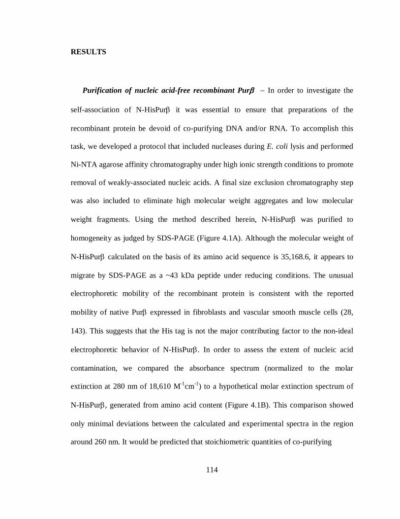
114
RESULTS
Purification of nucleic acid-free recombinant Purββββ − In order to investigate the
self-association of N-HisPurβ it was essential to ensure that preparations of the
recombinant protein be devoid of co-purifying DNA and/or RNA. To accomplish this
task, we developed a protocol that included nucleases during E. coli lysis and performed
Ni-NTA agarose affinity chromatography under high ionic strength conditions to promote
removal of weakly-associated nucleic acids. A final size exclusion chromatography step
was also included to eliminate high molecular weight aggregates and low molecular
weight fragments. Using the method described herein, N-HisPurβ was purified to
homogeneity as judged by SDS-PAGE (Figure 4.1A). Although the molecular weight of
N-HisPurβ calculated on the basis of its amino acid sequence is 35,168.6, it appears to
migrate by SDS-PAGE as a ~43 kDa peptide under reducing conditions. The unusual
electrophoretic mobility of the recombinant protein is consistent with the reported
mobility of native Purβ expressed in fibroblasts and vascular smooth muscle cells (28,
143). This suggests that the His tag is not the major contributing factor to the non-ideal
electrophoretic behavior of N-HisPurβ. In order to assess the extent of nucleic acid
contamination, we compared the absorbance spectrum (normalized to the molar
extinction at 280 nm of 18,610 M-1cm-1) to a hypothetical molar extinction spectrum of
N-HisPurβ, generated from amino acid content (Figure 4.1B). This comparison showed
only minimal deviations between the calculated and experimental spectra in the region
around 260 nm. It would be predicted that stoichiometric quantities of co-purifying
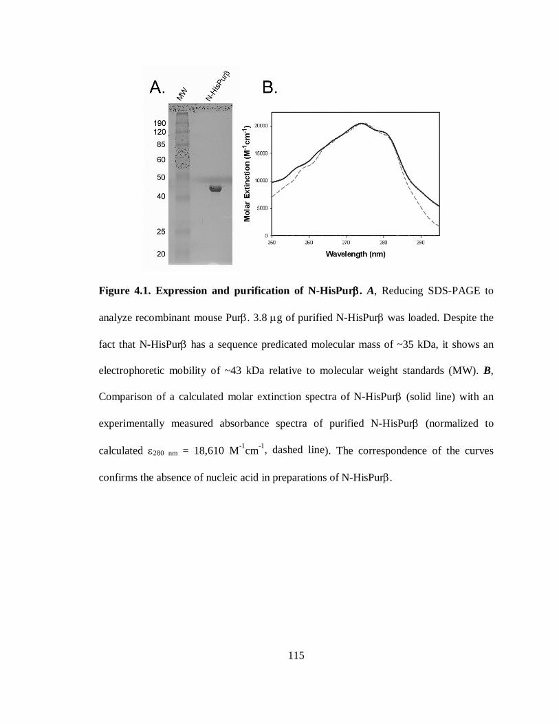
115
Figure 4.1. Expression and purification of N-HisPurββββ. A, Reducing SDS-PAGE to
analyze recombinant mouse Purβ. 3.8 µg of purified N-HisPurβ was loaded. Despite the
fact that N-HisPurβ has a sequence predicated molecular mass of ~35 kDa, it shows an
electrophoretic mobility of ~43 kDa relative to molecular weight standards (MW). B,
Comparison of a calculated molar extinction spectra of N-HisPurβ (solid line) with an
experimentally measured absorbance spectra of purified N-HisPurβ (normalized to
calculated ε280 nm = 18,610 M-1cm-1, dashed line). The correspondence of the curves
confirms the absence of nucleic acid in preparations of N-HisPurβ.
116
nucleic acids would result in a large spectral difference in this range since nucleoside-5′-
monophosphates have a molar extinction coefficient on the average of 104 M-1cm-1 at or
near 260 nm (32). Furthermore, absorbance spectra of phenol-chloroform extracts of
purified N-HisPurβ preparations showed no species with a λmax of 260 nm (data not
shown). Collectively, these data indicate that the preparations of N-HisPurβ used in this
study were free of co-purifying nucleic acids.
Buffer E was chosen as the buffer condition for all sedimentation and light-scattering
experiments. We have determined that the solubility of N-HisPurβ relies heavily on ionic
strength and reducing agent concentration (data not shown). Dialysis of protein at
moderate concentrations (~ 1 mg/ml) leads to loss of protein (likely deposition on vessel
surfaces, as sample can be recovered by addition of high salt buffers). The salt
concentration of buffer E (200 mM NaCl) was determined to be the minimum quantity to
limit this effect and to maximize stability.
Hydrodynamic analysis of N-HisPurββββ by size-exclusion coupled light scattering
techniques − A series of physical techniques that make use of macromolecular light
scattering phenomena were used to investigate the hydrodynamic properties of N-
HisPurβ in solution. SEC-LLS-DLS is a well-suited means of investigating the
hydrodynamic character of proteins. It is non-destructive and each individual light
scattering detection technique can be performed in series after a size fractionation step.
The use of SEC as a preliminary step to light scattering can eliminate some of the
ambiguity created in performing weight-average measurements (81, 279, 306).
117
Figure 4.2. Molecular size measurements of N-HisPurββββ in solution by light
scattering techniques. N-HisPurβ was applied to a Superose 6 column and eluted with
buffer E at high pressure. Elution was monitored by refractive index (RI) changes and
analyzed by LLS (A) and DLS (B) to determine the solution characteristics of the
recombinant protein. A, The RI traces (normalized to molar mass values, solid lines) and
LLS molar mass measurements (points) of N-HisPurβ (dark gray) show an asymmetric
elution pattern suggesting that the protein elutes as a mixture of self-associating and
monomeric species, as compared to a bovine serum albumin standard (light gray). The
118
weight average molar mass of the elution peak determined by LLS was 67.7 ± 4.12 kDa.
B, As determined by DLS, a dependence of the weight average RH on loading quantity
was also noted. Solid lines represent RI traces whereas individual data points represent
the DLS determined weight average hydrodynamic radius measurements of eluting N-
HisPurβ collected at different loading concentration (19.9 pmoles in 350 µl, dark gray;
5.1 pmoles in 100 µl, light gray). Again, RI trace values were normalized to RH values.
119
The results of applying the SEC-LLS to solutions of N-HisPurβ reveal an asymmetric
distribution of refractive index (RI) and molar mass measurements in the sole eluting
peak of N-HisPurβ, as compared to a bovine serum albumin standard (Figure 4.2A).
These results are consistent with a polydisperse mixture that elutes as a single, albeit
asymmetric peak. On the sole basis of RI signal, one might surmise that the asymmetric
shape of the elution peak could arise from interaction of N-HisPurβ with the column
matrix. Although this could conceivably cause a tailing effect on elution, it would not
cause a broadening in the distribution of weight-average molar mass measurements as is
seen for N-HisPurβ (Figure 4.2A). Instead, this effect is likely due to polydispersity. The
number average of the weight average molar mass measurements across the elution peak
is 67.7 ± 4.12 kDa corresponding to a 6.08% degree of polydispersity. Based on the
number average molar mass, this suggests that N-HisPurβ exists as an interacting mixture
of monomers and dimers (expected dimeric Mr of 70,337.2).
Weight average hydrodynamic radii (RH) determinations on SEC fractionated N-
HisPurβ by DLS were also consistent with a self-associating system. It was found that the
number average RH across the top 10% of the eluting peak (region of peak where scatter
signal is strongest and RH values are approximately constant) is dependent upon the
loading quantity of N-HisPurβ (Figure 4.2B). The resultant RH values for loading
quantities of 19.9 pmoles and 5.1 pmoles were 4.3 nm and 3.8 nm, respectively. From
these data, it is apparent that the weight average RH of the eluting solution is dependent
upon a mass-action governed self-association of N-HisPurβ. These RH values are very
different from the calculated RH = 2.72 nm for an equivalent non-compressible sphere
120
with a molecular mass of 70,337.3 Da. The RH value of 4.3 nm is expected to result in a
frictional coefficient ratio (ƒ/ƒ0) of 1.56, as determined by analysis with the Dynamics
Software (Protein Solutions). Collectively, findings based on light scattering suggest the
N-HisPurβ participates in a monomer-dimer equilibrium and that the dimeric form
assumes an asymmetric shape in solution.
Sedimentation velocity analysis of recombinant Purββββ − Quantitative hydrodynamic
and thermodynamic analyses aimed at investigating the oligomeric structure of N-
HisPurβ in solution were performed by analytical ultracentrifugation. Sedimentation
velocity experiments were carried out to investigate the hydrodynamic properties of
recombinant Purβ and to validate the observations made by SEC-LLS-DLS studies. The
apparent sedimentation coefficient distribution function, g(s*), for solutions of N-
HisPurβ at four loading concentrations ranging from 4.7 µM to 43.5 µM is shown in
Figure 4.3A. Analysis of the normalized weight average sedimentation coefficient (s20°,w)
from g(s*) distributions shows an increase as a function of loading concentration (Figure
4.3B).
The presence of a single peak in the g(s*) data shown in Figure 4.3A may not imply a
single sedimenting species. This is because the effects of diffusion in these experiments
may hide heterogeneity, especially in the case where the sedimentation coefficients of all
sedimenting species vary only slightly (216). However, the ensemble of data suggests a
diffuse mixture of species whose sedimentation is dynamic and dependent upon the
changing radial concentration distribution over time (267).
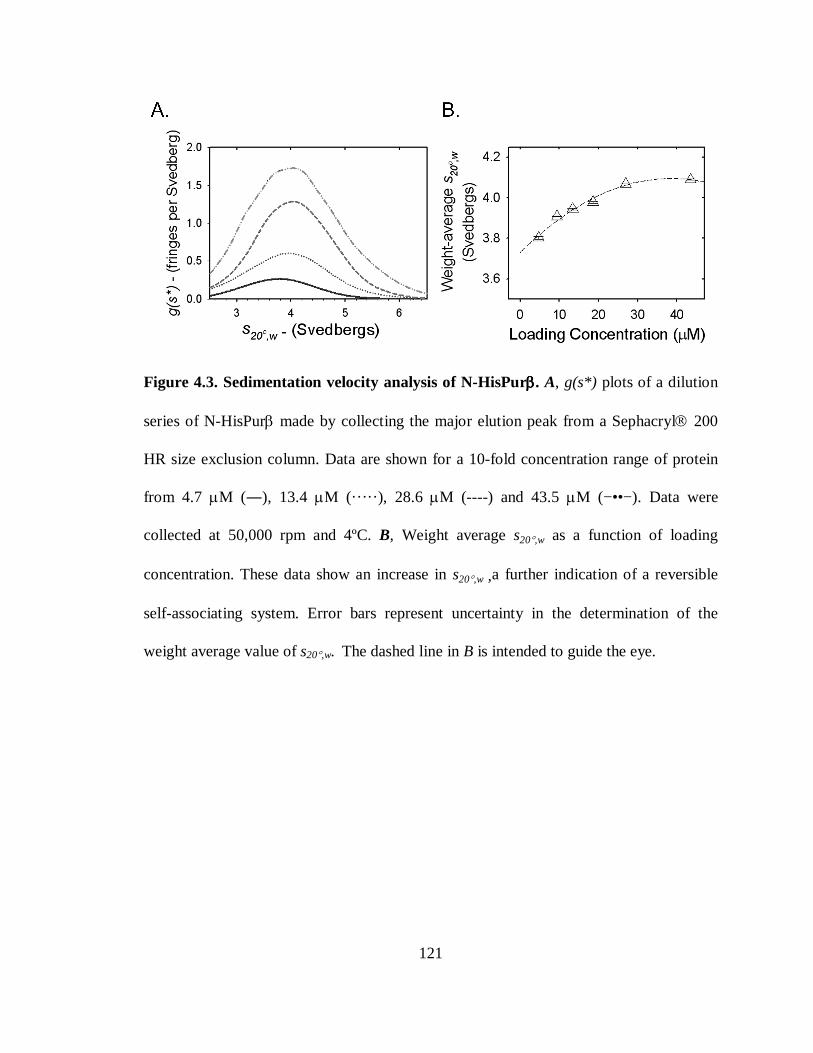
121
Figure 4.3. Sedimentation velocity analysis of N-HisPurββββ. A, g(s*) plots of a dilution
series of N-HisPurβ made by collecting the major elution peak from a Sephacryl 200
HR size exclusion column. Data are shown for a 10-fold concentration range of protein
from 4.7 µM (―), 13.4 µM (·····), 28.6 µM (----) and 43.5 µM (−••−). Data were
collected at 50,000 rpm and 4ºC. B, Weight average s20°,w as a function of loading
concentration. These data show an increase in s20°,w ,a further indication of a reversible
self-associating system. Error bars represent uncertainty in the determination of the
weight average value of s20°,w. The dashed line in B is intended to guide the eye.
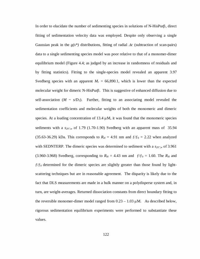
122
In order to elucidate the number of sedimenting species in solutions of N-HisPurβ, direct
fitting of sedimentation velocity data was employed. Despite only observing a single
Gaussian peak in the g(s*) distributions, fitting of radial ∆c (subtraction of scan-pairs)
data to a single sedimenting species model was poor relative to that of a monomer-dimer
equilibrium model (Figure 4.4; as judged by an increase in randomness of residuals and
by fitting statistics). Fitting to the single-species model revealed an apparent 3.97
Svedberg species with an apparent Mr = 66,890.1, which is lower than the expected
molecular weight for dimeric N-HisPurβ. This is suggestive of enhanced diffusion due to
self-association (M ~ s/DT). Further, fitting to an associating model revealed the
sedimentation coefficients and molecular weights of both the monomeric and dimeric
species. At a loading concentration of 13.4 µM, it was found that the monomeric species
sediments with a s20°,w of 1.79 (1.70-1.90) Svedberg with an apparent mass of 35.94
(35.63-36.29) kDa. This corresponds to RH = 4.91 nm and ƒ/ƒ0 = 2.22 when analyzed
with SEDNTERP. The dimeric species was determined to sediment with a s20°,w of 3.961
(3.960-3.968) Svedberg, corresponding to RH = 4.43 nm and ƒ/ƒ0 = 1.60. The RH and
ƒ/ƒ0 determined for the dimeric species are slightly greater than those found by light-
scattering techniques but are in reasonable agreement. The disparity is likely due to the
fact that DLS measurements are made in a bulk manner on a polydisperse system and, in
turn, are weight-averages. Returned dissociation constants from direct boundary fitting to
the reversible monomer-dimer model ranged from 0.23 – 1.03 µM. As described below,
rigorous sedimentation equilibrium experiments were performed to substantiate these
values.
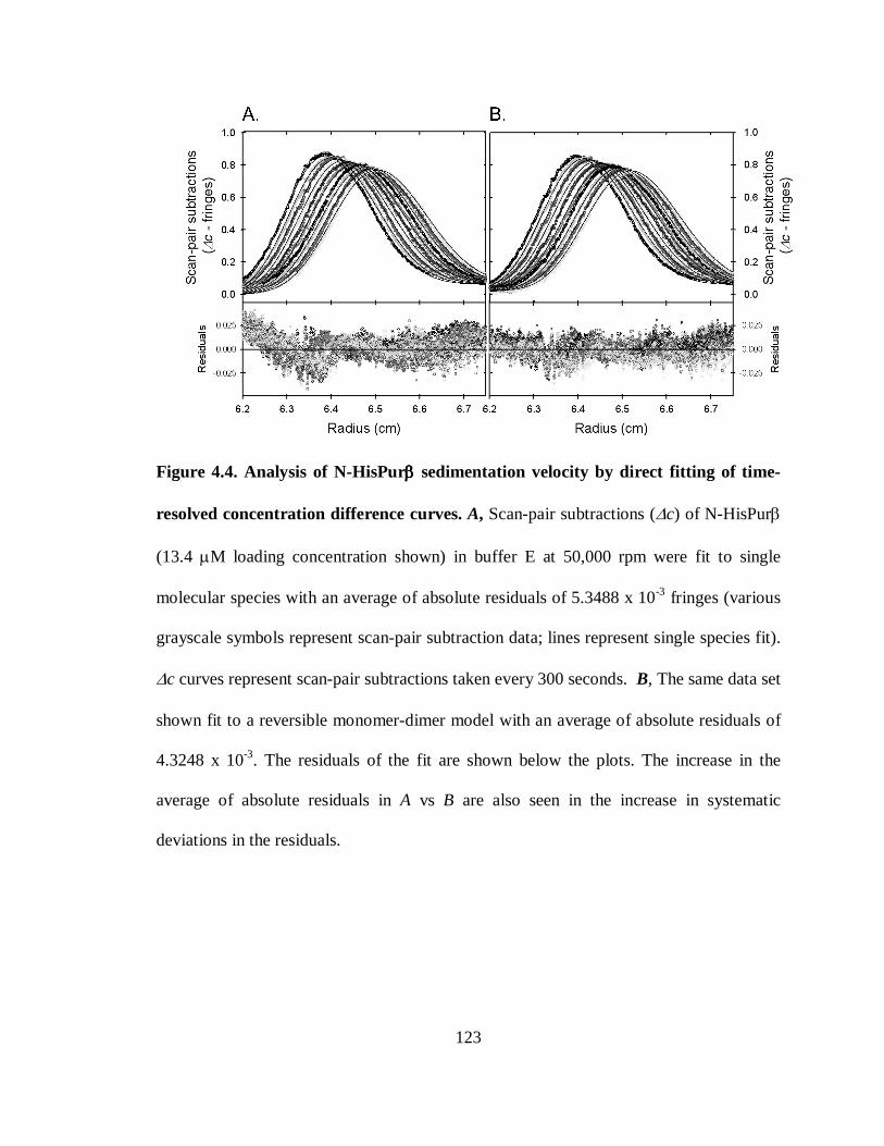
123
Figure 4.4. Analysis of N-HisPurββββ sedimentation velocity by direct fitting of time-
resolved concentration difference curves. A, Scan-pair subtractions (∆c) of N-HisPurβ
(13.4 µM loading concentration shown) in buffer E at 50,000 rpm were fit to single
molecular species with an average of absolute residuals of 5.3488 x 10⁻³ fringes (various
grayscale symbols represent scan-pair subtraction data; lines represent single species fit).
∆c curves represent scan-pair subtractions taken every 300 seconds. B, The same data set
shown fit to a reversible monomer-dimer model with an average of absolute residuals of
4.3248 x 10⁻³. The residuals of the fit are shown below the plots. The increase in the
average of absolute residuals in A vs B are also seen in the increase in systematic
deviations in the residuals.

124
The disparities between the experimentally derived and calculated RH values, as well
as the value of ƒ/ƒ0 for the dimeric species are suggestive that the shape of dimeric N-
HisPurβ is markedly different from that of a condensed sphere. Molecular shape
calculations predict that N-HisPurβ dimer is elliptical in solution, either prolate or oblate,
with axial ratios of either a/b = 7.01 or 8.05 for a prolate or oblate ellipsoid respectively.
These calculations were made using a degree of hydration of 0.4294 g H2O/g N-HisPurβ
which represents the predicted maximum degree of hydration based on amino acid
composition of the protein using the Teller method (160) which is based on the
assumption that all amino acids are maximally hydrated. Given this degree of hydration,
the molecular dimensions would be 23.57 nm × 3.36 nm (2a × 2b) for the prolate
prediction and 12.90 nm × 1.60 nm (2a × 2b) for the oblate ellipsoid. However, studies
suggest that the actual degree of hydration of proteins is generally lower than maximal
values, presumably due to folding and exclusion of water in the hydrophobic core of
proteins. Kuntz showed that corrections for folding on the degree of hydration of
proteins were on the order of 10% (90% of the maximum calculated value) (159),
however values of 0.30-0.35 g H2O/g protein are generally used in instances where the
actual degree of hydration is not known. Unfortunately, accurate hydrodynamic
modeling cannot be accomplished without prior knowledge of either the degree of
hydration or the axial ratios of the hydrated molecule (109). If a 10% decrease in
hydration of dimeric N-HisPurβ due to folding is assumed, the resulting hydration value
is 0.349 g H2O/g protein, and predicts axial ratios of 7.29 and 8.43 for a prolate and
oblate ellipsoid, respectively. Molecular dimensions arising from these values would
125
then be 23.91 x 3.28 nm (2a x 2b) for the prolate case, and 12.94 x 1.54 nm (2a x 2b) for
the oblate prediction. These values indicate that the projected dimensions differ only
modestly with the inclusion of this assumption-based correction.
Sedimentation equilibrium analysis of recombinant Purββββ − In order to validate the
monomer-dimer self-association model for N-HisPurβ, and confirm the equilibrium
constant that characterizes this association, sedimentation equilibrium studies were
performed. A 10-fold range of concentrations (from 4 – 40 µM) of size-fractionated N-
HisPurβ in buffer E were sedimented at three different rotor speeds until equilibrium was
attained (representative scans are shown in Figure 4.6). A careful and systematic analysis
of experiments in which protein concentration, rotor speeds and buffer conditions were
altered indicated that we were limited in the range of conditions, which would result in
interpretable data. We were unable to run N-HisPurβ at concentrations lower than 4 µM
due to low radial absorbance distributions resulting in values below the signal to noise
ratio of the instrument (data not shown). Attempts at lowering the reducing agent
concentration to lower baseline absorbance, in an effort to collect low concentration
absorbance datasets at 230 nm, resulted in formation of higher order oligomers as
assessed by SEC (data not shown). We believe that these were artifactual oxidation
products. Protein concentrations and rotor speeds were chosen such that non-ideal
solution conditions were avoided. Further, buffer conditions were selected such that N-
HisPurβ solubility was optimal. It has been found by our laboratory that preparations of
N-HisPurβ are considerably less stable in buffered solutions containing less than 200 mM
monovalent salt.
126
Figure 4.5. Analysis of concentration-dependant N-HisPurββββ solution non-ideality by
sedimentation equilibrium. Solutions of N-HisPurβ at various concentrations were
analyzed by sedimentation equilibrium at rotor speeds of 22,000, 28,000, and 35,000
rpm. Radial concentration distributions for each loading concentration from all three rotor
speeds were globally fit to a single-ideal species to ascertain the weight-average apparent
molecular weight of the samples. These values (as a ratio of the apparent molecular
weight to the known monomeric molecular weight, MwApp/MCal) are shown plotted versus
the initial loading concentration. Data indicate that the apparent solution molecular
weight approaches that of a dimer at loading concentrations less than 40 µM,
representative of self-association. Loading concentrations greater than 40 µM show a
decline in MwApp/MCal values, suggestive of electrostatic repulsion non-ideality.
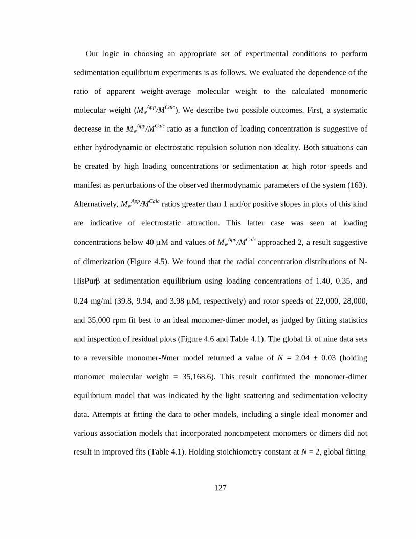
127
Our logic in choosing an appropriate set of experimental conditions to perform
sedimentation equilibrium experiments is as follows. We evaluated the dependence of the
ratio of apparent weight-average molecular weight to the calculated monomeric
molecular weight (MwApp/MCalc). We describe two possible outcomes. First, a systematic
decrease in the MwApp/MCalc ratio as a function of loading concentration is suggestive of
either hydrodynamic or electrostatic repulsion solution non-ideality. Both situations can
be created by high loading concentrations or sedimentation at high rotor speeds and
manifest as perturbations of the observed thermodynamic parameters of the system (163).
Alternatively, MwApp/MCalc ratios greater than 1 and/or positive slopes in plots of this kind
are indicative of electrostatic attraction. This latter case was seen at loading
concentrations below 40 µM and values of MwApp/MCalc approached 2, a result suggestive
of dimerization (Figure 4.5). We found that the radial concentration distributions of N-
HisPurβ at sedimentation equilibrium using loading concentrations of 1.40, 0.35, and
0.24 mg/ml (39.8, 9.94, and 3.98 µM, respectively) and rotor speeds of 22,000, 28,000,
and 35,000 rpm fit best to an ideal monomer-dimer model, as judged by fitting statistics
and inspection of residual plots (Figure 4.6 and Table 4.1). The global fit of nine data sets
to a reversible monomer-Nmer model returned a value of N = 2.04 ± 0.03 (holding
monomer molecular weight = 35,168.6). This result confirmed the monomer-dimer
equilibrium model that was indicated by the light scattering and sedimentation velocity
data. Attempts at fitting the data to other models, including a single ideal monomer and
various association models that incorporated noncompetent monomers or dimers did not
result in improved fits (Table 4.1). Holding stoichiometry constant at N = 2, global fitting
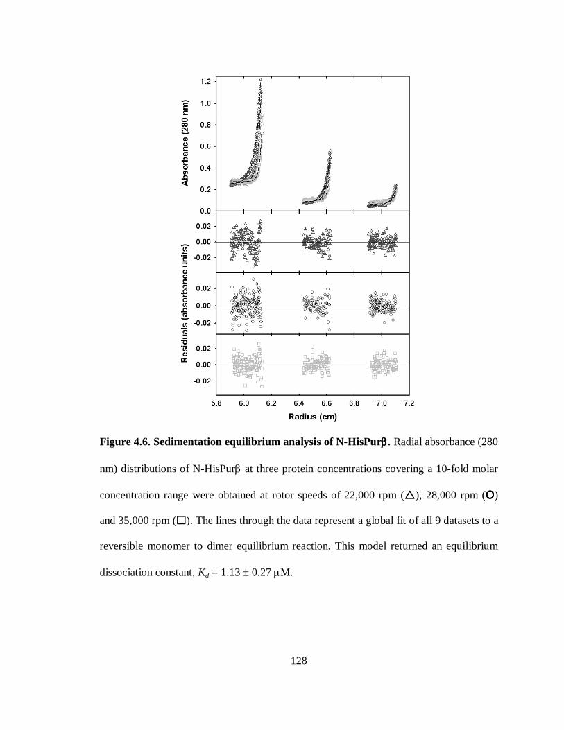
128
Figure 4.6. Sedimentation equilibrium analysis of N-HisPurββββ. Radial absorbance (280
nm) distributions of N-HisPurβ at three protein concentrations covering a 10-fold molar
concentration range were obtained at rotor speeds of 22,000 rpm (����), 28,000 rpm (OOOO)
and 35,000 rpm (����). The lines through the data represent a global fit of all 9 datasets to a
reversible monomer to dimer equilibrium reaction. This model returned an equilibrium
dissociation constant, Kd = 1.13 ± 0.27 µM.
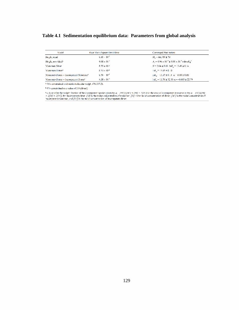
129
Table 4.1 Sedimentation equilibrium data: Parameters from global analysis
130
of the data yielded a dissociation constant, Kd = 1.13 µM ± 0. 27 µM (Table 4.1; ± 1
standard deviation), which is reasonably consistent with direct sedimentation velocity
boundary fitting analyses.
The effects of salt concentration on N-HisPurβ self-association energetics were not
explored in the present study. As mentioned previously, solubility of N-HisPurβ appears
to be an issue when salt concentrations are reduced below 200 mM. Salt effects above
200 mM have yet to be rigorously tested.
DISCUSSION
In this study, we utilized hydrodynamic techniques to characterize the quaternary
structure of N-HisPurβ in the absence of nucleic acids. The various hydrodynamic and
thermodynamic techniques employed here suggest that N-HisPurβ exists in a monomer-
dimer equilibrium characterized by a Kd = 1.13 ± 0. 27 µM. Preliminary sedimentation
studies on N-HisPurβ showed signs of contaminating incompetent species (data not
shown) that appeared to be artifacts, namely disulfide oxidation products, generated by
concentrating techniques and consequently it was our feeling that utilization of a variety
of robust hydrodynamic and thermodynamic techniques coupled with a preemptive size-
fractionation step were warranted to distinguish between a possible mixture of
thermodynamically tight dimer with incompetent monomer and a measurably reversible
monomer-dimer equilibrium. Omission of the SEC step in preliminary sedimentation
131
equilibrium experiments resulted in returned Kd values for dimer dissociation ranging
from 3.7 to 19.5 µM, whereas inclusion of this step limited the range to 0.70 to 1.54 µM.
Moreover, fitting our data to models that included incompetent dimer did not improve the
fitting statistics (Table 4.1). Hence, we believe that we have a fully reversible interacting
system. The value of Kd reported here should be interpreted as an apparent upper limit to
the actual value, since all measurements of Kd were made at concentrations greater than
the apparent value of 1 µM (Figure 4.7) due to low optical densities. It is likely that
equilibrium measurements at concentrations lower than those utilized here will have to be
made by a sensitive orthogonal technique, such as fluorescence polarization spectroscopy
(128).
Hydrodynamic radius determinations either made directly by dynamic light scattering
or by extrapolation from sedimentation data were in sound agreement. Molecular shape
calculations were consistent with an asymmetric shape of dimeric N-HisPurβ in solution.
This finding is not surprising based on the fact that Purβ is composed of 22.2% glycine
(143), which likely contributes to a lack of secondary structural elements and a somewhat
disordered tertiary structure, and may explain difficulties in obtaining higher resolution
structural information. A further interesting finding is that the ƒ/ƒ0 ratio determined for
the monomeric species is greater than that of the dimer, which suggests that dimerization
results in partial condensation of the overall structure. The implications of this finding
are discussed below.
We also addressed the concern that the N-terminal hexahistidine tag present on the
recombinant protein might adversely affect the functional activity of Purβ by comparing
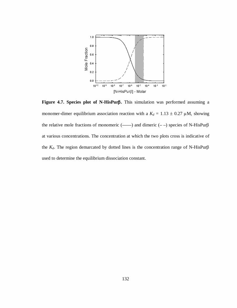
132
Figure 4.7. Species plot of N-HisPurββββ. This simulation was performed assuming a
monomer-dimer equilibrium association reaction with a Kd = 1.13 ± 0.27 µM, showing
the relative mole fractions of monomeric (——) and dimeric (– –) species of N-HisPurβ
at various concentrations. The concentration at which the two plots cross is indicative of
the Kd. The region demarcated by dotted lines is the concentration range of N-HisPurβ
used to determine the equilibrium dissociation constant.
133
the ssDNA-binding activity of native and recombinant N-HisPurβ using a quantitative
ELISA approach (145). The ssDNA-binding activity of N-HisPurβ was indistinguishable
compared to Purβ derived from either mouse embryo fibroblasts or vascular smooth
muscle cells (data not shown). Also, non-specific metal ion-mediated dimerization of
hexahistidine can be eliminated as a complicating factor since all measurements were
made in the presence of 0.5 mM EDTA.
Our interest in Pur proteins stems from their putative involvement in repressing the
transcription and translation of genes that mark the phenotypic status of myofibroblasts,
vascular smooth muscle cells, and cardiomyocytes (28, 104, 142, 145, 148). Evidence for
formation of transient ssDNA structures within the asymmetric purine/pyrimidine tract of
the 5′-SMαA promoter (9) has also fueled investigation into the mechanism of DNA-
binding by Pur proteins. It has been suggested that Pur proteins bind PUR-elements as
either hetero- or homodimers (141), although the stoichiometry, mechanism, and
thermodynamics of nucleoprotein assembly have not yet been determined by rigorous
physical investigation. In the case of Purα, self-association has been reported to be
mediated by an RNA molecule of unknown nature (91). This was an intriguing finding,
as it suggested that RNA may influence the ability of Purα to stably self-associate and to
bind to ssDNA. Curiously, despite sharing 70% amino acid sequence identity, we have
shown in this study that Purβ dimerizes in the absence of any nucleic acid. It is possible
that this disparity is due to a distinct functional difference between Purα and Purβ as
implied by previous gain-of-function studies (145, 148), or could be reflective of
different experimental approaches (e.g. pull-down assay versus sedimentation
134
equilibrium analysis). Moreover, the apparent binding affinities of Purα and Purβ for
different PUR-elements are reported to be on the order of 1 nM (145, 310), whereas the
affinity of Purβ self-association is three orders of magnitude weaker. As such, Purβ at
concentrations below 100 nM is predicted to be largely monomeric as depicted in the
molecular species plot in Figure 4.7. This suggests that other factors (such as an RNA
ligand in the specific case of Purα) may be required to help facilitate dimerization in a
cellular milieu if the concentration of protein is limiting. On the other hand, it remains to
be resolved as to what extent Purα and Purβ share similar intrinsic homodimerization
ability in the absence of nucleic acid and whether or not heterodimeric complexes
associate with enhanced or reduced affinity relative to their homodimeric counterparts.
In conclusion, we report that recombinant Purβ participates in a monomer-dimer
equilibrium governed by an apparent upper limit dissociation constant of ~1 µM. The
ability to dimerize in a reversible fashion may represent an important regulatory
mechanism, allowing mass-action governed self-association to play either a positive or
negative role in nucleic acid-binding. As indicated by frictional coefficient ratios,
dimerization may result in structural reorganization of N-HisPurβ that may permit
nucleic acid recognition and binding. Self-association of transcription factors as a
prerequisite to DNA-binding is not unprecedented. For example, the STAT proteins
require phosphorylation-dependent dimerization prior to nuclear localization and binding
to cytokine responsive gene promoters (112). Similarly, intracellular estrogen receptor
DNA-binding activity is apparently dependent upon ligand-mediated dimerization (80,
282, 295). However, in the case of Purβ, dimerization does not appear to require post-
135
translational modification or ligand binding per se as self-association is governed by
protein concentration in vitro. However, this fact does not rule out the prospect that post-
translational modifications may alter the affinity of homodimerization in vivo. Although
the absolute intracellular concentration is not known, it has been reported that levels of
Purβ increase dramatically in cardiovascular cell types undergoing phenotypic changes
(9). This entices speculation that repressive effects of Purβ may depend upon its
expression and accumulation within the nucleus to levels that drive self-association and
permit ssDNA-binding. Future studies will focus on determining the affinity and
stoichiometry of relevant Pur nucleoprotein complexes with the use of quantitative
biophysical approaches such as those described herein.
136
CHAPTER V. THERMODYNAMIC ANALYSIS OF SEQUENCE-
SPECIFIC SINGLE-STRANDED DNA-BINDING BY
RECOMBINANT PUR ββββ REVEALS A COOPERATIVE
MECHANISM OF NUCLEOPROTEIN ASSEMBLY
The following is a description of original and unpublished work.
INTRODUCTION
Classical models of transcription regulation imply that assembly and processivity of
transcriptional machinery at gene promoter regions is coordinated by the pre-assembly of
trans-acting factors at specific regions of the promoter, and that this pre-assembly is
dictated by a combination of the nucleotide sequence of the promoter and environmental
cues. By-and-large, these models propose that binding of specific DNA sequences by
transcription factors involves the recognition of particular arrangements of chemical
functional groups along the major or minor groove of duplex DNA that coincide with
compatible amino acid chemical groups arranged on the surface of the protein. However,
the detection of large structural re-arrangements, and altered nuclease sensitivities in
promoter regions of genes coincident with apparent transcription factor binding has
suggested that chromatin structure and/or DNA conformation also plays a prominent role
in regulating gene transcription (for review see (277)). Furthermore, the detection of
stable, single-stranded DNA structures, and the identification of interacting sequence-
specific ssDNA-binding transcription factors that are critical for regulated transcription of
137
numerous genes implies that many eukaryotic genes do not conform to classical models
of gene regulation, and likely utilize more complex mechanisms that incorporate
combinations of changes in chromatin structure, and the binding of protein factors to
double-stranded, and transiently formed single-stranded and non-B-DNA structures.
Collectively these findings question the view that genomic promoter DNA acts as a static
lattice for the coordinated assembly of transcription factors to bind and enforce
transcriptionally active or inactive states, and instead it appears likely that, for some
genes, the same sequence of promoter DNA is able to bind select sets of transcription
factors dependent upon the dynamic interconversion of double-stranded and single-
stranded conformations.
Single-stranded promoter character, as well as reliance on sequence-specific ssDNA-
binding proteins for regulated transcription is fairly common, and has been observed for
such notable genes as c-myc (10, 11, 191, 192, 203, 280, 288), major histocompatibility
complex class II (69, 183), platelet-derived growth factor A-chain (298, 301, 322), apo-
very low density lipoprotein II (263), low density lipoprotein receptor (221), fas cell
surface receptor (263), androgen receptor (39, 101), epidermal growth factor receptor
(130, 312), procollagen α1 (68), smooth muscle α-actin (11, 48, 275), and β-myosin
heavy chain (129). Common to many of the promoters of these genes are the presence of
asymmetrically distributed nucleotide tracts that, in other studies, have been shown to be
prone to forming non-B-DNA structures. For instance, polypurine/polypyrimidine, or
alternating purine/pyrimidine stretches have been shown to not only adopt Z-DNA (204),
and H-DNA (235) conformations, but to cause loss of base-pairing in the neighboring
138
transition residues (105). Often, single-stranded character in these regions can be
detected through the use of ssDNA-specific reagents (9, 130, 192, 229, 235, 274, 285,
298, 301), further suggesting that these conformations are stable entities. The correlation
of non-B-DNA-prone sequences and detectable single-stranded DNA in regions of
activated transcription suggests that alternate DNA conformations are an important
component of transcriptional regulation.
Despite mounting evidence supporting their existence and utility, the manner in
which sequence-specific ssDNA-binding proteins are able to function in a predominantly
double-stranded genome remains largely unknown. It has been postulated (179), and
shown that, in certain instances, negative supercoiling generated by transcriptional
machinery-associated helicase activity promotes localized Z-DNA formation or
unwinding of upstream sequences harboring atypical nucleotide stretches, as mentioned
above. This characteristic coincides with regions of transcriptional activity, but is not
universal, as it appears limited to a certain subset of genes (33, 161). A transcription-
induced topological strain mechanism has been attributed to the case of the far upstream
element (FUSE) of the c-myc promoter in which upstream nucleotide unpairing was
noted for both supercoiled and linearized plasmid templates in reconstituted transcription
assays (153), and in vivo (154). Structural interconversion, in turn, permits interaction
with several sequence-specific ssDNA-binding proteins including FUSE-binding protein
(FBP, (75)), and FBP-interacting repressor (FIR, (178)). Thus, in the case of c-myc
FUSE-protein interactions, sequence-specific ssDNA-binding by FBP and FIR is
apparently opportunistic, relying on pre-formation of single-stranded DNA structures for
139
assembly, despite reports that FBP is capable of helix destabilization in linear dsDNA
constructs containing FUSE sequences (8). Conceptually, this mechanism may not apply
to all promoter regions, especially those in which maintenance of a transcriptionally
repressed state requires ssDNA-interacting transcription factors. It is envisioned that in
these cases, single-stranded conformations are created and stabilized by binding of
protein factors by virtue of their inherent energetics of nucleoprotein complex assembly;
i.e., ssDNA nucleoprotein complex formation is thermodynamically competitive to non-
B-DNA duplex formation. Transcriptional regulation by sequence-specific ssDNA-
binding proteins at promoters containing sequences prone to Z-DNA conformations, or
other high-energy states with reduced melting temperatures, suggests that helix
destabilization may be a mechanism employed by many ssDNA-dependent transcription
factors to achieve promoter occupation and transcriptional regulation. This notion is
supported by findings that have shown that many sequence-specific ssDNA-binding
proteins bind to supercoiled plasmid DNA and not to linearized plasmids (8, 153, 154,
192, 288, 310).
Purβ is a sequence specific ssDNA/RNA- binding transcription/translation factor that,
along with Y-box protein MSY1 (mouse YB-1) and Pur protein family co-member Purα,
acts as a repressor of smooth muscle α-actin (SMαA) gene expression. The protein
encoded by this gene is an essential cytoskeletal contractile apparatus protein required for
regulation of vascular tone and cellular migration (79, 210, 231, 257). Therefore SMαA
and has been identified as being critical for maintenance of the differentiated contractile
phenotype of vascular smooth muscle cells (VSMCs) and myofibroblasts (166, 226, 243,
140
271). The repressive activities of Purβ, Purα and MSY1 on the SMαA promoter have
implicated them in facilitating smooth muscle cell dedifferentiation and vascular wall
remodeling, processes common to numerous vascular pathologies including
atherosclerosis (175). It has been demonstrated that each transcription factor exerts its
repressive effect by binding to the proximal muscle-specific CAT (MCAT) enhancer
element of the SMαA promoter that has been shown to possess a high level of
purine/pyrimidine base asymmetry (48, 82, 143, 275), and to transiently adopt single-
stranded conformations in response to dedifferentiating stimuli (9). Biochemical studies
have confirmed that Purα and Purβ preferentially bind the purine-rich strand, whereas
MSY1 shows specific affinity for the opposing pyrimidine-rich strand, and furthermore, a
network of protein-protein interactions between each transcription factor exists (141,
148) as well as nucleic acid independent self-association, in the case of Purβ [190].
Current models for the achievement of transcriptional repression by these proteins
propose that binding of Purα and/or Purβ to the purine-rich strand and MSY1 to the
pyrimidine-rich strand destabilizes B-DNA helical structures and maintains the enhancer
region in a single-stranded conformation, thus disrupting dsDNA-dependent transcription
enhancer 1 (TEF-1) binding and function (28, 275).
Based on our recent work which showed that recombinant Purβ self-dimerizes with a
dissociation constant of 1.13 µM (222), and previous reports describing marked increases
in Purβ protein levels in VSMCs undergoing phenotypic changes consistent with SMαA
repression (104, 271), it was hypothesized that Purβ dimerization may be a prerequisite
to ssDNA-binding and serve as a regulatory mechanism for Purβ function. In the present
141
study, our goal was to delineate the mechanism of sequence recognition and ssDNA
binding by Purβ, which has been shown to be the more dominant Pur protein repressor in
the context of SMαA gene expression (19, 145), to a sequence representative of the
proximal SMαA MCAT enhancer element. We have used a thermodynamic approach to
examine nucleoprotein stoichiometry, mechanism, and binding energetics to gauge the
plausibility of helix destabilization as a possible mechanism by which Purβ binds specific
ssDNA sequences in a generally double-stranded environment. Our results indicate that
binding of Purβ to the purine-rich strand (promoter element; PE) within the SMαA
MCAT enhancer involves facilitated cooperative assembly of Purβ monomers (in the 300
pM range) to form higher order nucleoprotein structures with stoichiometries of 2:1
(Purβ:PE). The resolved energetics of binding of Purβ to the purine rich strand of SMαA
enhancer, by way of quantitative ssDNA footprinting, suggests that maintenance of a
single-stranded state within this region might require auxiliary activities; either the
involvement of Purα, the concurrent cooperative assembly of MSY1 on the pyrimidine-
rich strand, the reduction of local melting temperatures by induction of topological stress,
or a combination thereof.
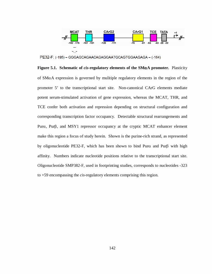
142
Figure 5.1. Schematic of cis-regulatory elements of the SMααααA promoter. Plasticity
of SMαA expression is governed by mulitiple regulatory elements in the region of the
promoter 5’ to the transcriptional start site. Non-canonical CArG elements mediate
potent serum-stimulated activation of gene expression, whereas the MCAT, THR, and
TCE confer both activation and repression depending on structural configuration and
corresponding transcription factor occupancy. Detectable structural rearrangements and
Purα, Purβ, and MSY1 repressor occupancy at the cryptic MCAT enhancer element
make this region a focus of study herein. Shown is the purine-rich strand, as represented
by oligonucleotide PE32-F, which has been shown to bind Purα and Purβ with high
affinity. Numbers indicate nucleotide positions relative to the transcriptional start site.
Oligonucleotide SMP382-F, used in footprinting studies, corresponds to nucleotides -323
to +59 encompassing the cis-regulatory elements comprising this region.
143
MATERIALS AND METHODS
Chemicals, protein reagents, and oligonucleotide probes. All chemicals used in this
study were of reagent grade or better. Recombinant Purα and Purβ were expressed as
amino-terminally labeled hexahistidine tag fusion proteins (referred to in this report as N-
HisPurα, and N-HisPurβ, respectively), purified from E. coli expression cultures, and
quantified as described in previous publications (149, 222). AKR-2B nuclear extracts
were obtained from cell monolayers cultured under exponential growth conditions as
described previously (143). Protein concentrations of nuclear extracts were assessed by
bicinchonic acid assay (Sigma-Aldrich) using high purity BSA (Boehringer-Mannheim)
as a protein standard. Preparation and validation of epitope-specific rabbit polyclonal
antibodies against mouse Purα (anti-Purα 291) and mouse Purβ (anti-Purβ 302) has been
described previously (141). Synthetic oligonucleotides were purchased from Sigma-
Genosys. Yeast tRNA was purchased from Sigma. Enzyme reagents were purchased
from commercial sources; T4 polynucleotide kinase was purchased from New England
Biolabs, DNase I, and Accuprime SupermixTM (Taq polymerase) were purchased from
Invitrogen Corp. Fresh stocks of [γ-32P]ATP (6000 Ci/mmole) were obtained from
Perkin-Elmer. Sequenase dideoxy-NTP sequencing kit was purchased from Unites States
Biochemical Corp. SYBR-Gold nucleic acid stain was purchased from Invitrogen.
Preparation of ssDNA for quantitative DNase I footprinting. Methods for the
purification of ssDNA were based on protocols originally developed and reported by
144
others (121, 139). A plasmid vector containing the full-length mouse SMαA promoter,
termed VSMP8 (296) was used as a template for PCR-based amplification of a 382 base
fragment comprising the forward strand of the proximal SMαA promoter, termed
SMP382-F (bases –323 to +59 relative to the transcriptional start site). To facilitate both
amplification and purification of the forward single strand consisting of the purine-rich
sequence of the proximal MCAT enhancer element, PCR primers were designed such
that the reverse strand primer was 5’-biotinylated (SMP8p1122s-R-5btn; 5’-biotin-
GGCTACTTACCCTGACAGCGACT-3’), whereas the forward strand primer was
unmodified (SMP8p741s-F; 5’-TTCTGAGGAATGTGCAAACCGTG-3’). PCR
amplification of the 382 bp fragment from 1 ng/µL of VSMP8 template was carried out
using Accuprime SupermixTM reagent according to the manufacturer’s instructions. The
incorporation of the biotinylated reverse strand primer allowed for the isolation of
useable quantities of SMP382-F through implementation of biotin-streptavidin affinity
based techniques. Briefly, double-stranded PCR product (typically 500 µL) was applied
to 1 mg of Streptavidin MagneSphere paramagnetic particle resin (Promega Corp.), pre-
equilibrated in 20 mM Tris pH 8.8, and incubated at 4°C for 16 h. The PCR-product-
resin complex was washed twice with 20 mM Tris pH 8.8. Non-biotinylated SMP382-F
was eluted by incubating the resin complex in 0.2 N NaOH for 5 min at 20±1°C. The
eluant solution was neutralized by the addition of 1/10 volume of 5 M ammonium
acetate. SMP382-F was precipitated by the addition of 2 volumes of isopropanol at -
20°C, washed with 70% ethanol, and redissolved in nuclease-free water. Concentration
of SMP382-F was approximated by absorbance measurement at 260 nm, assuming an
145
extinction coefficient of 3,630,200 M-1cm-1 as calculated using web-based software
(http://biophysics.idtdna.com) based on methods described by Tataurov and colleagues
(208, 284).
End-labeling of oligonucleotides with [γγγγ-32P]ATP. Single-stranded DNA
oligonucloetides for use in electrophoretic mobility shift assays (PE32-F), DNase I
footprinting assays (SMP382-F), and primer-labeled dideoxy sequencing reactions
(SMP8sp741s-F) were enzymatically labeled on their 5’ termini with [γ-32P]ATP using
T4 polynucleotide kinase activity as directed by the manufacturer. Reactions were
performed at 37°C for 90 min. Upon completion, the enzyme was heat inactivated by
incubating reaction mixtures at 70°C for 10 min. Unincorporated [γ-32P]ATP was
removed by buffer exchange over G-25 Microspin columns (Pharmacia). Extent of
labeling was assessed by scintillation counting of purified probes using a Perkin-Elmer
Tri-Carb® scintillation counter. For purposes of clarity, oligonucleotides carrying a 5’-
32PO4 radiolabel will be marked with an “*” (for example, PE32-F* denotes 5’-32PO4-
labeled PE32-F).
Qualitative electrophoretic mobility (super)shift assay. Binding reactions for
qualitative electrophoretic mobility shift assays (EMSA) were carried out in EMSA
binding buffer (50 mM Tris•HCl pH 7.5, 100 mM KCl, 5% (v/v) glycerol, 0.5 mM
dithiothreitol, 2 µg/mL dT32, 50 µg/mL BSA) plus 0.5 mM ethylenediaminetetracetic
acid (EDTA) to prevent nuclease digestion of probe by nuclear extracts. Recombinant
proteins and nuclear extracts were diluted to the indicated concentrations prior to addition
of PE32-F* (5’-GGGAGCAGAACAGAGGAATGCAGTGGAAGAGA-3’) to a final
146
concentration of 1 nM. Binding reactions were incubated for 16 h at 4°C to allow
formation of nucleoprotein complexes prior to the addition of super-shifting antibodies,
anti-Purα 291 and anti-Purβ 302, to the indicated concentrations. Final volumes of
reactions were 40 µL. Reactions were incubated at 4°C for an additional 2 h. Complexes
were resolved electrophoretically by loading 10 µL of binding reactions (typically 5,000
– 10,000 cpm) on a 1.5 mm thick 10% polyacrylamide gel (75:1
acrylamide:bisacrylamide) cast and run in TBE buffer (25 mM Tris, 25 mM boric acid,
0.5 mM EDTA). Gels were pre-run at 4 Watts per gel for 1 h prior to loading and
running for 75 min at 4 Watts at room temperature. Upon completion, gels were
disassembled and dried on Whatman filter paper in a Bio-Rad slab gel dryer for 45 min at
65°C. Phosphorimaging of the dried gels was performed by exposing to phosphor
storage screens (Molecular Dynamics) for 24-48 h, prior to developing with a Bio-Rad
PersonalFXTM phosphorimager. In certain cases, dried gels were visualized by exposure
to X-Omat film for 6-16 h at -80°C prior to development.
An agarose gel electrophoresis-based assay was used to gauge the extent of
binding of recombinant proteins to plasmid DNA, and is based on experiments described
previously (310). Briefly, VSMP8 (7.9 kb) and parent plasmid vector pBLCAT3 (4.3 kb)
were purified by double cesium chloride gradient centrifugation, and digested at a final
DNA concentration of 50 ng/µL, with HindIII (Roche) according to the manufacturers
instruction. Enzyme was inactivated by incubation at 65°C for 10 min. Digested and
supercoiled plasmids were mixed at an original plasmid concentration of 1 nM (2.84 and
5.21 ng/mL for pBLCAT3 and VSMP8, respectively) with 25, 50, 100, or 200 nM
147
recombinant Pur protein in buffer (50 mM Tris•HCl pH 7.5, 100 mM KCl, 5% (v/v)
glycerol, 0.5 mM dithiothreitol, 2 µg/mL dT32, 50 µg/mL BSA) for 16 h at 20±1°C.
Samples (30 ng total DNA) were electrophoresed on a 0.7% SeaKem LE agarose
(Cambrex) gel cast and run in 0.5X traditional TBE Buffer (44.5 mM Tris-Borate pH 8.3,
1 mM EDTA) at 5 V/cm for approximately 2 h at 20±1°C. Gels were stained with
SYBR-Gold stain diluted 1:10,000 in 0.5X traditional TBE for 30 min prior to image
capture with a Bio-Rad GelDoc XR imaging system.
Fluorescence anisotropy measurements. Fluoresceinated (3’) oligonucleotide probe
PE32-F (termed PE32-F-3FLC, 5’-
GGGAGCAGAACAGAGGAATGCAGTGGAAGAGA-Fluoroscein-3’) was titrated
with N-HisPurβ in buffer consisting of 50 mM HEPES pH 7.8, 300 mM NaCl, 1.5 mM
MgCl2, 2 mM β-mercaptoethanol, and allowed to equilibrate for approximately 16 h at
20±1 °C in the absence of light. Probe concentrations were varied. Steady-state
fluorescence anisotropy measurements were made on a Quantamaster-6
spectrofluorometer (Photon Technologies International, South Brunswick, NJ) equipped
with a 75 W xenon arc lamp excitation source, excitation and emission monochromators,
and automatic excitation and emission polarizers in a T-format. Slit-widths were varied
to maintain constant observed emission intensities between samples with different probe
concentrations. Parallel and perpendicular emission intensities were collected with
horizontally polarized exciting light in order to first calculate the instrument correction
factor, G, given by G = IHV/IHH where IHV and IHH are the intensities measured through
the vertical and horizontal polarizers when excited with horizontally polarized light.
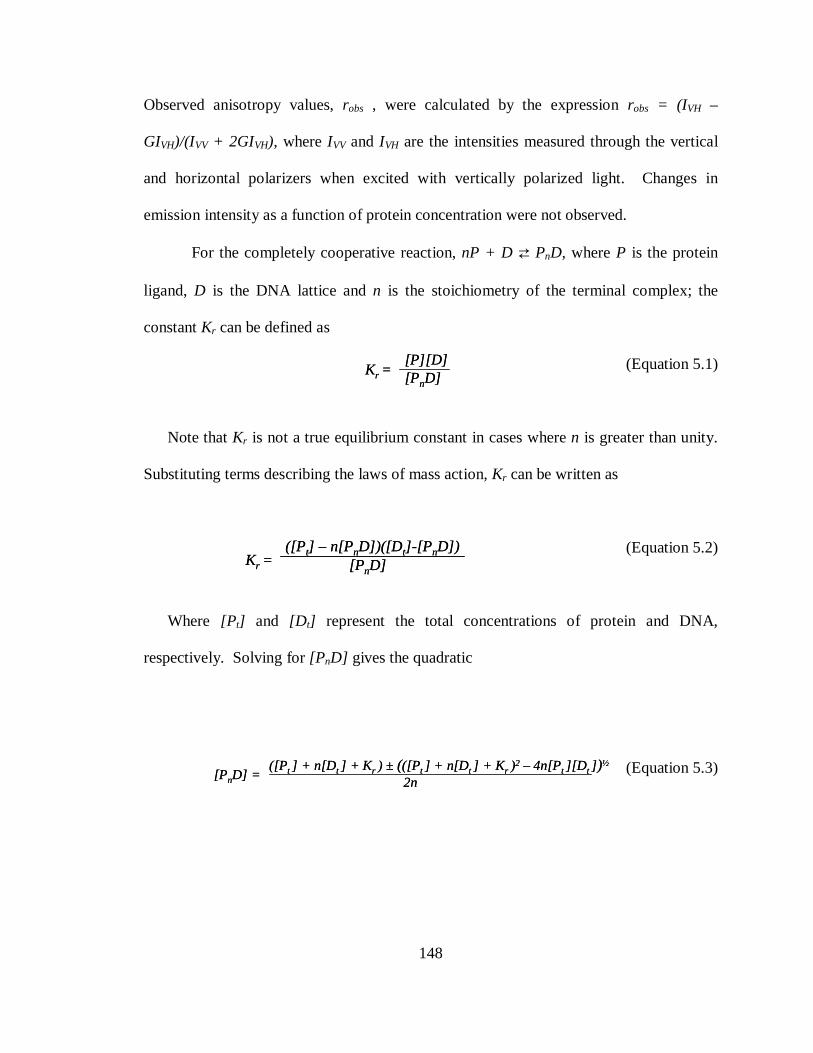
148
Observed anisotropy values, robs , were calculated by the expression robs = (IVH –
GIVH)/(IVV + 2GIVH), where IVV and IVH are the intensities measured through the vertical
and horizontal polarizers when excited with vertically polarized light. Changes in
emission intensity as a function of protein concentration were not observed.
For the completely cooperative reaction, nP + D ⇄ PnD, where P is the protein
ligand, D is the DNA lattice and n is the stoichiometry of the terminal complex; the
constant Kr can be defined as
(Equation 5.1)
Note that Kr is not a true equilibrium constant in cases where n is greater than unity.
Substituting terms describing the laws of mass action, Kr can be written as
(Equation 5.2)
Where [Pt] and [D t] represent the total concentrations of protein and DNA,
respectively. Solving for [PnD] gives the quadratic
(Equation 5.3)
Kr =[P][D][PnD]
Kr =[P][D][PnD]
Kr =([P t] – n[PnD])([D t]-[P nD])
[PnD]Kr =([P t] – n[PnD])([D t]-[P nD])
[PnD]
[PnD] =([Pt ] + n[D t ] + K r ) ± (([Pt ] + n[D t ] + K r )
2 – 4n[Pt ][D t ] )½
2n[PnD] =
([Pt ] + n[D t ] + K r ) ± (([Pt ] + n[D t ] + K r )2 – 4n[Pt ][D t ] )½
2n
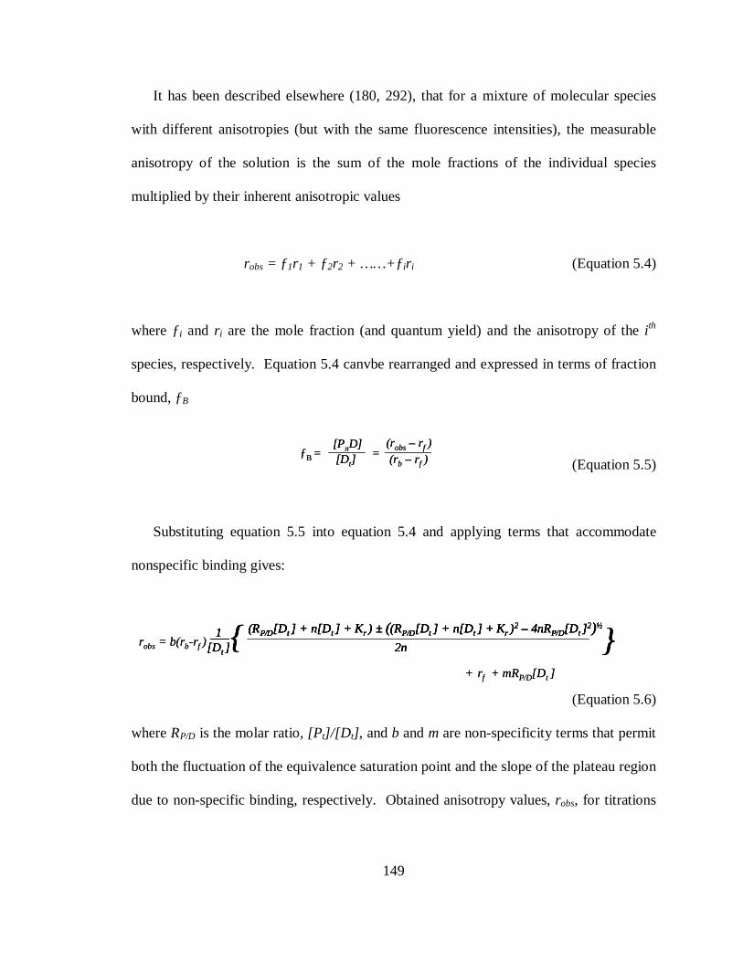
149
It has been described elsewhere (180, 292), that for a mixture of molecular species
with different anisotropies (but with the same fluorescence intensities), the measurable
anisotropy of the solution is the sum of the mole fractions of the individual species
multiplied by their inherent anisotropic values
robs = ƒ1r1 + ƒ2r2 + ……+ƒir i (Equation 5.4)
where ƒi and r i are the mole fraction (and quantum yield) and the anisotropy of the i th
species, respectively. Equation 5.4 canvbe rearranged and expressed in terms of fraction
bound, ƒB
(Equation 5.5)
Substituting equation 5.5 into equation 5.4 and applying terms that accommodate
nonspecific binding gives:
(Equation 5.6)
where RP/D is the molar ratio, [Pt]/[D t] , and b and m are non-specificity terms that permit
both the fluctuation of the equivalence saturation point and the slope of the plateau region
due to non-specific binding, respectively. Obtained anisotropy values, robs, for titrations
ƒB = = [PnD][D t]
(robs– rf )(rb – rf )
ƒB = = [PnD][D t]
(robs– rf )(rb – rf )
robs= b(rb-rf )(RP/D[D t ] + n[D t ] + K r ) ± ((RP/D[D t ] + n[D t ] + K r )
2 – 4nRP/D[D t ]2)½
2n1
[D t ]{ }
+ rf + mRP/D[D t ]
robs= b(rb-rf )(RP/D[D t ] + n[D t ] + K r ) ± ((RP/D[D t ] + n[D t ] + K r )
2 – 4nRP/D[D t ]2)½
2n1
[D t ]{ }(RP/D[D t ] + n[D t ] + K r ) ± ((RP/D[D t ] + n[D t ] + K r )
2 – 4nRP/D[D t ]2)½
2n
(RP/D[D t ] + n[D t ] + K r ) ± ((RP/D[D t ] + n[D t ] + K r )2 – 4nRP/D[D t ]
2)½
2n1
[D t ]{ }
+ rf + mRP/D[D t ]
150
of N-HisPurβ against fixed concentrations of PE32-F-3FLC were plotted against known
values of RP/D, and fit to equation 5.6 to obtain best-fit values of n. Fitting was
performed with Prism 5 software (GraphPad Software, Inc., San Diego, CA).
Quantitative electrophoretic mobility shift assay. Binding reactions for quantitative
EMSAs were carried out in EMSA binding buffer and allowed to equilibrate for 16 h at
20 ± 1 °C. Methods of quantitative EMSA for the estimation of binding stoichiometry
are based on protocols published previously (84, 85, 223, 224) termed the serial-dilution
method. Briefly, solutions of N-HisPurβ (5 nM) and PE32-F* (1 nM) were serially-
diluted 1.1:1 fold in EMSA binding buffer to obtain a complete dilution series. The
serial-dilution method relies on the redistribution of nucleoprotein complex
concentration, as a result of serial-dilution, based on the ideal law of chemical
equilibrium without changing the ratio of total protein concentration to total DNA
concentration. This redistribution is then monitored by native gel electrophoresis as
described above for qualitative EMSA, with the only exception being that gels are run for
45 min to prevent streaking due to complex dissociation. For the general cooperative
binding mechanism nP + D ⇄ PnD, the macroscopic association constant Ka is defined
as:
Ka = [PnD]/[ P free]n[D] (Equation 5.7)
151
In these expressions P represents N-HisPurβ, D represents the probe PE32-F*, and n
represents the stoichiometry of the nucleoprotein complex. Rearrangement of the
definition of Ka gives:
ln([PnD]/[D]) = nln[P free] + lnKa (Equation 5.8)
Densitometry of phosphorimages was carried out using ImageQuant software
(Molecular Dynamics) to determine [PnD] and [D] , whereas [Pfree] was estimated from
the relationship:
[Pfree] = [P input] – n[PnD] (Equation 5.9)
In which case [P input] is the input concentration (a known quantity) and n is an integer
estimate of the stoichiometry. Measured values of [PnD] , [D] , and estimated values of
[Pfree] were used to calculate a value of n (slope from regression of the linear plot of
ln([PnD]/[D]) versus ln[Pfree] ). The estimated integer estimate value of n was iteratively
changed until the integer estimate and returned regression value of n converged. The
value of lnKa was also estimated from the intercept of the converged linear plot. Linear
regression was performed using Prism 5 software.
Direct titration methods were used to estimate macroscopic binding affinities and for
the detection of cooperativity of N-HisPurβ binding to PE32-F*. Briefly, 2X solutions of
N-HisPurβ were prepared in EMSA binding buffer by 2/3 fold serial dilution of a 20 nM
152
master stock solution. An equal volume of 2X PE32-F* in EMSA binding buffer was
added to N-HisPurβ solutions so that final concentrations of each were at 1X with PE32-
F* concentration being constant across all binding reactions. In order to maintain validity
of the assumption [Pfree] ≈ [Ptotal] , PE32-F* concentration was kept at 25 pM for EMSA
used for rigorous thermodynamic investigations. Free and bound probe was separated by
native gel electrophoresis as described above, with 5-10 µL of reaction mixture (usually
700 – 2000 cpm) loaded in each lane. Dried gels were exposed 72-96 h to phosphor
storage screens. Quantification of binding was carried out by measuring the optical
density of each electrophoretic species (band) using ImageQuant software (Molecular
Dynamics). Species density values were then used to determine fractional species
saturation, Θi, where i is equal to the number of protein ligands bound to the ssDNA
lattice (i = 0, 1, or 2 for a system with a finite stoichiometry of 2:1) by applying the
following expression:
Θi = I i / Σ I i (Equation 5.10)
where I i refers to the integrated optical density of the ith species, and the summation is
over all of the bands in a particular lane of the gel. Estimation of binding parameters was
carried out by a statistical mechanical method described previously by Brenowitz and
Senear (250), and makes use of the two-site model depicted in Table 5.1. The probability
of a particular species existing can be expressed by
(Equation 5.11)
fs = e(-∆Gs)/RT)[P free]
n
Σe(-∆Gs)/RT)[P free] nS=1
nfs = e(-∆Gs)/RT)[P free]
n
Σe(-∆Gs)/RT)[P free] nS=1
n
Σe(-∆Gs)/RT)[P free] nS=1
n
153
where ∆Gs is the relative free energy change observed upon formation of the s
configuration compared to the reference state, R is the gas constant, T is the absolute
temperature, n is the number of N-HisPurβ monomers bound in the s configuration, and
the summation is over all species. Microscopic association constants are related to their
corresponding microscopic free energies through the familiar relationship, ∆Gi = -RTlnki.
Thus for a two-site system, the fractional saturation, Θi, of a given species can be
expressed as the sum of the probabilities of configurations in which the adopted
stoichiometry equals i, giving
(Equation 5.12)
(Equation 5.13)
(Equation 5.14)
As indicated by Brenowitz and Senear, the microscopic constants k1, k2, and kc only
appear in two combinations in all three equations and can be replaced by substituting
macroscopic constants K1 = (k1 + k2), and K2 = k1k2kc. Thus, global fitting of species-
specific isotherms resolves only the macroscopic constants K1 and K2, from which
microscopic constants can only be extracted in instances when cooperativity is
Θ0 = 1 + (k1 + k2 )[P free] + k1k2kc[P free ]2
1Θ0 = 1 + (k1 + k2 )[P free] + k1k2kc[P free ]
21 + (k1 + k2 )[P free] + k1k2kc[P free ]2
1
Θ1 = 1 + (k1 + k2 )[P free] + k1k2kc[P free ]2
(k1 + k2 )[P free]Θ1 = 1 + (k1 + k2 )[P free] + k1k2kc[P free ]
21 + (k1 + k2 )[P free] + k1k2kc[P free ]2
(k1 + k2 )[P free]
Θ2 = 1 + (k1 + k2 )[P free] + k1k2kc[P free ]2
(k1k2kc )[Pfree] 2
Θ2 = 1 + (k1 + k2 )[P free] + k1k2kc[P free ]21 + (k1 + k2 )[P free] + k1k2kc[P free ]2
(k1k2kc )[Pfree] 2
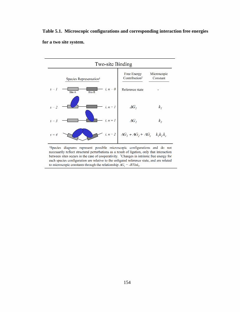
154
Table 5.1. Microscopic configurations and corresponding interaction free energies
for a two site system.
155
nonexistent or binding sites are identical. Fractional saturation curves were also fit to the
well known phenomenological Hill equation (305) to gauge the extent of cooperative
interactivity present in nucleoprotein complex formation. For this purpose complete
fractional saturation was calculated by:
Ῡ = 1 – ([Dfree]/[D input]) (Equation 5.15)
and fit to the familiar equation:
Ῡ = [P free]αH / (Kd
αH + [P free]αH) (Equation 5.16)
where αH is the Hill coefficient and Kd represents the macroscopic dissociation constant
(Kd = Ka-1). Nonlinear least-squares fitting was performed using Prism software.
Goodness of fit was assessed by visual inspection of residuals and by monitoring of
fitting statistics.
Quantitative DNase I ssDNA footprinting. To monitor binding of N-HisPurβ
monomers to individual sites of the purine-rich strand of the SMαA MCAT enhancer
element, quantitative DNase I footprinting was performed based on methods described by
Ackers and coworkers (1, 22, 23, 251), with the following modifications. All binding
reactions were carried out in buffer containing 50 mM Tris-HCl pH 7.5, 100 mM KCl, 1
mM dithiothreitol, 1 mM CaCl2, 2.5 mM MgCl2, 2 µg/mL dT32, and 50 µg/mL BSA at 20
± 1°C and allowed to equilibrate for 16 h. Each reaction contained 20,000 cpm of freshly
156
labeled SMP382-F* template, at a final concentration estimated to be well enough below
N-HisPurβ binding affinity to maintain validity of the [Pfree] ≈ [Ptotal] assumption (< 25
pM). N-HisPurβ was added to each reaction at the indicated concentrations to cover a
range from approximately 10-13 to 10-8 M in a final volume of 200 µL. After equilibrium
had been reached, template digestion was initiated by the addition of 5 µL of a DNase I
solution in assay buffer to reach a final concentration of 1.0 Units/mL. Digestion was
allowed to proceed for 2 min at 20 ± 1°C and was stopped by the addition of 700 µL of
stop solution (97% ethanol, 0.57 M ammonium acetate, 50 µg/mL yeast tRNA) and
incubated in a dry ice-ethanol bath for 30 min. Single-stranded DNA was pelleted by
centrifugation, washed twice with 70% ethanol, and air dried. Pellets were dissolved in 5
µL of buffer containing 98% formamide, 10 mM EDTA, 0.1% bromophenol blue, and
0.1% xylene cyanol. Samples were heated to 95°C for 5 min prior to being
eletrophoresed for 130 min at 65 Watts on a 0.4 mm thick sequencing gel consisting of
8% polyacrylamide (29:1 acrylamide:bisacrylamide), and 8M urea, cast and run in TBE
buffer. Gels were pre-run at 65 Watts for at least 2 h or until a gel temperature of ≥ 50°C
was attained. End-labeled primer dideoxy-NTP sequencing reactions performed with
double-stranded SMP382 PCR product as a template and SMP8p741s-F* as the extension
primer were also electrophoresed in order to identify sequences of interest on the
resulting footprints. Dideoxy-NTP sequencing reactions were performed according to the
manufacturer’s instructions for end-labeled primer sequencing (USB Corp.), and
typically 10,000-15,000 cpm per reaction were loaded on the sequencing gels. Following
electrophoresis, gels were dried in a Bio-Rad slab gel dryer on Whatman filter paper at
157
75°C for 45 min. Dried gels were exposed to phosphor storage screens for 72-96 h and
phosphorimaged as described above. Densitometry of phosphorimages was performed
using ImageQuant software (Molecular Dynamics) in order to determine values for
fractional protection (Fp) of a given sequence:
Fp = 1 – [(Iq, site i/Iq, control)]/[(I r, site i/Ir, control)] (Equation 5.17)
where I is again the relative densitometric intensity, q refers to any lane of the gel with
finite N-HisPurβ concentration, r refers to the reference lane containing no protein, site
refers to the ssDNA-binding site in question, and control refers to a region of the gel
whose intensity is independent of protein ligand concentration (bases -218 to -210 of
SMP382-F*). First line, crude analysis of fractional protection data was done by fitting
data to the familiar Langmuir isotherm, primarily to determine the upper and lower
endpoints of fractional protection since binding of protein ligands at specific sites, even at
saturating conditions, does not provide complete protection:
ƒ = u • {k[Pfree]/(1+k[P free])} + l (Equation 5.18)
Where k refers to the microscopic association constant (assuming no interaction
between sites), u and l refer to the upper and lower endpoints, respectively. Fractional
protection values were converted to values of fractional saturation, Ῡ, using the following
expression and the values of u and l from equation 5.19:
158
Ῡ = (f – l) / (u – l) (Equation 5.19)
Resolution of binding mechanism and microscopic association and interaction free
energies. Constructions of individual site binding expressions were done using a
statistical thermodynamics approach that has been described and applied previously (1).
Briefly the probability of the MCAT enhancer element existing in any one of the
microscopic configurations depicted in Table 5.2, ƒs, can be expressed as indicated in
equation 5.11. Microscopic association constants are related to microscopic free energies
through the relationship, ∆Gi = -RTlnki. The fractional saturation, Ῡ, of a given site can
be expressed as the sum of the probabilities of configurations in which the indicated site
is occupied. Applying this treatment for each of the models depicted in Table 2 gives
expressions describing fractional saturation at each site, A and/or B, in terms of their
microscopic association constants and N-HisPurβ monomer concentration:
Obligate Dimer (Equation 5.20)
Identical, independent (Equation 5.21)
Identical, interacting (Equation 5.22)
ῩA,B = k1kdi[P free]
2
1 + k1kdi[P free]2
ῩA,B = k1kdi[P free]
2
1 + k1kdi[P free]2
k1kdi[P free]2
1 + k1kdi[P free]2
ῩA,B = k1[Pfree] + k1
2[P free] 2
1 + 2k1[Pfree] + k 12[Pfree]
2ῩA,B =
k1[Pfree] + k12[P free] 2
1 + 2k1[Pfree] + k 12[Pfree]
2
ῩA,B = k1[P free] + k 1
2kc[P free]2
1 + 2k1[Pfree] + k12kc[Pfree]
2ῩA,B =
k1[P free] + k 12kc[P free]
2
1 + 2k1[Pfree] + k12kc[Pfree]
2
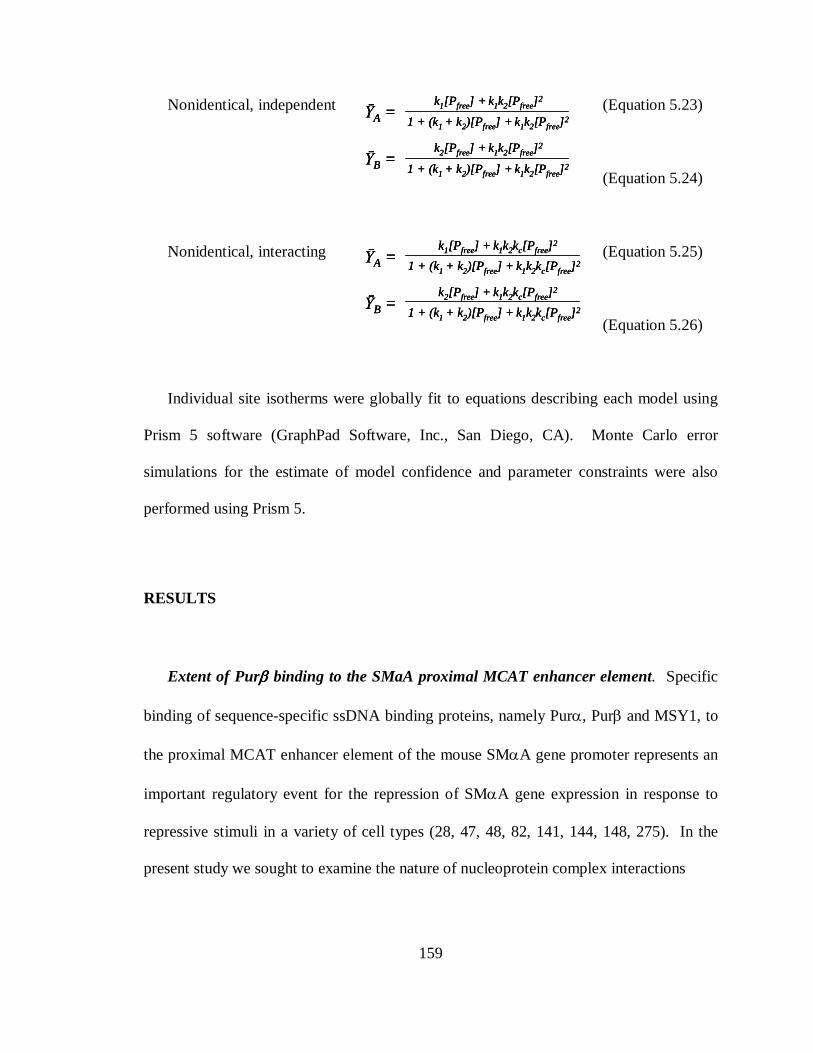
159
Nonidentical, independent (Equation 5.23)
(Equation 5.24)
Nonidentical, interacting (Equation 5.25)
(Equation 5.26)
Individual site isotherms were globally fit to equations describing each model using
Prism 5 software (GraphPad Software, Inc., San Diego, CA). Monte Carlo error
simulations for the estimate of model confidence and parameter constraints were also
performed using Prism 5.
RESULTS
Extent of Purββββ binding to the SMaA proximal MCAT enhancer element. Specific
binding of sequence-specific ssDNA binding proteins, namely Purα, Purβ and MSY1, to
the proximal MCAT enhancer element of the mouse SMαA gene promoter represents an
important regulatory event for the repression of SMαA gene expression in response to
repressive stimuli in a variety of cell types (28, 47, 48, 82, 141, 144, 148, 275). In the
present study we sought to examine the nature of nucleoprotein complex interactions
ῩA = k1[Pfree] + k1k2[Pfree]
2
1 + (k1 + k2)[Pfree] + k1k2[Pfree]2
ῩB = k2[Pfree] + k1k2[Pfree]
2
1 + (k1 + k2)[Pfree] + k1k2[Pfree]2
ῩA = k1[Pfree] + k1k2[Pfree]
2
1 + (k1 + k2)[Pfree] + k1k2[Pfree]2
ῩA = k1[Pfree] + k1k2[Pfree]
2
1 + (k1 + k2)[Pfree] + k1k2[Pfree]2
ῩB = k2[Pfree] + k1k2[Pfree]
2
1 + (k1 + k2)[Pfree] + k1k2[Pfree]2
ῩB = k2[Pfree] + k1k2[Pfree]
2
1 + (k1 + k2)[Pfree] + k1k2[Pfree]2
ῩA = k1[Pfree] + k1k2kc[Pfree]
2
1 + (k1 + k2)[Pfree] + k1k2kc[Pfree]2
ῩB = k2[Pfree] + k1k2kc[Pfree]
2
1 + (k1 + k2)[Pfree] + k1k2kc[Pfree]2
ῩA = k1[Pfree] + k1k2kc[Pfree]
2
1 + (k1 + k2)[Pfree] + k1k2kc[Pfree]2ῩA =
k1[Pfree] + k1k2kc[Pfree]2
1 + (k1 + k2)[Pfree] + k1k2kc[Pfree]2
ῩB = k2[Pfree] + k1k2kc[Pfree]
2
1 + (k1 + k2)[Pfree] + k1k2kc[Pfree]2ῩB =
k2[Pfree] + k1k2kc[Pfree]2
1 + (k1 + k2)[Pfree] + k1k2kc[Pfree]2
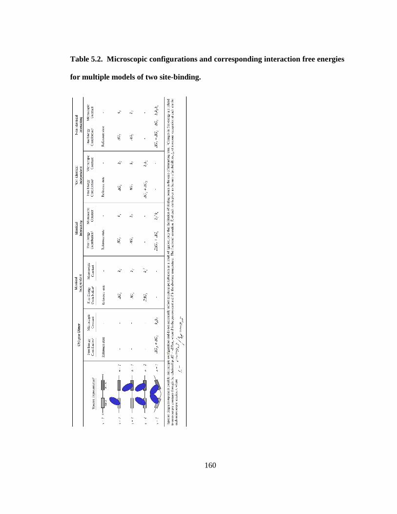
160
Table 5.2. Microscopic configurations and corresponding interaction free energies
for multiple models of two site-binding.
161
formed between Purβ, which has been shown to be the dominant Pur protein repressor of
SMαA at the proximal MCAT enhancer element in relevant cell-culture models (148,
149), and the purine-rich strand of the fore mentioned enhancer element. Towards this
aim, we used a combination of techniques that examine the effects of N-HisPurβ
concentration on the solution and electrophoretic properties of the representative single-
stranded oligonucleotide probe PE32-F. Both the electrophoretic mobility and
fluorescence anisotropy of PE32-F were cumulatively affected by increasing
concentrations of N-HisPurβ (Figure 5.2, panels A and B) indicative of binding.
Electrophoretic analysis of N-HisPurβ:PE32-F* nucleoprotein complexes suggest that N-
HisPurβ binds to PE32-F* (2 nM) in a sequential manner, as indicated by the appearance
of at least three discrete molecular species, and shifting of the higher mobility species to
complexes with lower mobility upon titration of N-HisPurβ over a concentration range of
0.41 nM to 400 nM (Figure 5.2, panel A). The co-appearance of the two highest mobility
species (speculated as adopting 1:1 and 2:1 complex stoichiometries, respectively) at
lower protein concentrations suggests that the 2:1 complex represents a high affinity
complex, possibly relying on cooperative facilitated assembly on a preformed 1:1
complex, as the higher mobility complex disappears at higher protein concentrations, and
the 2:1 species predominates over the applied N-HisPurβ concentration range. The
appearance of higher molecular weight species (3:1 and greater) at the highest N-HisPurβ
concentrations shows that these species likely represent low affinity, and possibly non-
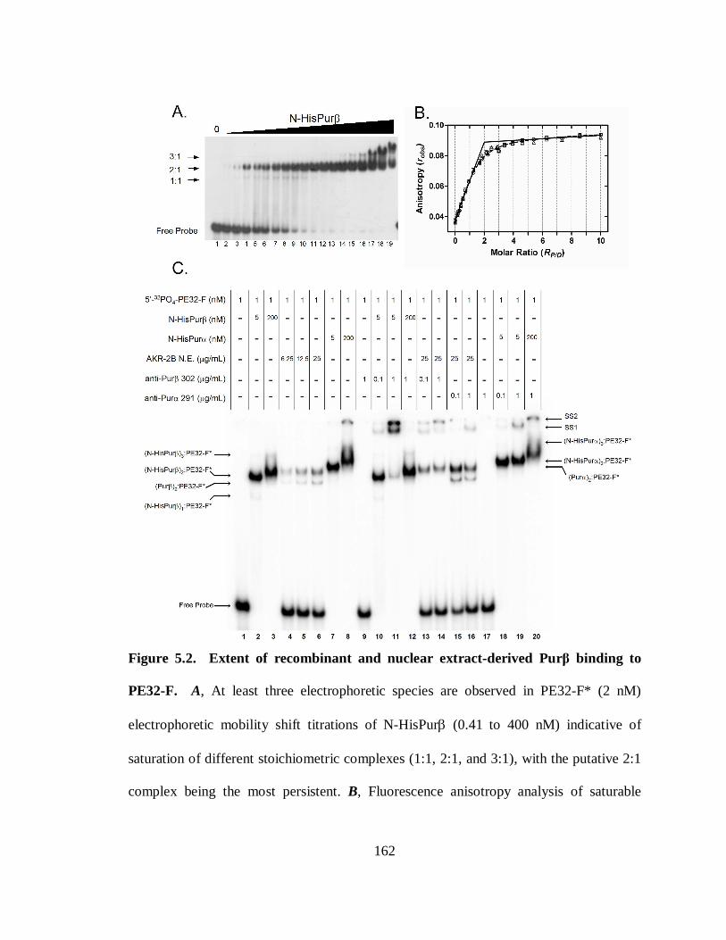
162
Figure 5.2. Extent of recombinant and nuclear extract-derived Purβ binding to
PE32-F. A, At least three electrophoretic species are observed in PE32-F* (2 nM)
electrophoretic mobility shift titrations of N-HisPurβ (0.41 to 400 nM) indicative of
saturation of different stoichiometric complexes (1:1, 2:1, and 3:1), with the putative 2:1
complex being the most persistent. B, Fluorescence anisotropy analysis of saturable
163
binding of N-HisPurβ to fluorescent probe PE32-F-3FLC (50 nM). Data was fit to
equation 5.6 to determine the equivalency point (dashed line). Fixing of Kr to near-zero
values (infinite affinity, solid line) verified the equivalency transition at an RP/D value of
2:1. Symbols represent titrations from different preparations of N-HisPurβ, indicating
similar number of active binding sites. C, Qualitative electrophoretic mobility (super)shift
assay of PE32-F* nucleoprotein complexes. The relative electrophoretic mobilities of
nucleoprotein complexes containing PE32-F* and either recombinant, or nuclear extract-
derived Pur proteins were compared to identify the predominant and presumably
biologically relevant species. The predominant species observed with 5 nM N-HisPurβ
and 1 nM PE32-F* (lane 2) displays mobility similar to that seen from nuclear extracts
(lanes 4-6), as verified by specific supershifting with anti-Purβ 302 antibody (lanes 13
and 14). Similar findings were observed for Purα (compare lane 7 with lanes 4-6, and
supershift lanes 16 and 17). The indicated identities of observed complexes are
hypothetical.
164
specific interactions of N-HisPurβ with free ssDNA sites and/or protein-protein
interaction sites on preformed nucleoprotein complexes.
Interpretations of the preliminary electrophoretic mobility shift results described
above were verified by experiments in which binding of N-HisPurβ to fluorescein-
labeled PE32-F (PE32-F-3FLC) at saturating conditions (probe concentration >50 times
the reported dissociation constant of approximately 1 nM) was monitored by changes in
the measured anisotropy of the probe. The results of this analysis are shown in panel B
of Figure 5.2. Under saturating binding conditions as applied in this analysis, it is evident
that N-HisPurβ can saturate PE32-F-3FLC to a specific terminal stoichiometry of 2:1, as
indicated by the returned stoichiometery values from non-linear least-squares regression
of both data sets to equation 5.6, and the equivalency transition point when the apparent
affinity value, Kr, is fixed at values approaching zero (near-infinite affinity, solid line).
Curvature in the best fit line at values of RP/D near the equivalence point when Kr is not
fixed (dashed line) supports the notion that either the total concentration of probe is not
sufficiently high enough to ensure that every protein molecule is bound to the probe at
lower protein concentrations, a condition that is only satisfied when Dtotal / Kd >> 10, in
the case of a completely cooperative system (228), that a non-specific binding event
occurs at higher concentrations leading to higher order stoichiometries, or both. Three
lines of evidence support the latter possibility: 1) Affinities of Purβ for single-stranded
PUR-elements have been reported to be in the 0.5 to 1.5 nM range (148, 310), whereas in
the experiments performed here, PE32-F-3FLC were held at 50 nM. 2) A positive slope
in the plateau region indicates that additional ligand binding is occurring at higher protein
165
concentrations. This observation could also be explained by an increase in the solution
viscosity as a function of protein concentration, however an anisotropy change of PE32-
F-3FLC in the presence of increasing concentrations of BSA was not observed nor did
the anisotropy of PE32-F3FLC change in the context of a double-stranded probe with
increasing concentrations of N-HisPurβ (data not shown), both suggesting that protein
concentration viscosity gradients are not significant in these concentration ranges. 3) The
electrophoretic mobility shift profile of PE32-F* at high N-HisPurβ concentrations
displayed in Figure 5.2A shows that species formed at these conditions are likely of
higher order (greater than 2:1), as indicated by the presence of two faster moving species
at lower protein concentrations. Collectively these data show that N-HisPurβ has the
capacity to form complexes with PE32-F or PE32-F-3FLC beyond a 1:1 stoichiometry,
and the extent of binding follows, most stably, the formation of a 2:1 complex but does
not appear terminable; however the extent of binding that is biologically relevant remains
uncertain.
To assess the extent of PE32-F binding by Purβ in the context of the nuclear
environment, and to compare this to levels observed with recombinant N-HisPurβ under
controlled conditions, comparative, qualitative EMSAs were performed. Figure 5.2C
demonstrates the electrophoretic mobilities of PE32-F*-containing nucleoprotein
complexes derived from equilibration or PE32-F* (1 nM) with recombinant N-HisPurβ,
N-HisPurα, and AKR-2B MEF nuclear extract-derived proteins. Consistent with
observations made in panel A, N-HisPurβ, at a concentration of 5 nM shifts PE32-F* into
two visible bands (lane 2), speculated to be N-HisPurβ1:PE32-F* (lower band) and N-
166
HisPurβ2:PE32-F* (upper band) based on findings presented above. N-HisPurα (5 nM,
lane 7) shows a single shifted band dissimilar to N-HisPurβ , but likely corresponds to N-
HisPurα2:PE32-F* despite slight differences in mobilities compared to the N-HisPurβ
equivalents. Mobility disparities are consistent with differences in the monomeric
molecular weights and calculated charges at pH 8. AKR-2B MEF nuclear extracts shift
PE32-F* into a lower band, and an upper group of closely migrating bands that do not
resolve well under the electrophoretic conditions applied here (lanes 4-6). Longer runs
suggest that, indeed, this upper grouping is composed of several closely migrating bands,
however, their complete resolution is hindered due to streaking of bands with longer gel-
run times (data not shown). Incubation of nuclear extract binding reactions with an
antibody that specifically binds Purβ (anti-Purβ 302, lanes 13 and 14) clearly identifies
the lower band as the Purβ:PE32-F* complex, as indicated by the complete
disappearance of this band in lanes 13 and 14, and the appearance of supershifted
complexes, SS1 and SS2. Additionally, the presence of anti-Purβ 302 causes some loss
of the faster migrating portion of the upper doublet, suggesting that this species might be
heteromeric Purα/β:PE32-F* complexes. Incubation of nuclear extract binding reactions
with an antibody that specifically binds Purα (anti-Purα 291, lanes 15 and 16) further
divulge the compositions of the slower migrating bands generated by nuclear extract
material. The upper band(s) in lanes 4-6 likely correspond to Purα:PE32-F*, or
heteromeric Purα/β:PE32-F* complexes due to, again, loss of the faster migrating portion
of the doublet, and some loss of the slower migrating portion upon antibody addition, and
the appearance of supershifted complexes. Persistence of some slower migrating species
167
in the presence of 1 µg/mL anti-Purα 291 suggests that these bands might represent
nucleoprotein complexes of unknown composition, or that anti-Purα 291 possesses lower
affinity for Purα-containing nucleoprotein complexes than does anti-Purβ 302 for Purβ-
containing nucleoprotein complexes, a possibility supported by supershifts conducted on
recombinant Pur:PE32-F complexes (compare lanes 11 and 19). Previous studies have
suggested that Purβ is the dominant Pur protein repressor in the context of SMαA
expression (145, 148), despite the fact that the intense slower moving complex observed
contains Purα, as indicated by reactivity with anti-Purα 291 antibody. This finding
suggests that either Purα is more abundant than Purβ in asynchronous AKR-2B nuclear
extracts, or displays higher affinity for PE32-F. Previous studies have shown that Purα
binds with higher affinity than Purβ to both PE32-F (148), and to a 24mer representative
of the c-myc PUR element (310).
Comparing the predominant N-HisPurβ shifted complex of PE32-F* (speculated as
N-HisPurβ2:PE32-F*, lane 2) and the lower, Purβ-containing band generated by
equilibration with nuclear extract-derived proteins (lower band, lanes 4-6), it is further
conjectured that the latter is likely Purβ2:PE32-F*. This inference is based on the similar
relative mobilities of the two complexes. Slight differences in electrophoretic mobility
between the recombinant and endogenous nucleoprotein complexes are consistent with
molecular weight and charge differences between N-HisPurβ (35,168.6 Da, -5.05) , and
Purβ (33,901.3 Da, -5.15). In conclusion, the predominant shifted species observed when
5 nM N-HisPurβ is equilibrated with 1 nM PE32-F* is equivalent to the nuclear extract
168
derived-endogenous Purβ:PE32-F* complex. However, the exact stoichiometry of this
complex can only be inferred from anisotropy experiments, described above, as being
2:1.
Direct assessment of the predominant electrophoretic nucleoprotein complex
generated from equilibration of 5 nM N-HisPurβ with 1 nM PE32-F* was performed
using a serial dilution-coupled EMSA. Briefly, a reaction mixture containing N-HisPurβ
and PE32-F* at the forementioned concentrations, was serially diluted at a ratio of 1.1 to
1 with buffer to create a series of reaction samples with the same molar ratio of
components, but at differing concentrations, and a distribution of reversibly interacting
species governed by laws of mass-action. Quantification of molecular species by
densitometric methods subsequent to electrophoretic separation permits determination of
the system stoichiometry by way of a value convergence method, as described in the
methods section of this paper. Implementation of this method, as shown in Figure 5.3,
shows that the persistent, and stable high-affinity nucleoprotein complex formed between
N-HisPurβ and PE32-F* adopts a stoichiometry of 2 to 1, as convergence between
estimated values of n and those obtained by linear regression of the ln[PnD/Dfree] vs
ln[Pfree] plot occurred at n = 2.
Affinity of specific N-HisPurββββ/ssDNA interactions. The reported ssDNA binding
characteristics that unify Pur proteins, is their preference for interaction with purine-rich
sequences, but also their affinity for such sequences (148, 310). However, an extensive
and thermodynamically rigorous assessment of the free energy of ssDNA-binding by Pur
proteins has not yet been reported. To address this fact, we performed direct titrations of
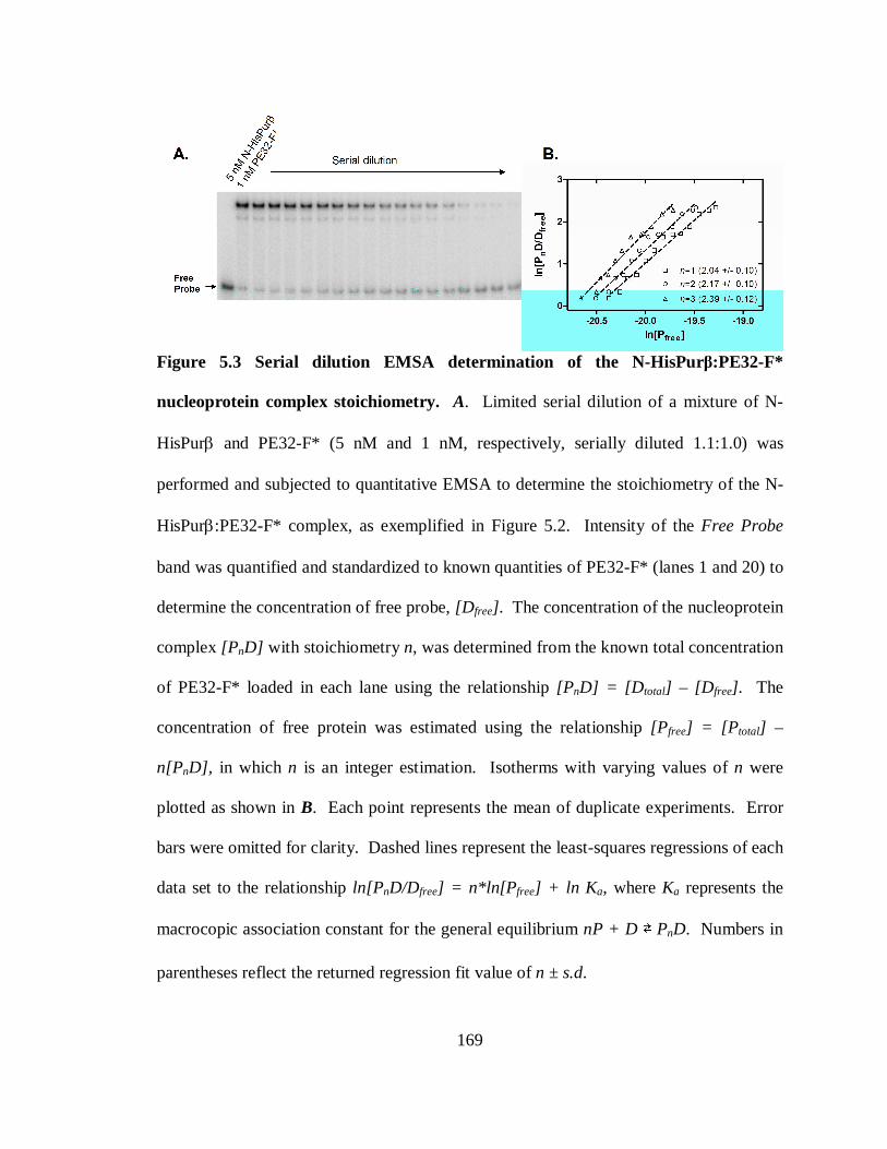
169
Figure 5.3 Serial dilution EMSA determination of the N-HisPurβ:PE32-F*
nucleoprotein complex stoichiometry. A. Limited serial dilution of a mixture of N-
HisPurβ and PE32-F* (5 nM and 1 nM, respectively, serially diluted 1.1:1.0) was
performed and subjected to quantitative EMSA to determine the stoichiometry of the N-
HisPurβ:PE32-F* complex, as exemplified in Figure 5.2. Intensity of the Free Probe
band was quantified and standardized to known quantities of PE32-F* (lanes 1 and 20) to
determine the concentration of free probe, [D free] . The concentration of the nucleoprotein
complex [PnD] with stoichiometry n, was determined from the known total concentration
of PE32-F* loaded in each lane using the relationship [PnD] = [D total] – [D free]. The
concentration of free protein was estimated using the relationship [P free] = [P total] –
n[PnD], in which n is an integer estimation. Isotherms with varying values of n were
plotted as shown in B. Each point represents the mean of duplicate experiments. Error
bars were omitted for clarity. Dashed lines represent the least-squares regressions of each
data set to the relationship ln[PnD/Dfree] = n*ln[P free] + ln K a, where Ka represents the
macrocopic association constant for the general equilibrium nP + D ⇄ PnD. Numbers in
parentheses reflect the returned regression fit value of n ± s.d.
170
N-HisPurβ against 25 pM PE32-F*, a condition that maintains validity of the assumption
[Pfree] ≈ [Ptotal] , which is necessary for mathematical modeling of binding reactions
(228). Molecular species distributions at equilibrium were separated by native gel
electrophoresis and quantified as described in the methods section. Results of this
analysis are shown in Figure 5.4. Panel A shows the electrophoretic mobility shift profile
of PE32-F* as a result of binding in the presence of increasing concentrations of N-
HisPurβ. Visualization and densitometric analysis confirms the presence of three
separate bands or pixel intensity peaks (Figure 5.4B) corresponding to free probe, and
two shifted complexes that are (N-HisPurβ)1:PE32-F* (1:1 complex) and (N-
HisPurβ)2:PE32-F* (2:1 complex) as identified in the previous section. Of special
interest is the transient nature of the 1:1 complex with respect to N-HisPurβ
concentration. Comparing lane intensity profiles in Figure 5.4B for increasing N-
HisPurβ concentrations shows that the 1:1 complex is not a protein preparation
contaminant as the peak intensity reaches a maxima at moderate N-HisPurβ
concentrations and drops-off as PE32-F* becomes saturated. Furthermore, this peak
pattern has been theoretically assigned to systems that adopt cooperative, two-site
binding mechanisms (27). Peak integration for each species provided the isotherms
observed in Figure 5.4C. Species specific isotherms Θ0, Θ1, and Θ2 were globally fit to
equations 5.12, 5.13 and 5.14, respectively, to resolve values for the macroscopic
association constants K1, and K2. This approach yielded K1 = 3.43 (± 0.368) x 108 M-1,
and K2 = 6.06 (± 0.191) x 1018 M-1. It has been previously demonstrated that in instances
in which the nature of two-site binding of protein ligands to DNA is unknown (i.e.,
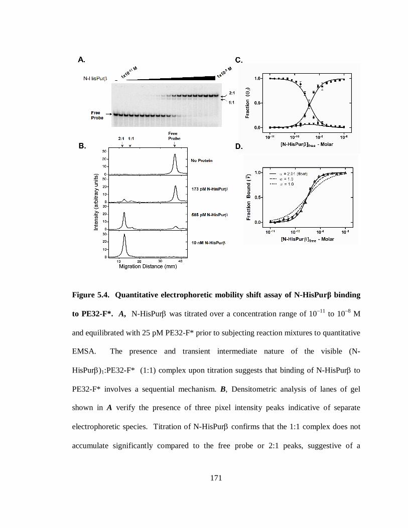
171
Figure 5.4. Quantitative electrophoretic mobility shift assay of N-HisPurβ binding
to PE32-F*. A, N-HisPurβ was titrated over a concentration range of 10−11 to 10−8 M
and equilibrated with 25 pM PE32-F* prior to subjecting reaction mixtures to quantitative
EMSA. The presence and transient intermediate nature of the visible (N-
HisPurβ)1:PE32-F* (1:1) complex upon titration suggests that binding of N-HisPurβ to
PE32-F* involves a sequential mechanism. B, Densitometric analysis of lanes of gel
shown in A verify the presence of three pixel intensity peaks indicative of separate
electrophoretic species. Titration of N-HisPurβ confirms that the 1:1 complex does not
accumulate significantly compared to the free probe or 2:1 peaks, suggestive of a

172
sequential and cooperative binding mechanism. C, Individual band intensities are plotted
as a function of [N-HisPurβfree] (●, free Probe; ▼, 1:1 complex; ■, 2:1 complex). Each
point represents the mean ± s.d. of quadruplicate experiments. Lines are global nonlinear
least squares fits of individual species isotherms to equations 5.12-5.14. D, Band
intensity data from A presented as Fraction Bound, Ῡ, (Ῡ = ([D total] – [D free]) / [D total])
versus [N-HisPurβfree] , were fit to the phenomenological Hill equation (equation 5.16).
The Hill coefficient, αH, was held constant at values of 1.5 (dashed line) and 1.0 (dotted
line) to reflect the dependency of this variable on goodness of fit.
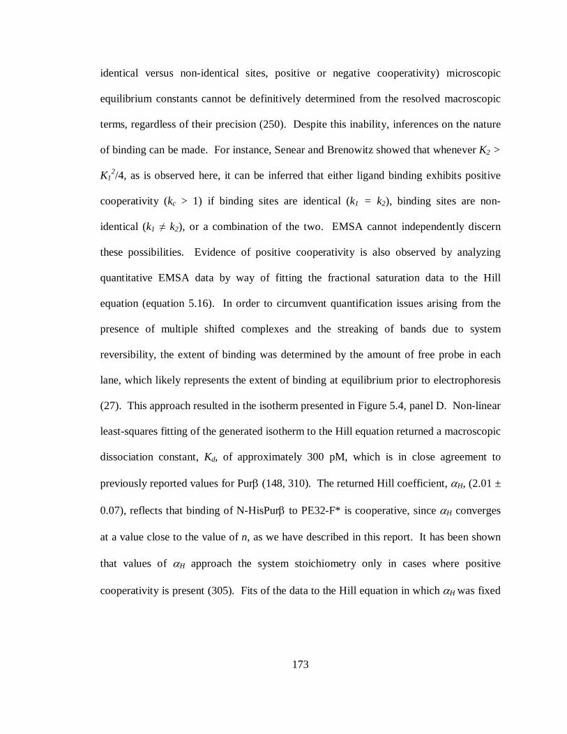
173
identical versus non-identical sites, positive or negative cooperativity) microscopic
equilibrium constants cannot be definitively determined from the resolved macroscopic
terms, regardless of their precision (250). Despite this inability, inferences on the nature
of binding can be made. For instance, Senear and Brenowitz showed that whenever K2 >
K12/4, as is observed here, it can be inferred that either ligand binding exhibits positive
cooperativity (kc > 1) if binding sites are identical (k1 = k2), binding sites are non-
identical (k1 ≠ k2), or a combination of the two. EMSA cannot independently discern
these possibilities. Evidence of positive cooperativity is also observed by analyzing
quantitative EMSA data by way of fitting the fractional saturation data to the Hill
equation (equation 5.16). In order to circumvent quantification issues arising from the
presence of multiple shifted complexes and the streaking of bands due to system
reversibility, the extent of binding was determined by the amount of free probe in each
lane, which likely represents the extent of binding at equilibrium prior to electrophoresis
(27). This approach resulted in the isotherm presented in Figure 5.4, panel D. Non-linear
least-squares fitting of the generated isotherm to the Hill equation returned a macroscopic
dissociation constant, Kd, of approximately 300 pM, which is in close agreement to
previously reported values for Purβ (148, 310). The returned Hill coefficient, αH, (2.01 ±
0.07), reflects that binding of N-HisPurβ to PE32-F* is cooperative, since αH converges
at a value close to the value of n, as we have described in this report. It has been shown
that values of αH approach the system stoichiometry only in cases where positive
cooperativity is present (305). Fits of the data to the Hill equation in which αH was fixed
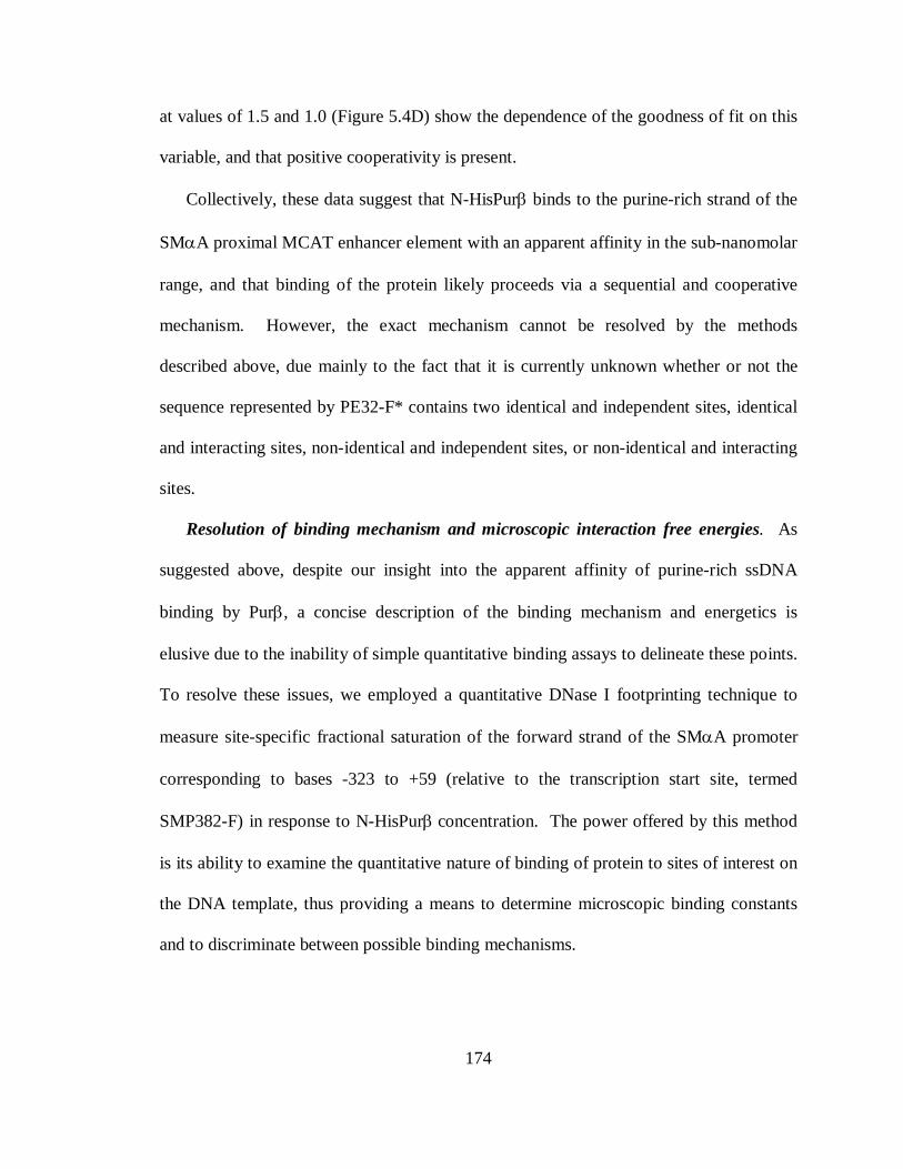
174
at values of 1.5 and 1.0 (Figure 5.4D) show the dependence of the goodness of fit on this
variable, and that positive cooperativity is present.
Collectively, these data suggest that N-HisPurβ binds to the purine-rich strand of the
SMαA proximal MCAT enhancer element with an apparent affinity in the sub-nanomolar
range, and that binding of the protein likely proceeds via a sequential and cooperative
mechanism. However, the exact mechanism cannot be resolved by the methods
described above, due mainly to the fact that it is currently unknown whether or not the
sequence represented by PE32-F* contains two identical and independent sites, identical
and interacting sites, non-identical and independent sites, or non-identical and interacting
sites.
Resolution of binding mechanism and microscopic interaction free energies. As
suggested above, despite our insight into the apparent affinity of purine-rich ssDNA
binding by Purβ, a concise description of the binding mechanism and energetics is
elusive due to the inability of simple quantitative binding assays to delineate these points.
To resolve these issues, we employed a quantitative DNase I footprinting technique to
measure site-specific fractional saturation of the forward strand of the SMαA promoter
corresponding to bases -323 to +59 (relative to the transcription start site, termed
SMP382-F) in response to N-HisPurβ concentration. The power offered by this method
is its ability to examine the quantitative nature of binding of protein to sites of interest on
the DNA template, thus providing a means to determine microscopic binding constants
and to discriminate between possible binding mechanisms.
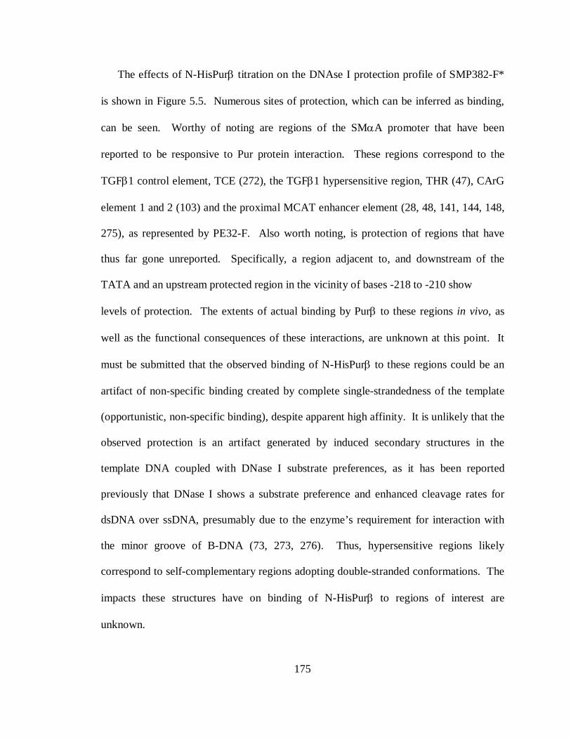
175
The effects of N-HisPurβ titration on the DNAse I protection profile of SMP382-F*
is shown in Figure 5.5. Numerous sites of protection, which can be inferred as binding,
can be seen. Worthy of noting are regions of the SMαA promoter that have been
reported to be responsive to Pur protein interaction. These regions correspond to the
TGFβ1 control element, TCE (272), the TGFβ1 hypersensitive region, THR (47), CArG
element 1 and 2 (103) and the proximal MCAT enhancer element (28, 48, 141, 144, 148,
275), as represented by PE32-F. Also worth noting, is protection of regions that have
thus far gone unreported. Specifically, a region adjacent to, and downstream of the
TATA and an upstream protected region in the vicinity of bases -218 to -210 show
levels of protection. The extents of actual binding by Purβ to these regions in vivo, as
well as the functional consequences of these interactions, are unknown at this point. It
must be submitted that the observed binding of N-HisPurβ to these regions could be an
artifact of non-specific binding created by complete single-strandedness of the template
(opportunistic, non-specific binding), despite apparent high affinity. It is unlikely that the
observed protection is an artifact generated by induced secondary structures in the
template DNA coupled with DNase I substrate preferences, as it has been reported
previously that DNase I shows a substrate preference and enhanced cleavage rates for
dsDNA over ssDNA, presumably due to the enzyme’s requirement for interaction with
the minor groove of B-DNA (73, 273, 276). Thus, hypersensitive regions likely
correspond to self-complementary regions adopting double-stranded conformations. The
impacts these structures have on binding of N-HisPurβ to regions of interest are
unknown.
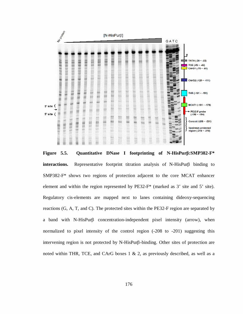
176
Figure 5.5. Quantitative DNase I footprinting of N-HisPurβ:SMP382-F*
interactions. Representative footprint titration analysis of N-HisPurβ binding to
SMP382-F* shows two regions of protection adjacent to the core MCAT enhancer
element and within the region represented by PE32-F* (marked as 3’ site and 5’ site).
Regulatory cis-elements are mapped next to lanes containing dideoxy-sequencing
reactions (G, A, T, and C). The protected sites within the PE32-F region are separated by
a band with N-HisPurβ concentration-independent pixel intensity (arrow), when
normalized to pixel intensity of the control region (-208 to -201) suggesting this
intervening region is not protected by N-HisPurβ-binding. Other sites of protection are
noted within THR, TCE, and CArG boxes 1 & 2, as previously described, as well as a

177
previously uncharacterized upstream region (-218 to -210), and a region adjacent to the
TATA box.
178
A goal of our approach was to quantitatively interrogate binding of N-HisPurβ to the
region encompassing the proximal MCAT enhancer (bases -195 to -164). Close
examination of this region by densitometric methods yields two sites of protection
afforded by N-HisPurβ titration. These sites have been termed the 3’, and 5’ sites, and
are labeled as such in Figure 5.5. It should be noted that these sites are separated by a
region that is not protected when compared to the control region (-208 to -201), and is
marked by an arrow. Based on our knowledge of the stoichiometry of the nucleoprotein
complex formed between N-HisPurβ and this sequence, this nature of protection appears
appropriate.
Mathematical expressions describing various models of interactions between N-
HisPurβ and the SMαA MCAT enhancer element deemed appropriate as based on the
measured stoichiometry of the N-HisPurβ:PE32-F nucleoprotein complex were generated
by methods outlined previously (1). For complexes with a saturating stoichiometry of 2:1
five possible models can be proposed. 1) The first possibility is that of a preformed
(obligate) dimer assembling on a single binding site. Other possible models involve
sequential assembly of monomers on the ssDNA lattice in which binding sites are: 2)
identical and independent, 3) identical and interacting, 4) non-identical and independent,
and 5) non-identical and interacting. The various macromolecular configurations for
each ligation state allowed by the restrictions of each binding model are depicted in Table
2 along with the corresponding free energy contributions and equilibrium constants used
for constructing expressions of N-HisPurβ binding.
179
Several assumptions have been made in order to constrain and mathematically define
the models described above: A) Binding of N-HisPurβ to the MCAT enhancer region
(bases -195 to -164 of the SMαA promoter) is independent of binding of N-HisPurβ to
other regions of the promoter, outside of this vicinity. It should be submitted that this
assumption may not be valid based on the protection profile observed, in which multiple
sites display binding. However, at this time the complete complex stoichiometries
obtained at these sites are unknown. B) DNA-independent self association of N-HisPurβ,
under the conditions used here, is defined by the pre-determined equilibrium constant of
kdi = 884955 M-1 (222), and is negligible in cases of sequential monomeric assembly,
where half saturation of sites is in the subnanomolar range (see results section). It should
be noted that the reported dimerization constant for N-HisPurβ was determined under
different solution conditions (higher ionic strength) than those applied in the present
study, and the absolute effects of salt identity and concentration on self-association have
not been explored thoroughly, due mainly to solubility problems associated with N-
HisPurβ at concentrations necessary for measurement by analytical centrifugation.
However, since we have observed that sodium chloride concentrations as high as 1M do
not alter the hydrodynamic properties of N-HisPurβ as judged by size-exclusion
chromatography (149), we have assumed that self-dimerization is not significantly
perturbed under the conditions applied here. C) Where applicable, identical binding sites
exhibit equal intrinsic binding free energy changes upon ligand binding, as illustrated
with ∆G1 designated as this free energy change for both sites. D) Likewise, non-
identical sites exhibit non-equivalent intrinsic binding free energy changes upon ligand
180
binding, and as such, are designated with individual parameters ∆G1 and ∆G2 to reflect
this prediction. E) In binding pathways possessing intersite interaction, the difference in
the change in free energy between binding of each monomer to a ssDNA site individually
and the total free energy change observed upon facilitated binding is represented as ∆Gc.
Microscopic species-specific free energy changes and microscopic equilibrium constant
assignments are shown in Table 2. The construction of expressions describing site
specific fractional saturation was performed as described in the methods section.
Densitometric quantification of N-HisPurβ binding to the identified 3’, and 5’ sites of
the proximal MCAT enhancer element was performed to generate individual site binding
isotherms, and are displayed in Figure 5.6. As can be seen, isotherms generated by
binding of N-HisPurβ by these sites are non-identical, suggesting that binding does not
proceed through an obligate dimer pathway, and that sites are also non-identical. Global
fitting of individual site isotherms to the model-specific expressions outlined in the
methods section (equations 5.20-5.26), provided a means to further discriminate between
models based on goodness of fit. From this approach, it can be seen that binding of N-
HisPurβ to the 3’ and 5’ sites of the proximal MCAT enhancer element proceeds through
a sequential mechanism, in which sites are non-identical and interacting, as judged by
random distribution of residuals and fit statistics. More specifically, binding of the 3’ site
occurs with highest affinity, and that the 3’ and 5’ sites are non-identical and interacting,
with binding to the 5’ site being cooperatively facilitated. This model is in line with the
results of the qualitative and quantitative electrophoretic mobilility shifts presented in
previous sections, which suggested that binding of N-HisPurβ proceeds via a sequential
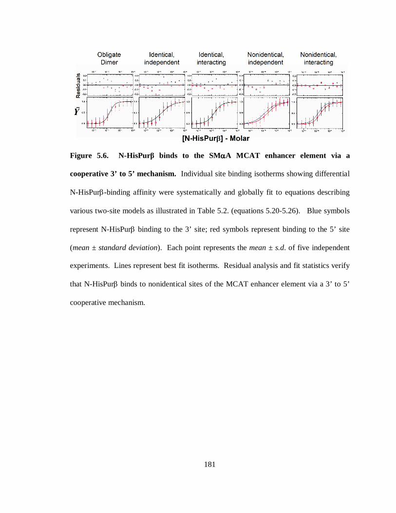
181
Figure 5.6. N-HisPurβ binds to the SMααααA MCAT enhancer element via a
cooperative 3’ to 5’ mechanism. Individual site binding isotherms showing differential
N-HisPurβ-binding affinity were systematically and globally fit to equations describing
various two-site models as illustrated in Table 5.2. (equations 5.20-5.26). Blue symbols
represent N-HisPurβ binding to the 3’ site; red symbols represent binding to the 5’ site
(mean ± standard deviation). Each point represents the mean ± s.d. of five independent
experiments. Lines represent best fit isotherms. Residual analysis and fit statistics verify
that N-HisPurβ binds to nonidentical sites of the MCAT enhancer element via a 3’ to 5’
cooperative mechanism.
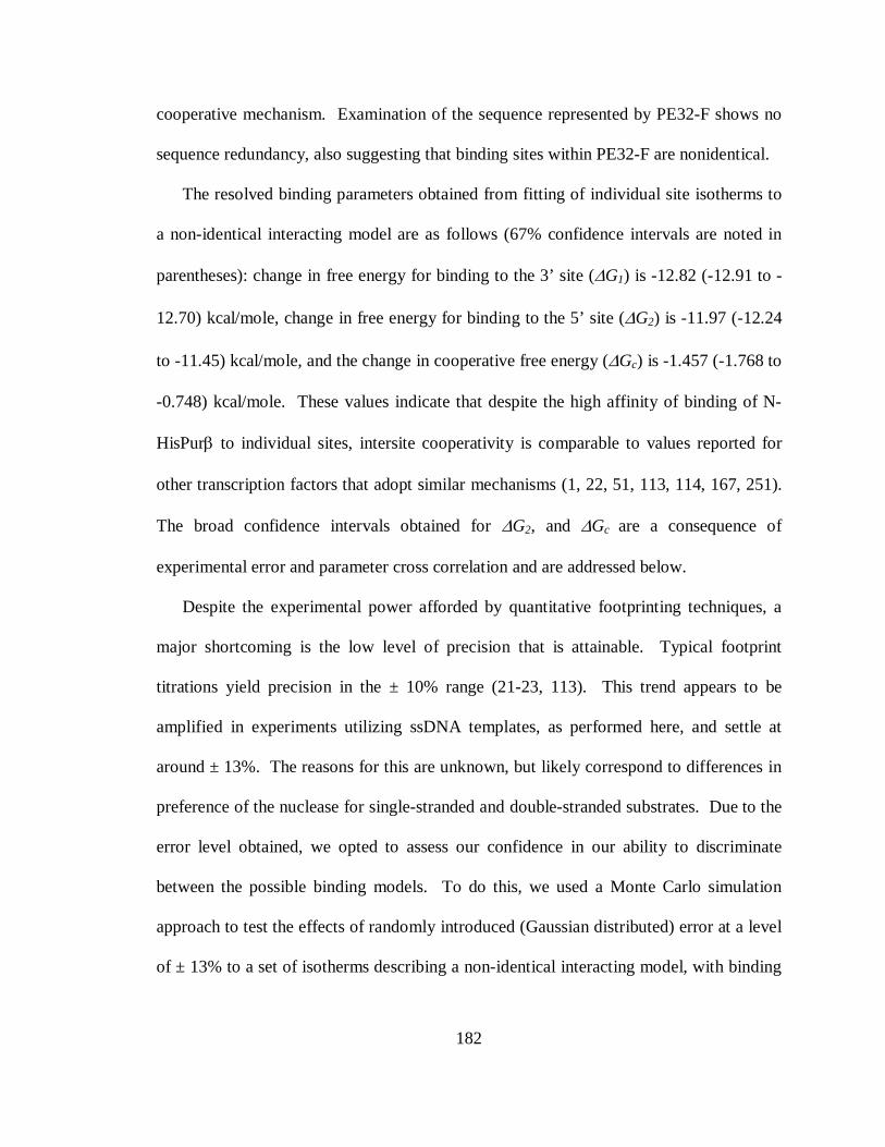
182
cooperative mechanism. Examination of the sequence represented by PE32-F shows no
sequence redundancy, also suggesting that binding sites within PE32-F are nonidentical.
The resolved binding parameters obtained from fitting of individual site isotherms to
a non-identical interacting model are as follows (67% confidence intervals are noted in
parentheses): change in free energy for binding to the 3’ site (∆G1) is -12.82 (-12.91 to -
12.70) kcal/mole, change in free energy for binding to the 5’ site (∆G2) is -11.97 (-12.24
to -11.45) kcal/mole, and the change in cooperative free energy (∆Gc) is -1.457 (-1.768 to
-0.748) kcal/mole. These values indicate that despite the high affinity of binding of N-
HisPurβ to individual sites, intersite cooperativity is comparable to values reported for
other transcription factors that adopt similar mechanisms (1, 22, 51, 113, 114, 167, 251).
The broad confidence intervals obtained for ∆G2, and ∆Gc are a consequence of
experimental error and parameter cross correlation and are addressed below.
Despite the experimental power afforded by quantitative footprinting techniques, a
major shortcoming is the low level of precision that is attainable. Typical footprint
titrations yield precision in the ± 10% range (21-23, 113). This trend appears to be
amplified in experiments utilizing ssDNA templates, as performed here, and settle at
around ± 13%. The reasons for this are unknown, but likely correspond to differences in
preference of the nuclease for single-stranded and double-stranded substrates. Due to the
error level obtained, we opted to assess our confidence in our ability to discriminate
between the possible binding models. To do this, we used a Monte Carlo simulation
approach to test the effects of randomly introduced (Gaussian distributed) error at a level
of ± 13% to a set of isotherms describing a non-identical interacting model, with binding
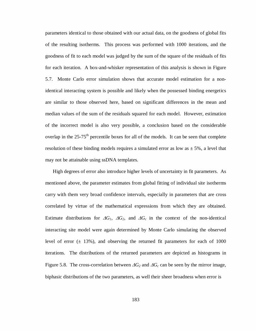
183
parameters identical to those obtained with our actual data, on the goodness of global fits
of the resulting isotherms. This process was performed with 1000 iterations, and the
goodness of fit to each model was judged by the sum of the square of the residuals of fits
for each iteration. A box-and-whisker representation of this analysis is shown in Figure
5.7. Monte Carlo error simulation shows that accurate model estimation for a non-
identical interacting system is possible and likely when the possessed binding energetics
are similar to those observed here, based on significant differences in the mean and
median values of the sum of the residuals squared for each model. However, estimation
of the incorrect model is also very possible, a conclusion based on the considerable
overlap in the 25-75th percentile boxes for all of the models. It can be seen that complete
resolution of these binding models requires a simulated error as low as ± 5%, a level that
may not be attainable using ssDNA templates.
High degrees of error also introduce higher levels of uncertainty in fit parameters. As
mentioned above, the parameter estimates from global fitting of individual site isotherms
carry with them very broad confidence intervals, especially in parameters that are cross
correlated by virtue of the mathematical expressions from which they are obtained.
Estimate distributions for ∆G1, ∆G2, and ∆Gc in the context of the non-identical
interacting site model were again determined by Monte Carlo simulating the observed
level of error (± 13%), and observing the returned fit parameters for each of 1000
iterations. The distributions of the returned parameters are depicted as histograms in
Figure 5.8. The cross-correlation between ∆G2 and ∆Gc can be seen by the mirror image,
biphasic distributions of the two parameters, as well their sheer broadness when error is
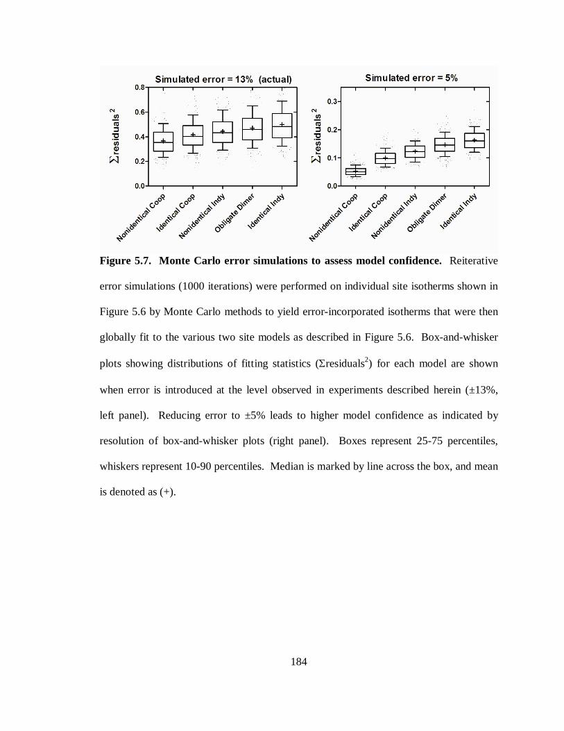
184
Figure 5.7. Monte Carlo error simulations to assess model confidence. Reiterative
error simulations (1000 iterations) were performed on individual site isotherms shown in
Figure 5.6 by Monte Carlo methods to yield error-incorporated isotherms that were then
globally fit to the various two site models as described in Figure 5.6. Box-and-whisker
plots showing distributions of fitting statistics (Σresiduals2) for each model are shown
when error is introduced at the level observed in experiments described herein (±13%,
left panel). Reducing error to ±5% leads to higher model confidence as indicated by
resolution of box-and-whisker plots (right panel). Boxes represent 25-75 percentiles,
whiskers represent 10-90 percentiles. Median is marked by line across the box, and mean
is denoted as (+).
185
simulated at ± 13%. The biphasicity persists until error is reduced to ± 5%, however the
width of the distributions is significant for all levels of introduced error. It has been
shown previously that resolution of cross-correlated values requires the isolated
determination of one of the parameters by implementation of reduced valency templates
(1). Unfortunately, construction of templates with reduced numbers of binding sites
requires extensive sequence knowledge of the binding site in question, and that binding to
this site can be abolished by deletion or mutation of the template. The absolute sequence
identity of a Pur protein binding site remains speculative at this time. Footprints obtained
by nuclease digestion often overestimate the size of binding sites due to steric effects
(reviewed in (108)), and thus do not provide enough resolution to unequivocally identify
the N-HisPurβ nucleotide binding site observed here, except to say that one exists on the
3’ and 5’ ends of the PE32-F region. The matter of binding site characterization is under
current investigation by our laboratory.
Binding of N-HisPurββββ to supercoiled DNA. The inability of purified recombinant
Purβ to bind 32 base-pair double-stranded representations of the SMαA proximal MCAT
enhancer element (dsPE32-F) has been previously demonstrated (148, 149). Calculations
of the free energy of strand separation (melting) of dsPE32-F by the nearest-neighbor
method proposed by Breslauer (24), using thermodynamic values reported by SantaLucia,
Jr. (239), suggest that the process requires 58.4 kcal/mole. This value is in accordance
with experimental values obtained by our group (data not shown) using methods
previously described (215). We have reported here that additive binding of N-HisPurβ to
a sequence corresponding to PE32-F liberates (∆GTotal = ∆G1 + ∆G2 + ∆Gc) 26.25
186
kcal/mole (26.92 to 24.90 kcal/mole at 66.7% confidence interval). These findings
suggest that occupation of the forward strand of the SMαA proximal MCAT enhancer
sequence by Purβ cannot proceed in a pure thermodynamic competition with the
complementary reverse strand, but must require auxiliary factors that induce localized
helix destabilization. Supercoiling requirements for binding of sequence-specific
ssDNA-binding proteins have been demonstrated for FBP binding to FUSE (8, 153, 154,
192, 288), and recombinant glutathione-S-transferase fused (GST)-fused Purβ binding to
the c-myc promoter PUR element (310). We sought to examine the ability of N-HisPurβ
to bind double-stranded sequences of DNA containing the proximal MCAT enhancer of
SMαA in the context of a supercoiled versus relaxed environment, and compare these
characteristics to those of N-HisPurα, for which dsDNA-binding properties have already
been examined (310). To achieve this, we utilized an agarose gel-based mobility shift
assay, and employed the full-length SMαA promoter:reporter construct VSMP8 in both a
supercoiled and linearized (HindIII digested) configuration, to determine if torsional
stress created by supercoiling creates localized destabilization of sequences, in turn
making them accessible for binding by recombinant Pur proteins. For comparison, the
parent-vector of VSMP8, termed pBLCAT3, was used to determine if binding events
observed were specific for SMαA promoter elements. Figure 5.9 shows the results of
this analysis. As can be seen in the upper-left panel of Figure 5.9, both N-HisPurβ and
N-HisPurα bind to all supercoiled catamers of VSMP8, although N-HisPurα shows
slightly greater affinity than does N-HisPurβ as judged by comparing the level of shifting
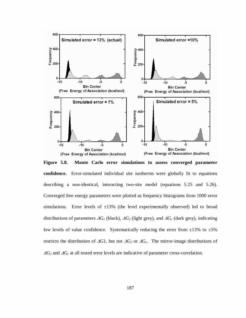
187
Figure 5.8. Monte Carlo error simulations to assess converged parameter
confidence. Error-simulated individual site isotherms were globally fit to equations
describing a non-identical, interacting two-site model (equations 5.25 and 5.26).
Converged free energy parameters were plotted as frequency histograms from 1000 error
simulations. Error levels of ±13% (the level experimentally observed) led to broad
distributions of parameters ∆G1 (black), ∆G2 (light grey), and ∆Gc (dark grey), indicating
low levels of value confidence. Systematically reducing the error from ±13% to ±5%
restricts the distribution of ∆G1, but not ∆G2 or ∆Gc. The mirror-image distributions of
∆G2 and ∆Gc at all tested error levels are indicative of parameter cross-correlation.
188
observed in corresponding lanes containing the same amount of each protein. Binding of
N-HisPurβ and N-HisPurα to supercoiled DNA was not dependent upon SMαA
promoter components, as, both proteins bound to catamers of pBLCAT3 (Figure 5.9,
upper-right panel) with similar affinity as VSMP8. A similar trend was observed for
GST-Purα binding to pUC19 with affinities similar to a pUC19-derived construct
containing the c-myc PUR-element (310). To test if binding was supercoiling-dependent,
we performed the same binding analysis with HindIII-digested plasmids. Digestion of
VSMP8 yields two observable fragments, a ~4.9 kbp fragment containing mostly
pBLCAT3-derived sequence plus ~800 bp of SMαA intron 1 sequence, and a ~2.9 kbp
fragment containing ~ 1kb of upstream elements of the SMαA gene promoter, as well as
~1.9 kb of exon 1 and a portion of intron 1. As with supercoiled dsDNA, both
recombinant Pur proteins bound SMαA promoter-derived sequences and parent vector-
derived sequences with affinities similar to what was observed for supercoiled sequences
(Figure 5.9, lower panels), albeit with a much lower affinity that what was observed for
ssDNA sequences.
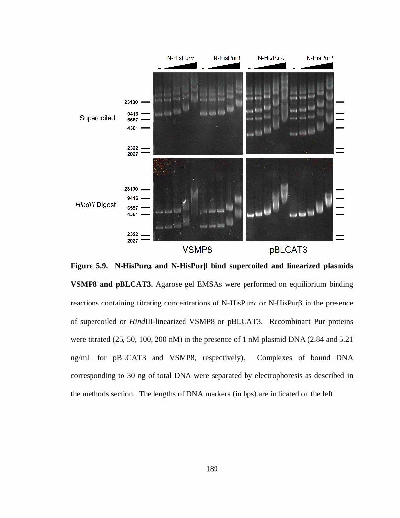
189
Figure 5.9. N-HisPurαααα and N-HisPurβ bind supercoiled and linearized plasmids
VSMP8 and pBLCAT3. Agarose gel EMSAs were performed on equilibrium binding
reactions containing titrating concentrations of N-HisPurα or N-HisPurβ in the presence
of supercoiled or HindIII-linearized VSMP8 or pBLCAT3. Recombinant Pur proteins
were titrated (25, 50, 100, 200 nM) in the presence of 1 nM plasmid DNA (2.84 and 5.21
ng/mL for pBLCAT3 and VSMP8, respectively). Complexes of bound DNA
corresponding to 30 ng of total DNA were separated by electrophoresis as described in
the methods section. The lengths of DNA markers (in bps) are indicated on the left.
190
DISCUSSION
The modulation of gene transcription by duplex-to-single-strand interconversions
represents an emerging regulatory scheme for eukaryotic gene transcription, as it permits
exclusive binding of distinct sets of trans-acting protein factors to particular
conformations that in turn regulate expression in a positive or negative fashion. In
substantiation of this notion, the involvement of sequence-specific ssDNA-binding
proteins has been identified to be of vital importance for the coordinated transcription of
genes involved in a wide array of biological functions, including tissue growth and
development, immune response, cell cycle progression, as well as cancer development, as
described in the introduction.
In the present study, we have focused on examining the mechanism of sequence-
specific ssDNA binding by Purβ, a transcriptional regulator of, most notably, muscle-
specific genes in mammals, such as SMαA and αMHC (48, 82, 103, 143, 275). A
culmination of promoter deletion, gain of function, and loss of function analyses has
identified binding of Purβ to the purine-rich strand of the SMαA proximal MCAT
enhancer element as a critical step in repression of SMαA expression (148). Hence, a
complete consideration of the enhancer element-binding mechanism employed by Purβ
will aid our understanding of how this apparently sequence-specific ssDNA-binding
protein contributes to SMC transdifferentiation. Recent studies by our lab have indicated
191
that Purβ self-associates with a dimerization constant of approximately 1 µM (222),
leading us to hypothesize that dimerization, representing a regulatory mechanism, is a
prequesite to ssDNA-binding.
To test our hypothesis, we sought first to determine the binding stoichiometry of a
physiologically relevant high affinity nucleoprotein complex formed between
recombinant Purβ (N-HisPurβ) and a 32mer oligonucleotide representative of the purine
rich strand of the SMαA proximal MCAT enhancer element (PE32-F) that has exhibited
high affinity binding to Purβ (28, 141, 144, 148, 275). In line with our hypothesis, we
measured the stoichiometry of a N-HisPurβ:PE32-F high affinity nucleoprotein complex
to be 2:1 (Figure 5.3). Furthermore, this complex was deemed to be physiologically
relevant based on mobility comparisons with nuclear extract-derived Pur protein
complexes (Figure 5.2). Previous reports investigating nucleic acid-binding properties of
Purα have found similarly that high affinity ribo- and deoxyribonucleoprotein complexes
containing recombinant forms of Purα adopt stoichiometries greater than 1:1 (91).
However, studies investigating mechanisms of GST-Purα and GST-Purβ binding to a c-
myc promoter PUR-element derived ssDNA oligonucleotide (24mer, termed MF0677)
have indicated that both recombinant Pur proteins bind this element with a 1:1
stoichiometry (310). It is currently unknown to what extent nucleotide sequence and
lattice length dictate Pur protein nucleoprotein complex stoichiometry, or if fusion
domains interfere with facilitated assembly of higher order complexes.
Independent experiments aimed at delineating the mechanism of PE32-F binding by
N-HisPurβ indicated that nucleoprotein assembly proceeds via a sequential binding
192
mechanism, and that binding is cooperative. These findings contradict our hypothesis
which stated that ssDNA binding was preceded by homodimer formation. Quantitative
EMSA binding experiments (Figure 5.4D) revealed that half-saturation of PE32-F by N-
HisPurβ occurs at monomer concentrations of approximately 0.3 nM. Accordingly,
nucleic acid-free self-association (based on a Kd of ~1 µM) at this monomer
concentration would give rise to dimer concentrations of approximately 75 fM. This
level of affinity would be quite high for any reversibly associating system. Close
inspection of N-HisPurβ EMSA titration data shows not only the existence of an
intermediary complex at concentrations near the half-saturation point, also an obligate
dimer binding model, but that the species distribution patterns (Figure 5.4B) closely
follow predicted distributions of a cooperative system (27). A statistical mechanics
approach, assuming a two-site system (based on the determined 2:1 stoichiometry) also
detected the presence of positive inter-site cooperativity (Figure 5.4C). Resolution of
thermodynamic binding parameters was impossible by this method due to the fact that
prior knowledge of the nature of the bindings sites was absent, except to say that
cooperative free energy (∆Gc) was negative (i.e. kc > 1).
To better resolve the binding energetics of N-HisPurβ for the SMαA proximal
MCAT enhancer element, we used quantitative DNase I footprinting of a 382 base
fragment of the forward strand of the SMαA promoter. The high utility of this assay
comes from its ability to singly measure fractional occupation of a particular binding site.
As such we were able to distinguish between binding mechanisms with limited
confidence, due primarily to the level of precision obtained with this type of analysis
193
(Figure 5.7). However, coupling of the footprinting approach to quantitative EMSA
analyses which detected positive cooperativity of binding, and analysis of the proximal
MCAT enhancer element which shows no sequence redundancy, collectively points to a
mechanism in which N-HisPurβ binds to the indicated sequence via a cooperative
mechanism in which the sites are non-identical. As with model confidence, resolved
parameters show very broad value constraints which are dictated, again, by low
experimental precision, as well as parameter cross-correlation (Figure 5.8). This latter
issue persists in all multisite cooperative systems, and is typically dealt with by
implementation of DNA templates with deleted sites, so as to remove uncertainty caused
by cooperativity (22, 23, 250). Applying values of footprinting resolved parameters,
assuming their accuracy, to equations 5.12-5.14 shows that these values only marginally
define the species-specific saturation curves obtained from quantitative EMSA (shown in
Figure 5.10). Reasons for these disparities are not completely clear, but may be a
reflection of experimentally induced disruption of equilibrium during performance of
mobility shift assays; specifically dissociation of nucleoprotein complexes upon loading
of equilibrated reaction in to gel wells or during electrophoresis. This effect would
manifest most drastically at concentrations below and near the half-saturation value.
Other possibilities for differences in resolved parameters would be due to increased
affinity for interacting multisite templates compared to that of the isolated sites in PE32-
F. Affinity of Purα for long purine-rich ssDNA sequences, such as the bovine papilloma
virus type I origin (100mer) has been measured on the order of 1 x 10⁻¹⁰ M, despite
knowledge of the complex stoichiometry (136). Similarly, poor constraints on
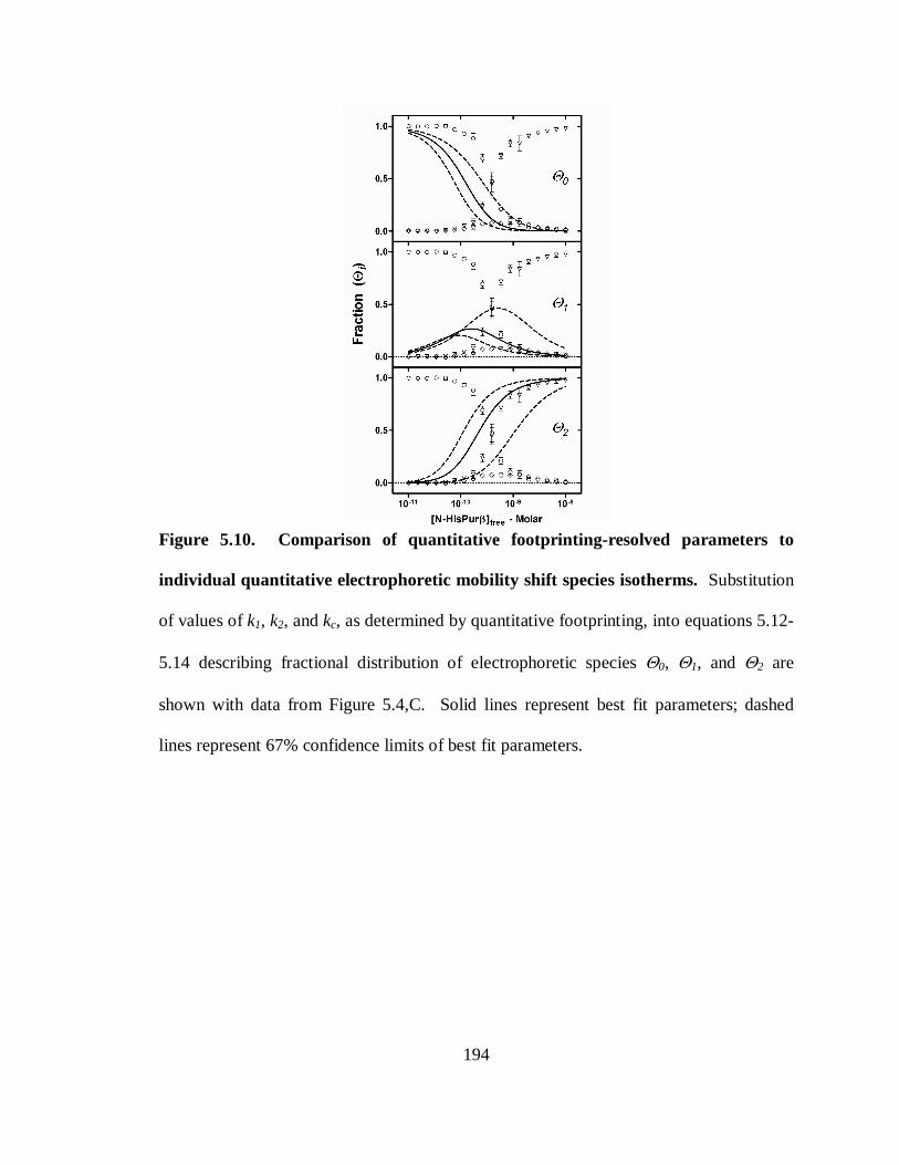
194
Figure 5.10. Comparison of quantitative footprinting-resolved parameters to
individual quantitative electrophoretic mobility shift species isotherms. Substitution
of values of k1, k2, and kc, as determined by quantitative footprinting, into equations 5.12-
5.14 describing fractional distribution of electrophoretic species Θ0, Θ1, and Θ2 are
shown with data from Figure 5.4,C. Solid lines represent best fit parameters; dashed
lines represent 67% confidence limits of best fit parameters.
195
microscopic values of k2, and kc have repercussions in the inference of macroscopic
constant K1 and K2. As is shown in Figure 5.8, these parameters have very wide
estimation distributions. Furthermore, it has been shown by Brenowitz and colleagues
(22) that without microscopic values determined by way of reduced valency promoters,
resolved cooperative free energy terms represent a lower limit to the actual cooperative
free energy of the system, and the greater the cooperative free energy that exists in a
system, the more difficult it is to resolve individual site interaction free energies. These
trends underlie the need to define the nucleotide binding site determinants for Pur
proteins such that mutant promoters can be constructed.
Cooperative binding mechanisms are common for ssDNA-binding proteins,
particularly those involved in DNA replication, recombination, and RNA transcription,
such as T4 bacteriophage gp32 (4), E.coli SSB protein (236), and the Adenovirus DNA-
binding protein (198, 323). By virtue of their abilities to cooperatively assemble
nucleoprotein filaments, these proteins thus possess helix destabilizing activities
necessary for their cellular functions. Cooperativity for sequence-specific ssDNA binding
proteins, on the other hand, is minimally described in the literature at this point. Reasons
for this are unclear, but may be due to the relative rarity of sequence-specific ssDNA-
binding proteins, or the limited number of thermodynamically rigorous assays able to
resolve site-specific binding isotherms and detect cooperativity. Binding of the yeast
telomere protection protein Pot1 has been ascribed a cooperative mechanism, and
similarly to what we have determined for N-HisPurβ reported here, sequential monomer
binding of Pot1 to specific telomeric sites proceeds in a 3’ to 5’ direction (167).
196
Functional cooperative binding of Pot1 to yeast telomeres has been deemed necessary for
telomeric nucleoprotein filament assembly and, in turn, protection of the chromosome
ends from cellular nuclease damage. Cooperative binding of genomic promoter ssDNA
sequences by Pur proteins may serve a similar helix destabilizing function, or to impart a
potent functional response over a small change in cellular concentration.
Models of SMαA proximal MCAT enhancer repression have proposed that formation
and maintenance of a single-stranded non-B-DNA conformation by Purα, Purβ, and
MSY1 binding to nucleotides adjacent to the MCAT sequence causes disruption of a
double-stranded TEF-1 binding site, and prevents binding of this transcriptional activator
(28, 275). Enforcement of the repressive state must then require destabilization of the
helix, either by thermodynamic favorability of ssDNA-binding protein occupation, or by
environmental factors such as topological stress of the duplex DNA that permit
occupation. To test this prediction, we examined the binding properties of N-HisPurβ to
supercoiled and linear sequences of dsDNA (Figure 5.9). Unexpectedly, N-HisPurβ
displayed binding to specific and non-specific sequences of supercoiled and linearized
dsDNA. Clearly this type of assay cannot detect if binding is occurring at specific sites
within the DNA, but it does suggest that, under these conditions, sequence selectivity of
N-HisPurβ is low. Furthermore, the resolution of this assay does not permit us to
differentiate between two possible binding modes for either N-HisPurβ or N-HisPurα;
dsDNA binding versus ssDNA binding. The lower level of affinity observed in these
experiments may indicate that dsDNA binding is a secondary activity for Pur proteins, or
that ssDNA-binding/helix destabilization is occurring with a lower level of observable
197
affinity due to a need to force the equilibrium in favor of ssDNA-binding by increasing
protein concentration and activity, overcoming annealing favorability.
Previous studies cast doubt on the notion of dsDNA binding by Pur proteins,
primarily reports by Darbinian and colleagues (56) which showed that GST-Purα
possesses strand displacement capabilities in the context of a short 15mer (26.7%
guanine) oligonucleotide annealed to M13 single-stranded plasmids. Wortman and
colleagues (310) showed that GST-Purα is capable of displacing short (16mer)
pyrimidine-rich strands of telomeric-repeats DNA, and that displacement occurs by virtue
of contacts between the protein and the (24mer) purine-rich strand, and that binding of
GST-Purα increased potassium permanganate sensitivity to dsDNA, and permits binding
of gp32, both suggestive of helix unwinding. Both groups localized strand displacement
activity to the core DNA-binding domain of Purα, which is highly homologous to Purβ
(143, 145), consisting of alternating basic-aromatic Class I and acidic leucine-rich Class
II repeats. It is unknown whether or not strand displacement capabilities observed by
these groups were reliant upon the ssDNA overhangs present in both sets of experiments,
as strand displacement with blunt-ended fragments was not demonstrated. Wortman, et
al (310) showed that binding of GST-Purα to linear dsDNA required the C-terminal
domain which consists of the psycho motif, as well as the glutamine- and glutamate-rich
domains. Glutamine-rich domains have been historically implicated in transcriptional
activation by transcription factors possessing them (97), however other studies have
implicated glutamine-rich tracts in DNA distortion and helix unwinding activity in
Drosophila melanogaster GAGA factor (GAF) (307). The means by which helix
198
unwinding activity is afforded by polyglutamine tracts is unknown, but may be
attributable to the hydrogen-bonding capacity contributed by the high local
concentrations of amides inherent to these peptides resulting in reduced melting
temperatures of stretches of dsDNA in a manner similar to what is observed for
formamide (14). Interestingly, human Purβ, which is divergent from human Purα in that
it lacks the C-terminal glutamine-rich domain, failed to bind linearized pUC19 in
experiments described by Wortman, et al. (310). Our results differ in this regard, as we
have shown here that recombinant mouse Purβ binds to linearized plasmid DNA (Figure
5.9, lower panels), despite also lacking a C-terminal glutamine-rich domain.
Discrepancies in dsDNA-binding activity may arise from primary amino acid sequence
differences in the N-terminus of human versus mouse Purβ, namely the presence of two
polyglycine tracts in mouse Purβ that are absent in the human homolog (Figure 1.2).
Polyglycine, and glycine-rich domains have been found in numerous proteins with
observable helix-destabilization character including the UP1 subunit of heterogeneous
ribonucleoprotein A1 (46, 86), and nucleolin (99). However, mouse Purα, which also
contains an N-terminal polyglycine tract (different in length and position compared to
mouse Purβ), loses considerable linear dsDNA-binding activity when the C-terminal
domain containing the psycho motif, glutamine-rich, and glutamate-rich domains is
deleted. Thus terminal domains of mouse Purα and Purβ may direct helix-destabilizing
properties differently.
The question remains how Pur proteins bind to ssDNA sequences in a double-
stranded environment. Ourselves and others before us have proposed that binding of
199
sequence-specific ssDNA-binding proteins to dsDNA is facilitated by either pre-
formation of single-stranded structures or a reduction in the annealing free energy of a
localized region that make thermodynamic competition for site occupation feasible. This
theory has been substantiated previously for FBP, a transcriptional activator, which
requires transcription-induced negative supercoiling and unwinding in FUSE for FBP
binding (153, 154). Models of repression of MCAT enhancer element-dependent
transcription of SMαA suggest that this mechanism may not be feasible for Pur protein
occupation, as transcription is presumably silent in the Pur protein occupied state, and
thus so is transcription-induced negative supercoiling. Moreover, binding of recombinant
Pur proteins to relaxed linear dsDNA supports this argument. If binding of ssDNA in the
context of a dsDNA lattice by N-HisPurβ proceeds via a thermodynamic competition
(bubble formation) target sequences would have to possess less than ~26 kcal/mole of
annealing free energy. Average base-pairing free energy for a single base-pair in dsDNA
of infinite length is on the order of -1.8 kcal/mole at 20°C (239). Thus, average
sequences of approximately 14 basepairs might be prone to strand displacement by direct
thermodynamic competition with N-HisPurβ. Localized melting of subdomains in large
linear DNA fragments have been detected by microscopy techniques at temperatures as
low as 64°C (234, 262) compared to a measured melting temperature of ~83°C for
dsPE32-F (data not shown). Unfortunately, nearest-neighbor melting temperature
predictions would estimate the melting temperature of a dsDNA of infinite length to also
be infinite. Hence it is thought that localized melting is due to DNA subdomain
architecture. Depressed melting temperatures in linear dsDNA are routinely believed to
200
be due to richness in A/T basepairs (187); however size and relaxed topology has also
been shown to have inverse effects on diffusion coefficients of dsDNA (230). Whether
or not limited Brownian motion (degrees of translational, rotational, and vibrational
freedom) as a consequence of extended topology dictates localized melting temperature
depression of linear dsDNA is not known, but transient base unpairing, or breathing,
within localized regions may be a means of escaping this entropic limitation. DNA
breathing has been detected at temperatures well below the melting temperature of an
oligonucleotide (102, 156), but the transient nature of these occurrences make
opportunistic binding by Pur proteins to opened duplexes unlikely, as binding is stably
observed despite being reversible. Presumed non-specific binding of Pur proteins to
ssDNA with varying degrees of affinity has been documented previously (56, 136, 145,
148, 310). These possibilities have the potential to explain non-specific binding of N-
HisPurβ to linear dsDNA as observed here (Figure 5.9, lower panels). Nevertheless, the
nature of dsDNA binding by Pur proteins needs to be investigated, most suitably by
nuclease and chemical footprinting techniques that are sensitive to ssDNA formation.
As mentioned previously, repression of MCAT enhancer dependent expression of
SMαA relies on the ssDNA-binding activities of Purα, Purβ, and MSY1, as well as a
network of protein-protein interactions between the three factors (28, 141, 148).
Recessed, pyrimidine strand displacement has been described for Purα (56, 310),
whereas MSY1 (YB-1) has exhibited strand displacement of blunt-ended, Y-box-
containing, short double-stranded oligonucleotides, cisplatin-modified double-stranded
oligonucleotides, as well as engineered fork and bubble structures (95, 123). Strand
201
separation activity for YB-1, although independent of ATP, has been shown to be
elevated in the presence of ATP. Furthermore, self-association becomes limited from a
possible monomer-trimer-hexamer-dodecamer equilibrium to that of primarily monomer-
dimer upon addition of ATP (95). These results suggest that ATP has allosteric effects
on MSY1 (YB-1) activity and protein-protein interactions. Furthermore, sequence
specificity of N-HisPurβ has been shown to be modulated by MSY1 (145). Hence,
ssDNA-recognition, binding activity, strand separation, ATP hydrolysis and
transcriptional repression achieved by Purα, Purβ, and MSY1 in vivo may be the result of
collaborative and cooperative activities and interactions at the MCAT enhancer element
of the SMαA promoter that have yet to be meticulously defined, and will likely require
high-resolution biophysical and structural studies to do so. Worth noting is that
collaboration of Purα and MSY1 has been found at gene promoters other than SMαA
(36, 37, 238).
In conclusion, we have found that recombinant, purified Purβ binds to the purine-rich
strand of the SMαA proximal MCAT enhancer element in a 3’ to 5’ sequential and
cooperative manner, with a stoichiometry of 2:1. Apparent affinity (half-saturation) is on
the order of 0.3 nM, while rigorous thermodynamic interrogation has shown that free
energy of binding is -26.25 kcal/mole. This free energy value suggests that Purβ is only
able to thermodynamically compete for binding to short stretches of oligonucleotides if at
all. The involvement of co-repressors Purα and MSY1 in maintaining repressive single-
stranded conformations of the SMαA MCAT enhancer element are the goals of future
studies, as well as determining specific nucleotide binding determinants in this element,

202
as well as other Pur protein-responsive sequences. The intended outcome of these efforts
is a better understanding of how transcriptional repression is enforced by these factors.
203
CHAPTER VI. TOWARDS THE CHARACTERIZATION OF THE
MINIMAL SINGLE-STRANDED DNA BINDING SITE OF PUR ββββ
WITHIN THE SM ααααA PROXIMAL MCAT ENHANCER ELEMENT
The following is a description of original and unpublished work.
INTRODUCTION
The identification of trans-acting regulatory proteins involved in the regulation of
gene expression has facilitated the elucidation of gene regulatory mechanisms in both
prokaryotes and eukaryotes. The involvement of specific proteins in the spatial and
temporal regulation of multiple genes has suggested that subsets of tissue-specific genes
may utilize similar combinations of trans-, and cis-acting regulatory elements of
transcriptional control to ensure proper timing and levels of expression. The ability to
identify additional gene targets of transcription factors that enact tissue-specific or
choreographed transcriptional regulation strengthens our capacity to understand
developmental process and disease progression by better describing gene regulatory
networks and identifying additional target genes. This ability relies, however, on clear
definitions of transcription factor binding site in terms of nucleotide sequences.
Transcription factor binding site definitions are typically represented by consensus
sequences; nucleotide sequences that signify the statistical predominance of nucleotides
at given positions within the binding site. The process of defining a nucleic acid-binding
204
protein consensus sequence requires the accumulation of numerous pieces of information,
most importantly the binding site size or footprint, and the sequences of all demonstrated
targets of binding. Stringent nucleotide preferences of transcription factors furnish robust
consensus sequences that are very useful for identifying possible interaction targets,
whereas diverse sequence tolerance and promiscuous binding can render derived
consensus sequences as inaccurate depictions of nucleotide preferences whose utilization
can be misleading. Inaccuracy of consensus sequences has led to the use of more
sophisticated algorithms of defining and visualizing sequence preference profiles for
transcription factors. Sequence logos have circumvented problems associated with
nucleic acid binding proteins exhibiting relatively indiscriminate binding properties for
which consensus sequences are inappropriate (245, 246). Nevertheless, transcription
factor binding site leniency remains problematic for defining nucleotide preferences and
identifying possible binding sites.
The goal of the work presented in this chapter was to characterize the minimal
ssDNA binding site of the sequence specific SSB Purβ. Purβ is one of three known
sequence-specific SSB transcription factors that repress expression of SMαA, an
important cytoskeletal contractile protein whose expression is important for cellular
contractile functions (243) and for the differentiation status of numerous cell types (127,
241). The abundance of SMαA protein levels in differentiated SMCs versus SMC
undergoing phenotypic modulation towards fibroblastic cell character has made SMαA a
hallmark of SMC differentiation (210, 315). Downregulation of SMαA has been
implicated as a contributing factor towards dysfunctional vessel wall remodeling (175)
205
and the vulnerability of atheroma in relation to plaque rupture (88, 151). SMαA
promoter deletion analysis has identified a cryptic enhancer element in the 5’ region of
the promoter (28, 48, 82, 270, 275) that possesses a high degree of purine/pyrimidine
asymmetry and exhibits transient structural interconversions in response to stimulus that
activates SMαA expression (9). This element contains a core MCAT motif shown to
bind TEF-1 when in double-stranded configurations which is necessary for gene
activation. Binding sites for sequence-specific SSBs Purβ, Pur protein family member,
Purα, and Y-box protein MSY1 have been detected on opposing strands of this element,
with Purα and/or Purβ occupying the purine-rich strand and MSY1 occupying the
pyrimidine-rich strand (28). Recent gain-and-loss of function studies (148) and
chromatin immunoprecipitation techniques (149) have pointed to Purβ as the dominant
Pur protein repressor in the context of SMαA gene regulation in certain cell types.
The critical nature of Purβ functions in regard to SMαA repression and its phenotypic
consequences have yielded a need to identify other targets of Purβ repression. Both Purα
and Purβ have been implicated in the regulation of numerous genes, both at the
transcriptional and translational levels, and in the case of Purα, the regulation of cellular
and viral DNA replication and cell cycle progression (92, 132). Diverse functions in
pathological blood vessel remodeling, cancer, and viral pathogenesis has revealed the
need for a full description of Pur protein regulatory objectives. Projection of possible
genomic and transcriptomic targets of Purβ has been hampered by the lack of a defined
consensus sequence for this multifunctional protein.
206
In the present study we have examined the in vitro binding characteristics of
recombinant mouse Purβ to oligonucleotides representative of the SMαA MCAT
enhancer element to map the binding sites within this region. Recent reports by our lab
have detected two binding sites within the region encompassing nucleotides -195 to -164
of the SMαA promoter in relation to the transcription start site with low resolution
(Chapter V). Here we show that these two sites map to nucleotides -195 to -190 (5’ site)
and -171 to -166 (3’ site) of the SMαA promoter. Furthermore, these sites resemble
consensus sequences reported previously for Purα which is 5’-GGGAGA-3’ (10, 11,
310), with only slight degeneracy at each site.
MATERIALS AND METHODS
Chemicals, protein reagents, and oligonucleotide probes. All chemicals used in this
study were of reagent grade or better. Recombinant Purβ was expressed as an amino-
terminally labeled hexahistidine tag fusion proteins (referred to in this report as N-
HisPurβ), purified from E. coli expression cultures, and quantified as described
previously in Chapter IV and in a previous publication (222). Preparation and validation
of the epitope-specific rabbit polyclonal antibody against mouse Purβ (anti-Purβ 302) has
been described previously (141). Synthetic oligonucleotides were purchased from
Sigma-Genosys.
Competitive ssDNA-binding assay. To monitor the extent of recombinant Purβ
binding to PE32-F in the presence of various oligonucleotide competitors, a
207
discontinuous solid-phase DNA-binding assay was performed as previously described
with some minor modifications (145). Biotinylated PE32-F was immobilized on
streptavidin-coated microtiter wells (Streptawells™, Roche) by application of 100 µL of
1 nM 3’-biotinylated PE32-F (PE32-bF) in buffer consisting of 25 mM
tris(hydroxymethyl)aminomethane (Tris)-HCl pH 7.5, 150 mM NaCl, 1.5 mM MgCl2 at
20 ± 1°C for 1 h with moderate shaking. Solutions were removed and wells were washed
three times with 300 µL wash buffer consisting of 25 mM Tris-HCl pH 7.5, 150 mM
NaCl, 1.5 mM MgCl2, 0.05% (v/v) Tween 20. Blocking of non-specific binding was
accomplished by incubation of 250 µL of blocking buffer consisting of 25 mM Tris-HCl
pH 7.5, 150 mM NaCl, 1.5 mM MgCl2, 20 µg/ml bovine serum albumin (BSA), at 20 ±
1°C for 1 h with moderate shaking. Wells were again washed three times with 300 µL of
wash buffer. Competitive binding solutions consisting of 1 nM N-HisPurβ and titrated
concentrations of competing oligonucleotides in buffer consisting of 50 mM Tris-HCl pH
7.5, 100 mM KCl, 0.5 mM dithiothreitol, 2 µg/ml polydeoxythymidine (dT32), 50 µg/ml
BSA were added to the wells (100 µL/well) and incubated overnight at 4°C. The
sequences of competing oligonucleotides are listed in Table 6.1. The next morning wells
were again washed as before. Quantities of N-HisPurβ nucleoprotein complexes
remaining after competition were detected by addition of 100 µL of primary antibody
solution containing 1 µg/ml anti-Purβ 302 polyclonal antibody in binding buffer
consisting of 25 mM Tris-HCl pH 7.5, 150 mM NaCl, 1.5 mM MgCl2, 0.05% Tween 20,
2 µg/ml BSA, for 1 h at 20 ± 1°C. This was followed by triplicate washing and addition
208
of 100 µL of secondary antibody solutions containing goat-anti-rabbit horseradish
peroxidase conjugate (ExtraAvidin-HRP, Sigma) diluted 1:10,000 in binding buffer to
each well and incubation for 1 h at 20 ± 1oC. Wells were washed three times and 100 µl
of 2,2'-azino-bis(3-ethylbenzthiazoline-6-sulfonic acid) substrate solution (ABTS,
Chemicon) was added. After satisfactory color development by incubation at room
temperature (for approximately 5 min), 100 µL of 1% (w/v) sodium dodecylsulfate was
added to stop the reactions. Solution absorbance readings at 405 nm were obtained with
a microplate reader. Self-competitor controls (PE32-F) were used to verify complete
competition absorbance values (Amin), whereas wells with no competitor were used to
obtain maximum absorbance values (Amax). These controls were included on each plate
to permit normalization of absorbance values necessary for comparison of results from
multiple plates. Normalized absorbance values (ANorm) were calculated using the
expression ANorm, i = (Ai-Amin)/(Amax-Amin), where Ai is the absorbance of well i.
Determinations of competitor concentrations necessary for 50% inhibition of complex
formation, IC50, were performed by nonlinear least-squares fitting to the following
expression:
(Equation 6.1)
where αH is the Hill coefficient which permits variability of the slope of the transition.
Non-linear least-squares fitting was performed using Prism 5 software (GraphPad
Software, Inc., San Diego, CA).
ANorm=1 + 10(LogIC50 – Log[Competitor])αH
1ANorm=
1 + 10(LogIC50 – Log[Competitor])αH
1
1 + 10(LogIC50 – Log[Competitor])αH
1
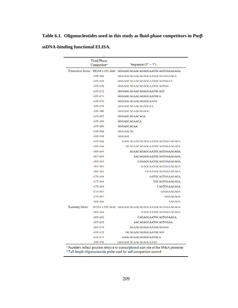
209
Table 6.1. Oligonucleotides used in this study as fluid-phase competitors in Purββββ
ssDNA-binding functional ELISA.
210
RESULTS
Investigations into genomic and RNA targets of Pur proteins have been impeded by
the inability of researchers to definitively describe a consensus binding site for these
proteins. The detection of Pur protein binding to a variety of ss/dsDNA and RNA
sequences has only made this pursuit more difficult, due to the fact that Purα and Purβ
display extraordinary sequence promiscuity. This apparent promiscuity likely arises from
a combination of factors but may arise artifactually, however, due to a poor
understanding of the nature of Pur protein nucleoprotein complexes with respect to
stoichiometry and affinity, as it is envisioned that sequence dictates these parameters.
Therefore, we sought to systematically examine the base specificity of Purβ in the
context of a system that has been thermodynamically predefined (described in Chapter
V). Towards this end we have examined N-HisPurβ2:PE32-F nucleoprotein complex
stability in the presence of oligonucleotide competitors designed to systematically dissect
nucleotide stability contributions. We have employed a discontinuous solid-phase DNA-
binding functional enzyme-linked immunosorbent assay (ELISA) to address this goal, as
this assay permits simultaneous testing of competitive oligonucleotides in a high-
throughput fashion, and direct comparison of results to gauge the effects of each
competitor.
To identify nucleotides that contribute to overall nucleoprotein complex stability, we
designed three series of competitive oligonucleotides for use in our functional ELISA.
Table 6.1 details the sequence identity of the competitors and the rationale of our design.
211
Briefly, we designed a series of binucleotide truncation mutants of PE32-F (-195 to -164
of the SMαA promoter) that have deletions proceeding from the 3’ end (-195 series), and
from the 5’ end (-164 series). Recent studies have shown the importance of nucleotides
near positions -195 to -192 and -171 to -164 of the SMαA promoter in both transfection-
based reporter assays (28, 48) and direct ssDNA-binding assays (275). Therefore,
truncations from either end of PE32-F (-195/-164) should interfere with the ability of the
oligonucleotide to compete for N-HisPurβ binding. The results obtained by incorporation
of these oligonucleotide competitors in functional ELISAs are shown in Figure 6.1.
Panel A and B show the competition isotherms generated by this approach for the -195
series and -164 series, respectively. Nonlinear least-squares fitting of these isotherms to
a phenomenological inhibition equation (Equation 6.1) yields IC50 values for comparing
relative affinities for N-HisPurβ. We used self-competition (PE32-F) as a positive
control and IC50 reference point for comparison. As shown in Figure 6.1, panel C,
deletions from either the 5’ or 3’ end of PE32-F results in loss of affinity for N-HisPurβ,
and the extent of truncation directly coincides with loss of affinity, as indicated by
increasing IC50 values. Consistent with the notion that N-HisPurβ2:PE32-F nucleoprotein
complexes are stabilized by cooperative interactions between sites (Chapter V), deletion
of either putative binding site results in a loss of the competitors ability to displace N-
HisPurβ from the solid phase. Interestingly, deletions from the 5’ end appear to be better
tolerated than those from the 3’ end. This contradicts previous findings that N-HisPurβ
shows higher affinity for the 3’ site of PE32-F than for the 5’ site (Chapter V). However,
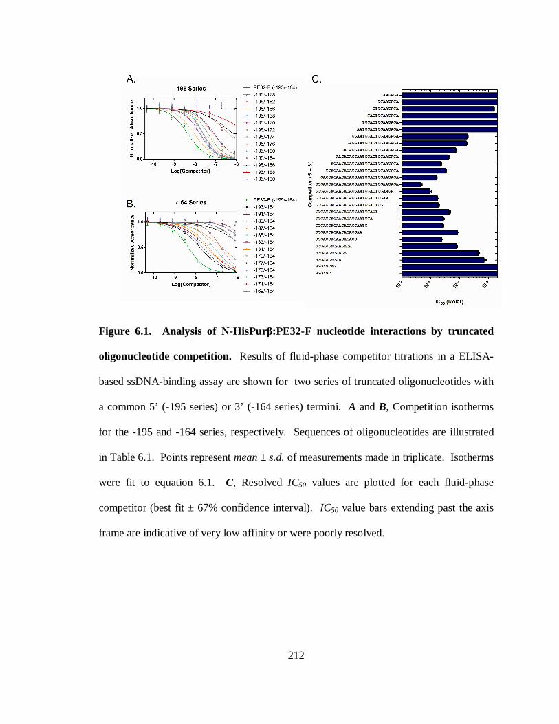
212
Figure 6.1. Analysis of N-HisPurβ:PE32-F nucleotide interactions by truncated
oligonucleotide competition. Results of fluid-phase competitor titrations in a ELISA-
based ssDNA-binding assay are shown for two series of truncated oligonucleotides with
a common 5’ (-195 series) or 3’ (-164 series) termini. A and B, Competition isotherms
for the -195 and -164 series, respectively. Sequences of oligonucleotides are illustrated
in Table 6.1. Points represent mean ± s.d. of measurements made in triplicate. Isotherms
were fit to equation 6.1. C, Resolved IC50 values are plotted for each fluid-phase
competitor (best fit ± 67% confidence interval). IC50 value bars extending past the axis
frame are indicative of very low affinity or were poorly resolved.
213
it may suggest that 5’ nucleotides of the putative PUR-consensus (GGGAGA) are more
critical to binding than are those in the 3’ end.
Based on results shown in Figure 6.1, it was found that oligonucleotides with a
minimum length of 18-20 nt consistently conferred the best competitive response in our
functional ELISA. This finding, coupled with a need to confirm sequence specificity and
discount possible length effects associated with observed IC50 profiles, led us to design a
series of competitive oligonucleotides of consistent length (20 nt) that scan the entirety of
PE32-F (Table 6.1). Using these oligonucleotides in the competitive functional ELISA
provided the results shown in Figure 6.2. Similar to what was observed for the truncation
series, oligonucleotides representing the ends of PE32-F competed the best for N-
HisPurβ binding. This result suggests that the competitive effect we observed in Figure
6.1 is sequence-specific and is not purely dependent upon oligonucleotide length. This
data also shows that the two binding sites are not equivalent. However, this data also
contradicts previous findings which show the 3’ site to possess greater inherent affinity
for N-HisPurβ binding than the 5’ site, as discussed in Chapter V.
DISCUSSION
Binding of Pur proteins to nucleic acids has been shown to potentiate extensive
cellular consequences ranging from cell cycle arrest to transcriptional and translational
regulation. The diverse functional aspects of Pur proteins make them important
molecular target candidates for the prevention of viral replication, cardiovascular disease
and cancer (132). However, a fundamental understanding of mechanisms employed by
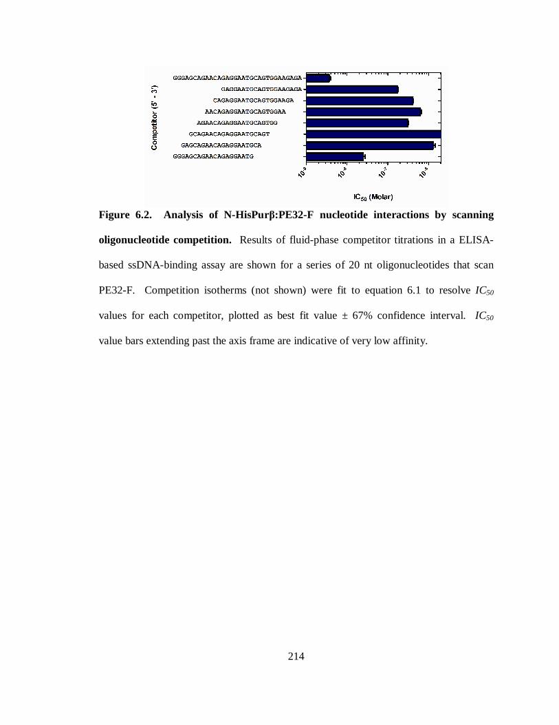
214
Figure 6.2. Analysis of N-HisPurβ:PE32-F nucleotide interactions by scanning
oligonucleotide competition. Results of fluid-phase competitor titrations in a ELISA-
based ssDNA-binding assay are shown for a series of 20 nt oligonucleotides that scan
PE32-F. Competition isotherms (not shown) were fit to equation 6.1 to resolve IC50
values for each competitor, plotted as best fit value ± 67% confidence interval. IC50
value bars extending past the axis frame are indicative of very low affinity.
215
Pur proteins in regulating these processes have not been achieved, due in part to an
inadequate description of Pur protein target sequences.
The diversity of nucleotide sequences that have been used to probe for Pur protein
binding and involvement, as well as differences in interaction affinities associated with
the use of these probes (Table 1.1) illustrates the difficulties associated with definitive
binding site identification. Furthermore, a lack of nucleoprotein complex stoichiometries
reported for these probes, with few exceptions, makes estimation of the number of
binding sites in each sequence difficult to estimate. The possibility of multiple
heterogeneous binding sites existing on a single probe obviously exists. Therefore a
systematic approach to delineation of binding site identity of any proteins requires
preexisting knowledge regarding complex stoichiometry, affinity, and/or footprint
information. Few systematic approaches to identifying nucleotide sequence components
critical for Pur proteins have been armed with any of this information. One such study,
aimed at the identification of a Purα consensus sequence made use of this information in
regards to the c-myc-associated PUR-element (10, 11, 310). In this series of papers,
Edward Johnson and colleagues systematically showed that Purα binds to a core
consensus sequence of GGGAGA with what is likely a 1:1 stoichiometry (reviewed in
Chapter I).
Armed with equivalent thermodynamic data regarding the binding of recombinant N-
HisPurβ to the proximal MCAT enhancer element of the SMαA promoter (Chapter V),
we sought to test the consensus sequence prediction of Johnson and colleagues in regards
to Purβ nucleotide preferences, and to see if they differ from those of Purα. Previous
216
reports have implicated nucleotides flanking the core MCAT sequence of the SMαA
promoter as being necessary for Pur protein-mediated repression of reporter expression
(28, 48) and for direct binding of Purα and Purβ to oligonucleotides representative of this
region (28, 275). Figure 6.3 summarizes the results of these findings. In the present
study, we used a convenient enzyme-linked immunosorbent ssDNA-binding assay to
gauge the effects of competitive fluid-phase mutant oligonucleotides to on N-
HisPurβ2:PE32-F nucleoprotein complex assembly.
Our results indicate that nucleotides on either end of PE32-F are critical to
nucleoprotein complex stability. This finding is based on the competition profiles of 5’
and 3’ truncation series of oligonucleotides harboring successive doublet deletions
(Figure 6.1). The findings were further corroborated by employment of a series of
scanning oligonucleotides, where the intention was to discount length effects that could
possibly explain the competition results of the truncation series. Indeed, these
oligonucleotides also suggest the involvement of terminal nucleotides in N-
HisPurβ2:PE32-F nucleoprotein complex stability (Figure 6.2). The presence of
degenerate GGGAGA core consensus sequences in each end of PE32-F suggest that these
regions indeed represent N-HisPurβ binding sites and the N-HisPurβ shares nucleotide
preferences with those reported for Purα.
Previous finding by our lab have shown regions of DNase I protection afforded by N-
HisPurβ that correspond to the 5’ and 3’ regions of the MCAT enhancer element
spanning from nucleotides -195 to -164 of the SMαA promoter when in a single-stranded
conformation (Figure 5.5). Unfortunately, the resolution of this footprinting approach
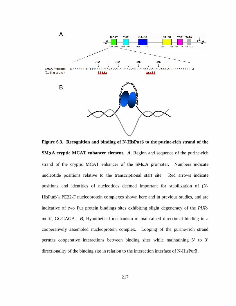
217
Figure 6.3. Recognition and binding of N-HisPurβ to the purine-rich strand of the
SMααααA cryptic MCAT enhancer element. A, Region and sequence of the purine-rich
strand of the cryptic MCAT enhancer of the SMαA promoter. Numbers indicate
nucleotide positions relative to the transcriptional start site. Red arrows indicate
positions and identities of nucleotides deemed important for stabilization of (N-
HisPurβ)2:PE32-F nucleoprotein complexes shown here and in previous studies, and are
indicative of two Pur protein bindings sites exhibiting slight degeneracy of the PUR-
motif, GGGAGA. B, Hypothetical mechanism of maintained directional binding in a
cooperatively assembled nucleoprotein complex. Looping of the purine-rich strand
permits cooperative interactions between binding sites while maintaining 5’ to 3’
directionality of the binding site in relation to the interaction interface of N-HisPurβ.
218
did not allow for precise determination of ligating nucleotides within these sites. The
results did indicate that N-HisPurβ binds to these sites in a successive 3’ to 5’ and
cooperative manner suggesting that these sites are non-equivalent, a finding consistent
with sequences analysis showing non-redundancy of these sites. Results from the present
study also show that the 5’ and 3’ binding sites within PE32-F are non-equivalent with
respect to N-HisPurβ binding; however our results also indicate that N-HisPurβ binds to
the 5’ site with greater affinity than to the 3’ site, in contrast to results obtained by
quantitative footprinting. It is possible that this discrepancy arises from isolation of the
two binding sites by the use of the 32 nt oligonucleotide PE32-F. Quantitative
footprinting experiments described in Chapter V were performed using 382 nt template
representations of the SMαA promoter region containing other known sites of Pur protein
interaction (47, 148, 272). It is possible binding of N-HisPurβ to the 5’ and 3’ sites of the
region encompassing the MCAT enhancer element is thermodynamically linked to
binding at other sites that may influence the interaction energetics observed in these
experiments. N-HisPurβ-mediated nuclease protection at sites outside of nucleotides -
195 to -164 indicates that this might be a possibility.
Identification of N-HisPurβ binding sites within the MCAT enhancer element of
SMαA raises structural questions of how nucleoprotein assembly is accomplished.
Numerous dimeric transcription factors typically display palindromic binding site
sequences. For example bHLH dimers bind palindromic E-boxes (5’-NCANTGN-3’) by
monomer recognition of major groove C-G basepair constituents on successive half-turns
of the B-DNA helix (202). The sequence represented by PE32-F contains direct repeats

219
of the putative N-HisPurβ binding sites. Therefore, the manner by which two monomers
of N-HisPurβ cooperatively bind two direct repeats of the identified binding sequence
represents a structural dilemma. Figure 6.3, panel B shows how binding might occur and
suggests that looping of the intervening ssDNA sequences may transpire. This is purely
speculation, however, and requires high resolution structural data for validation.
In conclusion, the results described herein provide direct information regarding the
nucleotide contributions stabilizing nucleoprotein complexes formed between
recombinant Purβ and oligonucleotide representations of the proximal MCAT enhancer
element of the SMαA gene promoter. These results provide insight into possible
experimental methods of reducing Purβ binding affinity in cultured cells and in vivo by
mutating putative nucleotide contacts, as well as in the detection of additional Purβ
binding targets genome-wide. These approaches may not only aid in our understanding
of mechanisms of SMαA repression involved in phenotypic reprogramming of VSMCs,
but also unveil the extents of Pur protein function in physiological events and disease
progression, and provide a means to prevent or manage clinical consequences of Pur
protein-ssDNA interactions.
220
CHAPTER VII. CONCLUDING REMARKS
The studies described in this dissertation have helped to characterize the mechanism
implemented by Purβ to assemble a repressive nucleoprotein component of SMαA
transcriptional regulation as it might occur in vivo and in cell-culture models of
myofibroblast transdifferentiation and VSMC dedifferentiation. Equally as important,
these studies have also shown that Pur protein-mediated repression of SMαA
transcription can serve as a model system for examining the cause and effect relationship
between sequence-specific SSB transcription factors and structural interconversions in
regulating gene expression.
Generally speaking, the results of these studies have shown that Purβ, the dominant
Pur protein repressor of SMαA transcription in MEFs, utilizes cooperative interactions to
facilitate and stabilize nucleoprotein assembly at the proximal MCAT enhancer element
of the SMαA promoter. It remains to be established whether or not this mechanism is
accurate in the context of a repressive nucleoprotein entity, one that enacts repression of
SMαA transcription in vivo, as the mechanism described herein was delineated with
purified recombinant Purβ, in the absence of co-repressors Purα and MSY1. Loss-of-
function studies described here, suggest that full-repression of SMαA expression in
MEFs requires collaborative, synergistic activities of both Purα and Purβ. Furthermore,
the inability of recombinant Purβ to solely establish ssDNA-binding in the presence of
short complementary strands (strand displacement) also suggests that Purβ is an
221
opportunistic SSB transcription factor, and that co-repressor involvement might be
necessary to destabilize base-pairing within the SMαA MCAT enhancer element, to
allow for repressive nucleoprotein complex assembly.
Future efforts in continuation of the studies described in this dissertation would be
aimed towards delineating the roles of Purα and MSY1 in nucleoprotein complex
assembly and helix destabilization as mentioned above. A combination of biophysical
assessment of isolated nucleoprotein assemblies for Purα and MSY1 with their respective
ssDNA strands, and footprinting techniques involving dsDNA templates in the presence
of Purα, Purβ, and MSY1 (in isolation and combination) would likely reveal the
thermodynamic parameters stabilizing nucleoprotein assembly and detail how assembly
occurs in the context of a double-stranded environment. Similarly, probing of the
secondary structure of the SMαA promoter region with ssDNA-sensitive reagents, in the
contexts of supercoiled and linearized plasmid dsDNA constructs, may help to explain if
topological stress facilitates stable non-B-DNA structures that promote sequence-specific
SSB recruitment and occupation, events crucial to negative regulation of SMαA
expression. Collectively, these future studies would help deduce the cause and effect
relationship between sequence-specific SSB repressors and promoter structural
interconversion in the regulation of SMαA gene transcription and other genes that rely on
ssDNA-binding transcription factors for regulated expression. In addition, they would
provide a technical foundation for examining these systems.
222
COMPREHENSIVE BIBLIOGRAPHY
1. Ackers GK, Johnson AD, Shea MA (1982) Quantitative model for gene
regulation by lambda phage repressor. Proc Natl Acad Sci U S A.79(4):1129-33.
2. Aikawa M, Rabkin E, Voglic SJ, Shing H, Nagai R, Schoen FJ, Libby P (1998)
Lipid lowering promotes accumulation of mature smooth muscle cells expressing smooth
muscle myosin heavy chain isoforms in rabbit atheroma. Circ Res.83(10):1015-26.
3. Aikawa M, Sakomura Y, Ueda M, Kimura K, Manabe I, Ishiwata S, Komiyama
N, Yamaguchi H, Yazaki Y, Nagai R (1997) Redifferentiation of smooth muscle cells
after coronary angioplasty determined via myosin heavy chain expression.
Circulation.96(1):82-90.
4. Alberts BM, Frey L (1970) T4 bacteriophage gene 32: a structural protein in the
replication and recombination of DNA. Nature.227(5265):1313-8.
5. Autieri MV, Keleman SE, Wendt KW (2003) AIF-1 is an actin-polymerizing and
Rac1-activating protein that promotes vascular smooth muscle cell migration. Circ
Res.92:1107-14.
6. Ban C, Chung S, Park DS, Shim YB (2004) Detection of protein-DNA interaction
with a DNA probe: distinction between single-strand and double-strand DNA-protein
interaction. Nucleic Acids Res.32:e110 1-8.
7. Barr SM, Johnson EM (2001) Ras-induced colony formation and anchorage-
independent growth inhibited by elevated expression of Puralpha in NIH3T3 cells. J Cell
Biochem.81(4):621-38.
223
8. Bazar L, Meighen D, Harris V, Duncan R, Levens D, Avigan M (1995) Targeted
melting and binding of a DNA regulatory element by a transactivator of c-myc. J Biol
Chem.270(14):8241-8.
9. Becker NA, Kelm RJ, Jr., Vrana JA, Getz MJ, Maher III LJ (2000) Altered
sensitivity to single-strand-specific reagents associated with the genomic vascular smooth
muscle α-actin promoter during myofibroblast differentiation. J Biol Chem.275:15384-
91.
10. Bergemann AD, Johnson EM (1992) The HeLa pur factor binds single-stranded
DNA at a specific element conserved in gene flanking regions and orgins of DNA
replication. Mol Cell Biol.12:1257-65.
11. Bergemann AD, Ma ZW, Johnson EM (1992) Sequence of cDNA comprising the
human pur gene and sequence-specific single-stranded-DNA-binding properties of the
encoded protein. Mol Cell Biol.12:5673-82.
12. Bergwerff M, Verberne ME, DeRuiter MC, Poelmann RE, Gittenberger-de Groot
AC (1998) Neural crest cell contribution to the developing circulatory system:
implications for vascular morphology? Circ Res.82(2):221-31.
13. Bissell DM, Wang SS, Jarnagin WR, Roll FJ (1995) Cell-specific expression of
transforming growth factor-beta in rat liver. Evidence for autocrine regulation of
hepatocyte proliferation. J Clin Invest.96(1):447-55.
14. Blake RD, Delcourt SG (1996) Thermodynamic effects of formamide on DNA
stability. Nucleic Acids Res.24(11):2095-103.
224
15. Blank RS, McQuinn TC, Yin KC, Thompson MM, Takeyasu K, Schwartz RJ,
Owens GK (1992) Elements of the smooth muscle alpha-actin promoter required in cis
for transcriptional activation in smooth muscle. Evidence for cell type-specific regulation.
J Biol Chem.267(2):984-9.
16. Blank RS, Owens GK (1990) Platelet-derived growth factor regulates actin
isoform expression and growth state in cultured rat aortic smooth muscle cells. J Cell
Physiol.142(3):635-42.
17. Blank RS, Thompson MM, Owens GK (1988) Cell cycle versus density
dependence of smooth muscle alpha actin expression in cultured rat aortic smooth muscle
cells. J Cell Biol.107(1):299-306.
18. Bloomfield VA (1981) Quasi-elastic light scattering applications in biochemistry
and biology. Annu Rev Biophys Bioeng.10:421-50.
19. Bochkarev A, Bochkareva E (2004) From RPA to BRCA2: lessons from single-
stranded DNA binding by the OB-fold. Curr Opin Struct Biol.14(1):36-42.
20. Bockman DE, Sohal GS (1998) A new source of cells contributing to the
developing gastrointestinal tract demonstrated in chick embryos.
Gastroenterology.114(5):878-82.
21. Brenowitz M, Senear DF, Kingston RE (2001) DNase I footprint analysis of
protein-DNA binding. Current protocols in molecular biology / edited by Frederick M
Ausubel [et al.Chapter 12:Unit 12 4.
22. Brenowitz M, Senear DF, Shea MA, Ackers GK (1986) "Footprint" titrations
yield valid thermodynamic isotherms. Proc Natl Acad Sci U S A.83(22):8462-6.
225
23. Brenowitz M, Senear DF, Shea MA, Ackers GK (1986) Quantitative DNase
footprint titration: a method for studying protein-DNA interactions. Methods
Enzymol.130:132-81.
24. Breslauer KJ, Frank R, Blocker H, Marky LA (1986) Predicting DNA duplex
stability from the base sequence. Proc Natl Acad Sci U S A.83(11):3746-50.
25. Brunelli S, Tagliafico E, De Angelis FG, Tonlorenzi R, Baesso S, Ferrari S,
Niinobe M, Yoshikawa K, Schwartz RJ, Bozzoni I, Ferrari S, Cossu G (2004) Msx2 and
Necdin combined activities are required for smooth muscle differentiation in
mesoangioblast stem cells. Circ Res.94:1571-8.
26. Bucala R, Spiegel LA, Chesney J, Hogan M, Cerami A (1994) Circulating
fibrocytes define a new leukocyte subpopulation that mediates tissue repair. Molecular
medicine (Cambridge, Mass.1(1):71-81.
27. Cann JR (1989) Phenomenological theory of gel electrophoresis of protein-
nucleic acid complexes. J Biol Chem.264(29):17032-40.
28. Carlini LE, Getz MJ, Strauch AR, Kelm RJ, Jr. (2002) Cryptic MCAT enhancer
regulation in fibroblasts and smooth muscle cells. J Biol Chem.277:8682-92.
29. Carroll SL, Bergsma DJ, Schwartz RJ (1986) Structure and complete nucleotide
sequence of the chicken alpha-smooth muscle (aortic) actin gene. An actin gene which
produces multiple messenger RNAs. J Biol Chem.261(19):8965-76.
30. Carroll SL, Bergsma DJ, Schwartz RJ (1988) A 29-nucleotide DNA segment
containing an evolutionarily conserved motif is required in cis for cell-type-restricted
226
repression of the chicken alpha-smooth muscle actin gene core promoter. Mol Cell
Biol.8(1):241-50.
31. Carson JA, Fillmore RA, Schwartz RJ, Zimmer WE (2000) The smooth muscle
gamma-actin gene promoter is a molecular target for the mouse bagpipe homologue,
mNkx3-1, and serum response factor. J Biol Chem.275(50):39061-72.
32. Cavaluzzi MJ, Borer PN (2004) Revised UV extinction coefficients for
nucleoside-5'-monophosphates and unpaired DNA and RNA. Nucleic Acids
Res.32(1):e13
33. Champ PC, Maurice S, Vargason JM, Camp T, Ho PS (2004) Distributions of Z-
DNA and nuclear factor I in human chromosome 22: a model for coupled transcriptional
regulation. Nucleic Acids Res.32(22):6501-10.
34. Chang CF, Gallia GL, Muralidharan V, Chen NN, Zoltick P, Johnson E, Khalili K
(1996) Evidence that replication of human neurotropic JC virus DNA in glial cells is
regulated by the sequence-specific single-stranded DNA-binding protein Pur alpha. J
Virol.70(6):4150-6.
35. Chaponnier C, Goethals M, Janmey PA, Gabbiani F, Gabbiani G,
Vandekerckhove J (1995) The specific NH2-terminal sequence Ac-EEED of alpha-
smooth muscle actin plays a role in polymerization in vitro and in vivo. J Cell
Biol.130(4):887-95.
36. Chen NN, Chang CF, Gallia GL, Kerr DA, Johnson EM, Krachmarov CP, Barr
SM, Frisque RJ, Bollag B, Khalili K (1995) Cooperative action of cellular proteins YB-1
227
and Purα with the tumor antigen of the Human JC Polyomavirus determines their
interaction with the viral lytic control element. Proc Natl Acad Sci USA.92:1087-91.
37. Chen NN, Khalili K (1995) Transcriptional regulation of human JC polyomavirus
promoters by cellular proteins YB-1 and Pur alpha in glial cells. J Virol.69(9):5843-8.
38. Chen S, Lechleider RJ (2004) Transforming growth factor-β-induced
differentiation of smooth muscle from a neural crest stem cell line. Circ Res.94:1196-
202.
39. Chen S, Supakar PC, Vellanoweth RL, Song CS, Chatterjee B, Roy AK (1997)
Functional role of a conformationally flexible homopurine/homopyrimidine domain of
the androgen receptor gene promoter interacting with Sp1 and a pyrimidine single strand
DNA-binding protein. Mol Endocrinol.11(1):3-15.
40. Chen Y, Kelm RJ, Jr., Budd RC, Sobel BE, Schneider DJ (2004) Inhibition of
apoptosis and caspase-3 in vascular smooth muscle cells by plasminogen activator
inhibitor type-1. J Cell Biochem.92(1):178-88.
41. Chepenik LG, Tretiakova AP, Krachmarov CP, Johnson EM, Khalili K (1998)
The single-stranded DNA binding protein, Pur-alpha, binds HIV-1 TAR RNA and
activates HIV-1 transcription. Gene.210:37-44.
42. Cho A, Graves J, Reidy MA (2000) Mitogen-activated protein kinases mediate
matrix metalloproteinase-9 expression in vascular smooth muscle cells. Arterioscler
Thromb Vasc Biol.20(12):2527-32.
228
43. Clement S, Hinz B, Dugina V, Gabbiani G, Chaponnier C (2005) The N-terminal
Ac-EEED sequence plays a role in alpha-smooth-muscle actin incorporation into stress
fibers. Journal of cell science.118(Pt 7):1395-404.
44. Clement S, Stouffs M, Bettiol E, Kampf S, Krause KH, Chaponnier C, Jaconi M
(2007) Expression and function of alpha-smooth muscle actin during embryonic-stem-
cell-derived cardiomyocyte differentiation. Journal of cell science.120(Pt 2):229-38.
45. Clouthier DE, Comerford SA, Hammer RE (1997) Hepatic fibrosis,
glomerulosclerosis, and a lipodystrophy-like syndrome in PEPCK-TGF-beta1 transgenic
mice. J Clin Invest.100(11):2697-713.
46. Cobianchi F, SenGupta DN, Zmudzka BZ, Wilson SH (1986) Structure of rodent
helix-destabilizing protein revealed by cDNA cloning. J Biol Chem.261(8):3536-43.
47. Cogan JG, Subramanian SV, Polikandriotis JA, Kelm RJ, Jr., Strauch AR (2002)
Vascular smooth muscle α-actin gene transcription during myofibroblast differentiation
requires Sp1/3 protein binding proximal to the MCAT enhancer. J Biol Chem.277:36433-
42.
48. Cogan JG, Sun S, Stoflet ES, Schmidt LJ, Getz MJ, Strauch AR (1995) Plasticity
of vascular smooth muscle α-actin gene transcription. J Biol Chem.270:11310-21.
49. Cole JL (2004) Analysis of heterogeneous interactions. Methods
Enzymol.384:212-32.
50. Coles LS, Bartley MA, Bert A, Hunter J, Polyak S, Diamond P, Vadas MA,
Goodall GJ (2004) A multi-protein complex containing cold shock domain (Y-box) and
229
polypyrimidinetract binding proteins forms on the vascularendothelial growth factor
mRNA. Eur J Biochem.271:648-60.
51. Connaghan-Jones KD, Heneghan AF, Miura MT, Bain DL (2007)
Thermodynamic analysis of progesterone receptor-promoter interactions reveals a
molecular model for isoform-specific function. Proc Natl Acad Sci U S A.104(7):2187-
92.
52. Corjay MH, Thompson MM, Lynch KR, Owens GK (1989) Differential effect of
platelet-derived growth factor- versus serum-induced growth on smooth muscle alpha-
actin and nonmuscle beta-actin mRNA expression in cultured rat aortic smooth muscle
cells. J Biol Chem.264(18):10501-6.
53. Creazzo TL, Godt RE, Leatherbury L, Conway SJ, Kirby ML (1998) Role of
cardiac neural crest cells in cardiovascular development. Annual review of
physiology.60:267-86.
54. Da Silva N, Bharti A, Shelley CS (2002) hnRNP-K and Pur(alpha) act together to
repress the transcriptional activity of the CD43 gene promoter. Blood.100(10):3536-44.
55. Damon DH (2005) Sympathetic innervation promotes vascular smooth muscle
differentiation. Am J Physiol Heart Circ Physiol.288:H2785-H91.
56. Darbinian N, Gallia GL, Khalili K (2001) Helix-destabilizing properties of the
human single-stranded DNA- and RNA-binding protein Purα. J Cell Biochem.80:589-95.
57. Darbinian N, Gallia GL, King J, Del Valle L, Johnson EM, Khalili K (2001)
Growth inhibition of glioblastoma cells by human Pur(alpha). J Cell Physiol.189(3):334-
40.
230
58. Darbinian N, Gallia GL, Kundu M, Shcherbik N, Tretiakova A, Giordano A,
Khalili K (1999) Association of Purα and E2F-1 suppresses transcriptional activity of
E2F-1. Oncogene.18:6398-402.
59. Darbinian N, Sawaya BE, Khalili K, Jaffe N, Wortman B, Giordano A, Amini S
(2001) Functional interaction between cyclin T1/cdk9 and Puralpha determines the level
of TNFalpha promoter activation by Tat in glial cells. Journal of
neuroimmunology.121(1-2):3-11.
60. Darby I, Skalli O, Gabbiani G (1990) Alpha-smooth muscle actin is transiently
expressed by myofibroblasts during experimental wound healing. Laboratory
investigation; a journal of technical methods and pathology.63(1):21-9.
61. Davies MJ (1996) Stability and instability: two faces of coronary atherosclerosis.
The Paul Dudley White Lecture 1995. Circulation.94(8):2013-20.
62. Davis-Smyth T, Duncan RC, Zheng T, Michelotti G, Levens D (1996) The far
upstream element-binding proteins comprise an ancient family of single-strand DNA-
binding transactivators. J Biol Chem.271(49):31679-87.
63. Desmouliere A, Chaponnier C, Gabbiani G (2005) Tissue repair, contraction, and
the myofibroblast. Wound Rep Reg.13:7-12.
64. Desmouliere A, Geinoz A, Gabbiani F, Gabbiani G (1993) Transforming growth
factor-beta 1 induces alpha-smooth muscle actin expression in granulation tissue
myofibroblasts and in quiescent and growing cultured fibroblasts. J Cell Biol.122(1):103-
11.
231
65. Desmouliere A, Redard M, Darby I, Gabbiani G (1995) Apoptosis mediates the
decrease in cellularity during the transition between granulation tissue and scar. The
American journal of pathology.146(1):56-66.
66. Desmouliere A, Rubbia-Brandt L, Abdiu A, Walz T, Macieira-Coelho A,
Gabbiani G (1992) Alpha-smooth muscle actin is expressed in a subpopulation of
cultured and cloned fibroblasts and is modulated by gamma-interferon. Experimental cell
research.201(1):64-73.
67. Desmouliere A, Rubbia-Brandt L, Grau G, Gabbiani G (1992) Heparin induces
alpha-smooth muscle actin expression in cultured fibroblasts and in granulation tissue
myofibroblasts. Laboratory investigation; a journal of technical methods and
pathology.67(6):716-26.
68. Dhalla AK, Ririe SS, Swamynathan SK, Weber KT, Guntaka RV (1998) chk-YB-
1b, a Y-box binding protein activates transcription from rat alpha1(I) procollagen gene
promoter. Biochem J.336 ( Pt 2):373-9.
69. Didier DK, Schiffenbauer J, Woulfe SL, Zacheis M, Schwartz BD (1988)
Characterization of the cDNA encoding a protein binding to the major histocompatibility
complex class II Y box. Proc Natl Acad Sci U S A.85(19):7322-6.
70. Ding Y, Osugi T, Kuo CH, Tanaka H, Do E, Irie Y, Miki N (1997)
Characterization of a nuclear factor that enhances DNA binding activity of SSCRE-
BP/PUR alpha, a single-stranded DNA binding protein. Neurochem Int.31(1):45-54.
232
71. Dixon JL, Stoops JD, Parker JL, Laughlin MH, Weisman GA, Sturek M (1999)
Dyslipidemia and vascular dysfunction in diabetic pigs fed an atherogenic diet.
Arterioscler Thromb Vasc Biol.19(12):2981-92.
72. Doucet C, Brouty-Boye D, Pottin-Clemenceau C, Canonica GW, Jasmin C,
Azzarone B (1998) Interleukin (IL) 4 and IL-13 act on human lung fibroblasts.
Implication in asthma. J Clin Invest.101(10):2129-39.
73. Drew HR, Travers AA (1984) DNA structural variations in the E. coli tyrT
promoter. Cell.37(2):491-502.
74. Du Q, Tomkinson AE, Gardner PD (1997) Transcriptional regulation of neuronal
nicotinic acetylcholine receptor genes. A possible role for the DNA-binding protein
Puralpha. J Biol Chem.272(23):14990-5.
75. Duncan R, Bazar L, Michelotti G, Tomonaga T, Krutzsch H, Avigan M, Levens
D (1994) A sequence-specific, single-strand binding protein activates the far upstream
element of c-myc and defines a new DNA-binding motif. Genes Dev.8(4):465-80.
76. Engelse MA, Lardenoye JH, Neele JM, Gimbergen JM, de Vries MR, Lamfers
MLM, Pannekok H, Quax PHA, de Vries CJM (2002) Adenoviral activin A expression
prevents intimal hyperplasia in human and murine blood vessels by maintaining the
contractile smooth muscle phenotype. Circ Res.90:1128-34.
77. Evdokimova V, Ruzanov P, Anglesio MS, Sorokin AV, Ovchinnikov LP,
Buckley J, Triche TJ, Sonenberg N, Sorensen PH (2006) Akt-mediated YB-1
phosphorylation activates translation of silent mRNA species. Mol Cell Biol.26(1):277-
92.
233
78. Fabunmi RP, Sukhova GK, Sugiyama S, Libby P (1998) Expression of tissue
inhibitor of metalloproteinases-3 in human atheroma and regulation in lesion-associated
cells: a potential protective mechanism in plaque stability. Circ Res.83(3):270-8.
79. Fatigati V, Murphy RA (1984) Actin and tropomyosin in smooth muscles. J Biol
Chem.259:14383-8.
80. Fawell SE, White R, Hoare S, Sydenham M, Page M, Parker MG (1990)
Inhibition of estrogen receptor-DNA binding by the "pure" antiestrogen ICI 164,384
appears to be mediated by impaired receptor dimerization. Proc Natl Acad Sci U S
A.87(17):6883-7.
81. Folta-Stogniew E, Williams KR (1999) Determination of Molecular Masses of
Proteins in Solution: Implementation of an HPLC Size Exclusion Chromatography and
Laser Light Scattering Service in a Core Laboratory. J Biomol Techniques.10:51-63.
82. Foster DN, Min B, Foster LK, Stoflet ES, Sun S, Getz MJ, Strauch AR (1992)
Positive and negative cis-regulatory elements mediate expression of the mouse vascular
smooth muscle α-actin gene. J Biol Chem.267:11995-2003.
83. Frid MG, Kale VA, Stenmark KR (2002) Mature vascular endothelium can give
rise to smooth muscle cells via endothelial-mesenchymal transdifferentiation: in vitro
analysis. Circ Res.90(11):1189-96.
84. Fried M, Crothers DM (1981) Equilibria and kinetics of lac repressor-operator
interactions by polyacrylamide gel electrophoresis. Nucleic Acids Res.9(23):6505-25.
85. Fried MG, Daugherty MA (1998) Electrophoretic analysis of multiple protein-
DNA interactions. Electrophoresis.19(8-9):1247-53.
234
86. Fukuda H, Katahira M, Tsuchiya N, Enokizono Y, Sugimura T, Nagao M,
Nakagama H (2002) Unfolding of quadruplex structure in the G-rich strand of the
minisatellite repeat by the binding protein UP1. Proc Natl Acad Sci U S A.99(20):12685-
90.
87. Fukuda T, Ashizuka M, Nakamura T, Shibahara K, Maeda K, Izumi H, Kohno K,
Kuwano M, Uchiumi T (2004) Characterization of the 5'-untranslated region of YB-1
mRNA and autoregulation of translation by YB-1 protein. Nucleic Acids Res.32(2):611-
22.
88. Gabbiani G, Kocher O, Bloom WS, Vandekerckhove J, Weber K (1984) Actin
expression in smooth muscle cells of rat aortic intimal thickening, human atheromatous
plaque, and cultured rat aortic media. J Clin Invest.73(1):148-52.
89. Gabbiani G, Ryan GB, Majne G (1971) Presence of modified fibroblasts in
granulation tissue and their possible role in wound contraction. Experientia.27(5):549-50.
90. Gallia GL, Darbinian N, Jaffe N, Khalili K (2001) Single-stranded nucleic acid-
binding protein, Purα, interacts with RNA homologous to 18S ribosomal RNA and
inhibits translation in vitro. J Cell Biochem.83:355-63.
91. Gallia GL, Darbinian N, Johnson EM, Khalili K (1999) Self-association of Purα
is mediated by RNA. J Cell Biochem.74:334-48.
92. Gallia GL, Johnson EM, Khalili K (2000) Purα: a multifunctional single-stranded
DNA- and RNA-binding protein. Nucleic Acids Res.28:3197-205.
235
93. Gallia GL, Safak M, Khalili K (1998) Interaction of the single-stranded DNA-
binding protein Puralpha with the human polyomavirus JC virus early protein T-antigen.
J Biol Chem.273(49):32662-9.
94. Gan Q, Yoshida T, Li J, Owens GK (2007) Smooth muscle cells and
myofibroblasts use distinct transcriptional mechanisms for smooth muscle alpha-actin
expression. Circ Res.101(9):883-92.
95. Gaudreault I, Guay D, Lebel M (2004) YB-1 promotes strand separation in vitro
of duplex DNA containing either mispaired bases or cisplatin modifications, exhibits
endonucleolytic activities and binds several DNA repair proteins. Nucleic Acids
Res.32(1):316-27.
96. Geary RL, Wong JM, Rossini A, Schwartz SM, Adams LD (2002) Expression
profiling identifies 147 genes contributing to a unique primate neointimal smooth muscle
cell phenotype. Arterioscler Thromb Vasc Biol.22(12):2010-6.
97. Gerber HP, Seipel K, Georgiev O, Hofferer M, Hug M, Rusconi S, Schaffner W
(1994) Transcriptional activation modulated by homopolymeric glutamine and proline
stretches. Science.263(5148):808-11.
98. Getz MJ, Elder PK, Benz J, B.W., Stephens RE, Moses HL (1976) Effect of cell
proliferation on levels and diversity of poly(A)-containing mRNA. Cell.7:255-65.
99. Ghisolfi L, Joseph G, Amalric F, Erard M (1992) The glycine-rich domain of
nucleolin has an unusual supersecondary structure responsible for its RNA-helix-
destabilizing properties. J Biol Chem.267(5):2955-9.
236
100. Gordon D, Reidy MA, Benditt EP, Schwartz SM (1990) Cell proliferation in
human coronary arteries. Proc Natl Acad Sci U S A.87(12):4600-4.
101. Grossmann ME, Tindall DJ (1995) The androgen receptor is transcriptionally
suppressed by proteins that bind single-stranded DNA. J Biol Chem.270(18):10968-75.
102. Gueron M, Kochoyan M, Leroy JL (1987) A single mode of DNA base-pair
opening drives imino proton exchange. Nature.328(6125):89-92.
103. Gupta M, Sueblinvong V, Gupta MP (2007) The single-strand DNA/RNA-
binding protein, Purbeta, regulates serum response factor (SRF)-mediated cardiac muscle
gene expression. Canadian journal of physiology and pharmacology.85(3-4):349-59.
104. Gupta M, Sueblinvong V, Raman J, Jeevanandam J, Gupta MP (2003) Single-
stranded DNA-binding proteins Purα and Purβ bind to a purine-rich negative regulatory
element of the α-myosin heavy chain gene and control transcriptional and translational
regulation of gene expression. J Biol Chem.278:44935-48.
105. Ha SC, Lowenhaupt K, Rich A, Kim YG, Kim KK (2005) Crystal structure of a
junction between B-DNA and Z-DNA reveals two extruded bases.
Nature.437(7062):1183-6.
106. Haas S, Steplewski A, Siracusa LD, Amini S, Khalili K (1995) Identification of a
sequence-specific single-stranded DNA binding protein that suppresses transcription of
the mouse myelin basic protein gene. J Biol Chem.270(21):12503-10.
107. Haas S, Thatikunta P, Steplewski A, Johnson EM, Khalili K, Amini S (1995) A
39-kD DNA-binding protein from mouse brain stimulates transcription of myelin basic
protein gene in oligodendrocytic cells. J Cell Biol.130(5):1171-9.
237
108. Hampshire AJ, Rusling DA, Broughton-Head VJ, Fox KR (2007) Footprinting: a
method for determining the sequence selectivity, affinity and kinetics of DNA-binding
ligands. Methods.42(2):128-40.
109. Harding SE (1995) On the hydrodynamic analysis of macromolecular
conformation. Biophys Chem.55:69-93.
110. Hautmann MB, Madsen CS, Owens GK (1997) A transforming growth factor beta
(TGFbeta) control element drives TGFbeta-induced stimulation of smooth muscle alpha-
actin gene expression in concert with two CArG elements. J Biol Chem.272(16):10948-
56.
111. Hautmann MB, Thompson MM, Swartz EA, Olson EN, Owens GK (1997)
Angiotensin II-induced stimulation of smooth muscle alpha-actin expression by serum
response factor and the homeodomain transcription factor MHox. Circ Res.81(4):600-10.
112. Hebenstreit D, Horejs-Hoeck J, Duschl A (2005) JAK/STAT-dependent gene
regulation by cytokines. Drug News Perspect.18(4):243-9.
113. Heneghan AF, Connaghan-Jones KD, Miura MT, Bain DL (2006) Cooperative
DNA binding by the B-isoform of human progesterone receptor: thermodynamic analysis
reveals strongly favorable and unfavorable contributions to assembly.
Biochemistry.45(10):3285-96.
114. Heneghan AF, Connaghan-Jones KD, Miura MT, Bain DL (2007) Coactivator
assembly at the promoter: efficient recruitment of SRC2 is coupled to cooperative DNA
binding by the progesterone receptor. Biochemistry.46(39):11023-32.
238
115. Herault Y, Chatelain G, Brun G, Michel D (1993) The PUR element stimulates
transcription and is a target for single strand-specific binding factors conserved among
vertebrate classes. Cell Mol Biol Res.39(8):717-25.
116. Herring BP, Kriegel AM, Hoggatt AM (2001) Identification of Barx2b, a serum
response factor-associated homeodomain protein. J Biol Chem.276(17):14482-9.
117. Holycross BJ, Blank RS, Thompson MM, Peach MJ, Owens GK (1992) Platelet-
derived growth factor-BB-induced suppression of smooth muscle cell differentiation.
Circ Res.71(6):1525-32.
118. Hoskins RA, Carlson JW, Kennedy C, Acevedo D, Evans-Holm M, Frise E, Wan
KH, Park S, Mendez-Lago M, Rossi F, Villasante A, Dimitri P, Karpen GH, Celniker SE
(2007) Sequence finishing and mapping of Drosophila melanogaster heterochromatin.
Science.316(5831):1625-8.
119. Howell DC, Laurent GJ, Chambers RC (2002) Role of thrombin and its major
cellular receptor, protease-activated receptor-1, in pulmonary fibrosis. Biochemical
Society transactions.30(2):211-6.
120. Hu Y, Zhang Z, Torsney E, Afzal AR, Davison F, Metzler B, Xu Q (2004)
Abundant progenitor cells in the adventitia contribute to atherosclerosis of vein grats in
ApoE-deficient mice. J Clin Invest.113:1258-65.
121. Hultman T, Stahl S, Hornes E, Uhlen M (1989) Direct solid phase sequencing of
genomic and plasmid DNA using magnetic beads as solid support. Nucleic Acids
Res.17(13):4937-46.
239
122. Hungerford JE, Little CD (1999) Developmental biology of the vascular smooth
muscle cell: building a multilayered vessel wall. Journal of vascular research.36(1):2-27.
123. Ise T, Nagatani G, Imamura T, Kato K, Takano H, Nomoto M, Izumi H, Ohmori
H, Okamoto T, Ohga T, Uchiumi T, Kuwano M, Kohno K (1999) Transcription factor Y-
box binding protein 1 binds preferentially to cisplatin-modified DNA and interacts with
proliferating cell nuclear antigen. Cancer research.59(2):342-6.
124. Ishida Y, Kondo T, Takayasu T, Iwakura Y, Mukaida N (2004) The essential
involvement of cross-talk between IFN-gamma and TGF-beta in the skin wound-healing
process. J Immunol.172(3):1848-55.
125. Itoh H, Wortman MJ, Kanovsky M, Uson RR, Gordon RE, Alfano N, Johnson
EM (1998) Alterations in Purα Levels and Intracellular Localization in the CV-1 Cell
Cycle. Cell Growth Differentiation.9:651-65.
126. Itoh S, Umemoto S, Hiromoto M, Toma Y, Tomachika Y, Aoyagi S, Tanaka M,
Fujii T, Matsuzaki M (2002) Importance of NAD(P)H oxidase-mediated oxidative stress
and contractile type smooth muscle myosin heavy chain SM2 at the early stage of
atherosclerosis. Circulation.105:2288-95.
127. Iwasaki H, Isayama T, Ichiki T, Kikuchi M (1987) Intermediate filaments of
myofibroblasts. Immunochemical and immunocytochemical analyses. Pathology,
research and practice.182(2):248-54.
128. Jameson DM, Seifried SE (1999) Quantification of protein-protein interactions
using fluorescence polarization. Methods.19(2):222-33.
240
129. Ji J, Tsika GL, Rindt H, Schreiber KL, McCarthy JJ, Kelm RJ, Jr., Tsika R (2007)
Purα and Purβ Collaborate with Sp3 To Negatively Regulate β-Myosin Heavy Chain
Gene Expression during Skeletal Muscle Inactivity. Mol Cell Biol.27(4):1531-43.
130. Johnson AC, Jinno Y, Merlino GT (1988) Modulation of epidermal growth factor
receptor proto-oncogene transcription by a promoter site sensitive to S1 nuclease. Mol
Cell Biol.8(10):4174-84.
131. Johnson AD, Owens GK (1999) Differential activation of the SMalphaA
promoter in smooth vs. skeletal muscle cells by bHLH factors. Am J Physiol.276(6 Pt
1):C1420-31.
132. Johnson EM (2003) The Pur protein family: clues to function from recent studies
on cancer and AIDS. Anticancer Res.23:2093-100.
133. Johnson EM, Chen PL, Krachmarov CP, Barr SM, Kanovsky M, Ma ZW, Lee
WH (1995) Association of human Purα with the retinoblastoma protein, Rb, regulates
binding to the single-stranded DNA Purα recognition element. J Biol Chem.270:24352-
60.
134. Juliano RL, Dixit VR, Kang H, Kim TY, Miyamoto Y, Xu D (2005) Epigenetic
manipulation of gene expression: a toolkit for cell biologists. J Cell Biol.169:847-57.
135. Jung F, Johnson AD, Kumar MS, Wei B, Hautmann M, Owens GK, McNamara C
(1999) Characterization of an E-box-dependent cis element in the smooth muscle alpha-
actin promoter. Arterioscler Thromb Vasc Biol.19(11):2591-9.
241
136. Jurk M, Weissinger F, Lottspeich F, Schwarz U, Winnacker EL (1996)
Characterization of the single-strand-specific BPV-1 origin binding protein, SPSF I, as
the HeLa Pur alpha factor. Nucleic Acids Res.24(14):2799-806.
137. Kahler CM, Schratzberger P, Wiedermann CJ (1997) Response of vascular
smooth muscle cells to the neuropeptide secretoneurin. Arterioscler Thromb Vasc
Biol.17:2029-35.
138. Kanai Y, N. Dohmae, N. Hirokawa (2004) Kinesin transports RNA: isolation and
characterization of an RNA-transporting granule. Neuron.43:513-25.
139. Kasuga T, Cheng J, Mitchelson KR (1995) Metastable single-strand DNA
conformational polymorphism analysis results in enhanced polymorphism detection. PCR
methods and applications.4(4):227-33.
140. Kawai-Kowase K, Owens GK (2007) Multiple repressor pathways contribute to
phenotypic switching of vascular smooth muscle cells. Am J Physiol Cell
Physiol.292(1):C59-69.
141. Kelm RJ, Jr., Cogan JJ, Elder PK, Strauch AR, Getz MJ (1999) Molecular
interactions between single-stranded DNA-binding proteins associated with an essential
MCAT element in the mouse smooth muscle α-actin promoter. J Biol Chem.274(
20):14238-45.
142. Kelm RJ, Jr., Elder PK, Getz MJ (1999) The single-stranded DNA-binding
proteins Purα, Purβ, and MSY1 specifically interact with an exon derived mouse
vascular smooth muscle α-actin messenger RNA sequence. J Biol Chem.274(53):38268-
75.
242
143. Kelm RJ, Jr., Elder PK, Strauch AR, Getz MJ (1997) Sequence of cDNAs
encoding components of vascular actin single-stranded DNA-binding factor 2 establish
identity to Purα and Purβ. J Biol Chem.272:26726-33.
144. Kelm RJ, Jr., Sun S, Strauch AR, Getz MJ (1996) Repression of transcriptional
enhancer factor-1 and activator protein-1-dependent enhancer activity by vascular actin
single-stranded DNA binding factor 2. J Biol Chem.271(39):24278-85.
145. Kelm RJ, Jr., Wang SX, Polikandriotis JA, Strauch AR (2003) Structure/function
analysis of mouse Purβ, a single-stranded DNA-binding repressor of vascular smooth
muscle α-actin gene transcription. J Biol Chem.278:38749-57.
146. Khalili K, Valle LD, Muralidharan V, Gault WJ, Darbinian N, Otte J, Meier E,
Johnson EM, Daniel DC, Kinoshita Y, Amini S, Gordon J (2003) Purα is essential for
postnatal brain development and developmentally coupled cellular proliferation as
revealed by genetic inactivation in the mouse. Mol Cell Biol.23(19):6857-75.
147. Kim JH, Bushel PR, Kumar CC (1993) Smooth muscle alpha-actin promoter
activity is induced by serum stimulation of fibroblast cells. Biochem Biophys Res
Commun.190(3):1115-21.
148. Knapp AM, Ramsey JE, Wang SX, Godburn KE, Strauch AR, Kelm RJ, Jr.
(2006) Nucleoprotein interactions governing cell type-dependent repression of the mouse
smooth muscle alpha-actin promoter by single-stranded DNA-binding proteins Pur alpha
and Pur beta. J Biol Chem.281(12):7907-18.
149. Knapp AM, Ramsey JE, Wang SX, Strauch AR, Kelm RJ, Jr. (2007) Structure-
function analysis of mouse Pur beta II. Conformation altering mutations disrupt single-
243
stranded DNA and protein interactions crucial to smooth muscle alpha-actin gene
repression. J Biol Chem.282(49):35899-909.
150. Kocher O, Gabbiani G (1986) Cytoskeletal features of normal and atheromatous
human arterial smooth muscle cells. Human pathology.17(9):875-80.
151. Kolodgie FD, Burke AP, Farb A, Gold HK, Yuan J, Narula J, Finn AV, Virmani
R (2001) The thin-cap fibroatheroma: a type of vulnerable plaque: the major precursor
lesion to acute coronary syndromes. Current opinion in cardiology.16(5):285-92.
152. Koppel DE (1972) Analysis of Macromolecular Polydispersity in Intensity
Correlation Spectroscopy: The Method of Cumulants. J Chem Phys.57(11):4814-20.
153. Kouzine F, Liu J, Sanford S, Chung HJ, Levens D (2004) The dynamic response
of upstream DNA to transcription-generated torsional stress. Nature structural &
molecular biology.11(11):1092-100.
154. Kouzine F, Sanford S, Elisha-Feil Z, Levens D (2008) The functional response of
upstream DNA to dynamic supercoiling in vivo. Nature structural & molecular
biology.15(2):146-54.
155. Krachmarov CP, Chepenik LG, Barr-Vagell S, Khalili K, Johnson EM (1996)
Activation of the JC virus Tat-responsive transcriptional element by association of the
Tat protein of human immunodefficiency virus 1 with cellular protein Purα. Proc Natl
Acad Sci.93:14112-7.
156. Krueger A, Protozanova E, Frank-Kamenetskii MD (2006) Sequence-dependent
base pair opening in DNA double helix. Biophys J.90(9):3091-9.
244
157. Kumar MS, Hendrix JA, Johnson D, Owens GK (2003) Smooth muscle α-actin
gene requires two E-boxes for proper expression in vivo and is a target of class I basic
helix-loop-helix proteins. Circ Res.92:840-7.
158. Kumar MS, Owens GK (2003) Combinatorial control of smooth muscle-specific
gene expression. Arterioscler Thromb Vasc Biol.23:737-47.
159. Kuntz ID (1971) Hydration of macromolecules. III. Hydration of polypetides. J
Am Chem Soc.93(2):514-6.
160. Kuntz ID (1971) Hydration of macromolecules. IV. Polypeptide conformation in
frozen solutions. J Am Chem Soc.93(2):516-8.
161. Lancillotti F, Lopez MC, Arias P, Alonso C (1987) Z-DNA in transcriptionally
active chromosomes. Proc Natl Acad Sci U S A.84(6):1560-4.
162. Lasham A, Lindridge E, Rudert F, Onrust R, Watson J (2000) Regulation of the
human fas promoter by YB-1, Puralpha and AP-1 transcription factors. Gene.252(1-2):1-
13.
163. Laue TM (1992) Short column sedimentation equilibrium analysis for rapid
characterization of macromolecules. In: Technical Information DS-835, Spinco Business
Unit: Palo Alto, CA.
164. Laue TM, Shah BD, Ridgeway TM, Pelletier S (1992) Computer-Aided
Interpretation of Analytical Sedimentation Data for Proteins. In: Analytical
Ultracentrifugation in Biochemistry and Polymer Science, Harding SE, Rowe AJ, Horton
JC, (eds.), pp. 90-125, The Royal Chemistry Society: Cambridge, UK.
245
165. Layne MD, Yet SF, Maemura K, Hsieh CM, Liu X, Ith B, Lee ME, Perrella MA
(2002) Characterization of the mouse aortic carboxypeptidase-like protein promoter
reveals activity in differentiated and dedifferentiated vascular smooth muscle cells. Circ
Res.90(6):728-36.
166. Leavitt J, Gunning P, Kedes L, Jariwalla R (1985) Smooth muscle α-actin is a
transformation-sensitive marker for mouse NIH 3T3 and Rat-2 Cells. Nature.316:840-2.
167. Lei M, Baumann P, Cech TR (2002) Cooperative binding of single-stranded
telomeric DNA by the Pot1 protein of Schizosaccharomyces pombe.
Biochemistry.41(49):14560-8.
168. Lemaitre V, Soloway PD, D'Armiento J (2003) Increased medial degradation with
pseudo-aneurysm formation in apolipoprotein E-knockout mice deficient in tissue
inhibitor of metalloproteinases-1. Circulation.107(2):333-8.
169. Lezon-Geyda K, Najfeld V, Johnson EM (2001) Deletions of PURA, at 5q31, and
PURB, at 7p13, in myelodysplastic syndrome and progression to acute myelogenous
leukemia. Leukemia.15(6):954-62.
170. Li G, Chen YF, Kelpke SS, Oparil S, Thompson JA (2002) Estrogen attenuates
itegrin-β3-dependent adventitial fibroblast migration after inhibition of osteopontin
production in vascular smooth muscle cells
Circulation.101:2949-55.
171. Li G, Oparil S, Kelpke SS, Chen YF, Thompson JA (2002) Fibroblast growth
factor receptor-1 signalling induces osteopontin expression and vascular smooth muscle
cell-dependent adventitial fibroblast migration in vitro. Circulation.106:854-9.
246
172. Li S, Wang D-Z, Wang Z, Richardson JA, Olson EN (2003) The serum response
factor coactivator myocardin is required for vascular smooth muscle development. Proc
Natl Acad Sci.100:9366-70.
173. Li Y, Koike K, Ohashi S, Funakoshi T, Tadano M, Kobayashi S, Anzai K,
Shibata N, Kobayashi M (2001) Pur alpha protein implicated in dendritic RNA transport
interacts with ribosomes in neuronal cytoplasm. Biological & pharmaceutical
bulletin.24(3):231-5.
174. Libby P, Ridker PM, Maseri A (2002) Inflammation and atherosclerosis.
Circulation.105:1135-43.
175. Libby P, Theroux P (2005) Pathophysiology of coronary artery disease.
Circulation. (111):3481-8.
176. Limesand SW, Jeckel KM, Anthony RV (2004) Puralpha, a single-stranded
deoxyribonucleic acid binding protein, augments placental lactogen gene transcription.
Mol Endocrinol.18(2):447-57.
177. Liu H, Barr SM, Chu C, Kohtz DS, Kinoshita Y, Johnson EM (2005) Functional
interaction of Purα with the Cdk2 moiety of cyclin A/Cdk2. Biochem Biophys Res
Commun.328(4):851-7.
178. Liu J, He L, Collins I, Ge H, Libutti D, Li J, Egly JM, Levens D (2000) The FBP
interacting repressor targets TFIIH to inhibit activated transcription. Molecular
cell.5(2):331-41.
179. Liu LF, Wang JC (1987) Supercoiling of the DNA template during transcription.
Proc Natl Acad Sci U S A.84(20):7024-7.
247
180. Lundblad JR, Laurance M, Goodman RH (1996) Fluorescence polarization
analysis of protein-DNA and protein-protein interactions. Mol Endocrinol.10(6):607-12.
181. Ma C, Chegini N (1999) Regulation of matrix metalloproteinases (MMPs) and
their tissue inhibitors in human myometrial smooth muscle cells by TGF-beta1.
Molecular human reproduction.5(10):950-4.
182. Ma ZW, Bergemann AD, Johnson EM (1994) Conservation in human and mouse
Pur alpha of a motif common to several proteins involved in initiation of DNA
replication. Gene.149(2):311-4.
183. MacDonald GH, Itoh-Lindstrom Y, Ting JP (1995) The transcriptional regulatory
protein, YB-1, promotes single-stranded regions in the DRA promoter. J Biol
Chem.270(8):3527-33.
184. Mack CP, Owens GK (1999) Regulation of smooth muscle alpha-actin expression
in vivo is dependent on CArG elements within the 5' and first intron promoter regions.
Circ Res.84(7):852-61.
185. Mallat Z, Gojova A, Marchiol-Fournigault C, Esposito B, Kamate C, Merval R,
Fradelizi D, Tedgui A (2001) Inhibition of transforming growth factor-beta signaling
accelerates atherosclerosis and induces an unstable plaque phenotype in mice. Circ
Res.89(10):930-4.
186. Manabe I, Owens GK (2001) Recruitment of serum response factor and
hyperacetylation of histones at smooth muscle-specific regulatory regions during
differentiation of a novel P19-derived in vitro smooth muscle differentiation system. Circ
Res.88:1127-34.
248
187. Marmur J, Doty P (1962) Determination of the base composition of
deoxyribonucleic acid from its thermal denaturation temperature. J Mol Biol.5:109-18.
188. Matsumoto K, Wolffe AP (1998) Gene regulation by Y-box proteins: coupling
control of transcription and translation. Trends Cell Biol.8:318-23.
189. McDonald OG, Wamhoff BR, Hoofnagle MH, Owens GK (2006) Control of SRF
binding to CArG box chromatin regulates smooth muscle gene expression in vivo. J Clin
Invest.116(1):36-48.
190. Melnikova IN, Yang Y, Gardner PD (2000) Interactions between regulatory
proteins that bind to the nicotinic receptor beta4 subunit gene promoter. European journal
of pharmacology.393(1-3):75-83.
191. Michelotti EF, Tomonaga T, Krutzsch H, Levens D (1995) Cellular nucleic acid
binding protein regulates the CT element of the human c-myc protooncogene. J Biol
Chem.270(16):9494-9.
192. Michelotti GA, Michelotti EF, Pullner A, Duncan RC, Eick D, Levens D (1996)
Multiple single-stranded cis elements are associated with activated chromatin of the
human c-myc gene in vivo. Mol Cell Biol.16(6):2656-69.
193. Mikawa T, Fischman DA (1992) Retroviral analysis of cardiac morphogenesis:
discontinuous formation of coronary vessels. Proc Natl Acad Sci U S A.89(20):9504-8.
194. Min BH, Foster DN, Strauch AR (1990) The 5'-flanking region of the mouse
vascular smooth muscle alpha-actin gene contains evolutionarily conserved sequence
motifs within a functional promoter. J Biol Chem.265(27):16667-75.
249
195. Minty A, Kedes L (1986) Upstream regions of the human cardiac actin gene that
modulate its transcription in muscle cells: presence of an evolutionarily conserved
repeated motif. Mol Cell Biol.6(6):2125-36.
196. Miwa T, Boxer LM, Kedes L (1987) CArG boxes in the human cardiac alpha-
actin gene are core binding sites for positive trans-acting regulatory factors. Proc Natl
Acad Sci U S A.84(19):6702-6.
197. Miwa T, Kedes L (1987) Duplicated CArG box domains have positive and
mutually dependent regulatory roles in expression of the human alpha-cardiac actin gene.
Mol Cell Biol.7(8):2803-13.
198. Monaghan A, Webster A, Hay RT (1994) Adenovirus DNA binding protein: helix
destabilising properties. Nucleic Acids Res.22(5):742-8.
199. Mosse PR, Campbell GR, Campbell JH (1986) Smooth muscle phenotypic
expression in human carotid arteries. II. Atherosclerosis-free diffuse intimal thickenings
compared with the media. Arteriosclerosis (Dallas, Tex.6(6):664-9.
200. Muralidharan V, Sweet T, Nadraga Y, Amini S, Khalili K (2001) Regulation of
Puralpha gene transcription: evidence for autoregulation of Puralpha promoter. J Cell
Physiol.186(3):406-13.
201. Muralidharan V, Tretiakova A, Steplewski A, Haas S, Amini S, Johnson E,
Khalili K (1997) Evidence for inhibition of MyEF-2 binding to MBP promoter by MEF-
1/Pur alpha. J Cell Biochem.66(4):524-31.
250
202. Nair SK, Burley SK (2003) X-ray structures of Myc-Max and Mad-Max
recognizing DNA. Molecular bases of regulation by proto-oncogenic transcription
factors. Cell.112(2):193-205.
203. Negishi Y, Nishita Y, Saegusa Y, Kakizaki I, Galli I, Kihara F, Tamai K,
Miyajima N, Iguchi-Ariga SM, Ariga H (1994) Identification and cDNA cloning of
single-stranded DNA binding proteins that interact with the region upstream of the
human c-myc gene. Oncogene.9(4):1133-43.
204. Nordheim A, Rich A (1983) Negatively supercoiled simian virus 40 DNA
contains Z-DNA segments within transcriptional enhancer sequences.
Nature.303(5919):674-9.
205. Notredame C, Higgins DG, Heringa J (2000) T-Coffee: A novel method for fast
and accurate multiple sequence alignment. J Mol Biol.302(1):205-17.
206. Ohashi S, Koike K, Omori A, Ichinose S, Ohara S, Kobayashi S, Sato TA, Anzai
K (2002) Identification of mRNA/protein (mRNP) complexes containing Puralpha,
mStaufen, fragile X protein, and myosin Va and their association with rough endoplasmic
reticulum equipped with a kinesin motor. J Biol Chem.277(40):37804-10.
207. Osugi T, Taniura H, Ikemoto M, Miki N (1991) Effects of chronic exposure of
NG108-15 cells to morphine or ethanol on binding of nuclear factors to cAMP-response
element. Biochem Biophys Res Commun.174(1):25-31.
208. Owczarzy R, Tataurov AV, Wu Y, Manthey JA, McQuisten KA, Almabrazi HG,
Pedersen KF, Lin Y, Garretson J, McEntaggart NO, Sailor CA, Dawson RB, Peek AS
251
(2008) IDT SciTools: a suite for analysis and design of nucleic acid oligomers. Nucleic
Acids Res.
209. Owens GK (1995) Regulation of differentiation of vascular smooth muscle cells.
Physiol Rev.75:487-517.
210. Owens GK, Kumar MS, Wamhoff BR (2003) Molecular regulation of vascular
smooth muscle cell differentiation markers in development and disease. Physiol
Rev.84:767-801.
211. Pace CN, Vajdos F, Fee L, Grimsley G, Gray T (1995) How to measure and
predict the molar absorption coefficient of a protein. Protein Sci.4(11):2411-23.
212. Paddison PJ, Caudy AA, Hannon GJ, Conklin DS (2002) Short hairpin RNAs
(shRNAs) induce sequence-specific silencing in mammalian cells. Genes Devel.16:948-
58.
213. Pecora R (1972) Quasi-elastic light scattering from macromolecules. Annu Rev
Biophys Bioeng.1:257-76.
214. Penberthy WT, Zhao C, Zhang Y, Jessen JR, Yang Z, Bricaud O, Collazo A,
Meng A, Lin S (2004) Pur alpha and Sp8 as opposing regulators of neural gata2
expression. Developmental biology.275(1):225-34.
215. Petruska J, Goodman MF, Boosalis MS, Sowers LC, Cheong C, Tinoco I, Jr.
(1988) Comparison between DNA melting thermodynamics and DNA polymerase
fidelity. Proc Natl Acad Sci U S A.85(17):6252-6.
216. Philo JS (1997) An improved function for fitting sedimentation velocity data for
low-molecular-weight solutes. Biophys J.72(1):435-44.
252
217. Philo JS (2000) A method fo directly fitting the time derivative of sedimentation
velocity data and an alternative algorithm for calculating sedimentation coefficient
distribution functions. Analytical Biochemistry.279:151-63.
218. Pidkovka NA, Cherepanova OA, Yoshida T, Alexander MR, Deaton RA, Thomas
JA, Leitinger N, Owens GK (2007) Oxidized phospholipids induce phenotypic switching
of vascular smooth muscle cells in vivo and in vitro. Circ Res.101(8):792-801.
219. Powell DW, Mifflin RC, Valentich JC, Crowe SE, Saada JI, West AB (1999)
Myofibroblasts. I. Paracrine cells important in health and disease. Am J Physiol.277:C1-
C9.
220. Rahmouni AR, Wells RD (1989) Stabilization of Z DNA in vivo by localized
supercoiling. Science.246(4928):358-63.
221. Rajavashisth TB, Taylor AK, Andalibi A, Svenson KL, Lusis AJ (1989)
Identification of a zinc finger protein that binds to the sterol regulatory element.
Science.245(4918):640-3.
222. Ramsey JE, Daugherty MA, Kelm RJ, Jr. (2007) Hydrodynamic studies on the
quaternary structure of recombinant mouse Purbeta. J Biol Chem.282(3):1552-60.
223. Rasimas JJ, Kar SR, Pegg AE, Fried MG (2007) Interactions of human O6-
alkylguanine-DNA alkyltransferase (AGT) with short single-stranded DNAs. J Biol
Chem.282(5):3357-66.
224. Rasimas JJ, Pegg AE, Fried MG (2003) DNA-binding mechanism of O6-
alkylguanine-DNA alkyltransferase. Effects of protein and DNA alkylation on complex
stability. J Biol Chem.278(10):7973-80.
253
225. Reddy S, Ozgur K, Lu M, Chang W, Mohan SR, Kumar CC, Ruley HE (1990)
Structure of the human smooth muscle alpha-actin gene. Analysis of a cDNA and 5'
upstream region. J Biol Chem.265(3):1683-7.
226. Regan CP, Adams PJ, Madsen CS, Owens GK (2000) Molecular mechanisms of
decreased smooth muscle differentiation marker expression after vascular injury. J Clin
Invest.106:1139-47.
227. Rich A, Nordheim A, Wang AH (1984) The chemistry and biology of left-handed
Z-DNA. Annual review of biochemistry.53:791-846.
228. Rippe K (1997) Analysis of protein-DNA binding at equilibrium. BIF
Futura.12:20-6.
229. Ritchie S, Boyd FM, Wong J, Bonham K (2000) Transcription of the human c-Src
promoter is dependent on Sp1, a novel pyrimidine binding factor SPy, and can be
inhibited by triplex-forming oligonucleotides. J Biol Chem.275(2):847-54.
230. Robertson RM, Laib S, Smith DE (2006) Diffusion of isolated DNA molecules:
dependence on length and topology. Proc Natl Acad Sci U S A.103(19):7310-4.
231. Ronnov-Jessen L, Petersen OW (1996) A function for filamentous α-smooth
muscle actin: retardation of motility in fibroblasts. J Cell Biol.134:67-80.
232. Ross R (1999) Atherosclerosis - an inflammatory disease. New England J
Med.340:115-25.
233. Ross R (1999) Atherosclerosis is an inflammatory disease. American heart
journal.138(5 Pt 2):S419-20.
254
234. Russell AP, Holleman DS (1974) The thermal denaturation of DNA: average
length and composition of denatured areas. Nucleic Acids Res.1(8):959-78.
235. Rustighi A, Tessari MA, Vascotto F, Sgarra R, Giancotti V, Manfioletti G (2002)
A polypyrimidine/polypurine tract within the Hmga2 minimal promoter: a common
feature of many growth-related genes. Biochemistry.41(4):1229-40.
236. Ruyechan WT, Wetmur JG (1975) Studies on the cooperative binding of the
Escherichia coli DNA unwinding protein to single-stranded DNA.
Biochemistry.14(25):5529-34.
237. Sadakata T, Kuo C, Ichikawa H, Nishikawa E, Niu SY, Kumamaru E, Miki N
(2000) Puralpha, a single-stranded DNA binding protein, suppresses the enhancer activity
of cAMP response element (CRE). Brain research.77(1):47-54.
238. Safak M, Gallia GL, Khalili K (1999) Reciprocal interaction between two cellular
proteins, Puralpha and YB-1, modulates transcriptional activity of JCVCY in glial cells.
Mol Cell Biol.19(4):2712-23.
239. SantaLucia J, Jr. (1998) A unified view of polymer, dumbbell, and
oligonucleotide DNA nearest-neighbor thermodynamics. Proc Natl Acad Sci U S
A.95(4):1460-5.
240. Sappino AP, Masouye I, Saurat JH, Gabbiani G (1990) Smooth muscle
differentiation in scleroderma fibroblastic cells. The American journal of
pathology.137(3):585-91.
241. Sappino AP, Schurch W, Gabbiani G (1990) Differentiation repertoire of
fibroblastic cells: expression of cytoskeletal proteins as marker of phenotypic
255
modulations. Laboratory investigation; a journal of technical methods and
pathology.63(2):144-61.
242. Sartore S, Chiavegato A, Faggin E, Franch R, Puato M, Ausoni S, Pauletto P
(2001) Contribution of adventitial fibroblasts to neointima formation and vascular
remodeling. Circ Res.89:1111-21.
243. Schildmeyer LA, Braun R, Taffet G, Debiasi M, Burns AE, Bradley A, Schwartz
RJ (2000) Impaired vascular contractility and blood pressure homeostasis in the smooth
muscle α-actin null mouse. FASEB J.14:2213-20.
244. Schneider DJ, Absher PM, Ricci MA (1997) Dependence of augmentation of
arterial endothelial cell expression of plasminogen activator inhibitor type 1 by insulin on
soluble factors released from vascular smooth muscle cells. Circulation.96(9):2868-76.
245. Schneider TD (2002) Consensus sequence Zen. Applied bioinformatics.1(3):111-
9.
246. Schneider TD, Stephens RM (1990) Sequence logos: a new way to display
consensus sequences. Nucleic Acids Res.18(20):6097-100.
247. Schulick AH, Taylor AJ, Zuo W, Qiu CB, Dong G, Woodward RN, Agah R,
Roberts AB, Virmani R, Dichek DA (1998) Overexpression of transforming growth
factor beta1 in arterial endothelium causes hyperplasia, apoptosis, and cartilaginous
metaplasia. Proc Natl Acad Sci U S A.95(12):6983-8.
248. Schwartz SM, Stemerman MB, Benditt EP (1975) The aortic intima. II. Repair of
the aortic lining after mechanical denudation. The American journal of
pathology.81(1):15-42.
256
249. Sen S, Lahiri A, Majumdar R (1992) Melting characteristics of highly supercoiled
DNA. Biophys Chem.42(3):229-34.
250. Senear DF, Brenowitz M (1991) Determination of binding constants for
cooperative site-specific protein-DNA interactions using the gel mobility-shift assay. J
Biol Chem.266(21):13661-71.
251. Senear DF, Brenowitz M, Shea MA, Ackers GK (1986) Energetics of cooperative
protein-DNA interactions: comparison between quantitative deoxyribonuclease footprint
titration and filter binding. Biochemistry.25(23):7344-54.
252. Serini G, Bochaton-Piallat ML, Ropraz P, Geinoz A, Borsi L, Zardi L, Gabbiani
G (1998) The fibronectin domain ED-A is crucial for myofibroblastic phenotype
induction by transforming growth factor-beta1. J Cell Biol.142(3):873-81.
253. Serini G, Gabbiani G (1999) Mechanisms of myofibroblast activity and
phenotypic modulation. Experimental cell research.250(2):273-83.
254. Shah NM, Groves AK, Anderson DJ (1996) Alternative neural crest cell fates are
instructively promoted by TGFβ superfamily members. Cell.85:331-43.
255. Shelley CS, Da Silva N, Teodoridis JM (2001) During U937 monocytic
differentiation repression of the CD43 gene promoter is mediated by the single-stranded
DNA binding protein Pur alpha. Br J Haematol.115(1):159-66.
256. Shelley CS, Teodoridis JM, Park H, Farokhzad OC, Bottinger EP, Arnaout MA
(2002) During differentiation of the monocytic cell line U937, Pur alpha mediates
induction of the CD11c beta 2 integrin gene promoter. J Immunol.168(8):3887-93.
257. Sheterline P, Sparrow JC (1994) Actin. Protein Profile.1:1-121.
257
258. Shimizu RT, Blank RS, Jervis R, Lawrenz-Smith SC, Owens GK (1995) The
smooth muscle alpha-actin gene promoter is differentially regulated in smooth muscle
versus non-smooth muscle cells. J Biol Chem.270(13):7631-43.
259. Shimotai Y, Minami H, Saitoh Y, Onodera Y, Mishima Y, Kelm RJ, Jr., Tsutsumi
K (2006) A binding site for Pur alpha and Pur beta is structurally unstable and is required
for replication in vivo from the rat aldolase B origin. Biochem Biophys Res
Commun.340(2):517-25.
260. Singleton CK, Klysik J, Stirdivant SM, Wells RD (1982) Left-handed Z-DNA is
induced by supercoiling in physiological ionic conditions. Nature.299(5881):312-6.
261. Sinha S, Hoofnagle MH, Kingston PA, McCanna ME, Owens GK (2004)
Transforming growth factor-β signaling contributes to development of smooth muscle
cells from embryonic stem cells. Am J Physiol Cell Physiol.287:C1560-C8.
262. Skowronski J, Furtak K, Klysik J, Panusz H, Plucienniczak A (1978) The 1360 bp
long basic repeat unit of calf satellite I DNA contains GC rich nucleus of about 140 bp.
Nucleic Acids Res.5(11):4077-85.
263. Smidt MP, Russchen B, Snippe L, Wijnholds J, Ab G (1995) Cloning and
characterisation of a nuclear, site specific ssDNA binding protein. Nucleic Acids
Res.23(13):2389-95.
264. Spencer JA, Baron MH, Olson EN (1999) Cooperative transcriptional activation
by serum response factor and the high mobility group protein SSRP1. J Biol
Chem.274(22):15686-93.
258
265. Stacey DW, Hitomi M, Kanovsky M, Gan L, Johnson EM (1999) Cell cycle arrest
and morphological alterations following microinjection of NIH3T3 cells with Purα.
Oncogene.18:4254-61.
266. Stafford III WF (1992) Boundary analysis in sedimentation transport experiments:
a procedure for obtaining sedimentation coefficient distributions using the time derivative
of the concentration profile. Anal Biochem.203(2):295-301.
267. Stafford III WF (1994) Sedimentation Boundary Analysis of Interacting Systems:
Use of the Apparent Sedimentation Coefficient Distribution Function. In: Modern
Analytical Ultracentrifugation, Schuster T, Laue TM, (eds.), pp. 119-37, Birkhauser:
Boston.
268. Stafford WF, Sherwood PJ (2004) Analysis of heterologous interacting systems
by sedimentation velocity: curve fitting algorithms for estimation of sedimentation
coefficients, equilibrium and kinetic constants. Biophys Chem.108(1-3):231-43.
269. Stenina OI, Poptic EJ, DiCorleto PE (2000) Thrombin activates a Y box-binding
protein (DNA-binding protein B) in endothelial cells. J Clin Invest.106(4):579-87.
270. Stoflet ES, Schmidt LJ, Elder PK, Korf GM, Foster DN, Strauch AR, Getz MJ
(1992) Activation of a muscle-specific actin gene promoter in serum-stimulated
fibroblasts. Mol Biol Cell.3(10):1073-83.
271. Subramanian SV, Kelm RJ, Jr., Polikandriotis JA, Orosz CG, Strauch AR (2002)
Reprogramming of vascular smooth muscle α-actin gene expression as an early indicator
of dysfunctional remodeling following heart transplant. Cardiovasc Res.54:539-48.
259
272. Subramanian SV, Polikandriotis JA, Kelm RJ, Jr., David JJ, Orosz CG, Strauch
AR (2004) Induction of vascular smooth muscle α-actin gene transcription in
transforming growth factor β−1-activated myofibroblasts mediated by dynamic interplay
between the Pur repressor proteins and Sp1/Smad coactivators. Mol Biol
Cell.15(10):4532-43.
273. Suck D, Oefner C (1986) Structure of DNase I at 2.0 A resolution suggests a
mechanism for binding to and cutting DNA. Nature.321(6070):620-5.
274. Sun D, Guo K, Rusche JJ, Hurley LH (2005) Facilitation of a structural transition
in the polypurine/polypyrimidine tract within the proximal promoter region of the human
VEGF gene by the presence of potassium and G-quadruplex-interactive agents. Nucleic
Acids Res.33(18):6070-80.
275. Sun S, Stoflet ES, Cogan JG, Strauch AR, Getz MJ (1995) Negative regulation of
the vascular smooth muscle α-actin gene in fibroblasts and myoblasts: disruption of
enhancer function by sequence-specific single-stranded DNA-binding proteins. Mol Cell
Biol.15:2429-36.
276. Sutton DH, Conn GL, Brown T, Lane AN (1997) The dependence of DNase I
activity on the conformation of oligodeoxynucleotides. Biochem J.321 ( Pt 2):481-6.
277. Swamynathan SK, Nambiar A, Guntaka RV (1998) Role of single-stranded DNA
regions and Y-box proteins in transcriptional regulation of viral and cellular genes. Faseb
J.12(7):515-22.
260
278. Swartz EA, Johnson AD, Owens GK (1998) Two MCAT elements of the SM
alpha-actin promoter function differentially in SM vs. non-SM cells. Am J Physiol.275(2
Pt 1):C608-18.
279. Takagi T (1990) Application of low-angle laser light scattering detection in the
field of biochemistry. J Chromatography.506:409-16.
280. Takai T, Nishita Y, Iguchi-Ariga SM, Ariga H (1994) Molecular cloning of
MSSP-2, a c-myc gene single-strand binding protein: characterization of binding
specificity and DNA replication activity. Nucleic Acids Res.22(25):5576-81.
281. Takeji M, Moriyama T, Oseto S, Kawada N, Hori M, Imai E, Miwa T (2006)
Smooth muscle alpha-actin deficiency in myofibroblasts leads to enhanced renal tissue
fibrosis. J Biol Chem.281(52):40193-200.
282. Tamrazi A, Carlson KE, Daniels JR, Hurth KM, Katzenellenbogen JA (2002)
Estrogen receptor dimerization: ligand binding regulates dimer affinity and dimer
dissociation rate. Mol Endocrinol.16(12):2706-19.
283. Tang WW, Ulich TR, Lacey DL, Hill DC, Qi M, Kaufman SA, Van GY, Tarpley
JE, Yee JS (1996) Platelet-derived growth factor-BB induces renal tubulointerstitial
myofibroblast formation and tubulointerstitial fibrosis. The American journal of
pathology.148(4):1169-80.
284. Tataurov AV, You Y, Owczarzy R (2008) Predicting ultraviolet spectrum of
single stranded and double stranded deoxyribonucleic acids. Biophys Chem.133(1-3):66-
70.
261
285. Tewari DS, Cook DM, Taub R (1989) Characterization of the promoter region
and 3' end of the human insulin receptor gene. J Biol Chem.264(27):16238-45.
286. Thiele BJ, Doller A, Kahne T, Pregla R, Hetzer R, Regitz-Zagrosek V (2004)
RNA-binding proteins heterogeneous nuclear ribonucleoprotein A1, E1, and K are
involved in post-transcriptional control of collagen I and III synthesis. Circ
Res.95(11):1058-66.
287. Tomasek JJ, Gabbiani G, Hinz B, Chaponnier C, Brown RA (2002)
Myofibroblasts and mechano-regulation of connective tissue remodelling. Nature
reviews.3(5):349-63.
288. Tomonaga T, Levens D (1996) Activating transcription from single stranded
DNA. Proc Natl Acad Sci U S A.93(12):5830-5.
289. Tretiakova A, Gallia GL, Shcherbik N, Jameson B, Johnson EM, Amini S, Khalili
K (1998) Association of Puralpha with RNAs homologous to 7 SL determines its binding
ability to the myelin basic protein promoter DNA sequence. J Biol Chem.273(35):22241-
7.
290. Tretiakova A, Otte J, Croul SE, Kim JH, Johnson EM, Amini S, Khalili K (1999)
Association of JC virus large T antigen with myelin basic protein transcription factor
(MEF-1/Puralpha) in hypomyelinated brains of mice transgenically expressing T antigen.
J Virol.73(7):6076-84.
291. Tretiakova A, Steplewski A, Johnson EM, Khalili K, Amini S (1999) Regulation
of myelin basic protein gene transcription by Sp1 and Puralpha: evidence for association
of Sp1 and Puralpha in brain. J Cell Physiol.181(1):160-8.

262
292. Tretyachenko-Ladokhina V, Ross JB, Senear DF (2002) Thermodynamics of E.
coli cytidine repressor interactions with DNA: distinct modes of binding to different
operators suggests a role in differential gene regulation. J Mol Biol.316(3):531-46.
293. Viglasky V, Antalik M, Adamcik J, Podhradsky D (2000) Early melting of
supercoiled DNA topoisomers observed by TGGE. Nucleic Acids Res.28(11):E51.
294. Voloshin ON, Shlyakhtenko LS, Lyubchenko Yu L (1989) Localization of melted
regions in supercoiled DNA. FEBS letters.243(2):377-80.
295. Wang H, Peters GA, Zeng X, Tang M, Ip W, Khan SA (1995) Yeast two-hybrid
system demonstrates that estrogen receptor dimerization is ligand-dependent in vivo. J
Biol Chem.270(40):23322-9.
296. Wang J, Niu W, Nikiforov Y, Naito S, Chernausek S, Witte D, LeRoith D,
Strauch A, Fagin JA (1997) Targeted overexpression of IGF-I evokes distinct patterns of
organ remodeling in smooth muscle cell tissue beds of transgenic mice. J Clin
Invest.100(6):1425-39.
297. Wang SX, Elder PK, Zheng Y, Strauch AR, Kelm RJ, Jr. (2005) Cell cycle-
mediated regulation of smooth muscle α-actin gene transcription in fibroblasts and
vascular smooth muscle cells involves multiple adenovirus E1A-interacting cofactors. J
Biol Chem.280:6204-14.
298. Wang Z, Lin XH, Qiu QQ, Deuel TF (1992) Modulation of transcription of the
platelet-derived growth factor A-chain gene by a promoter region sensitive to S1
nuclease. J Biol Chem.267(24):17022-31.
263
299. Wang Z, Rao PJ, Castresana MR, Newman WH (2005) TNF-α induces
proliferation or apoptosis in human saphenous vein smooth muscle cells depending on
phenotype. Am J Physiol Heart Circ Physiol.288:H293-H301.
300. Wang Z, Wang DZ, Pipes GC, Olson EN (2003) Myocardin is a master regulator
of smooth muscle gene expression. Proc Natl Acad Sci U S A.100(12):7129-34.
301. Wang ZY, Lin XH, Nobyuoshi M, Qui QQ, Deuel TF (1992) Binding of single-
stranded oligonucleotides to a non-B-form DNA structure results in loss of promoter
activity of the platelet-derived growth factor A-chain gene. J Biol Chem.267(19):13669-
74.
302. Warnhoff BR, Bowles DK, McDonald OG, Sinha S, Somlyo AP, Somlyo AV,
Owens GK (2004) L-type votage-gated Ca2+ channels modulate expression of smooth
muscle differentiation marker genes via a Rho kinase/myocardin/Srf-dependent
mechanism. Circ Res.95:406-14.
303. Wei Q, Miskimins WK, Miskimins R (2005) Stage-specific expressions of myelin
basic protein in oligodendrocytes involves Nkx2.2-mediated repression that is relieved by
the Sp1 transcription factor. J Biol Chem.280:16284-94.
304. Weintraub H, Davis R, Tapscott S, Thayer M, Krause M, Benezra R, Blackwell
TK, Turner D, Rupp R, Hollenberg S, et al. (1991) The myoD gene family: nodal point
during specification of the muscle cell lineage. Science.251(4995):761-6.
305. Weiss JN (1997) The Hill equation revisited: uses and misuses. Faseb
J.11(11):835-41.
264
306. Wen J, Arakawa T, Philo JS (1996) Size-exclusion chromatography with on-line
light-scattering, absorbance, and refractive index detectors for studying proteins and their
interactions. Anal Biochem.240(2):155-66.
307. Wilkins RC, Lis JT (1999) DNA distortion and multimerization: novel functions
of the glutamine-rich domain of GAGA factor. J Mol Biol.285(2):515-25.
308. Wilson WW (2003) Light scattering as a diagnostic for protein crystal growth--a
practical approach. J Struct Biol.142(1):56-65.
309. Woodcock-Mitchell J, Mitchell JJ, Low RB, Kieny M, Sengel P, Rubbia L, Skalli
O, Jackson B, Gabbiani G (1988) Alpha-smooth muscle actin is transiently expressed in
embryonic rat cardiac and skeletal muscles. Differentiation; research in biological
diversity.39(3):161-6.
310. Wortman MJ, Johnson EM, Bergemann AD (2005) Mechanism of DNA binding
and localized strand separation by Purα and comparison with Pur family member, Purβ.
Biochem Biophys Acta.1743:64-78.
311. Wortman MJ, Krachmarov CP, Kim JH, Gordon RG, Chepenik LG, Brady JN,
Gallia GL, Khalili K, Johnson EM (1999) Interaction of HIV-1 Tat with Purα in nuclei of
human glial cells: characterization of RNA-mediated protein-protein binding. J Cell
Biochem.77:65-74.
312. Wu J, Lee C, Yokom D, Jiang H, Cheang MC, Yorida E, Turbin D, Berquin IM,
Mertens PR, Iftner T, Gilks CB, Dunn SE (2006) Disruption of the Y-box binding
protein-1 results in suppression of the epidermal growth factor receptor and HER-2.
Cancer research.66(9):4872-9.
265
313. Yagil G (1991) Paranemic structures of DNA and their role in DNA unwinding.
Crit Rev Biochem Mol Biol.26(5-6):475-559.
314. Yoshida T (2008) MCAT elements and the TEF-1 family of transcription factors
in muscle development and disease. Arterioscler Thromb Vasc Biol.28(1):8-17.
315. Yoshida T, Owens GK (2005) Molecular determinants of vascular smooth muscle
cell diversity. Circ Res.96:280-91.
316. Zambrano N, De Renzis S, Minopoli G, Faraonio R, Donini V, Scaloni A, Cimino
F, Russo T (1997) DNA-binding protein Pur alpha and transcription factor YY1 function
as transcription activators of the neuron-specific FE65 gene promoter. Biochem J.328 ( Pt
1):293-300.
317. Zeng L-H, Okamura K, Tanaka H, Miki N, Kuo C-H (2005) Concomitant
translocation of Purα with its binding proteins (PurBPs) from nuclei to cytoplasm during
neuronal development. Neroscience Res.51:105-9.
318. Zhang A, David JJ, Subramanian SV, Liu X, Fuerst MD, Zhao X, Leier CV,
Orosz CG, Kelm RJ, Jr., Strauch AR (2008) Serum response factor neutralizes Pur alpha-
and Pur beta-mediated repression of the fetal vascular smooth muscle alpha-actin gene in
stressed adult cardiomyocytes. Am J Physiol Cell Physiol.294(3):C702-14.
319. Zhang A, Liu X, Cogan JG, Fuerst MD, Polikandriotis JA, Kelm RJ, Jr., Strauch
AR (2005) YB-1 coordinates vascular smooth muscle alpha-actin gene activation by
transforming growth factor beta1 and thrombin during differentiation of human
pulmonary myofibroblasts. Mol Biol Cell.16(10):4931-40.
266
320. Zhang H, Facemire CS, Banes AJ, Faber JE (2002) Different α-adrenoreceptors
mediate migration of vascular smooth muscle cells and adventitial fibroblasts in vitro.
Am J Physiol Heart Circ Physiol.282:H2364-H70.
321. Zhang M, Smith EP, Kuroda H, Banach W, Chernausek SD, Fagin JA (2002)
Targeted expression of a protease-resistant IGFBP-4 mutant in smooth muscle of
transgenic mice results in IGFBP-4 stabilization and smooth muscle hypotrophy. J Biol
Chem.277(24):21285-90.
322. Zhang Q, Pedigo N, Shenoy S, Khalili K, Kaetzel DM (2005) Purα activates
PDGF-A gene transcription via interactions with a G-rich, single-stranded region of the
promoter. Gene.348:25-32.
323. Zijderveld DC, van der Vliet PC (1994) Helix-destabilizing properties of the
adenovirus DNA-binding protein. J Virol.68(2):1158-64.

This document was created with Win2PDF available at http://www.win2pdf.com.The unregistered version of Win2PDF is for evaluation or non-commercial use only.This page will not be added after purchasing Win2PDF.


