Embed Size (px)
Citation preview
© 2014 Shokuhfar et al. This work is published by Dove Medical Press Limited, and licensed under Creative Commons Attribution – Non Commercial (unported, v3.0) License. The full terms of the License are available at http://creativecommons.org/licenses/by-nc/3.0/. Non-commercial uses of the work are permitted without any further
permission from Dove Medical Press Limited, provided the work is properly attributed. Permissions beyond the scope of the License are administered by Dove Medical Press Limited. Information on how to request permission may be found at: http://www.dovepress.com/permissions.php
International Journal of Nanomedicine 2014:9 3737–3748
International Journal of Nanomedicine Dovepress
submit your manuscript | www.dovepress.com
Dovepress 3737
O r I g I N a l r e s e a r c h
open access to scientific and medical research
Open access Full Text article
http://dx.doi.org/10.2147/IJN.S67344
Biophysical evaluation of cells on nanotubular surfaces: the effects of atomic ordering and chemistry
correspondence: Tolou shokuhfarDepartment of Mechanical engineering-engineering Mechanics, Multi scale Technologies Institute, Michigan Technological University, 815 rl smith Building, 1400 Townsend Drive, houghton, MI 49931, Usa Tel +1 906 370 7657Fax +1 906 487 2822email [email protected]
Tolou shokuhfar1–3
azhang hamlekhan1
Jen-Yung chang1
chang Kyoung choi1
cortino sukotjo4
craig Friedrich1
1Department of Mechanical engineering-engineering Mechanics, Multi scale Technologies Institute, Michigan Technological University, houghton, MI, 2Department of Physics, University of Illinois at chicago, chicago, Il, 3Department of Mechanical and Industrial engineering, University of Illinois at chicago, Il, 4Department of restorative Dentistry, comprehensive Dental Implant center, University of Illinois at chicago, college of Dentistry, chicago, Il, Usa
Journal name: International Journal of NanomedicineJournal Designation: Original ResearchYear: 2014Volume: 9Running head verso: Shokuhfar et alRunning head recto: Biophysical evaluation of cells on nanotubular surfacesDOI: http://dx.doi.org/10.2147/IJN.S67344
Abstract: After the implantation of a biomaterial in the body, the first interaction occurs between
the cells in contact with the biomaterial surface. Therefore, evaluating the cell–substrate interface
is crucial for designing a successful implant. In this study, the interaction of MC3T3 osteoblasts
was studied on commercially pure and alloy (Ti6Al4V) Ti surfaces treated with amorphous
and crystalline titanium dioxide nanotubes. The results indicated that the presence of nanotubes
increased the density of osteoblast cells in comparison to bare surfaces (no nanotubes). More
importantly, our finding shows that the chemistry of the substrate affects the cell density rather
than the morphology of the cells. A novel approach based on the focused ion beam technique
was used to investigate the biophysical cell–substrate interaction. The analysis revealed that
portions of the cells migrated inside the crystalline nanotubes. This observation was correlated
with the super hydrophilic properties of the crystalline nanotubes.
Keywords: nanotubes, osteoblasts, titanium dioxide
BackgroundAs a biomaterial is exposed to in vitro condition or in vivo physiologic environment,
proteins that are present in the cell culture media or fluids of the body adsorb to its
surface in less than 1 second. Then, the functional groups (ligands) of the adsorbed
protein bond with cell surface receptors (integrins).1 Particularly, adsorption of vit-
ronectin and fibronectin on the surface, results in formation of an intermediate layer
that enhances cell adhesion.2,3 A hydrophilic surface shows greater protein adsorption
than a hydrophobic surface4 which leads to an increase of desirable cellular behavior
compared with a hydrophobic surface.5–8 The presence of nanoscale features on the
surface of implants can enhance the growth and attachment (measured as cell density)
of osteoblast bone-forming cells.9,10 Such enhancement is due to an increase of surface
area that provides more area for cell–substrate interaction, more surface energy, more
protein adsorption, integrin clustering, and as a result, higher cell adhesion.11,12 Since
nanometer features can mimic the natural environment to which cells are adapted,13
various surface modifications such as sandblasting, coatings, and plasma spraying
have been introduced to enhance the biomimicry environment. However, commonly
used ceramic coatings have undesirable mechanical properties such as cracking and
delamination from the metal substrate. For instance, in titanium (Ti) and hydroxyapa-
tite (HA) plasma spraying, the resulting coating is not the same material as the bulk,
due to vaporization, condensation, and substrate temperature variations during high
temperature deposition. As a result, there will be residual stress in the coating/substrate
International Journal of Nanomedicine 2014:9submit your manuscript | www.dovepress.com
Dovepress
Dovepress
3738
shokuhfar et al
interface due to expansion coefficient mismatch which leads
to cracking and breakdown of the coating.14
Hence, development of novel surface modifications with
more robust and flexible structures containing nano features will
be highly promising for better osseointegration, cell–implant
interaction, and implant life cycle. TiO2 nanotubes that cover the
surface of Ti implants, and which are fabricated from the bulk
material instead of as a coating, offer this flexibility and can
relevantly mimic the natural environment of the bone-forming
cells. Considering that bone cells in the human body interact
with fluid that flows around them in interstitial spaces,15,16 the
presence of space between tubes is beneficial for transport of
waste and nutrients, and therefore cell metabolism.17 Since
these electrochemically etched nanotubes are from the bulk,
there is no interface, leading to much better design and analysis.
The electrochemical etching process is performed in ambient
temperature, leaving no residual stress due to heating.18,19
The effect of nanotubes’ wettability, diameter, crystallin-
ity, and alloying elements has been investigated in several
studies.20–24 Wettability is sharply increased after anodization
of flat (without nanotube) Ti, which enhances protein adsorp-
tion from body fluids in contact with implant surface and con-
sequently enhances cell adhesion.25–27 Popat et al’s28 results
indicated that in comparison to flat Ti surfaces, nanotubular
surfaces provided higher cell adhesion, alkaline phosphatase
activity, and extracellular matrix (ECM) production as well
as enhanced calcium (Ca) and phosphorus (P). In addition,
they investigated the biocompatibility of TiO2 nanotubes by
implanting disks with nanotubular titania surfaces in rats.
Their results showed no inflammatory response or fibrous
tissue formation in the tissues surrounding the Ti implant
due to the presence of TiO2 nanotubes.
Bjursten et al29 showed that TiO2 nanotube surfaces have
nine-fold higher bone-implant interlock compared with
sandblasted surfaces. Their histological evaluation showed
enhanced bone–implant contact area and increased Ca–P
levels on TiO2 nanotube surfaces. Hazan et al30 evaluated the
effect of titania surface topography on fibroblast behavior.
They compared three different surface structures including
nanotubes, thin films, and foams of TiO2 and concluded that
TiO2 nanotubes showed the best properties for higher cell
density. Yao et al13 compared three different surface structures
(smooth, nanoparticulate, and nanotubular) and observed that
the Ca deposition by osteoblasts was highest on surfaces with
TiO2 nanotubular features. They explained that this finding
is based on the fact that the nanotubes provide more surface
area and reactive sites for fibronectin protein adsorption
which mediates osteoblast adhesion.
Diameter of the nanotubes drastically affects cellular
response31,32 since it defines the position of transmembrane
integrins of attached cells. Integrins transmit the force to
actin filaments and cause cytoskeletal tension and con-
sequently cell morphology and signaling is affected.33
Reports on the effect of nanotube diameter on cell growth
and adhesion are often contrasting. Park et al34 reported
the optimized nanotube diameter to be 15 nm for adhesion
of mesenchymal stem cells (MSC) while Oh et al35 and
Brammer et al17 reported that 100 nm diameter nanotubes
provide the highest MC3T3-E1 mouse osteoblast cell den-
sity. Oh et al36 also reported that the 100 nm tube diameter
resulted in extremely elongated human MSCs, guiding
them to differentiate into specific osteoblast cells. Bauer
et al32 investigated the effect of change in dimension and
constructive material on MSCs’ response attachment and
proliferation. They concluded that change in diameter is
more effective on cell response compared to change in
surface chemistry and length.
The present investigation focuses on the effects of
substrate chemistry and atomic ordering on the cell growth
and attachment on commercially pure (cp)-Ti and alloy-Ti
surfaces with and without nanotubes. MC3T3 osteoblast
cells were cultured on the substrates and their growth was
characterized by optical microscopy, scanning electron
microscopy (SEM), and focused ion beam (FIB) milling
and imaging. This is the first study that utilizes the FIB
technique to investigate the biophysical behavior of cells
on nano textured surfaces such as TiO2 nanotubes. In par-
ticular, the architecture of the cell/substrate interface was
visualized by SEM and FIB to obtain better insight into
the morphological and functional features of interfacial
osteoblast cell attachment.
MethodsThe substrates for cell growth were 2×3 cm flat sheets with
thickness of 0.25 mm. Polishing was performed with 0.06 mm
colloidal SiO2. Prior to nanotube formation through anod-
ization etching, the polished substrates were sequentially
sonicated in acetone, isopropyl alcohol, and methanol, then
rinsed with deionized water, and dried in an N2 stream. The
electrolyte was 0.2 weight (wt) % solution of NH4F in 49
mL ethylene glycol and 1 mL deionized water. A constant
60 V DC was used for all experiments. After anodization, the
samples were rinsed with deionized water and dried in an N2
stream. The experimental setup consisted of a two-electrode
arrangement, with copper (Cu) mesh as the counter electrode
during different sets of anodization. The spacing between
International Journal of Nanomedicine 2014:9 submit your manuscript | www.dovepress.com
Dovepress
Dovepress
3739
Biophysical evaluation of cells on nanotubular surfaces
the substrates and the counter electrode was approximately
25 mm. The anodization was carried out at room temperature.
Post-anodization annealing treatment was conducted using a
rapid thermal annealer. A heating/cooling rate of 308°C/min
was used, and the substrates were annealed in air at 450°C
for 3 hours.
To investigate cell density of MC3T3-E1 mouse osteo-
blasts (CRL-2593, subclone 4, ATCC, Manassas, VA,
USA),37 each 1 mL vial of purchased cell was mixed with
10 mL of alpha Minimum Essential Medium (a-MEM;
Invitrogen, USA), 10 vol % fetal bovine serum (FBS; Invit-
rogen), and 1 vol % penicillin–streptomycin (PS; Invitro-
gen). The cell suspension was incubated at 37°C and 5 vol
% CO2 environment until saturation point of approximately
3,000 cells/mm2 was reached after 72 hours of incubation.
Confluent cells were seeded onto the substrates of interest,
placed on a 30-well polystyrene plate and stored in a 37°C
CO2 incubator for 24, 48, and 72 hours to observe the cell
morphology and count the number of cells attached as a
function of incubation time.
Field emission scanning electron microscopy (FE-SEM)
Hitachi S-4700 was used for surface characterization of the
substrates. The length of the nanotubes was determined from
cross-section images. Moreover, FE-SEM was used to image
the adhered cells. Using energy dispersive spectroscopy
(EDS) detector of the FE-SEM, chemical composition data
were collected before and after cell culture to investigate the
composition of individual nanotubes, confirm alloy nanotube
composition characteristics of Ti, Al, and V, and to detect
Ca and P deposition after cell adhesion. Extensive care was
taken for the preparation of samples for the EDS analysis.
The samples were washed several times to eliminate any
entrapped media.
After evaluating the cell density and adhesion on TiO2
nanotubes, the cell count experiments were conducted.
In order to count the attached cells, samples were rinsed
with phosphate buffered saline (PBS), fixed, rinsed again
and immediately imaged. Only the attached cells were
counted toward the cell density. The high-resolution imag-
ing gave the capability of distinguishing between non-flat
and well-spread flat cells and the rinsing followed by rapid
fixation removed the non-adherent cells, resulting in a more
accurate cell count assay. Five random fields were counted
per substrate and all experiments were run in triplicate,
repeated at least three times. The mean numbers of total
attached cells were calculated from the total cell number
counted from five random square areas (1.0×1.0 mm2) of
each of the 18 different substrates. The mean numbers of
attached cells on the surfaces of the control and nanotube
covered samples were compared for 24, 48, and 72 hours
of incubation.
For the FIB investigations, the cells were fixed on the
nanotube substrates. Substrates with fixed cells were inserted
into the specimen chamber of a Hitachi SA-2000 FIB. The
rough milling conditions to open a trench in the cell, used
ion currents of 5 to 7 nA at 30 kV. Lower beam currents of
100 to 670 pA were used to polish the cross section. Beam
dwell time for milling was 3 μs.
Results Samples of cp-Ti and Ti6Al4V were used to investigate the
effect of nanotubes on adhesion and cell density of osteo-
blasts due to their biocompatibility and preferred mechanical
and biological properties.38–40
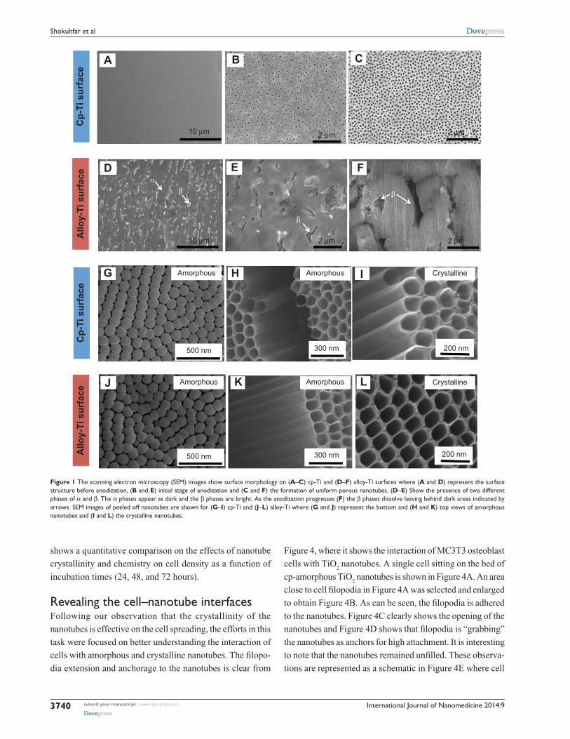
In order to investigate the biphasic nature of nano-
tubes, the samples were etched for 30 seconds in 0.5% HF
(Figure 1). At the initial stage of anodization of cp-Ti samples,
a uniform porous structure was formed (Figure 1A–B), which
eventually led to the formation of nanotubes (Figure 1C).
Figure 1G–L shows the nanotubes after being carefully
peeled off from the substrate using a diamond knife to observe
their bottom and top morphologies. These images indicate a
non-collapsed nanotube structure after anodization duration
of 4 hours. No significant morphological difference can be
seen between amorphous nanotubes (Figures 1H and K)
and crystalline nanotubes (Figures 1I and L).
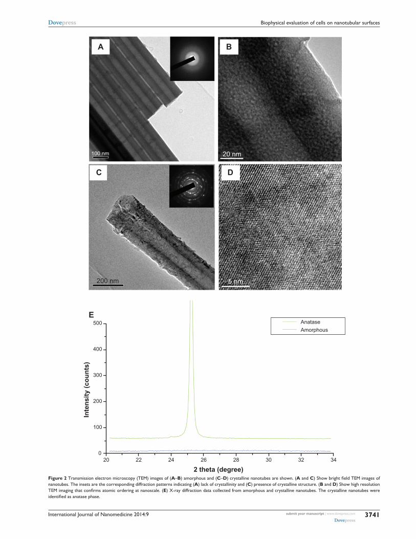
In order to confirm the presence of atomic ordering
in annealed nanotubes, transmission electron microscopy
(TEM) and X-ray diffraction (XRD) was utilized (Figure 2).
Figure 2A shows a bright field TEM image of amorphous
nanotubes. The fuzzy diffraction pattern with no obvious
spots of rings (the inset in Figure 2A) indicates lack of crys-
tallinity. High resolution TEM imaging (Figure 2B) confirms
the absence of atomic ordering at nanoscale. Similar TEM
imaging for annealed nanotubes (Figure 2C and D) shows the
presence of diffraction spots and atomic ordering at nanoscale
which indicates formation of crystalline structures. The XRD
data collected from amorphous and crystalline nanotubes
(Figure 2E) indicate that the crystalline nanotubes have an
anatase phase. As expected, no obvious peak was observed
for amorphous nanotubes. Since the TEM investigation of
nanotubes grown on alloy-Ti and cp-Ti did not show any
significant difference, the TEM images shown in Figure 2
are applicable for both.
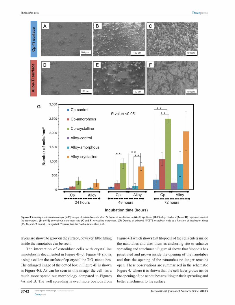
Figure 3A–F shows SEM images of osteoblast cells after
72 hours of incubation on prepared substrates. Figure 3G
International Journal of Nanomedicine 2014:9submit your manuscript | www.dovepress.com
Dovepress
Dovepress
3740
shokuhfar et al
shows a quantitative comparison on the effects of nanotube
crystallinity and chemistry on cell density as a function of
incubation times (24, 48, and 72 hours).
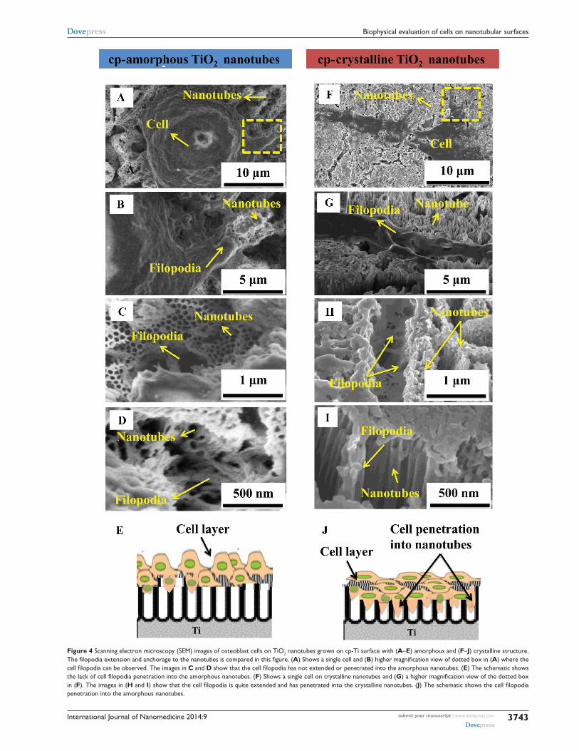
revealing the cell–nanotube interfacesFollowing our observation that the crystallinity of the
nanotubes is effective on the cell spreading, the efforts in this
task were focused on better understanding the interaction of
cells with amorphous and crystalline nanotubes. The filopo-
dia extension and anchorage to the nanotubes is clear from
Figure 4, where it shows the interaction of MC3T3 osteoblast
cells with TiO2 nanotubes. A single cell sitting on the bed of
cp-amorphous TiO2 nanotubes is shown in Figure 4A. An area
close to cell filopodia in Figure 4A was selected and enlarged
to obtain Figure 4B. As can be seen, the filopodia is adhered
to the nanotubes. Figure 4C clearly shows the opening of the
nanotubes and Figure 4D shows that filopodia is “grabbing”
the nanotubes as anchors for high attachment. It is interesting
to note that the nanotubes remained unfilled. These observa-
tions are represented as a schematic in Figure 4E where cell
Figure 1 The scanning electron microscopy (seM) images show surface morphology on (A–C) cp-Ti and (D–F) alloy-Ti surfaces where (A and D) represent the surface structure before anodization, (B and E) initial stage of anodization and (C and F) the formation of uniform porous nanotubes. (D–E) show the presence of two different phases of a and β. The a phases appear as dark and the β phases are bright. as the anodization progresses (F) the β phases dissolve leaving behind dark areas indicated by arrows. seM images of peeled off nanotubes are shown for (G–I) cp-Ti and (J–L) alloy-Ti where (G and J) represent the bottom and (H and K) top views of amorphous nanotubes and (I and L) the crystalline nanotubes.
AC
p-Ti
sur
face
Allo
y-Ti
sur
face
Cp-
Ti s
urfa
ceA
lloy-
Ti s
urfa
ce
10 µm
10 µm
500 nm 300 nm 200 nm
200 nm300 nm
Amorphous
Amorphous
Amorphous Crystalline
CrystallineAmorphous
500 nm
B
2 µm
C
D E F
G H I
J K L
2 µm
2 µm
β
β
βα
2 µm
International Journal of Nanomedicine 2014:9 submit your manuscript | www.dovepress.com
Dovepress
Dovepress
3741
Biophysical evaluation of cells on nanotubular surfaces
A
100 nm 20 nm
200 nm 5 nm
B
C D
E
Inte
nsity
(cou
nts)
500
400
300
200
100
020 22 24 26 28 30 32 34
2 theta (degree)
AnataseAmorphous
Figure 2 Transmission electron microscopy (TeM) images of (A–B) amorphous and (C–D) crystalline nanotubes are shown. (A and C) Show bright field TEM images of nanotubes. The insets are the corresponding diffraction patterns indicating (A) lack of crystallinity and (C) presence of crystalline structure. (B and D) show high resolution TEM imaging that confirms atomic ordering at nanoscale. (E) X-ray diffraction data collected from amorphous and crystalline nanotubes. The crystalline nanotubes were identified as anatase phase.
International Journal of Nanomedicine 2014:9submit your manuscript | www.dovepress.com
Dovepress
Dovepress
3742
shokuhfar et al
layers are shown to grow on the surface, however, little filling
inside the nanotubes can be seen.
The interaction of osteoblast cells with crystalline
nanotubes is documented in Figure 4F–J. Figure 4F shows
a single cell on the surface of cp-crystalline TiO2 nanotubes.
The enlarged image of the dotted box in Figure 4F is shown
in Figure 4G. As can be seen in this image, the cell has a
much more spread out morphology compared to Figures
4A and B. The well spreading is even more obvious from
Figure 4H which shows that filopodia of the cells enters inside
the nanotubes and uses them as anchoring site to enhance
spreading and attachment. Figure 4I shows that filopodia has
penetrated and grown inside the opening of the nanotubes
and thus the opening of the nanotubes no longer remains
open. These observations are summarized in the schematic
Figure 4J where it is shown that the cell layer grows inside
the opening of the nanotubes resulting in their spreading and
better attachment to the surface.
A
100 µm
100 µm
100 µm
100 µm
100 µm
100 µm
Cp-
Ti s
urfa
ceA
lloy-
Ti s
urfa
ce
B C
D E F
G 3,000
2,500
2,000
1,500
1,000Alloy-crystalline
Alloy-amorphous
* * * *
* ** *
* *
Alloy-control
Cp-crystalline
Cp-amorphous
Cp-controlP-value <0.05
500
0
Cp Alloy
24 hours 48 hours
Incubation time (hours)
72 hours
Cp Alloy Cp Alloy
Num
ber o
f cel
ls/m
m2
Figure 3 scanning electron microscopy (seM) images of osteoblast cells after 72 hours of incubation on (A–C) cp-Ti and (D–F) alloy-Ti where (A and D) represent control (no nanotubes), (B and E) amorphous nanotubes and (C and F) crystalline nanotubes. (G) Density of adhered Mc3T3 osteoblast cells as a function of incubation times (24, 48, and 72 hours). The symbol **means that the P-value is less than 0.05.
International Journal of Nanomedicine 2014:9 submit your manuscript | www.dovepress.com
Dovepress
Dovepress
3743
Biophysical evaluation of cells on nanotubular surfaces
Figure 4 scanning electron microscopy (seM) images of osteoblast cells on TiO2 nanotubes grown on cp-Ti surface with (A–E) amorphous and (F–J) crystalline structure. The filopodia extension and anchorage to the nanotubes is compared in this figure. (A) shows a single cell and (B) higher magnification view of dotted box in (A) where the cell filopodia can be observed. The images in C and D show that the cell filopodia has not extended or penetrated into the amorphous nanotubes. (E) The schematic shows the lack of cell filopodia penetration into the amorphous nanotubes. (F) shows a single cell on crystalline nanotubes and (G) a higher magnification view of the dotted box in (F). The images in (H and I) show that the cell filopodia is quite extended and has penetrated into the crystalline nanotubes. (J) The schematic shows the cell filopodia penetration into the amorphous nanotubes.
International Journal of Nanomedicine 2014:9submit your manuscript | www.dovepress.com
Dovepress
Dovepress
3744
shokuhfar et al
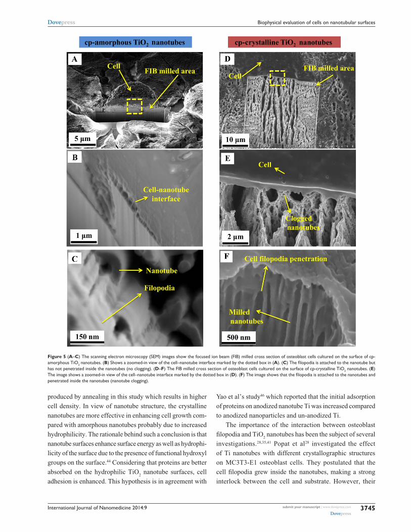
In order to investigate the growth of filopodia within the
hollow section of the amorphous and crystalline nanotubes,
FIB technique was used (Figure 5). Low energy beam of Ga
ions was used to carefully cut through sections of cells and
reveal the extent of interaction with the underlying nanotube
surface. Figure 5A shows an FIB milled osteoblast cell that is
attached to cp-amorphous TiO2 nanotubes. Closer examina-
tion demonstrates that the nanotubes have kept their morphol-
ogy intact under the cells (Figure 5B), while filopodia has not
grown into the hollow section of the nanotubes (Figure 5C).
However, the free end of cp-crystalline nanotubes is clogged
(Figures 5D and E) and the cell filopodia has grown into the
nanotubes (Figure 5F).
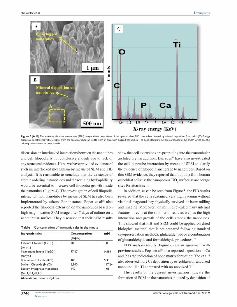
Figure 6 shows SEM images of the FIB milled cells
revealing that even after removal of the cell top section by
FIB the nanotubes remain clogged (Figures 6A and B). The
EDS chemical analysis showed that the bond between the
nanotubes and the adjacent osteoblast cell layer is composed
of Ca and P elements (Figure 6C), which appears to mimic
the bond in the bone tissue itself. Figures 5 and 6 indicate
that on the substrate surface, cell filopodia and the ECM
remained attached following separation from the milled top
section of the cell. These observations reveal a very close
interfacial contact or direct contact between the osteoblasts
and the nanotubes followed by Ca and P elements’ deposi-
tion on the nanotubes (Figure 6C). These elements are the
primary components of bone matrix and their deposition
onto nanotubes, which is an indication that nanotubes have
maintained osteoblast functionality. The proposed mor-
phological structure of deposited Ca and P elements under
SEM imaging is in agreement with the conclusions made by
Das et al.41 The low intensity of the peaks is due to the fact
that the EDS data were taken after only 3 days of culture.
In addition, it should be noted that the Ca and P elemental
peaks do not originate from the media. The concentration
of Ca and P elements in the media is much lower than other
inorganic components (Table 1). However, the EDS analysis
does not indicate any peaks regarding other elements such as
potassium (K) and sodium (Na). Moreover, the samples were
washed several times to eliminate any entrapped media in
nanotubes before EDS analysis. Therefore, the only reason
for the presence of Ca and P elemental peaks is stimulation
of cells by nanotube.
DiscussionFE-SEM images (Figure 1) confirm formation of nanotubes
on both cp-Ti and alloy-Ti. The nanotubes have an aver-
age diameter of 100 nm and an average length of 1 μm. In
agreement with the reports from Macak et al21 only the Al rich
a phase of Ti alloy showed stable porous structures. The V
rich β phases etch faster and dissolve, leaving behind dark
areas indicated by arrows in Figure 1F. Irrespective of the
compositional elements in the substrate, the grown nanotubes
are uniform and homogeneous.
TEM and XRD results (Figure 2) show that right after
anodization, as-anodized nanotubes have an amorphous
structure which transforms into an anatase phase during
annealing.
FE-SEM images of osteoblasts incubated over surfaces
demonstrate that the presence of nanotubes on cp-Ti substrate
has been effective in increasing the number of attached
cells (compare Figure 3A with B). Comparison between
Figures 3B and C shows a considerable increase in cell
density and spreading for crystalline nanotubes. A similar
observation can be made for the cells grown on an alloy-Ti
surface (Figure 3D–F) however the density of cells is much
lower on alloy-Ti.
Figure 3G shows the cell density increases as a function
of incubation time. Also, the cell density is always higher
on cp-Ti surfaces. The highest cell density (~2,500/mm2)
was obtained after 72 hours incubation of crystalline nano-
tubes grown on a cp-Ti surface which is ~60% higher than
amorphous nanotubes grown on cp-Ti. The cell density after
72 hours of incubation on crystalline nanotubes grown on
cp-Ti is about 20% higher than similar nanotubes that were
grown on alloy-Ti. Although the difference is not statistically
significant, this observation is also consistent for the cells
incubated for 24 and 48 hours suggesting that the chemistry
of nanotubes can have an effect on cell density. The reduction
in cell density on alloy-Ti surfaces may be due to slightly
lower biocompatibility of the alloying elements (Al and V)
and their oxides.42,43 In addition, the difference in cell density
between the amorphous and crystalline nanotubes can be
explained by hydrophilicity of the nanotubes as discussed
in the next section.
Droplet contact angle is highly affected by surface
treatments.44 After the anodization process, the droplet con-
tact angles decreased in the range of 55%~70% compared to
the bare substrate. This indicates that the creation of nanotube
structures makes the surface more hydrophilic. Remarkably,
the crystalline nanotubes had the most hydrophilic properties
among all the other substrates. On the crystalline nanotube
substrates, water droplet contact angles decreased 40%~56%
compared to the amorphous nanotube substrates.
In general, annealing metal generates a hydrophobic
surface.45 Conversely, more hydrophilic surfaces were
International Journal of Nanomedicine 2014:9 submit your manuscript | www.dovepress.com
Dovepress
Dovepress
3745
Biophysical evaluation of cells on nanotubular surfaces
produced by annealing in this study which results in higher
cell density. In view of nanotube structure, the crystalline
nanotubes are more effective in enhancing cell growth com-
pared with amorphous nanotubes probably due to increased
hydrophilicity. The rationale behind such a conclusion is that
nanotube surfaces enhance surface energy as well as hydrophi-
licity of the surface due to the presence of functional hydroxyl
groups on the surface.44 Considering that proteins are better
absorbed on the hydrophilic TiO2 nanotube surfaces, cell
adhesion is enhanced. This hypothesis is in agreement with
Yao et al’s study46 which reported that the initial adsorption
of proteins on anodized nanotube Ti was increased compared
to anodized nanoparticles and un-anodized Ti.
The importance of the interaction between osteoblast
filopodia and TiO2 nanotubes has been the subject of several
investigations.28,35,41 Popat et al28 investigated the effect
of Ti nanotubes with different crystallographic structures
on MC3T3-E1 osteoblast cells. They postulated that the
cell filopodia grew inside the nanotubes, making a strong
interlock between the cell and substrate. However, their
Figure 5 (A–C) The scanning electron microscopy (seM) images show the focused ion beam (FIB) milled cross section of osteoblast cells cultured on the surface of cp-amorphous TiO2 nanotubes. (B) shows a zoomed-in view of the cell–nanotube interface marked by the dotted box in (A). (C) The filopodia is attached to the nanotube but has not penetrated inside the nanotubes (no clogging). (D–F) The FIB milled cross section of osteoblast cells cultured on the surface of cp-crystalline TiO2 nanotubes. (E) The image shows a zoomed-in view of the cell–nanotube interface marked by the dotted box in (D). (F) The image shows that the filopodia is attached to the nanotubes and penetrated inside the nanotubes (nanotube clogging).
International Journal of Nanomedicine 2014:9submit your manuscript | www.dovepress.com
Dovepress
Dovepress
3746
shokuhfar et al
discussion on interlocked interactions between the nanotubes
and cell filopodia is not conclusive enough due to lack of
any structural evidence. Here, we have provided evidence of
such an interlocked mechanism by means of SEM and FIB
analysis. It is reasonable to conclude that the existence of
atomic ordering in nanotubes and the resulting hydrophilicity
would be essential to increase cell filopodia growth inside
the nanotubes (Figure 4). The investigation of cell filopodia
interaction with nanotubes by means of SEM has also been
implemented by others. For instance, Popat et al28 also
reported the filopodia extension on the nanotubes based on
high magnification SEM image after 7 days of culture on a
nanotubular surface. They discussed that their SEM results
show that cell extensions are protruding into the nanotubular
architecture. In addition, Das et al41 have also investigated
the cell–nanotube interaction by means of SEM to clarify
the evidence of filopodia anchorage to nanotubes. Based on
this SEM evidence, they reported that filopodia from human
osteoblast cells use the nanoporous TiO2 surface as anchorage
sites for attachment.
In addition, as can be seen from Figure 5, the FIB results
revealed that the cells sustained very high vacuum without
visible damage and they physically survived ion beam milling
and imaging. Moreover, ion milling revealed many internal
features of cells at the submicron scale as well as the high
interaction and growth of the cells among the nanotubes.
This showed that FIB and SEM could be applied on dried
biological material that is not prepared following standard
cryopreservation methods, glutaraldehyde or a combination
of glutaraldehyde and formaldehyde procedures.47
EDS analysis results (Figure 6) are in agreement with
previous studies. Popat et al28 also reported deposition of Ca
and P as the indication of bone matrix formation. Yao et al13
also observed more Ca deposition by osteoblasts on anodized
nanotube-like Ti compared with un-anodized Ti.
The results of the current investigation indicate the
formation of ECM on the nanotubes initiated by deposition of
Figure 6 (A, B) The scanning electron microscopy (seM) images show close views of the cp-crystalline TiO2 nanotubes clogged by mineral deposition from cells. (C) energy dispersive spectroscopy (eDs) signal from the area marked as a in (B) from an area with clogged nanotubes. The deposited minerals are composed of ca and P, which are the primary components of bone matrix.
Table 1 concentration of inorganic salts in the media
Inorganic salts Concentration (mg/L)
mM
calcium chloride (cacl2) (anhyd.)
200 1.8
Magnesium sulfate (MgsO4) (anhyd.)
97.67 0.814
Potassium chloride (Kcl) 400 5.33sodium chloride (Nacl) 6,800 117.24sodium Phosphate monobasic (Nah2PO4-h2O)
140 1.01
Abbreviation: anhyd., anhydrous.
International Journal of Nanomedicine 2014:9 submit your manuscript | www.dovepress.com
Dovepress
Dovepress
3747
Biophysical evaluation of cells on nanotubular surfaces
Ca and P.28 This production of ECM is the result of osteoblast
migration and healthy cellular activity in both crystalline
and amorphous nanotubular surfaces. In fact, it is possible
to consider the nanotubes as anchors for the cell filopodia to
“grab” onto and have a facilitated migration along the surface.
This anchorage benefit of nanotubes together with the strong
hydrophilic properties in crystalline nanotubes appear to have
increased osteoblast density in annealed nanotubes and higher
interactions with them. Therefore, the observations made in
the present investigation can be promising for the develop-
ment of implants with high-cohesion osseointegration.
ConclusionThe osteoblast cell culture experiments indicated that the
presence of nanotube morphology increased the total cell
density and spreading. This increase was correlated with
the anchoring effect of nanotubes for the cell filopodia to
“grab” onto, facilitating migration along the surface. The
early deposition of Ca and P onto nanotubes indicates that
nanotubes have maintained osteoblast functionality.
It was observed that the chemical composition of the
substrate strongly affected the cell density. In particular,
the total cell density of nanotubes grown on a cp-Ti surface
was higher in comparison to the cell density of nanotubes
grown on an alloy-Ti surface. This behavior can be explained
by slightly lower biocompatibility of the alloying elements
(Al and V in this case) and their oxides.
Interestingly, the high surface wettability due to
crystallinity is highly influential on further cell spreading
and extension of the cell filopodia into the hollow space of
nanotubes. In particular, the cell’s filopodia extended into
the crystalline nanotubes while much less penetration was
observed for the cells grown on amorphous nanotubes. This
was explained by the super hydrophilicity of crystalline nano-
tubes’ surfaces in comparison to amorphous nanotubes.
The anchorage benefit of nanotubes together with their
strong hydrophilic properties appear to cause increased
osteoblast density in annealed nanotubular surfaces in
comparison with anodized ones. This research can enable the
determination of an optimal implant surface modification where
implant–bone interlock and bone-cell density is highest.
AcknowledgmentsThe authors thank Mr Owen Mills and the Applied Chemi-
cal and Morphological Analysis Laboratory at Michigan
Technological University for providing microscopy sup-
port to conduct this research. We would also like to thank
Dr William Hendrickson, director of Research Resource
Center at UIC for providing laboratory space to carry out
some of the analysis related to the in vitro experiments.
DisclosureThe authors have no conflicts of interest to report.
References 1. Massia SP. Cell-extracellular matrix interactions relevant to vascular
tissue engineering. In: Landes Bioscience. Tissue Engineering of Vas-cular Prosthetic Grafts. 1999.
2. Webster TJ, Ergun C, Doremus RH, Siegel RW, Bizios R. Specific proteins mediate enhanced osteoblast adhesion on nanophase ceramics. J Biomed Mater Res. 2000;51(3):475–483.
3. Schneider G, Burridge K. Formation of focal adhesions by osteoblasts adhering to different substrata. Exp Cell Res. 1994;214(1):264–269.
4. Steele JG, Dalton BA, Johnson G, Underwood PA. Polystyrene chem-istry affects vitronectin activity – an explanation for cell attachment to tissue-culture polystyrene but not to unmodified polystyrene. J Biomed Mater Res. 1993;27(7):927–940.
5. Dalton BA, McFarland CD, Gengenbach TR, Griesser HJ, Steele JG. Polymer surface chemistry and bone cell migration. J Biomater Sci Polym Ed. 1998;9(8):781–799.
6. Webb K, Hlady V, Tresco PA. Relationships among cell attachment, spreading, cytoskeletal organization, and migration rate for anchorage-dependent cells on model surfaces. J Biomed Mater Res. 2000;49(3): 362–368.
7. Takebe J, Itoh S, Okada J, Ishibashi K. Anodic oxidation and hydro-thermal treatment of titanium results in a surface that causes increased attachment and altered cytoskeletal morphology of rat bone marrow stromal cells in vitro. J Biomed Mater Res. 2000;51(3):398–407.
8. Liao HH, Andersson AS, Sutherland D, Petronis S, Kasemo B, Thomsen P. Response of rat osteoblast-like cells to microstructured model surfaces in vitro. Biomaterials. 2003;24(4):649–654.
9. Doroudian G, Curtis MW, Gang A, Russell B. Cyclic strain dominates over microtopography in regulating cytoskeletal and focal adhesion remodeling of human mesenchymal stem cells. Biochem Biophys Res Commun. 2013;430(3):1040–1046.
10. Rabiee SM, Mortazavi SM, Moztarzadeh F, et al. Association of a synthetic bone graft and bone marrow cells as a composite biomaterial. Biotechnology and Bioprocess Engineering. 2009;14(1):1–5.
11. Yu WQ, Zhang YL, Jiang XQ, Zhang FQ. In vitro behavior of MC3T3-E1 preosteoblast with different annealing temperature titania nanotubes. Oral Dis. 2010;16(7):624–630.
12. von der Mark K, Park J, Bauer S, Schmuki P. Nanoscale engineering of biomimetic surfaces: cues from the extracellular matrix. Cell Tissue Res. 2010;339(1):131–153.
13. Yao C, Slamovich EB, Webster TJ. Enhanced osteoblast functions on anodized titanium with nanotube-like structures. J Biomed Mater Res A. 2008;85A(1):157–166.
14. Sun L, Berndt CC, Gross KA, Kucuk A. Material fundamentals and clinical performance of plasma-sprayed hydroxyapatite coatings: a review. J Biomed Mater Res. 2001;58(5):570–592.
15. Tami AE, Schaffler MB, Knothe Tate ML. Probing the tissue to sub-cellular level structure underlying bone’s molecular sieving function. Biorheology. 2003;40(6):577–590.
16. Nourmohammadzadeh M, Lo JF, Bochenek M, et al. Microfluidic Array with Integrated Oxygenation Control for Real-Time Live-Cell Imag-ing: Effect of Hypoxia on Physiology of Microencapsulated Pancreatic Islets. Anal Chem. 2013;85(23):11240–11249.
17. Brammer KS, Oh S, Cobb CJ, et al. Improved bone-forming functional-ity on diameter-controlled TiO
2 nanotube surface. Acta Biomater. 2009;
5(8):3215–3223.18. Roy P, Berger S, Schmuki P. TiO
2 Nanotubes: Synthesis and Applica-
tions. Angew Chem Int Ed Engl. 2011;50(13):2904–2939.
International Journal of Nanomedicine
Publish your work in this journal
Submit your manuscript here: http://www.dovepress.com/international-journal-of-nanomedicine-journal
The International Journal of Nanomedicine is an international, peer-reviewed journal focusing on the application of nanotechnology in diagnostics, therapeutics, and drug delivery systems throughout the biomedical field. This journal is indexed on PubMed Central, MedLine, CAS, SciSearch®, Current Contents®/Clinical Medicine,
Journal Citation Reports/Science Edition, EMBase, Scopus and the Elsevier Bibliographic databases. The manuscript management system is completely online and includes a very quick and fair peer-review system, which is all easy to use. Visit http://www.dovepress.com/testimonials.php to read real quotes from published authors.
Dovepress
International Journal of Nanomedicine 2014:9submit your manuscript | www.dovepress.com
Dovepress
Dovepress
3748
shokuhfar et al
19. Ghicov A, Schmuki P. Self-ordering electrochemistry: a review on growth and functionality of TiO
2 nanotubes and other self-aligned MOx
structures. Chem Commun (Camb). 2009;(20):2791–2808.20. Park J, Bauer S, Schmuki P, von der Mark K. Narrow Window in Nano-
scale Dependent Activation of Endothelial Cell Growth and Differentia-tion on TiO
2 Nanotube Surfaces. Nano Lett. 2009;9(9):3157–3164.
21. Macak JM, Tsuchiya H, Taveira L, Ghicov A, Schmuki P. Self-organized nanotubular oxide layers on Ti-6A1-7Nb and Ti-6A1-4V formed by anodization in NH
4F solutions. J Biomed Mater Res A. 2005;
75A(4):928–933.22. Bauer S, Park J, von der Mark, Schmuki P. Improved attachment of
mesenchymal stem cells on super-hydrophobic TiO2 nanotubes. Acta
Biomater. 2008;4(5):1576–1582.23. Hamlekhan A, Butt A, Patel S, et al. Fabrication of Anti-Aging TiO
2
Nanotubes on Biomedical Ti Alloys. PLoS One. 2014;9(5):1–10.24. Patel SB, Hamlekhan A, Royhman D, et al. Enhancing Surface Charac-
teristics of Ti-6Al-4V for Bio-implants Using Integrated Anodization and Thermal Oxidation. J Mater Chem B. 2014;2(23):3597–3608.
25. Tan AW, Pingguan-Murphy B, Ahmad R, Akbar SA. Review of titania nanotubes: Fabrication and cellular response. Ceramics International. 2012;38(6):4421–4435.
26. Butt A, Hamlekhan A, Patel SB, et al. A Novel Investigation of the Formation of TiO
2 Nanotubes on Thermally Formed Oxide of Ti6-
Al-4V. J Oral Implantol. Epub 2014 March 15.27. Azhang Hamlekhan, AB, Sweetu Patel, et al. Optimization of
Anodization and Annealing Condition Enhances TiO2 Nanotubular
Surface Hydrophilicity. TMS 2014 Supplemental Proceedings. John Wiley & Sons, Inc, 2014.
28. Popat KC, Leoni L, Grimes CA, Desai TA. Influence of engineered titania nanotubular surfaces on bone cells. Biomaterials. 2007;28(21): 3188–3197.
29. Bjursten LM, Rasmusson L, Oh S, Smith GC, Brammer KS, Jin S. Titanium dioxide nanotubes enhance bone bonding in vivo. J Biomed Mater Res A. 2010;92A(3):1218–1224.
30. Hazan R, Sreekantan S, Khalil AA, Nordin IM, Mat I. Surface Engi-neering of titania for Excellent Fibroblast 3T3 Cell-Metal Interaction. Journal of Physical Science. 2009;20(1):35–47.
31. Gongadze E, Kabaso D, Bauer S, Park J, Schmuki P, Iglic A. Adhesion of Osteoblasts to a Vertically Aligned TiO
2 Nanotube Surface. Mini
Rev Med Chem. 2013;13(2):194–200.32. Bauer S, Park J, Faltenbacher J, et al. Size selective behavior of mes-
enchymal stem cells on ZrO2 and TiO
2 nanotube arrays. Integr Biol
(Camb). 2009;1(8–9):525–532.
33. Chen CS. Mechanotransduction – a field pulling together? J Cell Sci. 2008;121(20):3285–3292.
34. Park J, Bauer S, von der Mark K, Schmuki P. Nanosize and vitality: TiO2
nanotube diameter directs cell fate. Nano Lett. 2007;7(6):1686–1691.35. Oh S, Daraio C, Chen LH, Pisanic TR, Finones RR, Jin S. Significantly
accelerated osteoblast cell growth on aligned TiO2 nanotubes. J Biomed
Mater Res A. 2006;78A(1):97–103.36. Oh S, Brammer KS, Li YS, Teng D, Engler AJ, Chien S, Jin S. Stem
cell fate dictated solely by altered nanotube dimension. Proc Natl Acad Sci U S A. 2009;106(7):2130–2135.
37. Wang D, Christensen K, Chawla K, Xiao G, Krebsbach PH, Franceschi RT. Isolation and characterization of MC3T3-E1 preosteoblast subclones with distinct in vitro and in vivo differentiation mineralization potential. J Bone Miner Res. 1999;14(6):893–903.
38. Luckey HA, Kubli F. Titanium alloys in surgical implants. Phoenix, Ariz: ASTM International, 1983.
39. Brunette DM, Tengvall P, Textor M, Thomsen P. Titanium in medicine. New York: Springer, 2001.
40. Lee J, Hurson S, Tadros H, Schupbach P, Susin C, Wikesjo UM. Crestal remodelling and osseointegration at surface-modified commercially pure titanium and titanium alloy implants in a canine model. J Clin Periodontol. 2012;39(8):781–788.
41. Das K, Bose S, Bandyopadhyay A. TiO2 nanotubes on Ti: Influence
of nanoscale morphology on bone cell-materials interaction. J Biomed Mater Res A. 2009;90A(1):225–237.
42. Eisenbarth E, Velten D, Muller M, Thull R, Breme J. Biocompatibility of beta-stabilizing elements of titanium alloys. Biomaterials. 2004; 25(26):5705–5713.
43. Wapner KL. Implications of metallic corrosion in total knee arthro-plasty. Clin Orthop Relat Res. 1991;(271):12–20.
44. Shin DH, Shokuhfar T, Choi CK, Lee SH, Friedrich C. Wettability changes of TiO
2 nanotube surfaces. Nanotechnology. 2011;
22(31):1–7.45. Shinde VR, Lokhande CD, Mane RS, Han SH. Hydrophobic and
textured ZnO films deposited by chemical bath deposition: annealing effect. Applied Surface Science. 2005;245(1–4):407–413.
46. Yao C, Perla V, McKenzie JL, Slamovich EB, Webster TJ. Anodized Ti and Ti(6)Al(4)V Possessing Nanometer Surface Features Enhances Osteoblast Adhesion. Journal of Biomedical Nanotechnology. 2005; 1(1):68–73.
47. Leser V, Drobne D, Pipan Z, Milani M, Tatti F. Comparison of different preparation methods of biological samples for FIB milling and SEM investigation. J Microsc. 2009;233(2):309–319.


