Embed Size (px)
Citation preview

QP Code: 3834
F.Y.B.Sc. in Biotechnology Semester II Examination (April 2018)
Model Answers
Chemistry I- Biorganic chemistry
Q.1. Do as directed: (Any fifteen) 15
Define the following terms:
1) Carbohydrates
Carbohydrates are poly hydroxy aldehydes or ketones with a general formula
Cn(H2O)n, where n 3
2) Dipeptide
A dipeptide holds two interacting amino acids with one peptide bond
3) Isoelectric pH
At isoelectric pH the amino acids exists as a zwitter ions and they bear no net charge.
4) Isomers
Isomers are the compounds having the same empirical formula and the structure, but
they differ from one another by a configuration around only one carbon atom.
5) Unsaturated fatty acid
Unsaturated Fatty acids are carboxylic acids with atleast one or more double bonds in
its hydrocarbon chain.
6) Pitch of the helix- is the height of one complete helix turn, measured parallel to the axis of
the helix.
7) Saponification
When animal fat/oil reacts with aqueous NaOH or KOH, they form soap and glycerol.
Since this reaction leads to the formation of soap, it is called the Saponification.
Draw structures of the following biomolecules:
8) Purine base. Any one base

9) D- glyceraldehydes
CHO-CHOH-CH2OH
10)
D-glucose
11)
Serine
12)
Acetone
Name the following:
13) An acidic amino acid- Aspartic acid, glutamic acid
14) One keto sugar- Fructose, ribulose
15) Sterol of biological significance - cholesterol
16) Fibrous protein present in hair - Keratin
17) Sugar present in DNA. -deoxyribose
18) Parent compound of phospholipids- Phosphatidic acid
19) The transport protein present in the red blood cell - Albumin
20) Type of bond present between base pair G-C – Hydrogen bond
Q2. a) Compare and contrast: unsaturated fatty acid and saturated fatty acids
Fatty acids are carboxylic acid with hydrocarbon side chain. General formula is R-
COOH. The hydrocarbon side chain may be saturated or unsaturated in nature
SFA USFA
Saturated fatty acids don’t have
unsaturation or double bonds in their
structure.
Unsaturated fatty acids have one or
more double bonds in their structure
Their general formula is R- COOH -
SFA s is found in animal sources. USFA s is found in plant based sources.
They are solids at room temperature
Eg: ghee , butter
They are liquids at room temperature,
They are popularly called as oils
8
Butyric acid- CH3 –CH2-CH2- COOH EG: Palmitolic acid has one double
bond- C16:9
Palmitic acid- CH3 –(CH2)14- COOH
Oleic acid has one double bond-
C18:9
Lauric acid.- CH3 –(CH2)10- COOH
Leinoleic acid has three double bonds-
C18: 9,12, 15
,
Aarachidonic acid has four double
bond- C20:5,8,11,14
4M for each category
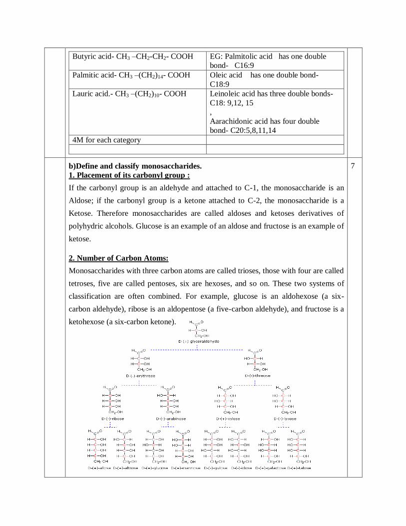
b)Define and classify monosaccharides.
1. Placement of its carbonyl group :
If the carbonyl group is an aldehyde and attached to C-1, the monosaccharide is an
Aldose; if the carbonyl group is a ketone attached to C-2, the monosaccharide is a
Ketose. Therefore monosaccharides are called aldoses and ketoses derivatives of
polyhydric alcohols. Glucose is an example of an aldose and fructose is an example of
ketose.
2. Number of Carbon Atoms:
Monosaccharides with three carbon atoms are called trioses, those with four are called
tetroses, five are called pentoses, six are hexoses, and so on. These two systems of
classification are often combined. For example, glucose is an aldohexose (a six-
carbon aldehyde), ribose is an aldopentose (a five-carbon aldehyde), and fructose is a
ketohexose (a six-carbon ketone).
7

(6M for classification and any three str )
OR
c) Compare and contrast:
i. Maltose and Lactose (4M)
Maltose Lactose
it’s the principle sugar found in malt it’s the principle sugar found in milk.
Lactose is an important component used
by dairy and Brewery
Lactose is an important component used
by dairy and pharma industry
Maltose is composed of -D glucose
linked to D glucose by means of a α
1→4 linkage
Lactose is composed of β -D galactose
linked to D glucose by means of a β
1→4 linkage.
ii. Starch and cellulose (4M)
Starch
Cellulose
Homopolymer of
α-D glucose
Amylose- unbranched
Amylopectin- branched
Homopolymer of
βD glucose
Storage polysaccharide found in
plants
Stuctural polysaccharide
found in plants
Bond between monomer is mostly
α1→4 ( amylose)
Bond between monomer is
β1→4
α1→6 linkage is found in
amylopectin, branched starch unit
unbranched
8


d.)What are phospholipids? Draw structures of Lecithin and Cephalin.
Phospholipids are derived from phosphatidic acid. They contain glycerol backbone
esterified with two fatty acids at 1,2 position and a phosphoric acid and a nitrogen
base.
(4M)
Eg: Phosphatidyl ethanol amine, Phosphatidyl serine, Phosphatidyl inositol,
phosphatidylcholin (lecithin), and cephalin.
Lecithin/ phosphatidylcholin, found in egg yolk and other membrane lipids.
R= fatty acid
(4M for Lecithin / Cephalin, any one structure )
7
Q3. a). Explain N-terminal sequencing of proteins using Edmans and Sanger’s
Methods.
Edman degradation, developed by Pehr Edman, is a method of sequencing amino
acids in a peptide. In this method, the amino-terminal residue is labeled and cleaved
from the peptide without disrupting the peptide bonds between other amino acid
residues.
Phenyl isothiocyanate is reacted with an uncharged N-terminal amino group, under
8

mildly alkaline conditions, to form a cyclical phenylthiocarbamoyl derivative.
Under acidic conditions, this derivative of the terminal amino acid is cleaved as a
thiazolinone derivative. The thiazolinone amino acid is then selectively extracted into
an organic solvent and treated with acid to form the more stable phenylthiohydantoin
(PTH)- amino acid derivative that can be identified
using chromatography or electrophoresis. This procedure can then be repeated again to
identify the next amino acid (4M)
Sanger’s reagent was FDNB.FDNB specifically binds to the n terminal residue and
forms a DNP amino acid. Chromatography is used to identify this amino acid. (4M)
b) Classify amino acids on the basis of the polarity of the R group.
The twenty amino acids are generally classified as
1. Nonpolar (hydrophobic, meaning they do not readily interact with water),
2. Polar(uncharged and
3. Hydrophilic, meaning they readily interact with water), or charged (either
positively or negatively). (1M)
The chemical composition of the R-group determines their classification. The
various properties of the R-groups greatly influence how the amino acids
interact with each other and their environment.
(One example of each category- 6M)
7

OR
c) Schematically represent the titration curve of an amino acid and describe
the different stages.
Titration helps in understanding of occurrence of different ionic forms of amino acids.
It is a graphical representation of pH changes (Y axis) plotted against corresponding
volume of NaOH of known concentration (X axis).
a) Nature of amino acids in particular their ionisable groups determines the PI of
protein. PI is affected by the presence of an additional –COOH in acidic amino
acid or an – NH2 groups in a basic amino acids. At isoelectric point, the protein
exists as a zwitter ion or dipolar ion. (2M)
b) Draw titration curve ( 2M)
8

The titration curve for alanine is shown in Figure . At a pH lower than 2, both the
carboxylate and amine functions are protonated, so the alanine molecule has a net
positive charge. At a pH greater than 10, the amine exists as a neutral base and the
carboxyl as its conjugate base, so the alanine molecule has a net negative charge. At
intermediate pH's the zwitterion concentration increases, and at a characteristic pH,
called the isoelectric point (pI), the negatively and positively charged molecular
species are present in equal concentration.
This behavior is general for simple (difunctional) amino acids. Starting from a fully
protonated state, the pKa's of the acidic functions range from 1.8 to 2.4 for -CO2H, and
8.8 to 9.7 for -NH3(+)
. The isoelectric points range from 5.5 to 6.2.
Titration curves show the neutralization of these acids by added base, and the change
in pH during the titration. (4M)
d.) Explain the term conjugated Proteins. Give examples and state their
functions.
Many proteins contain only amino acids and no other chemical groups, and they are
called simple proteins.
A conjugated protein is a protein that functions in interaction with non-protein
chemical group, which is attached by covalent bond or weak interactions.
Conjugated proteins, upon hydrolysis yield, some other chemical component in
addition to amino acids.
7

The non-amino part of a conjugated protein is usually called its prosthetic group.
(3M)
Conjugated proteins are classified on the basis of the chemical nature of their
prosthetic groups.
Examples of conjugated proteins-
1. Glycoproteins- phosphoproteins, , flavoproteins, Glycoproteins are generally
the largest and most abundant group of conjugated proteins. They range from
glycoproteins in cell surface membranes that constitute the glycocalyx, to
important antibodies produced by leukocytes.
2. Hemoproteins- Hemoglobin contains the prosthetic group known as heme.
Each heme group contains an iron ion (Fe2+
) which forms a co-ordinate bond
with an oxygen molecule (O2), allowing hemoglobin to transport oxygen
through the bloodstream. As each of the four protein subunits of hemoglobin
possesses its own prosthetic heme group, each hemoglobin can transport four
molecules of oxygen.
3. Lipoproteins-present on Cell membrane
4. Metalloproteins- Enzymes are complexed with metals- hexokinase, PFK etc
5. phytochromes/ cytochromes- take part in electron transport,
6. opsins and chromoproteins. (any four class and example 4M)
Q4. a) Give a brief account of types of RNA and discusses their functions.
Types of RNA Function(s)
1) Messenger RNA Transfer genetics information
from genes to ribosome to
synthesize proteins
2) Heterogeneous nuclear
RNA
Serve as precursor for mRNA
and other RNAs
3) Transfer RNA Transfer amino acid to mRNA
for protein synthesis
4) Ribosomal RNA Provides structural framework
for ribosomes
5) Small nuclear RNA Involved in mRNA processing
6) Small nucleolar RNA Plays a key role in the
processing of rRNA molecules
7) Small cytoplasmic RNA Involved in the selection of
proteins for export.
8) Transfer-messenger
RNA
Mostly present in bacteria. Add
short peptide tags to proteins to
facilitate the degradation of
incorrectly synthesized proteins.
8

b) Explain denaturation and renaturation of DNA in detail.
Denaturation of DNA is a loss of biological activity and is accompanied by cleave of
hydrogen bonds holding the complementary sequences of nucleotides together.
This result in a separation of the double helix into two constituent polynucleotide
chains. (2M)
Diagrammatic representation –(2M)
Effect of pH on denaturation ( 1M )
Effect of Temperature on denaturation (1M)
Renaturation- ( 1M )
The denatured DNA can reformulate hydrogen bonds between complementary single
strand, making it likely to reform double helix structure again. This process is called
as renaturation
7
OR
c) Give an account of different conformations of DNA double helix.
Double helical DNA exist in many different forms like, A, B, C, D, E and Z. Out of
which A, B and Z are important form. ( 1 M )
Characteristics A-DNA B-DNA Z-DNA
Helix sense Right-
handed
Right-
handed
Left-handed
Helix- diameter 25.5⁰A 23.7⁰A 18.4⁰A
Rise per base pair 2.3⁰A 3.4⁰A 3.8⁰A
Base pair per turn
of helix
11 10 12
Helix pitch 25.30⁰A 35.36⁰A 45.60⁰A
Shape Broadest Intermediate Narrowest
Any 4 points for each form -6M
8
d) Explain clover leaf structure of tRNA using a suitable diagram.
Neat labeled clover-leaf model of tRNA- 4 Marks
Descriptions – each arm of tRNA- 3Marks
7
5. Write a short note on: (Any three) 15
1) Types of Triacylglycerol
Triacylglycerol- Neutral or depot fat. In Triacylglycerol, central glycerol molecule is
condensed with three fatty acids. (1M)
If all the three fatty condensed are of the same type the molecule is called as simple
TG and if more than one type of fatty acid is condensed, the molecule is known as
mixed TG.(2M)
TG is the fat reserve of the body. It can be hydrolysed and fatty acids can be beta
oxidized whenever needed. Glycerol serves as a 3 carbon skeleton reserve, which
undergoes gluconeogenesis in cases of hypoglycemia. ( 2M structure, 2M types, 2M
function)

(2M)
2) Structure and function of Starch
Structure:
Starch is a homopolymer of α-D glucose.
Bond between monomer is mostly α1→4 and this unbranched unit is known as
Amylose. α1→6 linkage is found in amylopectin.
Function:
It is the storage polysaccharide found in plants.
It is also important in food industry.
( 4M structure, 1M function )
3) Denaturation of proteins
Disorganization of native protein structure is called denaturation.
Physical agents - heat, violent shaking, X rays, UV rays
Chemical agents- Acids, alkalies, heavy metals pb,Hg, salicylate, organic solvents
cause denaturation. (2M)
Properties:
Loss of native protein structure
Primary structure is retained
Denatured protein is easily digested
Peptide linkage is resistant to denaturing forces
Higher viscosity of denatured protein
Lower surface tension in denatured protein (3M)
4) Chargaff’s rule.
Chargaff's rules state that DNA from any cell of all organisms should have a 1:1 ratio
(base Pair Rule) of pyrimidine and purine bases and, more specifically, that the
amount of guanine should be equal to cytosine and the amount of adenine should be
equal to thymine.
The ratio of A+T/G+C, known as dissymmetry ration varies greatly from one species
of DNA to the other
5) Functions of Nucleotides
i) Act as building blocks/ precursors of DNA and RNA
ii) Nucleotide triphosphate especially ATP , act as universal currency of energy in
biological system.iii) Adenine nucleotides are components of coenzymes of B-
complex vitamins-FAD,NAD.iv) Act as metabolic regulators.v) Act as chemical
messenger-cAMP


