Embed Size (px)
Citation preview
This article was downloaded by: [Mahatma Gandhi University]On: 14 December 2014, At: 20:42Publisher: Taylor & FrancisInforma Ltd Registered in England and Wales Registered Number: 1072954 Registered office: Mortimer House,37-41 Mortimer Street, London W1T 3JH, UK
Click for updates
Soil and Sediment Contamination: An InternationalJournalPublication details, including instructions for authors and subscription information:http://www.tandfonline.com/loi/bssc20
Biosurfactant Facilitated Biodegradation Of QuinalphosAt High Concentrations By Pseudomonas aeruginosaQ10Ambilli M. Naira, Sharrel Rebelloa, Rishad K.Sa, Aju K. Asoka & Jisha M.S.a
a School of Biosciences, Mahatma Gandhi University, Kottayam, Kerala, IndiaAccepted author version posted online: 06 Dec 2014.
To cite this article: Ambilli M. Nair, Sharrel Rebello, Rishad K.S, Aju K. Asok & Jisha M.S. (2014): Biosurfactant FacilitatedBiodegradation Of Quinalphos At High Concentrations By Pseudomonas aeruginosa Q10, Soil and Sediment Contamination: AnInternational Journal, DOI: 10.1080/15320383.2015.988205
To link to this article: http://dx.doi.org/10.1080/15320383.2015.988205
Disclaimer: This is a version of an unedited manuscript that has been accepted for publication. As a serviceto authors and researchers we are providing this version of the accepted manuscript (AM). Copyediting,typesetting, and review of the resulting proof will be undertaken on this manuscript before final publication ofthe Version of Record (VoR). During production and pre-press, errors may be discovered which could affect thecontent, and all legal disclaimers that apply to the journal relate to this version also.
PLEASE SCROLL DOWN FOR ARTICLE
Taylor & Francis makes every effort to ensure the accuracy of all the information (the “Content”) containedin the publications on our platform. However, Taylor & Francis, our agents, and our licensors make norepresentations or warranties whatsoever as to the accuracy, completeness, or suitability for any purpose of theContent. Any opinions and views expressed in this publication are the opinions and views of the authors, andare not the views of or endorsed by Taylor & Francis. The accuracy of the Content should not be relied upon andshould be independently verified with primary sources of information. Taylor and Francis shall not be liable forany losses, actions, claims, proceedings, demands, costs, expenses, damages, and other liabilities whatsoeveror howsoever caused arising directly or indirectly in connection with, in relation to or arising out of the use ofthe Content.
This article may be used for research, teaching, and private study purposes. Any substantial or systematicreproduction, redistribution, reselling, loan, sub-licensing, systematic supply, or distribution in anyform to anyone is expressly forbidden. Terms & Conditions of access and use can be found at http://www.tandfonline.com/page/terms-and-conditions
ACCEPTED MANUSCRIPT
ACCEPTED MANUSCRIPT 1
Biosurfactant Facilitated Biodegradation Of Quinalphos At High
Concentrations By Pseudomonas aeruginosa Q10
Ambilli M. Nair; Sharrel Rebello; Rishad K.S; Aju K. Asok and Jisha M.S.*
School of Biosciences, Mahatma Gandhi University, Kottayam, Kerala, India
Tel: +91-481-2731035, Fax: +91 481 2731002.Email: [email protected];
Abstract
Previous studies indicate that high concentration of pesticides and its associated toxic effects are
high at their point source of application. Use of pesticide degrading bacteria at point sources
could augment degradation and thereby reduce toxic effects associated with its persistence in
soil. Quinalphos, an organophosphorus insecticide, though ranked 'moderately hazardous' in
WHO’s acute hazard ranking still continues to be used extensively in developing countries. The
presence of a chloride radical usually makes this pesticide sparingly soluble in water and hence
difficult to degrade. The present study aimed to isolate autochthonous bacterial strains capable of
utilizing quinalphos as carbon source. Primary screening of pesticide contaminated soil by
enrichment culture and degradation analysis by UV-VIS spectrophotometry led to isolation of 12
different bacterial isolates of which 3 efficient isolates of Pseudomonas sp, Serratia sp and
Pseudomonas aeruginosa with degradation rate 86%, 82%, 94% respectively were selected. GC-
MS studies with P.aeruginosa confirmed formation of 2-hydroxy quinoxaline and
phosphorothioic acid as a result of biodegradation. The present study succeeded to isolate
autochthonous bacterial strains capable of utilizing high concentrations of quinalphos as carbon
source in a shorter incubation period. This strain also possessed biosurfactant production ability
which makes quinalphos available to cells at higher concentrations.
Key words: Biosurfactant; Quinalphos; Gas Chromatography; Mass Spectrometry.
Dow
nloa
ded
by [
Mah
atm
a G
andh
i Uni
vers
ity]
at 2
0:42
14
Dec
embe
r 20
14
ACCEPTED MANUSCRIPT
ACCEPTED MANUSCRIPT 2
Introduction
The unprecedented population explosion and subsequent increased food demand, prompted man
to adopt various improved crop yielding strategies. The use of high breeding crop varieties,
better fertilizers and extensive pesticides helped man to achieve his goal of high crop yield, but it
also resulted in the massive disposal of xenobiotic pesticides into our environment. Apart from
its inhibitory role on various pests, pesticides often lead to several short-term and long-term
adverse effects on man. In developing countries like India, pesticides serves as a key pest control
mode, but they often result in the accumulation of this xenobiotic in our food products (Gupta,
2004),water resources (Sharma, 2007) and eventually sneak into the foetus (Whyatt and Barr,
2001) which is a matter of great concern. The rate of still births, spontaneous abortions,
premature births and birth defects are higher in those exposed to these pesticides.
Among the various pesticides, organophosphates such as quinalphos are widely used in Indian
agriculture over certain crops such as cotton, groundnut and rice (Jena et al., 1990; Reddy and
Ghewande, 1986).Quinalphos has contact and stomach action against a wide range of pests from
Lepidoptera, Diptera, Coleoptera and Hemiptera. Quinalphos acts by inhibiting
acetylcholinesterase, which in turn block the transmission of the nerve impulse at the nerve
endings. Among 323 reported events of pesticide toxicity in three villages of India, 83.6% was
associated with signs and symptoms of mild to severe poisoning, 10% of the pesticide
application sessions were associated with three or more neurotoxic/systemic signs and symptoms
typical of poisoning by organophosphates, which were used in 47% of the applications (Mancini
et al., 2005).Thus, studies on the degradation and toxicity of this pesticide gains much relevance.
Dow
nloa
ded
by [
Mah
atm
a G
andh
i Uni
vers
ity]
at 2
0:42
14
Dec
embe
r 20
14
ACCEPTED MANUSCRIPT
ACCEPTED MANUSCRIPT 3
Efforts are currently under way to develop safe, convenient and economically feasible methods
for pesticide detoxification (Kulshrestha and Kumari, 2010).Isolation of indigenous bacteria
capable of metabolizing organophosphorus compounds has received considerable attention
because these bacteria provide an environmentally friendly method of in situ detoxification
(Mulchandani et al., 1999).However, isolation of better strains that can degrade higher
concentrations of quinalphos at a faster rate is great need with the increasing rates of pesticide
posing rates.
Materials and methods
Enrichment of soil samples and isolation of Quinalphos degrading bacteria
Quinalphos 25EC (emulsified concentrate) supplied by Sandoz (India) Limited was used in the
present study.All other chemicals used were of analytical reagent grade. A minimal media broth
(MMB) containing quinalphos as sole source of carbon was formulated to isolate quinalphos
degrading bacteria. The medium composition was as follows (g/l) (pH 7.0 ± 0.2): KH2PO4-1.0
g/l, K2HPO4 -1.0 g/l, NH4NO3-1.0 g/l, MgSO4.7H2O-0.2 g/l, CaCl2-0.02 g/l, NaCl-1 g/l.The
mineral salt medium was also supplemented with 2ml/l of stock trace elements to provide
bacteria with essential trace elements.Trace element solution stock was prepared in distilled
water with composition FeSO4.7H2O (0.1 g/l), MnCl4.4H2O (0.1 g/l) and ZnSO4.7H2O (0.1 g/l).
Bacteria capable of utilizing quinalphos as sole source of carbon was isolated from pesticide
contaminated soils by enrichment in MMB. Five grams of soil sample was added to 100 ml of
minimal medium containing 0.001 g/l (w/v) quinalphos and incubated at 28 ± 2°C for 7-8 days
at 120 rpm. After eight days, 10 ml of supernatant was transferred to 90 ml each of fresh medium
with 0.005 g/l and 10 g/l quinalphos each and incubated. Subculturing was done six times from
Dow
nloa
ded
by [
Mah
atm
a G
andh
i Uni
vers
ity]
at 2
0:42
14
Dec
embe
r 20
14
ACCEPTED MANUSCRIPT
ACCEPTED MANUSCRIPT 4
each concentration of quinalphos to fresh minimal medium of the same quinalphos
concentration. After six serial transfers, 1ml of the supernatant from the final enrichment flask
were taken; plated directly on minimal media agar with 10 g/l quinalphos by spread plate
technique and incubated at 30°C for 3-4 days. Single colonies obtained were maintained for
further analysis.
Primary screening of quinalphos degrading bacteria
The ability of the bacterial isolates to utilize quinalphos was assessed by checking the quinalphos
level left over in the culture medium after incubation period. One ml of 1 Optical Density (OD)
culture of isolated bacterial strains were inoculated into the flasks with 10g/l quinalphos
supplemented MMB and incubated at 28 ± 2°C for 8 days on a shaker at 120 rpm. After
incubation, the cell free supernatant was extracted three times with an equal volume of
chloroform and absorbance was measured at 350 nm in Hitachi U-2800 UV-VIS
Spectrophotometer (Chiranjeevi et al., 1997).The percentage of quinalphos leftover was
calculated and the percentage of degradation was computed. Since quinalphos was the sole
carbon source in the medium, the decrease in the amount of quinalphos left over in the medium
after incubation indicated a greater level of degradation.
Identification of the selected cultures
The selected bacterial isolates were identified based on their morphological, physiological and
biochemical characteristics according to Bergey’s Manual of Determinative Bacteriology (Holt
et al., 1994).The 16S rDNA typing of the most efficient isolate Q10 was done using universal
eubacterial primers Fd1 and Rd1 primers amplifying the 16S rDNA gene in most of bacteria
(Weisburg et al., 1991).Chromosomal DNA was isolated from 5 ml of nutrient broth culture of
Dow
nloa
ded
by [
Mah
atm
a G
andh
i Uni
vers
ity]
at 2
0:42
14
Dec
embe
r 20
14
ACCEPTED MANUSCRIPT
ACCEPTED MANUSCRIPT 5
Q10 incubated at 37 ˚C for 16 hours (Wilson, 2001).The amplification was performed using
Personal Thermal cycler (Biorad, USA).The 1.5 kb amplicon was purified with PCR clean up
kit- 100 (Chromus biotech, Bengaluru).The purified amplicon was sequenced using big dye
terminator v3.2 cycle sequencing chemistry for ABI Bioprism (Applied Biosystems).The
sequence was analyzed using the BLAST (www.ncbi.nlm.nih.gov) search algorithm and aligned
to their nearest neighbors. The sequence was deposited in the NCBI Gene Bank database under
accession number JX256192.
Optimization of quinalphos degradation
The quinalphos biodegradation process was optimized at different operational conditions such as
pH (5.0 to 9.0), incubation temperature (25–37°C) and nitrogen sources in MMB media
supplemented with 10g/l quinalphos. In place of (NH4NO3), equivalent quantity of other nitrogen
compounds (on nitrogen basis) were used viz, NaNO3 (2.124 g/l) or KNO3 (2.527 g/l) or L-
Asparagine (1.65 g/l) or Urea (0.75 g/l). The isolates were inoculated at different operational
conditions in 100 ml of MMB media in 500 ml Erlenmeyer flasks for 8 days and the degradation
rates were assessed spectrophotometrically as described earlier.
The growth profile of quinalphos degrading organism in optimized conditions of quinalphos
degradation were analysed by constructing growth curves. The growth profile was analysed at 8
hour intervals in MMB with 10g/l quinalphos for 8 days.
GC-MS Analysis of Quinalphos Degradation
The various products of quinalphos degradation were extracted with hexane and analysed by Gas
Chromatography (Shimadzu GC-2010) using Capillary column DB-1 (30 M X 0.25 mm X 0.25
Dow
nloa
ded
by [
Mah
atm
a G
andh
i Uni
vers
ity]
at 2
0:42
14
Dec
embe
r 20
14
ACCEPTED MANUSCRIPT
ACCEPTED MANUSCRIPT 6
mm) and ECD detector (Agilent Technologies, USA). The operating conditions were as follows:
nitrogen (carrier gas) flow rate 0.79 ml min-1, injector temperature 250°C, column temperature
350°C and detector temperature 300°C. The degradation of quinalphos by the strain Q10 was
measured at 0th hour, 20th hour and after 8 days.
The metabolites formed as a result of degradation of quinalphos by Q10 after 8 days of
incubation were identified using GC-MS analysis. The sample preparation was similar to that
used for GC-analysis. The sample was analyzed using Shimadzu GC-MS QP 2010 Plus. The
conditions used in the MS analysis were mass scan 40-400 m/z, ion source temperature 200°C,
interface temperature 280 °C and solvent cut time of 2.50 min.
Biosurfactants in degradation
Siegmund-Wagner Medium previously developed for the screening of anionic biosurfactant
rhamnolipid from Pseudomonas sp was used (Siegmund and Wagner, 1991). The biosurfactant
production was detected by the formation of dark blue halo around the colony after 24 – 48 hours
of incubation. Rhamnolipid concentration of cell free broths were assessed by quantification of
L-rhamnose by the 6-deoxy-hexose method after 8 days of incubation (Chandrasekaran and
Bemiller, 1980). Extracellular glycolipids concentration was evaluated in triplicate by measuring
the concentration of rhamnose. Briefly, 333 µl of the culture supernatant was extracted twice
with 1ml diethyl ether, the ether fractions were evaporated to dryness and 0.5 ml of H2O was
added. To 100 µl of each sample 900 µl of 0.19 % orcinol reagent was added, heated for 30 min
at 80°C, cooled to room temperature and the OD421 was measured. The rhamnolipid
concentrations were calculated from a standard curve prepared with L-rhamnose and expressed
Dow
nloa
ded
by [
Mah
atm
a G
andh
i Uni
vers
ity]
at 2
0:42
14
Dec
embe
r 20
14
ACCEPTED MANUSCRIPT
ACCEPTED MANUSCRIPT 7
as rhamnose equivalents. All experiments were conducted in triplicates and controls were kept
under similar conditions.
Production of biosurfactant and calculation of CMC concentration
After adjusting pH to 2.0 using 6 N HCl, the culture supernatant was incubated at 4 C overnight
to precipitate the surfactant. The precipitated surfactant was isolated by centrifugation at 10,000
rpm for 20 min at 4 C. The precipitate was washed twice with aqueous HCl (pH 2.0), dissolved
in 1N NaOH and adjusted to pH 7. The biosurfactant mixture was extracted with ethyl acetate
(100 %) in a separating funnel and the organic phase was collected and concentrated using a
rotary evaporator at temperature of 40 C to yield the crude biosurfactant. The crude
biosurfactant was dissolved in 0.05 M NaHCO3, filtered and the pH was adjusted to 2.0 using 6
N HCl. The solution was kept at 4 C for 24 hours. The precipitate was finally collected by
centrifugation at 12,500 rpm for 15 minutes. The freeze dried sample was considered as the pure
form of biosurfactant (Yin et al., 2009).
The surface tension of the biosurfactant was measured with a KSV Sigma 701 tensiometer using
the Du Nouy ring method. The density of each sample was calculated using Hares apparatus
(Harkins et al., 1959).The critical micelle concentration (CMC) was determined by measuring
the surface tensions of dilutions of isolated biosurfactant in distilled water up to a constant value
of surface tension. Each result was the average of 10 determinations after stabilization. The value
of CMC was obtained from the plot of surface tension against surfactant concentration.
Dow
nloa
ded
by [
Mah
atm
a G
andh
i Uni
vers
ity]
at 2
0:42
14
Dec
embe
r 20
14
ACCEPTED MANUSCRIPT
ACCEPTED MANUSCRIPT 8
Statistical Analysis
The data represents the arithmetical averages of at least three replicates, and the error bars
indicate the standard deviations. Comparison between groups was performed by a one-way
analysis of variance with post hoc analysis by Tukey’s test. p < 0.05 was considered statistically
significant.
Results and Discussion
Isolation, screening and identification of quinalphos degrading bacteria
Saprophytic microorganism plays a vital role in the degradation and transformation of pesticides
in soil. During this study, 12 different bacterial strains capable of utilizing quinalphos as the sole
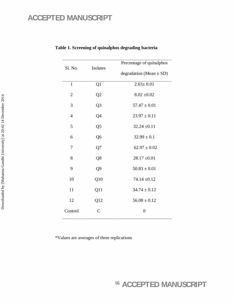
carbon source were isolated from soil. Spectrophotometric analysis of cell free broths indicated
that isolates Q3, Q7 and Q10 were capable of degrading quinalphos to 57.47 %, 65.97 % and
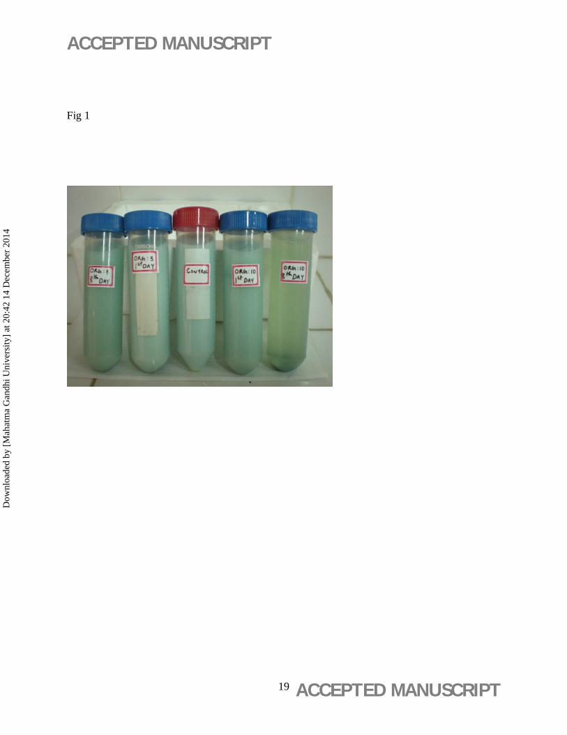
74.14 % respectively (table 1). A visible decrease in colour intensity of pesticide in the medium
was observed on the microbial growth as shown in fig 1. No decrease in pesticide concentration
was observed in uninoculated control flasks, thus proving the microbial role in pesticide
reduction.
The selected bacterial isolates Q3, Q7, Q10 were gram negative, aerobic, motile, nonsporing and
catalase positive rods. Q3 and Q10 showed fluorescence in King’s B medium. On the basis of
cultural and biochemical characteristics, the isolates Q3, Q7 and Q10 were identified as
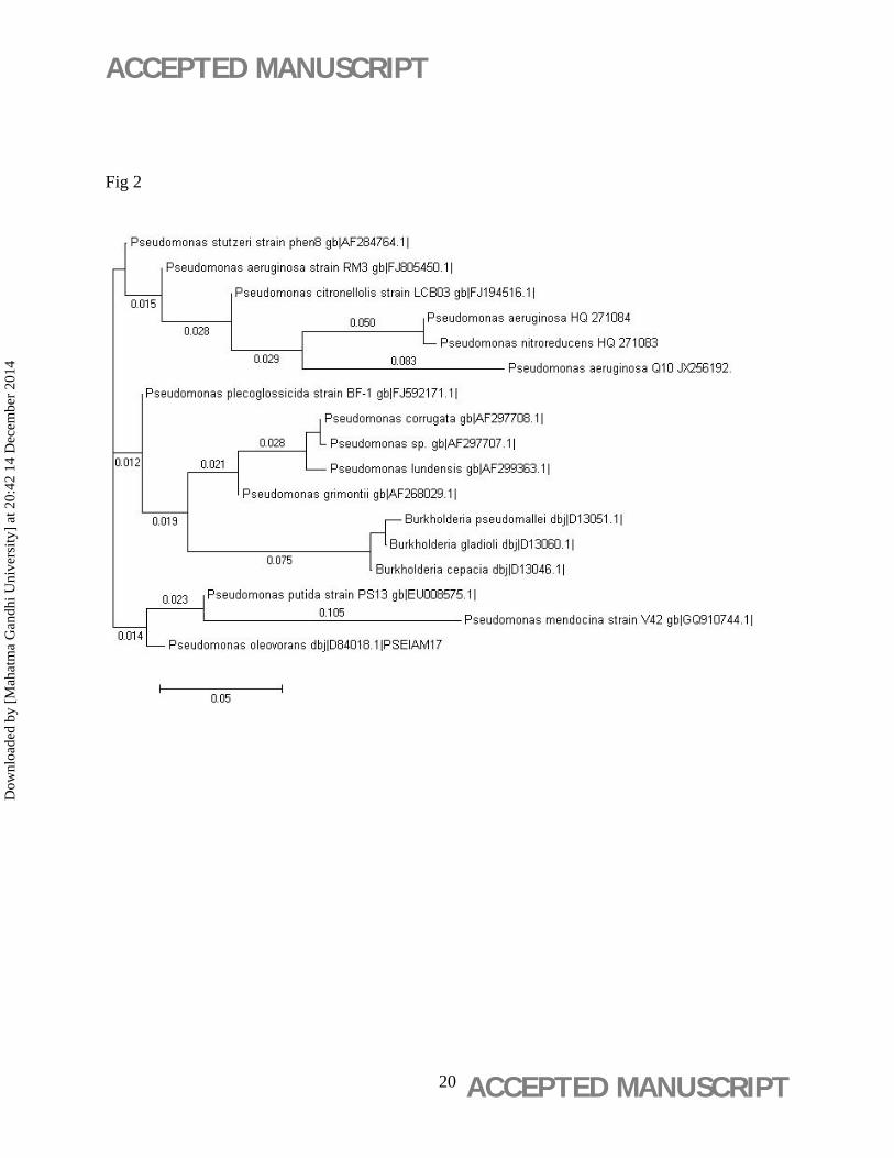
Pseudomonas aeruginosa, Serratia sp. and Pseudomonas sp. respectively. The 16S rDNA
sequence of the isolate Q10 was deposited in the NCBI gene bank under accession number
JX256192 and it showed 99% similarity to Pseudomonas aeruginosa on NCBI BLAST. The
Dow
nloa
ded
by [
Mah
atm
a G
andh
i Uni
vers
ity]
at 2
0:42
14
Dec
embe
r 20
14
ACCEPTED MANUSCRIPT
ACCEPTED MANUSCRIPT 9
nearest neighbors of the isolate Pseudomonas aeruginosa (Q10) were P.aeruginosa
(HQ271084) and P.nitroreducens (HQ271083) (fig 2).
Previous research reveals that microorganisms produce various hydrolases to degrade pesticides
and thus they can be effectively used to overcome pesticide associated pollution problems
(Bhadbhade et al., 2002). Pseudomonas sp. has been reported as a good candidate with
versatile bioremediatory potential capable of degrading wide array of xenobiotics such as
hydrocarbons, surfactants, phenols etc. The role of Pseudomonas sp. in the degradation of
malathion, methamidophos, cartap and cypermethrin (Jilani and Altaf Khan, 2004), difocol
(Sarkar et al., 2009) and 4-chloro benzoic acid were reported. This paper presents another
example of bioremediatory potential of Pseudomonas in quinalphos remediation.
Optimization of Quinalphos degradation & Microbial growth
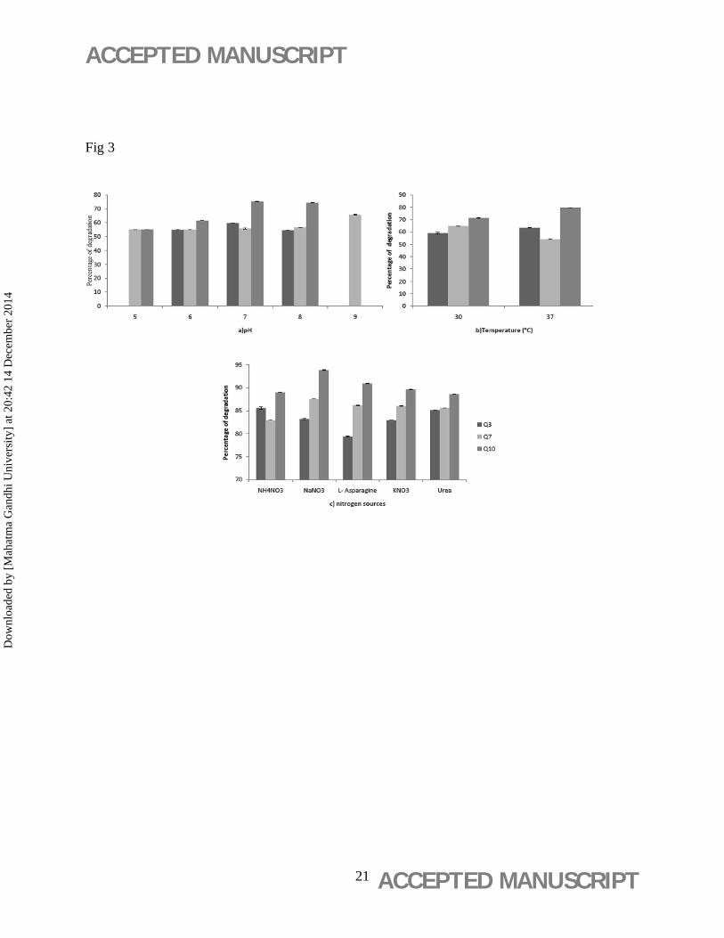
The rate of quinalphos degradation varied at different pH, temperature and nitrogen sources as
depicted in fig 3. During this study it was observed that a pH between 7 and 8 favored pesticide
degradation, which was similar to previous results (Devi S.G., 2006). This indicated that
quinalphos is biodegraded by an alkaline catalyzed hydrolysis reaction. The isolates Q3 and Q10
showed a greater growth and pesticide degradation at 37°C whereas Q7 showed better
degradation at 30°C. The nitrogen source of the medium greatly influenced the bacterial
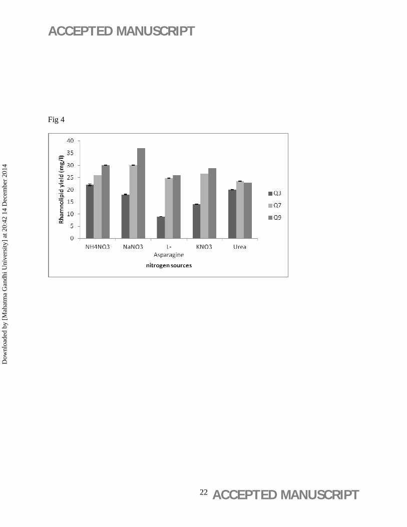
degradation of quinalphos. The supplementation of sodium nitrate instead of ammonium nitrate
resulted in 10- 20 % increase in rate of pesticide degradation than pH optimization step in the
case of Q7 & Q10 (fig 3). This increase in bacterial growth and pesticide degradation could be
attributed to the increased biosurfactant production in presence of NaNO3 as nitrogen source (fig
Dow
nloa
ded
by [
Mah
atm
a G
andh
i Uni
vers
ity]
at 2
0:42
14
Dec
embe
r 20
14
ACCEPTED MANUSCRIPT
ACCEPTED MANUSCRIPT 10
4). The influence of nitrogen sources on rhamnolipid production is previously reported (Rebello
et al., 2013).
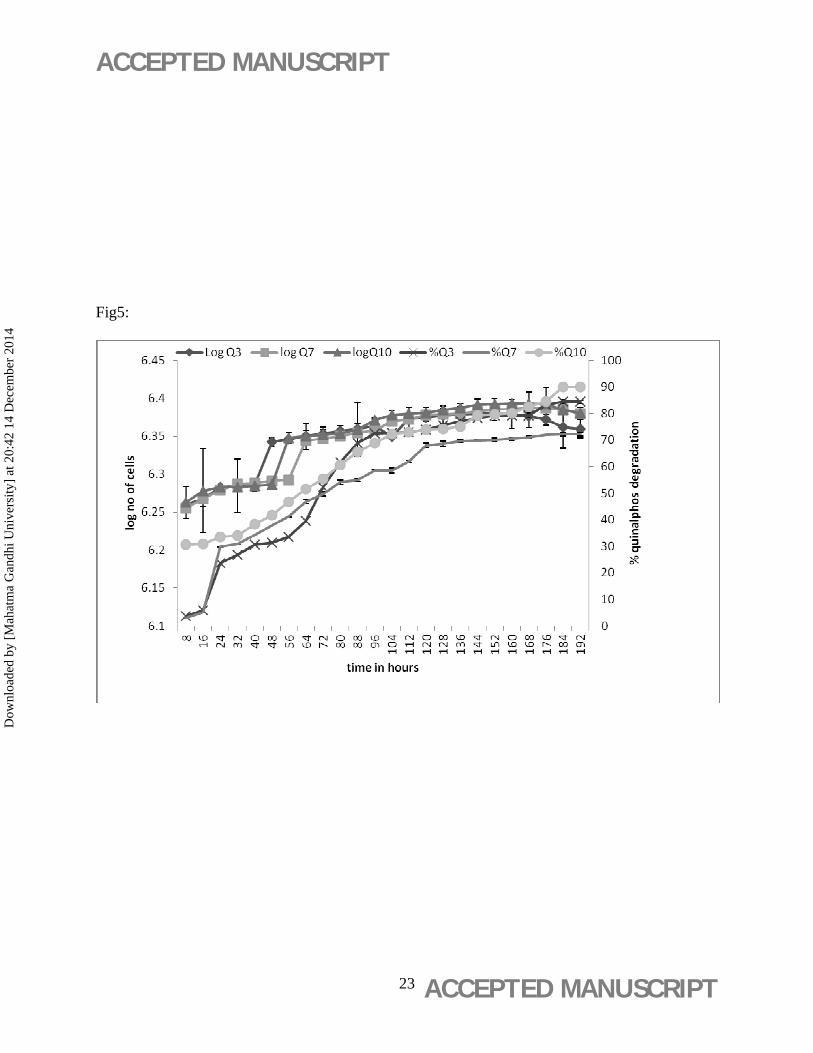
The isolates Q3, Q7 and Q10 were capable of degrading quinalphos to 85.6 %, 87.55 % and
93.87 % respectively after 8 day, under optimal degradation conditions. Extent of quinalphos
degradation and growth profile of the efficient bacterial isolate Q10 in MMB was plotted (fig 5).
Studies on biodegradation of quinalphos are meager compared to reports on its toxicity. Previous
studies reported that 83.71% degradation of 12 mg/l quinalphos was degraded by mixed cultures
of Pseudomonas and Bacillus sp in 10 days (Devi S.G., 2006). Bioremediation of quinalphos by
combined incubation with medicinal plants and Trichoderma leave back only traces of the
pesticide, but the process requires a long incubation time of 60 days (Subashini et al., 2007). The
current paper describes isolation of bacterial isolates capable of degrading of 85-94 % of 2.5 g/l
of quinalphos within 8 days of incubation. A comparison of the above studies reveals that the
strains used in this study were superior to the former in the time extent and quantity of pesticide
it degraded, implying a better optimization of quinalphos degradation. Thus, Q10 could serve as
an ideal candidate for quinalphos degradation capable of degrading even a high concentration of
pesticide in a shorter incubation time.
Degradation studies of Quinalphos
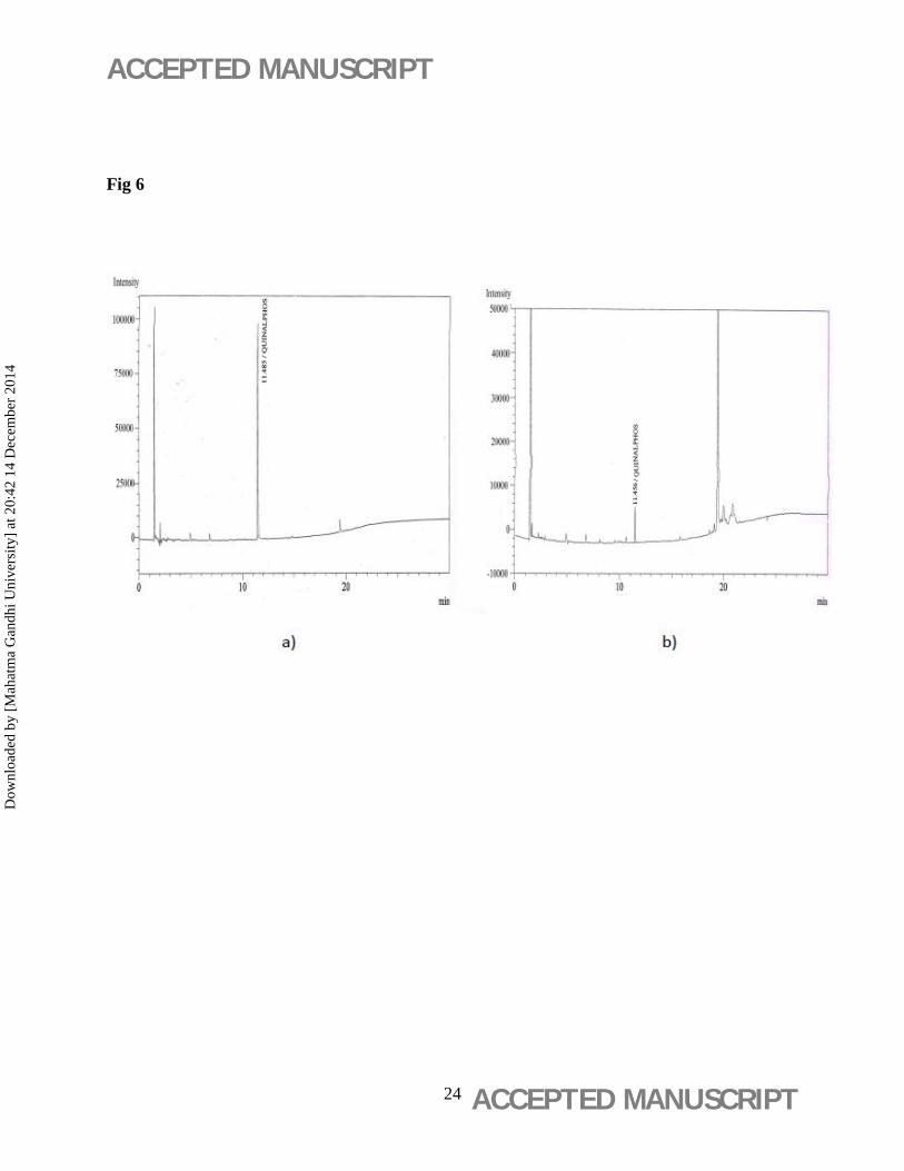
A 92.58% decrease in peak areas of quinalphos was observed in Q10 inoculated flasks on Gas
Chromatographic analysis (fig 6). The gas chromatographic analysis and the optical density
measurements confirmed substantial removal of quinalphos with simultaneous increase in
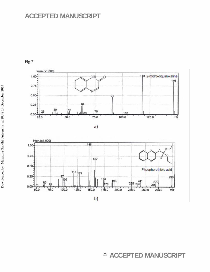
bacterial growth. Two new peaks were formed in the chromatogram of Q10 on 8th day (retention
times 3.90, 9.53), which were due to metabolites formed as a result of degradation (fig 7). The
Dow
nloa
ded
by [
Mah
atm
a G
andh
i Uni
vers
ity]
at 2
0:42
14
Dec
embe
r 20
14
ACCEPTED MANUSCRIPT
ACCEPTED MANUSCRIPT 11
additional peak at 9.53 min gave fraction with a molecular weight of 146 and was identified
as 2-hydroxyquinoxaline (fig. 7a). The peak at 3.9 min gave fraction with a molecular weight
of 298 and was identified as phosphorothioic acid (fig. 7b). Degradation or detoxification of
organophosphorus pesticides by the action of microorganisms is generally through the hydrolysis
of P–O alkyl and P–O aryl bonds. Such degradation makes the compound more vulnerable to
further metabolism (Ortiz-Hernadez and Sanchez-Salinas, 2010).
Biosurfactants in pesticide degradation
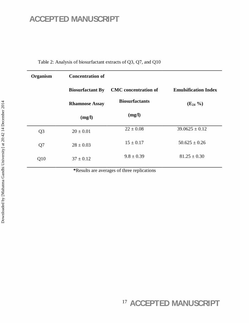
The three bacterial isolates Q3, Q7 & Q10 possessed biosurfactant production potential as per
halo on Siegmund Wagner agar plates. Quantization of rhamnolipids by rhamnose assay
indicated that the isolates produced rhamnolipids in quantities such that Q3< Q7<Q10 (table2).
The CMC values of the purified biosurfactants were found to range between 10- 22 mg/l during
our study (table 2).This agreed with former studies that suggested CMC values of rhamnolipids
could range between 10- 230 mg/l (Nitschke et al., 2005). The increased quinalphos degradation
ability by Q10 than other isolates could be due to its efficiency to produce more amounts of
rhamnolipids.Since the solubility of quinalphos is low in water, it is possible to claim that the
activity of biosurfactants is promising in the degradation of quinalphos. Quinalphos exhibits less
water solubility (20 mg/l) compared to other organophosphates such as monocrotophos (100%
solubility in water), which suggests the chance of its persistence in environment (Kaur et al.,
2013). But in the presence of biosurfactants, the solubility of quinalphos in water is increased.
During our study it was observed that the use of rhamnolipid at concentrations above CMC value
enhanced quinalphos solubility in water at greater rate than at concentrations below its CMC.
Dow
nloa
ded
by [
Mah
atm
a G
andh
i Uni
vers
ity]
at 2
0:42
14
Dec
embe
r 20
14
ACCEPTED MANUSCRIPT
ACCEPTED MANUSCRIPT 12
Previous studies indicate that addition of synthetic surfactants enhanced the solubility and
degradation of the pesticide-endosulfan by 94% (Jayashree and Vasudevan, 2007). The inherent
biosurfactant production ability of the isolates will help to solubilise insoluble compounds,
thereby increasing their bioavailability (Shreve et al., 1995).
Conclusion
Degradation of organophosphorus compounds has attracted considerable attention because of
their widespread use as pesticides and their high mammalian toxicity. The present study has
analyzed the bioremediation of pesticides using bacterial isolates, which seems to be a safe and
effective means of remediation. Considering the use, toxicity and demand of organophosphorous
pesticides in agriculture, studies on biodegradation of these pesticides are of immediate concern.
The studies carried out so far suggests that microorganisms endowed with the ability to degrade
toxic pollutants are a boon to humanity. Microbial degradation process to detoxify pesticides
contaminants can be effectively used to overcome the pollution problems. The effective
utilization of pesticide degrading isolates in immobilized forms in industrial treatment plants
could further increase its prospects in pesticide bioremediation. Construction of recombinant
strains for degradation of organophosphorous pesticides and their metabolites would be a
challenge. The current work successfully led to the isolation of Pseudomonas aeruginosa strain
unique in its ability to degrade large concentration of quinalphos in a shorter incubation time,
which is noteworthy based on its bioremediatory potential.
Acknowledgements
The authors are thankful to College of Agriculture, Vellayani for GC-MS analysis.
Dow
nloa
ded
by [
Mah
atm
a G
andh
i Uni
vers
ity]
at 2
0:42
14
Dec
embe
r 20
14
ACCEPTED MANUSCRIPT
ACCEPTED MANUSCRIPT 13
References
Bhadbhade, B.J., S.S. Sarnaik, and P.P. Kanekar. 2002. Bioremediation of an industrial effluent
containing monocrotophos. 0343-8651. 45:346-349.
Chandrasekaran, E.V., and J.N. Bemiller. 1980. Constituent analysis of glycosaminoglycans, p.
89-96, In R. L. Whistler, Wolfrom,M.L., ed. Methods Carbohydr. Chem, Vol. 8.
Academic Press.New York.
Chiranjeevi, P., T. Balaji, and G.R.K. Naidu. 1997. Spectrophotometric determination of
quinalphos with methylene green. 1018-4619. 6:536-543.
Devi S.G., K.B.P., Deopurkar R.L., Kale S.P. 2006. Isolation and 16S rDNA characterization of
two soil bacteria capable of degrading quinalphos – an organophosphorous insecticide.
The 22nd Annual International Conference on Soils, Sediments and Water.
Gupta, P.K. 2004. Pesticide exposure -Indian scene. 0300-483X. 198:83-90.
Harkins, W.D., A.E. Alexander, and A. Weissberger. 1959. Determination of surface and
interfacial tension., In A. Weissberger, ed. Physical Methods of Organic Chemistry., Vol.
I. Interscience Publishers, Sydney.
Holt, J.G., N.R. Krieg, P.H. Sneath, J.T. Staley, and S.T. Williams. 1994. Bergey's manual of
determinative bacteriology, 9 ed. Williams & Wilkins, Baltimore, London.
Jayashree, R., and N. Vasudevan. 2007. Effect of tween 80 added to the soil on the degradation
of endosulfan by Pseudomonas aeruginosa. International Journal of Environmental
Science & Technology. 4:203-210.
Jena, M., R.C. Dani, and S. Rajamani. 1990. Effectiveness of insecticides against rice gundhi
bug. Oryza. 27:96-98.
Jilani, S., and M. Altaf Khan. 2004. Isolation, characterization and growth response of pesticides
degrading bacteria. J. Biol. Sci. 4:15-20.
Kaur, P., P. Bansal, and D. Sud. 2013. Heterostructured Nanophotocatalysts for Degradation of
Organophosphate Pesticides from Aqueous Streams. Journal of the Korean Chemical
Society. 57.
Kulshrestha, G., and A. Kumari. 2010. Simultaneous degradation of mixed insecticides by mixed
fungal culture isolated from sewage sludge. 0021-8561. 58:11852-11856.
Dow
nloa
ded
by [
Mah
atm
a G
andh
i Uni
vers
ity]
at 2
0:42
14
Dec
embe
r 20
14
ACCEPTED MANUSCRIPT
ACCEPTED MANUSCRIPT 14
Mancini, F., A.H.C. Van Bruggen, J.L.S. Jiggins, A.C. Ambatipudi, and H. Murphy. 2005. Acute
pesticide poisoning among female and male cotton growers in India. Int J Occup Environ
Health. 11:221-232.
Mulchandani, A., I. Kaneva, and W. Chen. 1999. Detoxification of organophosphate nerve
agents by immobilized Escherichia coli with surface - expressed organophosphorus
hydrolase. 0006-3592. 63:216-223.
Nitschke, M., S.G. Costa, and J. Contiero. 2005. Rhamnolipid surfactants: an update on the
general aspects of these remarkable biomolecules. 8756-7938. 21:1593-1600.
Ortiz-Hernadez, M.L., and E. Sanchez-Salinas. 2010. Biodegradation of the organophosphate
pesticide tetrachlorvinphos by bacteria isolated from agricultural soils in Mexico. Rev Int
Contam Ambient. 26:27-38.
Rebello, S., A.K. Asok, S.V. Joseph, B.V. Joseph, L. Jose, S. Mundayoor, and M.S. Jisha. 2013.
Bioconversion of Sodium Dodecyl Sulphate to Rhamnolipid by Pseudomonas
aeruginosa: A Novel and Cost-Effective Production Strategy. Appl. Biochem.
Biotechnol. 169:418-430.
Reddy, P.S., and M.P. Ghewande. 1986. Major insect pests of groundnut and their management.
Pesticides. 20:52-56.
Sarkar, S., A. Satheshkumar, and R. Premkumar. 2009. Biodegradation of Dicofol by
Pseudomonas strains isolated from tea rhizosphere microflora. International Journal of
Integrative Biology. 5:164.
Sharma, B.K. 2007. Environmental Chemistry. 11 ed. Goel Publishing House, Meerut.
Shreve, G.S., S. Inguva, and S. Gunnam. 1995. Rhamnolipid biosurfactant enhancement of
hexadecane biodegradation by Pseudomonas aeruginosa. 1053-6426. 4:331-337.
Siegmund, I., and F. Wagner. 1991. New method for detecting rhamnolipids excreted by
Pseudomonas species during growth on mineral agar. Biotechnol. Tech. 5:265-268.
Subashini, H.D., S. Sekar, and V.R. Devi. 2007. Biodegradation of Pesticidal Residue Using
Traditional Plants with Medicinal Properties and Trichoderma. Research Journal of
Environmental Toxicology. 1:124-130.
Dow
nloa
ded
by [
Mah
atm
a G
andh
i Uni
vers
ity]
at 2
0:42
14
Dec
embe
r 20
14
ACCEPTED MANUSCRIPT
ACCEPTED MANUSCRIPT 15
Weisburg, W.G., S.M. Barns, D.A. Pelletier, and D.J. Lane. 1991. 16S ribosomal DNA
amplification for phylogenetic study. J Bacteriol. 173:697-703.
Whyatt, R.M., and D.B. Barr. 2001. Measurement of organophosphate metabolites in postpartum
meconium as a potential biomarker of prenatal exposure: a validation study. 0091-6765.
109:417.
Wilson, K. 2001. Preparation of Genomic DNA from Bacteria Current Protocols in Molecular
Biology. John Wiley & Sons, Inc.
Yin, H., J. Qiang, Y. Jia, J. Ye, H. Peng, H. Qin, N. Zhang, and B. He. 2009. Characteristics of
biosurfactant produced by Pseudomonas aeruginosa S6 isolated from oil-containing
wastewater. 0032-9592. 44:302-308.
Dow
nloa
ded
by [
Mah
atm
a G
andh
i Uni
vers
ity]
at 2
0:42
14
Dec
embe
r 20
14
ACCEPTED MANUSCRIPT
ACCEPTED MANUSCRIPT 16
Table 1. Screening of quinalphos degrading bacteria
*Values are averages of three replications
Sl. No. Isolates Percentage of quinalphos
degradation (Mean ± SD)
1 Q1 2.63± 0.01
2 Q2 8.02 ±0.02
3 Q3 57.47 ± 0.01
4 Q4 23.97 ± 0.11
5 Q5 32.24 ±0.11
6 Q6 32.99 ± 0.1
7 Q7 62.97 ± 0.02
8 Q8 28.17 ±0.01
9 Q9 50.83 ± 0.01
10 Q10 74.14 ±0.12
11 Q11 34.74 ± 0.12
12 Q12 56.08 ± 0.12
Control C 0 Dow
nloa
ded
by [
Mah
atm
a G
andh
i Uni
vers
ity]
at 2
0:42
14
Dec
embe
r 20
14
ACCEPTED MANUSCRIPT
ACCEPTED MANUSCRIPT 17
Table 2: Analysis of biosurfactant extracts of Q3, Q7, and Q10
Organism Concentration of
Biosurfactant By
Rhamnose Assay
(mg/l)
CMC concentration of
Biosurfactants
(mg/l)
Emulsification Index
(E24 %)
Q3 20 ± 0.01 22 ± 0.08 39.0625 ± 0.12
Q7 28 ± 0.03 15 ± 0.17 50.625 ± 0.26
Q10 37 ± 0.12 9.8 ± 0.39 81.25 ± 0.30
*Results are averages of three replications
Dow
nloa
ded
by [
Mah
atm
a G
andh
i Uni
vers
ity]
at 2
0:42
14
Dec
embe
r 20
14
ACCEPTED MANUSCRIPT
ACCEPTED MANUSCRIPT 18
Fig 1: Comparison of medium colour of Q3 & Q10 during 1st and 8th day of incubation with
respect to control
Fig 2: Phylogenetic tree of the isolates generated using neighbour joining method
Fig 3: Optimisation of quinalphos degradation by Q3, Q7 & Q10 at various temperature, pH and
nitrogen sources (p value < 0.05)
Fig 4: Rhamnolipid yield using different nitrogen sources during quinalphos degradation
Fig 5: Comparison of growth and quinalphos degradation by the isolate Q10
Fig 6: Gas chromatography analysis of the extract of Q10 inoculated mineral medium
containing quinalphos at retention time 11.485 min A) after 20 hours of incubation B) after
8 days of incubation.
Fig 7:Mass spectroscopic analysis of the gas chromatogram of biodegraded quinalphos
residues A) additional peak at 9.53 min showing 2- hydroxyquinoxaline (Molecular formula
C8H6N2O m/Z 146) formation B) additional peak at 3.9 min showing phosphorothioic acid
(Molecular formula C12H15N2O3 m/Z 298) formation.
Dow
nloa
ded
by [
Mah
atm
a G
andh
i Uni
vers
ity]
at 2
0:42
14
Dec
embe
r 20
14
ACCEPTED MANUSCRIPT
ACCEPTED MANUSCRIPT 19
Fig 1
Dow
nloa
ded
by [
Mah
atm
a G
andh
i Uni
vers
ity]
at 2
0:42
14
Dec
embe
r 20
14
ACCEPTED MANUSCRIPT
ACCEPTED MANUSCRIPT 20
Fig 2
Dow
nloa
ded
by [
Mah
atm
a G
andh
i Uni
vers
ity]
at 2
0:42
14
Dec
embe
r 20
14
ACCEPTED MANUSCRIPT
ACCEPTED MANUSCRIPT 21
Fig 3
Dow
nloa
ded
by [
Mah
atm
a G
andh
i Uni
vers
ity]
at 2
0:42
14
Dec
embe
r 20
14
ACCEPTED MANUSCRIPT
ACCEPTED MANUSCRIPT 22
Fig 4
Dow
nloa
ded
by [
Mah
atm
a G
andh
i Uni
vers
ity]
at 2
0:42
14
Dec
embe
r 20
14
ACCEPTED MANUSCRIPT
ACCEPTED MANUSCRIPT 23
Fig5:
Dow
nloa
ded
by [
Mah
atm
a G
andh
i Uni
vers
ity]
at 2
0:42
14
Dec
embe
r 20
14
ACCEPTED MANUSCRIPT
ACCEPTED MANUSCRIPT 24
Fig 6
Dow
nloa
ded
by [
Mah
atm
a G
andh
i Uni
vers
ity]
at 2
0:42
14
Dec
embe
r 20
14
ACCEPTED MANUSCRIPT
ACCEPTED MANUSCRIPT 25
Fig 7
Dow
nloa
ded
by [
Mah
atm
a G
andh
i Uni
vers
ity]
at 2
0:42
14
Dec
embe
r 20
14


