Embed Size (px)
Citation preview

doi:10.1182/blood-2011-09-380006Prepublished online March 23, 2012;2012 119: 4565-4576
Megias, Antonio Rodriguez and Jorge L. Martínez-TorrecuadradaPerez-Martinez, David Olmeda, Marco Marenchino, Marta Cañamero, Sagrario Ortega, Diego María Angeles Abéngozar, Sergio de Frutos, Sergio Ferreiro, Joaquím Soriano, Manuel lymphangiogenesis, and tumor growthBlocking ephrinB2 with highly specific antibodies inhibits angiogenesis,
http://bloodjournal.hematologylibrary.org/content/119/19/4565.full.htmlUpdated information and services can be found at:
http://bloodjournal.hematologylibrary.org/site/misc/rights.xhtml#repub_requestsInformation about reproducing this article in parts or in its entirety may be found online at:
http://bloodjournal.hematologylibrary.org/site/misc/rights.xhtml#reprintsInformation about ordering reprints may be found online at:
http://bloodjournal.hematologylibrary.org/site/subscriptions/index.xhtmlInformation about subscriptions and ASH membership may be found online at:
Copyright 2011 by The American Society of Hematology; all rights reserved.Washington DC 20036.by the American Society of Hematology, 2021 L St, NW, Suite 900, Blood (print ISSN 0006-4971, online ISSN 1528-0020), is published weekly
For personal use only. at Centro Nacional de Investigaciones Oncológicas on May 11, 2012. bloodjournal.hematologylibrary.orgFrom

VASCULAR BIOLOGY
Blocking ephrinB2 with highly specific antibodies inhibits angiogenesis,lymphangiogenesis, and tumor growthMarıa Angeles Abengozar,1 Sergio de Frutos,2 Sergio Ferreiro,3 Joaquím Soriano,4 Manuel Perez-Martinez,4 David Olmeda,5
Marco Marenchino,6 Marta Canamero,7 Sagrario Ortega,5 Diego Megias,4 Antonio Rodriguez,2 andJorge L. Martínez-Torrecuadrada1
1Proteomics Unit, Biotechnology Programme, Centro Nacional de Investigaciones Oncologicas (CNIO), Madrid, Spain; 2Molecular Biology Department, Facultyof Science, Universidad Autonoma de Madrid, Madrid, Spain; 3Animal Facility, CNIO, Madrid, Spain; 4Confocal Microscopy Unit, Biotechnology Programme,CNIO, Madrid, Spain; 5Transgenic Mice Unit, Biotechnology Programme, CNIO, Madrid, Spain; 6Spectroscopy and Nuclear Magnetic Resonance Unit,Structural Biology and Biocomputing Programme, CNIO, Madrid, Spain; and 7Comparative Pathology Unit, Biotechnology Programme, CNIO, Madrid, Spain
Membrane-anchored ephrinB2 and its re-ceptor EphB4 are involved in the forma-tion of blood and lymphatic vessels innormal and pathologic conditions. Eph/ephrin activation requires cell-cell interac-tions and leads to bidirectional signalingpathways in both ligand- and receptor-expressing cells. To investigate the func-tional consequences of blocking eph-rinB2 activity, 2 highly specific humansingle-chain Fv (scFv) Ab fragmentsagainst ephrinB2 were generated andcharacterized. Both Ab fragments sup-
pressed endothelial cell migration andtube formation in vitro in response toVEGF and provoked abnormal cell motil-ity and actin cytoskeleton alterations inisolated endothelial cells. As only one ofthem (B11) competed for binding of eph-rinB2 to EphB4, these data suggest anEphB-receptor–independent blockingmechanism. Anti-ephrinB2 therapy re-duced VEGF-induced neovascularizationin a mouse Matrigel plug assay. More-over, systemic administration of ephrinB2-blocking Abs caused a drastic reduction
in the number of blood and lymphaticvessels in xenografted mice and a con-comitant reduction in tumor growth. Ourresults show for the first time that spe-cific Ab-based ephrinB2 targeting mayrepresent an effective therapeutic strat-egy to be used as an alternative or incombination with existing antiangiogenicdrugs for treating patients with cancerand other angiogenesis-related diseases.(Blood. 2012;119(19):4565-4576)
Introduction
Angiogenesis and lymphangiogenesis are highly complex coordi-nated processes through which new blood and lymphatic vessels,respectively, arise from preexisting ones.1,2 Angiogenesis occursphysiologically during embryonic development, tissue repair (ie,wound healing), and menstruation, but it is also important in thepathogenesis of many diseases such as tumor growth and metasta-sis.3 Because most tumors cannot grow and disseminate in theabsence of new blood vessel formation, inhibition of the signalingpathways underlying pathologic angiogenesis is an importantpotential target for anticancer therapy.4 The process of lymphangio-genesis is less well understood, however, the mechanisms regulat-ing development and growth of lymphatic vessels have recentlyemerged as a very interesting field in cancer research, as theyrepresent an important venue for metastatic dissemination.5
The well-established role of VEGF in promoting tumor angio-genesis has led to the development of therapeutic agents, includingengineered mAbs and small molecule inhibitors such as bevaci-zumab (Avastin), sunitinib, and sorafenib6-8 which selectivelytarget VEGF/VEGF receptor pathways and have demonstratedpromising clinical utility in treating solid neoplasms.9 However,these VEGF-based treatments may result in only transient clinicalbenefits and tumors eventually develop a resistance to therapy andmay exhibit a progression to greater malignancy.10 Therefore, thereis an urgent need to develop new agents targeting other pathways
involved in angiogenesis that could be translated into more robusttherapeutic outcomes to replace or complement existing therapies.A group of signaling molecules, the ephrins and their receptors(Eph), has recently emerged as attractive therapeutic targetsbecause their pathways play critical roles in the development andmaturation of the blood and lymphatic vascular systems.11,12
Eph receptor tyrosine kinases and their ligands, ephrins, controlseveral cellular functions such as cell migration and cytoskeletalorganization.13,14 Ephrins are a family of cell-surface proteinslinked to the cell membrane either by a GPI anchor (class A) or by asingle transmembrane segment (class B). They act as activatingligands for the Eph receptors, which are divided into 2 classesdepending on the ephrins with which they interact, although limitedcrosstalk between both classes has also been described.15 Anintriguing feature of Eph-ephrin signaling is that both, receptorsand ligands, are able to transduce a downstream signaling cascadeon interaction, resulting in bidirectional cell-to-cell communica-tion. Eph-activated signaling is termed “forward” and ephrin-activated signaling is named “reverse.” Eph receptors initiateforward signal transduction by autophosphorylation of severaltyrosine residues located in the cytoplasmic part of the molecule inresponse to binding of clustered and membrane-attached ephrinligands on adjacent cells. The reverse signaling activity is mediatedby the C-terminal region of ephrins, which contains conserved
Submitted September 15, 2011; accepted March 11, 2012. Prepublished online asBlood First Edition paper, March 23, 2012; DOI 10.1182/blood-2011-09-380006.
The online version of this article contains a data supplement.
The publication costs of this article were defrayed in part by page chargepayment. Therefore, and solely to indicate this fact, this article is herebymarked ‘‘advertisement’’ in accordance with 18 USC section 1734.
© 2012 by The American Society of Hematology
4565BLOOD, 10 MAY 2012 � VOLUME 119, NUMBER 19
For personal use only. at Centro Nacional de Investigaciones Oncológicas on May 11, 2012. bloodjournal.hematologylibrary.orgFrom

tyrosines susceptible to phosphorylation16 and a PDZ-bindingdomain, which can recruit PDZ domain-containing proteins onphosphorylation.17 In addition, recent evidence has highlighted theimportance of processes such as endocytosis of Eph/ephrin mol-ecules coupled to the internalization of cell type-specific moleculeswith specialized functional roles such as AMPA receptors inneurons18 or VEGF receptors in endothelial cells.19,20
In the blood vasculature, ephrinB2 is expressed on arterialangioblasts, endothelial cells (ECs) and perivascular mesenchymalcells, whereas one of its binding partners, the receptor EphB4, isspecific of venous ECs.21 Targeted inactivation of either EphB4 orEfnB2 genes in mice leads to failure of embryonic vessel formationand early lethality, demonstrating their critical role during physi-ologic angiogenesis.22,23 This role of ephrinB2 also extends totumor-derived angiogenesis and tumor growth.19,24 Moreover,ephrinB2 controls lymphangiogenesis20 and it has been recentlyshown that an ephrinB2 blocking peptide was able to suppressVEGF-induced lymphatic endothelial sprouting.25 The develop-ment and remodeling of the lymphatic vasculature have beenreported to be regulated through interactions mediated by itsPDZ-binding domain12; this domain is also involved in theregulation of endothelial cell motility.26
Because the EphB4/ephrinB2 signaling is important in angiogen-esis, hampering this protein-protein interaction could have severalpotential medical applications in angiogenesis-based diseases suchas in tumor growth and metastasis. In fact, several groups havedemonstrated that the monomeric soluble extracellular domain ofEphB4 is able to block ephrinB2 interaction with its receptor,leading to inhibition of angiogenesis and tumor growth.27,28 Here,we bring for the first time the proof of concept of the antiangiogenicefficacy of blocking ephrinB2 with highly specific human Abs invitro and in vivo, showing a potent antitumor activity in differenthuman cancer xenografts models in mice, which might havepotential therapeutic applications.
Methods
Selection, production, and purification of anti-ephrinB2scFv Abs
All experimental procedures were performed in accordance with institu-tional animal welfare guidelines and approved by the Research Ethics andAnimal Welfare Committee of the Instituto de Salud Carlos III. Humanphage Ab libraries were used for panning as previously described.29 Therecombinant extracellular region of human ephrinB2 fused to Fc domain ofhuman IgG was used as the Ag source. This chimera was overexpressed bytransient transfection of HEK293-EBNA cells using linear polyethyleni-mine (PEI; Polysciences) as the transfection reagent and purified fromculture medium by affinity chromatography using protein A columns (GEHealthcare) on an AKTA system (GE Healthcare). Unique and specificsoluble scFvs were identified by ELISA and DNA sequencing.
The expression and purification of His- and c-myc–tagged scFvs fromselected bacterial clones were carried out as previously described.29
Endotoxin was removed from purified scFvs, using polymixin B-basedDetoxi-Gel Endotoxin Removing Gel (Thermo Scientific) according to themanufacturer’s instructions. The eluted fractions were assayed for endo-toxin content by using the Limulus Amebocyte Lysate (LAL) QCL-1000 kit(Lonza).
Endothelial cell tube formation assay
Ninety-six–well plates were coated with 50 �L/well Matrigel (BD Biosci-ences) which was allowed to solidify at 37°C for 30 minutes, according tothe manufacturer’s instructions. HUVECs were detached with 100mM
EDTA, washed twice in PBS, and resuspended in medium supplementedwith 10% FBS. The cell suspensions were preincubated with 100 �g/mLspecific scFvs or controls for 15 minutes and plated onto the surface of thepolymerized Matrigel (1.5 � 104 cells/well). After 6-hour incubation at37°C, 5% CO2, triplicate pictures were taken for each well using an invertedphase contrast microscope (Nikon). The number and length of the cord-likestructures were measured in randomly selected fields.
Cell migration assays
Cell invasion was tested in transwell Boyden chambers with a gelatin-coated polycarbonated filter (8-�m pore size, 0.1% w/v gelatin; CorningLife Sciences). A total of 1 � 105 cells were plated into the upper (pre-filter)well compartment containing 100 �L of complete medium supplementedwith 0.5% FBS, in the absence or presence of scFv (100 �g/mL � 3�M)and/or EphB4-Fc (2.5 �g/mL; R&D Systems). Lower (postfilter) wellcompartment were filled with complete medium supplemented with 10%FBS as a cell attractant. After a 48-hour incubation period at 37°C, 5% CO2,media were removed and cells were fixed with 4% paraformaldehyde (PFA)in PBS for 10 minutes at room temperature, and were then stained with1 mg/mL DAPI (4�-6-diamino-2-phenylindole) for 10 minutes. Finally, cellinvasion was analyzed by DAPI-positive nucleus counting at the top and thebottom of the chamber’s filter using a Leica TCS SP5 confocal microscopeequipped with a resonant scanner, a dry 20� Plan Apochromatic, 0.7 ANobjective, and Leica LAS AF (Version 2.4.1) for image capturing, and theImaris software (Bitplane) for image analysis.
Cell migration in scratch assays was also tested. Cells were grown toconfluence in 24-well plates. After 1-hour incubation with starving media(0.5% FBS), culture dishes were scratched with a pipette tip and cells werethen incubated in the absence or presence of 100 �g/mL of the correspond-ing scFv using 100 ng/mL VEGF (PeproTech) as proangiogenic stimulus.The rate of wound closure was calculated by capturing images over a24-hour period using a Leica AF6000 Workstation equipped with a dry 10�Plan Apochromatic, 0.4 AN objective, a Hamamatsu ORCA-AG CCDcamera and Leica LAS AF software (Version 2.4.1). Lateral migration wasmeasured as percentage of migrated area from time 0 to 24 hours using theLeica LAS AF software.
Random motility assays
Enhanced GFP (EGFP)–transfected HUVECs were treated with 100 �g/mLof the corresponding scFv or left untreated as control in complete mediumfor 24 hours at 37°C, 5% CO2. After Ab addition, the recording was startedby acquiring fluorescence images every 10 minutes using a Leica AF6000Workstation equipped with a dry 10� Plan Apochromatic, 0.4 ANobjective, a Hamamatsu ORCA-AG CCD camera, and Leica LAS AFsoftware (Version 2.4.1). Cell tracking analysis and statistics were per-formed with Imaris software (Bitplane).
Murine Matrigel plug angiogenesis assay
To test whether ephrinB2-specific scFvs were able to target neovasculature,growth factor–depleted Matrigel (BD Biosciences) in liquid form at 4°Cwas supplemented with heparin (6.6 �g/mL) and VEGF (250 ng/mL) andwas injected subcutaneously into the thoracic area of anesthetized 6-week-old female nu/nu mice. EphrinB2-specific scFvs and an irrelevant scFvwere labeled with Alexa Fluor 750 by using the SAIVI Rapid Antibody Kit(Invitrogen Life Technologies) following the manufacturer’s instructions.On day 5, when vasculature was macroscopically observed, mice wereinjected IV with 100 �g of each Alexa 750–labeled scFv. Eight hours afterinjection, animals were killed, plugs were excised, and briefly stored at 4°Cin PBS with 5% FBS before imaging acquisition. Fluorescence signal onplugs was acquired using an IVIS-200 small animal imaging system(Caliper Life Sciences).
To study the antiangiogenic capability of ephrinB2-specific scFvs,Matrigel plugs were implanted subcutaneously in nude mice as described inthis section, and groups of 6 animals each were treated intravenously with atotal dose of 15 mg/kg of specific and irrelevant scFvs distributed in3 injections over a period of 10 days. Groups of mice were also inoculated
4566 ABENGOZAR et al BLOOD, 10 MAY 2012 � VOLUME 119, NUMBER 19 For personal use only. at Centro Nacional de Investigaciones Oncológicas on May 11, 2012. bloodjournal.hematologylibrary.orgFrom

with Matrigel plugs supplemented with either vehicle (PBS) or VEGF(250 ng/mL) and left untreated as negative and positive controls, respec-tively. Then, Matrigel plugs were excised and divided into 2 parts: one wasprocessed for IHC and the other part was used to determine hemoglobinconcentration. For immunohistochemical analyses, plug pieces were fixedin 10% buffered formalin (Sigma-Aldrich), and embedded in paraffin.Identification of the endothelial cell population was carried out by CD34staining with a specific rabbit mAb (Abcam) and counterstained withhematoxylin. Slides were scanned and images were captured and countedusing the Carl Zeiss Mirax Scan and Axiovision 4.8 image processingsystem (Carl Zeiss MicroImaging). The other Matrigel blocks werehomogenized in distilled water, spun at 13 000g, and supernatants werecollected for hemoglobin measurement. Each supernatant was diluted 1:1with TMB (3,3�,5,5�-tetramethylbenzidine; Sigma-Aldrich) and opticaldensities at 620 nm were measured in a microplate reader (Bio-Rad). Valueswere normalized to the plug weight.
Tumor xenografts
Log-phase BxPC3 carcinoma pancreatic cells (1 � 106) in PBS wereimplanted subcutaneously into the flank of SCID mice on day 0. Tumorswere measured 3 times a week using a caliper, and tumor volume wascalculated using the formula: V � length � (width)2 � 0.5. When themean tumor volume reached � 30 mm3, mice were randomized into groupsof 10 and were treated with IV injections of B11 or 2B1 diluted in 0.2 mL ofPBS once every other day until reaching a total dose of 20 mg/kg. Controlanimals were given vehicle (PBS) alone. Tumor sizes were monitored atleast twice a week. SW620 colon carcinoma (5 � 106) or H460 lungcarcinoma (5 � 106) cells stably expressing mCherry were subcutaneouslyinjected in the back of female nu/nu mice (n � 4) on day 0. Treatment withB11 or 2B1 was initiated at day 4 following the schedule described in thissection. Tumor size was followed by fluorescence imaging at 610 nm twiceper week using an IVIS-200 small animal imaging system (Caliper Life
Figure 1. Characterization of selected ephrinB2-specific scFvs. (A) Specificity of scFvs for human ephrinB2-Fc (EB2) but not for other related members of the proteinfamily, mouse ephrinB1-Fc (EB1) and human ephrinB3-Fc (EB3) by Western blot. Commercial anti–mouse ephrinB2 polyclonal Ab was used as control. (B) Flow cytometricanalysis showing the specificity of B11 and 2B1 to ephrinB2 in native form. HEK293T cells overexpressing human ephrinB1 (left panel), ephrinB2 (middle panel), or ephrinB3(right panel) were subjected to flow cytometry for detection of B11 and 2B1 binding. As positive controls, specific antisera of each member of the ephrinB family (�eB1, �eB2,and �eB3) were used (lower histograms of each panel). Incubations omitting the specific Abs were used as negative controls. (C) SPR sensorgrams of ephrinB2-specificscFvs, B11 on the left panel and 2B1 on the right, binding to immobilized ephrinB2-Fc are shown. The sensograms were corrected for response differences between the activeand reference flow cells. EphrinB2-Fc was immobilized on CM5 chips and scFvs were passed over at concentration ranges noted on each sensorgram, giving affinity constants(KD) of 110nM for B11 and 630nM for 2B1. (D) Inhibition assays using SPR for detection of bound ephrinB2. Serial dilutions of scFv B11 (F) or 2B1 (E) were mixed with 0.2�MephrinB2-Fc and injected over an immobilized sEphB4 on a CM5 chip. The relative amount of ephrinB2 binding to sEphB4 was measured immediately after injection of eachsample and plotted as a function of scFv concentration. Mean values are shown with error bars indicating the SD (n � 3). (E) Activation of EphB4 receptor by tyrosinephosphorylation in HUVECs as a response to interaction with membrane-bound ephrinB2 in the absence or presence of ephrinB2-specific scFv in a cell-based assay.HEK293T cells overexpressing ephrinB2 were overlaid on HUVE cells with or without the corresponding scFv. Total (bottom panel) and phosphorylated EphB4s (top panel)after immunoprecipitation (IP) by Western blot (WB) with the respective Abs are shown. (F) Analysis of EphB4-induced ephrinB2 tyrosine phosphorylation in the absence orpresence of B11 and 2B1. HEK293T cells transfected with c-myc–tagged ephrinB2 were treated as indicated and the corresponding cell extracts were immunoprecipitated withanti-c-myc Ab. Total (bottom panel) and phosphorylated (top panel) ephrinB2 were detected by Western blot. The graph on the right showed average levels oftyrosine-phosphorylated ephrinB2, quantified from 3 different immunoblotting experiments. Graph shows normalized results (means � SD).
Ab-BASED BLOCKING OF ephrinB2 4567BLOOD, 10 MAY 2012 � VOLUME 119, NUMBER 19 For personal use only. at Centro Nacional de Investigaciones Oncológicas on May 11, 2012. bloodjournal.hematologylibrary.orgFrom
Sciences) and photons emitted (represented as photons per second persquare centimeter per steradian) from tumoral masses were quantified usingLiving Image software (Caliper Life Sciences).
More information on the methods and materials can be found insupplemental Methods (available on the Blood Web site; see the Supplemen-tal Materials link at the top of the online article).
Results
Identification and characterization of human Abs againstephrinB2
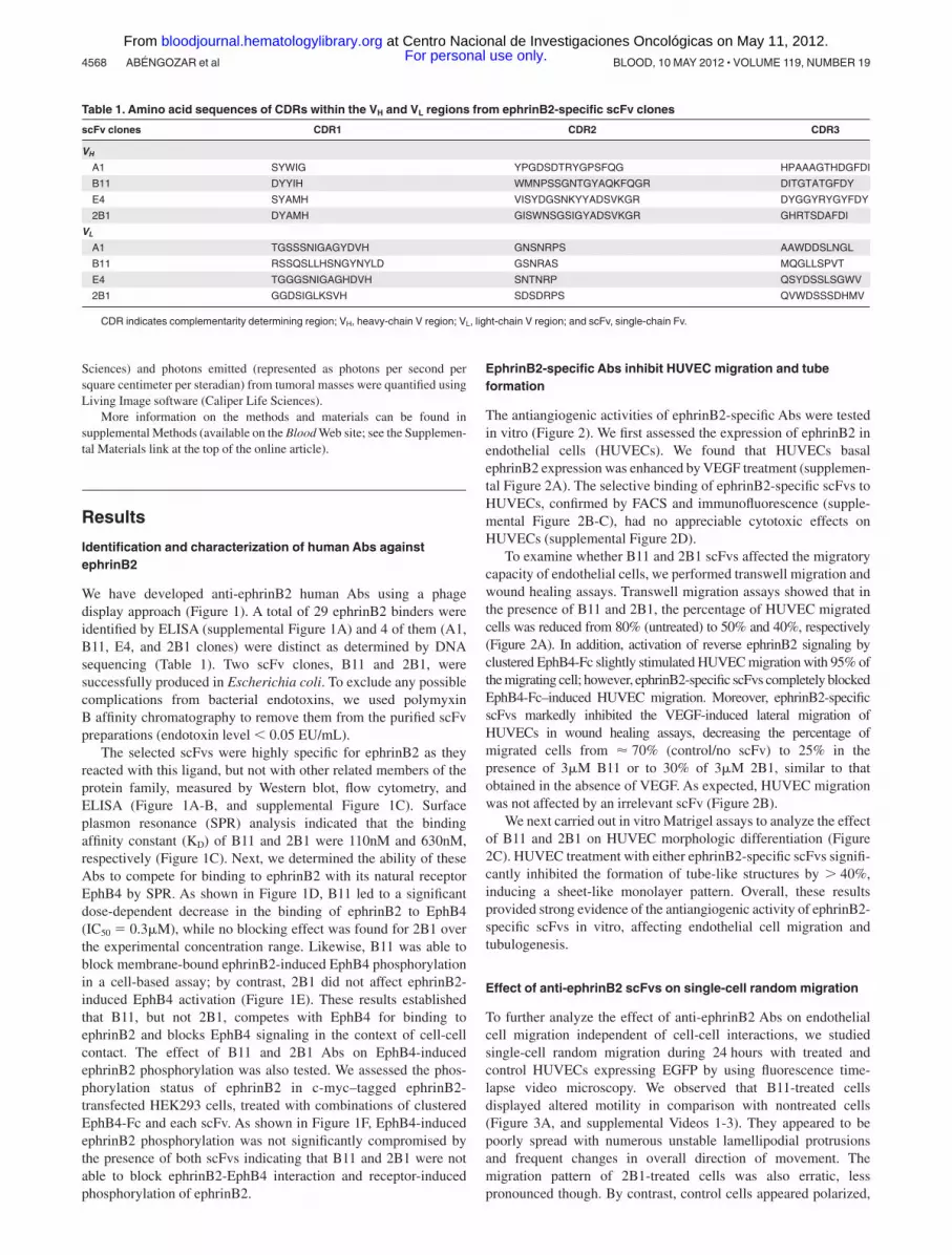
We have developed anti-ephrinB2 human Abs using a phagedisplay approach (Figure 1). A total of 29 ephrinB2 binders wereidentified by ELISA (supplemental Figure 1A) and 4 of them (A1,B11, E4, and 2B1 clones) were distinct as determined by DNAsequencing (Table 1). Two scFv clones, B11 and 2B1, weresuccessfully produced in Escherichia coli. To exclude any possiblecomplications from bacterial endotoxins, we used polymyxinB affinity chromatography to remove them from the purified scFvpreparations (endotoxin level � 0.05 EU/mL).
The selected scFvs were highly specific for ephrinB2 as theyreacted with this ligand, but not with other related members of theprotein family, measured by Western blot, flow cytometry, andELISA (Figure 1A-B, and supplemental Figure 1C). Surfaceplasmon resonance (SPR) analysis indicated that the bindingaffinity constant (KD) of B11 and 2B1 were 110nM and 630nM,respectively (Figure 1C). Next, we determined the ability of theseAbs to compete for binding to ephrinB2 with its natural receptorEphB4 by SPR. As shown in Figure 1D, B11 led to a significantdose-dependent decrease in the binding of ephrinB2 to EphB4(IC50 � 0.3�M), while no blocking effect was found for 2B1 overthe experimental concentration range. Likewise, B11 was able toblock membrane-bound ephrinB2-induced EphB4 phosphorylationin a cell-based assay; by contrast, 2B1 did not affect ephrinB2-induced EphB4 activation (Figure 1E). These results establishedthat B11, but not 2B1, competes with EphB4 for binding toephrinB2 and blocks EphB4 signaling in the context of cell-cellcontact. The effect of B11 and 2B1 Abs on EphB4-inducedephrinB2 phosphorylation was also tested. We assessed the phos-phorylation status of ephrinB2 in c-myc–tagged ephrinB2-transfected HEK293 cells, treated with combinations of clusteredEphB4-Fc and each scFv. As shown in Figure 1F, EphB4-inducedephrinB2 phosphorylation was not significantly compromised bythe presence of both scFvs indicating that B11 and 2B1 were notable to block ephrinB2-EphB4 interaction and receptor-inducedphosphorylation of ephrinB2.
EphrinB2-specific Abs inhibit HUVEC migration and tubeformation
The antiangiogenic activities of ephrinB2-specific Abs were testedin vitro (Figure 2). We first assessed the expression of ephrinB2 inendothelial cells (HUVECs). We found that HUVECs basalephrinB2 expression was enhanced by VEGF treatment (supplemen-tal Figure 2A). The selective binding of ephrinB2-specific scFvs toHUVECs, confirmed by FACS and immunofluorescence (supple-mental Figure 2B-C), had no appreciable cytotoxic effects onHUVECs (supplemental Figure 2D).
To examine whether B11 and 2B1 scFvs affected the migratorycapacity of endothelial cells, we performed transwell migration andwound healing assays. Transwell migration assays showed that inthe presence of B11 and 2B1, the percentage of HUVEC migratedcells was reduced from 80% (untreated) to 50% and 40%, respectively(Figure 2A). In addition, activation of reverse ephrinB2 signaling byclustered EphB4-Fc slightly stimulated HUVEC migration with 95% ofthe migrating cell; however, ephrinB2-specific scFvs completely blockedEphB4-Fc–induced HUVEC migration. Moreover, ephrinB2-specificscFvs markedly inhibited the VEGF-induced lateral migration ofHUVECs in wound healing assays, decreasing the percentage ofmigrated cells from � 70% (control/no scFv) to 25% in thepresence of 3�M B11 or to 30% of 3�M 2B1, similar to thatobtained in the absence of VEGF. As expected, HUVEC migrationwas not affected by an irrelevant scFv (Figure 2B).
We next carried out in vitro Matrigel assays to analyze the effectof B11 and 2B1 on HUVEC morphologic differentiation (Figure2C). HUVEC treatment with either ephrinB2-specific scFvs signifi-cantly inhibited the formation of tube-like structures by 40%,inducing a sheet-like monolayer pattern. Overall, these resultsprovided strong evidence of the antiangiogenic activity of ephrinB2-specific scFvs in vitro, affecting endothelial cell migration andtubulogenesis.
Effect of anti-ephrinB2 scFvs on single-cell random migration
To further analyze the effect of anti-ephrinB2 Abs on endothelialcell migration independent of cell-cell interactions, we studiedsingle-cell random migration during 24 hours with treated andcontrol HUVECs expressing EGFP by using fluorescence time-lapse video microscopy. We observed that B11-treated cellsdisplayed altered motility in comparison with nontreated cells(Figure 3A, and supplemental Videos 1-3). They appeared to bepoorly spread with numerous unstable lamellipodial protrusionsand frequent changes in overall direction of movement. Themigration pattern of 2B1-treated cells was also erratic, lesspronounced though. By contrast, control cells appeared polarized,
Table 1. Amino acid sequences of CDRs within the VH and VL regions from ephrinB2-specific scFv clones
scFv clones CDR1 CDR2 CDR3
VH
A1 SYWIG YPGDSDTRYGPSFQG HPAAAGTHDGFDI
B11 DYYIH WMNPSSGNTGYAQKFQGR DITGTATGFDY
E4 SYAMH VISYDGSNKYYADSVKGR DYGGYRYGYFDY
2B1 DYAMH GISWNSGSIGYADSVKGR GHRTSDAFDI
VL
A1 TGSSSNIGAGYDVH GNSNRPS AAWDDSLNGL
B11 RSSQSLLHSNGYNYLD GSNRAS MQGLLSPVT
E4 TGGGSNIGAGHDVH SNTNRP QSYDSSLSGWV
2B1 GGDSIGLKSVH SDSDRPS QVWDSSSDHMV
CDR indicates complementarity determining region; VH, heavy-chain V region; VL, light-chain V region; and scFv, single-chain Fv.
4568 ABENGOZAR et al BLOOD, 10 MAY 2012 � VOLUME 119, NUMBER 19 For personal use only. at Centro Nacional de Investigaciones Oncológicas on May 11, 2012. bloodjournal.hematologylibrary.orgFrom

exhibited more elongated cell shape and frequently continuedmigrating in the same direction without turning. Analysis of cellmigration showed that B11-treated cells displayed a significantlyreduced migration speed (Figure 3B) and they traveled a shorteraccumulated distance (Figure 3C), with a reduction in the index ofstraightness defined as the ratio of the displacement of a cell to theaccumulated distance (Figure 3D). 2B1-treated cells showed asmaller straightness index compared with control cells despite todisplay similar speed and migration length, indicating that only thecell directionality was affected.
Treatment with anti-ephrinB2 Abs leads to an increase instress fibers
To assess whether these motility changes were coupled with cellmorphology differences, we quantified cell shape changes bymeasuring the actin cytoskeleton (Figure 4). Anti-ephrinB2–treatedcells adopted a more round cell-shape morphology, whereas controlcells exhibited a more elongated phenotype (Figure 4A). Quantifi-cation of cell areas showed that the size of the cells treated with
ephrinB2-specific scFvs was significantly reduced by 40% com-pared with control cells (Figure 4B). In addition, densitometryanalysis of polymerized F-actin by fluorescence intensity showedthat treatment with B11 increased significantly the number andthickness of cytoplasmic actin-containing stress fiber (Figure 4C)compared with the untreated cells, indicating that actin cytoskel-eton dynamic was altered. 2B1 also induced actin stress fibers,albeit to a lesser extent than B11.
Impairment of VEGF-induced Matrigel angiogenesis in vivo byanti-ephrinB2 scFvs
The antiangiogenic properties of anti-ephrinB2 Abs in vivo wereanalyzed by Matrigel plug assays in athymic nude mice. First, wetested the ability of the anti-ephrinB2 scFvs to properly targetneovascularization developed within the VEGF-supplementedMatrigel (Figure 5A). Strong fluorescent signals were detected inthe Matrigel plugs from animals treated with Alexa 750–labeledB11, whereas mice injected with the 2B1–Alexa 750 conjugateshowed only moderate fluorescence intensities, indicating that
Figure 2. Characterization of antiangiogenic capability of ephrinB2-specific scFvs. (A) HUVEC migration (Transwell) assays in the absence or presence of B11 or 2B1and/or clustered EphB4-Fc are shown. FBS in the bottom chamber was used as migration stimuli. Cells crossing the Transwell membrane are in green, whereas the blue signalindicates cells that remain at the upper compartment. Results are expressed as the percentage � SD of migrated cells relative to total cells. *P � .001 and **P � .01 asdetermined by an unpaired Student t test. (B) Analysis of lateral migration of HUVECs by wound healing assays in vitro. HUVECs were grown to confluence when scratcheswere done. Cells were incubated with starving media in the absence (Control) or presence of 100 ng/mL VEGF as migrating stimulus. Cell migration was monitored over24 hours with 100 �g/mL ephrinB2-specific scFvs (B11 or 2B1) or an irrelevant scFv as indicated. Representative still photographs (4� magnification) taken 24 hours afterscratching are shown (original wound areas at time 0 are indicated by dotted lines). Quantification of lateral migration after 24 hours is shown in the bottom right panel. EachscFv was assayed at least 3 times and the corresponding values (means � SD) were represented as the percentage of migrated area from time 0. *P � .01 versus control.(C) Tubular formation assay. HUVECs were cultured on standard Matrigel in VEGF-stimulated conditions in the absence (control) or presence of anti-ephrinB2 (B11 or 2B1)scFvs or an irrelevant scFv. Representative microphotographs of tube formation after 6 hours of culture (4� magnification) are shown. Graph shows quantitative measure oftube formation. Each treatment was assayed at least 3 times, and the corresponding values (means � SE) were plotted as percentages of tube formation in the respectivecondition. *P � .001 versus control.
Ab-BASED BLOCKING OF ephrinB2 4569BLOOD, 10 MAY 2012 � VOLUME 119, NUMBER 19 For personal use only. at Centro Nacional de Investigaciones Oncológicas on May 11, 2012. bloodjournal.hematologylibrary.orgFrom

B11 targeted neovessels more efficiently than 2B1. We nextanalyzed their effect on angiogenesis. After Matrigel implantation,animals were treated intravenously with a total dose of 15 mg/kgbody weight of anti-ephrinB2 scFvs or an irrelevant scFv. After10 days, the Matrigel plugs were removed and the number ofneovessels was quantified by CD34 immunostaining and hematoxy-lin counterstaining (Figure 5C). Very few blood vessels werepresent in the B11-treated VEGF plugs, similar to those observed inthe absence of growth factors. An extensive amount of vessels wasdetected in the VEGF plugs either untreated or treated with anirrelevant scFv. A scanty pattern of endothelial structures wasobserved in the VEGF plugs treated with 2B1.
The hemoglobin content of the Matrigel plugs was alsomeasured to quantify functional vessel recruitment. Hemoglobinamount within the VEGF-supplemented plugs from B11-treatedmice was drastically reduced compared with untreated or treatedwith an irrelevant scFv (Figure 5B), whereas the 2B1-treated plugsexhibited a moderate but still significant reduction in hemoglobincontent. Taken together, these results suggest that anti-ephrinB2scFvs, B11 and 2B1 (albeit at a lower extent), were able toinhibit efficiently angiogenesis and functional blood vesselformation in vivo.
Anti-ephrinB2 scFvs inhibits tumor xenograft growth
We further examined the inhibitory capacity of anti-ephrinB2scFvs on tumor growth in nude or SCID mice bearing subcutaneoushuman tumor cell xenografts (Figures 6-7). First, we quantified theephrinB2 mRNA expressed in the carcinoma cells used (BxPC3,pancreas; SW620, colon; and H460, lung). Our results indicated
that SW620 cells expressed the highest levels of ephrinB2 mRNA,followed by BxPC3 cells with a moderate expression and finallyH460 with very low levels of ephrinB2 mRNA (supplementalFigure 3A). Subsequent immunohistochemical analysis of xeno-graft tissue sections confirmed those ephrinB2 expression levelsobserved at mRNA level and a vascular ephrinB2 expressionpattern in all xenografts tested (supplemental Figure 3B). Further-more, to study whether the treatment with anti-ephrinB2 Abs coulddirectly affect to the tumor cell proliferation and survival, thesecarcinoma cell lines were cultured in the presence of different Abconcentrations. No direct effect on tumor cell growth was observedin vitro in any of the cell lines at the concentrations tested(supplemental Figure 3B).
To evaluate the biodistribution, tumor vascular-targeting capa-bility and circulation time of anti-ephrinB2 scFvs, we did in vivotargeting experiments using near-infrared (IR) fluorescence imag-ing (supplemental Figure 4). The B11 and 2B1 scFvs werefluorescently labeled and IV injected into mice bearing H460tumors; this cancer cell line displayed the lowest level of ephrinB2,therefore most of the specific signal will come from the tumorvasculature rather than the tumor cell population. Both Abs showedselective accumulation in the tumors with peak signal intensitiesbetween 2 hours and 6 hours, albeit 2B1 exhibited a tumor uptakeat a lower level than B11 (� 2.0-fold). The signal intensitydecreased by 24 hours and remained detectable for at least 48 hours.Ab clearance via the kidneys and liver (data not shown) wasobserved throughout the experimental period (48 hours).
We first studied the effect of B11 and 2B1 on tumor growth byusing BxPC3 cells in SCID mice. The Abs were well tolerated by
Figure 3. Effect of ephrinB2-specific scFvs on single-cell random migration. Cell tracking analysis was carried out for 24 hours at a rate of 1 frame per 10 minutes.Several parameters of random motility were quantified on EGFP-transfected HUVEC culture: (A) 5 significant tracks of untreated, B11-, and 2B1-treated cells were plotted inblue, red, and green, respectively. Trajectories of each group of cells were standardized because they all begin at the same starting post. Colored dotted circles represent themaximum distance covered by a group of cells within 24 hours. (B) The speed of treated and untreated cells (�m/h). (C) The total length migrated by treated and untreated cells(�m) and (D) directionality scored as straightness index (distance from the origin/total distance ratio). n � 17, *P � .001, and **P � .05 versus untreated control cells.
4570 ABENGOZAR et al BLOOD, 10 MAY 2012 � VOLUME 119, NUMBER 19 For personal use only. at Centro Nacional de Investigaciones Oncológicas on May 11, 2012. bloodjournal.hematologylibrary.orgFrom

mice, with no detectable side effects. As shown in Figure 6A,B11 IV administration significantly inhibited tumor growth. Theratio of mean tumor volume of B11-treated mice over saline-treatedcontrol mice (T/C) after 30 days was 0.312, whereas 2B1-treatedmice on the same schedule exhibited a moderate reduction with aT/C value of 0.638. To study the mechanisms underlying theobserved tumor growth delay, an immunohistochemical study wascarried out on tumors (25 after implantation) to analyze theirapoptotic, proliferative, angiogenic, and lymphangiogenic status bystaining for caspase-3–active, Ki67, CD34, and LYVE1, respec-tively (Figure 6B). The percentage of apoptotic cells increasedaround 5-fold in the B11-treated group, whereas treatment with2B1 resulted in a lowest but still significant 2.5-fold increase. Onthe other hand, no significant differences were observed in tumorcell proliferation measured by Ki67 staining between treatedand untreated mice. Immunohistochemical analyses using Absagainst blood vascular endothelial marker CD34 revealed that theB11-treated tumors had significantly lower blood vessel densitythan control tumors, representing a reduction of 85%. Treatmentwith 2B1 also diminished the tumor vascularized area albeit at aslightly lower extent than B11 (63% reduction). Furthermore, weobserved marked differences in vessel morphology (Figure 6C). Incontrol tumors, the vasculature was mainly formed by tortuous anduneven vessels, with profuse branching and readily noticeablefilopodial extensions. In contrast, the tumor blood vessels in
anti-ephrinB2–treated mice showed clearly reduced-to-absentfilopodial protrusions and a smoother appearance, with lessbranching.
Finally, tumor lymphatic vessel density was analyzed byLYVE1 staining. Anti-ephrinB2 treatment resulted in a dramaticreduction of the lymphatic vasculature, observed both in the tumorperiphery and in the core of the treated tumors, particularly in thoseanimals treated with B11, in which the presence of lymphaticvessels was almost completely inhibited.
We next decided to determine whether the antitumor activity of B11and 2B1 observed in the BxPC3 model could be detected in other tumormodels. Administration of B11 to mice bearing mCherry-expressingSW620 colon tumor or H460 lung tumor xenografts resulted in a strongtumor growth inhibition as measured by monitoring the far-red fluores-cence at 610 nm (Figure 7). Compared with control tumors, SW620 andH460 xenografts showed an 85% and a 60% tumor reduction 23 dayspostimplantation. Tumor sections were further stained with anti-CD34and anti-LYVE1 Abs; CD34 staining showed that the blood vesseldensity was drastically decreased in the treated tumors, with a reductionof � 85% compared with control tumors. Likewise, the B11-treatedtumors almost lacked lymphatic vessels, as measured by levels ofLYVE1 staining. Treatment with 2B1 decreased moderately SW620and H460 tumor growths by 35% and 25%, respectively, relative tovehicle control. Quantification of vascular area density revealed asimilar decrease in blood and lymphatic vessels of 50% and 75%,correspondingly, in both xenograft models.
Taken together, these results suggest that the antitumor effectsexerted by the anti-ephrinB2 Abs were mediated by antiangiogenicand antilymphangiogenic mechanisms and that the observed tumorgrowth inhibition was not cancer cell type-specific and wasindependent of the levels of ephrinB2 expression in tumor cells.
Discussion
The critical role of ephrinB2-EphB4 in angiogenesis and lymphan-giogenesis provides the rationale for developing therapeutic re-agents aimed at interrupting these processes.30,31 In addition,ephrinB2 has recently emerged as an attractive angiogenic targetitself given that this protein has been shown to play an essential rolein the function of VEGFR2 and VEGFR3 in driving angiogenesisand lymphangiogenesis.19,20 Indeed, antagonizing EphB4-ephrinB2 binding seems to be effective in cancer-mediated angio-genesis from the therapeutic standpoint. Various molecules havebeen investigated for this purpose to date,32,33 including a mono-meric soluble extracellular domain of EphB4.27,28,34 However, ithas been described that EphB4 receptor interacts preferentiallywith ephrinB2 but it can also interact weakly with ephrinB1 andephrinB3,35 so the soluble EphB4 receptor might promiscuouslybind to different ephrin ligands and, therefore, the precise target ofsuch a therapeutic approach remains undefined. To achieve aselective and specific targeting of ephrinB2, we performed anAb-based strategy. In the present work, we were evaluated the useof highly specific human Ab fragments (scFv) directed againstephrinB2 to interfere ephrinB2-EphB4 signalings as an immuno-therapeutic strategy.
Angiogenesis and lymphangiogenesis are complex and tightlyorchestrated multistep processes that involve several biologicsystems such as cell proliferation, migration, and tube formation.Suppression at any step of these processes will inhibit theformation of new vessels. We have demonstrated antiangiogeniceffects of the 2 selected ephrinB2-specific Abs (B11 and 2B1) in
Figure 4. Increase of F-actin stress fibers by treatment with ephrinB2-specific scFvs. (A) Detection of polymerized actin was performed by stainingwith Alexa 568–labeled phalloidin on HUVEC cultures treated with B11, 2B1, orirrelevant scFv (C-) for 4 hours. (B) Cell area (�m2) and (C) fluorescenceintensity (arbitrary fluorescence unit [AFU]) were quantified using DefiniensSoftware XD Version 1.5 and were represented as the median of 150 cells(n � 150), upper and lower quartile, and upper and lower extreme for eachgroup. *P � .001 versus control.
Ab-BASED BLOCKING OF ephrinB2 4571BLOOD, 10 MAY 2012 � VOLUME 119, NUMBER 19 For personal use only. at Centro Nacional de Investigaciones Oncológicas on May 11, 2012. bloodjournal.hematologylibrary.orgFrom

vitro at 2 levels: inhibition of migration and tubular structureformation. Consistent with previously reported data,36 we de-scribed that activation of reverse signaling by clustered EphB4-Fcresulted in an increased motility of endothelial cells. Interestingly,this effect was completely abolished by both Abs. In the case ofB11, it blocks EphB4-ephrinB2 interaction, at least in part, bycompeting for receptor binding. However, the mechanism throughwhich 2B1 blocks motility remains unknown as this Ab does notapparently interfere with EphB4-ephrinB2 interaction; this factargues against a role of the EphB4 receptor in this process.Therefore, the exclusive inhibition of EphB4 activation does notcompletely explain the observed effects. Interestingly, EphB4-induced ephrinB2 phosphorylation was unaffected by the bindingof both Abs, suggesting that the antiangiogenic effects appeared tobe independent of ephrinB2 activation through tyrosine phosphory-lation. Hence, it is possible that the anti-ephrinB2 Abs themselvesmay inhibit ephrinB2 activation by forcing it into a conformationthat is not able to trigger the signal transduction cascade, just as theAb binding could inhibit ligand oligomerization and clustering37 orimpair reverse signaling through PDZ interactions. Additionalwork will be needed to conclusively address the precise mecha-nisms of these function-blocking Abs.
To further demonstrate that some of the effects observed werereceptor independent, EGFP-expressing endothelial cells werecultured at low density, so that ephrinB2 could not be substantiallyactivated through a mechanism involving cell-cell contact. SingleHUVECs treated with anti-ephrinB2 Abs in the absence of cell-cellinteractions showed an abnormal morphology and motility com-pared with untreated cells, suggesting that ephrinB2 might function
in a cell-autonomous fashion. Indeed, it has been recently described thatephrinB2 is able to modulate cellular motility and morphology indepen-dently of Eph-receptor binding in endothelial cells because blockingpeptides or ephrinB2 mutations that abolish interaction with EphB4 didnot lead to significant differences in motility behavior or cellularmorphology.26 Moreover, isolated ephrinB2-deficient cells presentedmorphologic changes consisting of insufficient spreading with many,but unstable lamellipodial protrusions and defects in motility (eg,disorganized migration with frequent changes of direction).38 So, as theabnormalities observed in anti-ephrinB2-treated endothelial cells aresimilar to those described when the reverse signaling through theC-terminal PDZ-binding motif of ephrinB2 is impaired or ephrinB2 isnot expressed, we speculate that the anti-ephrinB2 scFvs could block theEph-independent functions of ephrinB2 leading to an aberrant motilitythat would impair the VEGF-driven angiogenic properties of endothelialcells. The fact that B11 was more efficient in hampering migration than2B1 could be attributed to its higher affinity for ephrinB2.Also, becausethey recognize different epitopes of ephrinB2, the effectiveness of theblocking mechanism could be distinct.
In addition, the treatment with anti-ephrinB2 scFvs altered theactin cytoskeleton; consequently, the treated cells become morerigid, which may account for the inhibition of cellular motility. Incontrast, the activation of ephrin-B reverse signaling has beenshown to lead to actin stress fiber disassembly and cell migration.39
The anti-ephrinB2–Ab-induced cell rigidity could also explain whytreated cells formed epithelial sheets on the Matrigel instead ofassembling into capillary-like tubular structure in a tube formationassay. This induction of stress fibers in migratory endothelial cellshas also been described as the effect of a common mechanism
Figure 5. Inhibition of VEGF-induced neoangiogen-esis in a Matrigel plug model. (A) In vivo assessment ofthe vascular targeting capability of ephrinB2-specificscFvs labeled with Alexa 750 in mice bearing VEGF-containing Matrigel plugs. Representative fluorescentimages of Matrigel plugs taken from the killed mice at8 hours after IV injection of Alexa 750–conjugatedscFvB11 (top panel), Alexa 750–conjugated scFv2B1(middle panel), or an irrelevant Alexa 750–conjugatedscFv (bottom panel). Color-coded scale of fluorescencecounts (� 103) is shown at right. (B) Quantification ofhemoglobin in Matrigel plugs removed from the scFv-treated or untreated mice as indicated, after 10 days afterimplantation (n � 6). Data are presented as hemoglobinfold change over unstimulated and untreated group(negative control). *P � .001 and **P � .01 versus VEGF-stimulated and untreated positive control. (C) Represen-tative sections of the indicated Matrigel plugs excised onday 10. Endothelial cells were labeled with CD34 (brown),and the sections were counterstained with hematoxylin(blue). Quantification of CD34-positive area is shown inthe bottom right panel. Bars represent the mean percent-age of positive area � SD (n � 6). *P � .001 and**P � .01 versus VEGF-stimulated and untreated posi-tive control.
4572 ABENGOZAR et al BLOOD, 10 MAY 2012 � VOLUME 119, NUMBER 19 For personal use only. at Centro Nacional de Investigaciones Oncológicas on May 11, 2012. bloodjournal.hematologylibrary.orgFrom

exerted by a panel of diverse angiogenesis inhibitors that induce theinhibition of cellular motility.40
A very important goal of this work was to investigate thetherapeutic potential of the anti-ephrinB2 scFvs. We evaluated theirimpact on in vivo angiogenesis and found that physiologicangiogenesis was significantly inhibited, highlighting ephrinB2 asan important antiangiogenic target. The different scFv efficiencyobserved could be due to their distinct affinities; B11 has a higheraffinity and target vascular network more efficiently than the 2B1.To explore their therapeutic antiangiogenic potentials, we testedwhether anti-ephrinB2 Abs affected to pathologic angiogenesis,such as tumor angiogenesis, by using them in several xenograft
human tumor models. Systemic treatment of mice bearing theBxPC3 pancreatic carcinoma cell line with anti-ephrinB2 scFvsresulted in an effective inhibition of tumor xenograft growth; againB11 yielded the most potent effects probably because of its higheraffinity. Apoptosis was significantly increased in tumors fromanimals treated with anti-ephrinB2 Abs whereas cell proliferationwas not affected; the increased apoptosis over similar proliferationrates may account for the observed reduction in tumor growth intreated mice. Tumor xenografts showed a dramatic reduction in thenumber of blood and lymphatic vessels, indicating that anti-ephrinB2 Abs mediated angiogenic and lymphangiogenicsuppression. Similar results were obtained with other xenograft
Figure 6. In vivo antitumor activity of the ephrinB2-specific scFvs on BxPC3 xenograft. (A) Groups of SCID mice (n � 10) bearing BxPC3 tumors were treatedintravenously with either B11 or 2B1 at a total dose of 20 mg/kg or with an equal volume of PBS in alternating days starting when tumors were macroscopically noticeable.Tumor volumes were measured with calipers about twice a week and mean tumor volume was represented versus time for each group. Bars correspond to SD (*P � .001,**P � .01 versus PBS control). (B) BxPC3 tumor tissues excised on day 2 after treatments from mice injected with either PBS or 2B1 or B11 as indicated, were stained with H&Eor by IHC using Abs anti-caspase 3–active to assess apoptosis (brown, apoptotic cells; magnification, 200�), anti-Ki67 to assess proliferation (brown, proliferating cells;magnification, 200�), anti-CD34 to evaluate blood vessel density (brown, endothelial cells; magnification, 200�), and anti-LYVE-1 to measure lymphatic vessel density(brown, lymphatic endothelial cells; magnification, 200�). Representative examples of tumor sections in each experimental condition are shown in the photographs. Resultsare shown as the percentages of the positive cells or areas relative to the total number of cells or areas quantified with the AxioVison software. Each data point is derived from3 tumors and corresponds to the mean � SD. *P � .001, **P .01 versus PBS control. (C) Higher magnifications of CD34-positive blood vessels from BxPC3-bearing micetreated with PBS (left panel) or B11 (right panel). Arrowheads point to filopodial protrusions in untreated tumor vessels.
Ab-BASED BLOCKING OF ephrinB2 4573BLOOD, 10 MAY 2012 � VOLUME 119, NUMBER 19 For personal use only. at Centro Nacional de Investigaciones Oncológicas on May 11, 2012. bloodjournal.hematologylibrary.orgFrom

models of colorectal (SW620) and lung (H460) carcinoma cells inwhich the endogenous ephrinB2 levels substantially differed,suggesting that antitumoral activity was independent of the levelof ephrinB2 expression in tumor cells. Actually, anti-ephrinB2 Absfailed to inhibit the growth of cultured BxPC3, SW620, and H460cells, consistent with the hypothesis that anti-ephrinB2 scFvsaffect tumor growth through an antiangiogenic mechanismrather than through inhibiting tumor cell proliferation per se.Therefore, the observed induction of apoptosis would be theconsequence of eliminating blood vessels and reducing bloodsupply. The in vivo data presented herein also show that aneffective inhibition of tumor lymphatic vasculature can beachieved by targeting ephrinB2 with specific Abs, indicating animportant role for this molecule in the modulation of thelymphangiogenic process. Given that it has been extensivelyshown that the formation of lymphatic vessels into the tumorincreases the capacity of tumors to metastasize,41 it is temptingto speculate that the antilymphangiogenic activity of ephrinB2-specific Abs may restrain tumor spread by restricting tumor celldissemination through the lymphatic vasculature. Further workwill be needed to confirm this hypothesis.
Much remains to be learned about the mechanisms throughwhich ephrinB2-specific scFvs inhibit angiogenesis and lymphan-giogenesis . Recently, it has been demonstrated that ephrinB2 andVEGF signaling are mechanistically connected during both pro-cesses because ephrinB2 acts as a direct activator of VEGFR2 andVEGFR3 by controlling the internalization of both receptors.19,20
EphrinB2 mutant with an impaired PDZ target site resulted indecreased filopodial extensions in endothelial tip cells as aconsequence of failure to internalize VEGFR2. Furthermore, thetumor blood vessels in these mutant mice using an orthotopicglioma tumor model were characterized by a smooth and straightappearance lacking sprouts and filopodia.19 Interestingly, we ob-served a comparable vascular phenotype in the blood vessels oftumors treated with anti-ephrinB2 Abs, suggesting that the bindingof these scFvs would impair ephrinB2 reverse signaling throughPDZ interactions and would inhibit the internalization of VEGFR2and VEGFR3. Additional studies would be required to support thishypothesis.
In summary, we have described the generation and initialcharacterization of anti-ephrinB2 human scFvs that represent a newapproach to target angiogenesis and lymphangiogenesis for cancer
Figure 7. In vivo antitumor activity of the ephrinB2-specific scFvs on SW620 and H460 xenografts. (A) mCherry-expressing SW620 colon carcinoma cells (5 � 106) or(B) mCherry-expressing H460 lung carcinoma cells (5 � 106) were injected subcutaneously into nu/nu mice. Animals were noninvasively imaged for tumor growth at 610 nmusing the IVIS Imaging System and were split into 3 groups for each experiment: control (n � 4), which received PBS injections, B11 (n � 4) and 2B1 (n � 4) groups that wereinjected IV with 20 mg/kg of the corresponding scFv on alternating days, starting at day 4 postimplantation. Representative dorsal images of treated and untreated mice atindicated times are shown. Quantitative assessments of the tumor photon counts of treated and untreated groups are shown in the lower panels. Error bars represent the SD;*P � .001, **P � .01. (C) Immunohistochemical staining on tumor sections from mice bearing SW620 or (D) H460 xenografts after treatment with PBS, 2B1, or B11. Bloodvessel endothelial cells were stained with an anti-CD34 Ab (brown, endothelial cells; magnification, 200�) and lymphatic endothelial cells with an anti-LYVE-1 (brown,lymphatic endothelial cells; magnification, 200�). Representative photomicrographs of each experiment are shown. Quantification of the corresponding positive areas wasperformed with AxioVison software and plotted as percentage (mean � SD) of the stained area relative to the total tumor area. *P � .001, **P � .01.
4574 ABENGOZAR et al BLOOD, 10 MAY 2012 � VOLUME 119, NUMBER 19 For personal use only. at Centro Nacional de Investigaciones Oncológicas on May 11, 2012. bloodjournal.hematologylibrary.orgFrom

treatment and, possibly, other angiogenesis-dependent pathologies.Targeting ephrinB2 with specific Abs significantly inhibited vascu-lar organization and cell migration but not proliferation. Further-more, scFv-treated HUVECs showed an abnormal motility charac-terized by a reduced velocity and a loss of directionality. Thoseeffects may be regulated, at least in part, at the level of thecytoskeleton because treatment with anti-ephrinB2 Abs notablyincreased the formation of actin stress fibers. Moreover, these Abswere able to block physiologic angiogenesis and when adminis-tered to mice xenografted with different human tumor cells, theyinhibited tumor progression by impairing tumor angiogenesis andlymphangiogenesis. In light of these results, B11 Ab may besuitable as a lead for the development of improved antiangiogenictherapies of application in cancer or other angiogenesis-baseddiseases. In the future, it will also be of interest to investigatewhether anti-ephrinB2 Abs can complement or synergize withother established antiangiogenic agents.
Acknowledgments
The authors are grateful to Drs Keith Ashman, Fernando Pelaez,and Erwin Wagner for critical reading of the manuscript. Theythank the staff of the Comparative Pathology and Flow CytometryUnits of CNIO for excellent technical support; and Diana Romeroand Dr Luis Lombardía from the Molecular Diagnostics Unit ofCNIO for advice on qRT-PCR experiments.
This work was supported by grants from the Regional Govern-ment of Madrid, Spain (Angiobodies Programmes, S-BIO-0236-2006 and S2010/BMD-2312).
Authorship
Contribution: M.A.A. designed and performed experiments, ana-lyzed, and interpreted data; S.D.F. and A.R. acquired, analyzed, andinterpreted in vitro data and revised the manuscript; S.F. contrib-uted to the animal experiments; J.S., M.P.-M., and D.M. designed,performed, and analyzed data of confocal experiments; D.O. andS.O. performed animal experiments and revised the manuscript;M.M. performed Biacore experiments and analyzed data; M.C.analyzed and interpreted immunohistochemical data; and J.L.M.-T.designed research, analyzed and interpreted data, and wrote themanuscript.
Conflict-of-interest disclosure: The authors declare no compet-ing financial interests.
The current affiliation for M.A.A. is Chemical and PhysicalBiology Department, Centro de Investigaciones Biologicas (CIB),Madrid, Spain. The current affiliation for S.d.F. is PhysiologyDepartment, Universidad de Alcala, Madrid, Spain. The currentaffiliation for S.F. is Animal Facility, Faculty of Health Science,Universidad Rey Juan Carlos, Madrid, Spain.
Correspondence: Jorge L. Martínez-Torrecuadrada, ProteomicsUnit, Biotechnology Programme, CNIO, Melchor FernandezAlmagro 3, 28029 Madrid, Spain; e-mail: [email protected].
References
1. Risau W. Mechanisms of angiogenesis. Nature.1997;386(6626):671-674.
2. Oliver G. Lymphatic vasculature development.Nat Rev Immunol. 2004;4(1):35-45.
3. Carmeliet P, Jain RK. Angiogenesis in cancer andother diseases. Nature. 2000;407(6801):249-257.
4. Carmeliet P. Angiogenesis in life, disease andmedicine. Nature. 2005;438(7070):932-936.
5. Alitalo K, Tammela T, Petrova TV. Lymphangio-genesis in development and human disease. Na-ture. 2005;438(7070):946-953.
6. Ferrara N, Hillan KJ, Novotny W. Bevacizumab(Avastin), a humanized anti-VEGF monoclonalantibody for cancer therapy. Biochem BiophysRes Commun. 2005;333(2):328-335.
7. Sebolt-Leopold JS, English JM. Mechanisms ofdrug inhibition of signalling molecules. Nature.2006;441(7092):457-462.
8. Dancey JE, Chen HX. Strategies for optimizingcombinations of molecularly targeted anticanceragents. Nat Rev Drug Discov. 2006;5(8):649-659.
9. Jubb AM, Harris AL. Biomarkers to predict theclinical efficacy of bevacizumab in cancer. LancetOncol. 2010;11(12):1172-1183.
10. Paez-Ribes M, Allen E, Hudock J, et al. Antian-giogenic therapy elicits malignant progression oftumors to increased local invasion and distantmetastasis. Cancer Cell. 2009;15(3):220-231.
11. Adams RH, Klein R. Eph receptors and ephrinligands. essential mediators of vascular develop-ment. Trends Cardiovasc Med. 2000;10(5):183-188.
12. Makinen T, Adams RH, Bailey J, et al. PDZ inter-action site in ephrinB2 is required for the remod-eling of lymphatic vasculature. Genes Dev. 2005;19(3):397-410.
13. Kullander K, Klein R. Mechanisms and functionsof Eph and ephrin signalling. Nat Rev Mol CellBiol. 2002;3(7):475-486.
14. Pasquale EB. Eph-ephrin bidirectional signaling
in physiology and disease. Cell. 2008;133(1):38-52.
15. Himanen JP, Chumley MJ, Lackmann M, et al.Repelling class discrimination: ephrin-A5 binds toand activates EphB2 receptor signaling. Nat Neu-rosci. 2004;7(5):501-509.
16. Kalo MS, Yu HH, Pasquale EB. In vivo tyrosinephosphorylation sites of activated ephrin-B1 andephB2 from neural tissue. J Biol Chem. 2001;276(42):38940-38948.
17. Lin D, Gish GD, Songyang Z, Pawson T. The car-boxyl terminus of B class ephrins constitutes aPDZ domain binding motif. J Biol Chem. 1999;274(6):3726-3733.
18. Essmann CL, Martinez E, Geiger JC, et al. Serinephosphorylation of ephrinB2 regulates traffickingof synaptic AMPA receptors. Nat Neurosci. 2008;11(9):1035-1043.
19. Sawamiphak S, Seidel S, Essmann CL, et al.Ephrin-B2 regulates VEGFR2 function in devel-opmental and tumour angiogenesis. Nature.2010;465(7297):487-491.
20. Wang Y, Nakayama M, Pitulescu ME, et al.Ephrin-B2 controls VEGF-induced angiogenesisand lymphangiogenesis. Nature. 2010;465(7297):483-486.
21. Wang HU, Chen ZF, Anderson DJ. Molecular dis-tinction and angiogenic interaction between em-bryonic arteries and veins revealed by ephrin-B2and its receptor Eph-B4. Cell. 1998;93(5):741-753.
22. Gerety SS, Wang HU, Chen ZF, Anderson DJ.Symmetrical mutant phenotypes of the receptorEphB4 and its specific transmembrane ligandephrin-B2 in cardiovascular development. MolCell. 1999;4(3):403-414.
23. Adams RH, Wilkinson GA, Weiss C, et al. Rolesof ephrinB ligands and EphB receptors in cardio-vascular development: demarcation of arterial/venous domains, vascular morphogenesis, andsprouting angiogenesis. Genes Dev. 1999;13(3):295-306.
24. Yamanda S, Ebihara S, Asada M, et al. Role ofephrinB2 in nonproductive angiogenesis inducedby Delta-like 4 blockade. Blood. 2009;113(15):3631-3639.
25. Zheng W, Tammela T, Yamamoto M, et al. Notchrestricts lymphatic vessel sprouting induced byvascular endothelial growth factor. Blood. 2011;118(4):1154-1162.
26. Bochenek ML, Dickinson S, Astin JW, Adams RH,Nobes CD. Ephrin-B2 regulates endothelial cellmorphology and motility independently of Eph-receptor binding. J Cell Sci. 2010;123(Pt 8):1235-1246.
27. Martiny-Baron G, Korff T, Schaffner F, et al. Inhibi-tion of tumor growth and angiogenesis by solubleEphB4. Neoplasia. 2004;6(3):248-257.
28. Kertesz N, Krasnoperov V, Reddy R, et al. Thesoluble extracellular domain of EphB4 (sEphB4)antagonizes EphB4-EphrinB2 interaction, modu-lates angiogenesis, and inhibits tumor growth.Blood. 2006;107(6):2330-2338.
29. Martinez-Torrecuadrada J, Cifuentes G,Lopez-Serra P, Saenz P, Martinez A, Casal JI.Targeting the extracellular domain of fibroblastgrowth factor receptor 3 with human single-chainFv antibodies inhibits bladder carcinoma cell lineproliferation. Clin Cancer Res. 2005;11(17):6280-6290.
30. Adams RH, Alitalo K. Molecular regulation of an-giogenesis and lymphangiogenesis. Nat Rev MolCell Biol. 2007;8(6):464-478.
31. Cristofaro B, Emanueli C. Possible novel targetsfor therapeutic angiogenesis. Curr Opin Pharma-col. 2009;9(2):102-108.
32. Krasnoperov V, Kumar SR, Ley E, et al. NovelEphB4 monoclonal antibodies modulate angio-genesis and inhibit tumor growth. Am J Pathol.2010;176(4):2029-2038.
33. Koolpe M, Burgess R, Dail M, Pasquale EB.EphB receptor-binding peptides identified byphage display enable design of an antagonist
Ab-BASED BLOCKING OF ephrinB2 4575BLOOD, 10 MAY 2012 � VOLUME 119, NUMBER 19 For personal use only. at Centro Nacional de Investigaciones Oncológicas on May 11, 2012. bloodjournal.hematologylibrary.orgFrom

with ephrin-like affinity. J Biol Chem. 2005;280(17):17301-17311.
34. Scehnet JS, Ley EJ, Krasnoperov V, et al. Therole of Ephs, Ephrins, and growth factors in Ka-posi sarcoma and implications of EphrinB2 block-ade. Blood. 2009;113(1):254-263.
35. Chrencik JE, Brooun A, Kraus ML, et al. Struc-tural and biophysical characterization of theEphB4�ephrinB2 protein-protein interaction andreceptor specificity. J Biol Chem. 2006;281(38):28185-28192.
36. Steinle JJ, Meininger CJ, Chowdhury U, Wu G,Granger HJ. Role of ephrin B2 in human retinalendothelial cell proliferation and migration. CellSignal. 2003;15(11):1011-1017.
37. Toth J, Cutforth T, Gelinas AD, Bethoney KA,Bard J, Harrison CJ. Crystal structure of an eph-rin ectodomain. Dev Cell. 2001;1(1):83-92.
38. Foo SS, Turner CJ, Adams S, et al. Ephrin-B2controls cell motility and adhesion during blood-vessel-wall assembly. Cell. 2006;124(1):161-173.
39. Cowan CA, Henkemeyer M. The SH2/SH3 adap-
tor Grb4 transduces B-ephrin reverse signals.Nature. 2001;413(6852):174-179.
40. Keezer SM, Ivie SE, Krutzsch HC, Tandle A,Libutti SK, Roberts DD. Angiogenesis inhibitorstarget the endothelial cell cytoskeleton throughaltered regulation of heat shock protein 27 andcofilin. Cancer Res. 2003;63(19):6405-6412.
41. Cassella M, Skobe M. Lymphatic vessel activa-tion in cancer. Ann N Y Acad Sci. 2002;979:120-130.
4576 ABENGOZAR et al BLOOD, 10 MAY 2012 � VOLUME 119, NUMBER 19 For personal use only. at Centro Nacional de Investigaciones Oncológicas on May 11, 2012. bloodjournal.hematologylibrary.orgFrom