Embed Size (px)
Citation preview

Both linoleic and �-linolenic acid prevent insulin resistance but havedivergent impacts on skeletal muscle mitochondrial bioenergetics in obeseZucker rats
Sarthak Matravadia, Eric A. F. Herbst, Swati S. Jain, David M. Mutch, and Graham P. HollowayDepartment of Human Health and Nutritional Sciences, University of Guelph, Guelph, Ontario, Canada
Submitted 23 January 2014; accepted in final form 14 May 2014
Matravadia S, Herbst EA, Jain SS, Mutch DM, Holloway GP.Both linoleic and �-linolenic acid prevent insulin resistance but havedivergent impacts on skeletal muscle mitochondrial bioenergetics inobese Zucker rats. Am J Physiol Endocrinol Metab 307: E102–E114, 2014.First published May 20, 2014; doi:10.1152/ajpendo.00032.2014.—The ther-apeutic use of polyunsaturated fatty acids (PUFA) in preservinginsulin sensitivity has gained interest in recent decades; however, theroles of linoleic acid (LA) and �-linolenic acid (ALA) remain poorlyunderstood. We investigated the efficacy of diets enriched with eitherLA or ALA on attenuating the development of insulin resistance (IR)in obesity. Following a 12-wk intervention, LA and ALA bothprevented the shift toward an IR phenotype and maintained muscle-specific insulin sensitivity otherwise lost in obese control animals. Thebeneficial effects of ALA were independent of changes in skeletalmuscle mitochondrial content and oxidative capacity, as obese controland ALA-treated rats showed similar increases in these parameters.However, ALA increased the propensity for mitochondrial H2O2
emission and catalase content within whole muscle and reducedmarkers of oxidative stress (4-HNE and protein carbonylation). Incontrast, LA prevented changes in markers of mitochondrial content,respiratory function, H2O2 emission, and oxidative stress in obeseanimals, thereby resembling levels seen in lean animals. Together, ourdata suggest that LA and ALA are efficacious in preventing IR buthave divergent impacts on skeletal muscle mitochondrial content andfunction. Moreover, we propose that LA has value in preservinginsulin sensitivity in the development of obesity, thereby challengingthe classical view that n-6 PUFAs are detrimental.
linoleic acid; �-linoleic acid; polyunsaturated fatty acids; insulinresistance; mitochondria
SKELETAL MUSCLE, GIVEN ITS MASS and capacity for insulin-stimulated glucose uptake, has been implicated in the devel-opment of insulin resistance (IR) in obesity. The use of poly-unsaturated fatty acids (PUFA) as a treatment modality hasgained considerable interest, with a particular emphasis on theinsulin-sensitizing effects of long-chain n-3 PUFA [eicosapen-taenoic acid (EPA) and docosahexaenoic acid (DHA)] (33, 39,50). EPA and DHA are proposed to improve insulin sensitivitythrough remodeling of mitochondrial membrane phospholipidcomposition (28), reduced intramuscular accumulation of re-active lipid intermediates (33), and increased transcription ofgene targets involved in mitochondrial biogenesis and fattyacid oxidation (FAO) (24, 33, 39). Together, the prominenttheory surrounding n-3 PUFA and insulin sensitivity suggestsimprovements in mitochondrial bioenergetics.
Mitochondrial dysfunction is a central hypothesis in theprogression of skeletal muscle IR and is characterized tradi-tionally by reduced content or impairment of function affectingrates of FAO (26). However, given that increased mitochon-drial content can parallel the development of IR (15, 19, 53)and that the capacity of ATP production far exceeds reductionsin content (16, 21), the relationship between mitochondrialdysfunction and IR remains largely unresolved. Recently, al-terations in mitochondrial bioenergetics have also been asso-ciated with increased reactive oxygen species (ROS) emission-induced IR (2), whereas pharmacological and genetic ap-proaches that increase antioxidants prevent diet-induced IR (6,35). Therefore, therapies that improve mitochondrial oxidativephosphorylation or reduce mitochondrial ROS emission maybe particularly advantageous. Although n-3 PUFA appear idealin mitigating IR, improved insulin sensitivity with EPA andDHA occurs independent of improvements in mitochondrialcontent or function (33) and is known to increase mitochon-drial susceptibility to oxidative damage (i.e., lipid peroxida-tion) and propensity to emit ROS (33). Clearly, the mechanisticrelationship between long-chain n-3 PUFA and insulin sensi-tivity remains to be fully delineated.
Compared with EPA and DHA, little is known about therelationship between IR and �-linolenic acid (ALA). AlthoughALA can be endogenously converted into EPA/DHA, tracerstudies have revealed that the conversion efficiency is low(�8%) (44, 54). Therefore, it is conceivable that ALA andEPA/DHA have divergent effects on insulin sensitivity, al-though this remains to be shown. In contrast, n-6 PUFA havetraditionally been viewed as detrimental to insulin sensitivity inpart because they serve as precursors for the production ofproinflammatory eicosanoids (14); however, this view has beenchallenged, as accumulating evidence suggests that not all n-6PUFA are proinflammatory (25). Interestingly, linolenic acid(LA) may also influence mitochondrial function, as it is thepredominant fatty acyl moiety in the mitochondrial-specificphospholipid species cardiolipin (28). Current estimates sug-gest that n-6 PUFA are consumed in five- to 20-fold greateramounts than n-3 PUFA (7); however, the health benefits ofLA supplementation remain ambiguous. This highlights theneed to study LA and the mechanisms by which it mayinfluence IR in obesity.
Therefore, we investigated in young obese Zucker ratswhether LA- and ALA-enriched diets could prevent the ex-pected age-related decline in glucose homeostasis. Skeletalmuscle mitochondria exist in two spatially distinct subpopula-tions known as subsarcolemmal (SS) and the predominantintermyofibrillar (IMF) mitochondria. These subpopulationspossess unique characteristics (5, 17, 38, 41) and respond
Address for reprint requests and other correspondence: G. P. Holloway,Human Health and Nutritional Sciences, Univ. of Guelph, 491 Gordon St.,Guelph, ON, N1G 2W1 (e-mail: [email protected]).
Am J Physiol Endocrinol Metab 307: E102–E114, 2014.First published May 20, 2014; doi:10.1152/ajpendo.00032.2014.
0193-1849/14 Copyright © 2014 the American Physiological Society http://www.ajpendo.orgE102

differently to various metabolic perturbations in obesity andtype 2 diabetes (9, 19, 45) as well as changes in diet compo-sition (8, 36). Therefore, we also determined subpopulation-specific responses of SS and IMF mitochondria to LA- andALA-enriched diets and the necessity of adaptations withinthese mitochondria in mitigating IR. Altogether, our datasuggest that both LA and ALA prevented impairments inwhole body glucose homeostasis consistently seen with obeseZucker rats and have differential effects on SS mitochondrialprotein content and function.
MATERIALS AND METHODS
Animals. Five-week-old male lean (n � 48) and obese (n � 48)Zucker rats were purchased from Charles River Laboratories. Animalswere housed in a temperature-regulated room on a 12:12-h light-darkcycle with water available ad libitum. Control animals were givenunrestricted access to a control diet, whereas treated animals withineach genotype were pair-fed to match for caloric content. After 12 wk,animals were randomly assigned either to determine whole body andmuscle-specific insulin sensitivity (n � 6) or for assessments ofmitochondrial bioenergetics (n � 10). Anesthesia (60 mg/kg pento-barbital sodium injection), animal care, and housing procedures wereapproved by the University of Guelph Animal Care Committee.
Diets and feeding. All diets used in the present study were pur-chased through Research Diets (New Brunswick, NJ). Daily foodconsumption of lean and obese rats fed the control diet (no. AIN-93G;20% protein, 64% carbohydrate, and 16% fat) was recorded by weightto pair-feed rats given LA- (no. AIN-93G � 10% safflower oil; 20%protein, 54% carbohydrate, and 26% fat) and ALA-supplemented (no.AIN-93G � 10% flaxseed oil; 20% protein, 54% carbohydrate, and26% fat) diets. Diet fatty acid composition was confirmed by gaschromatography.
Whole body glucose and insulin tolerance. Four-hour-fasted ani-mals underwent an intraperitoneal glucose (IPGTT; 2 g/kg) andinsulin tolerance test (IPITT; 1.0 U/kg) separated by 48 h, as de-scribed previously (23).
Muscle-specific insulin signaling. To determine the phosphoryla-tion of proteins involved in insulin-mediated signaling by Westernblotting (described below), muscle was excised before and 15 minafter an intraperitoneal insulin injection (1.0 U/kg) and rapidly frozenin liquid nitrogen.
Skeletal muscle mitochondrial isolation. Isolation of SS and IMFmitochondria was achieved by differential centrifugation. The respec-tive speeds of centrifugation at each step were adapted from previouswork (11) as well as the chemical composition of the isolationbuffer (52). The exact protocols used in the present study werereported previously (32).
Mitochondrial bioenergetics. Rates of mitochondrial oxygen con-sumption and mitochondrial hydrogen peroxide (H2O2) emission weremeasured, as reported previously (32). In addition, separate experi-ments were performed to measure rates of oxygen consumption in thepresence of 25 �M palmitoyl-CoA � 2 mM malate � 750 �ML-carnitine. A submaximal (100 �M) ADP concentration was used todetermine per unit oxygen (P/O) ratios and a saturating ADP concen-tration (5 mM) to determine maximal palmitoyl-CoA-driven respira-tion.
Western blotting. Whole muscle homogenate (n � 6) as well asisolated SS and IMF mitochondrial samples were separated by elec-trophoresis using SDS-PAGE, transferred to polyvinylidene difluoridemembranes, and quantified, as reported previously (32). The follow-ing commercially available antibodies were used: total and phosphor-ylated (Thr308 and Ser473) Akt (Cell Signaling Technology), total andphosphorylated (Thr642) Akt substrate of 160 kDa (AS160; CellSignaling Technology), MitoProfile Total oxidative phosphorylation(OXPHOS) antibody cocktail (MitoSciences), adenine nucleotide
translocase 1 (ANT1; MitoSciences), ANT2 (Abcam, Cambridge,MA), manganese superoxide dismutase 2 (SOD2; Abcam), uncou-pling protein 3 (UCP3; Abcam), and 4-hydroxynonenal (4-HNE;Alpha Diagnostics). All samples were detected from the same West-ern blot by cutting gels and transferring onto a single membrane tolimit variability. Equal loading of protein was verified using Ponceaustaining.
Protein carbonylation. The commercially available Oxyblot Pro-tein Oxidation Detection Kit (Millipore; Billerica, MA) was used toassess protein carbonylation, as described previously (40).
Statistics. A one-way ANOVA, followed by a Newman-Keulsmultiple comparison post hoc analysis, was used to determine theeffects of LA and ALA supplementation within genotypes. It wasdetermined that diets did not affect markers of interest in lean animals,thus permitting the use of an unpaired Student’s t-test to comparediet-matched lean and obese Zucker rats for subsequent analyses(Figs. 1–7). P � 0.05 was considered statistically significant.
RESULTS
LA and ALA maintain whole body glucose homeostasis. LAand ALA did not alter glucose or insulin tolerance in leananimals (Fig. 1, A and C). In contrast, obese control rats hadelevated fasting blood glucose compared with control lean rats(13.9 � 2.1 vs. 5.0 � 0.2 mM), which resulted in an increasedAUC during both glucose and insulin intolerance tests. How-ever, when the baseline values were adjusted to take intoconsideration the obesity-related increase in fasting blood glu-cose, consumption of both ALA and LA prevented glucose andinsulin intolerance in obese animals (Fig. 1, C–F). Specifically,the baseline value during the IPGTT was constrained as thelowest individual glucose concentration within each genotype(lean � 3.9 mM; obese � 5.5 mM) (Fig. 1E), whereas duringthe IPITT, individual baseline values were set as the lowestblood glucose value of each animal. This method adjusts forthe elevated basal glycaemia of obese control rats, allowing fora more concrete assessment of glucose and insulin actionindependent of fasting blood glucose levels. Accordingly, wereport that obese control rats exhibit a substantially greaterAUC during both glucose (�70%) and insulin challenges(�84%) relative to lean controls, whereas no differences wereobserved between diet-matched animals fed LA and ALA (Fig.1, E and F). Altogether, these data suggest that both LA andALA prevented the development of insulin resistance in obeseZucker rats.
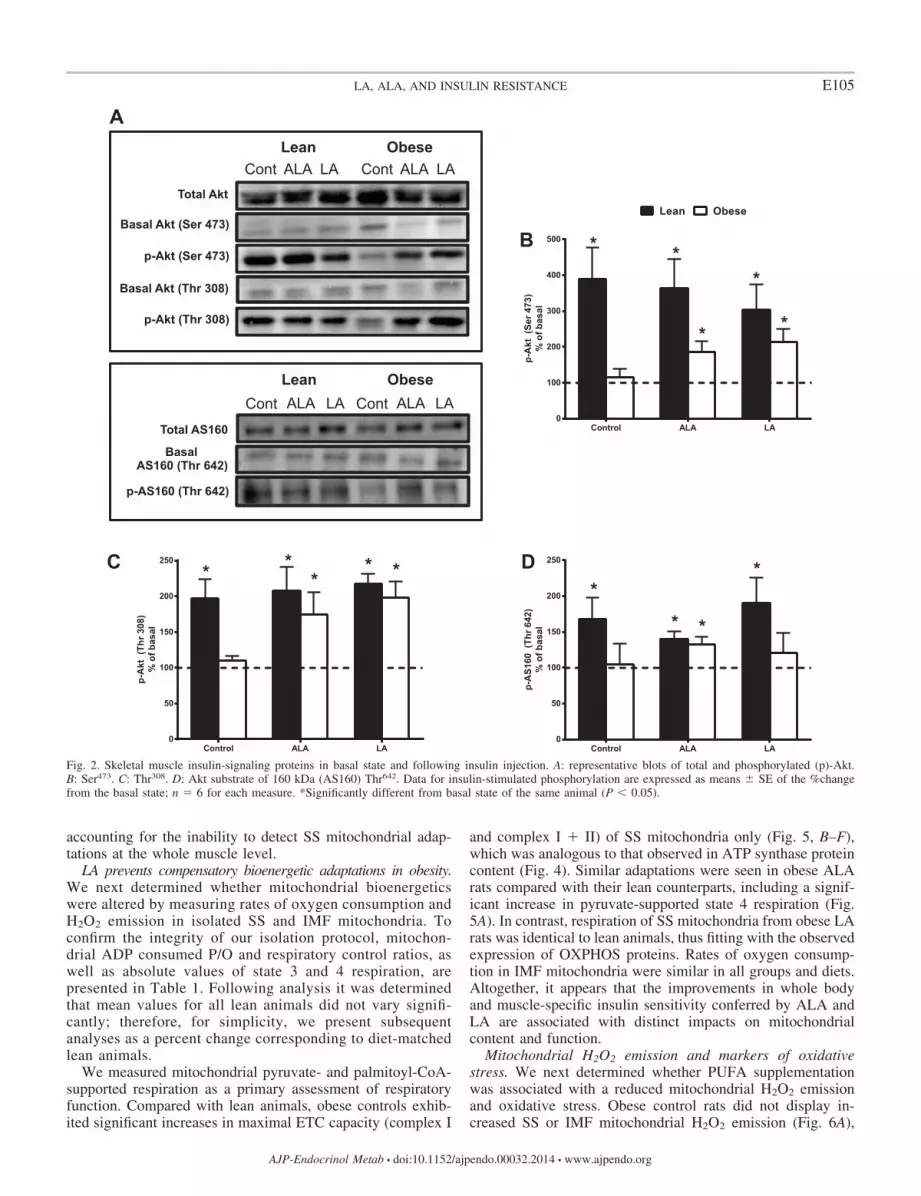
LA and ALA preserve skeletal muscle insulin signaling.Several parameters can influence whole body glucose andinsulin tolerance independent of skeletal muscle insulin sensi-tivity (e.g., glucose/insulin actions within adipose tissue, liver,and pancreas). Therefore, it was important to determine spe-cifically skeletal muscle insulin sensitivity in obese animalsfollowing LA and ALA supplementation. To determine this,we next investigated the ability of insulin to induce phosphor-ylation of proteins involved in the canonical insulin-signalingcascade. Within lean and obese animals there were no differ-ences in total content of Akt and AS160 protein (Fig. 2A). Inobese control animals, insulin failed to stimulate phosphoryla-tion of Akt at Ser473 (Fig. 2B) and Thr308 (Fig. 2C), as well asAS160 at Thr642 (Fig. 2D), above basal levels (Fig. 2A). Incontrast, obese rats supplemented with ALA maintained insu-lin-induced phosphorylation of Akt Ser473 (�100%) andThr308 (�75%) as well as AS160 Thr642 (�40%) (Fig. 2,B–D). Although LA evoked similar improvements in Akt
E103LA, ALA, AND INSULIN RESISTANCE
AJP-Endocrinol Metab • doi:10.1152/ajpendo.00032.2014 • www.ajpendo.org

phosphorylation at both sites, no changes were seen withAS160 (Thr642). These data, in combination with the IPITTresults, suggest that both LA and ALA maintain skeletalmuscle insulin signaling in obese Zucker rats.
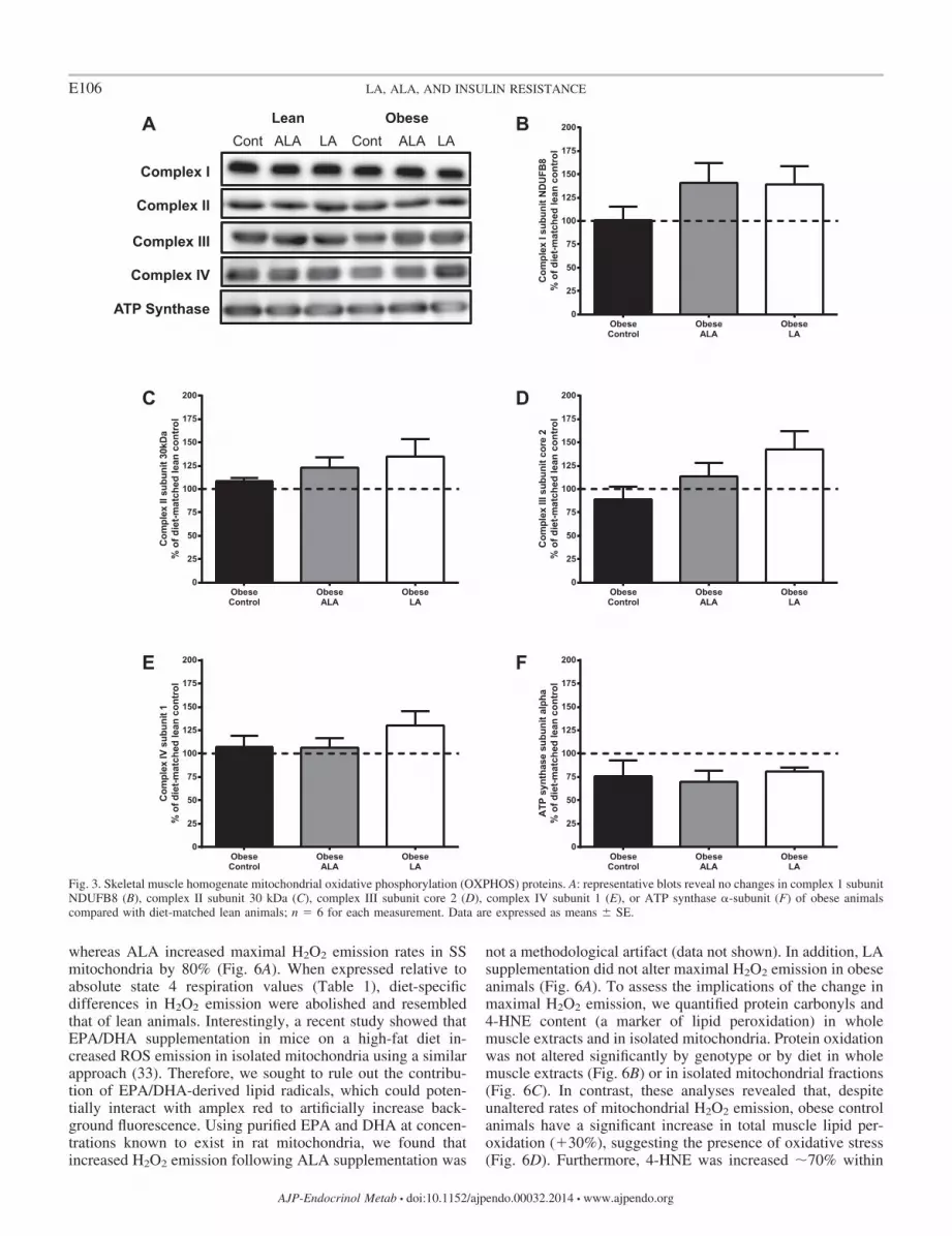
ALA preferentially increases electron transport chain pro-teins in SS mitochondria. The accumulation of OXPHOSproteins in whole muscle extracts was not different in obeserats following ALA or LA supplementation compared withcontrols (Fig. 3, A–F). Therefore, we reexamined OXPHOSprotein content in purified SS and IMF mitochondrial fractions.Compared with lean controls, obese control rats showed a
significant increase (�100%) in ATP synthase content in SSmitochondria only (Fig. 4, A and F). Interestingly, SS mito-chondria from obese ALA rats showed significant increases incomplex I subunit NUDFB8 (�100%), complex III subunitcore 2 (�80%), and ATP synthase (�150%) (Fig. 4, A, B, D,and F). In contrast, the content of electron transport chain(ETC) markers in mitochondria isolated from LA-supple-mented rats appeared identical to diet-matched leans (Fig. 4,A–F), suggesting an absence of compensatory adaptations.Finally, IMF mitochondria remained constant for all OXPHOSprotein targets measured across all groups (Fig. 4, A–F), likely
Fig. 1. Intraperitoneal glucose (IPGTT) and insulin tolerance tests (IPITT) for lean (A and C, respectively) and obese rats (B and D, respectively). �, Control(Cont) diet; �, �-linolenic acid (ALA) diet; o, linoleic acid (LA) diet. Area under the curve (AUC) values for IPGTT (E) and IPITT (F). Data expressed as means� SE; n � 6 for each measure. �Significantly different from obese control (P � 0.05); *significantly different from diet-matched lean animals (P � 0.05).
E104 LA, ALA, AND INSULIN RESISTANCE
AJP-Endocrinol Metab • doi:10.1152/ajpendo.00032.2014 • www.ajpendo.org
accounting for the inability to detect SS mitochondrial adap-tations at the whole muscle level.
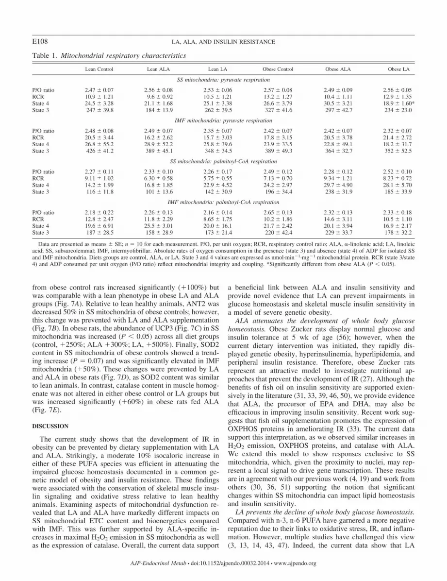
LA prevents compensatory bioenergetic adaptations in obesity.We next determined whether mitochondrial bioenergeticswere altered by measuring rates of oxygen consumption andH2O2 emission in isolated SS and IMF mitochondria. Toconfirm the integrity of our isolation protocol, mitochon-drial ADP consumed P/O and respiratory control ratios, aswell as absolute values of state 3 and 4 respiration, arepresented in Table 1. Following analysis it was determinedthat mean values for all lean animals did not vary signifi-cantly; therefore, for simplicity, we present subsequentanalyses as a percent change corresponding to diet-matchedlean animals.
We measured mitochondrial pyruvate- and palmitoyl-CoA-supported respiration as a primary assessment of respiratoryfunction. Compared with lean animals, obese controls exhib-ited significant increases in maximal ETC capacity (complex I
and complex I � II) of SS mitochondria only (Fig. 5, B–F),which was analogous to that observed in ATP synthase proteincontent (Fig. 4). Similar adaptations were seen in obese ALArats compared with their lean counterparts, including a signif-icant increase in pyruvate-supported state 4 respiration (Fig.5A). In contrast, respiration of SS mitochondria from obese LArats was identical to lean animals, thus fitting with the observedexpression of OXPHOS proteins. Rates of oxygen consump-tion in IMF mitochondria were similar in all groups and diets.Altogether, it appears that the improvements in whole bodyand muscle-specific insulin sensitivity conferred by ALA andLA are associated with distinct impacts on mitochondrialcontent and function.
Mitochondrial H2O2 emission and markers of oxidativestress. We next determined whether PUFA supplementationwas associated with a reduced mitochondrial H2O2 emissionand oxidative stress. Obese control rats did not display in-creased SS or IMF mitochondrial H2O2 emission (Fig. 6A),
Fig. 2. Skeletal muscle insulin-signaling proteins in basal state and following insulin injection. A: representative blots of total and phosphorylated (p)-Akt.B: Ser473. C: Thr308. D: Akt substrate of 160 kDa (AS160) Thr642. Data for insulin-stimulated phosphorylation are expressed as means � SE of the %changefrom the basal state; n � 6 for each measure. *Significantly different from basal state of the same animal (P � 0.05).
E105LA, ALA, AND INSULIN RESISTANCE
AJP-Endocrinol Metab • doi:10.1152/ajpendo.00032.2014 • www.ajpendo.org
whereas ALA increased maximal H2O2 emission rates in SSmitochondria by 80% (Fig. 6A). When expressed relative toabsolute state 4 respiration values (Table 1), diet-specificdifferences in H2O2 emission were abolished and resembledthat of lean animals. Interestingly, a recent study showed thatEPA/DHA supplementation in mice on a high-fat diet in-creased ROS emission in isolated mitochondria using a similarapproach (33). Therefore, we sought to rule out the contribu-tion of EPA/DHA-derived lipid radicals, which could poten-tially interact with amplex red to artificially increase back-ground fluorescence. Using purified EPA and DHA at concen-trations known to exist in rat mitochondria, we found thatincreased H2O2 emission following ALA supplementation was
not a methodological artifact (data not shown). In addition, LAsupplementation did not alter maximal H2O2 emission in obeseanimals (Fig. 6A). To assess the implications of the change inmaximal H2O2 emission, we quantified protein carbonyls and4-HNE content (a marker of lipid peroxidation) in wholemuscle extracts and in isolated mitochondria. Protein oxidationwas not altered significantly by genotype or by diet in wholemuscle extracts (Fig. 6B) or in isolated mitochondrial fractions(Fig. 6C). In contrast, these analyses revealed that, despiteunaltered rates of mitochondrial H2O2 emission, obese controlanimals have a significant increase in total muscle lipid per-oxidation (�30%), suggesting the presence of oxidative stress(Fig. 6D). Furthermore, 4-HNE was increased �70% within
Fig. 3. Skeletal muscle homogenate mitochondrial oxidative phosphorylation (OXPHOS) proteins. A: representative blots reveal no changes in complex 1 subunitNDUFB8 (B), complex II subunit 30 kDa (C), complex III subunit core 2 (D), complex IV subunit 1 (E), or ATP synthase �-subunit (F) of obese animalscompared with diet-matched lean animals; n � 6 for each measurement. Data are expressed as means � SE.
E106 LA, ALA, AND INSULIN RESISTANCE
AJP-Endocrinol Metab • doi:10.1152/ajpendo.00032.2014 • www.ajpendo.org

IMF mitochondria of obese controls but was reduced in SSmitochondria (Fig. 6E). Overall, in obesity, LA preventedchanges in 4-HNE content within whole muscle (Fig. 6D) andSS/IMF mitochondria (Fig. 6E), resembling lean healthyanimals. ALA supplementation also prevented increases in4-HNE content within whole muscle (Fig. 6D) and IMF mito-chondria (Fig. 6E) seen in obese control rats.
Mitochondrial ADP transport, uncoupling, and antioxidantproteins. Given the apparent discrepancy between maximal invitro mitochondrial H2O2 emission rates and in vivo markers ofoxidative stress, we next examined the expression of proteinsknown to influence mitochondrial H2O2 emission. ANT1 con-tent did not change in SS mitochondria across all groups anddiets (Fig. 7A). In contrast, ANT1 content in IMF mitochondria
Fig. 4. Changes in OXPHOS proteins of subsarcolemmal (SS) and intermyofibrillar (IMF) mitochondria. A: representative blots for complex 1 subunit NDUFB8(B), complex II subunit 30 kDa (C), complex III subunit core 2 (D), complex IV subunit 1 (E), and ATP synthase �-subunit (F); n � 10 for each measurement.Data are expressed as means � SE. *Significantly different from diet-matched lean animals (P � 0.05).
E107LA, ALA, AND INSULIN RESISTANCE
AJP-Endocrinol Metab • doi:10.1152/ajpendo.00032.2014 • www.ajpendo.org
from obese control rats increased significantly (�100%) butwas comparable with a lean phenotype in obese LA and ALAgroups (Fig. 7A). Relative to lean healthy animals, ANT2 wasdecreased 50% in SS mitochondria of obese controls; however,this change was prevented with LA and ALA supplementation(Fig. 7B). In obese rats, the abundance of UCP3 (Fig. 7C) in SSmitochondria was increased (P � 0.05) across all diet groups(control, �250%; ALA �300%; LA, �500%). Finally, SOD2content in SS mitochondria of obese controls showed a trend-ing increase (P � 0.07) and was significantly elevated in IMFmitochondria (�50%). These changes were prevented by LAand ALA in obese rats (Fig. 7D), as SOD2 content was similarto lean animals. In contrast, catalase content in muscle homog-enate was not altered in either obese control or LA groups butwas increased significantly (�60%) in obese rats fed ALA(Fig. 7E).
DISCUSSION
The current study shows that the development of IR inobesity can be prevented by dietary supplementation with LAand ALA. Strikingly, a moderate 10% isocaloric increase ineither of these PUFA species was efficient in attenuating theimpaired glucose homeostasis documented in a common ge-netic model of obesity and insulin resistance. These findingswere associated with the conservation of skeletal muscle insu-lin signaling and oxidative stress relative to lean healthyanimals. Examining aspects of mitochondrial dysfunction re-vealed that LA and ALA have markedly different impacts onSS mitochondrial ETC content and bioenergetics comparedwith IMF. This was further supported by ALA-specific in-creases in maximal H2O2 emission in SS mitochondria as wellas the expression of catalase. Overall, the current data support
a beneficial link between ALA and insulin sensitivity andprovide novel evidence that LA can prevent impairments inglucose homeostasis and skeletal muscle insulin sensitivity ina model of severe genetic obesity.
ALA attenuates the development of whole body glucosehomeostasis. Obese Zucker rats display normal glucose andinsulin tolerance at 5 wk of age (56); however, when thecurrent dietary intervention was initiated, they rapidly dis-played genetic obesity, hyperinsulinemia, hyperlipidemia, andperipheral insulin resistance. Therefore, obese Zucker ratsrepresent an attractive model to investigate nutritional ap-proaches that prevent the development of IR (27). Although thebenefits of fish oil on insulin sensitivity are supported exten-sively in the literature (31, 33, 39, 46, 50), we provide evidencethat ALA, the precursor of EPA and DHA, may also beefficacious in improving insulin sensitivity. Recent work sug-gests that fish oil supplementation promotes the expression ofOXPHOS proteins in ameliorating IR (33). The current datasupport this interpretation, as we observed similar increases inH2O2 emission, OXPHOS proteins, and catalase with ALA.We extend this model to show responses exclusive to SSmitochondria, which, given the proximity to nuclei, may rep-resent a local signal to drive gene transcription. These resultsare in agreement with our previous work (4, 19) and work fromothers (30, 36, 51) supporting the notion that significantchanges within SS mitochondria can impact lipid homeostasisand insulin sensitivity.
LA prevents the decline of whole body glucose homeostasis.Compared with n-3, n-6 PUFA have garnered a more negativereputation due to their links to oxidative stress, IR, and inflam-mation. However, multiple studies have challenged this view(3, 13, 14, 43, 47). Indeed, the current data show that LA
Table 1. Mitochondrial respiratory characteristics
Lean Control Lean ALA Lean LA Obese Control Obese ALA Obese LA
SS mitochondria: pyruvate respiration
P/O ratio 2.47 � 0.07 2.56 � 0.08 2.53 � 0.06 2.57 � 0.08 2.49 � 0.09 2.56 � 0.05RCR 10.9 � 1.21 9.6 � 0.92 10.5 � 1.21 13.2 � 1.27 10.4 � 1.11 12.9 � 1.35State 4 24.5 � 3.28 21.1 � 1.68 25.1 � 3.38 26.6 � 3.79 30.5 � 3.21 18.9 � 1.60*State 3 247 � 39.8 184 � 13.9 262 � 39.5 327 � 41.6 297 � 42.7 234 � 23.0
IMF mitochondria: pyruvate respiration
P/O ratio 2.48 � 0.08 2.49 � 0.07 2.35 � 0.07 2.42 � 0.07 2.42 � 0.07 2.32 � 0.07RCR 20.5 � 3.44 16.2 � 2.62 15.7 � 3.03 17.8 � 3.15 20.5 � 3.78 21.4 � 2.72State 4 26.8 � 55.2 28.9 � 52.2 25.8 � 39.6 23.9 � 33.5 22.8 � 49.1 18.2 � 31.7State 3 426 � 41.2 389 � 45.1 348 � 34.5 389 � 49.3 364 � 32.7 352 � 52.5
SS mitochondria: palmitoyl-CoA respiration
P/O ratio 2.27 � 0.11 2.33 � 0.10 2.26 � 0.17 2.49 � 0.12 2.28 � 0.12 2.52 � 0.10RCR 9.11 � 1.02 6.30 � 0.58 5.75 � 0.55 7.13 � 0.70 9.34 � 1.21 8.23 � 0.72State 4 14.2 � 1.99 16.8 � 1.85 22.9 � 4.52 24.2 � 2.97 29.7 � 4.90 28.1 � 5.70State 3 116 � 11.8 101 � 13.6 142 � 30.9 196 � 34.4 238 � 31.9 185 � 33.9
IMF mitochondria: palmitoyl-CoA respiration
P/O ratio 2.18 � 0.22 2.26 � 0.13 2.16 � 0.14 2.65 � 0.13 2.32 � 0.13 2.33 � 0.18RCR 12.8 � 2.47 11.8 � 2.29 8.65 � 1.75 10.2 � 1.86 14.6 � 3.11 10.5 � 1.10State 4 19.6 � 6.91 25.5 � 3.01 20.0 � 16.1 21.7 � 2.42 20.1 � 3.94 16.9 � 2.17State 3 187 � 28.5 158 � 28.9 173 � 21.4 220 � 42.4 229 � 33.7 178 � 32.2
Data are presented as means � SE; n � 10 for each measurement. P/O, per unit oxygen; RCR, respiratory control ratio; ALA, �-linolenic acid; LA, linoleicacid; SS, subsarcolemmal; IMF, intermyofibrillar. Absolute rates of oxygen consumption in the presence (state 3) and absence (state 4) of ADP for isolated SSand IMF mitochondria. Diets groups are control, ALA, or LA. State 3 and 4 values are expressed as nmol·min1·mg1 mitochondrial protein. RCR (state 3/state4) and ADP consumed per unit oxygen (P/O ratio) reflect mitochondrial integrity and coupling. *Significantly different from obese ALA (P � 0.05).
E108 LA, ALA, AND INSULIN RESISTANCE
AJP-Endocrinol Metab • doi:10.1152/ajpendo.00032.2014 • www.ajpendo.org

prevented the development of whole body glucose intoleranceand maintained muscle-specific insulin sensitivity and 4-HNEcontent in obese animals. In contrast to ALA, these improve-ments were independent of changes in H2O2 emission, OX-PHOS proteins, and antioxidant enzyme expression, raising thepossibility of a divergent mechanism for improving insulinsensitivity. However, our findings do not exclude the possibil-ity that LA remodels the membrane cardiolipin profile within
mitochondria (28), which is known to impact mitochondrialfunction (18, 28). Although previous reports have linked obe-sity with changes that would likely promote mitochondrialH2O2 emission, including increased ETC sensitivity to reduc-ing equivalents (34) and diminished sensitivity to ADP (49),we found no changes in maximal ADP-stimulated respirationfollowing LA supplementation. Therefore, it remains possiblethat LA may alter the dynamic response of mitochondria to
Max
imal
Pal
mito
yl-C
oA R
espi
ratio
n%
of d
iet-m
atch
ed le
an c
ontr
ol
Stat
e 4
Res
pira
tion
% o
f die
t-mat
ched
lean
con
trol
Fig. 5. Isolated SS and IMF mitochondrial respiration. Basal (A) and ADP-stimulated (B) pyruvate-supported respiration, maximal complex I (plus glutamate;C) and maximal electron transport chain respiration (plus succinate; D), and basal (E) and ADP-stimulated (F) states of palmitoyl-CoA respiration; n � 10 foreach measurement. Data expressed as means � SE. *Significantly different from diet-matched lean animals (P � 0.05).
E109LA, ALA, AND INSULIN RESISTANCE
AJP-Endocrinol Metab • doi:10.1152/ajpendo.00032.2014 • www.ajpendo.org

Fig. 6. Markers of oxidative stress. A: maximal succinate H2O2 emission in SS and IMF mitochondria. B and C: protein carbonylation in muscle homogenate(B) and SS and IMF mitochondria (C). D and E: 4-hydroxynonenal (4-HNE) content in muscle homogenate (D) and SS and IMF mitochondria (E) indicates lipidperoxidation. Representative blots shown in respective panels; n � 10 for each measurement. Data are expressed as means � SE. *Significantly different fromdiet-matched lean animals (P � 0.05).
E110 LA, ALA, AND INSULIN RESISTANCE
AJP-Endocrinol Metab • doi:10.1152/ajpendo.00032.2014 • www.ajpendo.org

Fig. 7. Changes in adenine nucleotide translocase 1 (ANT1; A), ANT2 (B), mitochondrial uncoupling protein 3 (UCP3; C), and manganese superoxide dismutase(SOD2; D) in SS and IMF mitochondria. For data shown in A–D, n � 10 for each measure. E: catalase content in muscle homogenate; n � 6 for all groups.Data are expressed as means � SE. *Significantly different from diet-matched lean animals (P � 0.05).
E111LA, ALA, AND INSULIN RESISTANCE
AJP-Endocrinol Metab • doi:10.1152/ajpendo.00032.2014 • www.ajpendo.org

submaximal substrate concentrations. Regardless of the elusivemechanism of action, the current data provide convincingevidence that LA prevented IR in obese Zucker rats.
Mitochondrial H2O2 emission and uncoupling and antioxi-dant proteins. The current study found increased ETC contentonly within SS mitochondria, which represents �20% of totalcellular mitochondrial volume (12, 20), accounting for theabsence of changes in whole muscle measurements. The cur-rent study cannot explain mechanistically why SS mitochon-dria preferentially respond, although this appears to be aconserved observation across cellular stresses (19, 36). How-ever, the increased expression of ETC proteins within the SSmitochondria likely contributed to the increase in maximalmitochondrial H2O2 emission, as normalization of emissionrates to state 4 respiration negated all differences. Therefore,the increase in ETC subunits following ALA supplementationmay be construed as a negative adaptation, as mitochondriallipid uptake and ROS emission have been causally linked to IR(2), possibly through ROS-mediated activation of the NF-B/IB/IKK� pathway, attenuating insulin signaling by serinephosphorylation of insulin receptor substrate 1 (48, 55). How-ever, this working model remains controversial, as chronicmitochondrial antioxidant treatment that improves cellular re-dox balance does not improve insulin sensitivity following ahigh-fat diet (40). Perhaps subtle increases in mitochondrialROS emissions are required for the transcriptional adaptationsthat are necessary to regulate metabolic homeostasis during ahigh-fat challenge. Our data indirectly support this model, asALA supplementation increased maximal H2O2 emission andOXPHOS proteins in SS mitochondria as well as musclecatalase content. Our results are consistent with the suggestionthat incorporating n-3 PUFA into mitochondrial membranesincreases the propensity for ROS production (1, 33). Further-more, in vivo markers of oxidative stress (4-HNE and proteincarbonylation) suggest that despite an increase in maximalH2O2 emission, ALA supplementation conserved redox bal-ance in whole muscle and mitochondrial samples. Given thatROS have several intracellular functions, including participa-tion in the complex signaling network involved in mitochon-drial biogenesis (22, 37), perhaps the increased OXPHOSexpression within SS mitochondria in the obese ALA group ismediated in part through tightly regulated redox signaling.
In contrast, we found no differences in maximal H2O2
emission or catalase content in lean and obese animals fed LA.Unlike the changes seen with obese control and ALA groups,mitochondria from the obese LA group were identical to theirlean counterparts, suggesting that compensatory bioenergeticadaptations are not necessary to preserve glucose homeostasisand muscle insulin signaling while consuming LA. Previouswork showed that arachidonic acid increased mitochondrialROS emission and was linked to mitochondrial dysfunction(10). Surprisingly, the precursor LA did not alter rates ofmitochondrial H2O2 emission, and therefore, prevention of IRoccurs through a mechanism not investigated in the currentstudy. Although speculative, LA was shown to drive a lipox-ygenase-mediated eicosanoid response, leading to productionof the PPAR�-activating 13-hydroperoxy-9,11-octadecadien-oic acid (29). Therefore, if LA evokes a PPAR-mediatedimprovement in glucose homeostasis, the mechanism by whichit acts may differ from that of n-3 PUFA. Alternatively, LAconsumption may involve primary adaptations within the liver,
pancreas, and white adipose tissue and secondary responseswithin muscle, a possibility that has not been explored in thecurrent study. Regardless, the current study provides evidencethat LA is beneficial at preventing the development of insulinresistance.
Perspectives and limitations. The current study providesinsight on the link between ALA and insulin sensitivity andevidence that LA supplementation represents additional thera-peutic potential. Although aspects of mitochondrial dysfunc-tion were very similar between obese control and ALA-sup-plemented rats, the preservation of skeletal muscle insulinsignaling and whole body glucose homeostasis highlights thevalue of this n-3 PUFA. The precise mechanism(s) by whichLA and ALA exert their preserving effects requires furtherelucidation and may involve changes in liver given the en-hanced glucose tolerance. Indeed, EPA/DHA may augmenthepatic IR and lipotoxicity by increasing FAO, inhibiting denovo lipogenesis and reducing proinflammatory cytokine pro-duction (42). Furthermore, within white adipose tissue, thesen-3 PUFAs are known to improve factors influencing IR, suchas adipocyte morphology, rates of endogenous FAO, andadipokine secretory profiles, as well as immunometabolic sta-tus (as recently reviewed in Ref. 42). Whether LA and ALAexert their effects through similar mechanisms remains to beshown.
In the current study, we were unable to uncouple the effectsof ALA from EPA and DHA; therefore, it is possible that theeffects seen with ALA are due to its conversion (albeit limited)into EPA/DHA. Future work using animal models that preventthe conversion of ALA into EPA/DHA will enable us to moredefinitively describe the independent role of ALA on skeletalmuscle insulin signaling. Also, in the current study, the ma-cronutrient composition of the LA and ALA diets was out ofnecessity different from control diets, having higher fat (26 vs.16% in control diet) and by default decreased carbohydratecontent (54 vs. 64% in control diet). Therefore, future studiesshould also determine whether the modest 10% increase/de-crease in dietary fat/carbohydrate could override the stronggenetic predisposition for an IR phenotype.
More importantly, our data challenge the traditional viewthat LA is harmful and welcome the reassessment of its use asa therapeutic strategy for preserving insulin sensitivity. Despiteobserving no changes in mitochondrial content, function, ormaximal H2O2 emission in isolated mitochondria, LA main-tained skeletal muscle insulin signaling similarly to ALA.Future investigations should focus on changes in membranephospholipid composition, as both n-3 and n-6 PUFA areknown to compete in the remodeling of membranes, includingmitochondria, and may also preferentially accumulate in dif-ferent tissues. The impacts of LA and ALA on IR maytranscend the boundaries of skeletal muscle and mitochondriabut nevertheless represent valuable therapeutic strategies forpreventing the development of an insulin-resistant phenotypein obesity.
ACKNOWLEDGMENTS
We thank Michael Zulyniak (University of Guelph) for confirming fattyacid composition of the experimental diets by gas chromatography.
E112 LA, ALA, AND INSULIN RESISTANCE
AJP-Endocrinol Metab • doi:10.1152/ajpendo.00032.2014 • www.ajpendo.org

GRANTS
This work was funded by the Ontario Ministry of Agriculture, Food, andRural Affairs, and infrastructure was purchased with the assistance of theCanadian Foundation for Innovation as well as the Ontario Research Fund.
DISCLOSURES
The authors report no conflicts of interest, financial or otherwise.
AUTHOR CONTRIBUTIONS
S.M., D.M.M., and G.P.H. conception and design of research; S.M., E.A.H.,S.S.J., and G.P.H. performed experiments; S.M., E.A.H., S.S.J., D.M.M., andG.P.H. analyzed data; S.M., D.M.M., and G.P.H. interpreted results of exper-iments; S.M., D.M.M., and G.P.H. prepared figures; S.M., D.M.M., and G.P.H.drafted manuscript; S.M., E.A.H., S.S.J., D.M.M., and G.P.H. edited andrevised manuscript; S.M., D.M.M., and G.P.H. approved final version ofmanuscript.
REFERENCES
1. Al-Gubory KH. Mitochondria: omega-3 in the route of mitochondrialreactive oxygen species. Int J Biochem Cell Biol 44: 1569–1573, 2012.
2. Anderson EJ, Lustig ME, Boyle KE, Woodlief TL, Kane DA, Lin CT,Price JW 3rd, Kang L, Rabinovitch PS, Szeto HH, Houmard JA,Cortright RN, Wasserman DH, Neufer PD. Mitochondrial H2O2 emis-sion and cellular redox state link excess fat intake to insulin resistance inboth rodents and humans. J Clin Invest 119: 573–581, 2009.
3. Baker PR, Lin Y, Schopfer FJ, Woodcock SR, Groeger AL, BatthyanyC, Sweeney S, Long MH, Iles KE, Baker LM, Branchaud BP, ChenYE, Freeman BA. Fatty acid transduction of nitric oxide signaling:multiple nitrated unsaturated fatty acid derivatives exist in human bloodand urine and serve as endogenous peroxisome proliferator-activatedreceptor ligands. J Biol Chem 280: 42464–42475, 2005.
4. Benton CR, Nickerson JG, Lally J, Han XX, Holloway GP, Glatz JF,Luiken JJ, Graham TE, Heikkila JJ, Bonen A. Modest PGC-1alphaoverexpression in muscle in vivo is sufficient to increase insulin sensitivityand palmitate oxidation in subsarcolemmal, not intermyofibrillar, mito-chondria. J Biol Chem 283: 4228–4240, 2008.
5. Bizeau ME, Willis WT, Hazel JR. Differential responses to endurancetraining in subsarcolemmal and intermyofibrillar mitochondria. J ApplPhysiol 85: 1279–1284, 1998.
6. Boden MJ, Brandon AE, Tid-Ang JD, Preston E, Wilks D, Stuart E,Cleasby ME, Turner N, Cooney GJ, Kraegen EW. Overexpression ofmanganese superoxide dismutase ameliorates high-fat diet-induced insulinresistance in rat skeletal muscle. Am J Physiol Endocrinol Metab 303:E798–E805, 2012.
7. Calder PC, Yaqoob P. Omega-3 polyunsaturated fatty acids and humanhealth outcomes. Biofactors 35: 266–272, 2009.
8. Chen LL, Zhang HH, Zheng J, Hu X, Kong W, Hu D, Wang SX,Zhang P. Resveratrol attenuates high-fat diet-induced insulin resistanceby influencing skeletal muscle lipid transport and subsarcolemmal mito-chondrial beta-oxidation. Metabolism 60: 1598–1609, 2011.
9. Chomentowski P, Coen PM, Radikova Z, Goodpaster BH, Toledo FG.Skeletal muscle mitochondria in insulin resistance: differences in inter-myofibrillar versus subsarcolemmal subpopulations and relationship tometabolic flexibility. J Clin Endocrinol Metab 96: 494–503, 2011.
10. Cocco T, Di Paola M, Papa S, Lorusso M. Arachidonic acid interactionwith the mitochondrial electron transport chain promotes reactive oxygenspecies generation. Free Radic Biol Med 27: 51–59, 1999.
11. Cogswell AM, Stevens RJ, Hood DA. Properties of skeletal musclemitochondria isolated from subsarcolemmal and intermyofibrillar regions.Am J Physiol Cell Physiol 264: C383–C389, 1993.
12. Ferreira R, Vitorino R, Alves RM, Appell HJ, Powers SK, Duarte JA,Amado F. Subsarcolemmal and intermyofibrillar mitochondria proteomedifferences disclose functional specializations in skeletal muscle. Pro-teomics 10: 3142–3154, 2010.
13. Ferrucci L, Cherubini A, Bandinelli S, Bartali B, Corsi A, LauretaniF, Martin A, Andres-Lacueva C, Senin U, Guralnik JM. Relationshipof plasma polyunsaturated fatty acids to circulating inflammatory markers.J Clin Endocrinol Metab 91: 439–446, 2006.
14. Fritsche KL. Too much linoleic acid promotes inflammation-doesn’t it?Prostaglandins Leukot Essent Fatty Acids 79: 173–175, 2008.
15. Hancock CR, Han DH, Chen M, Terada S, Yasuda T, Wright DC,Holloszy JO. High-fat diets cause insulin resistance despite an increase inmuscle mitochondria. Proc Natl Acad Sci USA 105: 7815–7820, 2008.
16. Holloszy JO. Skeletal muscle “mitochondrial deficiency” does not medi-ate insulin resistance. Am J Clin Nutr 89: 463S–466S, 2009.
17. Holloway GP, Benton CR, Mullen KL, Yoshida Y, Snook LA, HanXX, Glatz JF, Luiken JJ, Lally J, Dyck DJ, Bonen A. In obese ratmuscle transport of palmitate is increased and is channeled to triacylglyc-erol storage despite an increase in mitochondrial palmitate oxidation. AmJ Physiol Endocrinol Metab 296: E738–E747, 2009.
18. Holloway GP, Fajardo VA, McMeekin L, LeBlanc PJ. Unsaturation ofmitochondrial membrane lipids is related to palmitate oxidation in sub-sarcolemmal and intermyofibrillar mitochondria. J Membr Biol 245: 165–176, 2012.
19. Holloway GP, Gurd BJ, Snook LA, Lally J, Bonen A. Compensatoryincreases in nuclear PGC1alpha protein are primarily associated withsubsarcolemmal mitochondrial adaptations in ZDF rats. Diabetes 59:819–828, 2010.
20. Hoppeler H, Howald H, Conley K, Lindstedt SL, Claassen H, Vock P,Weibel ER. Endurance training in humans: aerobic capacity and structureof skeletal muscle. J Appl Physiol (1985) 59: 320–327, 1985.
21. Hulver MW, Berggren JR, Cortright RN, Dudek RW, Thompson RP,Pories WJ, MacDonald KG, Cline GW, Shulman GI, Dohm GL,Houmard JA. Skeletal muscle lipid metabolism with obesity. Am JPhysiol Endocrinol Metab 284: E741–E747, 2003.
22. Jain SS, Paglialunga S, Vigna C, Ludzki A, Herbst EA, Lally JS,Schrauwen P, Hoeks J, Tupling AR, Bonen A, Holloway GP. High-FatDiet-Induced Mitochondrial Biogenesis Is Regulated by MitochondrialDerived Reactive Oxygen Species Activation of CaMKII. Diabetes 63:1907–1913, 2014.
23. Jain SS, Snook LA, Glatz JF, Luiken JJ, Holloway GP, ThurmondDC, Bonen A. Munc18c provides stimulus-selective regulation of GLUT4but not fatty acid transporter trafficking in skeletal muscle. FEBS Lett 586:2428–2435, 2012.
24. Jelenik T, Rossmeisl M, Kuda O, Jilkova ZM, Medrikova D, Kus V,Hensler M, Janovska P, Miksik I, Baranowski M, Gorski J, HebrardS, Jensen TE, Flachs P, Hawley S, Viollet B, Kopecky J. AMP-activatedprotein kinase alpha2 subunit is required for the preservation of hepaticinsulin sensitivity by n-3 polyunsaturated fatty acids. Diabetes 59: 2737–2746, 2010.
25. Johnson GH, Fritsche K. Effect of dietary linoleic acid on markers ofinflammation in healthy persons: a systematic review of randomizedcontrolled trials. J Acad Nutr Diet 112: 1029–1041, 1041.e1–1041.e15,2012.
26. Kelley DE, He J, Menshikova EV, Ritov VB. Dysfunction of mitochon-dria in human skeletal muscle in type 2 diabetes. Diabetes 51: 2944–2950,2002.
27. Kemmer FW, Berger M, Herberg L, Gries FA, Wirdeier A, Becker K.Glucose metabolism in perfused skeletal muscle. Demonstration of insulinresistance in the obese Zucker rat. Biochem J 178: 733–741, 1979.
28. Khairallah RJ, Kim J, O’Shea KM, O’Connell KA, Brown BH,Galvao T, Daneault C, Des Rosiers C, Polster BM, Hoppel CL, StanleyWC. Improved mitochondrial function with diet-induced increase in eitherdocosahexaenoic acid or arachidonic acid in membrane phospholipids.PLoS One 7: e34402, 2012.
29. Konig B, Eder K. Differential action of 13-HPODE on PPARalphadownstream genes in rat Fao and human HepG2 hepatoma cell lines. JNutr Biochem 17: 410–418, 2006.
30. Koves TR, Noland RC, Bates AL, Henes ST, Muoio DM, CortrightRN. Subsarcolemmal and intermyofibrillar mitochondria play distinctroles in regulating skeletal muscle fatty acid metabolism. Am J PhysiolCell Physiol 288: C1074–C1082, 2005.
31. Kuda O, Jelenik T, Jilkova Z, Flachs P, Rossmeisl M, Hensler M,Kazdova L, Ogston N, Baranowski M, Gorski J, Janovska P, Kus V,Polak J, Mohamed-Ali V, Burcelin R, Cinti S, Bryhn M, Kopecky J.n-3 fatty acids and rosiglitazone improve insulin sensitivity throughadditive stimulatory effects on muscle glycogen synthesis in mice fed ahigh-fat diet. Diabetologia 52: 941–951, 2009.
32. Lally JS, Herbst EA, Matravadia S, Maher AC, Perry CG, Ventura-Clapier R, Holloway GP. Over-expressing mitofusin-2 in healthy maturemammalian skeletal muscle does not alter mitochondrial bioenergetics.PLoS One 8: e55660, 2013.
33. Lanza IR, Blachnio-Zabielska A, Johnson ML, Schimke JM, JakaitisDR, Lebrasseur NK, Jensen MD, Sreekumaran Nair K, Zabielski P.Influence of fish oil on skeletal muscle mitochondrial energetics and lipidmetabolites during high-fat diet. Am J Physiol Endocrinol Metab 304:E1391–E1403, 2013.
E113LA, ALA, AND INSULIN RESISTANCE
AJP-Endocrinol Metab • doi:10.1152/ajpendo.00032.2014 • www.ajpendo.org

34. Larsen S, Stride N, Hey-Mogensen M, Hansen CN, Andersen JL,Madsbad S, Worm D, Helge JW, Dela F. Increased mitochondrialsubstrate sensitivity in skeletal muscle of patients with type 2 diabetes.Diabetologia 54: 1427–1436, 2011.
35. Lee HY, Choi CS, Birkenfeld AL, Alves TC, Jornayvaz FR, JurczakMJ, Zhang D, Woo DK, Shadel GS, Ladiges W, Rabinovitch PS,Santos JH, Petersen KF, Samuel VT, Shulman GI. Targeted expressionof catalase to mitochondria prevents age-associated reductions in mito-chondrial function and insulin resistance. Cell Metab 12: 668–674, 2010.
36. Lionetti L, Mollica MP, Crescenzo R, D’Andrea E, Ferraro M, BiancoF, Liverini G, Iossa S. Skeletal muscle subsarcolemmal mitochondrialdysfunction in high-fat fed rats exhibiting impaired glucose homeostasis.Int J Obes 31: 1596–1604, 2007.
37. Lira VA, Benton CR, Yan Z, Bonen A. PGC-1� regulation by exercisetraining and its influences on muscle function and insulin sensitivity. AmJ Physiol Endocrinol Metab 299: E145–E161, 2010.
38. Mollica MP, Lionetti L, Crescenzo R, D’Andrea E, Ferraro M,Liverini G, Iossa S. Heterogeneous bioenergetic behaviour of subsar-colemmal and intermyofibrillar mitochondria in fed and fasted rats. CellMol Life Sci 63: 358–366, 2006.
39. Neschen S, Morino K, Dong J, Wang-Fischer Y, Cline GW, RomanelliAJ, Rossbacher JC, Moore IK, Regittnig W, Munoz DS, Kim JH,Shulman GI. n-3 Fatty acids preserve insulin sensitivity in vivo in aperoxisome proliferator-activated receptor-alpha-dependent manner. Dia-betes 56: 1034–1041, 2007.
40. Paglialunga S, van Bree B, Bosma M, Valdecantos MP, Amengual-Cladera E, Jorgensen JA, van Beurden D, den Hartog GJ, OuwensDM, Briede JJ, Schrauwen P, Hoeks J. Targeting of mitochondrialreactive oxygen species production does not avert lipid-induced insulinresistance in muscle tissue from mice. Diabetologia 55: 2759–2768, 2012.
41. Palmer JW, Tandler B, Hoppel CL. Biochemical differences betweensubsarcolemmal and interfibrillar mitochondria from rat cardiac muscle:effects of procedural manipulations. Arch Biochem Biophys 236: 691–702,1985.
42. Pinel A, Morio-Liondore B, Capel F. n-3 polyunsaturated fatty acidsmodulate metabolism of insulin-sensitive tissues: implication for theprevention of type 2 diabetes. J Physiol Biochem 70: 647–658, 2013.
43. Pischon T, Hankinson SE, Hotamisligil GS, Rifai N, Willett WC,Rimm EB. Habitual dietary intake of n-3 and n-6 fatty acids in relation toinflammatory markers among US men and women. Circulation 108:155–160, 2003.
44. Poudyal H, Panchal SK, Diwan V, Brown L. Omega-3 fatty acids andmetabolic syndrome: effects and emerging mechanisms of action. ProgLipid Res 50: 372–387, 2011.
45. Ritov VB, Menshikova EV, He J, Ferrell RE, Goodpaster BH, KelleyDE. Deficiency of subsarcolemmal mitochondria in obesity and type 2diabetes. Diabetes 54: 8–14, 2005.
46. Sener A, Zhang Y, Bulur N, Louchami K, Malaisse WJ, CarpentierYA. The metabolic syndrome of omega3-depleted rats. II. Body weight,adipose tissue mass and glycemic homeostasis. Int J Mol Med 24:125–129, 2009.
47. Serhan CN. Resolution phase of inflammation: novel endogenous anti-inflammatory and proresolving lipid mediators and pathways. Annu RevImmunol 25: 101–137, 2007.
48. Sinha S, Perdomo G, Brown NF, O’Doherty RM. Fatty acid-inducedinsulin resistance in L6 myotubes is prevented by inhibition of activationand nuclear localization of nuclear factor kappa B. J Biol Chem 279:41294–41301, 2004.
49. Smith BK, Perry CG, Herbst EA, Ritchie IR, Beaudoin MS, Smith JC,Neufer PD, Wright DC, Holloway GP. Submaximal ADP-stimulatedrespiration is impaired in ZDF rats and recovered by resveratrol. J Physiol591: 6089–6101, 2013.
50. Storlien LH, Kraegen EW, Chisholm DJ, Ford GL, Bruce DG, PascoeWS. Fish oil prevents insulin resistance induced by high-fat feeding inrats. Science 237: 885–888, 1987.
51. Takahashi M, Hood DA. Protein import into subsarcolemmal and inter-myofibrillar skeletal muscle mitochondria. Differential import regulationin distinct subcellular regions. J Biol Chem 271: 27285–27291, 1996.
52. Tonkonogi M, Sahlin K. Rate of oxidative phosphorylation in isolatedmitochondria from human skeletal muscle: effect of training status. ActaPhysiol Scand 161: 345–353, 1997.
53. Turner N, Bruce CR, Beale SM, Hoehn KL, So T, Rolph MS, CooneyGJ. Excess lipid availability increases mitochondrial fatty acid oxidativecapacity in muscle: evidence against a role for reduced fatty acid oxidationin lipid-induced insulin resistance in rodents. Diabetes 56: 2085–2092,2007.
54. Williams CM, Burdge G. Long-chain n-3 PUFA: plant v. marine sources.Proc Nutr Soc 65: 42–50, 2006.
55. Yuan M, Konstantopoulos N, Lee J, Hansen L, Li ZW, Karin M,Shoelson SE. Reversal of obesity- and diet-induced insulin resistance withsalicylates or targeted disruption of Ikkbeta. Science 293: 1673–1677,2001.
56. Zucker LM, Antoniades HN. Insulin and obesity in the Zucker geneti-cally obese rat “fatty”. Endocrinology 90: 1320–1330, 1972.
E114 LA, ALA, AND INSULIN RESISTANCE
AJP-Endocrinol Metab • doi:10.1152/ajpendo.00032.2014 • www.ajpendo.org


