Embed Size (px)
Citation preview

R2
Estrogen has Opposing Effects on Vascular Reactivity in
Obese, Insulin-Resistant Male Zucker Rats
Authors: Esther M. Brooks-Asplund, Ph.D.1; Artin A. Shoukas, Ph.D.1; Soon-Yul Kim, M.D.2;
Sean A. Burke1, Dan E. Berkowitz, M.D.2
Affiliations: Department of Biomedical Engineering1 and Department of Anesthesiology and
Critical Care Medicine2, The Johns Hopkins University School of Medicine, Baltimore, MD
Abbreviated Title: Estrogen affects vascular reactivity in male Zucker rats
Reprints & Correspondence: Esther M. Brooks-Asplund, Cato Research, 200 Westpark
Corporate Center, 4364 S. Alston Avenue, Durham, NC 27713; Phone: 919-361-2286, Ext #355;
Fax: 919-361-2290; Email address: [email protected].
Copyright 2002 by the American Physiological Society.
J Appl Physiol Articles in PresS. Published on January 11, 2002 as DOI 10.1152/japplphysiol.00559.2001

R2 2
ABSTRACT
The influence of chronic estradiol treatment on vascular function in obese, insulin-
resistant male Zucker rats was assessed. We hypothesized that estradiol administration would
improve endothelium-dependent vasodilation and attenuate vasoconstrictor responses commonly
observed in rodent models of obesity, hyperlipidemia, and insulin-resistance via an upregulation
of NOSIII protein expression. A sham operation or a 21-day release 17β-estradiol (0.1 mg)
pellet implantation was performed in male lean and obese Zucker rats. Maximal contractile
responses to phenylephrine (PE) and potassium chloride (KCl) were exaggerated in the untreated
obese Zuckers compared to the lean Zuckers, but estrogen treatment significantly attenuated this
response in the obese Zuckers. In contrast, estradiol reduced the PE concentration required to
evoke 50% of the maximal tension (EC50) in lean and obese rats. This effect was not a result of a
cyclooxygenase-dependent factor, because preincubation of the rings with 10-4M indomethacin
similarly reduced the contractile response to PE in a subset of LC and LE rats.
Endothelium-dependent (acetylcholine, ACh) and -independent vasodilation (sodium
nitroprusside, SNP) was determined following precontraction of the aorta with PE (≅10-6M).
Vasorelaxation to SNP was similar among all groups, but vasorelaxation to ACh was
significantly impaired in the obese Zuckers compared to the lean Zuckers. Estradiol improved
vasorelaxation in lean and obese Zuckers by decreasing EC50, but impaired function by
decreasing maximal vasorelaxation. The shift in EC50 corresponded to an upregulation in
NOSIII protein expression in the aorta of the estrogen-treated obese group. In conclusion,
estrogen treatment improves vascular reactivity in male insulin-resistant, obese Zucker rats
partially via an upregulation of NOSIII protein expression. However, these effects are

R2 3
counteracted by adverse factors, such as hyperlipidemia and potentially a release of an
endothelium-derived contractile agent contributing to exaggerated vascular sensitivity to PE.
Keywords: hormones, NIDDM, nitric oxide synthase, indomethacin, cyclooxygenase

R2 4
ABBREVIATIONS
ACh Acetylcholine
EC50 effective concentration for 50% of the maximal response
EDHF endothelium-derived hyperpolarizing factor
Emax concentration to evoke a maximal response
ERT estrogen replacement therapy
HERS Heart and Estrogen/progestin Replacement Study
HRT hormone replacement therapy
INDO indomethacin
KCl potassium chloride
LC lean control
LE lean plus estrogen treatment
L-NAME Nω-Nitro-L-Arginine Methyl Ester Hydrochloride
NIDDM non insulin-dependent diabetes mellitus
NO nitric oxide
OC obese control
OE obese plus estrogen treatment
PE phenylephrine
PKC protein kinase C
SNP sodium nitroprusside

R2 5
INTRODUCTION
In both healthy postmenopausal women (33) and those with underlying heart disease
(14), findings from observational studies performed in the last three decades indicate that
estrogen replacement therapy (ERT) reduces the rate of cardiovascular events. In male-to-
female transsexuals, ERT improves endothelium-dependent, flow-mediated vasodilation (30). In
support of these clinical studies, acute and chronic estradiol administration causes both
endothelium-dependent (11) and -independent vasodilation (11, 17). Estrogen has mixed effects
on vascular reactivity to contractile agents, attenuating contraction to certain agents (7, 36) and
potentiating contraction to others (10, 28). These findings are attributed the ability of estrogen to
influence nitric oxide (NO) availability, cyclooxygenase products, calcium handling, free radical
production, and lipid profile (9). Despite the plethora of evidence supporting a cardioprotective
effect by estrogen, findings from the recent Heart and Estrogen/progestin Replacement Study
(HERS), a prospective, randomized, placebo-controlled clinical trial, indicate that hormone
replacement therapy (HRT), both estrogen plus progestins, is not protective for secondary
prevention of cardiovascular disease until after 2 to 3 years of use (15). In fact, the use of HRT
actually increased risk for cardiovascular events in the first year of treatment while increasing
risk for venous thromboembolism by three-fold (12, 35). Similarly, in a recent case-control
study of postmenopausal estrogen use and risk of incident myocardial infarction in diabetic
women enrolled at Group Health Cooperative of Puget Sound (19), multivariate-adjusted relative
risk for myocardial infarction was 1.18 for current users of HRT of less than 2 years, 0.69 for
current users of HRT for 2 to 6 years, and 0.18 for users of HRT greater than 6 years. Although
this study was limited by the small number of subjects and low prevalence of HRT use, it
R2 6
corresponds to findings from the recent HERS study (15). Therefore, the role of estrogen in
cardiovascular disease prevention is unclear at this time.
There is a paucity of information regarding the influence of hormone replacement therapy
on heart disease and cardiovascular function in postmenopausal women with non-insulin
dependent diabetes mellitus (NIDDM). Estrogen improves insulin resistance and lipoprotein
profile in postmenopausal women with NIDDM (4), so it might also improve vascular function
in this cohort of women. The prevalence of NIDDM is approximately 15-30% in the population
of individuals aged 55 years or older in the United States, a population that is expected to grow
approximately 20% to 30% over the next 50 years according to recent estimates by the
U.S. Census Bureau. Epidemiological studies have reported that the presence of diabetes
increases the incidence of cardiovascular disease in both men and women (2, 24), results in
widespread microvascular (21) and macrovascular complications (25), and increases risk for
stroke (27). Furthermore, the existence of obesity, hypertension, hypercholesterolemia, and
dyslipidemia represent independent risk factors for cardiovascular disease progression.
Although partially explained by the increased survival rate in women than men without diabetes,
the presence of diabetes removes the normal sex difference in the prevalence of coronary heart
disease (3). Therefore, questions include the following: (1) is estrogen vascular protective in the
setting of insulin resistance and glucose intolerance? and (2) if not, what mechanism(s) or
factor(s) prevent cardioprotection by estrogen in the presence of insulin-resistance. In the
present study, we hypothesized that estrogen administration might protect against vascular
complications in an obese male rat model of NIDDM, the fatty Zucker rat. The male rat was
chosen to study the estrogen interaction with insulin-resistance, because the male obese rats
show a higher incidence of glucose intolerance than the female counterparts (16). Our primary

R2 7
focus was on the contributing role of a cyclooxygenase-dependent factor(s) and nitric oxide
(NO) as potential mechanisms in contributing to changes in vascular function. Our hypotheses
were: (1) estradiol administration would improve endothelium-dependent vasodilation to
acetylcholine (ACh) via an upregulation of NOSIII protein expression with a subsequent increase
in NO availability, and (2) estradiol administration would attenuate the exaggerated
vasoconstrictor responses commonly observed in humans and rodent models of obesity,
dyslipidemia, and insulin-resistance.
METHODS
Animals
A total of twenty-two male obese and thirty-two lean Zucker rats (Charles River
Laboratories, Inc.) at approximately 10 to 14 weeks of age were used for the present study. The
obese Zucker rat is a commonly used rat model for non-insulin-dependent diabetes mellitus
(NIDDM) showing similar characteristics as human NIDDM—obesity, hyperinsulinemia,
dyslipidemia, insulin resistance, and moderate hypertension (23).
Rats underwent estradiol pellet implantation or sham operation under halothane
anesthesia (1.0-1.5%). In half of the lean and obese Zuckers, a 21-day 17β-estradiol (0.1 mg)
pellet (Innovative Research) was implanted subcutaneously. The control rats underwent a sham
operation. The rats were housed separately and fed standard rat chow and water ad libitum.
Food consumption was monitored from the time of operation until the in vitro bioassay.
Aortic Ring Preparation and Vascular Tension Recordings
Two to three weeks after pellet or sham operation, control and estrogen-treated rats were
weighed and then euthanized by an overdose of halothane inhalation. A mid-sternal split was

R2 8
quickly performed and the descending thoracic aorta was carefully excised and placed in ice-cold
Krebs buffer (concentrations in mM: 118.3 NaCl, 4.7 KCl, 1.6 CaCl2, 1.2 KH2PO4, 25 NaHCO3,
1.2 MgSO4, and 11.1 dextrose [Sigma]). The aorta was cleaned of excess fat and the aortic rings
(1.5 mm to 2.0 mm) were placed in oxygenated chambers (95% O2 / 5% CO2) superfused with
Krebs buffer solution and maintained at 37°C, pH 7.4. Four to eight rings were used from each
rat for Experiments 1 and 2 (described below). A single ring from each rat was suspended
between two wire stirrups in one of the 25-ml organ chambers of the 8-chamber myograph
system. One stirrup was connected to a three-dimensional micromanipulator and the other to a
force transducer.
All of the rings were stretched to 3000 mg in 500 mg increments over a 1-hour period to
optimize the contractile response to potassium chloride (KCl). One dosage of KCl (60 mM) was
administered to verify vascular smooth muscle viability.
Experiment 1
Twenty-two obese and twenty-five lean rats were used for these experiments. One to
four rings from each rat were exposed to a given vasoactive agent. After 3 washes, cumulative
dose response curves for phenylephrine (PE) (10-9 to 10-5M) were obtained by administering the
drug in ½ log doses. Endothelium-dependent and -independent vasodilation was determined by
generating dose response curves to ACh and sodium nitroprusside (SNP), respectively.
Vasorelaxation evoked by Ach and SNP was expressed as “%Relaxation” determined by % of
inhibition to the pre-constricted tension evoked with the effective concentration for 50% of the
maximal response (EC50) to PE (range from 10-7M to 10-6M).

R2 9
Experiment 2
In a subset of each group of rats (n=5 for each subset), 2 to 4 rings from each rat were
incubated for 30 minutes with 10-5M Nω-Nitro-L-Arginine Methyl Ester Hydrochloride
(L-NAME), a nitric oxide synthase III (NOSIII) inhibitor, and 2 to 4 rings from the same rat
were maintained without L-NAME (control set of rings) in separate chambers prior to generation
of PE, ACh, and SNP dose-response curves as described in Experiment 1. Although L-NAME
has muscarinic receptor antagonistic properties, the concentration of L-NAME (10-5M) used in
these experiments should not significantly (≤10%) block muscarinic receptors and is
approximately an order of magnitude less than the L-NAME concentration reported to
competitively displace radioligand binding of muscarinic receptors by 50% (5).
Experiment 3
In a separate experiment, 3 lean control (LC) and 4 lean, estrogen-treated (LE) rats were
used to investigate the contribution of cyclooxygenase-dependent factors on vascular reactivity.
Three rings from each rat were incubated for 20 minutes with 10-4M indomethacin (INDO), a
cyclooxygenase inhibitor, and 2 rings without INDO (control set of rings) were maintained in
separate chambers prior to testing smooth muscle viability with one dose of 60mM KCL
followed by generation of a PE (10-9 to 10-5M) dose-response curve. Hormonal and lipid
measurements were not performed for this experiment.
Blood Chemistry and Hormonal Analysis
Blood samples were collected by a cardiac puncture in a heparinized vacutainer. Samples
were placed on ice, centrifuged at 2500 rpm for 20 minutes to obtain plasma fractions, stored at
-70°C, and later assayed for circulating 17β-estradiol, progesterone, and insulin concentrations
(Table 1). Insulin (Linco Research, Inc., St. Charles, MO), 17β-estradiol, and progesterone

R2 10
(Diagnostic Products Corporation, Los Angeles, CA) were analyzed by an 125I double-antibody
radioimmunoassay. Remaining plasma samples were assayed within one week for plasma
glucose, total cholesterol, and triglycerides using enzymatic determination kits purchased from
Sigma.
NOSIII Protein Expression
Eight to ten aortic rings (2 mm to 3 mm) per rat were snap frozen and stored at -80°C.
On a separate day, rings were homogenized in ice-cold lysis buffer (100 mM NaCl, 50 mM Tris,
pH 7.6, 1 mM EDTA, 0.1% SDS, 1% sodium deoxycholic acid, 1% Triton X-100, 1 µM PMSF,
50 µM NaF, 0.5 µM sodium orthovanadate, 10 µM leupeptin, and 20 µM aprotinin [Sigma
Chemical Co.), incubated for 30 minutes at room temperature, and centrifuged at 4°C for
30 minutes at 1000 rpm. A BCA protein assay (Pierce, Rockford, IL) was performed on the
soluble fraction (supernatant). Protein samples (10 µg) were fractionated by SDS-PAGE on 8-
16% wide-range gels (Novex, San Diego, CA). The proteins were then transferred to a PVDF
membrane (Novex) and blocked for 60 minutes at room temperature or overnight at 4°C with
PBS blocking buffer (600 ml PBS, 0.1% Tween-20 [Biorad], and 2.4g I-Block [Biorad]). The
following day, membranes were immunoblotted for one hour against mouse ecNOS (or NOSIII)
primary antibody (Transduction Laboratories, Lexington, KY), washed twice in PBS blocking
buffer, incubated with biotin-goat anti-mouse IgG (Zymed, San Francisco, CA), washed three
times with blocking buffer, incubated with conjugated streptavidin with alkaline phosphatase for
30 minutes, and then washed 3 times with PBS blocking buffer. An assay buffer was prepared
(250 ml ddH20, 2.4 ml of DEA, pH 10.0, and 50 mg of MgCl2) and used to wash the membrane
twice for 5 minutes. The membrane was exposed to Chemiluminescence substrate (CSPD,
Tropix, Bedford, MA) for 5 minutes and chemiluminescence was measured on Hyperfilm ECL

R2 11
(Amersham, England) developed with a Konica Medical Film Processor (QX-70, Konica
Corporation, Japan). Densitometric analysis was performed (ImageQuantNT, Personal
Densitometer SI, Molecular Dynamics, Inc.), and band density of NOSIII for LC, LE, and OE
was expressed a percentage of the density of the OC band for each film.
STATISTICAL ANALYSIS
Data was expressed as mean±SEM and the statistical significance level was set to
α=0.05. To calculate the vascular response of each rat to a specific vasoactive factor (SNP, Ach,
KCl, PE), the average vascular response of all the rings for each rat at each concentration of a
particular vasoactive agent was calculated. For determination of the mean vascular response
within a group of rats, the mean of these averages was calculated and used for statistical analyses
and data summaries. Vascular reactivity data was analyzed off-line using PRISM data analysis
software (GraphPad). EC50 and concentration to evoke a maximal response (Emax) were
calculated for the individual concentration-response curves using non-linear logistic regression
with the PRISM software. The mean value for the EC50 was reported as the negative logarithm
of the molar drug concentrations. Statistical differences for EC50 and Emax among the four
groups were determined using two-factor (obese vs. lean and estrogen vs. no estrogen) analysis
of variance. The influence of L-NAME on vascular responsiveness was analyzed by two-factor
ANOVA (L-NAME vs. no L-NAME, estrogen vs. control) within each of the two groups: obese
and lean. The influence of INDO on vascular responsiveness was analyzed by two-factor
ANOVA (INDO vs. no INDO, estrogen vs. control) for the lean rats. Differences between
groups at each dose concentration were not analyzed, because the study was not powered to
investigate this number of comparisons (approximately 9 comparisons). Differences among
groups and treatments for anthropometric data, blood hormone and chemical concentrations, and

R2 12
NOSIII were analyzed by two-factor ANOVA using Statview statistical software. Post hoc
comparisons using a Bonferonni correction were conducted for all overall significant tests. The
correlation between the percentage increase in NOSIII expression and serum estradiol was also
investigated.
RESULTS
Anthropometric, Plasma Hormone and Blood Chemistry Values
Anthropometric and blood chemistry values are presented in Table 1 for the obese control
(OC), estrogen-treated obese (OE), LC, and LE Zuckers. The data presented in Table 1 does not
include the 3 LC rats and 4 LE rats used for Experiment 3. For this separate experiment, mean
ages (±SEM) at the time of sacrifice for LE and LC rats were 15.0±0.6 and 15.7±0.3 years,
respectively. Mean weights (±SEM) at the time of sacrifice for LE and LC groups were
356.8±12.7 and 474.3±2.2 grams, respectively.
Obese Zuckers weighed significantly more than lean Zuckers before and after 2 to
3 weeks of exposure to 17β-estradiol. Obese Zuckers were approximately 1 week younger than
the lean Zuckers. Food intake was significantly greater in the obese Zuckers compared to the
lean Zuckers, and estrogen treatment significantly reduced food intake in the OE group only.
Although food intake was not significantly reduced in the LE group, weight gain over the 2 to
3 week period was significantly less for both LE and OE groups.
After estrogen administration, plasma estradiol concentrations increased to values
typically observed in female rats. A nearly significant interaction (p=0.0648) between drug
treatment (estradiol vs. control) and group (lean vs. obese) indicated subcutaneous estradiol
administration was more effective in raising serum estradiol concentration in the lean than in the
obese Zuckers.

R2 13
Insulin resistance in the OC and OE groups was characterized by marked elevations in
plasma insulin concentrations compared to LC and LE groups (p=0.0011). Non-fasting plasma
glucose concentrations were not significantly different between lean and obese Zuckers, and
estrogen treatment did not affect plasma glucose. Obesity significantly raised plasma
triglyceride and total cholesterol concentrations. Estrogen did not affect total cholesterol
concentrations, but estrogen significantly increased triglyceride concentrations in both LE and
OE groups relative to controls (LC and OC groups).
Vascular Response to Potassium Chloride (KCl)
Maximal tension development in response to a 60 mM bolus dose of KCl in LC and LE
groups was significantly lower compared to the OC group (Figure 1). Estrogen treatment
decreased the maximal contractile response to KCl for both the obese and lean Zuckers;
however, this reduction was only significant for the OE group (p<0.001).
Vascular Response to Phenylephrine
As presented in Figure 2 and Table 2, Emax in response to PE in the OC group
(2162±101 mg) was significantly greater compared to LC (1827±96 mg) and LE (1855±67 mg)
groups. Similar to KCl findings, chronic estradiol exposure significantly reduced Emax in the
OE group, such that Emax for the OE group (1811±53 mg) was similar to that of LC and LE
groups.
The PE concentration required to achieve 50% of the maximal contractile response was
significantly lower in the obese Zuckers (EC50 = -7.35±0.11 M and -7.51±0.07 M for OC and OE
groups, respectively) compared in the lean Zuckers (EC50 = -7.13±0.11 M and -7.34±0.08 M for
LC and LE groups, respectively). Estrogen enhanced the contractile response to PE in both the
lean and obese Zuckers by significantly shifting EC50 to a lower concentration.

R2 14
Incubation of the aortic rings for 15 minutes with L-NAME, a NOS inhibitor,
significantly increased Emax and reduced EC50 in response to PE for all four groups (Figure 3, A
and B). Preincubation of L-NAME prior to generation of PE dose-response curves resulted in
quantitatively similar shifts in both Emax and EC50 for all four groups.
When aortic rings from LC (n=3) and LE (n=4) groups were tested separately in
Experiment 3, maximal contraction to PE without INDO was similar between the two groups
(Emax=1908±217mg and 2076±124mg for LC and LE groups, respectively) (Figure 4).
Preincubation with INDO significantly decreased maximal contraction in both groups by a
similar extent (Emax=720±108mg and 1100±61mg for LC and LE groups, respectively).
Likewise, INDO treatment significantly shifted the EC50 for both LC and LE rats to a higher
concentration, thereby reducing vascular tension for a given dose of PE. Although EC50 for the
LE group of rats was lower than the LC group, this difference between LC and LE groups was
not significant as was observed in Experiments 2 and 3.
Vascular Response to Sodium Nitroprusside
Percentage relaxation to SNP was similar among the four groups of rats. Emax values for
LC, LE, OC, and OE groups were 103.0±2.1%, 102.5±2.5%, 100.7±1.9%, and 102.4±3.1%,
respectively. EC50 values (-log[SNP,M]) for LC, LE, OC, and OE groups were 8.34±0.17,
8.27±0.15, 8.49±0.14, and 8.28±0.21M, respectively. In no case did estrogen treatment and/or
the presence of the cardiovascular risk factors affect vasodilator responses to SNP. In contrast to
previously reported findings (29), the preincubation and presence of L-NAME did not alter
vascular responses to SNP in any group.

R2 15
Vascular Response to Acetylcholine
In general, endothelium-dependent relaxation to ACh was impaired in the obese control
rats compared to their lean counterparts. As presented in Figures 5 and 6, Emax for the OC
(82.30±4.82mg) group was significantly lower compared to the LC (91.29±1.97mg) group. The
EC50 for the OC (-7.16±0.13) group was significantly higher than LC (-7.42±0.06) group.
Estrogen treatment significantly reduced the maximal capacity of the aortic rings of the OE
(78.78±3.27mg) and LE (84.32±2.52mg) groups to dilate in response to ACh. However,
estrogen treatment enhanced ACh-mediated vasodilation by significantly shifting EC50 in LE
(-7.60±0.09) and OE (-7.40±0.11) groups to a lower drug concentration. Vasorelaxation to ACh
was abolished in the presence of L-NAME (data not presented) indicating that the ACh-mediated
vasodilation in the aorta of the male Zucker rats was primarily dependent on NO synthase
activity.
Protein Quantification
NOSIII protein expression was significantly greater (199.3≤32.9%) in the LC group
compared to the OC group (Figures 7 and 8). Estrogen treatment significantly increased
(191.4≤19.7%) expression of NOSIII from aortic rings in the OE group, but not in the LE group
(Figure 7). Representative bands from each of the four groups are presented in Figure 8. Serum
estradiol concentration was not significantly correlated to the percentage increase in NOSIII
expression (p=0.99, R=0.001).
DISCUSSION
To our knowledge, this is the first study to assess the influence of chronic estradiol on
vascular function in an obese, insulin-resistant rat model. Our original hypotheses were the
following: (1) chronic estradiol administration would improve endothelium-dependent

R2 16
vasodilation to ACh via an upregulation of NOSIII protein expression with a subsequent increase
in NO availability, and (2) estradiol administration would attenuate the exaggerated
vasoconstrictor responses commonly observed in humans and rodent models of obesity,
dyslipidemia, and insulin-resistance. Our hypotheses were partially confirmed. As
hypothesized, 2 to 3 weeks of estrogen treatment partially restored ACh-mediated vasodilation in
the obese Zuckers by increasing the vasodilatory responsiveness to ACh. Estrogen also reduced
the exaggerated contractile responses to KCl and PE in the obese Zucker rats. Contrary to our
hypotheses, estrogen reduced the maximal vasodilatory capacity to ACh, and enhanced
contractile responses to PE by reducing the PE concentration required to achieve 50% of the
maximal tension in both lean and obese Zucker rats. This shift in EC50 by estrogen does not
appear to occur via an upregulation or release of a cyclooxygenase-dependent factor, because
preincubation of aortic rings with INDO prior to generation of PE dose-response curves similarly
affected vascular responses in both LC and LE groups of rats.
Vasoconstrictor Effects
Similar to previous findings (32), the maximal contractile responses to KCl and PE were
exaggerated in the OC group compared to LC group. This effect has been attributed to increased
activation of voltage-dependent Ca2+ channels (32), impaired ability of insulin to attenuate
contractile responses to pressor agents (39), and increased oxidant stress (38) in the fatty Zucker
rat and rodent models of obesity and diabetes. As hypothesized, estrogen reduced the maximal
contractile responses to 10-6M KCl and PE in the OE group. It is unlikely that this effect was
due to the upregulation of NOSIII or a greater constituitive release of NO in the OE group,
because preincubation of L-NAME prior to generation of these dose-response curves resulted in
quantitatively similar shifts in both Emax and EC50 for OC and OE groups. Estrogen likely

R2 17
reduced the maximal pressor response to KCl and PE in the OE group by modulating a NOS-
independent cellular mechanism, such as calcium handling (40), free radical production (1),
protein kinase C (PKC) activity (18), cyclooxygenase product availability, or sensitivity of the
vasculature to insulin receptor activation. There are several impaired cellular pathways in the
insulin-resistant obese male Zuckers that may be modified by estrogen treatment. Lipid
peroxidation is elevated in the fatty, insulin-resistant Zucker rat (26), while estradiol reduces
lipid peroxidation via inhibition of superoxide radical generation and chain propagation (1). A
second possibility is that vascular PKC activity may be increased in the obese Zucker rats (22),
resulting in an exaggerated maximal contractile response to PE. In contrast, estrogen could
potentially reduce PKC-mediated aortic contraction (18), thereby counteracting the abnormal
response in the fatty Zucker rat.
Contrary to its influence on Emax, estrogen enhanced the exaggerated contractile response
to PE in both lean and obese Zuckers by significantly reducing EC50, the concentration of PE
required to evoke 50% of the maximal contractile response. Unlike Emax, estrogen treatment
similarly affected this surrogate marker of vascular reactivity in both the lean and obese Zuckers
and was independent of NOS activation. This shift in EC50 was not significant for the lean rats
in Experiment #3, but the lack of significance may be attributable to the small number of animals
used for this separate experiment. The presence of estrogen or a substance upregulated by
estrogen apparently enhanced the vascular reactivity to PE in the male Zucker rats. These
findings indicate that estrogen is sensitizing the early responsiveness of the vasculature to PE,
and at the same time limiting the maximal contractile capacity of the vessel. The potential
contribution of a cyclooxygenase-dependent product from the endothelium by estrogen was
investigated in a group of lean Zucker rats only. We did not find that estrogen increased the

R2 18
availability of a cyclooxygenase-dependent factor to contribute to the exaggerated sensitivity to
PE. In fact, the preincubation of 10-4 INDO evoked similar changes in both Emax and EC50 in
both LC and LE groups of rats (Figure 4). A second factor which may contribute to the
exaggerated vascular responses in the OE and LE groups is the elevated triglyceride
concentrations following estrogen treatment. Similar to previous findings in humans and
animals, estrogen treatment increased triglyceride concentrations in the male lean and obese
Zucker rats. The hyperlipidemia following estrogen administration worsens glomerular injury in
the Zucker rats by increasing low-density triglyceride-rich lipoproteins and albuminuria, despite
reducing food intake and weight gain (34). Elevated circulating triglyceride-rich lipoproteins
may have similar effects on other vascular beds. In a recently published randomized, placebo-
controlled study (20) in 20 postmenopausal women with NIDDM, 0.625 mg/day of conjugated
equine estrogen for 8 weeks did not significantly improve endothelium-dependent dilation of the
brachial artery following reactive hyperemia compared to placebo treatment. The failure by
estrogen to improve endothelium-dependent vasodilation corresponded to a significant increase
(~16%) in circulating triglyceride concentrations.
Vasodilator Effects
Prior to estrogen administration, endothelium-dependent vasodilation to ACh was
significantly impaired in obese Zuckers compared to the lean Zuckers. Emax was reduced and
EC50 was significantly increased in the OC group compared to the LC group (Figures 5 and 6).
Impaired endothelium-dependent relaxation is a common manifestation of obesity,
hypercholesterolemia, hyperlipidemia, insulin-resistance, and estrogen deficiency. Endothelial
dysfunction may result from reduced NO bioavailability, decreased endothelium-derived
relaxing factors other than NO (e.g. EDHF, prostacyclin), increased endothelium-derived
R2 19
contractile factors (e.g. endothelin, thromboxane A2, PGH2), increased superoxide production, or
a combination of these mechanisms. Since, L-NAME completely blocked vasodilation in
response to ACh in all groups, the ACh-mediated vasorelaxation in the aortic rings from our
population of animals occurred via NO rather than a cyclooxygenase-derived relaxing factor
(prostaglandin) or EDHF. The lack of a difference in vasorelaxation to SNP between LC and
OC groups indicates that an impaired sensitivity of the vessel to NO could not explain the
attenuated ACh-mediated relaxation in the OC group. These findings along with the reduced
expression of NOSIII in the aortic rings of the OC group indicate that ACh-induced NO release
was impaired in the OC group. Additionally, decreased bioavailability of NO associated with
increased oxidative stress may contribute to the endothelial dysfunction associated with diabetes
(26).
Two to three weeks of estradiol treatment partially restored the impaired ACh-mediated
vasodilation in the obese Zuckers by significantly reducing EC50 in the OE group. NOSIII
protein expression was significantly lower in the OC group compared to LC group, but estrogen
administration profoundly increased expression of NOSIII in the aortic rings of OE group.
These findings are consistent with the estrogen-mediated leftward shift in EC50 of the ACh
curves in the OE group. Estrogen treatment also shifted the ACh curve leftward in the LE group
via a reduction in EC50 despite a lack of an effect by estrogen on NOSIII protein expression.
These findings suggest that the improved endothelium-dependent vasodilation in response to
ACh in the LE and OE groups occurred via different mechanism. One explanation may be strain
differences in estrogen receptor density or responsiveness. In hypothalamic preoptic areas of the
brain, the density of estrogen receptors in obese Zucker rats is less than compared to lean Zucker
rats resulting in less sexual receptivity in the obese rats (31). Sexual behavior in the obese

R2 20
Zucker rats following estrogen treatment is also impaired, indicating a reduced receptor
responsiveness in this group of animals (8). Other possible mechanisms to explain the
differential response to estrogen treatment in lean and obese rats include the following: (1) a
relatively greater improvement in circulating insulin concentrations in the obese rats, and
(2) strain variations in intracellular signaling. Estrogen may enhance the expression of NOSIII
(a genomic based mechanism for enhanced endothelial function) or increases NOSIII activity
rapidly by a non-genomic mechanism (6). This enhanced NOSIII activity does not involve
increases in endothelial cytosolic Ca2+(6). Recently, Haynes et al. demonstrated that membrane
estrogen receptor activation increased NOSIII activity through a PI3-kinase–Akt-dependent
pathway resulting in phosphorylation of NOSIII and enhanced activity (13). Although we did
not interrogate this mechanism, it is possible that it may contribute to the leftward shift in the
ACh-mediated curve in the LE group.
In contrast to our original hypothesis and the aforementioned beneficial effects by
estrogen on endothelium-dependent vasodilation, maximal relaxation to estrogen was reduced
after estrogen treatment in both lean and obese male Zuckers. This finding was unexpected, but
could be explained by increased triglycerides that may impair ACh-induced vasodilation and
limit the maximal dilatory capacity of the vessel. The ACh-mediated vasodilation may be
confounded by the interaction between estrogen and PE. In a study by Vedernikov and
colleagues (37), estradiol treatment significantly increased ACh-induced relaxation in aortic
rings precontracted with KCl, but not in rings precontracted with PE. Therefore, although
estradiol improves Ach-mediated vasodilation via a shift in EC50, its effect on maximal
relaxation is attenuated in the presence of PE in the male Zucker rats.

R2 21
In contrast to previously reported findings (29), the preincubation and presence of
L-NAME did not alter vascular responses to SNP in any group. Removal of endogenous sources
of NO (e.g. endothelium removal or NOS inhibition) has consistently been shown to reduce the
vascular smooth muscle cell sensitivity to exogenous sources of NO. It is not clear why we did
not detect similar differences, but we speculate that additional rats may be needed to detect small
differences or shifts in vascular responsiveness.
Limitations
Confounding factors in the interpretation of the results of the present study include the
following: (1) gender differences in responsiveness to estrogen treatment, (2) higher baseline
estrogen concentrations in the OC group compared to the LC group, (3) the change in estradiol
concentrations following estrogen treatment, (4) the presence of testosterone in the control
Zuckers, (5) strain differences among rat models, and (6) distinguishing between the influence of
estrogen and the weight loss associated with estrogen administration.
Summary
In summary, estrogen had opposing effects on vascular reactivity in the male insulin-
resistant obese Zucker rat. Estrogen ameliorated vascular dysfunction in the obese, insulin-
resistant male Zuckers by improving the vasodilatory responsiveness to ACh and decreasing the
exaggerated maximal contractile responses to KCl and PE. These effects by estrogen likely
occurred via NO-dependent and independent mechanisms. These effects were not observed in
the lean Zuckers, indicating that estrogen interacts with signaling pathways that are dysregulated
in the obese Zucker, but not in the lean Zucker.
Estrogen adversely influenced vascular function in both lean and obese groups of
Zuckers. Estrogen treatment reduced the drug concentration required to achieve 50% of the

R2 22
maximal pressor response to PE. This effect was NO-independent, since the same relationship
existed with the pre-treatment of L-NAME prior to generation of the PE curve. Furthermore,
this finding was not a result of a cyclooxygenase-dependent factor, because preincubation of the
rings with INDO similarly affected vascular responses to PE in both LC and LE groups of rats.
Therefore, the mechanism by which estrogen imposes these adverse effects is unclear, but may
be a result of vascular damage resulting from increased circulating triglyceride concentrations.
This factor poses a risk for vasospasm, and might explain the increased risk of a cardiovascular
event in the first year following initiation of HRT in postmenopausal women with underlying
heart disease.

R2 23
ACKNOWLEDGMENTS
The authors would like to thank Dr. Patricia Hurn for her consultation throughout the
study, and Christin Sanders and Megan Williams for their assistance with data collection
procedures. These studies were fully supported by National Space and Biomedical Research
Institute grant #M592-125-2015.

R2 24
REFERENCES
1. Ayres S, Abplanalp W, Liu JH, and Ravi Subbiah MT. Mechanisms involved in the
protective effect of estradiol-17β on lipid peroxidation and DNA damage. Am J Physiol.
274 (Endocrinol. Metab. 37): E1002-E1008, 1998.
2. Barrett-Connor E and Wingard DL. Sex differential in ischemic heart disease mortality
in diabetics: a prospective population-based study. Am J of Epidemiol. 118 (4): 489-496,
1983.
3. Barrett-Connor EL, Cohn BA, Wingard DL and Edelstein SL. Why is diabetes mellitus a
stronger risk factor for fatal ischemic heart disease in women than men? JAMA. 265: 627-
631, 1991.
4. Brussaard H, Leuven JG, Frolich M, Kluft C, and Krans H. Short-term oestrogen
replacement therapy improves insulin resistance, lipids and fibrinolysis in
postmenopausal women with NIDDM. Diabetologia. 40 (7): 843-849, 1997.
5. Buxton ILO, Cheek DJ, Eckman D, Westfall DP, Sanders KM and Keef KD. NG-Nitro
L-arginine methyl ester and other alkyl esters of arginine are muscarinic receptor
antagonists. Circ Res. 72: 387-395, 1993.
6. Caulin-Glaser T, Garcfa-Cardena G, Sarrel P, Sessa WC, and Bender JR. 17β-Estradiol
regulation of human endothelial cell basal nitric oxide release, independent of cytosolic
Ca2+ mobilization. Circ Res. 81: 885-892, 1997.
7. Crews J and Khalil R. Gender-specific inhibition of Ca2+ entry mechanisms of arterial
vasoconstriction by sex hormones. Clin Exp Pharmacol Physiol. 26 (9): 707-715, 1999.
8. Doherty PC, Baum MJ, and Finkelstein JA. Evidence of incomplete behavioral sexual
differentiation in obese male Zucker rats. Physiol Behav. 34: 177-179, 1985.

R2 25
9. Duckles SP, Krause DN, and Miller VM. Effects of gonadal steroids on vascular
function. J. Pharmacol. Exp. Therap. 279 (1): 1-3, 1996.
10. Farhat M. and Ramwell P. Estradiol potentiates the vasopressor response of the isolated
perfused rat lung to the thromboxane mimic U-46619. J. Pharmacol. Exp. Ther. 261:
686-691, 1992.
11. Gilligan DM, Badar DM, Panza JA, Quyyumi AA, and Cannon III RO. Acute vascular
effects of estrogen in postmenopausal women. Circulation. 90: 786-791, 1994.
12. Grady D, Wenger N, Herrington D, Khan S, Furberg C, Hunninghake D, Vittinghoff E,
and Hulley S. Postmenopausal hormone therapy increases risk for venous
thromboembolic disease. The Heart and Estrogen/progestin Replacement Study. Ann
Intern Med. 132 (9): 689-696, 2000.
13. Haynes MP, Sinha D, Russel KS, Collinge M, Fulton D, Morales-Ruiz M, Sessa WC, and
Bender JR. Membrane estrogen receptor engagement activates endothelial nitric oxide
synthase via the PI3-Kinase-Akt pathway in human endothelial cells. Circ Res. 87: 677-
682, 2000.
14. Henderson BE, Paganini-Hill A, and Ross RK. Decreased mortality in users of estrogen
replacement therapy. Arch Intern Med. 151: 75-78, 1991.
15. Hulley S, Grady D, Bush T, Furberg C, Herrington D, Riggs B, and Vittinghoff E.
Randomized trial of estrogen plus progestin for secondary prevention of coronary heart
disease in postmenopausal women. JAMA. 280 (7): 605-653, 1998.
16. Ikeda H, Shino A, Matsuo T, Iwatsuka H, and Suzuoki Z. A new gentically obese-
hyperglycemic rat (Wistar fatty). Diabetes. 30: 1045-1050, 1981.

R2 26
17. Jiang C, Sarrel PM, Lindsay DC, Poole-Wilson PA, and Collins P. Endothelium-
independent relaxation of the rabbit coronary artery by 17β-estradiol in vitro. Br J
Pharmacol. 104: 1033-1037, 1991.
18. Kanashiro CA, Cockrell KL, Alexander BT, Granger JP, and Khalil RA. Pregnancy-
associated reduction in vascular protein kinase C activity rebounds during inhibition of
NO synthesis. Am. J. Physiol. 278: R295-R303, 2000.
19. Kaplan RC, Heckbert SR, Weiss NS, Wahl PW, Smith NL, Newton KM, and Psaty BM.
Postmenopausal estrogens and risk of myocardial infarction in diabetic women. Diabetes
Care. 21 (7): 1117-1121, 1998.
20. Koh KK, Kang MH, Jin DK, Lee S-K, Ahn JY, Hwang HY, Yang SH, Kim DS, Ahn TH,
and Shin EK. Vascular effects of estrogen in Type II diabetic postmenopausal women. J
Am Coll Cardiol. 38: 1409-1415, 2001.
21. Kollros PR and Konkle BA. Microvascular disease in diabetes mellitus. J Cardiovasc
Risk. 4: 70-75, 1997.
22. Kuboki K, Jiang ZY, Takahara N, Ha SW, Igarashi M, Yamauchi T, Feener EP, Herbert
TP, Rhodes CJ and King GL. Regulation of endothelial constituitive nitric oxide
synthase gene expression in endothelial cells and in vivo. A specific vascular action of
insulin. Circulation. 101: 676-681, 2000.
23. Kurtz TW, Morris RC, and Pershadsingh HA. The Zucker fatty rat as a genetic model of
obesity and hypertension. Hypertension. 13: 896-901, 1989.
24. Laakso M. Hyperglycemia and cardiovascular disease in Type 2 diabetes. Diabetes. 48:
937-942, 1999.

R2 27
25. Laakso M. and Lehto S. Epidemiology of macrovascular disease in diabetes. Diabetes
Review. 5: 294-315, 1997.
26. Laight D, Kengatharan K, Gopaul N, Anggard E, and Carrie M. Investigation of oxidant
stress and vasodepression to glycerol trinitrate in obese Zucker rat in vivo. Br. J.
Pharmacol. 125 (4): 895-901, 1998.
27. Lehto S, Ronnemaa T, Pyorala K, and Laakso M. Predictors of stroke in middle-aged
patients with non-insulin dependent diabetes. Stroke. 27: 63-68, 1996.
28. Miller VM and Vanhoutte PM. 17β-Estradiol augments endothelium-dependent
contractions to arachidonic acid in rabbit aorta. Am J Physiol. 258 (27): R1502-R1507,
1990.
29. Monacada S, Rees DD, Schultz R, and Palmer RMJ. Development and mechanism of a
specific supersensitivity to nitrovasodilators after inhibition of vascular nitric oxide
synthesis in vivo. Proc Natl Acad Sci USA. 88: 2166-2170, 1991.
30. New G, Duffy SJ, Harper RW, and Meredith IT. Estrogen improves acetylcholine-
induced but not metabolic vasodilation in biological males. Am J Physiol. 277 (Heart
Circ. Physiol. 45): H2341-H2347, 1999.
31. Olster DH and Auerbach ID. Deficits in progesterone-facilitated sexual behaviors and
forebrain estrogen and progestin receptors in obese female Zucker rats.
Neuroendocrinology. 72: 350-359, 2000.
32. Ouchi Y, Han S, Kim S, Akishita M, Kozaki K, Toba K, and Orimo H. Augmented
contractile function and abnormal Ca2+ handling in the aorta of Zucker obese rats with
insulin resistance. Diabetes. 45 (Suppl. 3): S55-S58, 1996.

R2 28
33. Stampfer MJ and Colditz GA. Estrogen replacement therapy and coronary heart disease:
a quantitative assessment of the epidemiologic evidence. Prev Med. 20: 47-63, 1991.
34. Stevenson F, Wheeldon C, Gades M, Kaysen G, Stern J, and van Goor H. Estrogen
worsens incipient hypertriglyceridemic glomerular injury in the obese Zucker rat. Kidney
Int. 57 (5): 1927-1935, 2000.
35. Teede H, McGrath B, Smolich J, Malan E, Kotsopoulos D, Liang Y and Peverill R.
Postmenopausal hormone replacement therapy increases coagulation activity and
fibrinolysis. Arterioscler Thromb Vasc Biol. 20 (5): 1404-1409, 2000.
36. Thomas G, Ito K, Zikic E, Bhatti T, Han C, and Ramwell P. Specific inhibition of the
contraction of the rat aorta by estradiol-17β . J Pharmacol Exp Ther. 273: 1544-1550,
1995.
37. Vedernikov Y, Liao Q, Jain V, Saade G, Chwalisz K, and Garfield R. Effect of chronic
treatment with 17β-estradiol and progesterone on endothelium-dependent and
endothelium-independent relaxation in isolated aortic rings from ovariectomized rats. Am
J Obstet Gynecol. 176 (3): 603-608, 1997.
38. Yan S, Schmidt A, Anderson GM, Zhang J, Brett J, Zou YS, Pinsky D, and Stern D.
Enhanced cellular oxidant stress by the interaction of advanced glycation end products
with their receptors/binding proteins. J Biol Chem. 269 (13): 9889-9897, 1994.
39. Zemel MB, Reddy S, and Sowers JR. Insulin attenuation of vasoconstrictor responses to
phenylephrine in Zucker lean and obese rats. Am J Hypertens. 4: 537-539, 1991.
40. Zhang F, Ram JL, Standley PR, and Sowers JR. 17β-Estradiol attenuates voltage-
dependent Ca2+ currents in A7r5 vascular smooth muscle cell line. Am J Physiol. 266
(Cell Physiol. 35): C975-C980, 1994.
R2 29
FIGURE LEGENDS
Figure 1. Maximal tension development in response to a 60 mM bolus dose of KCl in LC and
LE groups was significantly lower compared to the OC group (* p<0.05). Estrogen treatment
decreased the maximal contractile response to KCl for the obese and lean Zuckers; however, this
reduction was only significant for the OE group (* p<0.001).
Figure 2. The maximal tension development (Emax) to phenylephrine (10-9 M to 10-5 M) was
significantly greater in the OC group compared to LC and LE groups. The dose concentration of
PE required to achieve 50% of the maximal tension (EC50) was significantly lower in the OC
group compared to LC group. Two to three weeks of estrogen treatment significantly reduced
Emax in the OE group, and shifted EC50 to a lower concentration in both LE and OE groups.
Figure 3. Pre-treatment of the aortic rings with L-NAME, a NOSIII inhibitor, resulted in a
significant increase in Emax and decrease in EC50 in both lean (A) and obese (B) Zuckers.
Figure 4. Preincubation with 10-4M indomethacin, a cyclooxygenase enzyme inhibitor,
significantly and similarly reduced Emax and increased EC50 in lean Zucker rats (*, significantly
different than without INDO, p<0.05).
Figure 5. Vasodilation in response to acetylcholine (ACh), an endothelium-dependent
vasodilator, was significantly impaired in the OC group compared to LC group. Estrogen
treatment significantly improved vasodilation to ACh in the OE group by decreasing the drug
concentration required to achieve 50% of maximal relaxation. On the other hand, estrogen

R2 30
treatment significantly reduced the maximal capacity of the aortic rings of the OE and LE groups
to dilate in response to ACh.
Figure 6. Vasodilation in response to acetylcholine (ACh), an endothelium-dependent
vasodilator, was significantly impaired in the OC group compared to LC group. Estrogen
treatment significantly reduced the maximal capacity of the aortic rings of the OE and LE groups
to dilate in response to ACh (top panel). Estrogen treatment significantly improved vasodilation
to ACh in the OE group by decreasing the drug concentration required to achieve 50% of
maximal relaxation (bottom panel). (*, significantly different than LC group; †, significantly
different than LE group; § significantly different than OC group)
Figure 7. Endothelial-derived nitric oxide synthase (ecNOS or NOSIII) protein expression was
quantified by Western Blot analysis. NOSIII protein expression was significantly lower in the
OC group compared to LC and LE groups (*, p<0.05). Estrogen treatment significantly
increased NOSIII protein expression in the OE group (**, p<0.05) such that protein expression
for the OE group was similar to LC and LE groups of Zuckers.
Figure 8. Western blot analysis and SDS-PAGE. Representative bands for NOSIII (140 kDa
protein) protein expression for LC, LE, OC, and OE groups of Zucker rats.

R2 31
TABLES
Table 1. Anthropometric Characteristics and Blood Chemistry Profile of Experimental
Groups.
Lean Control Lean + E2 Obese Control Obese+E2
LC LE OC OE
Age, weeks 14.9±1.6 15.0±1.7 13.9±1.3* 14.1±1.4*
Pre-Weight, g 324.5±13.1 323.7±13.3 474.1±23.6* 478.5±17.8*
Post-Weight, g 349.2±12.5 306.8±13.7† 553.7±15.1* 490.7±17.5*†
Food Ingested, g 303.8±32.5 270.9±34.3 667.2±31.6* 479.2±44.0*†
Glucose, mg/dl 208.2±14.2 196.8±11.0 227.9±21.9 187.2±15.9
Estradiol, pg/ml 15.0±2.2 44.7±6.1† 21.8±6.6 30.5±6.3†
Progesterone, pg/ml 4.1±0.6 5.5±0.7 4.2±0.4† 5.7±0.9†
Triglycerides, mg/dl 68.4±15.0 112.1±17.6† 204.7±37.6* 307.0±40.6*†
Total Cholesterol, mg/dl 86.7±11.0 88.2±9.4 148.9±19.5* 194.8±36.7*
Insulin, mg/dl 2.7±0.6 1.8±0.5 39.6±13.2* 16.4±6.3*
Table values represent mean±SEM. * obese vs. lean Zuckers, p<0.05 † estradiol vs. control, p<0.05
R2
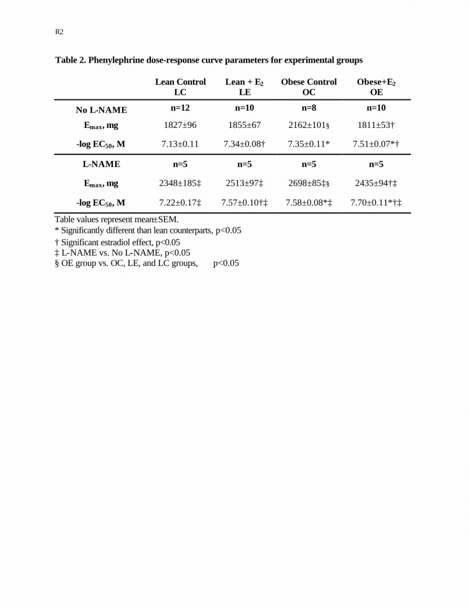
Table 2. Phenylephrine dose-response curve parameters for experimental groups
Lean Control
LC Lean + E2
LE Obese Control
OC Obese+E2
OE
No L-NAME n=12 n=10 n=8 n=10
Emax, mg 1827±96 1855±67 2162±101§ 1811±53†
-log EC50, M 7.13±0.11 7.34±0.08† 7.35±0.11* 7.51±0.07*†
L-NAME n=5 n=5 n=5 n=5
Emax, mg 2348±185‡ 2513±97‡ 2698±85‡§ 2435±94†‡
-log EC50, M 7.22±0.17‡ 7.57±0.10†‡ 7.58±0.08*‡ 7.70±0.11*†‡
Table values represent mean±SEM. * Significantly different than lean counterparts, p<0.05 † Significant estradiol effect, p<0.05 ‡ L-NAME vs. No L-NAME, p<0.05 § OE group vs. OC, LE, and LC groups, p<0.05

R2
Maximal Tension Development to 60mM KCl
Lean Obese
Ten
sion
(mg)
0
200
400
600
800
1000
1200
1400
1600
1800
2000
2200Control Estrogen
**

R2
-9 -8 -7 -6 -5
0
500
1000
1500
2000
2500
LC
LE
OCOE
log [Phenylephrine, M]
Ten
sion
(m
g)

R2
-9 -8 -7 -6 -5-500
0
500
1000
1500
2000
2500
3000
LC with L-NAME
LE with L-NAME
LC
LE
A
log [Phenylephrine, M]
Ten
sion
(mg)
-9 -8 -7 -6 -5-500
0
500
1000
1500
2000
2500
3000
3500
OC with L-NAME
OC
OE
OE with L-NAME
B
log [Phenylephrine, M]
Ten
sion
(mg)

R2
Influence of Indomethacin (INDO) on Maximal ContractileResponse (Emax) to Phenylephrine: Control vs. Estrogen
Control Estrogen0
500
1000
1500
2000
2500
No INDO
INDO
*
*
Ten
sion
(mg)
Influence of Indomethacin (INDO) on Vascular Sensitivityto Phenylephrine: Control vs. Estrogen
Control Estrogen5.0
5.5
6.0
6.5
7.0No INDO
INDO
* *
-log
EC
50

R2
-9 -8 -7 -6 -5
-20
0
20
40
60
80
100
LC
LE
OC
OE
log [Acetylcholine, M]
%R
elax
atio
n

R2
Emax: Response to ACh
Lean Obese75
80
85
90
95Control
Estrogen
**
*†§
Treatment
%R
elax
atio
n
EC50: Response to ACh
Lean Obese7.0
7.1
7.2
7.3
7.4
7.5
7.6
7.7 Control
Estrogen
*†
*
†§
Treatment
-log
[AC
h, M
]
*
†§

R2
NOSIII Protein Expression
Lean Obese
% o
f Obe
se C
ontr
ol G
roup
0
50
100
150
200
250
Control Estrogen
*
**

R2
NOSIII 140 kDa
LC LE OC OE


