Embed Size (px)
Citation preview

OPENACCESS
TRANSPARENTPROCESS
Research ArticleAntigen-specific CD8þ T cells in cerebral malaria
916
Brain microvessel cross-presentation is ahallmark of experimental cerebral malaria
Shanshan W. Howland1, Chek Meng Poh1,2, Sin Yee Gun1,2, Carla Claser1, Benoit Malleret1,Nilabh Shastri5, Florent Ginhoux1, Gijsbert M. Grotenbreg2,3,4, Laurent Renia1,2*
Keywords: brain; cross-presentation;
malaria; pathology; T cells
DOI 10.1002/emmm.201202273
Received November 14, 2012
Revised April 04, 2013
Accepted April 05, 2013
GSee accompanying article
http://dx.doi.org/10.1002/emmm.201302849
(1) Singapore Immunology Network, Agency for Scien
Research (A�STAR), Singapore, Singapore
(2) Department of Microbiology, Life Sciences Institute
of Medicine, National University of Singapore, Sing
(3) Faculty of Science, Department of Biological Sci
Institute, Yong Loo Lin School of Medicine, Na
Singapore, Singapore, Singapore
(4) Immunology Programme, Life Sciences Institute, Yo
Medicine, National University of Singapore, Singapo
(5) Division of Immunology and Pathogenesis, Departm
Cell Biology, University of California, Berkeley, CA, U
*Corresponding author: Tel: þ65 6407 0005; Fax: þ6
E-mail: [email protected]
� 2013 The Authors. Published by John Wiley and Sons,the terms of the Creative Commons Attribution License (Cin any medium, provided the original work is properly cite
Cerebral malaria is a devastating complication of Plasmodium falciparum infec-
tion. Its pathogenesis is complex, involving both parasite- and immune-mediated
events. CD8þ T cells play an effector role in murine experimental cerebral malaria
(ECM) induced by Plasmodium berghei ANKA (PbA) infection. We have identified a
highly immunogenic CD8 epitope in glideosome-associated protein 50 that is
conserved across rodent malaria species. Epitope-specific CD8þ T cells are
induced during PbA infection, migrating to the brain just before neurological
signs manifest. They are functional, cytotoxic and can damage the blood–brain
barrier in vivo. Such CD8þ T cells are also found in the brain during infection with
parasite strains/species that do not induce neuropathology. We demonstrate here
that PbA infection causes brain microvessels to cross-present parasite antigen,
while non-ECM-causing parasites do not. Further, treatment with fast-acting
anti-malarial drugs before the onset of ECM reduces parasite load and thus
antigen presentation in the brain, preventing ECM death. Thus our data suggest
that combined therapies targeting both the parasite and host antigen-presenting
cells may improve the outcome of CM patients.
INTRODUCTION
Malaria remains one of the most important global health
problems, affecting more than 200 million people and causing
655,000 deaths in 2010, most of them young children in Africa
(World Health Organization, 2011). The most severe patho-
logical complication of Plasmodium falciparum infection termed
human cerebral malaria (CM) is estimated to account for three-
quarters of the parasite’s death toll (Brewster et al, 1990).
Although not completely identical to the human disease, animal
ce, Technology and
, Yong Loo Lin School
apore, Singapore
ences, Life Sciences
tional University of
ng Loo Lin School of
re, Singapore
ent of Molecular and
SA
5 6464 2056;
Ltd on behalf of EMBO. ThisC BY 3.0), which permits ud.
models have complemented clinical studies and in vitro
experiments aimed at understanding the pathogenesis of CM.
The most established of these is the infection of susceptible
mice (e.g. C57BL and CBA backgrounds) with the ANKA strain
of Plasmodium berghei (PbA). In this model of experimental
cerebral malaria (ECM), at least 60% of susceptible mice
develop neurological symptoms (ataxia, paralysis, head
deviation, convulsions) culminating in coma and then death
6–12 days after inoculation with infected red blood cells
(Engwerda et al, 2005). ECM is characterized by intravascular
accumulation of infected red blood cells and leukocytes in the
brain, petechial hemorrhages and breakdown of the blood–
brain barrier (Thumwood et al, 1988).
Knockout mice have been instrumental in uncovering the cell
types involved in ECM. Mice deficient in CD4þ T cells, CD8þ T
cells, interferon-g (IFN-g) or its receptor are resistant to ECM,
while B-cell-deficient mice remain susceptible (Amani et al,
2000; Yanez et al, 1996). The role of CD4þ T cells in C57BL/6
mice is restricted to the earlier induction phase of ECM, as
antibody depletion of these cells prevented ECM if performed
4 days post-infection (p.i.) but not 6 days p.i.; in contrast, CD8þ
T-cell depletion at the later time point, just 1 day before the onset
of neurological symptoms, completely abrogated ECM death
is an open access article underse, distribution and reproduction
EMBO Mol Med (2013) 5, 916–931

www.embomolmed.org Research ArticleShanshan W. Howland et al.
(Belnoue et al, 2002). It has recently been shown that IFN-g
production by CD4þ T cells recruits CD8þ T cells to the brain
(Belnoue et al, 2008; Villegas-Mendez et al, 2012). Both perforin
and Granzyme B (GrB) are essential for ECM, suggesting that
damage to the blood–brain barrier may be a direct result of CD8þ
T-cell cytolysis (Haque et al, 2011; Nitcheu et al, 2003).
Although considerable evidence implicates cytotoxic CD8þ
T cells as the proximal cause of neuropathology in ECM, the
specificities of these cells has remained a mystery. Studies with
transgenic parasites bearing a model epitope from chicken
ovalbumin confirmed that parasite-specific, brain-sequestered
CD8þ T cells are indeed induced during infection (Lundie et al,
2008; Miyakoda et al, 2008). However, this immunodominant
model epitope may not reflect immune responses against native
malaria antigens. Further, such a transgenic system is not easily
comparable to the human CM situation and hinders comparative
studies between rodent malaria strains differing in their ability
to induce ECM. Despite (or perhaps because of) the�5500 genes
in P. berghei, not one CD8 epitope had yet been identified
in C57BL/6 mice at the start of this work, prompting us to
supply this deficiency.
Our epitope identification strategy builds upon an established
NFAT-lacZ reporter system for T-cell receptor (TCR) signalling
(Sanderson & Shastri, 1994). Whereas the original approach
fused T cells with partners bearing the NFAT-lacZ cassette, we
sequenced TCR genes from individual T cells to select an over-
represented pair to transduce into the reporter cells. By
screening the TCR-transduced reporter cells against a library
of antigen-presenting cells expressing PbA cDNA fragments, we
sought to identify the cognate antigen in the library member/s
able to induce lacZ expression (see schematic in Fig 1). To
improve our chances of finding a highly immunogenic epitope,
we focused our efforts on CD8þ T cells bearing the Vb8 gene
segment, which have been associated with ECM in susceptible
mice (Belnoue et al, 2002; Boubou et al, 1999).
RESULTS
TCR sequencing of brain-sequestered CD8R T cells reveals an
over-represented motif
We sorted Vb8.1,2þ CD8þ T cells from the brains of PbA-infected
C57BL/6 mice exhibiting neurological signs and subjected these to
single cell TCR sequencing. A clear motif emerged after a relatively
small number of TCR genes were sequenced. Of 18 Vb8.1 cells,
13 shared a ‘‘DWG’’ peptide sequence within the TCRb junction
(Table 1). These were paired with TCRa genes bearing a variety of
Va segments. Three cells from one mouse shared identical TCRa
and b genes, indicating clonal expansion. We therefore selected
this TCR pair to transduce into reporter cells bearing an NFAT-
lacZ cassette, creating the LR-BSL8.4a cell line, so as to begin
screening for the cognate antigen.
Glideosome-associated protein 50 contains the cognate
epitope
We created a library of EL4 cells (syngeneic for MHC genes
with C57BL/6) expressing fragments of cDNA isolated from
EMBO Mol Med (2013) 5, 916–931 �
blood-stage PbA. Pools of the library cells were incubated with
the LR-BSL8.4a TCR-transduced reporter cells in 96-well plates
that were then stained for lacZ expression. One well was found
to have 22 blue spots (versus a median of two in control wells,
Fig 2A and B). Individual clones from the positive library pool
were screened again, resulting in three positive wells with
thousands of blue cells (Fig 2C). The positive EL4 clones were all
sequenced and found to contain a fragment of P. berghei
glideosome-associated protein 50 (PbGAP50, amino acids 40–
119). Potential H-2Db and Kb epitopes within this fragment were
predicted with a computer algorithm and used to generate
peptide-MHC tetramers by a rapid peptide-exchange strategy
(Grotenbreg et al, 2008; Toebes et al, 2006). One tetramer was
able to label LR-BSL8.4a cells, leading us to conclude that the
TCR recognizes the H-2Db-restricted peptide SQLLNAKYL
(Fig 2D).
SQLLNAKYL-specific brain-sequestering CD8R T cells are
induced during PbA infection
Equipped with the SQLLNAKYL-H-2Db tetramer, we set out to
investigate the extent, kinetics and localization of the CD8þ
T-cell response during PbA infection. Leukocytes from the
spleens, blood and brains of naıve and infected C57BL/6 mice
were subjected to tetramer staining, with CD8þ T cells identified
by CD8 alpha staining and lack of CD16/32 staining (since CD8þ
T ells lack the Fc receptors CD16 and CD32) (Fig 3A–D). A
distinct population of tetramer-positive cells (typically 0.5–2%
of spleen CD8þ T cells and 2–9% of brain CD8þ T cells) was seen
in infected but not naıve mice, validating the epitope specificity.
Consistent with reports that the CD8 immune response in PbA is
primed by splenic CD11chi Clec9Aþ dendritic cells (deWalick
et al, 2007; Piva et al, 2012), we saw that SQLLNAKYL-specific T
cells appeared in some spleens as early as 5 days p.i. (Fig 3E),
whereas they expanded in the blood only from day 6 onwards
(Fig 3F). These cells started migrating to the brain after 6 days
but their numbers did not reach statistical significance until
7 days p.i. (Fig 3G), a time when some mice started exhibiting
neurological signs. On this day, there was no apparent
correlation between the number of specific T cells in the brain
and the clinical condition of the mice, suggesting that the
localization of these cells in the brain precedes the development
of ECM symptoms. Note that while SQLLNAKYL is predicted to
bind to both H-2Db and H-2Kb MHC molecules, the increase in
tetramer staining of T cells during infection was seen only with
the H-2Db and not the H-2Kb tetramer.
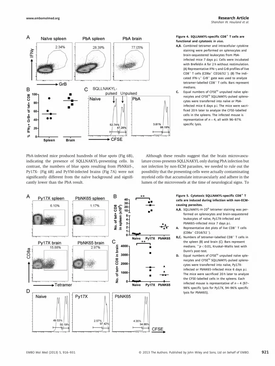
SQLLNAKYL-specific CD8R T cells are functional and cytotoxic
We further characterized the SQLLNAKYL-specific CD8þ T cells
by combining MHC tetramer analysis with intracellular staining
for IFN-g and GrB. A population of IFN-gþ GrBþ CD8þ T cells
was present in the spleen 7 days p.i., and most of the brain-
sequestered CD8þ T cells exhibited a similar cytokine produc-
tion profile (Fig 4A). A substantial proportion of the IFN-gþ
GrBþ CD8þ T cells were tetramer-positive: 3–9% in the spleen
and 4–12% in the brain (Supporting Information Fig S1). No ex
vivo restimulation had been performed, so these CD8þ T cells
must have recently encountered their cognate peptide-MHC
2013 The Authors. Published by John Wiley and Sons, Ltd on behalf of EMBO. 917

Research Article www.embomolmed.orgAntigen-specific CD8þ T cells in cerebral malaria
Figure 1. Schematic of antigen identification strategy. Single cell TCR sequencing is performed on Vb8.1,2þ CD8þ T cells sorted from the brains of PbA-infected
C57BL/6 mice with ECM symptoms. The selected pair of TCR genes is transduced into a reporter cell bearing an NFAT-lacZ cassette. The reporter cells, LR-BSL8.4a,
are used to screen a library of EL4 cells transduced to express fragments of PbA cDNA. Upon encountering the cognate peptide-MHC complex, the reporter cells
express lacZ and are detected as blue spots following b-galactosidase staining. EL4 library cells from positive wells are cloned and re-screened to narrow down to
single clones that are sequenced to identify the cognate antigen.
918
in vivo. At least 40% of the SQLLNAKYL-specific CD8þ T cells in
the spleen and at least 70% in the brain were IFN-gþ GrBþ
(Fig 4B), consistent with a role in ECM pathology. Since both
perforin and GrB have been demonstrated to be necessary for
ECM (Haque et al, 2011; Nitcheu et al, 2003), we examined
whether SQLLNAKYL-specific CD8þ T cells had the ability to kill
in an in vivo cytolysis assay. SQLLNAKYL-pulsed splenocytes
transferred into PbA-infected mice were almost obliterated
relative to unpulsed splenocytes (Fig 4C). Taken together, these
results point towards cytotoxic SQLLNAKYL-specific CD8þ T
cells being in the right place at the right time to presumably
damage the cells of the brain microvasculature.
� 2013 The Authors. Published by John Wiley and Sons, Ltd on behalf of EMBO.
SQLLNAKYL-specific CD8R T-cell response with non-ECM
parasites
While PbA induces ECM in C57BL/6 mice, there are other strains
and species of rodent malaria that do not; determining why
they differ is a key piece of the ECM etiology puzzle. The
SQLLNAKYL epitope is conserved in the published genome
sequence of P. yoelii 17X clone 1.1 non-lethal strain (Py17X;
Carlton et al, 2002) and in the GAP50 protein of P. yoelii 17X
clone YM lethal strain (PyYM) (PYYM_0822000). We sequenced
the GAP50 gene in the NK65 strain of P. berghei (PbNK65) and
found it to be identical to the PbA sequence. Py17X, PyYM and
PbNK65 strains do not induce ECM, and we asked whether the
EMBO Mol Med (2013) 5, 916–931

www.embomolmed.org Research ArticleShanshan W. Howland et al.
Table 1. Vb8.1 TCR sequences derived from brain-sequestered CD8 T cells during ECM
Cell code TRBV TRBJ TRBD TCRb junction TRAV TRAJ TCRa junction
8.16 13-3 1-1 1 CASSRDRANTEVFF
8.23 13-3 1-3 1 CASSDWGHSGNTLYF 17 57 CALEGRQGGSAKLIF
6.22 13-3 1-3 1 CASSEQGSGNTLYF 3-3 45 CAVSDAEGADRLTF
8.14 13-3 1-3 1 CASSERGSGNTLYF
8.4 13-3 2-1 2 CASSDWGAGAEQFF 4D-4 52 CAAEANTGANTGKLTF
8.28 13-3 2-1 2 CASSDWGAGAEQFF 4D-4 52 CAAEANTGANTGKLTF
8.31 13-3 2-1 2 CASSDWGAGAEQFF 4D-4 52 CAAEANTGANTGKLTF
8.9 13-3 2-1 1 CASSLPGQGWAEQFF
6.16 13-3 2-2 2 CASSDWGDTGQLYF
8.6 13-3 2-4 2 CASSADWGGQNTLYF 4D-3 48 CAGGGNEKITF
6.23 13-3 2-4 2 CASSDWGSQNTLYF 8-2 42 CATVTGGSNAKLTF
6.29 13-3 2-5 2 CASSDWGGQDTQYF 8D-1 35 CATGG#TGFASALTF
6.2 13-3 2-5 2 CASSDWGNQDTQYF 16D 57 CAMSPQGGSAKLIF
8.15 13-3 2-5 2 CASSDWGNQDTQYF 14-2 39 GGAKLTF
6.30 13-3 2-5 2 CASSDWGQDTQYF
6.1 13-3 2-5 2 CASSDWGTQDTQYF 12D-2 30 CALSDGTNAYKVIF
8.19 13-3 2-5 2 CASSDWGVQDTQYF 8D-1 56 CARPMATGGNNKLTF
6.17 13-3 2-7 1 CASSGTGTSSYEQYF
Single-cell TCR sequencing was performed on Vb8.1,2þ CD8þ T cells isolated from the brains of two mice 7 days after infection with PbA. Sequences were analyzed
with IMGT/V-QUEST and only Vb8.1 sequences (TRBV13-3 in IMGT notation) are reported here. Highlighted rows share a ‘DWG’ motif (underlined) in the TCRb
junction. TCRa sequences could not be obtained from some cells.
CD8þ T-cell response against the SQLLNAKYL epitope could be
missing or defective during infection with these strains, which
could account for the lack of neuropathology. Surprisingly,
MHC tetramer staining performed 7 days p.i. showed that not
only were SQLLNAKYL-specific CD8þ T cells induced during
infection with Py17X and PbNK65, they also accumulated in the
EMBO Mol Med (2013) 5, 916–931 �
brain (Fig 5A–C). Indeed, the SQLLNAKYL epitope may be
amongst the most immunodominant epitopes in Py17X, with the
proportion of tetramer-labelled CD8þ T cells exceeding 15% in
several brains (Fig 5A). While the numbers of tetramer-labelled
CD8þ T cells in the organs of PbNK65-infected mice did not
reach significance in this non-parametric three-group analysis,
they are comparable to those seen earlier with PbA (Fig 5B and
C). Furthermore, the specific CD8þ T cells were not cytolytically
defective in mice infected with the non-ECM strains, as
demonstrated in the in vivo cytotoxicity assay where transferred
SQLLNAKYL-pulsed cells were killed (Fig 5D).
PbA but not non-ECM parasites induces brain vessel
cross-presentation
The observation that SQLLNAKYL-specific CD8þ T cells were
not absent during infection with parasites that do not cause ECM
prompted us to consider whether there was a lack of target cells
for the cytolytic T cells to recognize. We and others have
proposed that the brain endothelium becomes activated during
Figure 2. Identification of PbGAP50 as the antigen recognized by a Vb8.1
TCR associated with ECM.
A–C. Well images of X-Gal-stained LR-BSL8.4a reporter cells incubated
overnight with a library of EL4 cells transduced with P. berghei cDNA
fragments. (A) A representative negative well. (B) The positive well in the
first round of screening, containing about 250 library clones. An inset
showing two blue cells at the original resolution is included. (C) A
positive well in the second round of screening, containing a single
library clone that was found to contain a fragment of PbGAP50.
D. Predicted MHC epitopes in the fragment were used to generate peptide-
MHC tetramers for staining LR-BSL8.4a cells. Open histogram: repre-
sentative non-cognate peptide-MHC tetramer. Filled histogram:
SQLLNAKYL-H-2Db tetramer.
2013 The Authors. Published by John Wiley and Sons, Ltd on behalf of EMBO. 919

Research Article www.embomolmed.orgAntigen-specific CD8þ T cells in cerebral malaria
Figure 3. SQLLNAKYL-specific CD8R T cells are induced during PbA infection and migrate to the brain just prior to the development of neurological signs.
SQLLNAKYL-H-2Db tetramer staining was performed on splenocytes, blood and brain-sequestered leukocytes of naıve and PbA-infected mice 5, 6 and
7 days p.i.
A–D. Representative dot plots of live cells, showing the gating of CD8þ T cells (CD8aþ CD16/32�) for tetramer analysis. (A) Naıve spleen. (B) Day 7 p.i. spleen.
(C) Naıve brain. (D) Day 7 p.i. brain.
E–G. Analysis of tetramer-labelled cells amongst CD8þ T cells in the spleen (E), blood (F) and brain (G). Bars represent medians. �p< 0.05, ��p<0.01, Kruskal–
Wallis test with Dunn’s post-test. Results are representative of three experiments.
920
infection, acquiring the ability to take up parasite material and
cross-present parasite antigens, thus becoming targets for killing
by specific CD8þ T cells (Belnoue et al, 2002; Nitcheu et al, 2003;
Pino et al, 2005; Renia et al, 2006). We realized that the LR-
BSL8.4a TCR-transduced reporter cells could potentially be used
to detect such cross-presentation. Based on established proto-
cols (Song & Pachter, 2003; Wu et al, 2003), we developed a
technique to isolate microvessel fragments from homogenized
mouse brains using dextran gradient centrifugation to remove
� 2013 The Authors. Published by John Wiley and Sons, Ltd on behalf of EMBO.
the myelin-rich brain parenchyma tissue, followed by size
fractionation to separate the vessel fragments from cells in
suspension (Fig 6A). Brain microvessels were isolated from
naıve mice and mice infected 7 days earlier with PbA, PbNK65 or
Py17X. After collagenase digestion of the vessel basal lamina,
they were then incubated with LR-BSL8.4a cells overnight and
stained for lacZ expression to determine if the reporter cells
had encountered their cognate epitope. While the background
staining from naıve mice amounted to only 10–20 blue spots,
EMBO Mol Med (2013) 5, 916–931
www.embomolmed.org Research ArticleShanshan W. Howland et al.
Figure 4. SQLLNAKYL-specific CD8R T cells are
functional and cytotoxic in vivo.
A,B. Combined tetramer and intracellular cytokine
staining were performed on splenocytes and
brain-sequestered leukocytes from PbA-
infected mice 7 days p.i. Cells were incubated
with Brefeldin A for 2 h without restimulation.
(A) Representative IFN-g and GrB profiles of live
CD8þ T cells (CD8aþ CD16/32�). (B) The indi-
cated IFN-gþ GrBþ gate was used to analyze
tetramer-labelled CD8þ T cells. Bars represent
medians.
C. Equal numbers of CFSEhi unpulsed naıve sple-
nocytes and CFSElo SQLLNAKYL-pulsed spleno-
cytes were transferred into naıve or PbA-
infected mice 6 days p.i. The mice were sacri-
ficed 20 h later to analyze the CFSE-labelled
cells in the spleens. The infected mouse is
representative of n¼4, all with 96–97%
specific lysis.
PbA-infected mice produced hundreds of blue spots (Fig 6B),
indicating the presence of SQLLNAKYL-presenting cells. In
contrast, the numbers of blue spots resulting from PbNK65-,
Py17X- (Fig 6B) and PyYM-infected brains (Fig 7A) were not
significantly different from the naıve background and signifi-
cantly lower than the PbA result.
EMBO Mol Med (2013) 5, 916–931 �
Although these results suggest that the brain microvascu-
lature cross-presents SQLLNAKYL only during PbA infection but
not infection by non-ECM parasites, we needed to rule out the
possibility that the presenting cells were actually contaminating
myeloid cells that accumulate intravascularly and adhere in the
lumen of the microvessels at the time of neurological signs. To
Figure 5. Cytotoxic SQLLNAKYL-specific CD8R T
cells are induced during infection with non-ECM-
causing parasites.
A,B. SQLLNAKYL-H-2Db tetramer staining was per-
formed on splenocytes and brain-sequestered
leukocytes of naıve, Py17X-infected and
PbNK65-infected mice 7 days p.i.
A. Representative dot plots of live CD8þ T cells
(CD8aþ CD16/32�).
B,C. Numbers of tetramer-labelled CD8þ T cells in
the spleen (B) and brain (C). Bars represent
medians. ��p< 0.01, Kruskal–Wallis test with
Dunn’s post-test.
D. Equal numbers of CFSEhi unpulsed naıve sple-
nocytes and CFSElo SQLLNAKYL-pulsed spleno-
cytes were transferred into naıve, Py17X-
infected or PbNK65-infected mice 6 days p.i.
The mice were sacrificed 20 h later to analyze
the CFSE-labelled cells in the spleens. Each
infected mouse is representative of n¼4 (97–
98% specific lysis for Py17X, 94–96% specific
lysis for PbNK65).
2013 The Authors. Published by John Wiley and Sons, Ltd on behalf of EMBO. 921

Research Article www.embomolmed.orgAntigen-specific CD8þ T cells in cerebral malaria
922
do this, we isolated the brain leukocytes, including monocytes/
macrophages and neutrophils (Belnoue et al, 2002) and tested
them for the ability to trigger lacZ expression in LR-BSL8.4a
cells. Although some cross-presentation was detected from the
brain leukocytes of PbA-infected mice, the signal was less than
one-tenth of the signal resulting from brain microvessels in the
same experiment (Fig 6C). Therefore, even if the microvessel
Figure 6.
� 2013 The Authors. Published by John Wiley and Sons, Ltd on behalf of EMBO.
preparations contain some myeloid cells, such cells cannot
be the primary source of the detected cross-presentation.
To investigate the role of myeloid cells in another way, we
employed the MAFIA transgenic mouse model for macrophage/
granulocyte depletion via a drug-inducible suicide gene (Burnett
et al, 2004). MAFIA mice treated with the drug 5, 6 and 7 days
p.i. were previously shown to have a �80% reduction of these
EMBO Mol Med (2013) 5, 916–931

www.embomolmed.org Research ArticleShanshan W. Howland et al.
3
myeloid cells in the blood and brain while remaining susceptible
to ECM (Claser et al, 2011). We observed no reduction in brain
microvessel cross-presentation in drug-treated MAFIA mice
when compared to either untreated MAFIA mice or C57BL/6
mice (Supporting Information Fig S2A). Drug treatment even
increased the number of blue spots, indicating that some
myeloid cells may either compete for parasite antigen or regulate
cross-presentation by the microvessels.
The role of collagenase digestion during the microvessel
isolation protocol is to break down the basal lamina,
allowing the exposed endothelial cells to protrude (Song &
Pachter, 2003). When we omitted collagenase during the
brain microvessel cross-presentation assay, the number of blue
spots decreased by 80–90% (Supporting Information Fig S2B),
suggesting that the cells responsible for cross-presentation may
be those surrounded by the basal lamina, i.e. the endothelial
cells or pericytes. We used scanning electron microscopy (SEM)
to visualize interactions between the microvessels and LR-
BSL8.4a cells. Note that both before (Fig 6D) and after (Fig 6E)
collagenase digestion, the microvessels are in the form of
multicellular tubes. After prolonged co-incubation (24 h in
Fig 6F), many of the vessels split open, presumably as
endothelial cells migrate outwards. We have captured images
of LR-BSL8.4a cells forming cell–cell junctions with both the
outer surface of an intact vessel (Fig 6E) as well as the lumenal
surface of a split vessel (Fig 6F). These cytoskeletal rearrange-
ments may represent interactions akin to immunological
synapses, with the cross-presenting cells being part of the
microvessel walls. From these lines of evidence, we infer that
cells constituting the blood–brain barrier, most probably
endothelial cells, cross-present parasite antigens during PbA
infection, thus becoming targets for CD8þ T-cell cytolysis. In
PbNK65, Py17X and PyYM infection, on the other hand, the
blood–brain barrier remains intact because there is little or no
cross-presentation.
Cross-presentation is associated with PbA parasite specific
accumulation in the brain
Infected red blood cell accumulation in the brain capillaries have
been recently strongly associated with the development of ECM
Figure 6. SQLLNAKYL is cross-presented by brain microvessels during PbA inf
A. Schematic of brain microvessel cross-presentation assay. Mouse brains were
of 30% dextran. The pellets were resuspended and passed over 40 mm cell
DNase I. After washing, the microvessel fraction from each brain was divided
cells overnight, prior to b-galactosidase staining.
B. Brain microvessel cross-presentation results from naıve mice and mice 7 days
group. ��p< 0.01, ���p<0.001, ANOVA with Bonferroni’s post-test on log-tr
C. Brain-sequestered leukocytes from naive mice and mice 7 days post-infecti
overnight prior to b-galactosidase staining. The brain microvessel cross-pre
time. Bars represent means of 3–5 mice/group. �p<0.05, ��p< 0.01, ����p<0
arising from each brain.
D,E. Scanning electron microscopy images of brain microvessel fragments from
D. Brain microvessel before collagenase digestion, not mixed with LR-BSL8.4a
E. Digested microvessel co-incubated with LR-BSL8.4a cells for 4 h.
F. Digested microvessel co-incubated with LR-BSL8.4a cells for 24 h. Curly bra
junctions LR-BSL8.4a cells have formed with microvessels.
EMBO Mol Med (2013) 5, 916–931 �
(Amante et al, 2010; Baptista et al, 2010; Claser et al, 2011;
McQuillan et al, 2011). We hypothesize that the difference in
cross-presentation may reflect a difference in sequestration
between ECM and non-ECM parasites. To test this, we used
the ECM-inducing line PbAluc and the non-ECM-inducing line
PyYMluc, which both possess the SQLLNAKYL epitope. Both
lines are tagged with luciferase allowing us to assess parasite
accumulation in deep tissues by measuring bioluminescence in
the heads or brains isolated from infected mice after injection of
the luciferase substrate (Supporting information Fig S3) (Claser
et al, 2011). We first confirmed that microvessels from mice
infected with the non-ECM line PyYMluc did not cross-present
the SQLLNAKYL epitope in contrast to those from mice infected
with PbA, the parental line of PbAluc (Fig 7A). When parasite
biological parameters were compared between PbAluc and
PyYMluc, we observed that at the time of ECM signs for PbAluc
(Day 7), PyYM parasitemia was higher than that of PbAluc and
no difference in bioluminescence were detected when the heads
of the infected animals were imaged (Fig 7B and C). However,
bioluminescence in the perfused brains of infected mice was one
log lower with PyYMluc compared to PbAluc (Fig 7D). This
suggests that local accumulation of PbA-infected red blood
cells may lead to increased contact to and/or cytoadherence to
brain microvessels, facilitating antigen uptake required for
cross-presentation.
SQLLNAKYL-specific CD8R T cells damage the blood–brain
barrier
Are the SQLLNAKYL-specific CD8þ T cells able to compromise
the blood–brain barrier by killing endothelial cells presenting
the epitope? To support this model, we devised an experiment to
demonstrate that SQLLNAKYL-specific CD8þ T cells contribute
to the neuropathology seen during ECM. Two groups reported
that treatment with anti-malarial drugs 1 day before ECM is
expected prevents the development of neurological signs even
though CD8þ T cells still accumulate in the brain (Baptista et al,
2010; Haque et al, 2011). We postulated that anti-malarial drug
treatment would severely reduce the amount of parasite antigen
available in the brain to be cross-presented by the micro-
vasculature, thus preventing ECM. However, if the microvas-
ection but not during infection by parasites that do not cause ECM.
minced, homogenized through a needle and centrifuged with an equal volume
strainers, and the retained microvessels were digested with collagenase and
between five wells of a 96-well filter plate and co-incubated with LR-BSL8.4a
post-infection with PbA, PbNK65 or Py17X. Bars represent means of 3–4 mice/
ansformed numbers. Results are representative of three experiments.
on with PbA or Py17X were purified and co-incubated with LR-BSL8.4a cells
sentation assay was performed on additional PbA-infected mice at the same
.0001, ANOVA with Bonferroni’s post-test on log-transformed total spot counts
mice infected 7 days previously with PbA. Scale bars indicate 1 mm.
cells.
ces indicate LR-BSL8.4a cells and arrowheads indicate apparent cell–cell
2013 The Authors. Published by John Wiley and Sons, Ltd on behalf of EMBO. 923

Research Article www.embomolmed.orgAntigen-specific CD8þ T cells in cerebral malaria
Figure 7. Cross-presentation of SQLLNAKYL is
associated with parasite accumulation in the
brain of infected mice.
A. Brain microvessel cross-presentation results
from naıve mice and mice 7 days post-infec-
tion with PyYMluc or PbA. Bars represent
means of 2–4 mice/group. ���p< 0.01, ANOVA
with Bonferroni’s post-test on log-transformed
total spot counts arising from each brain.
B–D. Parasitemia (B), head luminescence (C), and (D)
luminescence of isolated and perfused brains
from mice infected with PbAluc or PyYMluc.
Parasitemia levels were determined at Days 5–
7. Bars represent the mean� SD. ���p<0.01,
Mann–Whitney test. Luminescence was
measured at day 7 post-infection. Bars
represent the mean. ��p< 0.01, t-test on
values transformed with x0 ¼ log(xþ 1).
924
culature was then exposed to soluble SQLLNAKYL peptide via
repeated intravenous (i.v.) injection (see schedule in Fig 8A),
then the specific CD8þ T cells present in the brain vasculature
would presumably be able to recognize the peptide loaded onto
class I MHC molecules.
First, we investigated the effects of combined chloroquine
and artesunate drug treatment for a 24-h period starting 6 days
p.i. with PbA. Peripheral parasitemia levels dropped by an
order of magnitude after treatment, and as expected, the parasite
biomass in the brain (as measured by in vivo imaging of PbAluc
in a separate experiment) was likewise reduced (Supporting
Information Fig S4). This treatment also protected all mice
from developing ECM. As expected, immediately following
the treatment, there was no significant change in the number
of SQLLNAKYL-specific CD8þ T cells in the brain compared
to untreated mice (Fig 8B). Conversely, there was a drastic
reduction in brain microvessel cross-presentation of the
SQLLNAKYL epitope, down to essentially background levels
(Fig 8C). In an additional control experiment, we verified that
reduced endogenous presentation of the SQLLNAKYL epitope
was not due to an inhibition of MHC class I expression by the
drugs used to clear the parasites. Indeed, chloroquine has been
known to affect MHC molecules (Belizaire & Unanue, 2009;
Kurotaki et al, 2007). Thus, we devised a modified presentation
assay where the peptide SIINFEKL corresponding to the major
CD8 epitope of chicken ovalbumin was used to pulse brain
microvessels before mixing together with a reporter cell line
recognizing the OVA peptide in the context of MHC class I
molecule. We chose this peptide over the SQLLNAKYL peptide
� 2013 The Authors. Published by John Wiley and Sons, Ltd on behalf of EMBO.
to eliminate any cofounding factor due to endogenous
presentation of the SQLLNAKYL peptide. We observed that
exogenous addition of the OVA peptide to the microvessels from
untreated PbA-infected mice induces similar numbers of blue
spots compared to those from chloroquine/artesunate-treated
infected mice, with both groups giving a much higher response
than naive mice (Supporting Information Fig S5). This means
that more MHC class I molecules are expressed by brain
microvessels after PbA infection and drug treatment did not
affect MHC class I expression. All together, this indicates that in
situ parasite load controls parasite antigen presentation and is a
key factor for ECM to develop.
To further investigate whether CD8þ T-cell recognition of
SQLLNAKYL epitope presented in the brain in vivo contributes
to ECM pathology, SQLLNAKYL peptide or the irrelevant
SIINFEKL peptide (the immunodominant epitope from ovalbu-
min in C57BL/6 mice) was injected i.v. after drug treatment. We
observed that SQLLNAKYL-treated mice (eight out of nine)
displayed early signs of ECM (ruffled fur, hunching and
lethargy) but did not progress to paralysis or coma whether
or not additional peptide injections were administered. None of
the eight mice treated with the control peptide exhibited these
signs. On hindsight, the mild pathology was not surprising
because SQLLNAKYL-specific CD8þ T cells accounted for only
4–12% of the IFN-gþGrBþ CD8þ T cells in the brain (Supporting
Information Fig S1). Nevertheless, we sought a more sensitive
and objective method of detecting damage to the blood–brain
barrier and adopted the approach of Hermsen et al, who
intravenously injected folic acid into mice infected with
EMBO Mol Med (2013) 5, 916–931

www.embomolmed.org Research ArticleShanshan W. Howland et al.
Figure 8. SQLLNAKYL-specific CD8R T cells damage the blood–brain
barrier.
A. Experiment schedule for (D). Mice infected with PbA were treated with
chloroquine and artesunate (CQ/ART) between 6 and 7 days p.i., then
injected three times i.v., 4 h apart with 100 mg of either SQLLNAKYL
peptide or control SIINFEKL peptide. They were injected with folic acid
8 days p.i. to test the integrity of the blood–brain barrier.
B,C. Mice infected with PbA were untreated or treated with CQ/ART between
6 and 7 days p.i., then sacrificed soon after.
B. Tetramer staining was performed on the brain-sequestered leukocytes.
Bars represent medians. ns, not significant by Mann–Whitney test.
C. Microvessels were isolated from each brain and co-incubated with
LR-BSL8.4a cells overnight. The total number of blue spots after
b-galactosidase staining is reported. Bars represent means. ���p< 0.001,
Student t-test on log-transformed data.
D. Mice subjected to the protocol in (A) either convulsed and died within
90 min or survived until the next day. p¼0.0033, Fisher’s exact test.
E. Mice infected with Py17X were injected i.v. with 100 mg of either
SQLLNAKYL or SIINFEKL peptide 7 days p.i., then challenged with folic
acid 6 h later. p¼0.0047, Fisher’s exact test.
P. berghei K173. In C57BL/6 mice infected with this parasite, the
neurotoxin induced convulsions and death within 90 min of
injection, whereas an intact blood–brain barrier protected
uninfected mice (Hermsen et al, 1998). We performed the folic
acid challenge 8 days p.i. on PbA-infected mice that had
undergone drug treatment between 6 and 7 days p.i., followed
by three injections of SQLLNAKYL or control peptide (Fig 8A).
Ten out of 12 SQLLNAKYL-treated mice convulsed and died, in
contrast to only 2 out of 11 mice given the control peptide
(Fig 8D), a significant difference (p¼ 0.0033 by Fisher’s exact
test) indicating a relative risk of 4.58. We therefore conclude that
EMBO Mol Med (2013) 5, 916–931 �
CD8þ T cells recognizing the SQLLNAKYL epitope are able to
damage the blood–brain barrier in vivo and thus are likely to
contribute to ECM pathology.
Our earlier results indicated that Py17X infection induces a
strong, cytotoxic CD8 response against SQLLNAKYL (Fig 5), but
the lack of target cells presenting this epitope in the brain
microvasculature could explain the absence of ECM. We asked if
we could induce ECM-like pathology in Py17X-infected mice by
pulsing the brain microvasculature with SQLLNAKYL peptide.
Six hours following a single SQLLNAKYL i.v. injection 7 days
p.i., the mice displayed early signs of ECM and seven out of
eight died of convulsions after folic acid challenge (Fig 8E).
In contrast, all the SIINFEKL-injected control mice (n¼ 5) had
no such symptoms and survived the folic acid injection
(p¼ 0.0047, Fisher’s exact test). These results reinforce our
proposition that brain microvessel cross-presentation of parasite
antigens is a key step in ECM pathogenesis and distinguishes
ECM-inducing parasites from those that do not.
DISCUSSION
Investigations of ECM etiology have been hampered by the lack
of known CD8þ T-cell epitopes, motivating us to tackle the task
of epitope identification. To do so, we developed two essential
complimentary tools, a P. berghei antigen library in EL4 cells
suited for class I MHC haplotype H-2b presentation and a
reporter cell line, LR-BSL8.4a, that expresses a and b chains of
the TCR derived from FACS-sorted, clonally expanded brain-
sequestered T cells. Screening of the library pinpointed a
fragment of GAP50 as being the cognate antigen of this TCR, in
turn allowing us to identify the SQLLNAKYL epitope. Shortly
after we identified this CD8þ T epitope, Lau and colleagues
reported five other peptides that induced IFN-g secretion in
up to 1% of splenic CD8þ T cells from PbA-infected mice (Lau
et al, 2011). Unfortunately, they were unable to generate MHC
tetramers at that time, limiting further characterization to just
in vivo cytotoxicity. Moreover, they did not demonstrate
whether CD8þ T cells recognizing these epitopes are able to
damage the blood–brain barrier and mediate ECM. In this
work, we have developed a powerful toolkit for probing
ECM pathogenesis, consisting of the SQLLNAKYL peptide,
the corresponding peptide-MHC tetramer and the LR-BSL8.4a
reporter cell line, allowing us to study not just the specific
CD8þ T cells but also cross-presentation in the brain.
SQLLNAKYL-specific T cells accounted for 4–12% of the
activated CD8þ T cells in the brain. The true extent of the
CD8þ immune response against this epitope may be even
higher than estimated by MHC tetramer staining since low
affinity TCRs may not be labelled. For instance, reporter cells
transduced with another motif-containing Vb8.1 TCR (cell 6.2
in Table 1) expressed lacZ when incubated with SQLLNAKYL-
pulsed cells but were MHC tetramer-negative.
Through tetramer labelling, intracellular cytokine staining
and in vivo cytotoxicity assays, an incriminating picture of
SQLLNAKYL-specific CD8þ T cells emerged. They had the
means (GrBþ and cytotoxic) and opportunity (brain sequestration
2013 The Authors. Published by John Wiley and Sons, Ltd on behalf of EMBO. 925

Research Article www.embomolmed.orgAntigen-specific CD8þ T cells in cerebral malaria
926
7 days p.i.) to be responsible for the neuropathology seen during
PbA infection. We performed several experiments to adoptively
transfer SQLLNAKYL-specific CD8þ T cells (either sorted
from infected mice or from a generated CD8þ T-cell line) into
CD8-deficient mice but they were invariably deleted following
PbA infection. We thus provided evidence for the ability of
SQLLNAKYL-specific CD8þ T cells to damage the blood–brain
barrier by rapidly clearing PbA antigens with anti-malarial
drugs, then introducing soluble SQLLNAKYL peptide into the
circulation that can be loaded onto MHC molecules presented by
brain microvessel cells in vivo. One limitation of our approach is
that blood–brain barrier integrity was assessed by a very
sensitive assay, by injecting folic acid intravenously. Folate has
a direct epileptogenic effect on neurons, causing convulsions
and death if it can access the central nervous system via a breach
in the blood–brain barrier (Hommes & Obbens, 1972; Obbens &
Hommes, 1973). Without performing the folic acid assay, the
damage caused by CD8þ T cells of just this one specificity
was insufficient to recapitulate the full extent and range of
pathologies of ECM.
The presence of cytotoxic SQLLNAKYL-specific CD8þ T cells
alone does not ensure neurological damage, since such cells
were also found in mice infected with PbNK65 and Py17X,
which do not cause ECM. To take the criminal analogy further,
we then turned to the question of the ‘‘motive’’ for killing,
which in CD8þ T cells generally means interaction with cells
expressing the cognate peptide-MHC complex. Could a lack of
cross-presenting ‘‘victim’’ cells in the brain microvasculature
during infection with PbNK65, Py17X and PyYM account for the
failure to induce ECM? We devised a novel brain microvessel
cross-presentation assay and saw that the SQLLNAKYL epitope
is presented by the cerebral microvasculature during PbA but
not PbNK65 and Py17X infection. It remains to be seen whether
the absence of cross-presentation can be generalized to all
parasite antigens, and if so, which aspects of parasite biology or
host response account for this difference. Parasite load in the
brain was markedly reduced in PbNK65 compared to PbA
infection (Baptista et al, 2010), so a simple deficiency of parasite
material available for processing locally in the brain during the
first week of infection is part of the answer. This was confirmed
by using luciferase-tagged PbA and PyYM. PyYMluc had more
circulating parasites than PbAluc, but much fewer PyYM-
infected red blood cells accumulated in the brain following
perfusion than those of PbA. This lower accumulation in the
brain was associated with an absence of cross-presentation of
the SQLLNAKYL epitope by microvessels of PyYMluc-infected
mice. Our data also suggest that parasites that are merely
circulating in blood vessels in the brain may not be
phagocytosed by brain microvessel cells. Nevertheless, we
have demonstrated that treatment with chloroquine and
artesunate massively reduces the parasite load in the periphery
and more importantly in the brain, preventing antigen
presentation and ensuing ECM death.
We have provided the first evidence of malaria parasite
cross-presentation by brain microvessel cells. PbA parasites
do not infect the brain parenchyma. However, a fraction of PbA-
infected red blood cells accumulate intravascularly (Amante
� 2013 The Authors. Published by John Wiley and Sons, Ltd on behalf of EMBO.
et al, 2010; Claser et al, 2011) and cytoadhere to endothelial cells
(unpublished results). It is thus likely that endothelial cells are
acting as antigen-presenting cells (Razakandrainibe et al, 2012;
Renia et al, 2006). As shown previously, endothelial cells in
retinal wholemounts undergo apoptosis via a perforin-depen-
dent pathway during ECM, suggesting that brain CD8þ T cells
specifically recognize parasite-derived peptide-MHC complexes
on endothelial cells (Potter et al, 2006). Endothelial cells from
a number of organs have been shown to be capable of cross-
presentation, including the liver, pancreas, aorta and lymph
node (Bagai et al, 2005; Limmer et al, 2000; Lund et al, 2012;
Savinov et al, 2003). Recently, a non-canonical, TNF-mediated
mechanism by which CD8þ T cells can kill cross-presenting
endothelial cells was discovered (Wohlleber et al, 2012).
This mechanism is unlikely to play a major role in ECM since
TNF-a-deficient mice remain susceptible to ECM (Engwerda
et al, 2002), whereas IFN-g (Amani et al, 2000), perforin
(Nitcheu et al, 2003) and Granzyme B (Haque et al, 2011) are all
required for ECM.
In our protocol, the brain microvessel fragments were
isolated without relying on any molecular markers, and even
after collagenase digestion, the multicellular tubes preclude
analysis or sorting by flow cytometry. More extensive
digestion to yield a single cell suspension greatly decreases
viability (Song & Pachter, 2003). Therefore, while endothelial
cells are the major cell type present in the microvessel
preparation, we cannot yet conclude definitively that these
cells are responsible for the detected cross-presentation.
Nevertheless, we have ruled out contaminating myeloid
leukocytes as being the major cross-presenting population,
since when brain leukocytes are intentionally purified, they
stimulate the reporter cells an order of magnitude less than
brain microvessels. Furthermore, we observed no decrease
in brain microvessel cross-presentation from MAFIA mice
when they were treated to deplete most of the macrophages
and granulocytes. In addition to endothelial cells and
pericytes that are surrounded by basal lamina, blood vessels
in the brain are associated with astrocyte foot processes
and microglia. The observation that omitting collagenase
digestion of the basal lamina greatly decreases the detected
microvessel cross-presentation argues against astrocytes
and microglia being responsible, as do SEM images of reporter
cells interacting with both the ablumenal and lumenal surfaces
of vessel walls. Further studies using the TCR-transduced
reporter cells to decipher whether and how endothelial cells
cross-present are underway. This is of importance since elegant
studies show that endothelial cells from other organs, such as
the liver, cross-present antigens using different mechanisms,
kinetics and dynamics of antigen uptake compared to dendritic
cells (Kurts et al, 2010; Schurich et al, 2009).
The relevance of the murine ECM model to human CM has
been disputed, but we and others contend that understanding
the mechanism of ECM pathogenesis suggests lines of inquiry
for investigating human disease (Craig et al, 2012; Renia et al,
2010). Naturally acquired CD8þ immune responses against
several P. falciparum proteins have been detected (Chelimo
et al, 2011; Dodoo et al, 2011; Woodberry et al, 2009), and it
EMBO Mol Med (2013) 5, 916–931

www.embomolmed.org Research ArticleShanshan W. Howland et al.
would be informative to find out if GAP50 is also immunogenic
in humans. While the pathogenic role of CD8þ T cells has been
clearly demonstrated in ECM, evidence of their involvement in
human CM is lacking, with the main criticism being their
absence or rarity in post-mortem histology samples (Renia et al,
2012). However, even in an ECM-afflicted mouse brain with a
volume of about 400 mm3, there may only be a total of 50,000
CD8þ T cells following perfusion or exsanguination, implying
that on average, six 500 mm square, 5 mm thick sections would
have to be examined to find a single CD8þ T cell. Techniques
more sensitive than histology are definitively required to detect
malaria-specific CD8þ T-cell accumulation in human brains. On
the other hand, our work suggests an alternative approach for
investigating whether CD8þ T-cell cytolysis plays a role in
human CM, which is to search for evidence of brain microvessel
cross-presentation of parasite antigens. Human brain endothe-
lial cells cultured in vitro with P. falciparum parasites upregulate
many genes associated with inflammation and the immune
response, including Antigen Peptide Transporter 1 (TAP1) and
class I HLA molecules (Tripathi et al, 2009), suggesting
increased cross-presentation capability. Parasite engulfment
and transfer of parasite antigens to endothelial cell endosomes
have also been observed in co-culture experiments (Jambou
et al, 2010). It remains to be established if parasite-derived
epitopes in the context of class I MHC molecules can be detected
on endothelial cells in vitro or even in post-mortem brain tissue,
perhaps by peptide elution (Fissolo et al, 2009) or using a
reporter cell strategy such as we used here. Since human CM is
characterized by adhesion and sequestration of infected red
blood cells in brain capillaries, the local concentration of
parasite material available for cross-presentation could well be
higher than in the murine model. Based on our data, we propose
that fast-acting drugs able to decrease parasite load in vivo
should be used to prevent the antigen presentation and ensuing
lethal cascade.
MATERIALS AND METHODS
Mice
C57BL/6J female mice (5–8 weeks old) were used unless otherwise
stated. Macrophage Fas-induced Apoptosis (MAFIA) mice, which bear an
inducible suicide gene under the Csf1r promoter (Burnett et al, 2004),
were treated with the dimerizer drug AP20187 as previously described
(Claser et al, 2011). The mice were bred under specific pathogen-free
conditions in the Biomedical Resource Centre, Singapore. All animal
experiments and procedures were approved by the Institutional Animal
Care and Use Committee (IACUC) and complied with the guidelines of
the Agri-Food and Veterinary Authority (AVA) and the National Advisory
Committee for Laboratory Animal Research (NACLAR).
Parasites and infection
Five Plasmodium lines were used: P. berghei ANKA clone 15Cy1 (PbA), a
GFP-luciferase-transgenic derivative of PbA clone 15Cy1 (Franke-
Fayard et al, 2005) (PbAluc), P. berghei NK65 (PbNK65) uncloned line
(Yoeli & Most, 1965), P. yoelii yoelii 17XNL clone 1.1 (Py17X; Weiss
et al, 1989), and a GFP-luciferase-transgenic derivative of P. yoelii
EMBO Mol Med (2013) 5, 916–931 �
yoelii clone YM (PyYMluc; Mwakingwe et al, 2009). Mice were infected
by intraperitoneal injection of 106 infected red blood cells, from
stabilates prepared by passage in C57BL/6J mice and stored in liquid
nitrogen in Alseveer’s solution. In some experiments, parasitemia was
determined by flow cytometry (Malleret et al, 2011).
Creation of cDNA library
P. berghei blood stage cDNA fragments (average �400bp) were
produced by random priming with phosphorothioate-modified
primers as we previously described (Howland et al, 2011). A total of
1.2�105 clones enriched for in-frame inserts were produced by in-
fusion cloning into a specially constructed plasmid. The inserts were
then transferred into a lentiviral transfer plasmid based on pWPXL
(kindly provided by Dr. Didier Trono, Ecole Polytechnique Federale de
Lausanne, Switzerland), downstream of a GFP gene and a 2A self-
cleaving peptide. Details of the plasmids and lentivector production
are included in the Supporting Information. Lentiviral particles were
produced and used to transduce EL4 cells (ATCC). GFP-expressing EL4
cells were sorted, expanded and cryopreserved to constitute the
cDNA library.
Leukocyte isolation
Mice were terminally exsanguinated retro-orbitally under ketamine/
xylazine anesthesia before the brains and spleens were removed. We
have not observed a significant difference in CD8þ T-cell numbers in
the brain of infected mice between exsanguinated and perfused mice.
Heparinized blood was treated twice with ACK lysis buffer to remove
red blood cells. Spleens were mashed, passed through a 40mm cell
strainer and subjected to ACK lysis. Each brain was mashed, digested
for 30min at room temperature with 0.5mg/ml collagenase type 4
(Worthington) and 10mg/ml DNase I (Roche) in 10ml PBS, and then
passed through a 40mm cell strainer. After a brief centrifugation to
remove large debris, the cells were centrifuged at 1900g for 10min
over a 30% Percoll gradient. The cells in the pellet were treated with
ACK lysis buffer and washed.
Single cell TCR sequencing
Brain-sequestered leukocytes from PbA-infected mice displaying
neurological signs were labelled with aCD8a-APC, aVb8.1,2-FITC
(BD Biosciences) and DAPI. Live double-positive cells were sorted singly
into PCR tubes containing the reaction buffer. We adapted a published
protocol (Ozawa et al, 2008) of single cell human TCR sequencing
for mouse cells; further details are included in the Supporting
Information. The sequences were analyzed using IMGT/V-QUEST
(Brochet et al, 2008).
Generation of TCR-transduced reporter cells
Variable regions from the brain-sequestered leukocyte cell 8.4 were
assembled by PCR with the constant regions into a single open
reading frame, with the two chains separated by a 2A self-cleaving
peptide. LR-BSL8.4a cells were cloned after lentivector transduction of
these TCR genes into LR-Ø cells bearing an NFAT-lacZ cassette (see
Supporting Information).
Library screening
EL4 cells transduced with the PbA cDNA library were seeded at
250 cells/well in 96-well plates and allowed to grow up. About
2013 The Authors. Published by John Wiley and Sons, Ltd on behalf of EMBO. 927

Research Article www.embomolmed.orgAntigen-specific CD8þ T cells in cerebral malaria
The paper explained
PROBLEM:
CM, a severe neurological complication of infection by
P. falciparum, is the major cause of malaria mortality but remains
poorly understood. A murine model of CM using P. berghei ANKA
(PbA) infection represents a valuable tool for deciphering the
mechanisms of neurological damage, which involve both host
and parasite contributions. CD8þ T cells have been demonstrated
to play an effector role in ECM. However, the lack of known CD8þ
T-cell epitopes has impeded further understanding of ECM
pathogenesis. One unanswered question is what differentiates
PbA from other rodent Plasmodium parasites that do not cause
ECM.
RESULTS:
To discover the cognate antigen of a population of CD8þ T cells
sequestered in the mouse brain during ECM, we created and
screened a PbA cDNA library expressed in antigen-presenting
cells using a TCR-transduced reporter cell line. We thus identified
a class I MHC epitope in glideosome-associated protein 50 that
elicits a strong CD8þ T-cell response during PbA infection.
Unexpectedly, we found that cytotoxic, brain-migrating CD8þ T
cells recognizing this conserved epitope are also induced during
infection with Plasmodium parasites that do not cause ECM.
However, using the TCR-transduced reporter cell line, we
determined that this peptide-MHC complex was presented on
brain microvessels isolated from PbA-infected mice, but not from
mice infected with the non-ECM-causing parasites. These results
support a model of ECM pathogenesis where brain endothelial
cells cross-presenting PbA-derived epitopes become targets of
CD8þ T-cell-mediated cytolysis, leading to disruption of the
blood–brain barrier. We further showed that fast-acting drugs
that reduce the parasite load in vivo reduce presentation and
prevent ECM death.
IMPACT:
The epitope, peptide-MHC tetramer and TCR-transduced
reporter cell line developed in this work constitute a powerful
tool kit for further mechanistic studies of ECM pathogenesis. The
discovery that brain microvessel cross-presentation differenti-
ates PbA from non-ECM-causing parasites has important
implications for human CM, which develops in only a small
fraction of infected children. Interventions targeting parasite
accumulation in the brain and/or brain endothelial cell cross-
presentation pathways have potential therapeutic value.
928
3�104 library cells from each well were transferred to 96-well filter
plates (Pall 8029) and co-incubated with 3�104 LR-BSL8.4a cells
overnight. The plates were then stained for 6 h with X-gal as published
(Sanderson & Shastri, 1994), with solution changes accomplished by
centrifuging the plates briefly to drain them. Blue spots were imaged
and counted on a CTL ImmunoSpot Analyzer. Library cells giving rise to
the single positive well (out of 11 plates) were cloned by sorting; the
clones were then screened in the same manner.
Epitope identification
Potential H-2Kb and H-2Db epitopes were predicted and used to
produce PE-labelled tetramers by peptide exchange as previously
described (Grotenbreg et al, 2008). Malaria peptides were obtained
from Genscript while the SIINFEKL peptide from ovalbumin was
obtained from Mimotopes. LR-BSL8.4a cells were labelled with each
tetramer and analyzed by flow cytometry.
Tetramer staining
Spleen, blood or brain leukocytes were first stained with LIVE/DEAD
Violet (Life Technologies). Next, they were incubated with PE-labelled
SQLLNAKYL-H-2Db tetramer for 15min on ice before aCD8a-APC (BD)
and aCD16/32-APC-Cy7 (Biolegend) were added. After 30min
incubation on ice, the cells were washed and fixed in 1%
formaldehyde. For brains, the entire sample was acquired on a
MACSQuant Analyzer and the number of live CD8þ CD16/32�
tetramerþ cells is reported directly. For spleens, the number of cells
in this sub-population was calculated from the total splenocyte count.
� 2013 The Authors. Published by John Wiley and Sons, Ltd on behalf of EMBO.
Intracellular cytokine staining
Spleen and brain leukocytes were cultured in medium containing
10mg/ml Brefeldin A for 2 h before tetramer staining was performed,
substituting LIVE/DEAD Aqua and aCD8a-PerCP-Cy5.5 (Biolegend).
After overnight fixation in 2% formaldehyde at 48C, the cells were
permeabilized using 0.5% saponin and stained with aIFN-g-FITC (BD)
and aGranzymeB-PE-Cy7 (eBiosciences) for 20min at room tempera-
ture.
In vivo cytolysis assay
Naıve splenocytes were divided into two portions. One portion was
incubated with 10mg/ml SQLLNAKYL peptide for 1 h at 378C, then
washed and labelled with 0.5mM CFSE for 10min at 378C; the other
was not pulsed with peptide and labelled with 5mM CFSE. Equal
numbers of peptide-pulsed and unpulsed splenocytes (107 cells each)
were injected i.v. into naıve mice or mice infected 6 days previously.
The mice were sacrificed 20h later to analyze the CFSE-labelled cells
in the spleen.
Brain microvessel cross-presentation assay
The technique for isolating brain microvessels was adapted from
published protocols (Song & Pachter, 2003; Wu et al, 2003). Each
anesthetized mouse was terminally bled before the brain (without the
meninges and brain stem) was finely minced with 1ml of medium and
homogenized by passing five times through a 23-gauge needle. The
homogenate was mixed with an equal volume of 30% dextran (MW
�70,000, Sigma–Aldrich) in PBS and centrifuged at 10,000g for
EMBO Mol Med (2013) 5, 916–931

www.embomolmed.org Research ArticleShanshan W. Howland et al.
15min at 48C. The pellet was resuspended in PBS and passed through
a 40mm cell strainer that retains the microvessels. After washing, the
cell strainer was back-flushed with 2ml PBS over a 6-well plate to
collect the microvessels, which were rocked at room temperature
with 2% foetal bovine serum, 1mg/ml collagenase 4 and 10mg/ml
DNaseI for 90min. The digested microvessels were added to 5ml
medium, pelleted at 500g for 5min, resuspended in 500ml of
medium and divided between five wells of a 96-well filter plate.
LR-BSL8.4a cells (3�104 cells in 100ml) were added to each well
before the plate was incubated overnight, then stained with X-gal
as described earlier.
Scanning electron microscopy
Brain microvessels pre-incubated (or not) with LR-BSL8.4a cells were
allowed to settle on a poly-lysine pretreated glass coverslip for 15min,
fixed in 2.5% glutaraldehyde 0.1M phosphate buffer (pH 7.4) for 1 h
and washed two times in PBS, all at room temperature. After post-
fixation with 1% osmium tetroxide (Ted Pella) at for 1 h, cells were
washed in deionized water, dehydrated with a graded series of ethanol
immersions starting at 25–100% and critical point dried (CPD 030,
Leica). The glass coverslip was then laid on an adhesive film on an SEM
sample holder and firmly touched with an adhesive sample holder. The
surface on which cells were deposited and the adhesive surfaces were
coated with 5 nm of gold by sputter coating in a high-vacuum
sputtering device (SCD005 sputter coater, Leica). The coated samples
were examined with a field emission scanning electron microscope
(JSM-6701F, JEOL, Japan) at an acceleration voltage of 8 kV using the
in-lens secondary electron detector.
Drug treatment
Mice infected with PbA or its luciferase-expressing derivative were
injected i.p. with 0.8mg chloroquine diphosphate (Sigma–Aldrich) 6
and 7 days p.i. and with 0.1mg artesunate (Holly Pharmaceuticals) 6
and 6.5 days p.i. The artesunate was first dissolved in 5% sodium
bicarbonate solution, and then diluted with an equal volume of saline.
Ex vivo imaging of luciferase-expressing parasites in perfused brains
and live in vivo imaging of the head (Supporting Information Fig 3)
were performed as described previously (Claser et al, 2011).
Folic acid challenge
To test the integrity of the blood–brain barrier, mice were injected i.v.
twice with 5mg folic acid (25mg/ml in PBS and adjusted to
neutral pH with NaOH) 1 h apart and monitored for 90min. Mice
either convulsed and died within this time or survived until they were
euthanized the next day.
Statistical analysis
Tetramer staining results were analyzed non-parametrically, using the
Mann–Whitney test for two groups and the Kruskal–Wallis test with
Dunn’s post-test for multiple group comparison. For the brain
microvessel cross-presentation experiments, the total number of blue
spots arising from each brain was log-transformed allowing normal
distribution of the data, followed by analysis using the two-tailed t-
test for two groups and ANOVA with Bonferroni’s post-test for multiple
groups. Survival and death after folic acid challenge was analyzed
with Fisher’s exact test. All calculations were performed in GraphPad
Prism.
EMBO Mol Med (2013) 5, 916–931 �
Author contributionsSWH and LR prepared the manuscript. SWH, CC and LR
designed the experiments. SWH, CMP, CC and SYG performed
the experiments and analyzed the data. BM performed the SEM.
NS, FG and GMG provided valuable materials and conceptual
input as well as edited the manuscript.
AcknowledgementsThe authors would like to thank Didier Trono for providing
valuable materials, the SIgN Flow Cytometry Core and National
University of Singapore Electron Microscopy Unit (particularly
Prof. Mary Ng Mah Lee, Tan Suat Hoon and Lu Thong Beng)
for their technical excellence and Delphine Perriat for her
technical assistance. This work was supported by an intramur-
al grant from Singapore’s Agency for Science, Technology and
Research and the National Research Foundation Singapore
under its Research Fellowship Programme (NRF2007NRF-
RF001-226). The unpublished P. yoelii YM genome data were
produced by the Wellcome Trust Sanger Institute and are
available from www.genedb.org and plasmodb.org.
Supporting Information is available at EMBO Molecular
Medicine Online.
Conflict of interest statement: SWH, PCM and LR have filed a
provisional Singapore Patent Application No. 201208080-0
entitled ‘Immunogenic Plasmodium antigens and uses thereof’,
which includes GAP50. Apart from this, the authors declare
that they have no conflict of interest.
ReferencesAmani V, Vigario AM, Belnoue E, Marussig M, Fonseca L, Mazier D, Renia L
(2000) Involvement of IFN-gamma receptor-medicated signaling in
pathology and anti-malarial immunity induced by Plasmodium berghei
infection. Eur J Immunol 30: 1646-1655
Amante FH, Haque A, Stanley AC, de Labastida Rivera F, Randall LM, Wilson YA,
Yeo G, Pieper C, Crabb BS, de Koning-Ward TF, et al (2010) Immune-
mediated mechanisms of parasite tissue sequestration during experimental
cerebral malaria. J Immunol 185: 3632-3642
Bagai R, Valujskikh A, Canaday DH, Bailey E, Lalli PN, Harding CV, Heeger PS
(2005) Mouse endothelial cells cross-present lymphocyte-derived antigen
on class I MHC via a TAP1- and proteasome-dependent pathway. J Immunol
174: 7711-7715
Baptista FG, Pamplona A, Pena AC, Mota MM, Pied S, Vigario AM (2010)
Accumulation of Plasmodium berghei-infected red blood cells in the brain is
crucial for the development of cerebral malaria in mice. Infect Immun 78:
4033-4039
Belizaire R, Unanue ER (2009) Targeting proteins to distinct subcellular
compartments reveals unique requirements for MHC class I and II
presentation. Proc Natl Acad Sci USA 106: 17463-17468
Belnoue E, Kayibanda M, Vigario AM, Deschemin JC, van RN, Viguier M,
Snounou G, Renia L (2002) On the pathogenic role of brain-sequestered
alpha beta CD8þ T cells in experimental cerebral malaria. J Immunol 169:
6369-6375
Belnoue E, Potter SM, Rosa DS, Mauduit M, Gruner AC, Kayibanda M, Mitchell
AJ, Hunt NH, Renia L (2008) Control of pathogenic CD8þ T cell migration to
the brain by IFN-gamma during experimental cerebral malaria. Parasite
Immunol 30: 544-553
2013 The Authors. Published by John Wiley and Sons, Ltd on behalf of EMBO. 929

Research Article www.embomolmed.orgAntigen-specific CD8þ T cells in cerebral malaria
930
Boubou MI, Collette A, Voegtle D, Mazier D, Cazenave PA, Pied S (1999) T cell
response in malaria pathogenesis: selective increase in T cells carrying the
TCR V(beta)8 during experimental cerebral malaria. Int Immunol 11: 1553-
1562
Brewster DR, Kwiatkowski D, White NJ (1990) Neurological sequelae of
cerebral malaria in children. Lancet 336: 1039-1043
Brochet X, Lefranc MP, Giudicelli V (2008) IMGT/V-QUEST: the highly
customized and integrated system for IG and TR standardized V-J and V-D-J
sequence analysis. Nucleic Acids Res 36: W503-W508
Burnett SH, Kershen EJ, Zhang J, Zeng L, Straley SC, Kaplan AM, Cohen DA
(2004) Conditional macrophage ablation in transgenic mice expressing a
Fas-based suicide gene. J Leukoc Biol 75: 612-623
Carlton JM, Angiuoli SV, Suh BB, Kooij TW, Pertea M, Silva JC, Ermolaeva MD,
Allen JE, Selengut JD, Koo HL, et al (2002) Genome sequence and
comparative analysis of the model rodent malaria parasite Plasmodium
yoelii yoelii. Nature 419: 512-519
Chelimo K, Embury PB, Odada Sumba P, Vulule J, Ofulla AV, Long C, Kazura JW,
Moormann AM (2011) Age-related differences in naturally acquired T cell
memory to Plasmodium falciparum merozoite surface protein 1. PLoS ONE 6:
e24852
Claser C, Malleret B, Gun SY, Wong AY, Chang ZW, Teo P, See PC, Howland SW,
Ginhoux F, Renia L (2011) CD8þ T cells and IFN-gamma mediate the time-
dependent accumulation of infected red blood cells in deep organs during
experimental cerebral malaria. PLoS ONE 6: e18720
Craig AG, Grau GE, Janse CJ, Kazura JW, Milner DA, Jr, Barnwell JW, Turner GDH,
Langhorne J (2012) The role of animal models for research on severe
malaria. PLoS Pathog 8: e1002401
deWalick S, Amante FH, McSweeney KA, Randall LM, Stanley AC, Haque A, Kuns
RD, MacDonald KP, Hill GR, Engwerda CR (2007) Cutting edge: conventional
dendritic cells are the critical APC required for the induction of
experimental cerebral malaria. J Immunol 178: 6033-6037
Dodoo D, Hollingdale MR, Anum D, Koram KA, Gyan B, Akanmori BD, Ocran J,
du-Amankwah S, Geneshan H, Abot E, et al (2011) Measuring naturally
acquired immune responses to candidate malaria vaccine antigens in
Ghanaian adults. Malar J 10: 168
Engwerda C, Belnoue E, Gruner AC, Renia L (2005) Experimental models of
cerebral malaria. Curr Top Microbiol Immunol 297: 103-143
Engwerda CR, Mynott TL, Sawhney S, De Souza JB, Bickle QD, Kaye PM (2002)
Locally up-regulated lymphotoxin alpha, not systemic tumor necrosis factor
alpha, is the principle mediator of murine cerebral malaria. J Exp Med 195:
1371-1377
Fissolo N, Haag S, de Graaf KL, Drews O, Stevanovic S, Rammensee HG,
Weissert R (2009) Naturally presented peptides on major histocompatibility
complex I and II molecules eluted from central nervous system of multiple
sclerosis patients. Mol Cell Proteomics 8: 2090-2101
Franke-Fayard B, Janse CJ, Cunha-Rodrigues M, Ramesar J, Buscher P, Que I,
Lowik C, Voshol PJ, den Boer MAM, van Duinen SG, et al (2005) Murine
malaria parasite sequestration: CD36 is the major receptor, but cerebral
pathology is unlinked to sequestration. Proc Natl Acad Sci USA 102: 11468-
11473
Grotenbreg GM, Roan NR, Guillen E, Meijers R, Wang JH, Bell GW, Starnbach
MN, Ploegh HL (2008) Discovery of CD8þ T cell epitopes in Chlamydia
trachomatis infection through use of caged class I MHC tetramers. Proc Natl
Acad Sci USA 105: 3831-3836
Haque A, Best SE, Unosson K, Amante FH, de Labastida F, Anstey NM, Karupiah
G, Smyth MJ, Heath WR, Engwerda CR (2011) Granzyme B expression by
CD8þ T cells is required for the development of experimental cerebral
malaria. J Immunol 186: 6148-6156
Hermsen CC, Mommers E, van de WT, Sauerwein RW, Eling WM (1998)
Convulsions due to increased permeability of the blood–brain barrier in
experimental cerebral malaria can be prevented by splenectomy or anti-T
cell treatment. J Infect Dis 178: 1225-1227
Hommes OR, Obbens EAMT (1972) The epileptogenic action of Na-folate in
the rat. J Neurol Sci 16: 271-281
� 2013 The Authors. Published by John Wiley and Sons, Ltd on behalf of EMBO.
Howland SW, Poh CM, Renia L (2011) Directional, seamless, and restriction
enzyme-free construction of random-primed complementary DNA libraries
using phosphorothioate-modified primers. Anal Biochem 416: 141-143
Jambou R, Combes V, Jambou MJ, Weksler BB, Couraud PO, Grau GE (2010)
Plasmodium falciparum adhesion on human brain microvascular endothelial
cells involves transmigration-like cup formation and induces opening of
intercellular junctions. PLoS Pathog 6: e1001021
Kurotaki T, Tamura Y, Ueda G, Oura J, Kutomi G, Hirohashi Y, Sahara H, Torigoe
T, Hiratsuka H, Sunakawa H, et al (2007) Efficient cross-presentation by
heats shock protein 90-peptide complex-loaded dendritic cells via an
endosomal pathway. J Immunol 179: 1803-1426
Kurts C, Robinson BW, Knolle PA (2010) Cross-priming in health and disease.
Nat Rev Immunol 10: 403-414
Lau LS, Fernandez RD, Davey GM, de Koning-Ward TF, Papenfuss AT, Carbone
FR, Brooks AG, Crabb BS, Heath WR (2011) Blood-stage Plasmodium berghei
infection generates a potent, specific CD8þ T-cell response despite
residence largely in cells lacking MHC I processing machinery. J Infect Dis
204: 1989-1996
Limmer A, Ohl J, Kurts C, Ljunggren HG, Reiss Y, Groettrup M, Momburg F,
Arnold B, Knolle PA (2000) Efficient presentation of exogenous antigen by
liver endothelial cells to CD8þ T cells results in antigen-specific T-cell
tolerance. Nat Med 6: 1348-1354
Lund AW, Duraes FV, Hirosue S, Raghavan VR, Nembrini C, Thomas SN, Issa A,
Hugues S, Swartz MA (2012) VEGF-C promotes immune tolerance in B16
nelanomas and cross-presentation of tumor antigen by lymph node
lymphatics. Cell Rep 1: 191-199
Lundie RJ, de Koning-Ward TF, Davey GM, Nie CQ, Hansen DS, Lau LS, Mintern
JD, Belz GT, Schofield L, Carbone FR, et al (2008) Blood-stage Plasmodium
infection induces CD8þ T lymphocytes to parasite-expressed antigens,
largely regulated by CD8alphaþ dendritic cells. Proc Natl Acad Sci USA 105:
14509-14514
Malleret B, Claser C, Ong AS, Suwanarusk R, Sriprawat K, Howland SW, Russell
B, Nosten F, Renia L (2011) A rapid and robust tri-color flow cytometry assay
for monitoring malaria parasite development. Sci Rep 1: 118
McQuillan JA, Mitchell AJ, Ho YF, Combes V, Ball HJ, Golenser J, Grau GE, Hunt
NH (2011) Coincident parasite and CD8þ T cell sequestration is required for
development of experimental cerebral malaria. Int J Parasitol 41: 155-163
Miyakoda M, Kimura D, Yuda M, Chinzei Y, Shibata Y, Honma K, Yui K (2008)
Malaria-specific and nonspecific activation of CD8þ T cells during blood
stage of Plasmodium berghei infection. J Immunol 181: 1420-1428
Mwakingwe A, Ting LM, Hochman S, Chen J, Sinnis P, Kim K (2009) Noninvasive
real-time monitoring of liver-stage development of bioluminescent
Plasmodium parasites. J Infect Dis 200: 1470-1478
Nitcheu J, Bonduelle O, Combadiere C, Tefit M, Seilhean D, Mazier D,
Combadiere B (2003) Perforin-dependent brain-infiltrating cytotoxic CD8þ
T lymphocytes mediate experimental cerebral malaria pathogenesis. J
Immunol 170: 2221-2228
Obbens EA, Hommes OR (1973) The epileptogenic effects of folate derivatives
in the rat. J Neurol Sci 20: 223-229
Ozawa T, Tajiri K, Kishi H, Muraguchi A (2008) Comprehensive analysis of the
functional TCR repertoire at the single-cell level. Biochem Biophys Res
Commun 367: 820-825
Pino P, Taoufiq Z, Nitcheu J, Vouldoukis I, Mazier D (2005) Blood–brain barrier
breakdown during cerebral malaria: suicide or murder? Thromb Haemost
94: 336-340
Piva L, Tetlak P, Claser C, Karjalainen K, Renia L, Ruedl C (2012) Cutting edge:
Clec9Aþ dendritic cells mediate the development of experimental cerebral
malaria. J Immunol 189: 1128-1132
Potter S, Chan-Ling T, Ball HJ, Mansour H, Mitchell A, Maluish L, Hunt NH
(2006) Perforin mediated apoptosis of cerebral microvascular endothelial
cells during experimental cerebral malaria. Int J Parasitol 36: 485-496
Razakandrainibe R, Pelleau S, Grau GE, Jambou R (2012) Antigen presentation
by endothelial cells: what role in the pathophysiology of malaria? Trends
Parasitol 28: 151-160
EMBO Mol Med (2013) 5, 916–931
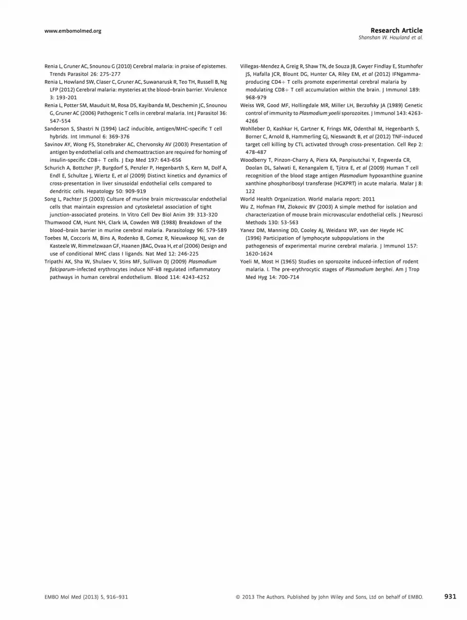
www.embomolmed.org Research ArticleShanshan W. Howland et al.
Renia L, Gruner AC, Snounou G (2010) Cerebral malaria: in praise of epistemes.
Trends Parasitol 26: 275-277
Renia L, Howland SW, Claser C, Gruner AC, Suwanarusk R, Teo TH, Russell B, Ng
LFP (2012) Cerebral malaria: mysteries at the blood–brain barrier. Virulence
3: 193-201
Renia L, Potter SM, Mauduit M, Rosa DS, Kayibanda M, Deschemin JC, Snounou
G, Gruner AC (2006) Pathogenic T cells in cerebral malaria. Int J Parasitol 36:
547-554
Sanderson S, Shastri N (1994) LacZ inducible, antigen/MHC-specific T cell
hybrids. Int Immunol 6: 369-376
Savinov AY, Wong FS, Stonebraker AC, Chervonsky AV (2003) Presentation of
antigen by endothelial cells and chemoattraction are required for homing of
insulin-specific CD8þ T cells. J Exp Med 197: 643-656
Schurich A, Bottcher JP, Burgdorf S, Penzler P, Hegenbarth S, Kern M, Dolf A,
Endl E, Schultze J, Wiertz E, et al (2009) Distinct kinetics and dynamics of
cross-presentation in liver sinusoidal endothelial cells compared to
dendritic cells. Hepatology 50: 909-919
Song L, Pachter JS (2003) Culture of murine brain microvascular endothelial
cells that maintain expression and cytoskeletal association of tight
junction-associated proteins. In Vitro Cell Dev Biol Anim 39: 313-320
Thumwood CM, Hunt NH, Clark IA, Cowden WB (1988) Breakdown of the
blood–brain barrier in murine cerebral malaria. Parasitology 96: 579-589
Toebes M, Coccoris M, Bins A, Rodenko B, Gomez R, Nieuwkoop NJ, van de
Kasteele W, Rimmelzwaan GF, Haanen JBAG, Ovaa H, et al (2006) Design and
use of conditional MHC class I ligands. Nat Med 12: 246-225
Tripathi AK, Sha W, Shulaev V, Stins MF, Sullivan DJ (2009) Plasmodium
falciparum-infected erythrocytes induce NF-kB regulated inflammatory
pathways in human cerebral endothelium. Blood 114: 4243-4252
EMBO Mol Med (2013) 5, 916–931 �
Villegas-Mendez A, Greig R, Shaw TN, de Souza JB, Gwyer Findlay E, Stumhofer
JS, Hafalla JCR, Blount DG, Hunter CA, Riley EM, et al (2012) IFNgamma-
producing CD4þ T cells promote experimental cerebral malaria by
modulating CD8þ T cell accumulation within the brain. J Immunol 189:
968-979
Weiss WR, Good MF, Hollingdale MR, Miller LH, Berzofsky JA (1989) Genetic
control of immunity to Plasmodium yoelii sporozoites. J Immunol 143: 4263-
4266
Wohlleber D, Kashkar H, Gartner K, Frings MK, Odenthal M, Hegenbarth S,
Borner C, Arnold B, Hammerling GJ, Nieswandt B, et al (2012) TNF-induced
target cell killing by CTL activated through cross-presentation. Cell Rep 2:
478-487
Woodberry T, Pinzon-Charry A, Piera KA, Panpisutchai Y, Engwerda CR,
Doolan DL, Salwati E, Kenangalem E, Tjitra E, et al (2009) Human T cell
recognition of the blood stage antigen Plasmodium hypoxanthine guanine
xanthine phosphoribosyl transferase (HGXPRT) in acute malaria. Malar J 8:
122
World Health Organization. World malaria report: 2011
Wu Z, Hofman FM, Zlokovic BV (2003) A simple method for isolation and
characterization of mouse brain microvascular endothelial cells. J Neurosci
Methods 130: 53-563
Yanez DM, Manning DD, Cooley AJ, Weidanz WP, van der Heyde HC
(1996) Participation of lymphocyte subpopulations in the
pathogenesis of experimental murine cerebral malaria. J Immunol 157:
1620-1624
Yoeli M, Most H (1965) Studies on sporozoite induced-infection of rodent
malaria. I. The pre-erythrocytic stages of Plasmodium berghei. Am J Trop
Med Hyg 14: 700-714
2013 The Authors. Published by John Wiley and Sons, Ltd on behalf of EMBO. 931


