Embed Size (px)
Citation preview

www.elsevier.com/locate/ygyno
Gynecologic Oncology
Pretreatment serum hemoglobin level and a preliminary investigation of
intratumoral microvessel density in advanced ovarian cancer
Annamaria Ferreroa,*, Paolo Zolaa, Simona Mazzolaa, Luca Fusoa, Ivana Sarottob,
Nicoletta Ravarinoc, Pier Giorgio Spanua, Maria Elena Jacomuzzia,
Alice Peroglio Carusa, Piero Sismondia
aDepartment of Gynecologic Oncology, University of Torino-Mauriziano bUmberto I Q Hospital, Torino, Italy, and Institute for Cancer Research
and Treatment, Candiolo (TO), ItalybDepartment of Pathology, Institute for Cancer Research and Treatment, Candiolo (TO), Italy
cDepartment of Pathology, Mauriziano bUmberto I Q Hospital, Torino, Italy
Received 9 December 2003
Available online 18 September 2004
Abstract
Objective. The primary aim of this study was to evaluate the prognostic and predictive value of pretreatment serum hemoglobin level (Hb)
in advanced ovarian cancer; second aim was to perform a preliminary investigation of intratumoral microvessel density (IMD).
Methods. The influence on survival and response to treatment of several clinico-pathological features, including Hb, was analyzed in 72
patients with advanced ovarian cancer. IMD was assessed in tumor specimens of 25 of the 72 patients to compare three different endothelial
markers: anti-FactorVIII, anti-CD31 and anti-CD34. In this subgroup of patients, a preliminary analysis of the prognostic and predictive
value of IMD, and its relationship with Hb and other clinico-pathological features, was performed.
Results. Hb z 12 g/dl was significantly associated with a better overall survival in univariate analysis (P = 0.0181) and was the only
independent prognostic variable in multivariate analysis (P = 0.0160). Hb was directly related to progression-free survival (P = 0.0240) and
complete response to treatment (P = 0.016). In the preliminary investigation of IMD, mean microvessel count did not show any significant
difference among the three endothelial markers used, but anti-CD34 revealed a more consistent staining reaction. The relationship between
IMD and complete response to treatment was found near to statistical significance (P = 0.05); Hb and IMD were inversely related (r =
�0.47; P = 0.045).
Conclusions. Hb has a prognostic and predictive value in advanced ovarian cancer. In our preliminary study, which was performed on a
limited number of patients, we found anti-CD34 to be an optimal marker for IMD determination, IMD to be a possible predictive factor of
complete response to treatment, and IMD and Hb to be inversely related.
D 2004 Elsevier Inc. All rights reserved.
Keywords: Hemoglobin; Angiogenesis; Ovarian cancer
Introduction
Ovarian carcinoma has the highest mortality rate among
gynecological malignancies and the majority (almost 70%)
of ovarian cancer patients are diagnosed at an advanced
stage of the disease [1]. Cytoreductive surgery combined
0090-8258/$ - see front matter D 2004 Elsevier Inc. All rights reserved.
doi:10.1016/j.ygyno.2004.07.053
* Corresponding author. Via G. Carducci 20, 10044 Pianezza (TO),
Italy. Fax: +39 011 5082683.
E-mail address: [email protected] (A. Ferrero).
with chemotherapy is the standard treatment of advanced
ovarian cancer [2]; carboplatin and paclitaxel association
is, by now, the standard first-line chemotherapy regimen
[3,4].
The identification of predictive factors of response to a
specific treatment, among already known prognostic factors,
should open a new way through a tailored therapy for each
patient [5,6].
Tumor anemia is a common symptom in cancer patients.
Several reports have documented the prognostic role of
95 (2004) 323–329

A. Ferrero et al. / Gynecologic Oncology 95 (2004) 323–329324
pretreatment serum hemoglobin level (Hb) in patients with a
variety of malignancies, but only two authors showed its
influence on survival in ovarian cancer [7–9]. Hb was also
reported to influence the response rates to radiotherapy and
to radio-chemotherapy in cervical cancer and other non-
gynecological tumors [10].
Angiogenesis plays a critical role in tumor growth. It is
the result of predominantly proangiogenic inputs; there is
strong evidence of a pivotal role of the hypoxic starting
point and VEGF [11]. Many studies analyzed the prognostic
role of intratumoral microvessel density (IMD) in ovarian
cancer: the results are not homogeneous and the endothelial
markers used not the same. While some authors found that
IMD influenced survival, others did not. The most
frequently used endothelial markers were anti-FactorVIII,
anti-CD31, and anti-CD34 [12–21]. Gasparini et al. [22]
showed a statistically significant inverse association
between IMD and response to chemotherapy, while Gad-
ducci et al. [23] demonstrated a positive association between
IMD and response to chemotherapy, mainly represented by
a paclitaxel–platinum regimen.
Primary aim of this study was to evaluate the influence of
Hb on survival and on response to treatment in advanced
ovarian cancer. Second aim was to perform a preliminary
investigation of IMD.
Materials and methods
Patients selections
Criteria for eligibility to the study were: histologically
confirmed epithelial ovarian cancer; advanced stages (III–
IV); no previous treatment; no previous or concurrent
malignancy; clinical, pathological and laboratory data
availability; follow-up informations.
Clinical records of patients treated at the Department of
Gynecologic Oncology, University of Turin, from 1997 to
2000 were collected. Seventy-two consecutive patients were
selected.
All the eligible patients underwent primary debulking
surgery followed by platinum-based chemotherapy. Forty of
the 72 patients received a regimen without paclitaxel: 24
carboplatin alone; 4 cisplatin + cyclophosphamide; 4
cisplatin + doxorubicin + cyclophosphamide. Thirty-two
of the 72 patients received a regimen with paclitaxel: 25
carboplatin + paclitaxel; 7 cisplatin + paclitaxel. The mean
number of cycles administered was 6.
One month after the last course of chemotherapy, response
to treatment was assessed by clinical examination, Ca125
evaluation and CT scan. Patients with clinical complete
response (CR) underwent second look surgery, if response
had to be confirmed.
At the end of the complete treatment program, Ca125
serum level evaluation and a clinical and gynecological
examination were performed every 3–4 months for the first
2 years; patients underwent instrumental evaluation accord-
ing to clinical status.
Clinico-pathological features analyzed
The following clinico-pathological features were ana-
lyzed: age, performance status (on the basis of the World
Health Organization), ascites at diagnosis, FIGO stage,
histologic type, tumor grade, peritoneal cytology, post-
operative residual disease, and pretreatment serum hemo-
globin level (Hb).
Intratumoral microvessel density determination
In 25 of the 72 patients, intratumoral microvessel density
(IMD) was assessed. IMD determination was performed on
formalin-fixed paraffin-embedded specimens with the meth-
odology described by Weidner [24] and by the International
Consensus in 1996 [25].
All histologic slides of hematoxylin and eosin stained
sections from the primary tumor were examined at low
magnification (10�) to select the most representative
tissue block of the invasive carcinoma for each patient.
Five-A-thick sections of the selected blocks were
obtained. Sections were deparaffinized in xylol and
rehydrated in alcohol, then exposed to 3% H2O2 for
15 min to block endogenous peroxide activity. After
pretreatment in a microwave for 10 min to unmask the
antigens, the microvessels were highlighted by immunos-
taining the sections with three different endothelial
markers using a standard immunoperoxidase technique.
The three markers were anti-FactorVIII, anti-CD31, and
anti-CD34 (Biogenex).
The immunostained sections were scanned at low
magnification (40� to 100�) to select those areas of invasive
carcinoma with the greatest numbers of distinctly highlighted
microvessels (i.e., neovascular bhotspotQ). All the vessels
within a 0.74 mm2 area of this neovascular bhotspotQ werecounted at high power (200�). Microvessel counts were
performed without knowledge of the clinical outcome.
Outcome measures and statistical analysis
The overall survival and the response to treatment have
been evaluated and have been compared in patients treated
with regimens containing paclitaxel or not.
In all the 72 patients, the influence on survival and
response to treatment of the selected clinico-pathological
features was analyzed. The mean microvessel counts with
the three different endothelial markers were compared. In
the 25 patients assessed for IMD, a preliminary analysis to
evaluate the relationship between IMD and overall survival
rate, response to treatment, and the other clinico-patholog-
ical variables was performed.
The overall survival (OS) was considered as the period of
time from diagnosis until the time of death or to the date of

Table 1
Clinico-pathological characteristics
Characteristics All the
patients
Patients evaluated
for IMD
Number
(mean)
%
(range)
Number
(mean)
%
(range)
Number 72 25
Age (58.4) (25–80) (58) (33–79)
Performance status WHO
0 39 54.2 10 40
1 25 34.7 9 36
2 8 11.1 6 24
Ascites
Absent 28 38.9 5 20
Present 44 61.1 20 80
FIGO stage
IIIA 13 18 2 8
IIIB 6 8.3 4 16
IIIC 48 66.7 19 76
IV 5 7 0 0
Histologic type
Serous 44 61.1 15 60
Mucinous 9 12.5 4 16
Endometrioid 7 9.7 0 0
Clear cells 1 1.4 0 0
A. Ferrero et al. / Gynecologic Oncology 95 (2004) 323–329 325
the last available follow-up. The progression-free survival
(PFS) was considered as the period from surgery to the first
documented relapse.
Responses to treatment were defined as CR, partial
response (PR), stable disease (SD) or progressive disease
(PD) according to standard criteria.
All clinical data were revised and recorded in a database.
Statistical analyses were performed with SPSS package
(SPSS Advanced Statistics for Window version 9).
OS and PFS curves were obtained using the Kaplan–
Meyer method. Log Rank, Breslow and Tarone–Ware
statistics were used to test the difference between two groups.
Responses were correlated to continue variables with the
Analysis of Variance and independent-samples Student’s t
test in a parametric setting. v2, Mann–Whitney and
Kruskal–Wallis statistics were adopted for non-parametric
distributions. To correlate continuous variables the para-
metric Pearson’s coefficient and the non-parametric Spear-
man test were used.
A multivariate analysis was performed according to Cox
proportional hazard model for statistical significant varia-
bles at the univariate level.
Undifferentiated 11 15.3 6 24Tumor grade
1 3 4.2 1 4
2 24 33.4 6 24
3 45 62.4 18 72
Peritoneal cytology
Negative 8 11.1 2 8
Positive 64 88.9 23 92
Postoperative residual disease
b2 cm 33 45.8 12 48
z2 cm 38 52.8 13 52
Results
Patient characteristics
The clinico-pathological characteristics of the 72 patients
are reported in Table 1. The numbers of patients treated with
or without paclitaxel are not statistically different (P =
0.239).
Forty-three (59.7%) patients had a complete response to
treatment (CR), while the remaining 29 (40.3%) had a partial
response or a stable disease or a progressive disease (PR + SD
+ PD). No statistically significant difference in the response
to treatment between patients treated with or without
paclitaxel were found: CR were 21 in patients treated with
paclitaxel and 22 in patients treated without paclitaxel (P =
0.454); PR + SD + PD were 11 in patients who received
paclitaxel and 18 in patients who did not receive paclitaxel
(P = 0.639). Median progression-free survival and overall
survival were similar in these two subgroups of patients.
Table 1 shows also the characteristics of the 25 patients
evaluated for IMD. Twelve (48%) of these patients had a
complete response to treatment, while the remaining 13
(52%) had a partial response or a stable disease or a
progressive disease.
Prognostic factors analysis
In our series age at diagnosis, performance status, FIGO
stage, histological type, and tumor grade did not influence
survival (data not shown).
A statistically significant association between overall
survival and ascites at diagnosis, postoperative residual
disease, and pretreatment serum hemoglobin level was
found (Table 2).
In Table 3 multivariate analysis for variables influencing
overall survival is reported: Hb was the only independent
prognostic variable (P = 0.0160). Hb was statistically
associated with progression-free survival too.
Fig. 1 shows the Kaplan–Meier curve for overall survival
in patients with pretreatment serum hemoglobin levels z12
and b12 g/dl; Fig. 2 shows the Kaplan–Meier curve for PFS
in the same patients.
Predictive factors analysis
As reported in Table 4, a statistically significant
association between complete response to treatment and
ascites at diagnosis, postoperative residual disease, and
pretreatment serum hemoglobin level was found.
IMD determination and preliminary analyses
Microvessel counts were similar with the three endothe-
lial markers. The mean microvessel count was anti-CD34 =
17.48; anti-CD31 = 17.31; anti-FactorVIII = 18.23.
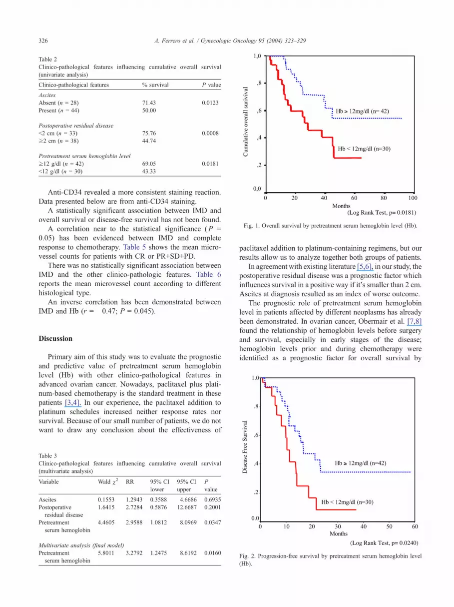
Table 2
Clinico-pathological features influencing cumulative overall survival
(univariate analysis)
Clinico-pathological features % survival P value
Ascites
Absent (n = 28) 71.43 0.0123
Present (n = 44) 50.00
Postoperative residual disease
b2 cm (n = 33) 75.76 0.0008
z2 cm (n = 38) 44.74
Pretreatment serum hemoglobin level
z12 g/dl (n = 42) 69.05 0.0181
b12 g/dl (n = 30) 43.33
Fig. 1. Overall survival by pretreatment serum hemoglobin level (Hb).
A. Ferrero et al. / Gynecologic Oncology 95 (2004) 323–329326
Anti-CD34 revealed a more consistent staining reaction.
Data presented below are from anti-CD34 staining.
A statistically significant association between IMD and
overall survival or disease-free survival has not been found.
A correlation near to the statistical significance (P =
0.05) has been evidenced between IMD and complete
response to chemotherapy. Table 5 shows the mean micro-
vessel counts for patients with CR or PR+SD+PD.
There was no statistically significant association between
IMD and the other clinico-pathologic features. Table 6
reports the mean microvessel count according to different
histological type.
An inverse correlation has been demonstrated between
IMD and Hb (r = �0.47; P = 0.045).
Discussion
Primary aim of this study was to evaluate the prognostic
and predictive value of pretreatment serum hemoglobin
level (Hb) with other clinico-pathological features in
advanced ovarian cancer. Nowadays, paclitaxel plus plati-
num-based chemotherapy is the standard treatment in these
patients [3,4]. In our experience, the paclitaxel addition to
platinum schedules increased neither response rates nor
survival. Because of our small number of patients, we do not
want to draw any conclusion about the effectiveness of
Table 3
Clinico-pathological features influencing cumulative overall survival
(multivariate analysis)
Variable Wald v2 RR 95% CI
lower
95% CI
upper
P
value
Ascites 0.1553 1.2943 0.3588 4.6686 0.6935
Postoperative
residual disease
1.6415 2.7284 0.5876 12.6687 0.2001
Pretreatment
serum hemoglobin
4.4605 2.9588 1.0812 8.0969 0.0347
Multivariate analysis (final model)
Pretreatment
serum hemoglobin
5.8011 3.2792 1.2475 8.6192 0.0160
paclitaxel addition to platinum-containing regimens, but our
results allow us to analyze together both groups of patients.
In agreement with existing literature [5,6], in our study, the
postoperative residual disease was a prognostic factor which
influences survival in a positive way if it’s smaller than 2 cm.
Ascites at diagnosis resulted as an index of worse outcome.
The prognostic role of pretreatment serum hemoglobin
level in patients affected by different neoplasms has already
been demonstrated. In ovarian cancer, Obermair et al. [7,8]
found the relationship of hemoglobin levels before surgery
and survival, especially in early stages of the disease;
hemoglobin levels prior and during chemotherapy were
identified as a prognostic factor for overall survival by
Fig. 2. Progression-free survival by pretreatment serum hemoglobin level
(Hb).

Table 4
Clinico-pathological features predictive of complete response to treatment
Clinico-pathological features CR
(%)
PR + SD + PD (%) P
value
Ascites
Absent (n = 28) 22 (78.6) 6 (21.4) 0.0092
Present (n = 44) 21 (47.7) 23 (52.3)
Postoperative residual disease
b2 cm (n = 33) 28 (84.8) 5 (15.2) 0.00009
z2 cm (n = 38) 15 (39.5) 23 (60.5)
Pretreatment serum hemoglobin level
z12 g/dl (n = 42) 30 (71.4) 12 (28.6) 0.016
b12 g/dl (n = 30) 13 (43.3) 17 (56.7)
Table 6
Mean microvessel count (CD34) according to histologic type
Histologic type Mean count DS SE
Serous (n = 15) 17.60 7.70 1.99
Mucinous (n = 4) 16.50 5.07 2.53
Indifferenziated (n = 6) 17.83 1.94 0.79
(ANOVA, P = 0.944).
A. Ferrero et al. / Gynecologic Oncology 95 (2004) 323–329 327
Munstedt et al. [9]. Like these two authors, in our series,
pretreatment serum hemoglobin levels z12 g/dl were
significantly associated with a better overall survival (P =
0.0181) and retained significance in multivariate analysis
(P = 0.0160). The reasons of these results are presently
unexplained. Obermair hypothesized a paraneoplastic phe-
nomenon. Tumor-released cytokines play a major role in the
development of tumor-associated anemia by hemolysis,
suppression of erythropoiesis, and impairment of erythro-
poietin response on erythroid progenitor cells; marked
tumor anemia might indicate the presence of biologically
aggressive tumor cell clones. Another possible explanation
is that hypoxia, due to low levels of circulating hemoglobin,
causes a worse response to treatment [7,10].
In our study, three factors were linked to a better
response to treatment: the postoperative residual disease
smaller than 2 cm, the absence of ascites at diagnosis and
pretreatment serum hemoglobin levels z12 g/dl. The
predictive value of Hb is a datum that still needs further
study. A considerable number of studies report radiotherapy
and chemotherapy to be more effective under well-oxy-
genated conditions. Anemia might contribute to reduce
tumor oxygenation via reduced oxygen delivery to the
tumor [26]. In vitro and animal models have shown that
cellular hypoxia, the consequence of anemia, may provide a
selection pressure for tumor cells with higher rate of
mutation, which may ultimately result in increased meta-
static potential, increased cellular growth, therapy resist-
ance, and decreased apoptotic potential [27].
In literature different endothelial markers have been used
to highlight microvessels with immunohistochemistry.
Hollingsworth et al. [13] initially performed counts with
CD34, Ulex and von Willebrand Factor (vWF, also
Table 5
Correlation between response to treatment and mean microvessel count
(CD34)
Type of response Mean count DS SE
CR (n = 12) 15.08 6.08 1.76
PR + SD + PD (n = 13) 19.68 5.71 1.58
Independent-samples Student’s t test, P = 0.05.
designated Factor VIII) antibodies. Subsequently, it was
noted that variability in staining was occurring with both
Ulex and vWF with misleading of low vessel counts and
understaining of endothelium in approximately 50% of the
slides. CD34 antibody stained consistently and reproducibly
and did no exhibit such problems. Alvarez et al. [21]
evaluated IMD with antibodies to von Willebrand Factor
(vWF) and CD31: vessel counts for vWF and CD31 were
highly correlated and comparable results for outcome were
seen for the two factors. In our study, three different
endothelial markers for IMD determination were used: anti-
Factor VIII, anti-CD31, and anti-CD34. The comparison did
not show any difference in terms of mean microvessel
count, but anti-CD34 revealed a more consistent staining
reaction. In agreement with Heiburg et al. [18,19], we
consider CD34 as an optimal marker for IMD determination
in tumoral neoangiogenesis and have used anti-CD34
staining for our analyses.
IMD was not a statistically significant prognostic factor
for our patient sample. Surely, the small number of patients
could be a cause, but the numbers of patients in some
already published studies are only slightly higher than ours.
Some, but not all, papers reported a statistically significant
association between low IMD and an increased overall
survival. However, in these studies, even patients at early
stages were included and in those stages IMD is usually
lower and the outcome is better [12].
The mean microvessel count in different histological
types was almost the same in our study, and moreover we
did not find an evident difference concerning mucinous
histotype such as happened in Orre et al. [28] and Gasparini
et al. [22] studies.
A correlation near to the statistical significance (P =
0.05) has been found between IMD and complete response
to chemotherapy. The mean microvessel count was 15.08
for patients with CR and 19.69 for PR, SD, PD. Gasparini et
al. [22] also showed a statistically significant inverse
association between IMD and response to chemotherapy,
while Gadducci et al. [23] found a positive association
between IMD and response to chemotherapy, mainly
represented by a paclitaxel–platinum regimen. Gadducci
hypothesized that the antineoplastic activity of paclitaxel
could be greater in highly vascularized tumors. Indeed
paclitaxel, besides causing tumor cell death, displays
antiangiogenic activity through organic and functional
damage of endothelial cells.
An interesting evidence seems to be the finding of an
inverse correlation between pretreatment serum hemoglo-

A. Ferrero et al. / Gynecologic Oncology 95 (2004) 323–329328
bin level and IMD. A low level of circulating hemoglobin
should increase IMD because of hypoxia and at the end
become a proangiogenic stimulation. The switch of
tumors from the avascular to the vascular phase is a
critical checkpoint in cancer progression. Folkman [29]
proposed that neovascularization of a tumor was required
to provide essential nutrients beyond the limit of simple
diffusion and to allow a growth higher than 2 mm. At
this time, tumor and host tissues produce a great variety
of angiogenic factors that promote the development of a
new capillary bed. The mechanism by which low
hemoglobin levels could be involved in this process
needs further elucidations by biochemical and molecular
studies, but a hypothesis is that the decreased oxygen
carrying capacity may lead to increased tumor hypoxia
than to neoangiogenic stimulation and tumor growth.
Neoangiogenesis may cause a misunderstanding in a
positive way: the major perfusion granted by the new
vessels may be seen as a new therapeutical option
because a greater dose of drugs may arrive directly into
the tumoral mass. Unfortunately, the first vascular phase
is followed by a second phase during which there is a
mechanical compression of new vessels due to the
increased volume of the neoplasm mass.
In conclusion, our study confirms the prognostic role of
pretreatment hemoglobin level, which has been demonstra-
ted to be a predictive factor of response to treatment as well.
We are continuing to evaluate some more patients to
measure IMD in a larger number of cases to analyze the
correlation among hemoglobin levels, IMD, and the
response to treatment.
We are looking for a tailored treatment for selected
ovarian cancer patients. Recently, new therapeutic
approaches have been studied as a consequence of bio-
logical research development [30]: angiogenesis inhibitors
could be the new frontier of treatment with genic therapies
and vaccines. Paclitaxel may inhibit angiogenesis. The
correction of anemia before the beginning of treatment
could improve response rates and survival.
Acknowledgments
We thank the pathologists Mauro Riso and Bruno
Torchio for their support.
References
[1] FIGO annual report. J Epidemiol Biostat 1998;3:75–102.
[2] Berek JS, et al. Advanced epithelial ovarian cancer: 1998 consensus
statements. Ann Oncol 1999;10(Suppl. 1):87–92.
[3] McGuire WP. Current status of taxane and platinum-based chemo-
therapy in ovarian cancer. J Clin Oncol 2003;21:133s–5s.
[4] Markman M. Optimizing primary chemotherapy in ovarian cancer.
Hematol Oncol Clin North Am 2003;17:957–68.
[5] Eisenhauer EA, Gore M, Neijt JP. Ovarian cancer: should we be
managing patients with good and bad prognostic factors in the same
manner? Ann Oncol 1999;10(Suppl. 1):S9–15.
[6] Friedlander ML. Prognostic factors in ovarian cancer. Semin Oncol
1998;25:305–15.
[7] Obermair A, Handisurya A, Kaider A, Sevelda P, Kolbl H, Gitsch G.
The relationship of pretreatment serum hemoglobin level to the
survival of epithelial ovarian carcinoma patients: a prospective review.
Cancer 1998;83:726–31.
[8] Obermair A, Petru E, Windbichler G, Peters-Engl C, Graf AH,
Stummvoll W, et al. Significance of pretreatment serum hemoglo-
bin and survival in epithelial ovarian cancer. Oncol Rep 2000;
7:639–44.
[9] Munstedt K, Kovacic M, Zygmunt M, Von Georgi R. Impact of
hemoglobin levels before and during chemotherapy on survival of
patients with advanced ovarian cancer. Int J Oncol 2003:837–43.
[10] Obermair A, Cheuk R, Horwood K, Janda M, Bachtiary B,
Schwanzelberger B, et al. Impact of hemoglobin levels before and
during concurrent chemoradiotherapy on the response of treatment in
patients with cervical carcinoma. Preliminary results. Cancer
2001;92:903–8.
[11] Abulafia O, Triest WE, Sherer DM. Angiogenesis in malignancies of
the female genital tract. Gynecol Oncol 1999;72:220–31.
[12] Obermair A, Preyer O, Leodolter S. Tumor angiogenesis and its
relation to prognosis in epithelial ovarian cancer. CME J Gynecol
Oncol 1999;4(2):169–77.
[13] Hollingsworth HC, Kohn EC, Steinberg SM, Rothenberg ML, Merino
MJ. Tumor angiogenesis in advanced stage ovarian carcinoma. Am J
Pathol 1995;147:33–41.
[14] Van Diest PJ, Zevering JP, Zevering LC, Baak JPA. Prognostic value
of microvessel quantification in cisplatin treated FIGO 3 and 4 ovarian
cancer patients. Pathol Res Pract 1995;191:25–30.
[15] Schoell WM, Pieber D, Reich O, Lahousen M, Janicek M, Guecer F,
et al. Tumor angiogenesis as a prognostic factor in ovarian carcinoma:
quantification of endothelial immunoreactivity by image analysis.
Cancer 1997;80:2257–62.
[16] Kohn EC. Angiogenesis in ovarian carcinoma: a formidable bio-
marker. Cancer 1997;80:2219–21.
[17] Abulafia O, Triest WE, Sherer DM. Angiogenesis in primary and
metastatic epithelial ovarian carcinoma. Am J Obstet Gynecol
1997;177:541–7.
[18] Heimburg S, Oehler MK, Kristen P, Papadopulos T, Caffier H. The
endothelial marker CD34 in the assessment of tumour vascularisation
in ovarian cancer. Anticancer Res 1997;17:3149–52.
[19] Heimburg S, Oehler MK, Papadopulos T, Caffier H, Kristen P, Dietl J.
Prognostic relevance of the endothelial marker CD34 in ovarian
cancer. Anticancer Res 1999;19:2527–30.
[20] Obermair A, Wasicky R, Kaider A, Preyer O, Losch A, Leodolter S, et
al. Prognostic significance of tumor angiogenesis in epithelial ovarian
cancer. Cancer Lett 1999;138:175–82.
[21] Alvarez AA, Krigman HR, Whitaker RS, Dodge RK, Rodriguez GC.
The prognostic significance of angiogenesis in epithelial ovarian
carcinoma. Clin Cancer Res 1999;5:587–91.
[22] Gasparini G, Bonoldi E, Viale G, Verderio P, Boracchi P, Panizzoni
GA, et al. Prognostic and predictive value of tumour angiogenesis in
ovarian carcinomas. Int J Cancer 1996;69:205–11.
[23] Gadducci A, Viacava P, Cosio S, Fanelli G, Fanucchi A, Cecchetti D,
et al. Intratumoral microvessel density, response to chemotherapy and
clinical outcome in patients with advanced ovarian carcinoma.
Anticancer Res 2003;23:549–56.
[24] Weidner N. Current pathologic methods for measuring intratumoral
microvessel density within breast carcinoma and other solid tumors.
Breast Cancer Res Treat 1995;36:169–80.
[25] Vermeulen PB, Gasparini G, Fox SB, Toi M, Martin L, McCulloch P,
et al. Quantification of angiogenesis in solid human tumours: an
international consensus on the methodology and criteria of evaluation.
Eur J Cancer 1996;32A(14):2474–84.
[26] Fyles AW, Milosevic M, Pintilie M, Syed A, Hill RP. Anemia,

A. Ferrero et al. / Gynecologic Oncology 95 (2004) 323–329 329
hypoxia and transfusion in patients with cervix cancer: a review.
Radiother Oncol 2000;57:13–9.
[27] Van Belle SJ, Cocquyt V. Impact of haemoglobin levels on the
outcome of cancers treated with chemotherapy. Crit Rev Oncol
Hematol 2003;47:1–11.
[28] Orre M, Lotfi-Miri M, Mamers P, Rogers PAW. Increased microvessel
density in mucinous compared with malignant serous and benign
tumours of the ovary. Br J Cancer 1998;77:2204–9.
[29] Folkman J. Clinical applications of research on angiogenesis. N Eng J
Med 1995;333:1757–63.
[30] DiSaia PJ, Bloss JD. Treatment of ovarian cancer: new strategies.
Gynecol Oncol 2003;90:s24–32.


