Embed Size (px)
Citation preview
At the Institut Pasteur - Rue Engeland 642 - 1180 Brussels - Belgium
Tel: +32 2 373 93 23 - Fax: +32 2 373 93 13 - www.inctr.org Des
ign:
GIC
Man
agem
ent
+
32
-2-7
43
15
40
-
ww
w.a
ssoc
iati
onh
q.co
m
INCTR Annual Meeting 2004 was supported by the Office of International Affairs of the National Cancer Institute,
Bethesda, MD, USA.
INCTR would like to thank the following companies for their sponsorship of the
Annual Meeting 2004
INC
TR
-A
nnua
l M
eeti
ng 2
004
Oct
ober
2nd
- 5
th, 2
004
- Ca
iro,
Egy
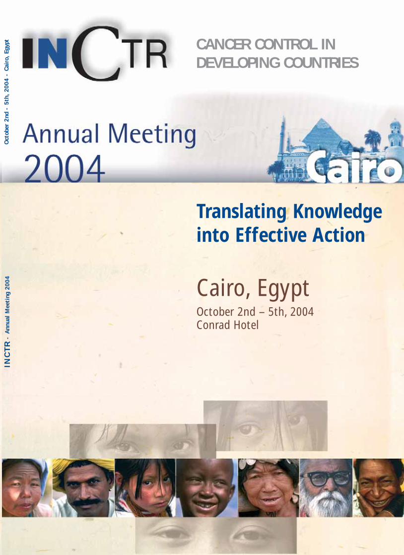
pt CANCER CONTROL INDEVELOPING COUNTRIES
Translating Knowledge into Effective Action
Cairo, Egypt October 2nd – 5th, 2004Conrad Hotel
Annual Meeting 2004
About INCTR INCTR is a non-profit organization whose Founder Members are the InternationalUnion Against Cancer and the Institut Pasteur, Brussels. The goals of theorganization are to assist in controlling cancer in developing countries through thedevelopment of infrastructure for cancer treatment and research, and throughcollaboration with physicians and scientists in such countries, to take advantage ofunique opportunities to improve our understanding of factors (genetic andenvironmental) that predispose people to various types of cancer and consequently,to allow the development of rational prevention strategies. Education is an integralelement of long-term collaborative projects relating to treatment or prevention andthe implementation of such projects, in many cases, will result in immediatebenefits to patients or individuals at high risk for cancer.
INCTR also emphasizes international collaboration, and promotes improvedcommunication among the wide range of professionals and volunteers working tocontrol cancer throughout the world.
About INCTR Egypt INCTR Egypt was established under the umbrella of the Egyptian Foundation forCancer Research. Its purpose is to assist in achieving the goals and objectives ofINCTR in Egypt and adjacent countries through selected projects relevant to cancerprevention and early detection and treatment. Educational and training programs forcancer specialists, nurses and other health professionals are high priorities andemphasis is given to regionally important cancers. INCTR Egypt also promotescollaborative efforts among institutions and organizations within Egypt and in theregion. The ultimate goal is to prevent cancer wherever possible, and to improvesurvival rates and the quality of life of patients who develop cancer.
phot
o C.
Dew
asm
e
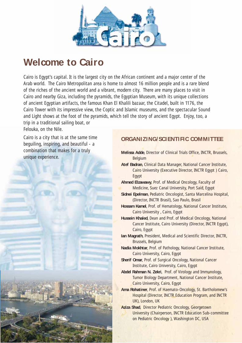
Cairo is Egypt’s capital. It is the largest city on the African continent and a major center of theArab world. The Cairo Metropolitan area is home to almost 16 million people and is a rare blendof the riches of the ancient world and a vibrant, modern city. There are many places to visit inCairo and nearby Giza, including the pyramids, the Egyptian Museum, with its unique collectionsof ancient Egyptian artifacts, the famous Khan El Khalili bazaar, the Citadel, built in 1176, theCairo Tower with its impressive view, the Coptic and Islamic museums, and the spectacular Soundand Light shows at the foot of the pyramids, which tell the story of ancient Egypt. Enjoy, too, atrip in a traditional sailing boat, orFelouka, on the Nile.
Cairo is a city that is at the same timebeguiling, inspiring, and beautiful - acombination that makes for a trulyunique experience.
Welcome to Cairo
ORGANIZING/SCIENTIFIC COMMITTEE
Melissa Adde, Director of Clinical Trials Office, INCTR, Brussels,Belgium
Atef Badran, Clinical Data Manager, National Cancer Institute,Cairo University (Executive Director, INCTR Egypt ) Cairo,Egypt
Ahmed Elzawawy, Prof. of Medical Oncology, Faculty ofMedicine, Suez Canal University, Port Said, Egypt
Sidnei Epelman, Pediatric Oncologist, Santa Marcelina Hospital,(Director, INCTR Brasil), Sao Paulo, Brasil
Hossam Kamel, Prof. of Hematology, National Cancer Institute,Cairo University , Cairo, Egypt
Hussein Khaled, Dean and Prof. of Medical Oncology, NationalCancer Institute, Cairo University (Director, INCTR Egypt),Cairo, Egypt
Ian Magrath, President, Medical and Scientific Director, INCTR,Brussels, Belgium
Nadia Mokhtar, Prof. of Pathology, National Cancer Institute,Cairo University, Cairo, Egypt
Sherif Omar, Prof. of Surgical Oncology, National CancerInstitute, Cairo University, Cairo, Egypt
Abdel Rahman N. Zekri, Prof. of Virology and Immunology,Tumor Biology Department, National Cancer Institute,Cairo University, Cairo, Egypt
Ama Rohatiner, Prof. of Haemato-Oncology, St. Bartholomew’sHospital (Director, INCTR Education Program, and INCTRUK), London, UK
Aziza Shad, Director Pediatric Oncology, GeorgetownUniversity (Chairperson, INCTR Education Sub-committeeon Pediatric Oncology ), Washington DC, USA
Annual Meeting Conrad Cairo, 1191 Corniche el Nil, 11221 Cairo, EgyptPhone: +20-2-580-8000, Fax: +20-2-580-8080
The Conrad Hotel Cairo is located on the river Nilewith convenient access to places of interest, including
the Egyptian Museum, the Citadel, the great Pyramidsof Giza and the Sphinx. It is located close to the busi-ness district, the World Trade Center, and to a largeshopping mall and cinema.
INCTR's Annual Meeting has become an important eventwhich serves to bring together INCTR Associate Membersfrom many different countries to strengthen internationalcollaboration in all aspects of cancer treatment andresearch, to report progress that has been made in INCTRprojects in the last year and to identify focal points fordiscussion that may lead to the development of new proj-ects. While it is essential to involve key figures in cancertreatment and research in these discussions, it is alsoimportant, in the interest of ensuring long term viabilityof programs, that young health professionals also partici-pate. Professional education - including continuing edu-cation - underlies much of the meeting content, andalthough primacy is given to an exchange of views amonghealth professionals from a variety of backgrounds, didac-tic elements are included in order to provide a foundationon which discussion can be based.
It must be recognized that cancer control, althoughfounded on the same basic principles throughout theworld, must contend with even greater obstacles in devel-oping countries than those present in more affluentnations - obstacles that ultimately arise from the eco-nomic difficulties faced by the populations susceptible tocancer, and the paucity of resources available to studypredisposing factors, prevention, early detection andtreatment. For these reasons, essential research relevantto cancer control in developing countries must, in part orin whole, be conducted in those countries themselves,where the pattern of cancer may be regionally unique,where the lifestyles, nutritional status and co-morbiditiesof potential and actual victims of cancer differ so pro-foundly, and where the availability or access to treatmentmay be poor or even absent. It is also critically impor-
tant to involve the entire family and local community inthe process of cancer control - particularly since successto a large degree is dependent upon the avoidance ofcancer, or its detection at the earliest possible stage of itsevolution - even before it has become a true "invasive"cancer. Both are dependent upon knowledge of the earlysymptoms and signs of cancer (by health professionalsand the population at large), and in those cases where itis known to be beneficial, screening of asymptomaticpopulations.
INCTR's Annual Meeting is unique in having, as its entirefocus, the problems encountered in developing countries,and in bringing together experts both from within thosecountries and from affluent nations to discuss possibleapproaches to the control of cancer, and how best toimplement and evaluate them. Cancer is a health prob-lem that is becoming more and more immediate as com-municable diseases are overcome, and populations ageand adopt the bad habits of affluent societies.
In addition to the INCTR Award Lectures, keynote lectures,and oral presentations and posters of the participants'own work, this year's meeting will feature a series of pre-sentations on cancers that are frequent in Africa and theMiddle East. The central element will be a workshop onvarious aspects of cancer control including institutionalleadership (particularly with respect to translating knowl-edge into effective action) and the importance, feasibilityand ethics of research in developing countries. As usual,the ever increasing role of technology in helping to con-trol cancer in developing countries will be discussed. Thisyear, there will be a special emphasis on informationtechnology (IT) and its role in improving the quality ofpatient care.
INTRODUCTION TO THE ANNUAL MEETING
HOTEL INFORMATION
TIM
E SE
SSIO
NM
EETI
NG
RO
OM
FRID
AY
1 O
CTO
BER
200
4 15
.00
- 17
.00
Regi
stra
tion
Conr
ad F
oyer
17.3
0 -
18.0
0M
eetin
g of
Con
fere
nce
Chai
rper
sons
Ra
Roo
m(in
vitat
ion
only)
18.3
0 -
20.0
0Ed
ucat
ion
Com
mitt
ee M
eetin
g Cl
eopa
tra R
oom
(com
mitt
ee m
embe
rs on
ly)
SATU
RD
AY
2 O
CTO
BER
200
4 07
.00
- 09
.00
INCT
R Br
eakf
ast
Conr
ad B
allro
om S
ectio
n 1
08
.00
- 09
.00
Regi
stra
tion
and
Post
er M
ount
ing
Conr
ad B
allro
om F
oyer
Nile
Bal
lroom
09
.00
- 11
.00
Open
ing
Rem
arks
and
Awa
rd L
ectu
res
Conr
ad B
allro
om S
ectio
n 2&
3Se
ssio
n 1
11.0
0 -
11.2
0Co
ffee
Brea
kCo
nrad
Bal
lroom
Foy
er11
.20
- 13
.00
Sess
ion
2: 'I
NCTR
Pro
gram
Rep
orts
' Co
nrad
Bal
lroom
Sec
tion
2&3
13.0
0 -
14.0
0IN
CTR
Lunc
h an
d Po
ster
View
ing
Nile
Ballr
oom
14
.00
- 15
.00
Mem
ber's
For
umCo
nrad
Bal
lroom
Sec
tion
2&3
15.0
0 -
15.2
0Co
ffee
Brea
k an
d Po
ster
View
ing
Nile
Ballr
oom
15
.20
- 17
.20
Sess
ion
3A: '
Prof
fere
d Pa
pers'
Adu
ltsCo
nrad
Bal
lroom
Sec
tion
2&3
15.2
0 -
17.2
0Se
ssio
n 3B
: 'Pr
offe
red
Pape
rs' P
edia
tric
Conr
ad B
all R
oom
Sec
tion
119
.00
- 19
.30
Open
ing
Cere
mon
yCo
nrad
Bal
lroom
Sec
tion
2&3
19.3
0 -
20.0
0IN
CTR
Conf
eren
ce R
ecep
tion
Conr
ad F
oyer
and
Bal
cony
20.0
0 -
23.0
0IN
CTR
Conf
eren
ce D
inne
rBB
Q Te
rrace
SUN
DA
Y 3
OCT
OB
ER 2
004
07.0
0 -
09.0
0IN
CTR
Brea
kfas
t Co
nrad
Bal
lroom
Sec
tion
108
.00
- 09
.00
Post
er V
iewin
gNi
le Ba
llroo
m
08.0
0 -
17.0
0Re
gist
ratio
n an
d Ho
spita
lity
Desk
Conr
ad B
allro
om F
oyer
09
.00
- 09
.40
Keyn
ote
Lect
ure:
Co
nrad
Bal
lroom
Sec
tion
2&3
'Can
cer C
are
in E
gypt
: Pas
t, Pr
esen
t and
Fut
ure'
09.4
0 -
12.0
0Se
ssio
n 4A
: 'Si
mul
tane
ous E
duca
tiona
l Ses
sions
: Ca
ncer
s of R
egio
nal I
mpo
rtanc
e' Ai
da a
nd C
leopa
tra R
oom
09.4
0 -
12.0
0Se
ssio
n 4B
: 'Si
mul
tane
ous E
duca
tiona
l Ses
sions
: Pe
diat
ric C
ance
rs'Co
nrad
Bal
lroom
Sec
tion
2&3
12.0
0 -
13.3
0Se
ssio
n 5:
'INC
TR C
onse
nsus
Pan
el Di
scus
sion'
Conr
ad B
allro
om S
ectio
n 2&
313
.30
- 14
.30
INCT
R Lu
nch
Nile
Ballr
oom
Free
afte
rnoo
n/in
form
al m
eetin
gs/ p
oste
rs vie
wing
17.0
0 -
19.0
0IN
CTR
Advis
ory
Boar
d M
eetin
g Ra
Roo
m(in
vitat
ion
only)
All t
he m
eetin
g ro
oms
are
loca
ted
on t
he f
irst
floor
TIM
E SE
SSIO
NM
EETI
NG
RO
OM
MO
ND
AY
4 O
CTO
BER
200
4 07
.00
- 09
.00
INCT
R Br
eakf
ast
Conr
ad B
allro
om S
ectio
n 1
08.0
0 -
17.0
0Re
gist
ratio
n an
d Ho
spita
lity
Desk
Conr
ad B
allro
om F
oyer
09
.00
- 09
.40
Keyn
ote
Lect
ure:
'Psy
chol
ogica
l Sup
port
for
Canc
er P
atien
ts a
nd th
eir F
amili
es'
Conr
ad B
allro
om S
ectio
n 2&
309
.40
- 11
.05
Sess
ion
6: ‘R
egio
nal a
nd G
loba
l Pro
gram
s and
Pe
rspec
tives
for C
ance
r Con
trol’
Conr
ad B
allro
om S
ectio
n 2&
311
.05
- 11
.30
Coffe
e Br
eak
Conr
ad F
oyer
11.3
0 -
13.0
0Se
ssio
n 7:
Wor
ksho
p: 'S
trate
gies
for C
ance
r Co
ntro
l in
Deve
lopi
ng C
ount
ries'
- Pa
rt 1.
Conr
ad B
allro
om S
ectio
n 2&
313
.00
- 14
.00
INCT
R Lu
nch
Conr
ad B
allro
om S
ectio
n 1
14.0
0 -
15.3
0 Se
ssio
n 8:
'Stra
tegy
Gro
up R
epor
ts'
Conr
ad B
allro
om S
ectio
n 2&
315
.30
- 15
.50
Coffe
e Br
eak
Conr
ad F
oyer
15.5
0 -
17.3
0Se
ssio
n 8
cont
inue
dCo
nrad
Bal
lroom
Sec
tion
2&3
18.0
0 -
19.3
0 IN
CTR
Bran
ch/ O
ffice
For
um (i
nvita
tion
only)
Cleo
patra
Roo
m
TUES
DA
Y 5
OCT
OB
ER 2
004
07.0
0 -
09.0
0IN
CTR
Brea
kfas
t Co
nrad
Bal
lroom
Sec
tion
108
.00
- 17
.00
Regi
stra
tion
and
Hosp
italit
y De
skCo
nrad
Bal
lroom
Foy
er
09.0
0 -
09.4
0Ke
ynot
e Le
ctur
e: 'R
eleva
nce
of M
olec
ular
Bio
logy
to
Can
cer C
ontro
l: Ne
w Fin
ding
s in
Brea
st C
ance
r' Conr
ad B
allro
om S
ectio
n 2&
309
.40
- 10
.40
Sess
ion
9A: '
Sim
ulta
neou
s Edu
catio
nal S
essio
ns.
Canc
ers o
f Reg
iona
l Im
porta
nce'
Conr
ad B
allro
om S
ectio
n 2&
309
.40
- 10
.40
Sess
ion
9B: '
Sim
ulta
neou
s Edu
catio
nal S
essio
ns.
Pedi
atric
Can
cers'
10.4
0 -
11.0
0Co
ffee
Brea
kCo
nrad
Foy
er11
.00
- 12
.00
Sess
ion
10: I
NCTR
Mul
tidisc
iplin
ary
Conf
eren
ce:
'Man
agem
ent o
f Hod
gkin
’s Ly
mph
oma
with
M
edia
stin
al In
volve
men
t'Co
nrad
Bal
lroom
Sec
tion
2&3
12.0
0 -
13.3
0Se
ssio
n 11
: Wor
ksho
p ' S
trate
gies
for C
ance
r Co
ntro
l in
Deve
lopi
ng C
ount
ries'
- Pa
rt 2.
Conr
ad B
allro
om S
ectio
n 2&
313
.30
- 14
.30
INCT
R Lu
nch
Conr
ad B
allro
om S
ectio
n 1
14.3
0 -
15.1
0 Se
ssio
n 12
: 'M
axim
izing
Res
ourc
es a
nd
Impr
ovin
g Co
mm
unica
tions
with
Info
rmat
ion
Tech
nolo
gy'
Conr
ad B
allro
om S
ectio
n 2&
315
.10
- 15
.30
Coffe
e Br
eak
Conr
ad F
oyer
15.3
0 -
17.0
0Se
ssio
n 12
con
tinue
dCo
nrad
Bal
lroom
Sec
tion
2&3
17.0
0 -
17.3
0Cl
osin
g Re
mar
ksCo
nrad
Bal
lroom
Sec
tion
2&3
WED
NES
DA
Y 6
OCT
OB
ER 2
004
07.0
0 -
09.0
0IN
CTR
Brea
kfas
t Co
nrad
Bal
lroom
Sec
tion
1al
l day
Tran
sfers
to th
e Ca
iro A
irpor
tM
ain
Entra
nce
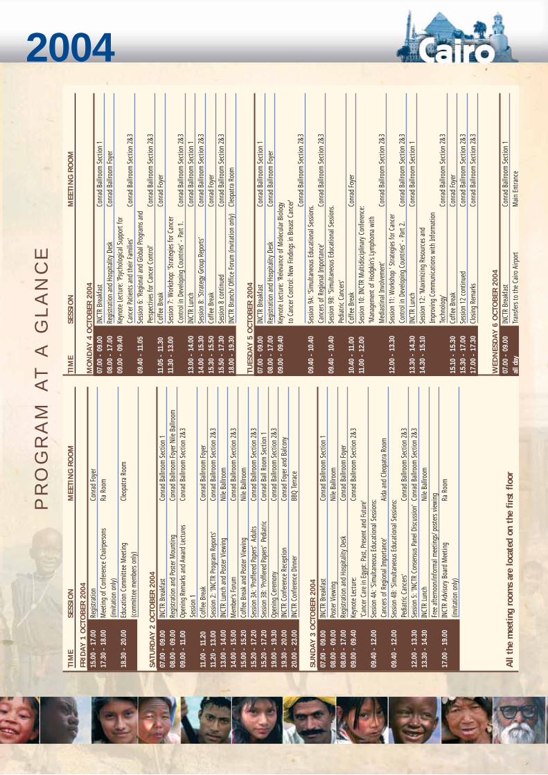
PR
OG
RA
M A
T A
GL
AN
CE
2004
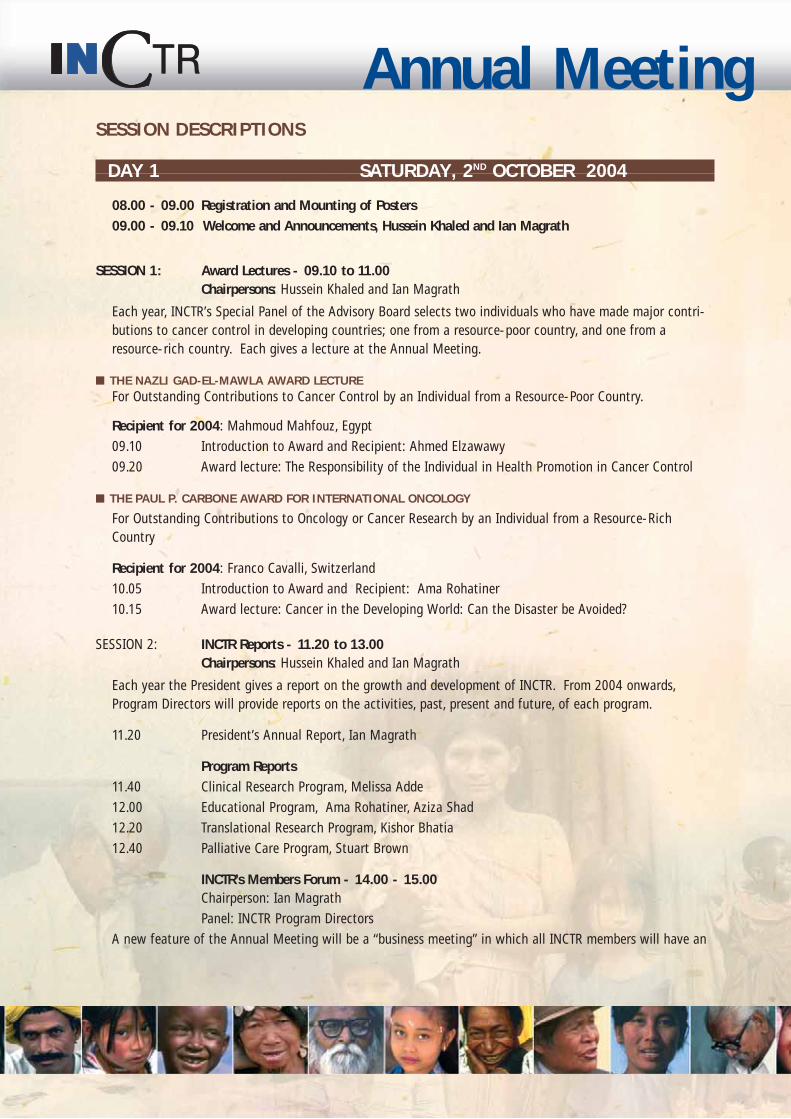
Annual Meeting SESSION DESCRIPTIONS
DAY 1 SATURDAY, 2ND OCTOBER 2004
08.00 - 09.00 Registration and Mounting of Posters09.00 - 09.10 Welcome and Announcements, Hussein Khaled and Ian Magrath
SESSION 1: Award Lectures - 09.10 to 11.00Chairpersons: Hussein Khaled and Ian Magrath
Each year, INCTR’s Special Panel of the Advisory Board selects two individuals who have made major contri-butions to cancer control in developing countries; one from a resource-poor country, and one from aresource-rich country. Each gives a lecture at the Annual Meeting.
■ THE NAZLI GAD-EL-MAWLA AWARD LECTURE For Outstanding Contributions to Cancer Control by an Individual from a Resource-Poor Country.
Recipient for 2004: Mahmoud Mahfouz, Egypt
09.10 Introduction to Award and Recipient: Ahmed Elzawawy
09.20 Award lecture: The Responsibility of the Individual in Health Promotion in Cancer Control
■ THE PAUL P. CARBONE AWARD FOR INTERNATIONAL ONCOLOGY
For Outstanding Contributions to Oncology or Cancer Research by an Individual from a Resource-RichCountry
Recipient for 2004: Franco Cavalli, Switzerland
10.05 Introduction to Award and Recipient: Ama Rohatiner
10.15 Award lecture: Cancer in the Developing World: Can the Disaster be Avoided?
SESSION 2: INCTR Reports - 11.20 to 13.00Chairpersons: Hussein Khaled and Ian Magrath
Each year the President gives a report on the growth and development of INCTR. From 2004 onwards,Program Directors will provide reports on the activities, past, present and future, of each program.
11.20 President’s Annual Report, Ian Magrath
Program Reports11.40 Clinical Research Program, Melissa Adde
12.00 Educational Program, Ama Rohatiner, Aziza Shad
12.20 Translational Research Program, Kishor Bhatia
12.40 Palliative Care Program, Stuart Brown
INCTR’s Members Forum - 14.00 - 15.00Chairperson: Ian Magrath
Panel: INCTR Program Directors
A new feature of the Annual Meeting will be a “business meeting” in which all INCTR members will have an
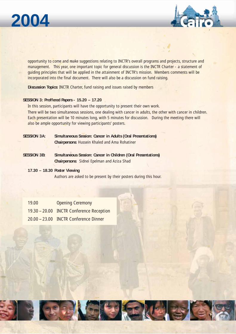
2004
opportunity to come and make suggestions relating to INCTR’s overall programs and projects, structure andmanagement. This year, one important topic for general discussion is the INCTR Charter - a statement ofguiding principles that will be applied in the attainment of INCTR’s mission. Members comments will beincorporated into the final document. There will also be a discussion on fund raising.
Discussion Topics: INCTR Charter, fund raising and issues raised by members
SESSION 3: Proffered Papers - 15.20 – 17.20In this session, participants will have the opportunity to present their own work.
There will be two simultaneous sessions, one dealing with cancer in adults, the other with cancer in children.Each presentation will be 10 minutes long, with 5 minutes for discussion. During the meeting there willalso be ample opportunity for viewing participants’ posters.
SESSION 3A: Simultaneous Session: Cancer in Adults (Oral Presentations)Chairpersons: Hussein Khaled and Ama Rohatiner
SESSION 3B: Simultaneous Session: Cancer in Children (Oral Presentations)Chairpersons: Sidnei Epelman and Aziza Shad
17.30 – 18.30 Poster Viewing Authors are asked to be present by their posters during this hour.
19.00 Opening Ceremony
19.30 – 20.00 INCTR Conference Reception
20.00 – 23.00 INCTR Conference Dinner
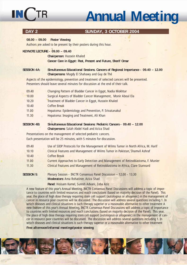
Annual Meeting DAY 2 SUNDAY, 3 OCTOBER 2004
08.00 – 09.00 Poster ViewingAuthors are asked to be present by their posters during this hour.
KEYNOTE LECTURE - 09.00 – 09.40Chairperson: Hussein Khaled
Cancer Care in Egypt: Past, Present and Future, Sherif Omar
SESSION 4A: Simultaneous Educational Sessions. Cancers of Regional Importance - 09.40 – 12.00Chairpersons: Magdy El Shahawy and Guy de Thé
Aspects of the epidemiology, prevention and treatment of selected cancers will be presented.Presenters should leave several minutes for discussion at the end of their talk.
09.40 Changing Pattern of Bladder Cancer in Egypt, Nadia Mokhtar
10.00 Surgical Aspects of Bladder Cancer Management, Monir Aboul Ela
10.20 Treatment of Bladder Cancer in Egypt, Hussein Khaled
10.40 Coffee Break
11.00 Hepatoma: Epidemiology and Prevention, P. Srivatanakul
11.30 Hepatoma: Imaging and Treatment, Ali Khan
SESSION 4B: Simultaneous Educational Sessions: Pediatric Cancers - 09.40 – 12.00Chairpersons: Salah Abdel Hadi and Aziza Shad
Presentations on the management of selected pediatric cancers.
Each presentation will be 25 minutes, with 5 minutes for discussion.
09.40 Use of SIOP Protocols for the Management of Wilms Tumor in North Africa, M. Harif
10.10 Clinical Features and Management of Wilms Tumor in Pakistan, Shamvil Ashraf
10.40 Coffee Break
11.00 Current Approaches to Early Detection and Management of Retinoblastoma, F. Munier
11.30 Clinical Features and Management of Retinoblastoma in Africa, Clare Stannard
SESSION 5: Plenary Session - INCTR Consensus Panel Discussion – 12.00 - 13.30 Moderators: Ama Rohatiner, Aziza Shad
Panel: Hossam Kamel, Suresh Advani, Zeba AzizA new feature of this year's Annual Meeting, INCTR Consensus Panel Discussions will address a topic of impor-tance to countries with limited resources and reach conclusions (based on majority decision of the Panel). Thisyear, the place of high dose therapy requiring stem cell support (autologous or allogeneic) in the management ofcancer in resource poor countries will be discussed. The discussion will address several questions including 1. Inwhich diseases and clinical situations is such therapy superior or a reasonable alternative to other treatment Anew feature of this year's Annual Meeting, INCTR Consensus Panel Discussions will address a topic of importanceto countries with limited resources and reach conclusions (based on majority decision of the Panel). This year,the place of high dose therapy requiring stem cell support (autologous or allogeneic) in the management of can-cer in resource poor countries will be discussed. The discussion will address several questions including 1. Inwhich diseases and clinical situations is such therapy superior or a reasonable alternative to other treatment
Free afternoon/informal meetings/poster viewing
2004DAY 3 MONDAY, 4 OCTOBER 2004
KEYNOTE LECTURE - 09.00 – 09.40Chairperson: Stuart Brown
Psychological Support for Cancer Patients and their Families, Jimmie Holland
SESSION 6: Regional and Global Programs and Perspectives on Cancer Control – 09.40- 11.05Chairpersons: Twalib Ngoma and Cecilia Sepulveda
These presentations will address aspects of cancer control in countries with limited resources and the per-spective of organizations created to address them.
09.40 Palliative Care in an African Setting, Ekie Kikule10.00 IARC’s Cervical Cancer Screening Program, R. Sankaranarayanan10.20 The Perspective of AORTIC, Paul Ndom10.35 The Perspective of CHALLENGE, Indraneel Mittra10.50 The Perspective of ICEDOC, Ahmed Elzawawy11.05 Coffee Break
SESSION 7: Workshop - Strategies for Cancer Control in Developing Countries – 11.30 – 13.00Part 1. Institutions, Approaches and Leadership
Chairpersons: Osama el-Khatib and Elmer Huerta
In part 1 of this workshop, the strategies of three international organizations concerned with cancer controlwill be presented and discussed.
11.30 WHO’s Cancer Control Program, Cecilia Sepulveda11.50 UICC’s Cancer Control Program, Odd Søreide12.10 IAEA’s Radiotherapy Programs, Victor Levin12.30 Panel Discussion: Institutional Leadership in Controlling Cancer on a Global Basis
Panel: Sherif Omar, Mohammed Al-Jarallah, Ben Anderson, Santiago Pavlovsky and above speakers
SESSION 8: Strategy Group Reports - 14.00 – 17.30Chairpersons: Eduardo Cazap and Melissa Adde
In this session, a representative from each strategy group will give a brief overview of the group’s ongoingand planned activities.
14.00 Retinoblastoma Strategy Group, Sidnei Epelman14.30 Leukemia Strategy Group, Suresh Advani15.00 Breast Cancer Strategy Group, Zeba Aziz15.30 Coffee Break15.50 Lymphoma Strategy Group (African), Muheez Durosinmi16.20 Cervical Cancer Strategy Group, Carlos Santos16.50 – 17.30 Discussion
Annual Meeting DAY 4 TUESDAY, 5 OCTOBER 2004
KEYNOTE LECTURE - 09.00 – 09.40Chairperson: Kishor Bhatia
Relevance of Molecular Biology to Cancer Control: New Findings in Breast Cancer, James Holland
SESSION 9A Simultaneous Educational Sessions. Cancers of Regional Importance - 09.40 – 10.40Chairpersons: Krishnan Nair and Alison Brown
09.40 Epidemiology and Prevention of Nasopharyngeal Carcinoma, Guy de Thé
10.10 Management of Nasopharyngeal Cancer, Hassan Awwad
SESSION 9B Simultaneous Educational Sessions. Pediatric Cancers - 09.40 – 10.40Chairpersons: Hassan El-Solh and Angelo Rosolen
09.40 Management of Osteosarcoma, Sidnei Epelman
10.10 Limb Sparing Surgery, Martin Malawer
SESSION 10 INCTR Multidisciplinary Conference: Management of Hodgkin’s Lymphoma with Mediastinal Involvement.
Panel: Aziza Shad, Henning Bredenfeld, K. Naresh, Ali Khan, Samy El-Badawy
A new element of the Annual Meeting is INCTR multidisciplinary conferences. Such conferences are a stan-dard feature of patient management in major cancer centers, in which multidisciplinary teams discuss opti-mal management of a particular patient for whom multimodality treatment or alternative modalities oftreatment are options. The pros and cons of various approaches are discussed by experts. For patients, theyensure that all relevant specialists have participated in the final decision, and for staff, they provide anopportunity to exchange views, and attempt to develop a consensus on optimal treatment. For more juniormembers they provide an educational experience.
SESSION 11 Workshop: Strategies for Cancer Control in Developing CountriesPart 2. Development of Capacity; Ethical Issues; Economic Issues
Chairpersons: Suresh Advani and Francis Crawley
In part 2 of this workshop, approaches to the building of capacity, and the financial problems faced in coun-tries with limited resources, both with respect to decisions regarding standard treatment, and supportingessential research (at a minimum, the investigation of the effectiveness of prevention and managementapproaches in a regional context) will be presented and discussed.
12.00 INCTR’s Program in Capacity Building for Research and Treatment, Ian Magrath
12.20 Who Supports the Cost of Research in Developing Countries?, Yasser Mostafa
12.40 The Dilemma of Treatment Cost versus Efficacy in Developing Countries, F. Crawley
200413.00 Panel Discussion: Faisal Sultan, El-Nasir Lalani, Sean Swarmer, Eduardo Cazap and
the above speakers
SESSION 12 Maximizing Resources and Improving Communications and Patient Care Via Information Technology - 14.30-17.00
Chairpersons: Faisal Sultan and Frans Dhaenens
Each year, INCTR includes a session on the use of technology in developing countries. This year, specialemphasis will be given to information technology, which has the potential to enhance quality and efficiencyand to reduce the cost of health services, research and education.
14.30 The Promise of Internet II: Grids, Portals and Semantic Sensitivity, Raj Shah
14.50 Enhancing Communication: the Power of Telemedicine, Ola Wagih Lowrance
15.10 Coffee Break
15.30 The Potential of PACS in Education and Consultation, Frans Dhaenens
15.50 Can Virtual Microscopy Improve Pathologists’ Diagnoses, Peter Dervan
16.10 Telemedicine Activities in India, Kishnan Nair
16.30 Improving Delivery of Care through Hospital Information Systems, Faisal Sultan
16.50 Discussion
17.00 Closing Remarks, Ian Magrath
Annual Meeting
INCTR gives two annual awards to individuals whohave made outstanding contributions to cancer treat-ment or research in one or more developing countries.The purpose of these awards is not simply to recog-nize and honor the recipients, although this is certain-ly an important element, but also to show, by theirexample, that much can be accomplished even whenresources are limited. It is hoped that their work andphilosophy brought through the award lectures to abroader audience than would otherwise be the casewill inspire others to greater efforts.
Each of the awards is named after a distinguishedoncologist. They began their careers when there wasso little knowledge about the causes of cancer thatpeople could only live in fear that they would one daybe a victim, while the diagnosis was usually hiddenfrom those unfortunate enough to develop cancer,because so little could be done for them. It is thanksto the resolution and fortitude of Dr. Nazli Gad-el-Mawla, Dr. Paul P. Carbone, and others like them, whoworked through a time when cancer specialists wereoften accused of prolonging the misery of cancer vic-tims rather than helping them, that today, at least inthe wealthier nations, more than half of those whodevelop cancer can be cured. Both Dr. Nazli and Dr.Carbone were responsible for training numerousyoung people, and so leave us a precious legacythrough which their work will be continued.
The Nazli Gad-el-Mawla Award is made for outstand-ing contributions to cancer control by an individualfrom a country with limited resources. Nazli Gad-el-Mawla was a pioneer Egyptian oncologist, who, as amember of a small group of oncologists working at theNational Cancer Institute in Cairo in the 1960s and70s, helped to build the institute into one of the pre-mier cancer centers in the Middle East. She foundedthe Department of Medical Oncology in 1970 and, aspart of it, developed a strong pediatric oncology pro-gram. She is known particularly for her work in thechemotherapy of cancer of the bilharzial bladder,
which accounts for some 25% of all cancer in Egypt,and in hematological malignancies. She was highlyrespected both by her colleagues in Egypt and also bythe international community of oncologists in whichshe became increasingly active throughout her career.
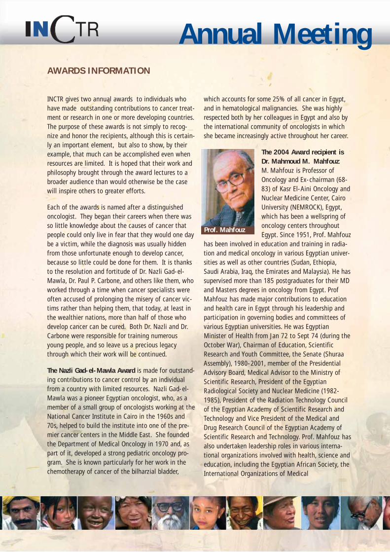
The 2004 Award recipient isDr. Mahmoud M. Mahfouz: M. Mahfouz is Professor ofOncology and Ex-chairman (68-83) of Kasr El-Aini Oncology andNuclear Medicine Center, CairoUniversity (NEMROCK), Egypt,which has been a wellspring ofoncology centers throughoutEgypt. Since 1951, Prof. Mahfouz
has been involved in education and training in radia-tion and medical oncology in various Egyptian univer-sities as well as other countries (Sudan, Ethiopia,Saudi Arabia, Iraq, the Emirates and Malaysia). He hassupervised more than 185 postgraduates for their MDand Masters degrees in oncology from Egypt. ProfMahfouz has made major contributions to educationand health care in Egypt through his leadership andparticipation in governing bodies and committees ofvarious Egyptian universities. He was EgyptianMinister of Health from Jan 72 to Sept 74 (during theOctober War), Chairman of Education, ScientificResearch and Youth Committee, the Senate (ShuraaAssembly), 1980-2001, member of the PresidentialAdvisory Board, Medical Advisor to the Ministry ofScientific Research, President of the EgyptianRadiological Society and Nuclear Medicine (1982-1985), President of the Radiation Technology Councilof the Egyptian Academy of Scientific Research andTechnology and Vice President of the Medical andDrug Research Council of the Egyptian Academy ofScientific Research and Technology. Prof. Mahfouz hasalso undertaken leadership roles in various interna-tional organizations involved with health, science andeducation, including the Egyptian African Society, theInternational Organizations of Medical
AWARDS INFORMATION
Prof. Mahfouz
2004
Parliamentarians, and the International Physicians forPrevention of Nuclear War (IPPNW) and the PugwashMovement of Science and International Affairs (theorganization being winner of the Nobel Peace Prize in1985). He has served or acted as consultant to variousUN committees, including the UN ScientificCommittee on the Effects of Atomic Radiation, 1961-1966, WHO Technical Committee on Cancer andRadiation, the International Atomic Energy Agency(IAEA) Division of Human Sciences (Medical Research)and the East Mediterranean Regional Office of WHO(EMRO). He has been the recipient of numerous hon-ors and awards, including the El-Gomhoria State Merit(1974), Egypt, the Art and Science Order (1992), Egypt,the State Merit Prize for Biological Sciences (1992),Egypt, Chevalier of the Legion D’Honneur (1982),France and Mubarak State Prize (2003), Egypt.
The Paul P. Carbone Award in InternationalOncology is made for outstanding contributions tooncology or cancer research by an individual from aresource-rich country. Paul P. Carbone was a pioneerAmerican oncologist, who, as the Associate Directorfor the Clinical Oncology Program at the NationalCancer Institute, Bethesda, played a critical role inthe development of cancer chemotherapy.Subsequently, he continued his work as the Director ofthe Cancer Center at the University of Madison,Wisconsin. From the beginning, he recognized notonly the needs of patients in developing countries, butalso the contribution that scientific research conduct-ed in such countries could and should make to theglobal efforts against cancer. Dr Carbone’s familyhave established a the Paul P. Carbone MD Foundationfor “the support of scientific, educational, and charita-ble endeavors that reflect Dr. Carbone's practice of theart and science of oncology and his lifelong dedicationto teaching and mentoring.”
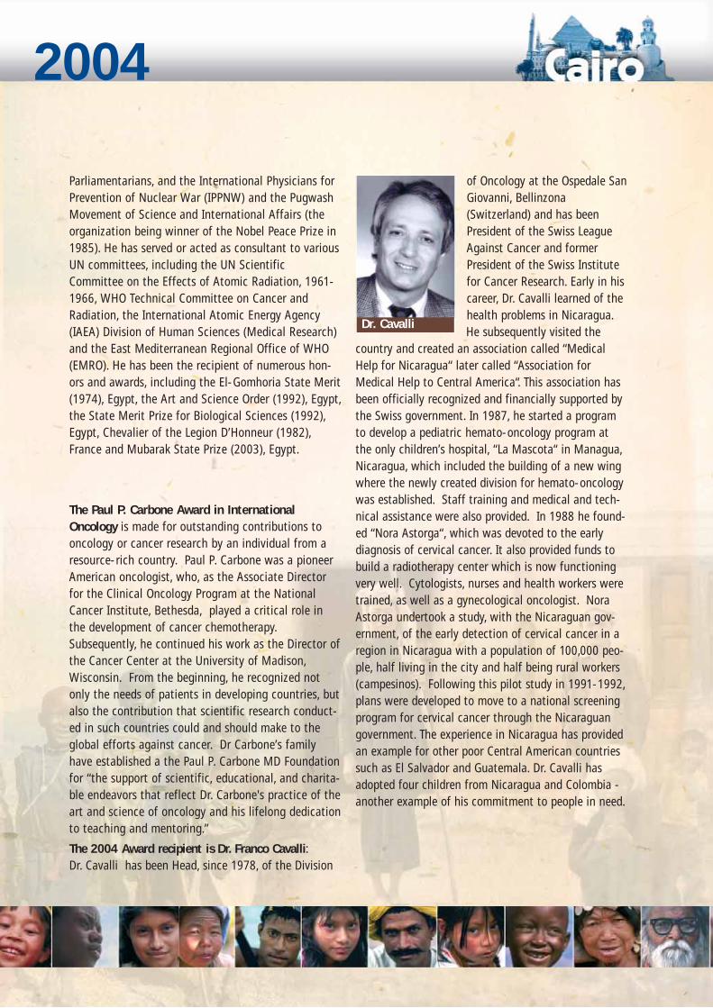
The 2004 Award recipient is Dr. Franco Cavalli: Dr. Cavalli has been Head, since 1978, of the Division
of Oncology at the Ospedale SanGiovanni, Bellinzona(Switzerland) and has beenPresident of the Swiss LeagueAgainst Cancer and formerPresident of the Swiss Institutefor Cancer Research. Early in hiscareer, Dr. Cavalli learned of thehealth problems in Nicaragua.He subsequently visited the
country and created an association called “MedicalHelp for Nicaragua“ later called “Association forMedical Help to Central America“. This association hasbeen officially recognized and financially supported bythe Swiss government. In 1987, he started a programto develop a pediatric hemato-oncology program atthe only children’s hospital, “La Mascota“ in Managua,Nicaragua, which included the building of a new wingwhere the newly created division for hemato-oncologywas established. Staff training and medical and tech-nical assistance were also provided. In 1988 he found-ed “Nora Astorga“, which was devoted to the earlydiagnosis of cervical cancer. It also provided funds tobuild a radiotherapy center which is now functioningvery well. Cytologists, nurses and health workers weretrained, as well as a gynecological oncologist. NoraAstorga undertook a study, with the Nicaraguan gov-ernment, of the early detection of cervical cancer in aregion in Nicaragua with a population of 100,000 peo-ple, half living in the city and half being rural workers(campesinos). Following this pilot study in 1991-1992,plans were developed to move to a national screeningprogram for cervical cancer through the Nicaraguangovernment. The experience in Nicaragua has providedan example for other poor Central American countriessuch as El Salvador and Guatemala. Dr. Cavalli hasadopted four children from Nicaragua and Colombia -another example of his commitment to people in need.
Dr. Cavalli
Annual Meeting
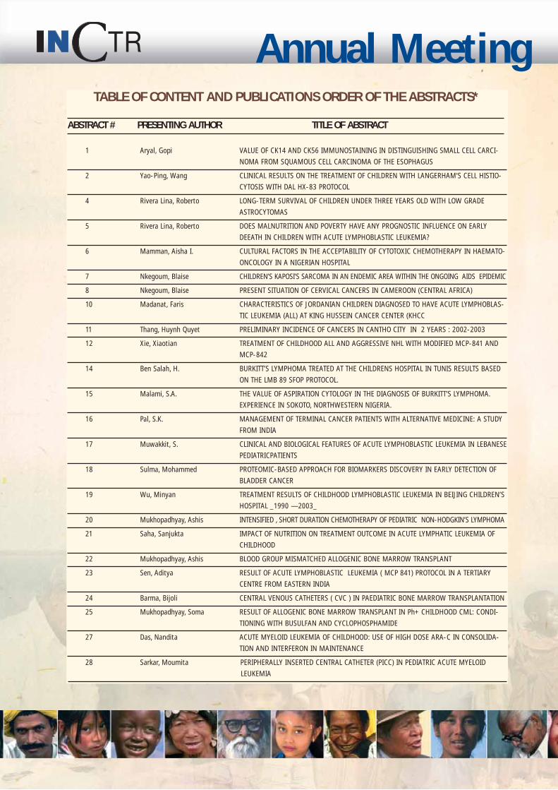
1 Aryal, Gopi VALUE OF CK14 AND CK56 IMMUNOSTAINING IN DISTINGUISHING SMALL CELL CARCI-
NOMA FROM SQUAMOUS CELL CARCINOMA OF THE ESOPHAGUS
2 Yao-Ping, Wang CLINICAL RESULTS ON THE TREATMENT OF CHILDREN WITH LANGERHAM'S CELL HISTIO-
CYTOSIS WITH DAL HX-83 PROTOCOL
4 Rivera Lina, Roberto LONG-TERM SURVIVAL OF CHILDREN UNDER THREE YEARS OLD WITH LOW GRADE
ASTROCYTOMAS
5 Rivera Lina, Roberto DOES MALNUTRITION AND POVERTY HAVE ANY PROGNOSTIC INFLUENCE ON EARLY
DEEATH IN CHILDREN WITH ACUTE LYMPHOBLASTIC LEUKEMIA?
6 Mamman, Aisha I. CULTURAL FACTORS IN THE ACCEPTABILITY OF CYTOTOXIC CHEMOTHERAPY IN HAEMATO-
ONCOLOGY IN A NIGERIAN HOSPITAL
7 Nkegoum, Blaise CHILDREN’S KAPOSI’S SARCOMA IN AN ENDEMIC AREA WITHIN THE ONGOING AIDS EPIDEMIC
8 Nkegoum, Blaise PRESENT SITUATION OF CERVICAL CANCERS IN CAMEROON (CENTRAL AFRICA)
10 Madanat, Faris CHARACTERISTICS OF JORDANIAN CHILDREN DIAGNOSED TO HAVE ACUTE LYMPHOBLAS-
TIC LEUKEMIA (ALL) AT KING HUSSEIN CANCER CENTER (KHCC
11 Thang, Huynh Quyet PRELIMINARY INCIDENCE OF CANCERS IN CANTHO CITY IN 2 YEARS : 2002-2003
12 Xie, Xiaotian TREATMENT OF CHILDHOOD ALL AND AGGRESSIVE NHL WITH MODIFIED MCP-841 AND
MCP-842
14 Ben Salah, H. BURKITT’S LYMPHOMA TREATED AT THE CHILDRENS HOSPITAL IN TUNIS RESULTS BASED
ON THE LMB 89 SFOP PROTOCOL.
15 Malami, S.A. THE VALUE OF ASPIRATION CYTOLOGY IN THE DIAGNOSIS OF BURKITT'S LYMPHOMA.
EXPERIENCE IN SOKOTO, NORTHWESTERN NIGERIA.
16 Pal, S.K. MANAGEMENT OF TERMINAL CANCER PATIENTS WITH ALTERNATIVE MEDICINE: A STUDY
FROM INDIA
17 Muwakkit, S. CLINICAL AND BIOLOGICAL FEATURES OF ACUTE LYMPHOBLASTIC LEUKEMIA IN LEBANESE
PEDIATRICPATIENTS
18 Sulma, Mohammed PROTEOMIC-BASED APPROACH FOR BIOMARKERS DISCOVERY IN EARLY DETECTION OF
BLADDER CANCER
19 Wu, Minyan TREATMENT RESULTS OF CHILDHOOD LYMPHOBLASTIC LEUKEMIA IN BEIJING CHILDREN’S
HOSPITAL _1990 — 2003_
20 Mukhopadhyay, Ashis INTENSIFIED , SHORT DURATION CHEMOTHERAPY OF PEDIATRIC NON-HODGKIN’S LYMPHOMA
21 Saha, Sanjukta IMPACT OF NUTRITION ON TREATMENT OUTCOME IN ACUTE LYMPHATIC LEUKEMIA OF
CHILDHOOD
22 Mukhopadhyay, Ashis BLOOD GROUP MISMATCHED ALLOGENIC BONE MARROW TRANSPLANT
23 Sen, Aditya RESULT OF ACUTE LYMPHOBLASTIC LEUKEMIA ( MCP 841) PROTOCOL IN A TERTIARY
CENTRE FROM EASTERN INDIA
24 Barma, Bijoli CENTRAL VENOUS CATHETERS ( CVC ) IN PAEDIATRIC BONE MARROW TRANSPLANTATION
25 Mukhopadhyay, Soma RESULT OF ALLOGENIC BONE MARROW TRANSPLANT IN Ph+ CHILDHOOD CML: CONDI-
TIONING WITH BUSULFAN AND CYCLOPHOSPHAMIDE
27 Das, Nandita ACUTE MYELOID LEUKEMIA OF CHILDHOOD: USE OF HIGH DOSE ARA-C IN CONSOLIDA-
TION AND INTERFERON IN MAINTENANCE
28 Sarkar, Moumita PERIPHERALLY INSERTED CENTRAL CATHETER (PICC) IN PEDIATRIC ACUTE MYELOID
LEUKEMIA
TABLE OF CONTENT AND PUBLICATIONS ORDER OF THE ABSTRACTS*
ABSTRACT # PRESENTING AUTHOR TITLE OF ABSTRACT
2004
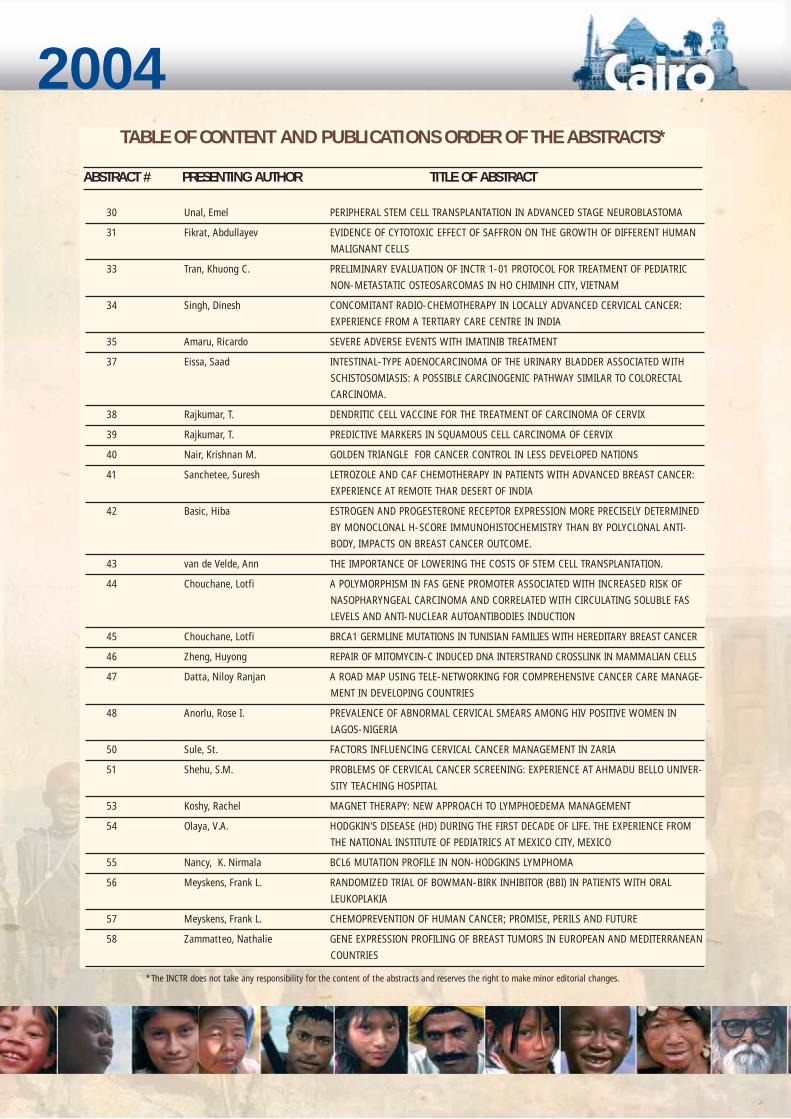
30 Unal, Emel PERIPHERAL STEM CELL TRANSPLANTATION IN ADVANCED STAGE NEUROBLASTOMA
31 Fikrat, Abdullayev EVIDENCE OF CYTOTOXIC EFFECT OF SAFFRON ON THE GROWTH OF DIFFERENT HUMAN
MALIGNANT CELLS
33 Tran, Khuong C. PRELIMINARY EVALUATION OF INCTR 1-01 PROTOCOL FOR TREATMENT OF PEDIATRIC
NON-METASTATIC OSTEOSARCOMAS IN HO CHIMINH CITY, VIETNAM
34 Singh, Dinesh CONCOMITANT RADIO-CHEMOTHERAPY IN LOCALLY ADVANCED CERVICAL CANCER:
EXPERIENCE FROM A TERTIARY CARE CENTRE IN INDIA
35 Amaru, Ricardo SEVERE ADVERSE EVENTS WITH IMATINIB TREATMENT
37 Eissa, Saad INTESTINAL-TYPE ADENOCARCINOMA OF THE URINARY BLADDER ASSOCIATED WITH
SCHISTOSOMIASIS: A POSSIBLE CARCINOGENIC PATHWAY SIMILAR TO COLORECTAL
CARCINOMA.
38 Rajkumar, T. DENDRITIC CELL VACCINE FOR THE TREATMENT OF CARCINOMA OF CERVIX
39 Rajkumar, T. PREDICTIVE MARKERS IN SQUAMOUS CELL CARCINOMA OF CERVIX
40 Nair, Krishnan M. GOLDEN TRIANGLE FOR CANCER CONTROL IN LESS DEVELOPED NATIONS
41 Sanchetee, Suresh LETROZOLE AND CAF CHEMOTHERAPY IN PATIENTS WITH ADVANCED BREAST CANCER:
EXPERIENCE AT REMOTE THAR DESERT OF INDIA
42 Basic, Hiba ESTROGEN AND PROGESTERONE RECEPTOR EXPRESSION MORE PRECISELY DETERMINED
BY MONOCLONAL H-SCORE IMMUNOHISTOCHEMISTRY THAN BY POLYCLONAL ANTI-
BODY, IMPACTS ON BREAST CANCER OUTCOME.
43 van de Velde, Ann THE IMPORTANCE OF LOWERING THE COSTS OF STEM CELL TRANSPLANTATION.
44 Chouchane, Lotfi A POLYMORPHISM IN FAS GENE PROMOTER ASSOCIATED WITH INCREASED RISK OF
NASOPHARYNGEAL CARCINOMA AND CORRELATED WITH CIRCULATING SOLUBLE FAS
LEVELS AND ANTI-NUCLEAR AUTOANTIBODIES INDUCTION
45 Chouchane, Lotfi BRCA1 GERMLINE MUTATIONS IN TUNISIAN FAMILIES WITH HEREDITARY BREAST CANCER
46 Zheng, Huyong REPAIR OF MITOMYCIN-C INDUCED DNA INTERSTRAND CROSSLINK IN MAMMALIAN CELLS
47 Datta, Niloy Ranjan A ROAD MAP USING TELE-NETWORKING FOR COMPREHENSIVE CANCER CARE MANAGE-
MENT IN DEVELOPING COUNTRIES
48 Anorlu, Rose I. PREVALENCE OF ABNORMAL CERVICAL SMEARS AMONG HIV POSITIVE WOMEN IN
LAGOS-NIGERIA
50 Sule, St. FACTORS INFLUENCING CERVICAL CANCER MANAGEMENT IN ZARIA
51 Shehu, S.M. PROBLEMS OF CERVICAL CANCER SCREENING: EXPERIENCE AT AHMADU BELLO UNIVER-
SITY TEACHING HOSPITAL
53 Koshy, Rachel MAGNET THERAPY: NEW APPROACH TO LYMPHOEDEMA MANAGEMENT
54 Olaya, V.A. HODGKIN'S DISEASE (HD) DURING THE FIRST DECADE OF LIFE. THE EXPERIENCE FROM
THE NATIONAL INSTITUTE OF PEDIATRICS AT MEXICO CITY, MEXICO
55 Nancy, K. Nirmala BCL6 MUTATION PROFILE IN NON-HODGKINS LYMPHOMA
56 Meyskens, Frank L. RANDOMIZED TRIAL OF BOWMAN-BIRK INHIBITOR (BBI) IN PATIENTS WITH ORAL
LEUKOPLAKIA
57 Meyskens, Frank L. CHEMOPREVENTION OF HUMAN CANCER; PROMISE, PERILS AND FUTURE
58 Zammatteo, Nathalie GENE EXPRESSION PROFILING OF BREAST TUMORS IN EUROPEAN AND MEDITERRANEAN
COUNTRIES
TABLE OF CONTENT AND PUBLICATIONS ORDER OF THE ABSTRACTS*
ABSTRACT # PRESENTING AUTHOR TITLE OF ABSTRACT
* The INCTR does not take any responsibility for the content of the abstracts and reserves the right to make minor editorial changes.
Annual Meeting
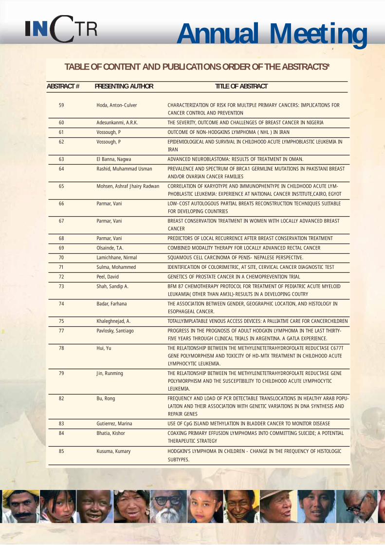
59 Hoda, Anton-Culver CHARACTERIZATION OF RISK FOR MULTIPLE PRIMARY CANCERS: IMPLICATIONS FOR
CANCER CONTROL AND PREVENTION
60 Adesunkanmi, A.R.K. THE SEVERITY, OUTCOME AND CHALLENGES OF BREAST CANCER IN NIGERIA
61 Vossough, P OUTCOME OF NON-HODGKINS LYMPHOMA ( NHL ) IN IRAN
62 Vossough, P EPIDEMIOLOGICAL AND SURVIVAL IN CHILDHOOD ACUTE LYMPHOBLASTIC LEUKEMIA IN
IRAN
63 El Banna, Nagwa ADVANCED NEUROBLASTOMA: RESULTS OF TREATMENT IN OMAN.
64 Rashid, Muhammad Usman PREVALENCE AND SPECTRUM OF BRCA1 GERMLINE MUTATIONS IN PAKISTANI BREAST
AND/OR OVARIAN CANCER FAMILIES
65 Mohsen, Ashraf Jhairy Radwan CORRELATION OF KARYOTYPE AND IMMUNOPHENTYPE IN CHILDHOOD ACUTE LYM-
PHOBLASTIC LEUKEMIA: EXPERIENCE AT NATIONAL CANCER INSTITUTE,CAIRO, EGYOT
66 Parmar, Vani LOW-COST AUTOLOGOUS PARTIAL BREATS RECONSTRUCTION TECHNIQUES SUITABLE
FOR DEVELOPING COUNTRIES
67 Parmar, Vani BREAST CONSERVATION TREATMENT IN WOMEN WITH LOCALLY ADVANCED BREAST
CANCER
68 Parmar, Vani PREDICTORS OF LOCAL RECURRENCE AFTER BREAST CONSERVATION TREATMENT
69 Olsainde, T.A. COMBINED MODALITY THERAPY FOR LOCALLY ADVANCED RECTAL CANCER
70 Lamichhane, Nirmal SQUAMOUS CELL CARCINOMA OF PENIS- NEPALESE PERSPECTIVE.
71 Sulma, Mohammed IDENTIFICATION OF COLORIMETRIC, AT SITE, CERVICAL CANCER DIAGNOSTIC TEST
72 Peel, David GENETICS OF PROSTATE CANCER IN A CHEMOPREVENTION TRIAL
73 Shah, Sandip A. BFM 87 CHEMOTHERAPY PROTOCOL FOR TREATMENT OF PEDIATRIC ACUTE MYELOID
LEUKAMIA( OTHER THAN AM3L)-RESULTS IN A DEVELOPING COUTRY
74 Badar, Farhana THE ASSOCIATION BETWEEN GENDER, GEOGRAPHIC LOCATION, AND HISTOLOGY IN
ESOPHAGEAL CANCER.
75 Khaleghnejad, A. TOTALLYIMPLATABLE VENOUS ACCESS DEVICES: A PALLIATIVE CARE FOR CANCERCHILDREN
77 Pavlosky, Santiago PROGRESS IN THE PROGNOSIS OF ADULT HODGKIN LYMPHOMA IN THE LAST THIRTY-
FIVE YEARS THROUGH CLINICAL TRIALS IN ARGENTINA. A GATLA EXPERIENCE.
78 Hui, Yu THE RELATIONSHIP BETWEEN THE METHYLENETETRAHYDROFOLATE REDUCTASE C677T
GENE POLYMORPHISM AND TOXICITY OF HD-MTX TREATMENT IN CHILDHOOD ACUTE
LYMPHOCYTIC LEUKEMIA.
79 Jin, Runming THE RELATIONSHIP BETWEEN THE METHYLENETETRAHYDROFOLATE REDUCTASE GENE
POLYMORPHISM AND THE SUSCEPTIBILITY TO CHILDHOOD ACUTE LYMPHOCYTIC
LEUKEMIA.
82 Bu, Rong FREQUENCY AND LOAD OF PCR DETECTABLE TRANSLOCATIONS IN HEALTHY ARAB POPU-
LATION AND THEIR ASSOCIATION WITH GENETIC VARIATIONS IN DNA SYNTHESIS AND
REPAIR GENES
83 Gutierrez, Marina USE OF CpG ISLAND METHYLATION IN BLADDER CANCER TO MONITOR DISEASE
84 Bhatia, Kishor COAXING PRIMARY EFFUSION LYMPHOMAS INTO COMMITTING SUICIDE; A POTENTIAL
THERAPEUTIC STRATEGY
85 Kusuma, Kumary HODGKIN'S LYMPHOMA IN CHILDREN - CHANGE IN THE FREQUENCY OF HISTOLOGIC
SUBTYPES.
TABLE OF CONTENT AND PUBLICATIONS ORDER OF THE ABSTRACTS*
ABSTRACT # PRESENTING AUTHOR TITLE OF ABSTRACT
2004
86 Al Lamki, Zakia PROMOTING QUALITY CARE AND RESEARCH CAPACITY IN PEDIATRIC ONCOLOGY IN A
DEVELOPING COUNTRY BY INTERNATIONAL COLLABORATION
88 Barnoya, Margarita RETINOBLASTOMA IN GUATEMALA
89 Sebastian, Paul SIMPLE, LOW COST RECONSTRUCTIVE OPTIONS IN ORAL CANCER SURGERY: 10-YEAR
EXPERIENCE FROM A REGIONAL CANCER CENTRE IN A DEVELOPING COUNTRY
90 Singh, Yogendra FEATURES OF BREAST CANCER IN NEPALESE YOUNG ADULTS
92 Leal, Carlos FACTORS RELATED WITH LATE DIAGNOSIS IN RETINOBLASTOMA. A PRELIMINARY REPORT
FROM MEXICO
93 Oluwabunmi Olapade-Olaopa, E. PROSTATE CANCER CONTROL: EVIDENCE FOR THE NEED FOR COMMUNITY-BASED EDU-
CATIONAL PROGRAMS IN SOUTHWEST NIGERIA
94 Sowedi, Muyingo INCREASING ACCESS TO CANCER TREATMENTS IN RESOURCE LIMITED COUNTRIES:
RESULTS FROM GLIVEC INTERNATIONAL PATIENT ASSISTANCE PROGRAM (GIPAP)
95 Zafad, S RETINOBLASTOMA, A REPROPECTIVE STUDY OF 55 CASES. EXEPRIENCE OF A SINGLE
INSTITUTION
96 Thavaraj, V MANAGEMENT OF RETINOBLASTOMA WITH CARBOPLATIN, ETOPOSIDE, VINCRISTINE AND
CYCLOPHOSPHAMIDE
97 Thavaraj, V CLINICAL PROFILE AND OUTCOME OF RETINOBLASTOMA CASES SEEN IN THE LAST 12 YEARS
98 Thavaraj, V FEASABILITY OF AWARENESS COMPAIGN PROGRAM IN THE EARLY DIAGNOSIS OF
RETINOBLASTOMA AT DISTRICT HOSPITAL IN RAJASTHAN IN INDIA
100 Gamal, Amira PANCREATICODUODENECTOMY FOR PERIAMPULLARY ADENOCARCINOMA. RESULTS OF
SURGICAL TREATMENT
101 Solimsn, A.S. DIFFERING DNA METHYLATION PATTERNS AND GENE MUTATION FREQUENCIES IN COL-
ORECTAL CARICINOMAS FROM MIDDLE EAST COUNTRIES
102 Zaghloul, Mohamed PHASE III RANDOMIZED CONTROLLED TRIAL FOR ADDING ADJUVANT CHERNORADIO-
THERAPY WITH GEMCITABINE AND CISPLATINUM TO POSTPERATIVE RADIOTHERAPY
(PORT) IN HIGH RISK BLADDER CANCER PATIENTS
103 Khalil, E. THE POSSIBILITY OF DEVELOPING A BLADDER CONSERVATION PROTOCOL FOR BLADDER
CANCER ASSOCIATED WITH SCHISTOMIASIS
105 Hussein, Hany IMPROVED SURVIVAL OF BURKITT'S LYMPHOMA IN RURAL AREAS OF EGYPT
106 Bahnassy, Abeer BIOLOGICAL DETERMINANTS OF PATIENTS RESPONSE TO TREATMENT IN CARCINOMA OF
THE UTERINE CERVIX
107 Al-Toubary, Y HLA CLASS II POLYMORPHISM IN EGYPTIAN CHILDREN WITH LYMPHOMAS
108 Bahnassy, Abeer THE POSSIBLE ROLE OF CELL CYCLE REGULATOR GENES IN THE MULTISTEP PROCESS OF
HP V-ASSOCIATED CERVICAL CARCINOMA
109 Dma, Yassin HER FAMILY EXPRESSION IN EGYPTIAN BREAST CANCER PATIENTS
110 Cowgill, Karen CASE-CONTROL STUDY OF NON-HODGKIN'S LYMPHOMA AND HEPATITIS C VIRUS INFEC-
TION IN EGYPT
111 El Bassuoni, M DNA DAMAGE IN CHILDHOOD B-CELL NON-HODGKIN'S LYMPHOMA
112 Azza, Kamel EXPRESSION OF CELL CYCLE REGULATORY PROTEINS IN BREAST CANCER
113 Rahnian, Abdel HEPATITIS C VIRUS GENOTYPING BY INNO-LIPA VERSUS TRUGENE SEQUENCING IN
EGYPTIAN PATIENTS
TABLE OF CONTENT AND PUBLICATIONS ORDER OF THE ABSTRACTS*
ABSTRACT # PRESENTING AUTHOR TITLE OF ABSTRACT
* The INCTR does not take any responsibility for the content of the abstracts and reserves the right to make minor editorial changes.
The INCTR Annual Meeting 2004 – Cancer Control inDeveloping Countries - has been appraised andapproved by the Accreditation Council of Oncology inEurope (ACOE). ACOE is a multidisciplinary body offull time specialists practicing in the field of oncologyand all recognized for their experience in educationand expertise in their field. ACOE accreditationacknowledges quality of the scientific program and its
educational value. The conference has been designat-ed for a maximum of 18 hours of European externalCME credits.
Delegates are kindly requested to complete the gener-al evaluation form before claiming their certificate ofattendance.
The conference secretariat will not issue or mail certifi-cates of attendance to participants after the conference.
CME ACCREDITATION AND CERTIFICATE OF ATTENDANCE
115 Zaghloul, Mohamed PROGNOSTIC FACTORS FOR BONE-OMLY METASTASIS IN BREAST CANCER PATIENTS
118 Lalani, Ema IMPLICATIONS OF INHIBITION OF CYCLOOXYGENASE-2 ON THE EXPRESSION OFMUC1
MUCIN IN TUMPUR CELL LINES: POSSIBLE IMPACT ON TUMUR INVASION
119 Lalani, Ema ErbB2:EGFR RATIO IS A CRITICAL PREDICTOR OF A MITOGENIC RESPONSE TO EGF IN A
PROSTATE CANCER MODEL
120 Lalani, Ema P13K ENZYMES ARE OVER-EXPRESSED IN PROSTATE CANCER (PC) AND ARE A PROG-
NOSTIC INDICATOR OF POOR SURVIVAL
121 Lalani, Ema NON-CLINICAL STUDIES INVOLVING SIGNAL TRANSDUCTION INHIBITORS IN ANDROGEN
SENSITIVE AND INSENSITIVE PROSTATE CANCER
122 Cavdar, Ayhan CHARACTERISTICS OF HODGKIN'S DISEASE IN TURKISH CHILDREN OF PRESCHOOL AGE
123 AbdulMuhsen, Y.Saleem LARYNGEAL CARCINOMA IN MOSUL
124 Al-Hamdani, ORAL CANCER IN MOSUL ANALZSIS OF 132 CASESRaad Yahya Mohamad
125 Al-Ramadani, Marah A PERSONAL ATTITUDES TOWARDS CANCER AMONG DOCTORS, NON-DOCTOR CANCER
CONFERENCE ATTENDANTS AND LAY PEOPLE IN MOSUL
126 Al-Ramdhani, Ayad H CANCER CONTROL PROGRAM IN IRAQ (1985 – 2000)
127 Al-Al-Saadoon, CENTRAL NERVOUS SYSTEMTUMOURS A PATHOLOGICAL STUDY OF 462 CASES IN
Rafah Saadi Abdullah NINEVAH PROVINCE
128 Al-Ramadhani, Ayad H CANCER PAIN RELIEF AND PALLIATIVE CARE IN IRAQ
129 Kharrufa, Nabeel D. MOSUL PAIN RELIEF AND PALIATIVE CARE CLINIC
131 Senan, Mohamed Tariq R. THYROID CANCER IN TOXIC GOITER
132 Ibrahim, Ali Attia LUNG CANCER IN MOSUL (1990-1999)
133 Naktal, Ahmad I. KNOWLEDGE & ATTITUDE TOWARD BREAST CANCER & BREAST SELF EXAMINATION IN
FEMALE SCHOOL TEACHERS IN MOSUL
134 Al-Ramdhani, Ayad H KNOWLEDGE & ATTITUDE TOWARD BREAST CANCER & BREAST SELF EXAMINATION INFEMALE DOCTORS IN MOSUL
135 Al-Sa'oor, E. Luay HODGKIN’S DISEASE IN MOSUL (1990-1999)
136 Ismail, Adel Mohamed CANCER IN MOSUL AMONG ADULTS AND CHILDREN: TEN YEARS STUDY 1990 – 1999
137 Al-hero, Khalid N.M. HAEMATOLOGICAL RESPONSE OF CHRONICE MYELOID LEUKAEMIA TO (IMATINIB)
138 Kotedia MARKERS OF METASTATIC POTENTIAL IN PROSTATE CANCER USING MAGNETIC
RESONANCE SPECTROSCOPY
TABLE OF CONTENT AND PUBLICATIONS ORDER OF THE ABSTRACTS*
ABSTRACT # PRESENTING AUTHOR TITLE OF ABSTRACT
* The INCTR does not take any responsibility for the content of the abstracts and reserves the right to make minor editorial changes.
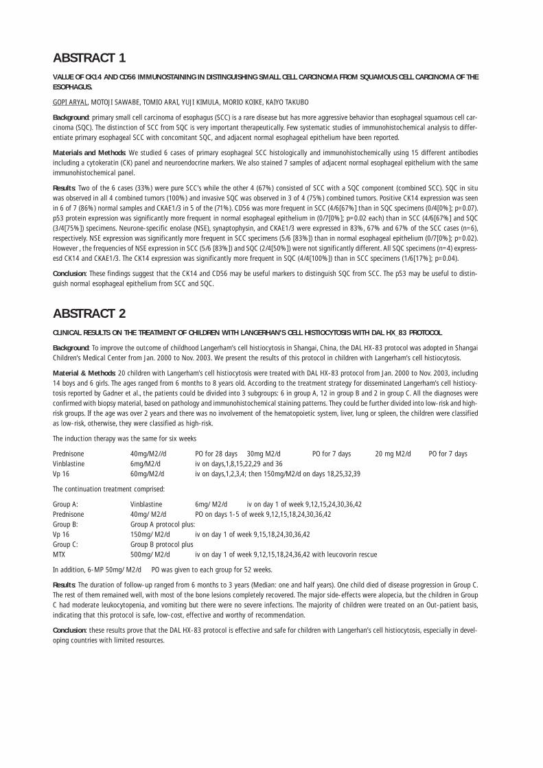
ABSTRACT 1VALUE OF CK14 AND CD56 IMMUNOSTAINING IN DISTINGUISHING SMALL CELL CARCINOMA FROM SQUAMOUS CELL CARCINOMA OF THEESOPHAGUS.
GOPI ARYAL, MOTOJI SAWABE, TOMIO ARAI, YUJI KIMULA, MORIO KOIKE, KAIYO TAKUBO
Background: primary small cell carcinoma of esophagus (SCC) is a rare disease but has more aggressive behavior than esophageal squamous cell car-cinoma (SQC). The distinction of SCC from SQC is very important therapeutically. Few systematic studies of immunohistochemical analysis to differ-entiate primary esophageal SCC with concomitant SQC, and adjacent normal esophageal epithelium have been reported.
Materials and Methods: We studied 6 cases of primary esophageal SCC histologically and immunohistochemically using 15 different antibodiesincluding a cytokeratin (CK) panel and neuroendocrine markers. We also stained 7 samples of adjacent normal esophageal epithelium with the sameimmunohistochemical panel.
Results: Two of the 6 cases (33%) were pure SCC’s while the other 4 (67%) consisted of SCC with a SQC component (combined SCC). SQC in situwas observed in all 4 combined tumors (100%) and invasive SQC was observed in 3 of 4 (75%) combined tumors. Positive CK14 expression was seenin 6 of 7 (86%) normal samples and CKAE1/3 in 5 of the (71%). CD56 was more frequent in SCC (4/6[67%] than in SQC specimens (0/4[0%]; p=0.07).p53 protein expression was significantly more frequent in normal esophageal epithelium in (0/7[0%]; p=0.02 each) than in SCC (4/6[67%] and SQC(3/4[75%]) specimens. Neurone-specific enolase (NSE), synaptophysin, and CKAE1/3 were expressed in 83%, 67% and 67% of the SCC cases (n=6),respectively. NSE expression was significantly more frequent in SCC specimens (5/6 [83%]) than in normal esophageal epithelium (0/7[0%]; p=0.02).However , the frequencies of NSE expression in SCC (5/6 [83%]) and SQC (2/4[50%]) were not significantly different. All SQC specimens (n=4) express-esd CK14 and CKAE1/3. The CK14 expression was significantly more frequent in SQC (4/4[100%]) than in SCC specimens (1/6[17%]; p=0.04).
Conclusion: These findings suggest that the CK14 and CD56 may be useful markers to distinguish SQC from SCC. The p53 may be useful to distin-guish normal esophageal epithelium from SCC and SQC.
ABSTRACT 2CLINICAL RESULTS ON THE TREATMENT OF CHILDREN WITH LANGERHAN'S CELL HISTIOCYTOSIS WITH DAL HX_83 PROTOCOL
Background: To improve the outcome of childhood Langerham’s cell histiocytosis in Shangai, China, the DAL HX-83 protocol was adopted in ShangaiChildren’s Medical Center from Jan. 2000 to Nov. 2003. We present the results of this protocol in children with Langerham’s cell histiocytosis.
Material & Methods: 20 children with Langerham’s cell histiocytosis were treated with DAL HX-83 protocol from Jan. 2000 to Nov. 2003, including14 boys and 6 girls. The ages ranged from 6 months to 8 years old. According to the treatment strategy for disseminated Langerham’s cell histiocy-tosis reported by Gadner et al., the patients could be divided into 3 subgroups: 6 in group A, 12 in group B and 2 in group C. All the diagnoses wereconfirmed with biopsy material, based on pathology and immunohistochemical staining patterns. They could be further divided into low-risk and high-risk groups. If the age was over 2 years and there was no involvement of the hematopoietic system, liver, lung or spleen, the children were classifiedas low-risk, otherwise, they were classified as high-risk.
The induction therapy was the same for six weeks
Prednisone 40mg/M2//d PO for 28 days 30mg M2/d PO for 7 days 20 mg M2/d PO for 7 daysVinblastine 6mg/M2/d iv on days,1,8,15,22,29 and 36Vp 16 60mg/M2/d iv on days,1,2,3,4; then 150mg/M2/d on days 18,25,32,39
The continuation treatment comprised:
Group A: Vinblastine 6mg/ M2/d iv on day 1 of week 9,12,15,24,30,36,42Prednisone 40mg/ M2/d PO on days 1-5 of week 9,12,15,18,24,30,36,42Group B: Group A protocol plus:Vp 16 150mg/ M2/d iv on day 1 of week 9,15,18,24,30,36,42Group C: Group B protocol plusMTX 500mg/ M2/d iv on day 1 of week 9,12,15,18,24,36,42 with leucovorin rescue
In addition, 6-MP 50mg/ M2/d PO was given to each group for 52 weeks.
Results: The duration of follow-up ranged from 6 months to 3 years (Median: one and half years). One child died of disease progression in Group C.The rest of them remained well, with most of the bone lesions completely recovered. The major side-effects were alopecia, but the children in GroupC had moderate leukocytopenia, and vomiting but there were no severe infections. The majority of children were treated on an Out-patient basis,indicating that this protocol is safe, low-cost, effective and worthy of recommendation.
Conclusion: these results prove that the DAL HX-83 protocol is effective and safe for children with Langerhan’s cell histiocytosis, especially in devel-oping countries with limited resources.
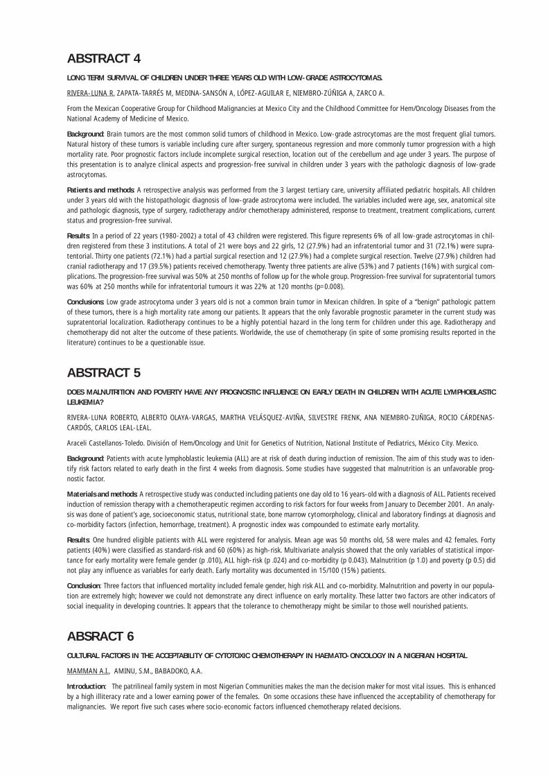
ABSTRACT 4LONG TERM SURVIVAL OF CHILDREN UNDER THREE YEARS OLD WITH LOW-GRADE ASTROCYTOMAS.
RIVERA-LUNA R, ZAPATA-TARRÉS M, MEDINA-SANSÓN A, LÓPEZ-AGUILAR E, NIEMBRO-ZÚÑIGA A, ZARCO A.
From the Mexican Cooperative Group for Childhood Malignancies at Mexico City and the Childhood Committee for Hem/Oncology Diseases from theNational Academy of Medicine of Mexico.
Background: Brain tumors are the most common solid tumors of childhood in Mexico. Low-grade astrocytomas are the most frequent glial tumors.Natural history of these tumors is variable including cure after surgery, spontaneous regression and more commonly tumor progression with a highmortality rate. Poor prognostic factors include incomplete surgical resection, location out of the cerebellum and age under 3 years. The purpose ofthis presentation is to analyze clinical aspects and progression-free survival in children under 3 years with the pathologic diagnosis of low-gradeastrocytomas.
Patients and methods: A retrospective analysis was performed from the 3 largest tertiary care, university affiliated pediatric hospitals. All childrenunder 3 years old with the histopathologic diagnosis of low-grade astrocytoma were included. The variables included were age, sex, anatomical siteand pathologic diagnosis, type of surgery, radiotherapy and/or chemotherapy administered, response to treatment, treatment complications, currentstatus and progression-free survival.
Results: In a period of 22 years (1980-2002) a total of 43 children were registered. This figure represents 6% of all low-grade astrocytomas in chil-dren registered from these 3 institutions. A total of 21 were boys and 22 girls, 12 (27.9%) had an infratentorial tumor and 31 (72.1%) were supra-tentorial. Thirty one patients (72.1%) had a partial surgical resection and 12 (27.9%) had a complete surgical resection. Twelve (27.9%) children hadcranial radiotherapy and 17 (39.5%) patients received chemotherapy. Twenty three patients are alive (53%) and 7 patients (16%) with surgical com-plications. The progression-free survival was 50% at 250 months of follow up for the whole group. Progression-free survival for supratentorial tumorswas 60% at 250 months while for infratentorial tumours it was 22% at 120 months (p=0.008).
Conclusions: Low grade astrocytoma under 3 years old is not a common brain tumor in Mexican children. In spite of a “benign” pathologic patternof these tumors, there is a high mortality rate among our patients. It appears that the only favorable prognostic parameter in the current study wassupratentorial localization. Radiotherapy continues to be a highly potential hazard in the long term for children under this age. Radiotherapy andchemotherapy did not alter the outcome of these patients. Worldwide, the use of chemotherapy (in spite of some promising results reported in theliterature) continues to be a questionable issue.
ABSTRACT 5DOES MALNUTRITION AND POVERTY HAVE ANY PROGNOSTIC INFLUENCE ON EARLY DEATH IN CHILDREN WITH ACUTE LYMPHOBLASTICLEUKEMIA?
RIVERA-LUNA ROBERTO, ALBERTO OLAYA-VARGAS, MARTHA VELÁSQUEZ-AVIÑA, SILVESTRE FRENK, ANA NIEMBRO-ZUÑIGA, ROCIO CÁRDENAS-CARDÓS, CARLOS LEAL-LEAL.
Araceli Castellanos-Toledo. División of Hem/Oncology and Unit for Genetics of Nutrition, National Institute of Pediatrics, México City. Mexico.
Background: Patients with acute lymphoblastic leukemia (ALL) are at risk of death during induction of remission. The aim of this study was to iden-tify risk factors related to early death in the first 4 weeks from diagnosis. Some studies have suggested that malnutrition is an unfavorable prog-nostic factor.
Materials and methods: A retrospective study was conducted including patients one day old to 16 years-old with a diagnosis of ALL. Patients receivedinduction of remission therapy with a chemotherapeutic regimen according to risk factors for four weeks from January to December 2001. An analy-sis was done of patient’s age, socioeconomic status, nutritional state, bone marrow cytomorphology, clinical and laboratory findings at diagnosis andco-morbidity factors (infection, hemorrhage, treatment). A prognostic index was compounded to estimate early mortality.
Results: One hundred eligible patients with ALL were registered for analysis. Mean age was 50 months old, 58 were males and 42 females. Fortypatients (40%) were classified as standard-risk and 60 (60%) as high-risk. Multivariate analysis showed that the only variables of statistical impor-tance for early mortality were female gender (p .010), ALL high-risk (p .024) and co-morbidity (p 0.043). Malnutrition (p 1.0) and poverty (p 0.5) didnot play any influence as variables for early death. Early mortality was documented in 15/100 (15%) patients.
Conclusion: Three factors that influenced mortality included female gender, high risk ALL and co-morbidity. Malnutrition and poverty in our popula-tion are extremely high; however we could not demonstrate any direct influence on early mortality. These latter two factors are other indicators ofsocial inequality in developing countries. It appears that the tolerance to chemotherapy might be similar to those well nourished patients.
ABSRACT 6 CULTURAL FACTORS IN THE ACCEPTABILITY OF CYTOTOXIC CHEMOTHERAPY IN HAEMATO-ONCOLOGY IN A NIGERIAN HOSPITAL
MAMMAN A.I., AMINU, S.M., BABADOKO, A.A.
Introduction: The patrilineal family system in most Nigerian Communities makes the man the decision maker for most vital issues. This is enhancedby a high illiteracy rate and a lower earning power of the females. On some occasions these have influenced the acceptability of chemotherapy formalignancies. We report five such cases where socio-economic factors influenced chemotherapy related decisions.
Methods: Five females aged 20, 20, 32, 24 and 58 years with acute lymphoblastic leukemia, chronic myeloid leukemia and non-Hodgkin’s lymphomaspresented between 1997 and 2004. One patient with CML was 7 months pregnant at the time of diagnosis. A second CML patient was breast-feed-ing a 7-month-old infant. A 20-year-old patient with CML was two weeks away from her wedding day. The 58 year old with non-Hodgkin lym-phoma was postmenopausal. The 22 year old with ALL became pregnant while in the maintenance phase of ALL chemotherapy and had to stop ther-apy. All patients were counseled on the chemotherapy option available and possible side effects. All 5 patients were housewives, only one - the preg-nant CML patient - had high school education.
Results: Out of the 5 patients, 4 did not receive chemotherapy because their husbands did not allow them. Cultural practice enforces breast-feedingfor a minimum of one year in rural settlements and termination of pregnancy is abhorred. This creates a dilemma between choice of therapy and pro-creation breast-feeding for the doctor. One patient stopped therapy for ALL because she became pregnant. One patient’s husband, the post-menopausal 58year old with NHL, insisted that she took all cycles of therapy. Only five patients were reported because many more patients wouldnot report to hospital because of ignorance and poverty.
Conclusion: Spouse participation is an important component of the Pre-therapy counseling services for patients with malignancy. The husband beingthe head of household holds the authority for decision-making in all matters. Girl-Child education: removes some of the obstacles like poverty, igno-rance while improving access to quality health care.
ABSTRACT 7CHILDREN’S KAPOSI’S SARCOMA IN AN ENDEMIC AREA WITHIN THE ONGOING AIDS EPIDEMIC
NKEGOUM B, GESSAIN A, AMA MOOR V, DOUMBE P, NDOM P, ESSAME O.
Faculty of Medicine and Biomedical Sciences, Yaoundé,Cameroon, Pasteur Institute Paris, France and Mother and child Chantal Biya Fundation,Yaoundé, Cameroon.Reference Hospital Yaoundé, Cameroon.
Introduction: Cameroon was an endemic area of Kaposi’s sarcoma even before the AIDS epidemic. 16 cases of children’s Kaposi’s sarcoma wereobserved between 1968 and 1974 by Jensen and al. An increased incidence of Kaposi’s sarcoma is noted among AIDS patients everywhere in the worldnowadays. The aim of this study was to show the anatomic, clinical and therapeutic aspects of Kaposi’s sarcoma in Cameroon (Central Africa).
Methods: This retrospective study was carried out over a period of 16 years (1987-2003). The pathological registers, clinical files, thesis, researchworks on Kaposi’s sarcoma in Cameroon were retrieved. We noted : the total number of Kaposi’s sarcoma within the period of the study, the ageand gender of the patients, the clinical signs, the treatment received and the prognosis.
Results: Among the 555 cases of Kaposi’s sarcoma histological diagnosed within the period of the study, 4% were aged 13months to 15 years. Themale to female ratio was 3/1. In most cases, children presented with massive cervical lymphadenopathy. All children had fever and weight loss. In afew cases, skin and lymph node involvement was also observed. Only three cases of HIV positive serology were observed within this series and theirmothers were also HIV positive. The pathological aspect observed was advanced stage Kaposi’s sarcoma. Less than ten chidren received full dosechemotherapy comprising cyclophosphamide, vincristine and prednisone. One child survived nine years after chemotherapy.
ABSTRACT 8PRESENT SITUATION OF CERVICAL CANCERS IN CAMEROON (CENTRAL AFRICA)
NKEGOUM B, NDOM P, DOH A, YOMI J, ESSAME O, DOUMBE P.
Faculty of Medicine and Biomedical Sciences-Yaoundé-Cameroon.
Introduction: Cervical cancers are the most common cancer in Cameroonian women. The aim of this retrospective study was to show the presentsituation of cervical cancers in Cameroon (Central Africa).
Methods: The material was made up of pathological laboratories' registers, clinical files, thesis, scientific publications and research works on cervi-cal cancers
Results: The sample of histologically diagnosed cervical cancers between 1986 and 2003 shows that there are about 165 cases per year. On the otherhand, more than 15.000 new cases of cancers are expected every year. Only 1500 are histologically diagnosed, for several reasons. On the whole,about 1650 new cases of cervical cancer should appear each year in Cameroon which has 15.000.000 inhabitants among women aged 20 and above.It is a young population (60% are less than 20 years old). The 40% left (i.e.6000.0000) are more than 20 years old. Among them, 51% ( i.e 3.060.000) are women, all exposed to cervical cancers. 70% live in rural areas, 3% are HIV positive but in most of the cases, HIV status is unknown. Cervicalcancers are 4th on the list of cancers in Cameroon. They form 11% of cancers in Cameroon, as do breast cancers. Cervical cancers are on top of thelist of cancers in Cameroonian women, and form 21,5% of them.
Cervical cancers are also the commonest cancers in Cameroonian women aged 50 years and above, and form 31 % (1/3) of them. 99,9 % are carci-nomas. Malignant lymphomas (mostly Burkitt ) make up the rest. Among carcinomas, squamous cell carcinomas and their variants predominate.Primitive adenocarcinoma of the cervix is exceptional. One HIV patient in this series had simultaneously cervical carcinoma and lymph node Kaposi’ssarcoma. The mean age of the patients is 59 years. All the patients have vaginal bleeding and 75% have an ulcerated and/or fungating cervix.
Conclusion: Despite the efforts of medical staff and journalists to decrease the incidence of clinical cervical cancers , through mass campaigns of Papsmear, UVIA and information, the situation has remained generally unchanged. However, in the 2 main towns (Yaounde and Douala), a positive changehas been observed with, at the moment, only exceptional cases recorded. Fight against cervical cancers in Cameroon needs a strong and rich nation-al programme which gets in touch with rural areas where the majority of women live.
ABSTRACT 10CHARACTERISTICS OF JORDANIAN CHILDREN DIAGNOSED TO HAVE ACUTE LYMPHOBLASTIC LEUKEMIA (ALL) AT KING HUSSEIN CANCERCENTER (KHCC).
MADANAT FM, KING HUSSEIN CANCER CENTER, AMMAN, JORDAN
Introduction: Jordan has a population of 5 million, 38% of whom are children under the age of 15. Acute lymphoblastic leukemia accounts for 25%of all childhood cancers in Jordan. Studies to characterize ALL in Jordanian children are lacking. This report aims to provide preliminary informationon the immunobiology and chromosomal abnormalities detected in children with ALL.
Methods: Between November 2003 and April 2004 (18 month period), 54 newly diagnosed Jordanian children with acute lymphoblastic leukemiawere referred to the newly named KHCC for management. Diagnosis of acute lymphoblastic leukemia was established by performing bone marrowaspirates and biopsies.
Results: Male to female ratio was 1.3 (31 males, 23 female). Ages ranged from 1 month to 15 years, median 4 years. Peak age was 3 – 4 years. Agedistribution as follows:< 1 year: (2); 1 -4:( 20); 5 – 9: (11); 10 -15:(11). Immunophenotypes:- Flow cytometry was performed on all patients andresults were as follows: precursor B cell 42 (78%), T cell 8 (15%), B cell 3 (5%), and bilineage 1 (2%). Cytogenetics:- Chromosomal studies were per-formed on 32 patients, 25 of whom had precursor B cell leukemia. Abnormalities detected among the precursor B group were as follows:- hyper-diploid in 3 , t ( 9:22) in 1 , t ( 1:19) in 2, t (4:11) in 3, t ( 12:21) in 1 , and trisomy 21 in 1. All t (4:11) cases occurred in children over 1 year ofageImmunophenotyping was performed using flow cytometry on marrow samples, and chromosomal studies were done using standard techniques,and FISH to detect specific chromosomal translocations. Conclusion: Age and sex distribution as well as immunophenotypes of Jordanian childrenwith acute lymphoblastic leukemia are similar to those reported from western countries. There appears to be a lower incidence of favourable cyto-genetic indicators, hyperdiploidy and t (12:21), and a higher incidence of adverse indicators like t (4:11). However a larger series is needed to sub-stantiate these findings.
ABSTRACT 11PRELIMINARY INCIDENCE OF CANCERS IN CANTHO CITY IN 2 YEARS: 2002-2003
HUYNH QUYET THANG , NGUYEN VAN QUI, NGUYEN TRUONG GIANG, HUYNH THAO LUAT, VO VAN KHA & DANG PHI HUNG.
Can Tho General Hospital, CAN THO - VIETNAM.
Introduction: Cancer is one of the most devastating diseases not only in developed countries but also in developing countries. In Viet Nam, the bur-den of cancer is increasing and becomes an important health problem for the whole country. Cancer registry is the unique source supplying data forevaluating the cancer burden of either a community or a country.
Purpose of study: We carried out the study of the population based cancer registry of Can Tho city in order to participate in the programme ofNational Cancer Control, to present the particularities of malignant diseases of the region and to establish a strategy of Cancer Control andManagement for the city.
Methods: 3293 cases of cancer were registered during the period of 2 years 2002-2003 among which 1437 males (43.6%) and 1856 females (56.4%).Information about each cancer case was registered. Data were collected from 18 hospitals and health care centers in the city and were analysed usingCANREG software version 3 for creating the CR (Crude Rate) and ASR (Age Standardized Rate) of cancers as well as of each kind of the most com-mon cancers.
Results: The cancer incidence for both sexes: 88.4/100000; males: 78,6/100000 and females: 97.8/100000.
- Of both sexes, the 10 most common cancers were: liver , colorectum , cervix uteri , breast , lung , stomach , non hodgkin lymphoma (NHL) , leukemia,ovary and skin.
- In male patients: Liver (ASR:24.4); Lung (ASR:18.1); Colorectum (ASR:18), Stomach (ASR:17.5); NHL(ASR:9.4); Leukemia(ASR:5.3); Penis (ASR:3.9);Panceas (ASR:3.3); Skin (ASR:2.9) and Bladder (ASR:2.7)
- In female patients: Cervix uteri (ASR:23.3); Breast (ASR:20); Colorectum (ASR:12.5), Ovary (ASR:8.3) Liver (ASR:7.9); Lung (ASR:7.4); NHL (ASR:7.3);Stomach (ASR: 6.4); Leukemia (ASR:5.4) and Thyroid (ASR:3.7).
Conclusion: The results of this study prove that the characteristics of CanTho cancer incidence were : a predominance of liver and lung cancers fol-lowed by colorectal cancer in male patients and the predominance of cervix uteri cancer followed by an abundance of breast cancer and colorectalcancer in female patients. These are useful data for participating in the programme of National Cancer Control and setting a strategy of CancerControl and Management in Can Tho city.
ABSTRACT 12TREATMENT OF CHILDHOOD ALL AND AGGRESSIVE NHL WITH MODIFIED MCP-841 AND MCP-842
XIAOTIAN XIE, YAOPING WANG,
Dept. Pediatric Hematology/Oncology of Tongji Hospital, Tongji University, Shanghai, China
Introduction: Some reports have shown that the outcomes of childhood ALL and aggressive NHL could be improved by treatment with MCP-841 andMCP-842. The use of radiotherapy may lead to unresolved impairment of the central nervous system. B cell linage of ALL and NHL may more sensi-tive to high-dose MTX (HD-MTX), and the treatment duration of MCP-842 for aggressive NHL might be prolonged using maintenance therapy suchas CHOP and COMP to improve the CCR rate. So, we tried to modify MCP-841 and MCP-842 in the treatment of childhood ALL and aggressive NHLin our hospital. We present the details of how we modified the protocols and the outcomes of the treatment for childhood ALL and aggressive NHLwith the modified protocols in our study in recent 10 years.
Methods: There were 69 cases in our study. 43 of ALL and 26 of aggressive NHL (13 stage III, 13 stage IV, and 18 B-cell, 8 T-cell) who were previ-ously untreated were enrolled on the study between March 1994 and March 2004. ALL and T-type NHL or stage IV NHL were treated with MCP-841,and non-T with stage III NHL were treated with MCP-842 respectively. We modified MCP-841 and MCP-842 as follows: (1) Using I2A (HD-AraC +CTX + 6MP) three cycles as induction2 therapy instead of cranial irradiation after CR, (2) Alternatively using HD-MTX for 5 to 7 cycles during themaintenance therapy, (3) Prolonging the period of therapy to two years with using CHOP and COMP as maintenance therapy and alternatively usingA or B regimen of MCP-842 in every 3-4 months during the maintenance, (4) Instead of DNR or ADR, we using E-ADR or THP in VDLP protocol forinduction and timed intensification therapy for the prophylaxis of heart impairment
Results: CR rate was 100% in ALL and 92.3% (24/26) in NHL. 4 cases (3 ALL, 1 NHL) were lost followed up, and the results of 65 cases were avail-able. Up to now, 48 cases (73.8%) were in the state of CR1 (30 ALL and 18 NHL). 20 cases of ALL and 10 cases of NHL had got CR1 for more than 3years (CCR), the CCR rate of ALL and NHL were 72.4% (21/29) and 77.8 (14/18) respectively. 10 cases of ALL and 4 cases of NHL had got CR1 for lessthan 3 years. No one died of the chemotherapy related complications.
Conclusion: Our study results showed that the modified MCP-841 and MCP-842 were more effective than the initial MCP for the treatment of child-hood ALL as well as advanced stage NHL. For the comprehensive measures we used to prevent and treat the complications associated with chemother-apy in our study, no patient died of chemotherapy related complications, this might be another reason for the outcome in our hospital being betterthan that in other reports.
ABSTRACT 14BURKITT’S LYMPHOMA TREATED AT THE CHILDRENS HOSPITAL IN TUNIS. RESULTS BASED ON THE LMB 89 SFOP PROTOCOL.
BEN SALAH H, BEN BECHER S, BEN ROMDHAN K.
Children’s hospital Tunis. Tunisia.
Introduction: Effective management of Burkitt’s lymphoma remains a challenge in developing countries. The aim of this study was to determinethe feasibilty and results of treating Tunisian children with Burkitt’s lymphoma according to an intensive protocol based on the French LMB 89Protocol.
Methods: 25 newly diagnosed patients (pts) admitted in our institution from May 96 to April 2001 for proven Burkitt’s lymphoma/ALL of the L3 FABcell type were enrolled on this retrospective study. Event- free survival ( EFS) was estimated according to Kaplan Meier method and subgroups werecompared by log rank test.
Results: The mean age was 74 months (ranged from 36 to 157 months). The male to female ratio was 2.7:1. Primary site was abdominal in 16 pts(64%), head and neck in 4 pts (16%), kidney (1pt), nodes (1pt) and 3 pts with ALL as defined in LMB 89 protocol. According to Murphy classification1 pt was stage I, 1 pt stage II, 14 pts stage III (56%), 6 pts stage IV (24% 3 pts with CSF+) and ALL in 3 cases (12%). 2 pts had a primary immunod-eficiency (8%). Mean follow up was 18 months (ranged from 1,5 to 57 months). 2 pts underwent hemodialysis for severe tumor lysis syndrome (8%).3 toxic deaths (12%) due to severe infection during induction therapy occurred. 1 pt was a poor responder (stage III) and died in PD. 21 pts achievedCR (85%). 3 pts relapsed (12%) on treatment and died (2 stage IV, 1 ALL). 18 pts are alive in first CR, all of them with a follow up superior to 12months. EFS was 73%. CNS involvement was the single predictive factor of failure (P=0.02).
Conclusion: The use of an intensive protocol is feasible in our institution. The EFS is lower than the one reported in a French study but comparableto other studies in developed countries despite the fact that a high rate of advanced stages where present in our patient population. Better resultswill be obtained with proper management of treatment related toxicity.
ABSTRACT 15THE VALUE OF ASPIRATION CYTOLOGY IN THE DIAGNOSIS OF BURKITT’S LYMPHOMA. EXPERIENCE IN SOKOTO, NORTHWESTERN NIGERIA.
MALAMI SA, JIYA NM, OJO BA1.
Usmanu Danfodiyo University Teaching Hospital, Sokoto and University of Ilorin, Nigeria.
Introduction: Scarcity of facilities for early diagnosis has hampered management of Burkitt’s lymphoma in our center. Fine needle aspiration cytology(FNAC), a cheap and well-tolerated outpatient procedure, was initiated to provide reliable and timely diagnosis. We present the results of this experience.
Methods: Twenty-seven previously untreated patients were enrolled in the study between January and December 2003. Using a 21G needle and a10-ml syringe satisfactory smears were obtained from suspicious masses in 20 patients, fixed in 95 % ethyl alcohol and stained with May GrunwaldGiemsa and Papanicolaou stains. They were then examined microscopically according to established cytomorphologic criteria for diagnosis of Burkitt’slymphoma. Clinical and radiologic methods were used to correlate the diagnosis. Information on the presentation, treatment and outcome on eachpatient was systematically collected and analysed.
Results: The mean age was 7.5 (range 3 to 12 years). The male to female ratio was 1.22:1.0. Correlation was good (19 / 20). There were no false pos-itives and one false negative. The diagnostic sensitivity is 95 %. The jaw was the commonest site involved. Late presentation, stages III and IV, wasthe rule (75 %). Five patients were removed from the hospital because the parents could not afford chemotherapy while eight defaulted after thefirst course of treatment. The mortality rate is 30 %.
Conclusion: FNAC can achieve a high degree of accuracy in establishing the diagnosis of Burkitt’s lymphoma. Health education is a necessity, andeconomic considerations call for urgent action to remove the burden of investigations and management from poor families to improve the outlookfor these patients.
ABSTRACT 16MANAGEMENT OF TERMINAL CANCER PATIENTS WITH ALTERNATIVE MEDICINE: A STUDY FROM INDIA
PAL S K 1 AND FATIMA S H 2
Department of Gastroenterology, Sanjay Gandhi Postgraduate Institute of Medical Sciences, Raebareli Road, Lucknow UP 226014, India1 Huma CancerSociety, Ganj Plaza, 42 Hazratganj, Lucknow UP 226001, India 2
Background: The cancer scenario in India is very different from the cancer problem from other parts of the world in terms of its challenges andmagnitude. Nearly 1 million new cancer cases are reported every year in India. Around 80% of cancer patients have late stage incurable diseasewhen first diagnosed. Treatment and palliative care resources are limited in our country. The biggest challenge before clinicians now concernthe management of the rising incidence of cancer in the developing world, with little prospect of more resources becoming available to fightthe disease.
Aim: An investigation was undertaken to access the efficacy of an alternative Ayurvedic cancer therapy HUMA in patients with histology confirmedmalignancy. These patients could not be offered conventional curative therapy because of the advanced nature of the disease or the patient had finan-cial constraints.
Materials and Methods: Ten biopsy proven cancer patients trying the alternative cancer therapy, HUMA, after leaving the tertiary care center werefollowed-up. Eight of these patients had disseminated intestinal tract malignancy. Evaluated by Kornofsky performance scale 7 of these patients hada score below 50. The patients did not receive any other therapy other than HUMA.
Results: Marked remission of the disease was observed in 3 patients. Complete remission of the disease was achieved in one patient. No adverse sideeffects were observed in any patients. In 6 patients the formulation was not very effective. Two patients who responded to this therapy have so farcompleted 24 months relapse free survival.
Conclusion: Use of Complementary and alternative therapies is widespread among cancer patients throughout the world. The reasons for adopt-ing these therapies are complex and are related to social and cultural contexts. In cases of severe illness, the hope to leave no stone unturnedis a powerful motivator. In India factors like ignorance, socioeconomics, and inadequate access to mainstream medical facilities are major fac-tors that play an important role for patients to opt for alternative therapies. The present investigation reveals that the poly-herbal formulationHUMA may be effective in the treatment of some terminal cancer patients. Proper evaluation of this alternative cancer therapy is thus required.Evidence based alternative cancer medicine if integrated properly with mainstream Oncology can play an important role in cancer managementin India.
ABSTRACT 17CLINICAL AND BIOLOGICAL FEATURES OF ACUTELYMPHOBLASTIC LEUKEMIA IN LEBANESE PEDIATRIC PATIENTS
MUWAKKIT S, RAZZOUK BI, MAHFOUZ R, ZAHED L, CHEIKH H, INATI A, HAIDAR J. AND ABBOUD MR.
Children Cancer Center, Beirut, Lebanon.
Introduction: The Children's Cancer Center of Lebanon(CCCL) performs free initial diagnostic tests for all newly diagnosed children with acute lym-phoblastic leukemia (ALL) from all centers in Lebanon upon request by their treating physicians.The objective of this study is to define the clinicaland biological features ofchildren with ALL whose initial studies were performed at the (CCCL).
Methods: 55 patients<18years of age who were diagnosed between April 2002 and May 2004 to have ALL, and who had their initial complete bloodcounts, flowcytometry, karyotype, andmolecular studies examined at the children cancer center of Lebanon were included.
Results: The mean age was 5.6 years, (median 4 years), (range 1-17 years). The male to female ratio was 1.3:1. Twelve patients (22%) presented withan age ≥10 years, (five patients were ≥14 years of age). Five patients(9%) had an initial total leukocytecount ≥ 50 x 109/L. Five patients (9%) had Tlineage ALL, and fifty patients (91%) had B lineage ALL. Among the Blineage ALL patients, CD10 (cALLa) expression was present in (89.8 %). Thirtypatients with B lineage ALL had successful cytogenetics and nine patients (30%) had a hyperdiploid karyotype(>50 chromosomes). Three patients hadtranslocations involving the MLL (11q23) gene, one patient had t(1;19), one patient had the Philadelphia chromosome t(9,22), and one patient hadt(12;22) . Molecular studies for TEL-AML1 [t (12; 21)] were performed in 18 B- lineage ALL patients and 4 were positive (22 %).
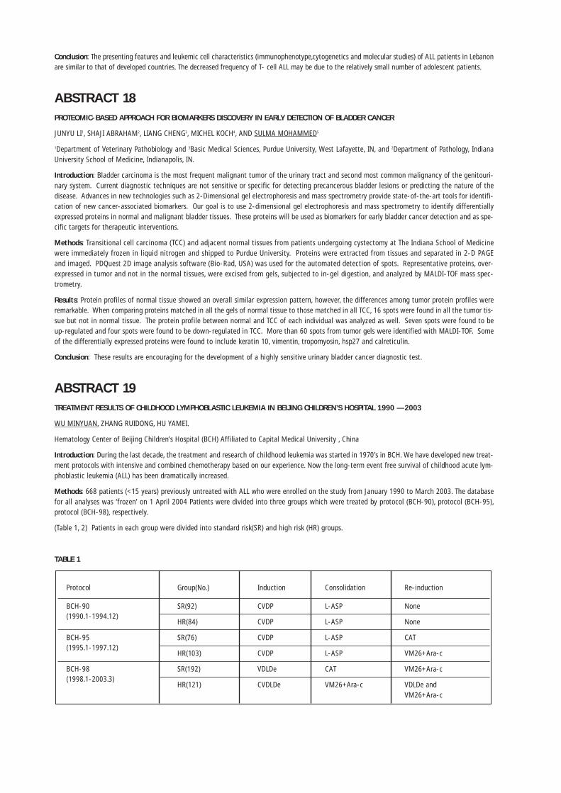
Conclusion: The presenting features and leukemic cell characteristics (immunophenotype,cytogenetics and molecular studies) of ALL patients in Lebanonare similar to that of developed countries. The decreased frequency of T- cell ALL may be due to the relatively small number of adolescent patients.
ABSTRACT 18PROTEOMIC-BASED APPROACH FOR BIOMARKERS DISCOVERY IN EARLY DETECTION OF BLADDER CANCER
JUNYU LI1, SHAJI ABRAHAM2, LIANG CHENG3, MICHEL KOCH4, AND SULMA MOHAMMED5
1Department of Veterinary Pathobiology and 3Basic Medical Sciences, Purdue University, West Lafayette, IN, and 2Department of Pathology, IndianaUniversity School of Medicine, Indianapolis, IN.
Introduction: Bladder carcinoma is the most frequent malignant tumor of the urinary tract and second most common malignancy of the genitouri-nary system. Current diagnostic techniques are not sensitive or specific for detecting precancerous bladder lesions or predicting the nature of thedisease. Advances in new technologies such as 2-Dimensional gel electrophoresis and mass spectrometry provide state-of-the-art tools for identifi-cation of new cancer-associated biomarkers. Our goal is to use 2-dimensional gel electrophoresis and mass spectrometry to identify differentiallyexpressed proteins in normal and malignant bladder tissues. These proteins will be used as biomarkers for early bladder cancer detection and as spe-cific targets for therapeutic interventions.
Methods: Transitional cell carcinoma (TCC) and adjacent normal tissues from patients undergoing cystectomy at The Indiana School of Medicinewere immediately frozen in liquid nitrogen and shipped to Purdue University. Proteins were extracted from tissues and separated in 2-D PAGEand imaged. PDQuest 2D image analysis software (Bio-Rad, USA) was used for the automated detection of spots. Representative proteins, over-expressed in tumor and not in the normal tissues, were excised from gels, subjected to in-gel digestion, and analyzed by MALDI-TOF mass spec-trometry.
Results: Protein profiles of normal tissue showed an overall similar expression pattern, however, the differences among tumor protein profiles wereremarkable. When comparing proteins matched in all the gels of normal tissue to those matched in all TCC, 16 spots were found in all the tumor tis-sue but not in normal tissue. The protein profile between normal and TCC of each individual was analyzed as well. Seven spots were found to beup-regulated and four spots were found to be down-regulated in TCC. More than 60 spots from tumor gels were identified with MALDI-TOF. Someof the differentially expressed proteins were found to include keratin 10, vimentin, tropomyosin, hsp27 and calreticulin.
Conclusion: These results are encouraging for the development of a highly sensitive urinary bladder cancer diagnostic test.
ABSTRACT 19TREATMENT RESULTS OF CHILDHOOD LYMPHOBLASTIC LEUKEMIA IN BEIJING CHILDREN’S HOSPITAL 1990 — 2003
WU MINYUAN, ZHANG RUIDONG, HU YAMEI.
Hematology Center of Beijing Children’s Hospital (BCH) Affiliated to Capital Medical University , China
Introduction: During the last decade, the treatment and research of childhood leukemia was started in 1970’s in BCH. We have developed new treat-ment protocols with intensive and combined chemotherapy based on our experience. Now the long-term event free survival of childhood acute lym-phoblastic leukemia (ALL) has been dramatically increased.
Methods: 668 patients (<15 years) previously untreated with ALL who were enrolled on the study from January 1990 to March 2003. The databasefor all analyses was ‘frozen’ on 1 April 2004 Patients were divided into three groups which were treated by protocol (BCH-90), protocol (BCH-95),protocol (BCH-98), respectively.
(Table 1, 2) Patients in each group were divided into standard risk(SR) and high risk (HR) groups.
TABLE 1
Protocol Group(No.) Induction Consolidation Re-induction
BCH-90 SR(92) CVDP L-ASP None(1990.1-1994.12)
HR(84) CVDP L-ASP None
BCH-95 SR(76) CVDP L-ASP CAT(1995.1-1997.12)
HR(103) CVDP L-ASP VM26+Ara-c
BCH-98 SR(192) VDLDe CAT VM26+Ara-c(1998.1-2003.3)
HR(121) CVDLDe VM26+Ara-c VDLDe andVM26+Ara-c
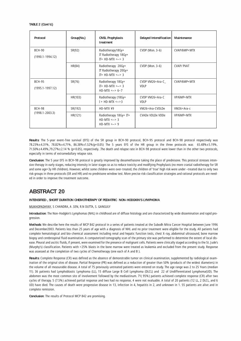
TABLE 2 (Cont’d.)
Results: The 5-year event-free survival (EFS) of the SR group in BCH-90 protocol, BCH-95 protocol and BCH-98 protocol respectively was78.23%±4.31%, 78.82%±4.71%, 86.38%±1.52%(p<0.05) The 5 years EFS of the HR group in the three protocols was 65.48%±5.19%,71.50%±4.49%,79.27%±2.14 % (p<0.05), respectively. .The death and relapse rate in BCH-98 protocol were lower than in the other two protocols,especially in terms of extramedullary relapse rate.
Conclusion: The 5 year EFS in BCH-98 protocol is greatly improved by dexamethasone taking the place of prednisone. This protocol stresses inten-sive therapy in early stages, reducing intensity in later stages so as to reduce toxicity and modifying Prophylaxis (no more cranial radiotherapy for SRand some age<3y HR children). However, whilst some children were over-treated, the children of ‘true’ high risk were under –treated due to only tworisk groups in three protocols (SR and HR) and no prednisone window test. More precise risk classification strategies and rational protocols are need-ed in order to improve the treatment outcome.
ABSTRACT 20INTENSIFIED , SHORT DURATION CHEMOTHERAPY OF PEDIATRIC NON-HODGKIN’S LYMPHOMA
MUKHOPADHYAY, S CHANDRA, A .SEN, R.N DUTTA, S. GANGULY
Introduction: The Non-Hodgkin’s Lymphomas (NHL) in childhood are of diffuse histology and are characterized by wide dissemination and rapid pro-gression.
Methods: We describe here the results of MCP 842 protocol in a series of patients treated at the Subodh Mitra Cancer Hospital between June 1996and December2003. Patients less than 25 years of age with a diagnosis of NHL and no prior treatment were eligible for the study. All patients hadcomplete hematological and bio-chemical assessment including renal and hepatic function tests, chest X-ray, abdominal ultrasound, bone marrowbiopsy and cerebrospinal fluid examination. A computerized tomography scan of the primary site was performed to determine the extent of local dis-ease. Pleural and ascitic fluids, if present, were examined for the presence of malignant cells. Patients were clinically staged according to the St. Jude’s(Murphy’s) classification. Patients with >25% blasts in the bone marrow were treated as leukemia and excluded from the present study. Responsewas assessed at the completion of two cycles of Chemotherapy (one each of A and B ).
Results: Complete Response (CR) was defined as the absence of demonstrable tumor on clinical examination, supplemented by radiological exam-ination of the original sites of disease. Partial Response (PR) was defined as a reduction of greater than 50% (products of the widest diameters) inthe volume of all measurable disease. A total of 75 previously untreated patients were entered on study. The age range was 2 to 25 Years (median11). 36 patients had Lymphoblastic Lymphoma (LL), 15 diffuse Large B Cell Lymphoma (DLCL) and 22 of Undifferentiated Lymphoma(UD). Theabdomen was the most common site of involvement followed by the mediastinum. 71( 95%) patients achieved complete response (CR) after twocycles of therapy. 5 (7.5%) achieved partial response and two had no response, 4 were not evaluable. A total of 20 patients (12 LL, 2 DLCL, and 6UD) have died. The causes of death were progressive disease in 13, infection in 4, hepatitis in 2, and unknown in 1. 55 patients are alive and incomplete remission.
Conclusion: The results of Protocol MCP 842 are promising.
Protocol Group(No.) CNSL Prophylaxis Delayed Intensification Maintenancetreatment
BCH-90 SR(92) Radiotherapy18Gy+ CVDP (Mon. 3-6) CVAP/6MP+MTXIT Radiotherapy 18Gy+
(1990.1-1994.12)IT+ HD-MTX <–-> 3
HR(84) Radiotherapy 20Gy+ CVDP (Mon. 3-6) CVAP/ PVATIT Radiotherapy 20Gy+ IT+ HD-MTX <–-> 3
BCH-95 SR(76) Radiotherapy 18Gy+ CVDP VM26+Ara-C_ CVAP/6MP+MTXIT+ HD-MTX <–-> 3 VDLP
(1995.1-1997.12)HD-MTX <–-> 6~7
HR(103) Radiotherapy (18Gy+ CVDP VM26+Ara-C VP/6MP+MTXI + HD-MTX <–->3 VDLP
BCH-98 SR(192) HD-MTX ¥9 VM26+Ara-CVDLDe VM26+Ara-c(1998.1-2003.3)
HR(121) Radiotherapy 18Gy+ IT+ CVADe VDLDe VDDe VP/6MP+MTXHD-MTX <–-> 3 HD-MTX <–-> 9
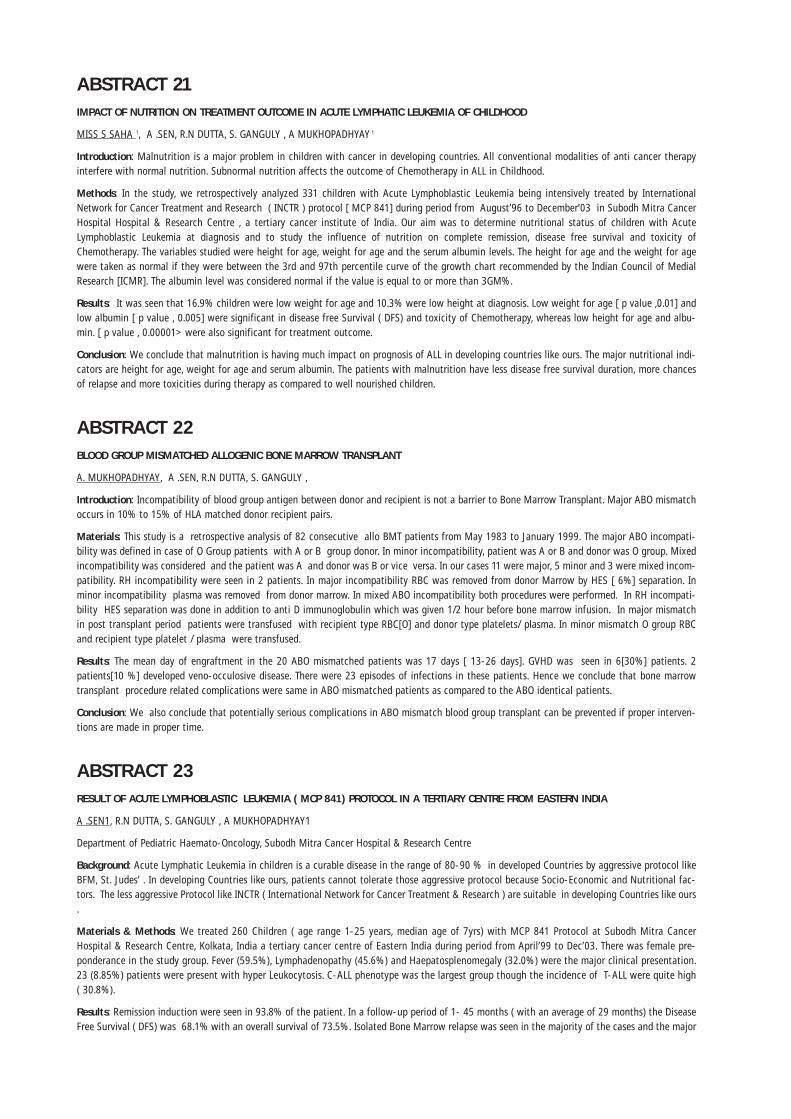
ABSTRACT 21IMPACT OF NUTRITION ON TREATMENT OUTCOME IN ACUTE LYMPHATIC LEUKEMIA OF CHILDHOOD
MISS S SAHA 1, A .SEN, R.N DUTTA, S. GANGULY , A MUKHOPADHYAY 1
Introduction: Malnutrition is a major problem in children with cancer in developing countries. All conventional modalities of anti cancer therapyinterfere with normal nutrition. Subnormal nutrition affects the outcome of Chemotherapy in ALL in Childhood.
Methods: In the study, we retrospectively analyzed 331 children with Acute Lymphoblastic Leukemia being intensively treated by InternationalNetwork for Cancer Treatment and Research ( INCTR ) protocol [ MCP 841] during period from August’96 to December’03 in Subodh Mitra CancerHospital Hospital & Research Centre , a tertiary cancer institute of India. Our aim was to determine nutritional status of children with AcuteLymphoblastic Leukemia at diagnosis and to study the influence of nutrition on complete remission, disease free survival and toxicity ofChemotherapy. The variables studied were height for age, weight for age and the serum albumin levels. The height for age and the weight for agewere taken as normal if they were between the 3rd and 97th percentile curve of the growth chart recommended by the Indian Council of MedialResearch [ICMR]. The albumin level was considered normal if the value is equal to or more than 3GM%.
Results: It was seen that 16.9% children were low weight for age and 10.3% were low height at diagnosis. Low weight for age [ p value ,0.01] andlow albumin [ p value , 0.005] were significant in disease free Survival ( DFS) and toxicity of Chemotherapy, whereas low height for age and albu-min. [ p value , 0.00001> were also significant for treatment outcome.
Conclusion: We conclude that malnutrition is having much impact on prognosis of ALL in developing countries like ours. The major nutritional indi-cators are height for age, weight for age and serum albumin. The patients with malnutrition have less disease free survival duration, more chancesof relapse and more toxicities during therapy as compared to well nourished children.
ABSTRACT 22BLOOD GROUP MISMATCHED ALLOGENIC BONE MARROW TRANSPLANT
A. MUKHOPADHYAY, A .SEN, R.N DUTTA, S. GANGULY ,
Introduction: Incompatibility of blood group antigen between donor and recipient is not a barrier to Bone Marrow Transplant. Major ABO mismatchoccurs in 10% to 15% of HLA matched donor recipient pairs.
Materials: This study is a retrospective analysis of 82 consecutive allo BMT patients from May 1983 to January 1999. The major ABO incompati-bility was defined in case of O Group patients with A or B group donor. In minor incompatibility, patient was A or B and donor was O group. Mixedincompatibility was considered and the patient was A and donor was B or vice versa. In our cases 11 were major, 5 minor and 3 were mixed incom-patibility. RH incompatibility were seen in 2 patients. In major incompatibility RBC was removed from donor Marrow by HES [ 6%] separation. Inminor incompatibility plasma was removed from donor marrow. In mixed ABO incompatibility both procedures were performed. In RH incompati-bility HES separation was done in addition to anti D immunoglobulin which was given 1/2 hour before bone marrow infusion. In major mismatchin post transplant period patients were transfused with recipient type RBC[O] and donor type platelets/ plasma. In minor mismatch O group RBCand recipient type platelet / plasma were transfused.
Results: The mean day of engraftment in the 20 ABO mismatched patients was 17 days [ 13-26 days]. GVHD was seen in 6[30%] patients. 2patients[10 %] developed veno-occulosive disease. There were 23 episodes of infections in these patients. Hence we conclude that bone marrowtransplant procedure related complications were same in ABO mismatched patients as compared to the ABO identical patients.
Conclusion: We also conclude that potentially serious complications in ABO mismatch blood group transplant can be prevented if proper interven-tions are made in proper time.
ABSTRACT 23RESULT OF ACUTE LYMPHOBLASTIC LEUKEMIA ( MCP 841) PROTOCOL IN A TERTIARY CENTRE FROM EASTERN INDIA
A .SEN1, R.N DUTTA, S. GANGULY , A MUKHOPADHYAY1
Department of Pediatric Haemato-Oncology, Subodh Mitra Cancer Hospital & Research Centre
Background: Acute Lymphatic Leukemia in children is a curable disease in the range of 80-90 % in developed Countries by aggressive protocol likeBFM, St. Judes’ . In developing Countries like ours, patients cannot tolerate those aggressive protocol because Socio-Economic and Nutritional fac-tors. The less aggressive Protocol like INCTR ( International Network for Cancer Treatment & Research ) are suitable in developing Countries like ours.
Materials & Methods: We treated 260 Children ( age range 1-25 years, median age of 7yrs) with MCP 841 Protocol at Subodh Mitra CancerHospital & Research Centre, Kolkata, India a tertiary cancer centre of Eastern India during period from April’99 to Dec’03. There was female pre-ponderance in the study group. Fever (59.5%), Lymphadenopathy (45.6%) and Haepatosplenomegaly (32.0%) were the major clinical presentation.23 (8.85%) patients were present with hyper Leukocytosis. C-ALL phenotype was the largest group though the incidence of T-ALL were quite high( 30.8%).
Results: Remission induction were seen in 93.8% of the patient. In a follow-up period of 1- 45 months ( with an average of 29 months) the DiseaseFree Survival ( DFS) was 68.1% with an overall survival of 73.5%. Isolated Bone Marrow relapse was seen in the majority of the cases and the major
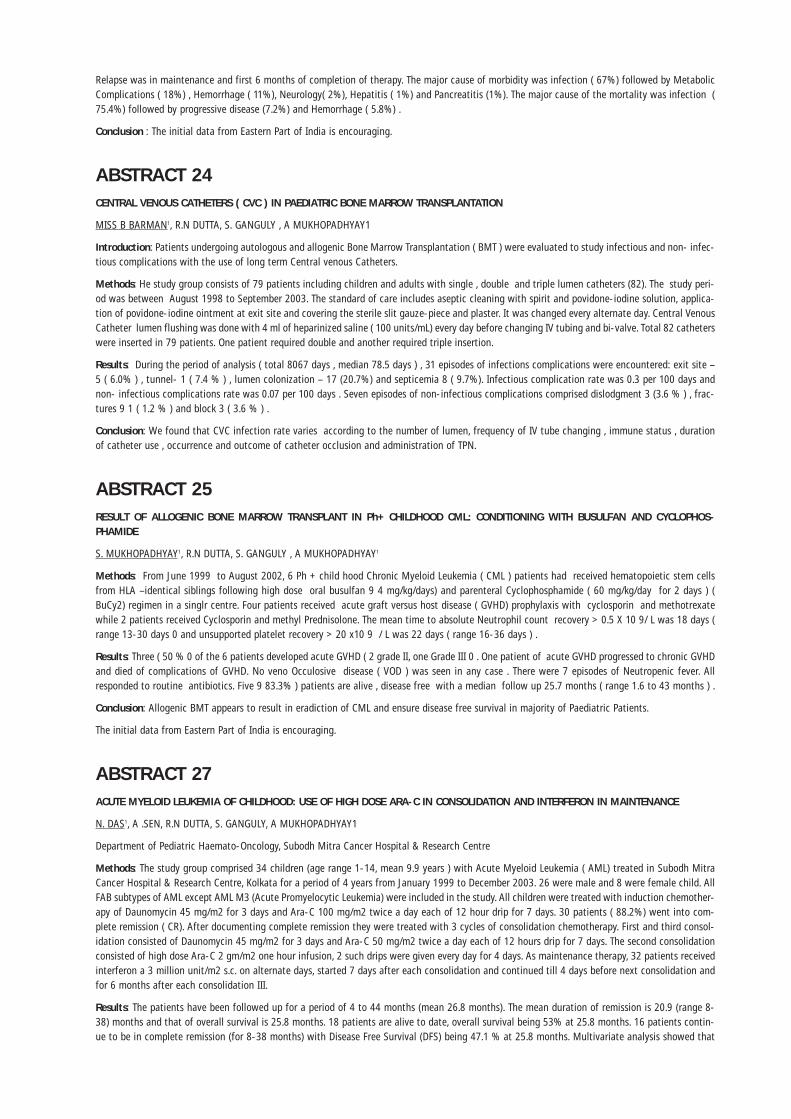
Relapse was in maintenance and first 6 months of completion of therapy. The major cause of morbidity was infection ( 67%) followed by MetabolicComplications ( 18%) , Hemorrhage ( 11%), Neurology( 2%), Hepatitis ( 1%) and Pancreatitis (1%). The major cause of the mortality was infection (75.4%) followed by progressive disease (7.2%) and Hemorrhage ( 5.8%) .
Conclusion : The initial data from Eastern Part of India is encouraging.
ABSTRACT 24CENTRAL VENOUS CATHETERS ( CVC ) IN PAEDIATRIC BONE MARROW TRANSPLANTATION
MISS B BARMAN1, R.N DUTTA, S. GANGULY , A MUKHOPADHYAY1
Introduction: Patients undergoing autologous and allogenic Bone Marrow Transplantation ( BMT ) were evaluated to study infectious and non- infec-tious complications with the use of long term Central venous Catheters.
Methods: He study group consists of 79 patients including children and adults with single , double and triple lumen catheters (82). The study peri-od was between August 1998 to September 2003. The standard of care includes aseptic cleaning with spirit and povidone-iodine solution, applica-tion of povidone-iodine ointment at exit site and covering the sterile slit gauze-piece and plaster. It was changed every alternate day. Central VenousCatheter lumen flushing was done with 4 ml of heparinized saline ( 100 units/mL) every day before changing IV tubing and bi-valve. Total 82 catheterswere inserted in 79 patients. One patient required double and another required triple insertion.
Results: During the period of analysis ( total 8067 days , median 78.5 days ) , 31 episodes of infections complications were encountered: exit site –5 ( 6.0% ) , tunnel- 1 ( 7.4 % ) , lumen colonization – 17 (20.7%) and septicemia 8 ( 9.7%). Infectious complication rate was 0.3 per 100 days andnon- infectious complications rate was 0.07 per 100 days . Seven episodes of non-infectious complications comprised dislodgment 3 (3.6 % ) , frac-tures 9 1 ( 1.2 % ) and block 3 ( 3.6 % ) .
Conclusion: We found that CVC infection rate varies according to the number of lumen, frequency of IV tube changing , immune status , durationof catheter use , occurrence and outcome of catheter occlusion and administration of TPN.
ABSTRACT 25RESULT OF ALLOGENIC BONE MARROW TRANSPLANT IN Ph+ CHILDHOOD CML: CONDITIONING WITH BUSULFAN AND CYCLOPHOS-PHAMIDE
S. MUKHOPADHYAY1, R.N DUTTA, S. GANGULY , A MUKHOPADHYAY1
Methods: From June 1999 to August 2002, 6 Ph + child hood Chronic Myeloid Leukemia ( CML ) patients had received hematopoietic stem cellsfrom HLA –identical siblings following high dose oral busulfan 9 4 mg/kg/days) and parenteral Cyclophosphamide ( 60 mg/kg/day for 2 days ) (BuCy2) regimen in a singlr centre. Four patients received acute graft versus host disease ( GVHD) prophylaxis with cyclosporin and methotrexatewhile 2 patients received Cyclosporin and methyl Prednisolone. The mean time to absolute Neutrophil count recovery > 0.5 X 10 9/ L was 18 days (range 13-30 days 0 and unsupported platelet recovery > 20 x10 9 / L was 22 days ( range 16-36 days ) .
Results: Three ( 50 % 0 of the 6 patients developed acute GVHD ( 2 grade II, one Grade III 0 . One patient of acute GVHD progressed to chronic GVHDand died of complications of GVHD. No veno Occulosive disease ( VOD ) was seen in any case . There were 7 episodes of Neutropenic fever. Allresponded to routine antibiotics. Five 9 83.3% ) patients are alive , disease free with a median follow up 25.7 months ( range 1.6 to 43 months ) .
Conclusion: Allogenic BMT appears to result in eradiction of CML and ensure disease free survival in majority of Paediatric Patients.
The initial data from Eastern Part of India is encouraging.
ABSTRACT 27ACUTE MYELOID LEUKEMIA OF CHILDHOOD: USE OF HIGH DOSE ARA-C IN CONSOLIDATION AND INTERFERON IN MAINTENANCE
N. DAS1, A .SEN, R.N DUTTA, S. GANGULY, A MUKHOPADHYAY1
Department of Pediatric Haemato-Oncology, Subodh Mitra Cancer Hospital & Research Centre
Methods: The study group comprised 34 children (age range 1-14, mean 9.9 years ) with Acute Myeloid Leukemia ( AML) treated in Subodh MitraCancer Hospital & Research Centre, Kolkata for a period of 4 years from January 1999 to December 2003. 26 were male and 8 were female child. AllFAB subtypes of AML except AML M3 (Acute Promyelocytic Leukemia) were included in the study. All children were treated with induction chemother-apy of Daunomycin 45 mg/m2 for 3 days and Ara-C 100 mg/m2 twice a day each of 12 hour drip for 7 days. 30 patients ( 88.2%) went into com-plete remission ( CR). After documenting complete remission they were treated with 3 cycles of consolidation chemotherapy. First and third consol-idation consisted of Daunomycin 45 mg/m2 for 3 days and Ara-C 50 mg/m2 twice a day each of 12 hours drip for 7 days. The second consolidationconsisted of high dose Ara-C 2 gm/m2 one hour infusion, 2 such drips were given every day for 4 days. As maintenance therapy, 32 patients receivedinterferon a 3 million unit/m2 s.c. on alternate days, started 7 days after each consolidation and continued till 4 days before next consolidation andfor 6 months after each consolidation III.
Results: The patients have been followed up for a period of 4 to 44 months (mean 26.8 months). The mean duration of remission is 20.9 (range 8-38) months and that of overall survival is 25.8 months. 18 patients are alive to date, overall survival being 53% at 25.8 months. 16 patients contin-ue to be in complete remission (for 8-38 months) with Disease Free Survival (DFS) being 47.1 % at 25.8 months. Multivariate analysis showed that
age > 5 yrs, male sex, absence of infection, absent Hepatospleenomegally and Lymphadenopathy, Hb> 10 gm%, TLC < 50.000, Platelet Count > 50.000,High Myeloperoxide (MPG) activity, Serum Albumin > 3 gm/dl and normal LDH level had a favorable influence on achievement of CR ( p value < 0.05). Absent Lymphadenopathy, Hb> 10 gm%, TLC < 50,000, Platelet Count > 50.000, Serum Albumin > 3 gm/dl and normal LDH had a favorable prog-nostic value for DFS (p value < 0.05).
Conclusion: The present data indicates the need for newer approach to improve long term survival in childhood AML.
ABSTRACT 28PERIPHERALLY INSERTED CENTRAL CATHETER (PICC) IN PEDIATRIC ACUTE MYELOID LEUKEMIA. A SINGLE TERTIARY CANCER CENTRE EXPERI-ENCE FROM EASTERN INDIA
MISS MOUMITA SARKAR1, R N DUTTA, S GANGULY, A MUKHOPADHYAY1
Department of Pediatric Haemato-Oncology, Subodh Mitra Cancer Hospital & Research Centre
Introduction: Peripherally inserted central catheter (PICC) is a recently developed procedure and plays a major role in the successful administrationof chemotherapeutic agents and supportive treatment in pediatric patients with Acute Myeloid Leukemia, other hematological malignancies as wellas solid tumors. These catheters are easily inserted by experienced Nurses. The PICC lines are usually placed in a superficial vein of the upper extrem-ity. The indications for peripherally inserted central catheterization are lack of peripheral venous access in small children, Infusion of hyperosmolarsolutions, Long term I.V. therapy in the home, hospital or clinics setting. The advantages of PICC are that they have a lower overall infection rate andare cost effective as compared with other central venous catheters. The other advantages are potential reduction of catheter sepsis, decrease in painand discomfort associated with frequent venipuncture, preservation of the peripheral vascular system of the upper extremities for time efficient,appropriate for future use at home .
Methods: We prospectively analyzed a total no of 50 PICCs in 48 Acute Myeloid Leukemia Patients age group ( 1.5 -14 years ) , inserted in cathetercare clinic at Subodh Mitra Cancer Hospital & Research Centre, during the period of Jan.2001 to Dec.2003. Our aim was to insert cost effective PICCfor facilating simple insertion as well as maintenance and to access its post insertion complications. we evaluated these 50 PICCs with a total of4200 access days. The rate of catheter related sepsis was 0.0007/100 days, coagulase negative staphylococci being the most common organism. Therate of non-infectious complications was 0.004/100 catheter days. The most common reasons for PICC removal were blockage 3 (6.0%) and infec-tion 2 (4.0%). We studied and observed that the rate of complications, cost of PICC insertion, care and maintenance were comparatively low as com-pared to tunneled catheters.
Conclusion: We can conclude that PICC is cost effective alternative to centrally inserted long term tunneled Catheter. It is also less traumatic, easyto implant, well tolerated by the patient and ultimately, it improved the quality of life.
ABSTRACT 30PERIPHERAL STEM CELL TRANSPLANTATION IN ADVANCED STAGE NEUROBLASTOMA (PRELIMINARY REPORT)
EMEL ÜNAL1, GÜLSAN YAVUZ1, NURDAN TAÇYILDIZ1, HANDAN UGUR1, EROL AYYILDIZ3, AYDAN IKINCIOGULLARI2, MUTLU ARAT3, SÜKRÜ CIN1
1Ankara University Medical School, Department of Pediatrics Division of Oncology, Ankara, Turkey 2 Division of Allergy and Immunology, 3Ibn-i SinaHospital Department of Haematology
Background: Neuroblastoma which is the most common extracranial solid tumor in children comprises 8 – 10 % of all childhood cancers and is themost common cancer diagnosed during infancy. The prevalence is approximately 1 case per 7.000 live births. Treatment of neuroblastoma still rep-resents one of the major challenges in pediatric oncology. Historically high risk retinablastoma patients have long-term survival probabilities of lessthan 15%. Therefore the treatment approach includes intensive induction chemotherapy, surgical excision of the primary tumor / local irradiation, ifnecessary followed by myeloablative consolidation therapy with stem cell rescue and targeted therapy for minimal residual disease.
Material and Method: In pediatric bone marrow transplant unit 3 advanced stage neuroblastoma cases underwent peripheral blood stem cell trans-plantation between January 2003 – March 2004. There were 3 girls whose ages varied between 3,5 – 5 years.
Results: According to INSS, they had stage IV disease with adrenal tumor in all cases, bone marrow infiltration, cortical bone involvement and addi-tionally, orbital metastasis in 1 case. Two cases were diagnosed based on the presence of tumor cells in the bone marrow accompanied by increasedurinary catecholamine metabolites. While the case with an orbital mass underwent orbitotomy with histopathology showing undifferentiated neu-roblastoma with poor stroma. Serum markers of Neuron Spesific Enolase (NSE) varied between 54-500 ng/ml, ferritin levels 186-556 ng/ml, LDH1708-5528 IU/l, urinary VMA levels varied between 17-114mg/dl. Myc-N amplifications could not be shown according to some technical problems.The patients were treated according to the Turkish Pediatric Oncology Group Neuroblastoma Protocol. They received chemotherapy regimens name-ly A3 (Vincristine 1.5mg/m2 (1. and 5.d.), Ifosfamide 1.8g/m2 (1-5d) + mesna, DTIC 250 mg/m2 (1-5d), Adriamycine 20 mg/m2 (1-3 d) alternatingwith A5 (Cisplatin) 30 mg/m2 (1-5 d), Cyclophosphamide 300mg/m2 (1-5 d), VP-16 150 mg/m2, (4. and 5. d). After completing 2 blocks (2 A3, 2 A5)of chemotherapy, the response rates were assessed which yielded 2 CR, 1 VGPR. Surgical excision was performed for adrenal medulla tumors. Noneof the patients received local irradiation. Priming therapy with 2.2g/m2 g/kg-day which was used formcyclophosphamide and GCSF 10 stem cell mobi-lization was followed by peripheral blood stem cell collection on d7-12 of the therapy. The mononuclear cell (MNC) yields were 12.08x10⁸/kg,13.3x108/kg, 9.2x10⁸/kg respectively and, CD34+ cells yield were 4.8x10⁶/kg, 5.8x10⁶/kg and 3.73x10⁶/kg, respectively. The conditioning regimenstarted 4-6 weeks after the priming which consisted of Carboplatin [300mg/m2/day:d-7,-6,-5,-4. (total 1200 mg/m2) 4h IV inf].; Etoposide[200mg/m2/day: d-7,-6,-5,-4. (total 800 mg/m2) 4h IV inf.]; Melphelan [50mg/m2/day: d-7,-6,-5,-4. (total 200 mg/m2) 30min IV inf.]; Days -3, -2and -1.: Resting; Day 0: Autologous peripheral blood stem cell infusion; After 4 hours G-CSF 10 mg/kg/day was commenced and was continued till
ANC>1000/mm3 was achieved. The myeloid engraftment, erythroid engraftment and thrombocyte engraftment occurred on days 13, 18 and 23(mean), respectively. Posttransplant +90 day, 13-cisretionic acid was commenced 160 mg/m2/day two weeks / 6 months for minimal residual dis-ease. The patients’ follow-up was 66-455 days with CR with no toxicity. The development of risk adapted and more effective therapy strategies mayimprove the long-term treatment results of these high risk patients.
ABSTRACT 31EVIDENCE OF CYTOTOXIC EFFECT OF SAFFRON ON THE GROWTH OF DIFFERENT HUMAN MALIGNANT CELLS
ABDULLAEV F.I,
Laboratory of Experimental Oncology, National Institute of Pediatrics , Mexico City, Mexico
Introduction: Cancer is a very important national health problem in Mexico, while a significant increase in the rate of total and childhood mortali-ty has been recorded in the last decades. Saffron (Crocus sativus L.,) has been widely used in traditional medicine since ancient times as a drugagainst different human diseases. In view of the renewed interest in cancer chemopreventive plant agents, it is important to study the cytotoxic effectof saffron on normal and malignant human cells.
Methods: 9 malignant and 4 normal human cell lines and ethanolic extract of saffron were used in our in vitro experiments. The cytotoxic effect ofsaffron extract on cell viability and on the growth of malignant and normal human cells were evaluated using the XTT assay, colony formation assayand a method for incorporation of radioactive precursors of DNA, RNA and protein synthesis. The percentage of growth inhibition was calculatedand IC50 values (concentration of drug to achieve 50% growth inhibition) were obtained graphically from the survival curves' distribution. Data wereanalysed using Statistical Analysis System software (SAS Institute Inc., Cary, NC 27511, USA Release 6.02). Values 0.05.<were considered significantwhen p
Results: We observed that, while saffron extracts produced no changes in cell viability of normal human lung fibroblasts, a dose-dependent inhibito-ry effect was observed on malignant cells. We found that MCF-7 cells were the most sensitive ones, followed by SKNH and HeLa cells IC50 (mg/ml)were: for MCF-7 –0.78; SKNM – 1,66, HeLa – 1,92 and for normal cells –19,99). Saffron extract inhibited colony formation of all tested malignanthuman cells (HeLa, A-204, HepG2, SW480, A549, WI-38VA) and had no effect on normal human cells (CCL210, CCD-18Lu and WI38). Saffron had adose-dependent inhibitory effect on DNA and RNA synthesis in malignant cells and no detectable effect in normal cells. In contrast to nucleic acidsynthesis, protein synthesis was not inhibited by saffron in malignant as well as in normal cells.
Conclusion: Our results reveal that the saffron extract possessed cytotoxic effects on viability and colony formation of different human malignantcells in vitro , while no marked effect was exerted on normal human cells. Several hypotheses for the antitumor actions of saffron have been pro-posed: inhibition of intracellular DNA and RNA synthesis; inhibition of free radical chain reaction; stability to irradiation; increase of intracellular SH-compounds; inhibition of genotoxicity; induction of apoptosis; inhibition of cell proliferation; inhibition of different cellular enzyme activities but theexact molecular mechanism of action of these natural agents is not clear at present.
ABSTRACT 33PRELIMINARY EVALUATION OF INCTR 1-01 PROTOCOL FOR TREATMENT OF PEDIATRIC NON-METASTATIC OSTEOSARCOMAS IN HO CHIMINH CITY, VIETNAM
HUONG C. TRAN, KHAI D. TRUONG, HUNG C. NGUYEN
HCMC CHILDREN HOSPITAL & CANCER CENTER, VN
Background: Osteosarcoma - the 9th commonest childhood cancer - is the most common primary bone tumor in HochiMinh City, VN ( CR:1.7; ASR:1.0 ).
The traditional approach has consisted of radical amputation followed by chemotherapy. CT regimens are controversial and mostly unachievable dueto the financial problems of child's family.
Treatment outcome was often not known. Limb salvage procedures are actually not feasible.
Chemo-surgery combination with INCTR the 1-01 protocol has been put in place since 2002 in the treatment of pediatric non-metastaticOsteosarcomas at the HCMC Children Hospital & Cancer Center.
Objectives: to apply the INCTR 1-01 protocol for treatment of non-metastatic Osteosarcomas in HoChiMinh City, VN and to assess the primaryresponse, the EFS, the toxicities and tolerabilities of the protocol.
Materials and Methods: Patients with newly diagnosed, non-metastatic Osteosarcoma were enrolled in 2002-2003 and based on the clinical fea-tures, medical imaging studies and histopathology of the bone tumor (open biopsy).- Prospective, open, non-randomized study.- Radical amputation is the current operation combined with an alternating chemotherapy regimen: Cisplatin, Doxorubicin and Ifosfamide ( INCTR1-01 protocol)Cisplatin 60mg/m2 + Doxorubicin 35mg/m2 IV D1-D2 on weeks 0, 6, 14, 22Ifosfamide 2000mg/m2 IV D1-D5 (with Mesna uroprotection) on weeks 3, 9, 19, 25- Two arms of therapeutic possibilities:
• Surgery + Adjuvant Chemotherapy ( 4 double cycles)• Induction Chemo + Surgery + Adjuvant Chemotherapy ( 4 double cycles)
Results: 17 cases of non metastasic Osteosarcomas were observed. The boys/girls ratio was 1.43:1. The median age was 13-15 years. 82.4% (15/17cases) presented in the around-the-knee sites. - Two therapeutic arms were recognized:
* 10 cases with primary amputation followed by adjuvant chemotherapy.* 7 cases with pre-op. and post-operative chemotherapy + amputation on week 12.
- 14/17 (82.4%) cases were high grade Osteosarcomas. 70% (12/17) had the central osteoblastic histology. - 82.4% had a tumor size ≥ 20 cm in diameter.- 5/7 cases had evidence of response ≥ 80% after induction chemotherapy.- Toxicities: Neutropenia G 3/4 (21%); Anemia G3/4 (12%); Thrombocytopenia G3/4 (7%) / the 3-4th cycles.
Non hematologic: Nausea/ vomiting ( 80%); Alopecia: almost universal; Mucositis (40%)Liver and kidney function: mild, acceptable.
- 5/17 cases are still being followed. The others were lost to follow-up.
Conclusions: The alternating regimen Ifos/ Dox-Cis for treatment of non-metastatic Osteosarcoma is easy to handle, less toxic, safe and well-toler-ated in child patients.The clinical survey of HCMC Children Hospital and Cancer Center is still ongoing and the DFS, OS survival will be on furthersurveillance
ABSTRACT 34CONCOMITANT RADIO-CHEMOTHERAPY IN LOCALLY ADVANCED CERVICAL CANCER: EXPERIENCE FROM A TERTIARY CARE CENTRE ININDIA
DINESH SINGH, ARUN GOEL, MALAY NANDY, KANIKA GUPTA, KAMLA SHARMA. DHARAMSHILA
Cancer Hospital & Research Centre, Vasundhara Enclave, Delhi 110096, India
Introduction: Cancer of uterine cervix is the commonest cancer of women in developing countries. Majority of patients present with locally advanceddisease. Such patients are not candidates for any radical surgery. Radical radiotherapy consisting of external beam radiotherapy and intracavitarybrachytherapy has been the mainstay of treatment for decades. Five randomized trials in the last decade showed that radiotherapy with concomi-tant chemotherapy (especially cisplatin based) leads to a long term survival benefit. With the results of these trials, it was recommended that con-comitant RT-CT (radio-chemotherapy) become the standard of care for locally advanced cervical cancer. Concerns have however been expressed aboutthe tolerability of such therapy especially in developing countries such as India where the majority of patients with cervical cancer come from lowsocioeconomic backgrounds and have poor nutrition and poor hygiene. At Dharamshila Cancer Hospital & Research Centre, we have been using RT-CT for more than five years and wanted to share our experience.
Methods: A retrospective analysis of data for patients with stage IIb, IIIa and IIIb squamous cell cervical cancer treated with RT-CT was carried out.Patients treated over a five year period from October 1998 to October 2003 were included in the study. During this period, 109 patients were treat-ed with RT-CT. Out of this, 28 were stage IIb, 15 were stage IIIa and 66 were stage IIIb. External radiotherapy was administered with a Telecobalt orLinear Accelerator to a dose of 5040 cGy (180cGy per fraction) by four field box technique using CT simulation and computerized planning. Weeklycisplatinum was administered at a dose of 30 mg/m2 in day care. After external RT, three fractions of Intracavitary brachytherapy were administeredusing a Microselectron HDR unit at a dose of 7.5 Gy to point A per at weekly intervals.
Results: Treatment was discontinued by 5 patients. Overall response rate was 93% (complete response 89%, partial response 4%). Grade 3 and 4toxicities (diarrhoea, vomiting, neutropenia, etc.) were observed in 36% of patients (39/109). With a median follow up of 30 months, the 5 yearactuarial disease free survival is 74.3% and actuarial 5 year overall survival is 81.6%. Late toxicities (grade III and IV) were observed in 11% ofpatients.
Conclusion: The experience at a tertiary cancer centre in India shows that concomitant radiotherapy and chemotherapy is feasible, well tolerated andeffective treatment for locally advanced cervical cancer even in developing countries. It gives complete response and survival rates that are superiorto the conventional approach of radiotherapy alone
ABSTRACT 35SEVERE ADVERSE EVENTS WITH IMATINIB TREATMENT
AMARU RICARDO, MACHICADO DAVID, AMARU JOSÉ, LIMACHI MARTHA, CUEVAS HERIBERTO.
Centro de Oncohematología Paolo Belli, Hospital Arco Iris, La Paz, Bolivia.
Unidad de Biología Celular, Facultad de Medicina, Universidad mayor de San Andrés.
Introduction: Imatinib (Gleevec) is an inhibitor of specific protein tyrosine kinases and the most extensively studied BCR-ABL tyrosine kinase ofchronic myelogenous leukemia (CML). Imatinib impairs BCR-ABL–mediated transfer of phosphate to its substrates and has had extraordinary activi-ty against CML.
Methods: Twenty four patients with CML were enrolled in this study: 20 in chronic phase, 2 in accelerated phase and 2 in blast crisis. The patientsreceived 400 mg/day in the chronic phase and 600 mg/day in accelerated phase and blast crisis.
We describe two severe adverse events: cerebral infarct and bilateral pleural effusion.
Results: A 56 years old patient in chronic phase of CML received imatinib for 2 months. Suddenly he developed right hemipalsy. CAT scan studies
revealed an infarct zone in the right medial cerebral artery region. Imatinib was discontinued and supportive measures were performed. Progressiveneurologic improvement was observed after 2 weeks and she has a 1/5 performance status after 6 months. No other adverse event was observedafter lowering the dose. A 40 years old in accelerated phase of CML presented to the ER with acute respiratory insufficiency after 12 months of ima-tinib treatment. CXR revealed massive bilateral pleural effusions. Clinical response was satisfactory with emergency thoracocentesis and imatinib dis-continuation. Imatinib was reinitiated after 2 weeks but pleural effusions recurred.
Conclusion: It is the first description of cerebral infarct as severe adverse events with imatinib treatment.
ABSTRACT 37INTESTINAL-TYPE ADENOCARCINOMA OF THE URINARY BLADDER ASSOCIATED WITH SCHISTOSOMIASIS: A POSSIBLE CARCINOGENICPATHWAY SIMILAR TO COLORECTAL CARCINOMA
EISSA S,**, NADJI M, **, NASSIRI M.
*Department of Pathology, NCI, Cario, Egypt,
**Department of Pathology University of Miami, Miami, Florida,
Introduction: Primary adenocarcinomas of the urinary bladder are relatively uncommon. We report 102 non-urachal examples of this tumor exhibit-ing an intestinal morphology, in a cohort of Egyptian patients with Schistosoma infestation.
Methods: Partial or total cystectomy specimens from 133 patients with proven primary non-urachal adenocarcinoma of the urinary bladder werereviewed. Based on the morphology, tumors were classified as intestinal (colonic), and non-intestinal (urothelial) types. Paraffin sections of eachtumor were stained by the avidin-biotin-peroxidase method using antibodies to cytokeratins 7 and 20 (CK-7, CK-20, Dako), as well as to recombi-nant wild type p53 protein (DO-7, Dako).
Results: Schistosoma eggs were identified in all specimens. A pure intestinal (colonic) morphology was seen in 101 tumors; one case was associatedwith foci of small cell carcinoma. Five tumors were predominantly composed of signet-ring cells and 12 demonstrated a "colloid" morphology withmucin lakes. When adjacent normal mucosa was present, 23% showed associated colonic metaplasia. Similarty, 24% of intestinal adenocarcinomashad areas, which resembled villous or tubular colonic adenomas. Cytokeratin 20 was expressed by 92 (90%) of intestinal, and 3 (10%) of urothelialadenocarcinomas. Conversely, CK-7 was expressed by 27 (87%) of urothelial and 17 (17%) of intestinal-type tumors. Nuclear accumulation of p53was observed in 84% and 48% of intestinal and urothelial adenocarcinomas, respectively.
Conclusion: Primary intestinal-type adenocarcinoma of the urinary bladder is morphologically and immunochemically identical to colonic adenocar-cinomas. Frequent association of this tumor with colonic metaplasia and colonic-type adenomas suggest possible carcinogenic pathways similar tothat observed in colorectal carcinomas.
ABSTRACT 38DENDRITIC CELL VACCINE FOR THE TREATMENT OF CARCINOMA OF CERVIX
RAJKUMAR T, PRIYA R, SELVALUXMI G.
Cancer Institute (WIA), Adyar, Chennai – 600020, India.
Introduction: Cancer of the cervix is the most common cancer among Indian women. The annual new cases has been projected to be 100,000/year.HPV plays an important role in the cervical tumourigenesis. In spite of adequate treatment nearly 30-50% of stage IIB and IIIB patients fail. It istherefore necessary to explore the possibility of additional methods of treatment to enhance the cure rates.
Methods: Peripheral blood monocytes were grown in the presence of IL4 and GM-CSF for 7 days, followed by maturation of the dendritic cells (DC)in the presence of TNF-a and ILb1 for an additional 2 days. The DC’s were primed with either Autologous tumour lysate or RNA or both to assess theefficacy of the method. FACS analysis was used to characterize the DC. MLR assay was used to assess the functional activity of the DC.
Results: Following the growth of the peripheral blood monocytes, the immature DC’s generated were found to be HLA DP,DQ,DR +++; CD86 low andCD14 negative. The mature DC’s after exposure to TNF-a and ILb1 were found to be HLA DP,DQ,DR +++; CD86 high and CD14 negative. The func-tional activity of the primed DC’s were found to be two fold greater than the unprimed DC.
Conclusion: The standardization of the protocol for the development of dendritic cells in vitro from peripheral blood monocytes has been complet-ed. A phase 1 study to evaluate the toxicity and efficacy in cervical cancer is under way.
ABSTRACT 39PREDICTIVE MARKERS IN SQUAMOUS CELL CARCINOMA OF CERVIX
RAJKUMAR T, GOPAL G, SELVALUXMI G, MAJHI U, RAJALEKSHMY KR.
Cancer Institute (WIA), Adyar, Chennai – 600020, India.
Introduction: Cancer of the cervix is the most common cancer among Indian women. The annual new cases has been projected to be 100,000/year.Although HPV plays an important role in the cervical tumourigenesis, it alone is not sufficient to induce the malignant change and additional fac-
tors are necessary. In addition, as most of the cases present in Stage IIB and IIIB (80%) and since nearly 90% are poorly differentiated cancers, addi-tional prognostic and predictive markers are needed.
Methods: Differential display was used to study the expression profile of tumour biopsy samples from patients, responding and not responding totreatment, obtained prior to radiotherapy and subsequent to treatment with Tele-radiation at 10Gy. One of the differentially expressed cDNA's whensequenced was identified to be CDC27 and Immuno-histochemistry was used to study the expression of this protein. Cervical cancer cell lines wereirradiated and their nuclei stained for expression of CDC27 and analyzed using FACS.
Results: Our study in identifying genes involved in radio-responsiveness of cervical cancer, using differential display has identified CDC27 gene asbeing differentially expressed. Immuno-histochemical analysis of pre- and post treatment tumour samples from 15 patients, treated with our stan-dard radical radiotherapy protocol, showed that there was down-regulation of expression of CDC27 protein in seven patients. All seven of thesepatients failed to achieve complete remission and showed progression and were deemed to have failed radiotherapy treatment. In contrast, of theeight patients who had either similar levels of CDC27 expression in both the pre- and post- treatment samples, or showed up-regulation in the post-treatment sample, 5 remain disease free (p=0.013).
Cervical cancer cell lines, C33A and SiHa, which exhibit differential sensitivity to radiation (C33A more sensitive; SiHa less sensitive), were also foundto have different CDC27 levels in the post-radiation (10Gy) sample, with SiHa showing marked down-regulation of nuclear levels of CDC27 comparedto C33A.
Conclusion: Our study has shown that CDC27 may have a role in radiation response. A larger study will be needed to confirm the value of CDC27protein as a predictive marker for radiation response in carcinoma of the cervix.
ABSTRACT 40GOLDEN TRIANGLE FOR CANCER CONTROL IN LESS DEVELOPED NATIONS
M. KRISHNAN NAIR
In less developed countries where infrastructure elements for proper implementation of NCCP are not available if the demographic status and avail-ability of health facilities would permit an alternate programme reaching out to the masses can be successfully implemented with minimal pressureson the scarce health service resources. In countries like India where infrastructure facilities for cancer control are minimal a cluster of activitiesdesigned on the basis of a Golden Triangle of education, investigative services and treatment would possibly achieve the similar results to cancer con-trol programme of a developed country when the general standard of literacy is high enough and people are basically health seeking. Such a pro-gramme was implemented in Kerala state in India whose demographic features are better in comparison to the rest of India.
The main objectives of public education were to teach avoidance of risk factors of cancer and to identify early warning signals of cancer. Differentcategories of lay public, intersectoral government workers, teachers and social workers were trained and enabled to pass on information about can-cer to the masses. Naturally, large numbers were required to achieve reach. In our programme, 100,000 were trained. The major educational pro-grammes undertaken were anti-tobacco education to 6.2 million schools children (school programme). 120,000 families (one lakh tobacco free home)and villagers all through the State as part of the Village Cancer Control programme (VCCP) through awareness camps. The more well informed laypublic, medical and paramedical personnel were instructed on early warning signals, investigative procedures such as FNA, Pap smear. Visual exami-nation of the cervix and some of the routine cancer tests which can be conducted in the peripheral clinics. They were also given training on policiesfor referral and facilities available in treatment centers and other relevant information.
Even though, Kerala has a large network of government and private hospitals with IP and lab facilities, since their orientation to cancer tests is min-imal, dedicated early detection facility had to be set up.
The RCC Trivandrum set up 2 early cancer detection centers (ECDC), one on Cochin and other in Palakkad with its own resource and staff and 4 morecenters with the assistance of NGOs. The work carried out by the center in Cochin will give an idea on the kind of work. All these 6 centres togeth-er refer approximately 5% of the cancer center to the RCC. Making use of the ECDC in Cochin, a District Cancer Control programme (DCCP) was alsobeen implemented there. ECDCs are centers where patient examination rooms, cytology laboratory and colposcopy facilities are available. Generalpublic are frequently appraised of the facilities and are invited for cancer related examinations. A more organized cancer control programme at dis-trict level is the DCCP. In DCCP programme the district is divided into segments and the doctors in the PHC conduct cancer detection camps in eachsegment at a rate of 3 camps per week. They make use of the labs in ECDC for all diagnostic tests. Another programme the VCCP (Village CancerControl Programme). This runs concurrently. Awareness programme on early warning signals of cancer by trained village level volunteers is conduct-ed 3 weeks prior to the cancer detection camps in that region. People who have suspicious signs will undergo physical examination and tests. Suchof those who have some sign pointing to a cancer or pre cancer will be referred to appropriate centers for investigation and treatment. The yield andcompliance in such programmes are high as the population is symptomatic, but there is considerable down staging justifying the effort. The inves-tigative support is provided by either the cancer centers or the nearby ECDC.
The facilities for treatment and palliative care had to be considerably augmented as part of this programme. The early detection should go hand inhand with therapy was one of the basic policies of this programme. Paediatric Oncology, Cancer surgery, Medical and hematologic oncology werestarted afresh in several institutions. Radiotherapy capacity was enhanced with considerable improvement in quality and private participation. Directreferrals to cancer centers increased consequent to human resource development for early detection.
Palliative care was also taken up as a major programme concurrently with therapy. Morphine was media available from 1991. 2 major Palliative carecenters and 45 subcentres were opened. Large members of doctors, social workers, nurses and pharmacists have been trained. This has given an excel-lent palliative care programme with consumption of nearly 50kg Morphine annually and 10,000 patient under care.
ABSTRACT 41LETROZOLE AND CAF CHEMOTHERAPY IN PATIENTS WITH ADVANCED BREAST CANCER: EXPERIENCE AT REMOTE THAR DESERT OF INDIA
SANCHETTEE, SURESH
Cancer Institute Jodhpur India
Introduction: In an effort to improve the outcome of locally advanced, recurrent or metastatic ER [+] ve breast cancer in resource poor country andarea. The use of low cost chemotherapy protocol was initiated at cancer institute, Jodhpur, India in year 2000. We present this protocol in patientswith advanced carcinoma breast.
Methods: 53 post menopausal women with locally advanced, recurrent or metastatic ER [+] ve biopsy proved were considered eligible between Jan.2000 to June 2003. Treatment consisted of daily 2.5mg. Letrozole and 6 cycles of cyclophosphamide, adriamycin and 5-flurouracil.
Results: The mean age was 55[range 45-64 years]. The overall response rate [ORR] was 70% including 20% complete response and 50 % partialresponse. The median time to response was 15 months. Leucopenia was the most common grade 1 toxicity. There was no non-hematological toxicity
Conclusion: The 70% ORR achieved with this regimen in resource poor set up is considered landmark in our community, where otherwise cancer isconsidered incurable. This study brings us a step closer to that elusive optimal regimen for advanced breast cancer.
ABSTRACT 42 ESTROGEN AND PROGESTERONE RECEPTOR EXPRESSION MORE PRECISELY DETERMINED BY MONOCLONAL H-SCORE IMMUNOHISTO-CHEMISTRY THAN BY POLYCLONAL ANTIBODY, IMPACTS ON BREAST CANCER OUTCOME.
BASIC HIBA 1, BILALOVIC N2, VRANIC S3, TATAREVIC A4.
1Institute of oncology University, Clinical Center Sarajevo, 2Institute of pathology University Clinical Center Sarajevo, 3Faculty of Medicine Universityof Sarajevo, Bosnia & Herzegovina.
Introduction: Hormonal receptor (HR) status is one of the most important prognostic and predictive factors of breast cancer. Estrogen (ER) and prog-esterone (PgR) receptor expression were measured by polyclonal immunohistochemistry in our patients previous years. Now, using monoclonal H-score determination, we wanted to analyze the correlation between these two methods, and its possible impact on therapy and outcome of the dis-ease.
Methods: We examined the relationship between the values of ER and PgR expression obtained by polyclonal antibody immunohistochemistry at thefirst diagnostic of breast cancer patients, given therapy according to it, and survival rate of patients. Than, now, paraffin-embedded tissue samplesof 43 randomly chosen such patients, median age 54 years (range 30-76 ys), treated at the Institute of Oncology University Clinical Center Sarajevo,were retrospectively analyzed using DACO ER 1D5, and PgR 636 monoclonal antibody testing. The difference between the methods, and probablyimplication on patient survival were analyzed.
Results: There was significant difference between both the first ER and PgR expression values and retrospectively measured H-score (p=0.009 for ER,and 0.024 for PgR). Among analyzed patients, there were 10 patients previously characterized as ER-negative, and 14 PgR-negative, who were HR-positive according to H-score testing, and who are supposed to be better survived if had had tamoxifen therapy. Overall survival (OS) significantlycorrelates with H-score pozitivity (for ER p=0.02, and PgR p=0.03), but this correlation was negative with previous value measured by polyclonal anti-body tests.
Conclusion: There is significant difference between the values of ER and PgR tested by two methods. Although polyclonal antibody test is more pre-cise, lack of stronger standardization of test methods could be the reason of inaccuracy, which influence on the therapy choice and impact on theoutcome of breast cancer.
ABSTRACT 43THE IMPORTANCE OF LOWERING THE COSTS OF STEM CELL TRANSPLANTATION.
VAN DE VELDE AL, GADISSEUR AP, STEEL E, SCHROYENS W, BERNEMAN ZN.
Antwerp University Hospital, Edegem, Belgium., The Belgian Hematological Society.
Introduction: Direct costs of stem cell transplantation include accomodation, nursing, physician’s fees, diagnostic tests and treatment. Indirect costsderive from the costs of a persons ability to use life in a productive way e.g. employment.
Transplants early in first remission cost less than those undertaken at later points in disease evolution. Nonmyeloablative conditioning regimens sup-port the development of graft versus tumour effects in patients not previously eligible for transplantation. They are associated with reduced trans-plant related toxicity. The greater cost of stem cell harvest from peripheral blood is more than offset by the reduced costs associated with a short-er hospital stay and outpatient treatment.
Methods: A total of 101 patients were enrolled in this phase II prospective Belgian study. Patients with lymphoid (L) malignancies were conditionedwith fludarabine phosphate (30 mg/m2/day for 4 days) plus cyclophosphamide (1 g/m2/day for 3 days), and patients with myeloid (M) malignanciesreceived fludarabine phosphate plus cytarabine (Ara-C; 2 g/m2/day). All patients received cyclosporine (3-5 mg/kg iv, daily) and ATG (10 mg/kg/dayfor 4 days [ATG 4, n = 36] or 2 days [ATG 2, n = 65]). Chimerism analyses were performed on days 30, 45, 60 and 90 to assess donor engraftment.
Results: 53 patients were evaluable, at a median follow up of 18 months. Engraftment rates were 100% in the ATG 2 groups. Treatment related mor-tality was 15% overall and higher in the lymphoid than in the myeloid groups. The rates of acute GVHD at day 90 were higher in patients with lym-phoid malignancies than in those with myeloid malignancies (38% vs 28%), and were higher in the L ATG 2 group (63%) than the L ATG 4 group(16%). At day 60, 48 patients were evaluable for T cell chimerism analysis. Full (> 90%) donor chimerism was observed in each group. Overall sur-vival was higher in patients with good prognostic factors (85% vs 54%).
Conclusion: These results confirm that nonmyeloablative conditioning is relatively safe in a high risk population, demonstrating low transplant relat-ed mortality and short hospital stay. In selected patients, the use of non myeloablative transplants may offer a cost-effective option, especially inthe developping country context.
ABSTRACT 44A POLYMORPHISM IN FAS GENE PROMOTER ASSOCIATED WITH INCREASED RISK OF NASOPHARYNGEAL CARCINOMA AND CORRELATEDWITH CIRCULATING SOLUBLE FAS LEVELS AND ANTI-NUCLEAR AUTOANTIBODIES INDUCTION
CHOUCHANE L, MAHFOUTH W, BEL HADJ JRAD B, GABBOUJ S, BEN AHMED S, JALBOUT M, AND BOUAOUINA N.
Faculty of Medicine of Monastir, Tunisia and EPS F. Hached Sousse, Tunisia
Introduction: Loss of FAS (CD95) expression is a common feature of malignant transformation which has been related to loss of epithelial cell dif-ferentiation and loss of sensitivity to apoptosis. We investigated the potential association between FAS promoter polymorphism and the genetic sus-ceptibility to the Epstein-Barr virus (EBV)-related nasopharyngeal carcinoma. The in vivo functional significance of the FAS polymorphism was inves-tigated by assessing the correlation between FAS genotypes and the serum-circulating FAS (sFAS) levels and the presence of autoantibodies tocytoskeleton and nuclear antigens frequently detected in nasopharyngeal carcinoma.
Methods: We determined the FAS polymorphism distributions by RFLP-PCR in 170 patients with nasopharyngeal carcinoma and in the corresponding170 sex and age-matched controls.We used ELISA and the immunofluorescence analysis to evaluate the soluble FAS (sFAS) levels and to character-ize the presence of IgG autoantibodies to the cytoskeleton and nuclear proteins in patients’ sera.
Results: A significantly increased risk of nasopharyngeal carcinoma associated with heterozygote FAS-A/G (OR = 2.13, p = 0.006) and homozygoteFAS-G/G (OR = 3.23, p = 0.0002) variants. The increased frequency of FAS-G/G genotype is correlated with the presence of antinuclear autoantibod-ies in patients with nasopharyngeal carcinoma (p = 0.022). Assessment of the concentration of soluble sFAS indicated that the level of sFAS in sub-jects carrying the FAS-A/A genotype was significantly higher than that of those carrying the G/G genotype (3.07 ng/ml vs 1.75 ng/ml, p = 0.0007).
Conclusion: The FAS promoter polymorphism associated not only with the increased risk of nasopharyngeal carcinoma in Tunisians but also withimmune response deregulation observed in this cancer.
ABSTRACT 45BRCA1 GERMLINE MUTATIONS IN TUNISIAN FAMILIES WITH HEREDITARY BREAST CANCER
CHOUCHANE L, AND THE BREAST-MED CONSORTIUM*
*(Steering committee: Y-J Bignon, N. Zammatteo, N. Ben Jaafar, A. Sefiani, A. Megarbané, A. Ben Ammar-El Gaied, G. Leclercq, C. Sibille, L. Chouchane)
Introduction: Germline mutations in the breast cancer susceptibility gene, BRCA1, account for a large proportion of hereditary breast cancer onset.Several studies have shown that the frequency of BRCA1 mutations differs widely between populations.
Methods: BRCA1 germline mutations were investigated in 13 Tunisian families with hereditary breast cancer having two or more affected first- or sec-ond-degree relatives with breast cancer. Mutation screening was performed using DNA sequencing of 12 exons of BRCA1 gene (exon 2; exons 9-19).
Results: BRCA1 gene analysis showed 13 different BRCA1 gene alterations. Ten (10) genetic variations were located in exon 11 (77%). Two BRCA1germline truncating mutations (916delTT and 3450 delCAAG) were found in 3 families (23%). The 916 delTT was shared by two apparently unrelat-ed families, which suggests that this mutation could be a recurrent mutation in the Tunisian population. The remaining identified mutations weremisense and silent mutations. Six of these were shared by several families.
Conclusion: Our data provides the identity of the first BRCA1 mutations in the Tunisian families with hereditary breast cancer. The 916 delTT BRCA1mutation could be a recurrent mutation in Tunisians
ABSTRACT 46REPAIR OF MITOMYCIN-C INDUCED DNA INTERSTRAND CROSSLINK IN MAMMALIAN CELLS
HUYONG ZHENG1, LEI LI2, YAMEI HU1,
Beijing Children’s Hospital, Capital University of Medical Science, Beijing, China1 and the University of Texas M. D. Anderson Cancer Center, Houston,TX, USA2
Introduction: Mitomycin-C (MMC) is an antitumor drug that exerts its antitumor activity and potent cytotoxicity by forming DNA interstrandcrosslinks (ICLs), preventing DNA replication and transcription. Recent clinical studies indicate that DNA repair capacity is strongly implicated in bothinherent tumor sensitivity and acquired drug resistance. On the other hand, the ability of DNA repair in normal cells reflects the density of drug tox-icity. Therefore, a detailed understanding of the cellular mechanisms to eliminating the critical DNA lesion is very important.
Methods: A single MMC crosslink was introduced into a luciferase reporter plasmid to block transcription of the reporter gene. Consequently,luciferase can only be expressed after removal of the ICL in the mammalian cells. A series of mammalian repair-proficient and nucleotide excisionrepair (NER) mutant cell lines were transfected with the ICL plasmid to test whether genetic defects in the NER pathway affect the ICL repair-medi-ated luciferase reactivation.
Results: The repair-proficient cells could repair the MMC crosslink in the absence of undamaged homologous sequences, indicating the existence ofan ICL repair pathway independent of homologous recombination. NER mutant cell lines were highly defective in the recombination-independentrepair of ICLs, while mutants of homologous recombination were found to be proficient. Rescue and sequencing of repaired plasmids indicated fre-quent base substitutions at or near the position of the MMC crosslink.
Conclusion: These results suggest that recombination-independent ICL pathway exist in mammalian cells and NER involve in the repair with an error-prone mechanism.
ABSTRACT 47A ROAD MAP USING TELE-NETWORKING FOR COMPREHENSIVE CANCER CARE MANAGEMENT IN DEVELOPING COUNTRIES
N. R. DATTA AND D. RAJASEKHAR
Department of Radiotherapy, Sanjay Gandhi Postgraduate Institute of Medical Sciences
Introduction: Cancer incidence has been projected to rise worldwide by around 50% in the next 20 years. Of this, 72.2% of the burden has been esti-mated due to the rise in the cancer incidence in developing countries. To face this unprecedented global health problem, it becomes essential to planout effective healthcare strategies. A comprehensive management strategy would involve a holistic approach for cancer prevention, early detection,cancer education, effective treatment facilities and palliative care. Proposed plan: The strategy for an integrated cancer care programme for devel-oping countries could involve a two–pronged approach (a) reduce the rising trend in cancer incidence (b) provision of effective treatment facility, Thereduction in cancer incidence could lead to a decline in new cancer cases and ease out the demand for investing in establishing expensive treatmentfacilities. This is feasible through effective cancer prevention and education strategy at grass root levels since more than 50% of the cancers in devel-oping world are related to preventable etiological factors. Since radiotherapy forms the mainstay of treatment in majority of patients, an effectivestrategy should be directed towards not just merely making a teletherapy unit available but also provide an access for patients to the state of the artradiation therapy facility. The road map with tele-networking: With the global explosion of telecommunication technology, a proposal has been pre-sented to create an integrated network of a proposed three–tier service consisting of primary; secondary and tertiary centres, catering not only toradiotherapy but to provide a comprehensive care. The primary centres could be the focal point for cancer prevention, education and mass screeningat the grass root levels. These centres could have a teletherapy unit so as to bring the treatment facility closer to patient’s door step, but linked tothe secondary centres for treatment simulation, radiation treatment planning and brachytherapy. The secondary centres could be also responsible formonitoring the programmes undertaken by the primary centers and also provide an input for radiotherapy being delivered at primary centres. The ter-tiary centres should form the focal point for state of the art technology and should have supportive equipments to fulfill their commitment towardsthis care for all the patients who need such a support and referred from the subsidiary centres. All these could be effectively linked through tele-net-working which would form a platform for tele-consultation, virtual classroom for teaching and training for the different medical, paramedical andtechnical staff at all centres, provide an access to all patients to highest standards of care, closely monitor all cancer related programme at a widernetwork and facilitate conduct of multi-centric trails for problems specific to the region.
Conclusion: To combat the growing cancer problem in developing countries is a challenge and to be able to provide access to all patients with thebest possible care is a commitment which would have appeared to be “Utopian” a few years ago. However, with the rapid explosion of telecommu-nication and telemedicine, efforts could be made to circumvent the problem of limited resources with a concept of effective and graded sharing ofthe technology.
ABSTRACT 48PREVALENCE OF ABNORMAL CERVICAL SMEARS AMONG HIV POSITIVE WOMEN IN LAGOS-NIGERIA
ANORLU RI1, IGWILO CI 1, AKANMU AS1, ODUNUKWE NN2, BANJO AAF, DIM SI1, ABUDU
College of Medicine, University of Lagos, Lagos University Teaching Hospital 1, Nigeria Institute of Medical Research 2, Lagos-Nigeria
Introduction: There are about 6-7 million people living with HIV/AIDS in Nigeria (pop. 120million). Over 50% of them are women. Cervical cancer isthe commonest genital cancer in Nigeria. There are many reports on the association of HIV with increased risk of cervical dysplasia. There are as yetno data on this subject in Nigeria. This cross sectional study was to determine the prevalence of abnormal cervical smears in HIV positive in Lagos.
Methods: Cervical smears were taken from 233 HIV positive women and 235 HIV negative women who attended the HIV clinic and the family plan-ning clinic respectively of the Lagos University Teaching Hospital during the period January-April 2004. Proportions were compared with the X2 test.
Results: Data was complete for analysis in 227 of HIV positive and 228 of HIV negative women. Mean age of HIV positive patients was 35.2+ 9.81years (range 20-65 years) and HIV negative was 34.5 +7.37years (range 22-51 years). Prevalence of squamous intraepithelial lesion (SIL) was higherin HIV positive than HIV negative 10.9 % vs 4.4% (X2, 7.04; p=0.00798). Prevalence of high grade SIL was higher in HIV positive than HIV negative7.9% vs 2.6% (X2, 6.38; p=0.0115). There was no difference in prevalence inflammatory smears,15.7% in HIV positive vs 16.2% in HIV negative(p=0.8129) .
Conclusion: Prevalence of cervical dysplasia is high in HIV positive women. In regions where there are no organised screening programmes for cer-vical cancer, every effort should be made to at least screen HIV positive women.
ABSTRACT 50FACTORS INFLUENCING CERVICAL CANCER MANAGEMENT IN ZARIA
SULE ST, AND SHEHU MS.
Department of Obstetrics and Gynaecology and Department of Pathology, Ahmadu Bello University Teaching Hospital, Zaria, Nigeria.
Introduction: Cervical cancer is the commonest genital tract malignancy and is a major cause of morbidity and mortality in this environment. Theobjective of this study was to identify factors that influence cervical cancer management in a developing country setting with a view to improvingthe outcome of management of this condition.
Methods: All case notes for patients managed for cervical cancer in Ahmadu Bello University Teaching Hospital, Zaria between January 1 1999 andDecember 31 2003, were retrieved and relevant information extracted. The analyses were carried out using MINTAB statistical software.
Results: 70 cases of cervical cancer were managed during the study period. The mean age of the patients was 47.61 years. Risk factors present inthese patients included high parity (48.57% were para 8 or higher), low age at first coitus (mean = 14.62 years), multiple sex partners (polygamousmarriages in 81.63% of cases and multiple marriages in 42.55%), and smoking (15.09%). Poor prognostic factors included delayed presentation -mean duration of symptoms before presentation was 12.59 months and there was no patient who presented with stage IA disease while only 4.35%presented with stage IB disease. Lack of funds for investigations and treatment was also a risk factor for poor prognosis (only 25% of the 24 patientswho needed blood transfusion were adequately transfused and only 21.74% of all patients had complete treatment, the rest defaulted). Defaultingwas not significantly associated with the patient’s age, duration of symptoms, distance from the patient’s residence or the distance from the refer-ring centre.
Conclusion: Public awareness about cervical cancer needs to be increased so that risk factors may be avoided where possible and medical attentionmay be sought early in the course of the disease. Governments and other funding agencies need to devote more funds for the management of thisdisease including prevention strategies and screening programs.
ABSTRACT 51PROBLEMS OF CERVICAL CANCER SCREENING: EXPERIENCE AT AHMADU BELLO UNIVERSITY TEACHING HOSPITAL, ZARIA, NIGERIA.
SHEHU SM, AND SULE ST.
Department of Pathology and Department of Obstetrics and Gyanecology, Ahmadu Bello University Teaching Hospital, Zaria, Nigeria.
Introduction: The problem of cervical cancer can be minimised by screening using cervical smears which detect premalignant lesions as well as earlystages of the tumour. This has been effectively carried out in developed countries, however, the situation is different in developing countries whereprevention of cervical cancer still remains a big problem. The objective of this study was to document the problems of cervical cancer screening ina developing country setting and recommend ways of improving these services.
Methods: Cervical smears and request cards sent for evaluation to the Department of Pathology Ahmadu Bello University Teaching Hospital, Zariabetween January 1 2001 and December 31 2003 were reviewed. The age distribution of the women that were screened, the quality of the smears andthe reporting terminology of the pathologists were evaluated.
Results: There were 816 cervical smears performed during the study period. The age range ofthe women that were screened was 21 to 78 years witha mean of 39.4 years. Majority of the women were in the age group 40 to 49 years (33.5%). The quality of the smears was satisfactory in 359 cases(44%) while 374 smears (46%) were limited by obscuring inflammation, red blood cells, and areas of thickness. The remaining 83 smears (10%) wereinadequate/unsatisfactory. The diagnosis was negative in 365 women (44.7%), inflammatory in 95 women (11.6%), CIN I and human papilloma virus(HPV) effects in 90 women (11%), inflammatory epithelial effects (dyskaryosis) in 74 women (9.1%), CIN II in 47 women (5.8%), CIN III in 29 women(3.6%), and malignant in 33 women (4%). None of the women with abnormal smears had colposcopy as this is not widely available in this environ-ment. Various reporting terminology were used by the pathologists including Bathesda, British, British Society for Cytology, and a combination ofthese and other types.
Conclusion: The number of cervical smears is low for the population being served and there is a need for increased public awareness on the bene-fits of this procedure. Cervical smears evaluated are of low quality and adequate training is required in order to improve the quality of smears in thiscentre. There is a need to standardise the reporting terminology in this centre, and the country at large, for proper evaluation and management ofwomen. Training in colposcopy should be an integral part of management of abnormal cervical smears.
ABSTRACT 53MAGNET THERAPY: NEW APPROACH TO LYMPHOEDEMA MANAGEMENT
RACHEL CHERIAN KOSHY, RENJU KURIAKOSE, CHERIAN M KOSHY, M. KRISHNAN NAIR
Regional Cancer Centre, Trivandrum, Kerala, India
Introduction: Lymphoedema, a progressive disabling consequence of malignancy is very difficult to manage especially when conservative measuresfail. A pilot study using magnet therapy was conducted in the physical rehabilitation unit of Palliative Care Division. The aim of the study was to eval-uate the effect of magnet therapy in lymphoedaema management with respect to reducing pain and oedema, augmenting joint mobility and senso-rimotor improvement.
Methods: 12 patients with unilateral lymphoedema secondary to malignancy were subjected to 7 sittings of magnet therapy by applying an electro-magnetic flux of 9 ohms on the proximal and distal parts of the affected limb separately for 3-5 minutes each on a daily basis for one week. Thecircumference of the limb was measured at specified levels before treatment and daily thereafter. Joint mobility, pain score, muscle power and sen-sory impairment were measured at the end of the sitting on the seventh day
Results: This pilot study included 10 females and 2 males. The mean age was 49.5 years. 7 patients had lymphoedema of upper limb and 5 patientslower limb lymphoedema. 7 patients were on exercise, hosiery and sleeve management. Prior to therapy the oedematous limb was at least 2 cmsgreater in circumference than the collateral limb. Both absolute and relative reduction in limb circumferences was noted after treatment. The absoluteupper and lower limb circumferences in the proximal and distal halves taken globally showed statistically significant reduction (p= 0.002). Relativereduction in limb circumference in proximal and distal parts varied between 18-19%. All patients had clinically significant pain relief. 16.7 % ofpatients who had Grade 4 power showed significant improvement in muscle power after therapy and 8.3% of patients reported sensory improvementafter magnet therapy. Joint mobility improved in 16.6% of patients after magnet therapy.
Conclusion: Magnet therapy, a non-invasive harmless relatively inexpensive mode of management may be a useful modality for treating lym-phoedema. This pilot study showed that magnet therapy is useful and well tolerated. The magnitude and duration of benefits produced could beaddressed only in prospective randomized trial in a larger population
ABSTRACT 54HODGKIN’S DISEASE (HD) DURING THE FIRST DECADE OF LIFE. THE EXPERIENCE FROM THE NATIONAL INSTITUTE OF PEDIATRICS AT MEX-ICO CITY.
OLAYA VA, VELAZQUEZ AM, RIVERA-LUNA R, CARDENAS CR.
Purpose: To describe the factors associated with HD in patients under 10 years old including the clinical, histologic, radiologic and follow up com-paring these findings with those over 10 years old.
Material & Methods: A retrospective analysis was done in order to have two groups, one under 10 years old (G1) with HD and a second group over10 years old (G2). The analysis include gender, clinical characteristics and serology for Epstein-Barr virus (EBV). Overall survival, survival by age groupand even free survival was evaluated among both groups.
Results: A total of 133 patients were evaluated, of which 43 (32.3%) belonged to G1 and 90 (67.7%) to G2. No statistical difference was noticebetween sex in both groups. “B” symptoms was present in G1 in 38% and 62% in G2. the most common primary site presentation was in the neckfor both groups without any significant difference. The duration of symptoms prior to diagnosis was for the G1 of 6.2 months with SD of 4.5 monthsand then with a value of .564 and CI of -1.19 to -1.01 and a p of 0.070. Definitely there was a predominance of nodular esclerosis for both groups(G1 65%, G2 56%). However in none of the histology’s there was any significance, neither in the clinical-image stages in both groups. The EBV titerfor G1 demonstrated a reactivity in 15% of the patients, while for G2 was 20.9% with and x2 of 0.588 and a p=0.29. In G1 the overall survival was87% and for G2 of 93% both at 160 months. The event free survival for G1 was of 94.6%.
Conclusion: No statistical differences were documented in either Groups in all parameters evaluated. In point that deserves mentioning is the reac-tivity for EBV reported in developed nations in children above 10 years old and the discrepancy with reports in Latin America in which it is observedmore commonly in patients under 10 years. This observation would explain the high incidence of HD in children under the 10 years old.
ABSTRACT 55BCL6 MUTATION PROFILE IN NON-HODGKINS LYMPHOMA
NIRMALA NANCY K, SASIKALAPS, MAJHI U, SHIRLEY S, RAJKUMAR T
Introduction: Bcl6 gene encoding a sequence specifictranscriptional repressor is altered by somatic hypermutation in 70% of DiffuseLarge B CellLymphomas (DLBCL) and 40% of Follicular Lymphomas (FL). These genealterations deregulate gene expression. So mapping functionally significant-mutations may help understand the mechanism of Bcl6 regulation and throw lighton the histogenesis of lymphomas associated with the mutation
Methods: Sixty lymph nodebiopsies (43 DLBCL and 17 FL) and the correspondingformalin-fixed, paraffin-embedded sections were studied. DNA iso-lated from thebiopsies was PCR amplified for Exon 1:10, 1:11 and 1:12 regions of the Bcl6gene, SSCP done , mutant bands cut, eluted and sequenced.Normal controls weresimilarily studied. Immunohistochemistry analysis with Bcl6 antibody was done tocorrelate the expression status with the muta-tion profile.
Results: 22/43 DLBCL and 11/17 FL cases revealed mutations in the Bcl6 gene. A significant increase in mutations isseen with age above 40 years.Most of the mutations were in the translocationbreakpoint cluster region.
27/36 DLBCLand 15/16 FL revealed Bcl6 overexpression . Some of the mutations resulted ingeneration of new binding sites for strong transcriptionfactors (TF), some inloss of binding sites for existing TF and some in altered binding sites.
Conclusion: Theability of Bcl6 to generate/ delete or alter binding sites for various TFsupport the concept that it may be involved inlymphoma-genesis
ABSTRACT 56
RANDOMIZED TRIAL OF BOWMAN-BIRK INHIBITOR (BBI) IN PATIENTS WITH ORAL LEUKOPLAKIA.
FRANK L. MEYSKENS JR., ANN KENNEDY, WILLIAM ARMSTRONG, AND DIANE MASADI.
Chao Family Comprehensive Cancer Center and UC Irvine, UCLA, and University of Pennsylvania.
Introduction: In epidemiologic studies soy bean intake is generally associated with a protective effect against cancer. BBI is one of the major com-ponents of soy beans and is active as an anticarcinogenesis compound in many experimental models in most major organ sites, including in the ham-ster cheek pouch model.
Methods: We have completed phase I and dose-escalation IIa non-randomized trials; no toxicity and over 30% of patients experienced regressionof lesions.
Results: We are now conducting a randomized, placebo-controlled phase IIb trial. A number of biomarkers including protease trial activity andneuprotein are serially measured.
Results: Sixty-five patients of the 130 required have been randomized to date. No toxicity has been seen, compliance to medication has been high(>90%), and collection of serum and buccal cells efficient.
Conclusion: A phase IIb randomized trial of BBI is underway. The rational and limitations will also be discussed.
ABSTRACT 57CHEMOPREVENTION OF HUMAN CANCER: PROMISE, PERILS AND FUTURE.
FRANK L. MEYSKENS JR.
University of California, Irvine, CA
Introduction: Epidemiologic observations regarding cancer risk and experimental investigations of carcinogenesis and its regulation suggest thathuman cancer is largely preventable.
Methods: Clinical trials to (chemo) prevent cancer or to cause regression of preneoplasias with promising dietary compounds and synthetic chem-icals have been completed in many organ sites.
Results: Randomized trials have demonstrated possible efficacy of retinoids (leukoplakia, second head and neck primaries, cervical intraepithelialneoplasia), tamoxifen (reduction of breast cancer is high risk women) and Finasteride (reduction of prostate cancer in high risk men), but toxicity hasabrogated use in the first two cases and a few higher grade malignancies in the third trial has raised concern. Additionally, retinoids have been uni-formly inactive in preventing lung cancers or reversing its precancers and β-carotene causes more lung cancer in smokers. However, favorable resultswith aspirin and calcium supplementation to prevent sporadic colon adenomas and cox-2 inhibitor to reverse colon adenomas in FAP patients areencouraging. Many other trials however such as fiber or antioxidant supplementation have been ineffective in preventing adenoma development.
Conclusions: New and better methods, including appropriate use of biomarkers and a more systematic phase I, II ab, III decision-making processshould led to greater advances in chemoprevention in the next decade.
ABSTRACT 58GENE EXPRESSION PROFILING OF BREAST TUMORS IN EUROPEAN AND MEDITERRANEAN COUNTRIES
«BREAST MED CONSORTIUM»: ZAMMATTEO N 10, BERTHOLET V10, DE LONGUEVILLE F10, BIGNON YJ 1-9, REMACLE J 2-10, MEGARBANE A 3, BEN JAAFARN 4, SEFIANI A 4, CHOUCHANE L 5, BEN AMMAR-EL GAIED A 6, LECLERCQ G 7, LACROIX M7, SIBILLE C 8, VIDAL V9
1 Centre Jean Périn, Clermont-Ferrand, France ; 2 University of Namur, Belgium; 3 University Saint Joseph, Beyrouth, Lebanon; 4 National Institut ofOncology, Rabat, Morocco; 5 Faculty of Medicine, Monastir, Tunisia; 6 Faculty of Sciences, Tunis, Tunisia ; 7 Institut Jules Bordet, Brussels, Belgium;8UCL, Brussels, Belgium, 9 Diagnogène S.A., Aurillac, France, 10Eppendorf Array Technologies S.A, Namur, Belgium.
Introduction: In a effort to evaluate the impacts of genetic factors involved in breast cancer on clinical outcome and prognosis of the disease, theBMC consortium has compared gene expression profiles of breast tumors between European and Mediterranean countries in order to improve theclassification of breast tumors and search for new prognosis markers in particular for early response to anti-cancerous treatment. Most currentlyavailable microarrays are expensive, which is mainly due to the high number of different DNA capture sequences that they carry. While high-densi-ty array are best suited for basic studies, their introduction into the clinical routine remains hypothetical due to their high variability. We describehere cheap, low-density microarray carrying only a few hundreds of capture sequences specific to markers whose importance in breast cancer is gen-erally recognized or suggested by the current medical literature.
Methods: The microarray comprised selected markers involved in breast cancer diagnosis, prognosis, response to therapeutic treatment and heredi-tary susceptibility. Total RNA was extracted from breast tumors with Trizol and its integrity was controlled by capillary electrophoresis (Bioanalyser,Agilent). 10 µg of total RNA was reverse transcribed, the cDNA hybridized on the array and the array scanned in fluorescence. The experiment wasperformed in triplicate. Human breast normal tissue was used as reference. The signal ratio of tumor versus reference was normalized according tointernal standards and housekeeping genes and a statistical test was used to determine significant ratio. Cluster analysis was finally performed onthe total number of genes of the array or on selected genes according a specific biological question (prognosis, hereditary susceptibility, etc.).
Results: 250 breast tumors are available for transcriptomic analysis of breast tumors. On a sample of 20 tumors obtained from Mediterranean andEuropean countries, two main branches are present: the first one corresponding to positive estrogen receptor (ER+) and negative ERBB2 (ERBB2-)and the second one being ER- and ERBB2+. Triplicates of the same tumor are found nearby in the tree due to the low variability of the assay.
Conclusion: These results show that Mediterranean and European tumors are not classified in separate groups. Tumors are rather classified accord-ing the predictive outcome of the disease.
ABSTRACT 59CHARACTERIZATION OF RISK FOR MULTIPLE PRIMARY CANCERS: IMPLICATIONS FOR CANCER CONTROL AND PREVENTION
HODA ANTON-CULVER PH D,
Professor, Epidemiology Division, College of Medicine, University of California Irvine, California 92697-7550
Introduction: There is an increased risk of multiple primary cancer after a first primary cancer, particularly among cases with a positive family his-tory in first- or second-degree relatives. Several risk factors may be involved in the etiology of multiple primaries either by initiating or promotingthese cancers
Methods: Using data from a population-based cancer registry we planned a study to investigate; a) risk factors that may be involved in the etiolo-gy of multiple primaries either by initiating or promoting these cancers b) The incidence of multiple primary cancers that may be influenced by sharedetiologic factors that operate in the pathogenesis of both neoplasms including genetic predisposition, treatment modality and possible environmen-tal exposures to carcinogenic agents.
Results: The results indicate that there is an increased proportion of multiple primary cancer (breast or ovary) probands in the familial group (11.32%)compared to the non-familial group (5.04%). Similar trends are observed in the ovarian cancer cohort ( 10.53% compared to 6.25%) as well as thecolorectal cancer cohort (12.5% in HNPCC families compared to 7.14% in the familial group and 3.25% in the sporadic group). Our results also indi-cate an association in the occurrence of second cancer specific for the first cancer site.
Conclusions: The characterization of specific and predictive factors in multiple primary cancer where there is a known candidate gene associatedwith the first primary cancer (such as breast, ovary, colorectal, melanoma and prostate), may not only provide clues as to causal mechanisms, but itmay potentially facilitate the planning of early detection, chemoprevention and intervention of these cancers.
ABSTRACT 60THE SEVERITY, OUTCOME AND CHALLENGES OF BREAST CANCER IN NIGERIA.
ADESUNKANMI ARK, ADELUSOLA KA, LAWAL OO, DUROSIMI MA
Obafemi Awolowo University Teaching Hospital Copmlex, Ile-Ife. Nigeria.
Background: Breast cancer is very common in Nigeria and often associated with poor prognosis. In order to determine the severity, outcome andchallenges associated with the management of cancer in Nigeria a survey of cases of carcinoma of breast was undertaken at the Obafemi AwolowoUniversity teaching Hospital Complex, Ile-Ife. Nigeria.
Material and Methods: The clinical records of 212 patients seen with breast cancer over an 8-year period (1996-2003) were reviewed. There were 2units to the Teaching Hospital Complex, serve the urban, semi-urban and rural communities of some part of Western Nigeria.
Results: The mean age was 48years (23-85 years). There were 211 female and 1 male. 2% were 20-30 years and 6% were 70 years and above. 110patients (52%) were traders or school Teachers (30%). No formal education in 71 (33.3%), 38 (18%) elementary education, 29 (14%) had second-ary education and 74 (34.7% tertiary education level. Mean menarche was 15.6 years (10-26years) and mean menopause of 43.9 years (39-62 years);206 patients (97.3%) were married with parity 0-11, mean of 4.74, mean age of first pregnancy 23.4 years (18-33years). Family history of breast can-cer in 15 (7%); mother or sister in 12 (80%).
Mean duration of symptoms 11.2 months (9 days-7 years), self detected in 195 (92%), associated with pain in 100 (47%). On the left breast in 113(53.3%), right in 95 (44.8%) and bilateral in 1%. Location was in the upper outer quadrant in 85 (40%), whole breast in 55 26%) and central or are-ola in 37 (17.5%). Loco regional features of advance cancer in 157 (74%), fungating 83 (39%) and clinical evidence of systemic metastasis 28 (13%).
Stage 1 in 13 patients (6%), Stage 2 in 28 (13.2%), Stage 3 in 41 (19.3%) and Stage 4 130 (61.3%).
Treatment modalities were surgery in 185 patients (87.3%), Neo adjuvant chemotherapy 65 (30.6%), Adjuvant Chemotherapy in 178 (84%), (drugcombinations were CMF, CMFP and CAF), antioestrogen therapy (Tamoxifen) in all patients and Radiotherapy 70 (33.2%). The treatment complianceafter surgery was very poor, with chemotherapy courses ranging from 1-12 courses and Tamoxifen only taken for 1-5 years in only7 13patients. Meanfollow-up period was 8.4 months (1 week –6 years), 92 patients were lost to follow up, 83 dead either in the hospital or at home, and 28 recentlydiagnosed were been seen in the clinic. Death and lost to follow-up were within a year of diagnosis in 91.4% and 88.5% respectively.
Causes of death not known in the majority, however, cardio respiratory failure from pulmonary or pleural metastasis was responsible in 63.2% ofcases, hepatic failure in 10.5%, CNS metastasis in 8.8% and septic problems, tetanus in 3.5%.
Conclusion: Breast cancer is very common in our area of practice in Nigeria, majority of our patients were young and pre-menopausal women, pre-sented in advance stage of cancer with ulceration, loco regional and systemic metastasis. Treatment compliance was very poor. Majority of thepatients were dead or lost to follow-up within a year of diagnosis.
ABSTRACT 61OUTCOME OF NON-HODGKINS LYMPHOMA ( NHL ) IN IRAN
P.VOSSOUGH1* , M .FARANOUSH2, GR. BAHOUSH
1 Proffessor of pediatric hematology and oncology . Iran medical university ,Ali asghar childrens hospital, 1 Assissted proffessor of pediatric hema-tology and oncology . Semnan medical university , Ali asghar childrens hospital
Introduction: Lymphoma are the third most common group of cancer in children and adolescence. Non – hodgkins lymphoma results from the malig-nant priliferation of cells of lymphocytic lineage . NHL accounts for 5-7%of malignant diseases in childhood and 60% of all chilhood lymphomas .Extent of disease at diagnosis is important for therapeutic approaches . Contrary to adult NHL , in pediatric group the disease is predominantly highgrade and extra nodal disease and incidence have increasd .Effective treatment of childhood NHL is based on chemotherapy .The aim of this studywas to evaluate epidemiology , and outcome of NHL in single institution .
Methods: We performed a retrospective simple sampling nonrandomised cross sectional descriptive review of files treating children and adolescentswith NHL over a 25 years period from 1974 –1999 in single institution . 383 cases were evaluated for age , sex primary site ,pathology ,staging ,outcome ,relapse , site of relapse and follow up after relapse . data were analyzed and response and survival rate were evaluated .
Results: According to cancer registrtion in this center lymphoma were the second most frequent malignancy . For younger children NHL was morefrequent than Hodgkins disease ( 80% vs 20% ) ,while the reverse was true for adolescents (45% vs 55% ) .NHL third most frequrnt malignancy.The ratio of male to female was approximately 2.5 ( M=70.75% , F= 29.25% ) in all histopathologyic feature . the average incidence age of NHLwas 6.3=- 0.3 yr with 95% CI ( most common in 2 – 10 yr , Age range 18 mo – 16 yr ) . No seasonal difference were seen in incidence of NHL. Primary site of tumoral involvement were abdomen (56.2% ) ,head and neck ( 21.1% ) , chest (6.2 % ) , multifocal ( 12.3% ) and others ( testis ,paravertebral , lymph node ,brain and bone 4.2% ) . Histologic type of NHL were small non cleaved cells ( 46.5% ) [ Burkits type 36.1 % , non burk-its 10.4 % ] , Lymphoblastic ( 16.6 % ) , Large cell ( 15 % ) , Lymphocytic ( 5.3 % ) and others ( 16.6 % ) . Staging of NHL were performed andinclude stage 1 ( 10.7%) , stage 2 ( 22.8% ) , stage 3 ( 17.4 % ) , stage 4 ( 49.1% ) . Bone marrow involvement in 35.8% of patients and mostcommon in burkits type of disease . Relapse were occurred in 21.67% of patients . Sites of relapse were include CNS (50.6%), Bone marrow ( 22.9% ) , Local relapse ( 19.3% ) , Testis ( 3.6 % ) and Orbital region ( 3.6 % ) . Final outcome of NHL patients were alive and off treatment in burkit41% and nonburkit 71% ( overall 61.5% ) , death 29.7% and ontreatment 8.8% . Prognosis of females with NHL was better than males .
Conclusion: The treatment of NHL in children has improved greatly in the last 15 yr . The best reported results indicate that approximately 90% ofall patients can be cured when treated optimally . In our center treatment results are encouraging and will focus on attempting to improve the out-come , reduce relapse and late effects.
ABSTRACT 62EPIDEMIOLOGICAL AND SURVIVAL IN CHILDHOOD ACUTE LYMPHOBLASTIC LEUKEMIA IN IRAN
Introduction: Overall childhood cancer is rare . ALL is the most common malignancy of childhood representing nearly one third of pediatric cancer.Annual incidence of ALL is about 30 cases per million population with a peak incidence in patients aged 2-5 years. There are many side effects asso-ciated with the treatment of childhood ALL and relapse and infection is the main problem of treatment . There are many factors that physicians con-sider when planning a treatment strategy . Treatment results for ALL have improved over the past decade but failure in treatment are more seen inlimited resource countries. The aim of this study was to evaluate epidemiological , survival and outlook of relapse in childhood ALL in a singleinstitution .
Materials: This is simple sampling nonrandomized cross sectional descriptive and retrospective study on the all patients who had ALL from 1974 -1999 . After excluding the incomplete medical files from the samples 1738 files were chosen for study .The patients were evaluated for age, sex , ini-tial WBC, FAB morphologic classification ,CSF ,protocol regimen, outcome , relapse , time of relapse and follow up of relapse . Data were analyzedwith SPSS . For correlations of variant with time and site of relapse Chi-square test and survival analysis were used.
Results: According to cancer registration in this center the most frequent malignancy among children were ALL (prevalence rate =38 %) with peakage of 5.8%+_ 0.2 years olds with 95% CI . 58% of children with ALL were males and 42% of were females F/M= 1.4 . Initial WBC count were66.7% in range of 4000-50000, in 10% greater than 100000 and 23.3% under 4000. FAB morphologic classification were 61.95% L1 ,28.5% L2 and9.55% L3 . Initial CSF were abnormal in 14.6% and Norma pattern in 95..83% . Chemotherapy regimen were 94.15% BFM ALL , 3.2% LSA2L2 and26.5% LMB0281.
The 5-year survival rate of these patients was 70% . Relapse occurred in 40% of patients with12%survival after6 years follow up of relapsed cases..The most common time of relapse was in the first 18 months from diagnosis . Bone marrow is the most common site of relapse then CNS and testis. Isolated relapse site occurred in 75% of cases. The prognosis of infants and children’s older than 10 years of age with ALL was poorer than otherage groups , that indicates age as a risk factor. There was no sex differences in survival rate in ALL patients .
Conclusion: Over the last 30-40 years the outlook for children with ALL has vastly improved . Development in supportive care ,new regimen for induc-tion of remission and consolidation therapy have improved the outlook for patients with ALL . The outlook though is influenced by age and factorsrelated to biology of the disease . today the cure rate for ALL is close to 80 percent . The overall results for childhood ALL at the authors hospi-tal are encouraging.
ABSTRACT 63ADVANCED NEUROBLASTOMA: RESULTS OF TREATMENT IN OMAN.
NAGWA A. EL BANNA
Introduction: Neuroblastoma is the third most common pediatric solid tumor. In Oman it affects 8% of the children with cancer. 60% of our patientshave advanced disease, known to have very poor prognosis.In order to improve response to treatment and survival rates of this high risk group, from1996, we tried intensive chemotherapy with or without high dose Melphelan(hdm) and stem cell rescue. Patients with stage 3 disease, and patientswith stage 4 diseases without bone involvement, treated with rapid COJEC with surgery for the residual tumor. Stage 4 disease; with bone involve-ment treated with N6 chemotherapy and surgery for the residual tumor. 5 patients had high dose Melphelan and stem cell rescue. 6 patients hadlocal radiotherapy and all patients had 6 courses of cisretenoic acid.
Results: 32 patients with advanced neuroblastoma have been treated from 1996 till 2004, 10 patients had stage 3 disease, all responded well totreatment.
All are in remission. 22 patients with Stage 4 disease, 15 patients treated with N6 chemotherapy: 8 had surgery for the residual disease, 5 had localradiotherapy, One had HDM. 12 of these children responded well, 7 of them relapsed and died of progressive disease. 3 had good partial response.. 7patients treated with rapid COJEC, 5 of them had HDM, one patient had relapse 6 months following HDM, treated with 6 courses of high dosecyclophosphamide and Topotecan, off treatment for 4 months . One patient died during surgery for residual disease.N6 chemotherapy is more toxicwith grade 3-4 myelosupression.
Conclusion: in spite of intensive treatment, advanced stage 4 neuroblastoma remains a disease with poor prognosis. Assessment of prognostic bio-logic factors is crucial to plan new modalities of treatment.
ABSTRACT 64PREVALENCE AND SPECTRUM OF BRCA1 GERMLINE MUTATIONS IN PAKISTANI BREAST AND/OR OVARIAN CANCER FAMILIES
M. U. RASHID1, F. SULTAN2, A. ZAIDI2, B. NAQVI2, A. R SHAKOORI3, A. AMIN4, U. HAMANN1
1 Deutsches Krebsforschungszentrum, Heidelberg, Germany
2 Shaukat Khanum Memorial Cancer Hospital and Research Centre (SKMCH & RC), Lahore, Pakistan
3 University of the Punjab, Lahore, Pakistan
4 Lombardi Cancer Centre, Georgetown University, Washington DC, United States
Introduction: In the Western world, mutations in a number of genes are known to cause susceptibility to breast and/or ovarian cancer includingBRCA1. Germline mutations in BRCA1 have been identified in individuals of many races and ethnic groups and the frequency of mutations variesbetween these groups. Little is known about the contribution of BRCA1 to hereditary breast and ovarian cancer in Pakistan, a country with one ofthe highest rate of breast cancer of any Asian population (excluding the Jewish population in Israel) and one of the highest rates of ovarian cancerworldwide.
Methods: One hundred and thirty-five families with one or more members affected with breast or ovarian cancer were recruited at the SKMCH & RCin Lahore, Pakistan between June 2001 and September 2003. All probands were subjected to an interview using a standardized questionnaire to assessfamily and personal histories of cancer. Clinical and pathological data as well as blood samples for the isolation of genomic DNA were collected.Comprehensive mutational analysis of the entire BRCA1 coding sequence was performed using single strand conformational polymorphism analysis(SSCP), denaturing high performance liquid chromatography (DHPLC), and the protein truncation test (PTT), followed by direct DNA sequencing ofvariant bands detected by these assays.
Results: Eleven different deleterious BRCA1 germline mutations were identified in twelve breast cancer families and in three breast-ovarian cancerfamilies (15/135, 11.1%). Among these were six frameshift mutations in exons 2, 7, 11, 17 and 20, four nonsense mutations in exons 11, 12, 15, and24, and one splice site mutation in intron 5. Three mutations are novel and may be specific for the Pakistani population. They were not detected inthe general population, suggesting that they are disease-causative. Three mutations were recurrent, two were found in two apparently unrelatedfamilies each and one in three apparently unrelated families. The average age of diagnosis of breast cancer of probands carrying BRCA1 mutations(n=12) and of non-carriers (n=105) was 30 years (range 22-40) and 33 years (range 22-74), respectively. One of the three breast-ovarian cancer fam-ilies harboured a mutation in a central region of the gene spanning nucleotides 2388 to 4190, whereas eight out of twelve breast cancer familiesharboured mutations 3´ to nucleotide 4191. In addition, twelve sequence variants including six missense mutations, one silent mutation and fiveintronic variants of unknown significance were detected in 18 families.
Conclusion: Our findings show that BRCA1 is implicated in a small fraction of Pakistani breast and/or ovarian cancer families suggesting
ABSTRACT 65CANCER CORRELATION OF KARYOTYPE AND IMMUNOPHENOTYPE IN CHILDHOOD ACUTE LYMPHOBLASTIC LEUKEMIA ; EXPERIENCEAT NATIONAL INSTITUTE , CAIRO, EGYPT.
ASHRAF KHAIRY, AZZA EL–SISSY ,FARIDA GADALLAH, , EMAN SEDHOM, EMAD EIBEID, SHEREEN IBRAHIM,NAHLA AL-SHARKAWY, HANY HSSEIN.
National Cancer Institute , Cairo University , Egypt.
Introduction: Ploidy distribution and recurrent translocations associated with specific morphology and immunophenotypic patterns are well recog-nized in ALL.Their prognostic value was confirmed by several studies .Some of these studies have also recognized the correlation between cytogeneticfindings and some clinical and haematological features as well as the stage of leukaemic cell maturation.This contributes significantly in designingthe potential therapeutic strategy. The aim of the current study: Is to identify the distribution of modal chromosomal numbers and frequency oftranslocations among the major immunophenotypic subgroups in Egyptian children with ALL, also to determine the correlation with the clinical pres-entation and Event free survival.
Methods: Patients with a diagnosis of Acute lymphoblastic leukemia were subjected to cytogenetic and immunophenotypic analysis . All the studypatients received a uniform chemotherapy protocol ,ALL-PNCI-III/98 applied at the NCI, Cairo university. Standard program for social sciences (SPSS)computer software is used for all analysis
Results: A total of 42 patients within the pediatric age group were included in this study with a diagnosis of ALL. Their mean age 9.5 ranging from(2.5-16 years). Among our patient population , great variability was observed in the distribution of chromosomal abnormalities in differentimmunophenotypic subgroups. The occurrence of pseudodiploidy in B lineage is a common finding 81%. The frequency of pseudodiploidy in our studywas 38.9% in CALLA positive early pre B, 28.5% in pre B cases, 71.4% in T cell cases ,and 100% in mature B cell cases. The EFS of the pseudodiploidgroup having pre B phenotype was 75% at 6 months ,dropping to 50% at 12 months ,where as the EFS of the pseudodiploid group having CALLApositive early pre B markers was 17% at 6 months ,dropping to o% at 12 month . 7/42 (16.6%) of cases were T cell, 71,4% 0f them carry pseudo-diploid karyotype ,all of them carry high risk features. Hyperdiploidy in the CALLA positive early pre B cases were associated with factors known toexert a favorable prognostic features as lower total leucocytic count , age between 2-10 years, lower incidence of organomegally and their event freesurvival was 83% at 6 months dropping to 66% at 12 months. Patients with >50 chromosome were encountered in 62.5% of hyperdipoid cases,80% of these cases were CALLA positive early pre B carrying good risk features. 50% of our normal karyotypic patients showed pre B phenotype, while42.8% showed CALLA positive early pre B. Their age, TLC, EFS, were almost comparable.
Conclusion: According to our data we concluded that the presence of CALLA has a positive impact on chromosomal pattern showing the best out-come among patients with hyperdiploid karyotype and least outcome among those with pseudodiploid karyotype. Pseudodiploid karyotype carry bet-ter outcome with pre B phenotype. Still normal karyotype carry intermediate outcome irrespective to the cell surface marker.
ABSTRACT 66LOW-COST AUTOLOGOUS PARTIAL BREAST RECONSTRUCTION TECHNIQUES SUITABLE FOR DEVELOPING COUNTRIES
PARMAR V, BADWE RA, UDUPA V.
Tata Memorial Hospital, Mumbai, India.
Introduction: Partial breast reconstruction is a feasible option in women with large operable primary tumors. Following encouraging results in oper-able breast cancer, breast conservation with or without partial breast reconstruction, is also gaining popularity in locally advanced breast cancer afterdownsizing with neo-adjuvant chemotherapy. We are presenting the short-term results of autologous breast reconstruction using latissimus dorsiflap (LD) in women with OBC and LABC performed by breast cancer surgeons in the Breast Unit at TMH. Long-term results and impact on overall out-come is still to be evaluated.
Methods: Sixty women with infiltrating duct carcinoma underwent BCT with primary autologous breast reconstruction during 1999 to 2003; 67.2%were operable (OBC) at presentation and 32.8% were locally advanced (LABC). Women with LABC and large OBC received neo-adjuvant CT (67.2%).All women received adjuvant radiotherapy after completing chemotherapy.
Results: Women aged 23-62 years with tumors ranging from 1-9cm underwent BCT with primary partial breast reconstruction. Gross positive cutmargins were seen 5%, while 5% were focally positive. All positive margins were re-excised to ensure negative margins. Extensive intraductal carci-noma was noted in 15%. Cosmesis was excellent to good in 69.7% and fair in the rest. Donor site morbidity was seen in 2 patients in the form ofwound dehiscence and 2 patients had minor recipient site infection treated conservatively. Till date, 8 patients have recurred, 2 local, 1 regional and5 distant (including 2 with associated local recurrence). In all, local recurrences occurred in 4/60 (6.6%). The average cost of reconstruction over andabove primary surgery was 100 -500US dollars per patient. The mean time taken per procedure was 4.7 hours and the mean duration of hospital staywas 6.7 days.
Conclusion: Autologous breast reconstruction with LD flap and local breast flaps is a very cost-effective and quick procedure with minimal morbid-ity, ideal for a developing country, and should be encouraged as a feasible technique and expertise should be learnt by all breast surgeons.
ABSTRACT 67BREAST CONSERVATION TREATMENT IN WOMEN WITH LOCALLY ADVANCED BREAST CANCER
BADWE RA, PARMAR V, HAWALDAR R, DINSHAW K, SARIN R, JALALI R, CHINOY R, THAKUR M, RAMANI S, NAIR R, GUPTA S.
Tata Memorial Hospital, Mumbai, India.
Introduction: Adequate randomized evidence is available to support breast conservation therapy (BCT) in operable breast cancer. BCT is also feasiblein locally advanced breast cancer (LABC) after down-sizing with neo-adjuvant chemotherapy. However, there is inadequate data to support the safe-ty of conservative surgery in locally advanced cancers.
Methods: We have critically analysed the data in 724 women with LABC at first presentation, treated between 1998 and 2003 at Tata MemorialHospital, Mumbai. All women were uniformly treated with a multimodality regimen comprising of neo-adjuvant chemotherapy followed by surgery
(modified radical mastectomy or breast conservation surgery), radiotherapy and hormone therapy (in receptor positive tumors).
Results: 72% women responded to neoadjuvant chemotherapy (22% CR and 50% PR), and breast conservation was technically feasible and per-formed in 24% cases. Margins were reported positive in 9.4% with gross presence of tumor at the resection margins in 2.7% requiring a revision sur-gery or mastectomy. The local relapse rates after conservative surgery was 7.6%. At a median follow up of 20 months, the disease-free survival afterconservation was 64% compared to 53% following mastectomy (p=0.0045). There was no significant difference in the local disease-free survivalbetween conservation and mastectomy.
Conclusion: Breast conservation therapy is a technically feasible and safe in women with locally advanced breast cancer after sufficient down-stag-ing with neo-adjuvant chemotherapy.
ABSTRACT 68PREDICTORS OF LOCAL RECURRENCE AFTER BREAST CONSERVATION TREATMENT
PARMAR V, BADWE RA, HAWALDAR R, DINSHAW K, SARIN R, JALALI R, CHINOY R, THAKUR M, RAMANI S, NAIR R, GUPTA S.
Tata Memorial Hospital, Mumbai, India.
Introduction: Breast conservation treatment (BCT) is the standard of care in operable breast cancer and, in consequence, is also being offered forlocally advanced breast cancer (LABC) after down-sizing with neo-adjuvant chemotherapy. Randomized evidence is currently available in favor of BCTin operable breast cancer. No such evidence is available for post-chemotherapy breast conservation in LABC. We have critically analysed our datafrom Tata Memorial hospital in 1668 women who underwent BCT during 1997-2003.
Method: Women with both operable and locally advanced breast cancer were included in the analysis. In operable breast cancer, breast conservationwas offered to all eligible women; in women with locally advanced breast cancer, breast conservation was offered if feasible after neo-adjuvantchemotherapy. Various patient and tumor variables were analysed. Predictors of positive margin status were tested by chi-square test for univariateanalysis, and by logistic regression analysis for multivariate analysis. Univariate and multivariate analysis of potential prognostic factors for localrecurrence and distant recurrence were performed using Kaplan-Meier survival analysis and Cox regression models for the whole group.
Results: Predictors of positive margin after excision of primary tumor on univariate analysis were presence of extensive intraductal carcinoma (EIC)(p<0.001), positive nodal status (p<0.001), lymphovascular emboli (LVE) (p=0.001), and age<40 years (p=0.027). On multivariate analysis only EIC andpositive nodal status remained as strong predictors of positive cut margin (p=<0.001 and p=0.002 respectively). Positive nodal status was also a sig-nificant predictor for local recurrence (p=0.001) and presence of LVE had a significant impact on distant metastases with a p value of <0.001. At amedian follow up of 26.5 months, the local recurrence after BCT in OBC was 3.7% and 8.7% in locally advanced cancers (p=0.0001).
Conclusion: Breast conservation is a feasible option in both operable and locally advanced breast cancer and should be offered to all women wherefeasible. Long term results of post-chemotherapy BCT are still awaited.
ABSTRACT 69COMBINED MODALITY THERAPY FOR LOCALLY ADVANCED RECTAL CANCER.
OLASINDE TA.
Radiotherapy and Oncology Centre, Ahmadu Bello University Teaching Hospital, Zaria, Nigeria.
Introduction: Rectal cancer is not a common tumour in Nigeria. It is commonly diagnosed at a late stage, as there is no screening programme in thecountry. The aim of this study was to improve local control and survival of patients with locally advanced rectal cancer, using Combined ModalityTherapy (CMT) with radiotherapy and chemotherapy either preoperatively or postoperatively.
Methods: 21 patients with histologically confirmed rectal cancers were treated at the relatively new radiotherapy and oncology centre between June2000 and December 2003. Patients were treated in supine position by anterior and posterior opposing fields using Cobalt-60 machine, which is theonly available megavoltage equipment. Total radiation dose of 45-50Gy in 25 fractions over 5 weeks was given to the pelvis. Chemotherapy of 5-Fluorouracil 500mg/m2 by infusion was given concomitantly with radiotherapy on Days 1,2 and 3 and Days 22, 23 and 24.
Results: A total of 21 patients were assessed (seventeen T3 and four T4 cancers). The male to female ratio was 4:3. There ages ranged from 30-60years. The mean age was 45 years. 19 (90.5%) patients developed erythema during treatment. 6(28.6%) developed impaired bowel functional changesand 3 (14.3%) had impaired sexual functional changes. The response rate was 90.4% with complete response in 6(28.6%) patients.
Conclusion: Rectal cancer is an increasingly considerable cause of cancer death and morbidity, due to immence adoption of the western lifestyle andpeople getting older. Although follow-up was short, long term outcome will be known later. Multidisciplinary approach should be adopted in man-agement, so that each modality is put to its best use for all patients
ABSTRACT 70SQUAMOUS CELL CARCINOMA OF PENIS - NEPALESE PERSPECTIVE.
NIRMAL LAMICHHANE, JIUSHENG DU, PRAKASH R. NEUPANE, BHAKTA M SHRESTHA.
Introduction: BP Koirala Memorial Cancer Hospital is recently established cancer hospital in Nepal. It started its services from year 1998. This hos-
pital is situated in middle part of Nepal and giving services to the majority of referred cancer patients from all over the Nepal. Carcinoma penis is amajor urological cancer treated in our hospital. We have Tele and interstitial radiotherapy facilities available in this centre. We can give best organpreserving services, if disease presents in time.
Methods: We retrospectively reviewed the patients of penile cancers treated in our hospital in the period of Jan. 1998 to Dec. 2003. We tried to findout commonest ethnic group of patients, site of disease, stage of disease, possibility of treatment option to be given and possibly to find out a wayfor early detection/ prevention.
Results: Altogether 40 cases were treated in that period of time. Madhese people (People form Terai region of Nepal) were the dominant populationaffected (35%). Age ranged from 22 to 75 years. Average visit was 7.6 months after the onset of symptoms. Most patients came with ulcerativelesions on the glans penis (60%) and 55% of them had palpable groin nodes. Main modality of treatment opted was partial penectomy with or with-out groin dissections. Eighteen percent of dissected groin ended in breakage in postoperative period.
Conclusions: Madhese people of low socioeconomic background are the most affected population. Upgrading of health education may reduce thetime to visit early for treatment. Educating the idea of early circumcisions in this mostly Hindu community will be the other program for prevention.At present, Penectomies are the main stay of management.
ABSTRACT 71IDENTIFICATION OF COLORIMETRIC, AT SITE, CERVICAL CANCER DIAGNOSTIC TEST
JUNYI LI1, LISA FLOWERS2, ROSE ANORLU3, KENYATTA STEPHENS2 AND SULMA MOHAMMED1
1 Department of Veterinary Pathobiology, Purdue University, 2 Department of Obstetrics and Gynecology, Emory University, 3 Department of Obstetricsand Gynecology, College of Medicine, Lagos University Teaching Hospital, Lagos, Nigeria.
Introduction: Cervical cancer is the third most prevalent cancer in the world and is most common cancer in African women. In developed countriesthe incidence of invasive cervical cancer has fallen drastically due to the availability of organized screening programs. The cytology-based screeningmethod widely used in developed countries (Pap smears) is unavailable in Africa due to lack of funds, laboratory facilities and trained personnel.Therefore, a less expensive and easy to administer (even by non-physician health care providers) on site diagnostic test is urgently needed. A colori-metric test that can be immediately interpreted on site will aid in diagnosis and even the treatment during a single physician visit. Our goal is to finda set of proteins that will be used as biomarkers in a colorimetric test (such as ELISA or substrate-enzyme) for early cervical cancer detection and asspecific targets for therapeutic interventions.
Methods: Cervical tumor and normal tissues from African American women undergoing preventive or treatment procedures at Emory University werecollected and immediately frozen in liquid nitrogen and shipped to Purdue University with approval of both institution’s Office of Human SubjectsResearch. Proteins were extracted and separated by 2-D PAGE and imaged. PDQuest 2D image analysis software was used for the automated detec-tion of spots. Representative proteins, overexpressed in tumor and not in the normal tissues, were excised from gels, subjected to in-gel digestionand analyzed by MALDI-TOF mass spectrometry.
Results: About one thousand spots were detected and analyzed in each gels. By comparing the content of individual protein spots between normaland cervical tumor, four hundred percent change in quantity of the spot was arbitrarily defined as differentially expressed. Sixty four spots werefound to be differentially expressed. Sixty spots were up-regulated and 4 spots were down-regulated. These spots as well as some spots which wereunique to either normal or tumor gels were excised and subjected to mass spectrometry analysis for protein identification.
Conclusion: Even though a long term prospective study involving a larger sample size is required to assess the biological potential of these biomarkers,we believe that this approach will prove be useful for early cancer detection
ABSTRACT 72GENETICS OF PROSTATE CANCER IN A CHEMOPREVENTION TRIAL
DAVID PEEL, FRANK MEYSKENS, JIM HESS AND HODA ANTON-CULVER
Epidemiology Division, College of Medicine, University of California, Irvine CA 92697
Introduction: Using subjects recruited as part of a chemoprevention trial of the polyamine inhibitor difluoromethylornithine (DFMO), the geneticsof various risk alleles for prostate cancer were evaluated. The genes that were tested for included genes related to androgen exposure and action(Cyp19 and the Androgen receptor); metabolic response to carcinogens (GSTM) and the enzyme (ODC) that is inhibited by the chemopreventiveagent DFMO.
Methods: Allele frequencies for these genes in 550 cases of prostate cancer and 150 unaffected population controls are given and compared withquestionnaire data collected on prostate specific antigen (PSA) levels, family history, tumor histology and age of diagnosis. Genetic data on singlealleles for these genes can be compared to data collected on the characteristics of each individual cancer case and control but for complex diseasestates such as prostate cancer where many alleles will probably be interacting and single gene interactions are difficult to assign a different approachis needed. Data collected on different genotypes has been aggregated into varying risk alleles so allowing multiple genotypes to be analyzed togeth-er with questionnaire data potentially aggregating small single gene interactions into a more detailed risk profile for prostate cancer. These twoapproaches are compared for data collected on PSA levels, tumor histology and age of diagnosis.
Results: Grouping of four risk alleles for these genes to provide a profile of overall high risk showed that this was predictive of high recent PSA lev-els and perhaps then would be a useful indication of patients most likely to have recurrent disease.
Conclusion: By profiling groups who perhaps have greater risk of developing disease or of having recurrent disease can aid in targeting resources toindividuals within a population who are most at risk. This is important where screening and early intervention will pay the most dividends in pre-venting occurrence and mortality from cancer.
ABSTRACT 73BFM 87 CHEMOTHERAPY PROTOCOL FOR TREATMENT OF PEDIATRIC ACUTE MYELOID LEUKAMIA( OTHER THAN AM3L)-RESULTS IN ADEVELOPING COUTRY
SHAH SA, ANAND A, PARIKH B J, PATEL KIRTI, TALATI SS,SHAH N
GUJARAT CANCER AND RESEARCH INSTITUTE,AHMEDABAD, INDIA.
Introduction: Background: before 1995, prognosis of childhood aml was poor at gujarat cancer and research institute. Most of the patients are com-ing from poor socioeconomical class. To improve the outcome of childhood acute myeloid leukamia (aml), protocol bfm 87 was started and we arepresenting the results of bfm 1987.
Methods: 42 patients of pediatric age group (<15 yrs.) Were diagnosed to have aml between january 1998 to december 2003. Out of which 24patients were put on bfm 87 chemotherapy protocol. Rest of the patients were either treated with supportive therapy or palliative therapy. Bfm 87protocol consists of 3 drug induction regimen with cytosar, daunomycin and etoposide, followed by consolidation therapy and two cycles of highdose cytosar.(hdac). It is followed by maintainance therapy with thioguanine and cytosar for 2 years. Intrathecal cytosar is given during consolida-tion. Prophylaxis cranial irradiation was not given to any patient.
Results: total 24 patients of pediatric age group were put on this protocol. Male to female ratio was 12:1. According to fab classification am2l wascommon variety seen. Treatment related mortality was seen in 5 patients. 4 patients died after induction chemotherapy while one patient died after2 cycles of high dose cytosar therapy.2 patients did not achieve remission. 17 patients achieved complete remission after induction chemotherapywhile 2 could not achive remission. Five patients were lost to follow up after induction chemotherapy. Out of remaining 12 patients, 6 patients hadrelapse of disease while 6 are in continuous complete remission (ccr). Three patients have completed treatment and are in cr.
Conclusion: BFM 87 protocol is feasible in pediatric aml in developing country like ours.about 35% patients are in ccr. If treatment related mortal-ity and lost to follow up cases can be reduced survival can further improve.
ABSTRACT 74THE ASSOCIATION BETWEEN GENDER, GEOGRAPHIC LOCATION, AND HISTOLOGY IN ESOPHAGEAL CANCER.
FARHANA BADAR, NATASHA ANWAR
SHAUKAT KHANUM MEMORIAL CANCER HOSPITAL AND RESEARCH CENTER (SKMCH AND RC), JOHAR TOWN, LAHORE, PAKISTAN.
Introduction: The purpose of our study is to examine relationships that exist between gender and geographic place of residence with histologic typefor esophagus cancer patients registered at SKMCH and RC.
Methods: A study of 415 consecutive subjects diagnosed with adenocarcinoma, which commonly arises in the distal esophagus, and squamous cellcarcinoma, which usually occurs in the upper two-thirds of the organ, at SKMCH and RC, from December 1994 to April 2004, was conducted. Thedata were stratified by histology, sex, and province.
Results: Among the 415 subjects, 232 (55.9%) were male and 183 (44.1%) female. The mean age at presentation was 51.3 years (range 8-86 years).Two hundred and thirty (55.4%) patients belonged to the province of Punjab, 133 (32.0%) to the North West Frontier Province (NWFP), 28 (6.7%) toBalochistan, and 24 (5.8%) to Sindh. Stratification by histology revealed 328 (79.0%) squamous cell carcinomas (SCC), and 87 (21.0%) adenocarci-nomas (AC). Among study subjects diagnosed with SCC, the mean age was 50.7 years and the male to female ratio 1.02:1; of those with AC, the meanage was 53.4 years and the male to female ratio 3.14:1. The relationship between histology and gender was found to be statistically significant, X2(1,N = 415) = 17.79, p < 0.001. Further analyses showed the ratios of SCC to AC to be 5.65:1 in the NWFP, and 2.83:1 in Punjab. The association betweenthe geographic areas investigated and two major histologies is represented by X2(3, N = 415) = 9.69, p = 0.021.
Conclusion: Our study found a preponderance of AC among males as compared to females. The findings reveal remarkable differences in the male tofemale ratio for the two major histologic forms of esophagus cancer diagnosed at SKMCH and RC. The association between geographic place of res-idence and histology was also found to be statistically significant. So as to substantiate our findings, we are currently exploring the feasibility of con-ducting a cohort study that evaluates the histologic features of esophagus cancer in cohorts that represent the unique geographic areas of Pakistan.
ABSTRACT 75TOTALLYIMPLATABLE VENOUS ACCESS DEVICES: A PALLIATIVE CARE FOR CANCERCHILDREN
Introduction: Although the use Totally Venous Access Devices (TIVAD) or port catheter in thepatients who need long term chemotherapy or hyperal-imentation has improved theoutcome of treatment dramatically during the last two decades the advantages andcomplications these catheter havenot been yet studied in ourcountry.
Methods: In a retrospective and descriptive study between March 1999 to March2004 100 patients have been operated for TIVAD catheter implan-
tation. Thesecatheters have been inserted under general anaesthesia and through internaljugular vein to superior vena cava by open surgery. The com-plications rate ofport catheter and the satisfaction of the patients, parents and medical teamswere evaluated and analysed.
Results: The meanage of the patients was 6.5 years (range: 3 months to 13 years). 57(57%) boysand 43(43%) girls. The underlying disease were61(61%) ALL, 7(7%) AML, 7(7%)neuroblastoma, 6(6%) lymphoma, 6(6%) germcell tumours, 4(4%) Wilm's tumour,2(2%) major thalassemias, 2(2%)haemolytic anaemia, 1(1%) Ewing's sarcoma, 1(1%)retinoblastoma, 1(1%) hemangioendothelioma, 1(1%) soft tissue sarcoma ofretroperitoneum, and1(1%) short gut syndrome. 12(12%) had complications 4(4%)had withdrawal occlusion that treated with irrigation, 3(3%) had occlusion byfibrin clotthat one opened by thrombolytic agent irrigation and two otherneeded surgery for excision of clot from catheter, 3(3%) leukemic patientsdevelopedsevere neutropenia and thereafter severe refractory infection anderosion of the skin overlying the reservoir site that needed complete removal oftheport catheter and 2(2%) patients had fever and chills at the beginning ofeach injection into the port and despite negative culture needed removal ofthecatheter. All of the patients, parents and medical staff who were involved inthe treatment these patients were fully satisfied with the use of theportcatheter.
Conclusions: 1-Placement of the TIVAD or port catheter is feasible in all age groups even inthe infant. 2-These catheters have no external compo-nent and so the body imageis preserved and the patients are fully comfortable without any restriction oftheir activity. 3- The thromboembolic andinfectious complications arenegligible if the catheters are placed by surgeons skilled in this field. 4-Themaximal longevity of these catheters yield thepatients more compliance so thetreatment will be more successful. 5- We recommend to use these cathetersroutinely in all patients who need longterm chemotherapy and hyperalimentation,specially in paediatric age group.6- The only disadvantage of these devices isthe high cost (200$) thatneed to deal with social health insurance and charityorganisations to cover this cost .
ABSTRACT 77PROGRESS IN THE PROGNOSIS OF ADULT HODGKIN LYMPHOMA IN THE LAST THIRTY-FIVE YEARS THROUGH CLINICAL TRIALS IN ARGENTI-NA. A GATLA EXPERIENCE.
S. PAVLOVSKY, F. LASTIRI, AND MEMBERS OF GATLA. FUNDALEU,
CENTRO DE INTERNACIÓN E INVESTIGACIÓN CLÍNICA “ANGÉLICA OCAMPO” (SP); INSTITUTO DE INVESTIGACIONES HEMATOLÓGICAS “MARIANO R.CASTEX” (FL) GATLA OPERATION OFFICE, BUENOS AIRES, ARGENTINA.
Introduction: To evaluate the improvement in complete remission (CR) rate, event-free survival (EFS) and overall survival (OS) throughout thirty fiveyears of GATLA protocols (1968-2003) in 1293 adult Hodgkin Lymphoma (HL) patients.
Methods: From 1968 up to 2003 a total of eight protocols were designed for early and advanced stages of HL. Our first protocol from 1968 to 1972(H-68) included COPP. The second protocol (H-72) randomized patients with CVPP regimen that included cyclophosphamide, (CPM) and vinblastine(VBL) IV only on day 1, both with prednisone and procarbazine PO on days 1 to 14 vs CVPP plus CCNU (CCVPP). In 1977 pts were randomized to thesame CVPP regimen with or without involved field radiotherapy (IFRT)(H-77). In 1986, stages I-II with low risk prognosis (age <45 years, no bulky dis-ease, <three areas involved) were randomized to 3 vs 6 cycles of reinforced CVPP (CPM and VBL on day 1 and 8) (H-86). Pts with high-risk stagesreceived other combinations of chemotherapy plus IFRT. In 1996 (H-96), ABVD was used in all the stages plus IFRT, the number of cycles and dosesof RT were adapted to the risk.
Results: For stages I-II, the CR rate went from 84% to 88% to 91% and 94% for the period 1968 to 1977, 1978 to 1987, 1988 to 1997 and 1998to 2003 respectively (P=0.043). The estimated EFS at 5 years was 50%, 61%, 78% and 87% for the four periods (P<0.001). The OS at 5 years was74%, 82%, 91% and 96% (P<0.001).
The CR rate in the four periods for stages III-IV was, 69%, 73%, 85% and 87% respectively (P<0.001). The EFS at five years was 38%, 39%, 66% and63% for the four periods respectively (P<0.001). The OS at 5 years for the four periods was 60%, 64%, 81% and 88% respectively (P<0.001).
Conclusion: During thirty-five years of GATLA clinical studies there was a continuous improvement in CR, EFS and OSV in all stages of adult HL. ABVDfor 3 or 6 cycles plus IFRT according to prognostic factors produced the higher CR rate, EFS and OS with less toxicity.
ABSTRACT 78THE RELATIONSHIP BETWEEN THE METHYLENETETRAHYDROFOLATE REDUCTASE C677T GENE POLYMORPHISM AND TOXICITY OF HD-MTXTREATMENT IN CHILDHOOD ACUTE LYMPHOCYTIC LEUKEMIA.
YU HUI1, JIN RUNMING1, BAI YAN1, XIONG ANXIU2, LIN WEN1, ZHANG ZHIQUAN1, XIAO YAN1.
DEPARTMENT OF PAEDIATRICS, UNION HOSPITAL, HUAZHONG UNIVERSITY OF SCIENCE AND TECHNOLOGY, WUHAN, CHINA1; DEPARTMENT OF PAE-DIATRICS THE CENTRAL HOSPITAL OF YICHANG, HUBEI, CHINA2
Introduction: MTX is one of the most widely used cytotoxic agents in childhood ALL, and side effect are quite common. Both the therapeutics andside effects of MTX are directly related to its folate antagonism. The MTHFR gene mutation reduces the activity of the MTHFR enzyme, which canmodif the balance in the folate metabolism pool. There may occur a combined effect between MTX and reduced activity caused by MTHFR gene muta-tion.In recent years, it has been found that patients with MTHFR677 TT genotype have higher frequency to develop toxicity when used MTX. A clin-ical trial was carried out to investigate the relationship between the MTHFR mutation and the toxicity caused by MTX in childhood ALL.
Methods: 42 childhood ALL patients were treated with high-dose MTX, and the number of patients developed toxicity was added up. The technology ofpolymerase chain reaction-restriction fragment length polymorphism( PCR—RFLP) was used to detect MTHFR gene C677T mutation in all 42 patients. Thetoxicity-developed frequencies of patients with different MTHFR677 genotype was calculated respectively and analyzed by SAS8.1 statistics software.
Results: Patients with MTHFR 677C —> T polymorphism have a relative risk of 11.3 (95%CI, 2.1—60.3; P<0.001) of developing MTX-related toxicitycompared to patients with MTHFR 677 wild genotype.
Conclusion: It could be hypothesized that patients with MTHFR 677C —> T polymorphism are predisposed to MTX toxicity, and the determination ofMTHFR 677 genotype could be a useful marker to predict MTX toxicity.
ABSTRACT 79THE RELATIONSHIP BETWEEN THE METHYLENETETRAHYDROFOLATE REDUCTASE GENE POLYMORPHISM AND THE SUSCEPTIBILITY TOCHILDHOOD ACUTE LYMPHOCYTIC LEUKEMIA.
JIN RUNMING1, YU HUI1, BAI YAN1, LIN WEN1 , XIONG ANXIU2, ZHANG ZHIQUAN1, XIAO YAN1, HUANG XIAOLING3, LIU YINAN3.
DEPARTMENT OF PAEDIATRICS UNION HOSPITAL,HUAZHONG UNIVERSITY OF SCIENCE AND TECHNOLOGY,WUHAN, CHINA1; DEPARTMENT OF PAEDI-ATRICS THE CENTRAL HOSPITAL OF YICHANG,HUBEI,CHINA2 AND WUHAN NO.13 HOSPITAL,WUHAN,CHINA3
Introduction: The etiology of most types of leukemia remains unknown. There may be a correlation between functional polymorphisms in the genefor the folate metabolizing enzyme, 5,10-methylenetetrahydrofolate reductase (MTHFR), and leukemogenesis because of the association betweenfolate status and susceptibility to genetic damage in dividing cells.
Methods: The technology of polymerase chain reaction-restriction fragment length polymorphism( PCR—RFLP) was used to detect MTHFR gene C677Tmutation and A1298C mutation in 51 childhood patients with ALL and 53 healthy controls(age- and sex-matched).
Results: The frequencies of MTHFR gene 677C —> T polymorphism was significantly lower among 51 ALL cases compared with 53 controls, confer-ring a 3.3-fold decrease in risk of ALL (OR=0.30; 95% CI=0.14 -0.68, P<0.05).We observed a 4.3-fold reduction in risk of ALL in individuals with theMTHFR 677CT and 1298AC polymorphism (OR=0.23; 95% CI=0.06-0.87, P<0.05).
Conclusion: The MTHFR gene mutation may play a protective role in the onset of childhood ALL.
ABSTRACT 82FREQUENCY AND LOAD OF PCR DETECTABLE TRANSLOCATIONS IN HEALTHY ARAB POPULATION AND THEIR ASSOCIATION WITH GENETICVARIATIONS IN DNA SYNTHESIS AND REPAIR GENES
RONG BU, MUNA M. IBRAHIM, MAHA AL-RASHEED, MARINA I GUTIÉRREZ, KISHOR BHATIA
Introduction: The incidence of follicular lymphoma (FL) differs significantly between various geographic regions and ethnic groups. For example,FL accounts for about 40% of all non-Hodgkin’s lymphomas in the West but 10% in Japan. BCL-2/JH translocation, which is present in >85% ofFL, is also detected in peripheral blood lymphocytes from about 50% of healthy individuals from the West. Notably, such translocation is signif-icantly less frequent (16%) in the Japanese population. NHL ranks among the most frequent malignancies in the Middle East but FL is not as com-mon. Although a significant fraction of NHL may represent progressive transformation from latent or non-diagnosed FL, it is also possible thatthe incidence of FL in this population is decreased. We therefore analysed the incidence and prevalence of the BCL-2/JH translocation in lym-phocytes from healthy individuals from Saudi Arabia. It is possible that the incidence/load of such aberrant translocations may be determined bya predisposition in the population to aberrant DNA recombinational events. We were hence interested in determining whether single nucleotidepolymorphisms (SNP) in genes involved in non-homologous recombination of DNA would influence the prevalence of this translocation in normalpopulation.
Methods: We analyzed peripheral blood lymphocytes from 92 healthy individuals. The presence of the translocation was examined using nested PCR.Five µg DNA, corresponding to approximately 1 X 10⁶ cells, were screened and the “load” expressed as the number of positive cells/million. Positiveand negative controls were included in each experiment. Nine SNPs in 5 genes that participate in DNA synthesis and repair, including MTHFR, RAD52,XRCC1, XRCC3 and XRCC4 were analyzed by PCR and restriction fragment length polymorphism.
Results: The sensitivity of the nested PCR assay for BCL-2/JH was determined by spiking DNA from a positive cell line (SUDHL6) in background humanDNA. We could reproducibly detect 20 positive cells in a background of 200,000 (10-4). The frequency of BCL-2/JH translocation in the Arab popula-tion was 50%. Most of these individuals (83%) carried <40 BCL-2/JH cells/million lymphocytes and only 17% demonstrated >40 positive cells/mil-lion. Notably, 18 individuals (20%) carried 2 to 5 distinct BCL-2/JH rearrangements, suggesting independent recombinatorial events. The allelic fre-quencies of MTHFR-677T was 14%, RAD52-2259T was 16% and XRCC4-3693G was 0.5%, all lower than reported in Western populations. However,the frequency of XRCC1-31647C was 7.4%, significantly higher than previously reported. The remaining frequencies were similar to the West. Weexpect to expand this study to over 200 available samples.
Conclusions: Our data suggest that the frequency of BCL-2/JH in Arabs is similar to that reported in the West and thus it may not explain thelow frequency of FL in Arabs. While SNPs in MTHFR, XRCC1, XRCC3 and XRCC4 do not associate with a higher “load” of BCL-2/JH, variant formsof RAD52 demonstrate a statistical trend. The incidence of BCL-2/JH in a population appears to be the result of genetic and environmental fac-tors.
ABSTRACT 83USE OF CPG ISLAND METHYLATION IN BLADDER CANCER TO MONITOR DISEASE
MARINA I GUTIÉRREZ, ABDUL K SIRAJ, HUSSEIN KHALED, KISHOR BHATIA.
KING FAHAD NATIONAL CENTRE FOR CHILDREN’S CANCER AND RESEARCH, RIYADH, SAUDI ARABIA; NATIONAL CANCER INSTITUTE, CAIRO, EGYPT.
Introduction: Transitional cell carcinomas (TCC) constitute the vast majority of bladder cancers in most of the world. On the other hand, squamouscell bladder carcinoma, a rare subtype in the Western world, is a common subtype in areas with endemic Schistosoma infection. Schistosomal infec-tion has been reported to influence DNA methylation. Our laboratory has recently elucidated the pattern and extent of CpG island hypermethylationin Egyptian bladder cancer.
Methods: We used methylation-specific PCR to develop a real time quantitative assay that can be applied to selected cancer-related genes fre-quently methylated in bladder cancer samples from Egypt. We will describe the application of this assay using methylated forms of E-cadherin todetermine the presence of occult disease in biological fluids.
Results: Methylation of at least one gene was detected in all squamous cell tumors except two, and 45% of samples had at least three methylatedgenes. The average methylation index was 0.24, corresponding to three of the 12 analysed genes. Schistosoma-associated tumours had more genesmethylated than non-Schistosoma tumors (average MI: 0.29 vs 0.14) (P=0.027). Although the extent of methylation in TCC (average MI: 0.16) waslower than in squamous cell carcinomas (SCC), the overall profile of methylation was similar, with Schistosoma-associated cases having a highermethylation index. We have further exploited the presence of methylated genes to develop a quantitative assay capable of monitoring the levels ofthis tumour marker (methylated DNA) in cell-free biological fluids. The sensitivity of this assay was 125 pg. The assay can thus detect an equivalenttumour load of 20cells/ml of urine/blood.
Conclusion: Our results provide a molecular profile of epigenetic lesions in Schistosomal and non-Schistosomal bladder cancer and support thehypothesis that parasite involvement associates with a greater degree of epigenetic changes in the bladder epithelium. We apply this information indeveloping an assay that can identify and quantify the presence of residual tumour cells in the urine of bladder cancer patients, providing a power-ful tool for further clinical application.
ABSTRACT 84 COAXING PRIMARY EFFUSION LYMPHOMAS INTO COMMITTING SUICIDE; A POTENTIAL THERAPEUTIC STRATEGY
SHAHAB UDDIN, AZHAR HUSSAIN, MARINA I GUTIERREZ, KISHOR BHATIA.
KFNCCC&R; KFSHR&C, P.O. Box 3354, MBC 98-16, Riyadh, 11211, Saudi Arabia.
Introduction: AIDS is a major health concern in developing countries in Africa and South Asia. Although effective and affordable antiretroviral ther-apy is now available to many of these populations, such therapy is unlikely to prevent AIDS associated malignant complications. AIDS associated lym-phomas respond poorly to dose-intensive chemotherapy, primary effusion lymphomas (PEL) for instance have a median survival of only 3 months. PELcells often carry genomes of both EBV and HHV-8. EBV and HHV-8 are known to activate the PI-3 kinase/AKT pathway. Therefore, the objective ofthis study was to analyze the signaling through PI-3 kinase and evaluate how does it affect cell survival/death.
Methods: We studied the PI-3/AKT pathway in several PEL cell lines, including BC1, BC3, BCBL1, BCP1 and HBL6. Western blot analyses using anti-bodies that recognize phosphorylation of the downstream targets of PI-3 kinase, including AKT, GSK-3 and FKHR, were used. Furthermore, LY294002,a small molecular inhibitor of PI-3 kinase was added to the cultures to block AKT activation. The influence of LY294002 on activation of apoptosis inthese lymphoma cells was also assessed. Apoptosis was measured using flow cytometry and annexin V/PI dual staining. Transduction of apoptotic sig-nal via the mitochondrial pathway was characterized by demonstration of activation of the caspase cascade, release of cytochrome C and the use ofinhibitors of caspase activity.
Results: Our data demonstrate that AKT is constitutively phosphorylated in PELs and that this activation cross talks with the mitochondrial apoptot-ic mechanisms. We show that inhibition of AKT phosphorylation results in activation of intrinsic apoptotic pathways and that this activation is reg-ulated by degradation of XIAP, a member of the inhibitor of apoptosis protein (IAP) family.
Conclusion: Our findings provide evidence that mitochondrial apoptosis in PEL cells is blocked via XIAP stabilization enforced by constitutive activa-tion of AKT pathway. Thus, AKT signaling is a critical process for PEL survival and blocking it will activate an intrinsic death pathway in PELs. Wedemonstrate the proof of principle that targeting this pathway by AKT inhibition provides a novel therapeutic approach for this aggressive lymphoma.
ABSTRACT 85HODGKIN'S LYMPHOMA IN CHILDREN - CHANGE IN THE FREQUENCY OF HISTOLOGIC SUBTYPES.
KUSUMA KUMARY P, CHELLAM VG, REKHA NAIR.
REGIONAL CANCER CENTRE,THIRUVANANTHAPURAM, INDIA.
Introduction: The distribution of histological subtypes of Hodgkin's Lymphoma[HL] is different in less developed countries when compared to devel-oped ones. In developed countries, nodular sclerosis[NC] is the common histologic subtype seen in children whereas mixed cellularity [MC] is com-mon in less developed countries. The purpose of this study is to find out whether there is any change in the frequency of histological subtypes of HLin our centre over the years.
Methods: Between 1984-2001, a total of 116 cases of HL up to the age of 14 were registered in the division. The hospital records were retrospec-tively reviewed for data including age, sex histopathology, stage and treatment modality. The pattern of disease in patients registered during the peri-od 1984-1993[group I] were compared with those registered during 1994-2001[group II].
Results: 48 patients were in group I and 68 in group II. M:F ratio was 4:1 in both groups. The % of early stage disease [stage I & IIA] were 56% and61% respectively for group I and II. 35% had 'B' symptoms in both groups. Mediastinal involvement was seen in 11% of patients in group I and 22%of patients in group II. The distribution of histologic subtype in group I was-MC 50%, NS 27%,and LP 22% and in group II-NS 41%,MC 30% and LP24%. Majority of the patients in group I received MOPP/COPP chemotherapy[90%] while 60% in group II received ABVDregimen. Overall survival was92% and 95% and disease free survival was 88% and 92% respectively for groups I and II.
Conclusion: Over the years there is a change in the frequency of histological subtype. Mixed cellularity was common in earlier period while nodularsclerosis is seen more commonly in the later period. However there was no statistically significant difference in the overall survival of the patients inthese two groups.
ABSTRACT 86PROMOTING QUALITY CARE AND RESEARCH CAPACITY IN PEDIATRIC ONCOLOGY IN A DEVELOPING COUNTRY BY INTERNATIONAL COL-LABORATION
ZAKIA AL-LAMKI, SHAH M. WASIFUDDIN, YASSER A. WALI, RAYHANNA AL-MJENI, CHANGPING LI, PREETHI GUNARATNE, MARIE-CLAUDE GINGRAS,AND JUDITH F. MARGOLIN
DEPARTMENT OF CHILD HEALTH, HEMATOLOGY/ONCOLOGY UNIT & DEPT. OF GENETICS, COLLEGE OF MEDICINE, SULTAN QABOOS UNIVERSITY,OMAN; TEXAS CHILDREN’S CANCER CENTER & HUMAN GENOME SEQUENCING CENTER, BAYLOR COLLEGE OF MEDICINE, HOUSTON, TEXAS, USA.
Introduction: Oman located at the tip of the Arabian peninsula has a Population of 2.4 million with 42% below 15 years. It has made significantprogress in the last 30 years in basic health care. Despite the noteworthy progress, Pediatric oncology service is available in only 2 tertiary centers.Sultan Qaboos University Hospital (SQUH) is the national referral center of Childhood Leukemias.
Methods: Collaborations and contacts were established with Texas children’s cancer center (TCCC) USA, Medical Research Council (MRC) UK and Universityof New Castle Upon Tyne UK. Health professional’s exchange visits, sharing of treatment & research protocols and workshops & seminars were conductedjointly in the last 10 years. Modern tele-communications tools such as e-mails, Tele Medicine link and conference calls were liberally used.
Results: Collaboration with TCCC resulted in training visits of 7 SQU staff members ranging from 1 week to 18 months. 3 clinicians visited for clin-ical & research related activities, 2 trainees were involved in molecular oncology bench work, one had training in advanced Cytogenetics in cancerand another one learnt clinical Data Research Management. 5 health professionals from TCCC made several visits to SQU for clinical, CME andresearch objectives. This historical collaboration crystallized into a joint research Project “Childhood Acute Lymphoblastic Leukemia: MolecularCorrelates of Clinical Outcomes” in May 2001, funded by His Majesty’s Research trust funds. 3 potential good prognosis markers were identified inchildren with high-risk non-relapse acute lymphoblastic leukemia. Apart from involving cutting edge science, the project has achieved training andtechnology transfer goals and also helped in establishing a childhood cancer Laboratory. It can handle RNA and DNA work, RT-PCR assays for chro-mosomal breakpoints, Subtractive Hybridizations and quantitative real time RT-PCR assays. A comprehensive children’s cancer registry, first ventureof its kind in the region, was established with the help of Prof. Louis Parker and team from New Castle Upon Tyne Medical school. Modern treat-ment protocols shared with MRC, POG/COG Groups revolutionized the outcome of treatment in childhood Leukemias in Oman with a quantum jumpfrom less than a double digit survival rate 15 years ago to more than 70 OS for ALL, currently.
Conclusion: Our results prove that, active personal and institutional International collaboration can bring about rapid improvements in clinical andResearch fields of pediatric Oncology in a developing country. We believe that this model could work for other countries and also for other speciali-ties. Under the changed international circumstances its importance increases as sensitivities could be circumvented due to the good will prevailingamong Oncologists across the globe for the cause of curing cancers.
ABSTRACT 88RETINOBLASTOMA IN GUATEMALA
BARNOYA, MARGARITA, LUNA-FINEMAN, SANDRA, AGUILAR, LISSETTE, SÁNCHEZ, EUGENIA.
UNIDAD NACIONAL DE ONCOLOGÍA PEDIÁTRICA, GUATEMALA CITY, GUATEMALA.
Introduction: Retinoblastoma is the 9th childhood cancer in developed countries. 98% are cured. An average of 350 new cases of childhood cancerare diagnosed yearly in Guatemala. Retinoblastoma is the third in frecuency. An average of 28 new cases per year are diagnosed. Early global stagestherapy aim towards conserving vision.
Methods: In an effort to unify the diagnosis and treatment modalities for patients with Retinoblastoma, we have integrated their therapy. With theaim of saving lives and vision, ophthalmologists from two institutes (Rodolfo Robles Eye Institute and National Ophthalmology Unit) follow theirpatients with pediatric oncologists (Unidad Nacional de Oncología Pediátrica). Patients from all states of Guatemala were presented depending onthe population of the specific state. 25 patients were referred from other institutions. 30 were diagnosed in 2000, 25 in 2001, 27 in 2002, 31 in2003 and 19 up to April of 2004. Patients were treated according to vision potential and global stage. Therapy includes local control with local diy-ode laser during chemotherapy with vincristine, etoposide and carboplatin. Patients with no vision but advanced disease received the samechemotherapy for 6 cycles and local control with surgery and/or EBRT. A total of 132 newly diagnosed patients were treated, April 2000 - april2004. 71 were male. 39 had bilateral disease. 93 had unilateral disease (53 OD and 40 OS). 24/39 (61%) of those with bilateral disease were diag-
nosed before age 2. 32/93 (32%) of those with unilateral disease were diagnosed before age 2. All patients were treated depending their Reese-Elsworth global stage.
Results: Of the patients with unilateral disease, RE staging was: 74 had V, 5 had IV, 2 had III, and 13 were unknown. Of those with bilateral disease,RE staging was: 25 were V, 5 were IV, 9 were III, 7 were II, 2 were I, and 10 were unknown. Global stage: 56 were 1 (limited to retina), 41 were II(lim-ited to orbit), 24 were III (extension outside the orbit) and 11 were IV (distant metastasis). 107 (81%) are alive and 25 (19%) are dead. However, 44pts. did not complete therapy (33 abandoned and 11 refused treatment). Survival of all 132 patients is 48% at 3 years. 46/56 (82%) patients withstage I are alive without disease.
Conclusions: Retinoblastoma is diagnosed in advanced stages (48% greater than global stage II). Nonetheless, early stages are very curable withadvanced targeted therapy. Advance stages remain of poor prognostic. Abandonment therapy is the most common cause of therapy failure. There isan urgent need for a nationwide early diagnostic educational program.
ABSTRACT 90FEATURES OF BREAST CANCER IN NEPALESE YOUNG ADULTS
SINGH1 Y, SAYAMI1 P, SAYAMI1 G, TSUBURA2 A.
TRIBHUVAN UNIVERSITY TEACHING HOSPITAL, KATHMANDU, NEPAL1 AND KANSAI MEDICAL UNIVERSITY, OSAKA, JAPAN2.
Introduction: The features of Nepalese breast cancer in young adults have not been studied. Breast cancers in <40 year of age group are usuallyignored and present with aggressive tumor biology.
Methods: Retrospective analysis of 90 breast cancer patients below the age of 40 year out of the total 321 patients (28%) in a period of 10 years(1991 to 2000) was carried out at the Department of Surgery , Tribhuvan University Teaching Hospital , Kathmandu . Immunohistochemical analy-sis of hormone receptor ,c-erbB-2 and p53 was done on paraffin- embedded tissue blocks at the Department of Pathology, Kansai MedicalUniversity, Osaka.
Results: The average age of young patients was 32.5 ± 8.2 year. The youngest patient was 22 year old female . The majority of the patients(90%) with a painless breast lump presented to the hospital quite late (mean 8.1 month).The mean tumor size was 6.1 ± 2.5cm ranging from1.2 to 20 cm. Infiltrating ductal carcinoma was the commonest histological type (87%). Axillary lymph node metastasis was detected in 74%of the cases . Low estrogen and progestrone receptor expression (35% and 40%, respectively ) and high expression of c-erbB-2 (53%) and p53(40%).
Conclusions: In Nepal breast cancers are seen frequently in young females with advanced stage when discovered. These breast cancer are aggres-sive in nature and may involve the mechanisms independent of estrogen exposure . Therefore, early suspicion of breast cancer is mandato-ry even in young Nepalese women.
ABSTRACT 92FACTORS RELATED WITH LATE DIAGNOSIS IN RETINOBLASTOMA. A PRELIMINARY REPORT FROM MEXICO
MARTHA FLORES-ROJO, CARLOS LEAL-LEAL, INCTR RETINOBLASTOMA STRATEGY GROUP.
INSTITUTO NACIONAL DE PEDIATRÍA, MÉXICO AND INCTR BRUSSELS, BELGIUM.
Introduction: The diagnosis of Retinoblastoma (RB) at advanced stages (extra ocular disease) is a common problem in developing countries whichseriously limits the possibilities of preserving the affected eye and protecting the life of the child; increasing at the same time the treatment costsand the late effects. The INCTR RB Strategy Group has developed a prospective multicentric study regarding the factors related with the late diag-nosis. The aim of this report is to present the partial results analyzing the differences between patients diagnosed in early and advanced stages, intime lag from the first symptom to first sought for help and diagnosis, and the possible related factors.
Methods: A prospective and descriptive study was carried out, we report the data obtained between July 2002 and May 2004 at the NationalPediatrics Institute (INP) from Mexico. A questionnaire was administered to parents/caregivers of children with RB who were within the 6 months ofstarting primary treatment at the INP. Questions included were related to demographics, family history and the history of the child’s illness. Medicalrecords from the patients were also reviewed to obtain information about the stage of the disease at the time of presentation and the date of firsttreatment at INP.
Results: Fifty seven participants were interviewed, 39 females (68.4%) and 18 males (31.6%); 34 (59.6%) were the mother from the child.Participant’s age range was from 17 to 55 years (median 32). Patients age ranged from 5 to 162 months (median 28); 38 (66.7%) unilateral dis-ease, and 19 (33.3%) bilateral; 41 (71.9%) had intraocular disease, 14 (24.6%) had advanced extra ocular disease, and 2 (3.5%) were not classifi-able at their presentation at INP. Time from first symptom to first sought for help range from 0 to 29 months (mean 3.5); time from first soughtfor help to first diagnosis ranged from 0 to 19 months (mean 3.9) and; time from first diagnosis to presentation at INP ranged form 0 to 3 months(mean .28). Early and advanced stages were compared, obtaining statistically significant differences in time in months from the apparition of thefirst symptom to the first diagnosis (sig.= .007), time from the first sought for advice to the diagnosis (Mann-W=146.5, sig.= .007); type of com-munity of residence (urban/rural) (sig.=.05); and maximum education of the father (sig.=.01). There were no differences in time from first symptomto the first sought for help (Mann-W= 279, sig.= .87); time from first diagnosis to the first evaluation at INP (Mann-W= 280.5, sig.= .89); distancefrom the residence of the family to the INP (Mann-W= 281, sig.= .90); socioeconomic level of the family (sig.= .58); and maximum education ofthe mother (sig.=.23).
Conclusion: These results suggest that time from first symptom to diagnosis is related to the stage of the disease. Late diagnosis is a complex phe-nomenon associated with demographic, social and doctor related factors. An early diagnosis program should include strategies directed towards theeducation of lag population to increase their awareness of the symptoms and decrease the time between the first symptom and the sought for help.General practitioners, pediatricians and ophthalmologist –particularly those from rural communities- should be included in continual medical edu-cation programs to enhance their ability to diagnose the disease and refer to its treatment to specialized centers. And finally, we have to build capac-ity for the treatment of RB in more centers all around the country so that demographic and economic barriers could be lessened. These results arepreliminary; a larger sample is needed to confirm the data obtained thus far.
ABSTRACT 94INCREASING ACCESS TO CANCER TREATMENTS IN RESOURCE LIMITED COUNTRIES: RESULTS FROM GLIVEC INTERNATIONAL PATIENTASSISTANCE PROGRAM (GIPAP) MUYINGO S*, BOSA H*. LUBEGA J*, KIBUUKA MD*, SABA J*MONIQUE BEN-AM**, PATRICIA GARCIA GONZALEZ***. *AXIOS INTERNATIONAL, **NOVARTIS PHARMA AG, ***THE MAX FOUNDATION
Background: In order to widen access to innovative cancer therapies, Novartis Pharma AG established the Glivec International Patient AssistanceProgram (GIPAP) in order to enable patients in resource limited settings with either Philadelphia chromosome–positive chronic myeloid leukemia(CML) in various phases and c-KitCD117 positive unresectable and/or metastatic gastrointestinal stromal tumors (GIST), to have access to Glivec. Thisabstract presents the results from the implementation of GIPAP program in 34 of the targeted countries.
Methods: Axios in collaboration with Novartis Pharma AG and the Max Foundation (TMF) identifies and qualifies cancer-treatment institutions inthe eligible countries for participation in the GIPAP program. An extensive review of institutional capacity including availability of qualified cancerspecialists, diagnostic and monitoring facilities, and experience in cancer management. The Max foundation is responsible for registering physiciansand approval of patients’ applications in line with predetermined medical and financial eligibility criteria. The data presented are obtained from col-lation of results since program inception in the targeted countries.
Results: 34 countries have had institutions identified and approved to date, 19 are still in dialogue, 16 are still inaccessible and 8 could not partic-ipate due to inadequate lack of capacity. 439 (98.9%) patients with CML and 1.1% with GIST) have been commenced on treatment with Glivec. Lackof adequate diagnostic, monitoring and appropriate human resource capacities have made some countries slow in enrolling into GIPAP. Forecastingand setting up appropriate distribution models remain the major challenges of scaling up.
Conclusions: GIPAP seems a promising model for widening access to cancer treatments in resource limited settings. Therefore there is need to scale upthe diagnostic, monitoring and human resource capacities for managing CML and GIST’s in order to widen access to treatment to many deserving patients.
ABSTRACT 96 MANAGEMENT OF RETINOBLASTOMA WITH CARBOPLATIN, ETOPOSIDE, VINCRISTINE AND CYCLOPHOSPHAMIDE.
V. THAVARAJ, S.H.M. NIZAMUDDIN4, H. KUMAR4, A. SETHI4, AVINASH4, A.G.HASIB2, B.K. MOHANTY2, P. LAL2, R SINGH3, S. GHOSE4, L.S. ARYA1
DEPARTMENTS OF PEDIATRICS1, RADIATION ONCOLOGY2, BIOSTATISTICS3, OPHTHALMOLOGY4, ALL INDIA INSTITUTE OF MEDICAL SCIENCES, NEWDELHI-110029, INDIA.
Aim : To evaluate carboplatin, etoposide, vincristine and cyclophosphamide in the management of retinoblastoma.
Method : Hundred cases of retinoblastoma attending the Pediatric Oncology Clinic at the AIIMS from 1st Jan 1996 to 31st Dec 2002 were diagnosedby histopathological examination of the enucleated eyes and / or ultrasound B scan, indirect ophthalmoscopy and examination under anaesthesia.They were investigated for the extent of disease by a contrast enhanced computerized tomography of the head and orbits, cerebrospinal fluidcytopathology and bone marrow biopsy with imprint smears.
Protocol : Carboplatin (560 mg/m2) and etoposide 200mg/m2 were infused in the first week and vincristine (1.5 mg/m2/ IV push) and cyclophos-phamide (150mg/m2/po) in the fourth week. Tweleve, 4 weekly cycles were given. Radiotherapy of 36 to 40 Gy/10 fractions over 4-6 weeks was given.Infants received only chemotherapy.
Statistics : Kaplan-Meier method was used for estimation of overall and disease free survival.
Results : Hundred cases were enrolled for this chemotherapy protocol. The mean age at presentation was 24.0 months ±28.46 SD (range 1 month –186 months). There were 64 boys and 36 girls. There were 38 unilateral retinoblastoma (21 right eye and 17 left eye) and 62 bilateral retinoblastoma.The mean duration of symptom before presenting to the clinic was 6.1 months ±7.54 SD. The family history of retinoblastoma either in parent or sib-ling was seen in 9 cases (9.2%) and history of other cancers in 7 cases (16.3%). According to St. Judes Classification, there was only one case in stageI, 74 cases in stage II (73.5%); 21 cases in stage III (22.2%), and 4 cases in stage III (4.3%). Seventy two cases (72.4%) underwent enucleation, 6cases (6.9%) underwent exenteration and 21 cases (21.4%) were subjected to primary chemoreduction. Seventy two (72.4%) cases are under followup and 28 cases (27.6%) have dropped out of the study. Out of the 72 cases (18 cases are ongoing chemotherapy, 54 cases were on combined ther-apy) have been evaluated for survival status. The median follow up is 25 months (range 1 month – 75 months). The overall survival is 73.8%, the dis-ease free survival is 75.6% at 5 years. The survival status of the retinoblastoma cases regarding duration of symptoms, involvement of choroid, opticnerve head and cut end of the optic nerve with tumor cells were analysed. Out of the 72 cases 31 (43%) cases achieved complete remission, 26 (36%)cases partial remission, 11 (15%) cases progressed during and/or after completion of therapy and 1 patient was status quo.
Conclusion : The five year survival of the retinoblastoma cases was better in early stage disease. The survival was affected if the cut end of the opticnerve was infiltrated with tumor cells. The protocol was highly effective in local control of the cases.
ABSTRACT 97CLINICAL PROFILE AND OUTCOME OF RETINOBLASTOMA CASES SEEN IN THE LAST 12 YEARS
V. THAVARAJ, S.H.M. NIZAMUDDIN4, H. KUMAR4, A. SETHI4, AVINASH4, A.G. HASIB2, B.K. MOHANTY2, P. LAL2, R SINGH3, S. GHOSE4, L.S. ARYA1
DEPARTMENTS OF PEDIATRICS1, RADIATION ONCOLOGY2, BIOSTATISTICS3, OPHTHALMOLOGY4, ALL INDIA INSTITUTE OF MEDICAL SCIENCES, NEWDELHI-110029, INDIA.
Aim : A descriptive prospective study of retinoblastoma cases seen in this Institution for the last 12 years is analysed.
Method : 569 cases of retinoblastoma were seen in Pediatric Oncology Clinic, AIIMS from 1st January 1990 to 31st December, 2002. A detailed his-tory, clinical examination were done. They were investigated for the extent of disease by the CECT scan of head and orbits, ultrasound B scan, indi-rect ophthalmoscopy and examination under anaesthesia. Mestastatic work up was also done.
Results : The mean age at presentation was 26.7±21.36 months (range 1 month – 186 months). There were 372 boys and 197 girls ( M:F :: 1.8:1). Therewere 217 (38.1%) bilateral and 352 (61.9%) unilateral cases. The mean duration of symptoms was 7.0 ±7.76 months. The family history of retinoblas-toma either in parent or sibling was seen in 34 cases (5.9%) and of other cancers was seen in (3.1%) of the cases. 336 cases were enucleated and 48cases were exenterated. Staging was done according to St. Jude’s classification. 6 cases in stage I (1.0%), 299 cases in stage II, (54.5%), 188 cases instage III (34.2%) and 57 cases in stage IV (10.0%). The CSF and bone marrow were involved in 20/366 (5.5%) and 19/370 (6.0%) respectively. Surgeryas a sole therapy was done in 28 cases, 86 cases received only radiotherapy and only chemotherapy was given in 123 cases and 167 cases receivedradiochemotherapy. Radiotherapy was given in 251 cases (palliative 54 and full dose 197 cases). 216 cases received VAC (Vincristine, Adriamycin,Cyclophosphamide) cycles and 72 received carbo/etoposide cycles (carboplatin /etoposide in wk 0 and vincristine / cyclophosphamide in wk 3).
Conclusion : Most of the cases were in late stages (44.2%). The survival of the retinoblastoma cases was better in early stage disease. Extraocularretinoblastoma cases had a poor survival. Better survival was with carboetoposide chemotherapy.
ABSTRACT 98FEASIBILITY OF AWARENESS COMPAIGN PROGRAM IN THE EARLY DIAGNOSIS OF RETINOBLASTOMA (RB) AT DISTRICT HOSPITAL DHOLPURIN RAJASTHAN IN INDIA
V. THAVARAJ1, K. PADMANJALI1, A. CHATURVEDI2, L.S. ARYA1, S. GHOSE2 AND P. SINGH*
RETINOBLASTOMA SERVICES DEPARTMENTS OF PEDIATRICS1 AND OPHTHALMOLOGY2ALL INDIA INSTITUTE OF MEDICAL SCIENCES (AIIMS)AND INDI-AN COUNCIL OF MEDICAL RESEARCH (ICMR)*
NEW DELHI, INDIA.
Aim: With the current treatment modalities available retinoblastoma (RB) is a curable malignant tumor of the eye in children if diagnosed and treat-ed early. The main objectives of the study were (1) to disseminate knowledge in the population about early symptoms and signs of RB and (2) toscreen for RB in the district hospital at Dholpur, Rajasthan, India.
Material and methods: Dholpur is 250km away from New Delhi on National Highway 8. A camp was organised jointly by the ICMR, AIIMS at dis-trict hospital Dholpur with the active help of their hospital authorities. Our RB group there of a pediatric oncologist and an ophthalmologist con-ducted an outpatient department (OPD) services on 150 children in the camp. Detailed history taken from the parents included any cancer in the fam-ily, decrease in vision, cat’s eye reflex and squint. The children were screened for RB by focal illumination, direct ophthalmoscopy on undilated pupils,indirect ophthalmoscopy under dilatation in suspected cases. All the children were examined for other diseases and ophthalmic conditions.
To disseminate the essential knowledge on RB, posters and pamphlets were prepared in vernacular (Hindi), highlighting the early signs and symptomsand officially released by their District Collector at the inauguration. The population were advised to consult with a paediatric oncologist or an oph-thalmologist and to get the eye examined for early RB at times of immunization at 6 weeks, 10 weeks, 14 weeks, 9 months and at 18 months of age.200 copies of pamphlets were distributed to 3500 people who attended the camp and doctors, nurses and paramedical personnel and were also sentto other sub centers and smaller peripheral hospital.
Results: There was a tremendous response and enthusiasm amongst people, parents and health personnel. There were no RB cases detected in the150 children screened. Though their other ailments were treated.
Conclusion and Recommendations: Awareness campaign in vernacular to diagnose RB early can be carried out and feasible in a district hospital set-ting. The experience could be replicated in other parts where the prevalence of RB is high by organising regular camps.
ABSTRACT 100PANCREATICODUODENECTOMY FOR PERIAMPULLARY ADENOCARCINOMA ,RESULTS OF SURGICAL TREATMENT
GAMAL AMIRA MD, NATIONAL CANCER INSTITUTE (NCI), CAIRO UNIVERSITY
Background and Purpose: Surgery remains the mainstay therapy for pancreatic and periampullary carcinoma PC and provides the only chance ofcure. Improvements of surgical technique, increased surgical experience and advances in anesthesia, intensive care and parentral nutrition have sub-stantially decreased surgical complications and increased the survival. The aim of this prospective study is to analyze the surgical procedures, mor-tality and morbidity and survival data after pancreaticoduodenectomy (PD) for periampullary carcinoma and to evaluate the benefit of preoperativebiliary drainage (PBD) and the technique of pancreaticogastrostomy (PG) and pylorus preserving pancreaticoduodenectomy (PPPD)
Patients and Methods: From September 2000 to September 2003, 20 patients with periampullary carcinoma in NCI, Cairo University and SouthEgypt Cancer Institute (SECI), Assiut University were operated upon, (19 patients underwent pancreaticoduodenectomy while one patient under-went total pancreaticectomy. Nine patients had pancreatic head tumors, 5 had ampullary tumors, 4 had tumors in the distal bile duct and 2 patientshad tumors in the second part of the duodenum. Pylorus preservation was done in 7patients. Pancreatic anastmosis was done through pancre-aticogastrostomy end to side in all cases except the case of total pancreatectomy (19), while biliary anastmosis was done as end-to-end hepatico-jejunostomy in 5 patients and end to side in 15 patients. Gastric anastmosis was fashioned as end-to-end gastrojejunostomy in 13 patients (includ-ing the 7patients with pylorus preservation) and as end to side in 7 patients. Eight patients were subjected to preoperative biliary drainage withinternal stents.
Results: The age of the patients ranged from 23 to 64 years and the mean age was 52.3.Twelve patients (60%) were males while 8 patients (40%)were females. Fourteen patients (70%) had stage I and II cancer while 5 had stage III disease (25%) and 1 case had stage IV disease. Three patientsdied in the perioperative period(15%) ;one due to reactionary hemorrhage from the portal vein, one due to hematemesis on the 3rd postoperativeday and the third died after one month due to uncontrolled diabetes (the case was total pancreatectomy) Postoperative morbidity in the form of,minor biliary leakage in 4 patients (20%); 3 healed conservatively and 1 was drained through interventional radiology. Leakage from gastrointestinalanastmosis occurred in 2 patients (10%); one healed conservatively and the other was explored and closure of the site of leakage was done. Fivepatients (25%) had mild wound infection (four of them with preoperative stenting). No complication was found in the pancreaticogastrostomy anast-mosis. Delayed gastric emptying occurred in 6 patients (30%)(in 4 of the patients with pylorus preservation and in 2 patients without pylorus preser-vation),. The mean hospital stay was 19 days. The patients were followed up between 7 and 35 months. The median overall survival was 24 monthswhile the median disease free survival was 22 month. At one year the overall survival (OS) was 93.8% while, the disease free survival (DFS) was 92%while at 24 month, the OS was 47.7% and the DFS was 32%. During follow up 4 patients died:2 with liver metastases after 10 and 24 months ofoperation and 2 due to unrelated causes after 16 and 20 months, 1 patient is living with liver metastases that was ablated with radiofrequency and2 living with recurrence after 14 and 22 months and 10 (50%) patients were completely free. Type of operation (PPPD), stenting (PBD), periampullarytumours (Ampulla, CBD and duodenum), -ye nodes and grade I tumours had better OS and DFS than PD. no stenting, pancreatic head cancer, +venodes and grade II and Ill tumours but not reaching statistical significance due to small number of cases.
Conclusions:
1- Surgical resection remains the only modality to offer the possibility of long-term survival. All patients deemed suitable for surgery should be accu-rately staged and referred for appropriate specialist assessment.
2- Staging protocols should include a combination of enhanced helical CT, dynamic MRI and MRCP, endosonography and laparoscopy.
3- Pylorus preserving pancresticoduodenctomy can be performed for better physiological function and does not compromise survival though is asso-ciated more with delayed gastric emptying
4- Pancreaticogastrostomy is easy to perform and safe as regards the incidence of pancreatic fistula.
5- Preoperative biliary drainage does not influence the incidence of postoperative complications except wound infection and though it can be per-formed safely in jaundiced patients, it should not be used routinely.
6- A combination of surgery and adjuvant therapy may hopefully lead to a new approach in the treatment of patients with periampullary carcinomaperhaps with a better survival.
ABSTRACT 101DIFFERING DNA METHYLATION PATTERNS AND GENE MUTATION FREQUENCIES IN COLORECTAL CARCINOMAS FROM MIDDLE EASTERNCOUNTRIES
SOLIMAN A. S.,1 MOKHTAR N.,2 OMAR S.,3 AL-MASRI N.,4 AND CHAN A. 5
Departments of Epidemiology1 and Pathology2, and Surgical Oncology3, The National Cancer institute of Cairo University, Cairo, Egypt; Department ofPathology Jordan University for Science and Technology, Irbid, Jordan; Department of Pathology,The University of Texas M. D. Anderson Cancer Center,Houston, Texas, U.S.A5
Background: The epidemiology of colorectal cancer (CRC) is known to differ among Middle Eastern and western countries, but the molecular char-acteristics have not been studied extensively. Molecular alterations in colorectal carcinogenesis in-clude the CpG island methylator phenotype (CIMP)pathway which results in tran-scriptional silencing of numerous genes, the microsatellite instability pathway, and mutation of ras and p53 genes.
Aim: To study the methylation patterns and gene mutations in CRC from select-ed Middle Eastern countries and compare the results to a westernpopulation.
Materials and Methods: Methylation of the p16 suppressor gene, methylated loci MINT1, MINT2, and MINT3I, and the hMLH1 mismatch repair genewas as-sessed by methylation-specific polymerase chain reaction amplification in 152 CRC from Egypt and Jordan. Microsatellite instability, hMLH1protein expression, K-ras mutation and p53 protein overexpression were also evaluated. The findings were compared to our previous series of 99United States cases.
Results: Univariate analysis showed that CRC from Egypt had the lowest methyl-ation rate in the 5 markers. After adjustment for age and CRC sitein logistic regres-sion analysis. Jordanian CRC had a higher frequency CIMP-High (OR 2.). 95% CI 1.1-7.9), especially involving the p16 gene (OR 3.3,95% CI 1.0-10.4), and p53 pro-1cm overexpression was more frequent in Jordanian CRC (OR 3.1, 95% CI 1.3-7.9).
Conclusion: CRC from Middle Eastern countries have differing methylation pat-terns and gene mutation frequencies, which indicate differences inmolecular patho-genesis and probably reflect heterogeneous environmental exposures.
ABSTRACT 102PHASE III RANDOMIZED CONTROLLED TRIAL FOR ADDING ADJUVANTCHERNORADIOTHERAPY WITH GEMCITABINE AND CISPLATINUM TO POSTOPERATIVERADIOTHERAPY (PORT) IN HIGH RISK BLADDER CANCER PATIENTS.
MOHAMED S. ZAGHLOUL, HUSSEIN M. KHALED, M. IOUTYEF AND H.WILLIAM
NATIONAL CANCER INSTITUTE, CAIRO UNIVERSITY, CAIRO, EGYPT.
Background: locally advanced bladder cancer patients have low survival rates after radical cystectomy. Postoperative radiotherapy though improvedthis survival, yet this improvement was not satisfactory.
Methods: A prospective randomized trial started on August 2002, aimed at accrual of 180 patients in 2 arms, included 68 locally advanced blad-der cancer patients so far. Patients who underwent radical cystectomy and had P3b, P4a, Gill and / or pelvic nodal involvement were randomizedinto 2 groups:
Group 1 (34 patients) received post-operative radiotherapy (PORT) 45 Gy/30 fractions /3 weeks. Group II (34 patients) received adjuvant chemora-diotherapy in the form of 2 courses of Gemcitabine I g/m2 Dl and D8 and cisplatin 70 mg/m2 D2, same regimen of PORT as in group I followed byanother 2 courses of Gemcitabine- cisplatin. The toxicities of both chemotherapy and radiotherapy were evaluated using WI-b scoring system. Theprimary end point is disease-free survival (DFS). Results: Group II patients received 131 cycles of chemotherapy, only 3 patients (9%) could not com-plete their prescribed regimen. Chemotherapy was tolerated in most of the patients with grade 1/2 toxicities. Grade3/4 anemia was observed in 10%of the cycles. No grade 3/4 thrombocytopenia or neutropenia were observed in these patients. Grade 3/4 vomiting was observed in 42% of the cycles,while grade 3 4 renal complications were observed in one patient only. Early radiation reactions were also tolerable in both groups. Only 18% of thepatients experienced grade 3 diarrhea with of no differences between the 2 treatment groups. Severe abdominal colics (grade3) were experienced by23% patients, that was higher in group 11(38%) than group 1 (6%) (p<0.001). Severe Tensmus (grade 3) were experienced by 2 patients (3%). Patientswere followed up for 3-22 months. The 15-months DFS rate for group I patients was 40±12% while that of group II patients was 66±11%, with nostatistical difference of significance (p0.4).The difference between DFS of Transitional Cell (TCC) and Squamous Cell Carcinoma (SCC) were not sta-tistically significant in both groups. Though the cumulative distant metastasis rate was higher in group I (23±11%) than group II (3 1±10%) yet, itdid not reach to level of significance.
Conclusions: Adjuvant chemoradiotherapy using Gemcitabine- cisplatin and PORT were tolerable with minimal severe toxicities. The 15-months dis-ease free survival was encouraging for such high risk patients
ABSTRACT 103THE POSSIBILITY OF DEVELOPING A BLADDER CONSERVATION PROTOCOL FOR BLADDER CANCER ASSOCIATED WITH SCHISTOSOMIASIS
KHALIL E., AWWAD, H., KHALID H.
NATIONAL CANCER INSTITUTE, CAIRO UNIVERSITY, CAIRO, EGYPT
Background. Radical cystectomy is still the treatment of choice of bladder cancer in Egypt. The procedure involves loss of the urinary bladder andrequires an elaborate urine diversion procedure. Recently, however, both the rate and intensity have been markedly diminished. This is expected toimprove the tolerance of the UB to radiotherapy particularly since radiotherapy techniques that permit safe delivery of high radiation doses have alsobeen developed. Moreover, chemotherapeutic agents active against both SQC and transitional cell cancer (TCC) are now available. These developmentsmotivated a phase II study testing the possibility of applying a bladder conservation protocol in patients with either SQC or TCC who are medicallyunfit for radical cystectomy
Methods: 55 patients with T2b-4a, N0 (or Nx), M0 bladder cancer (SQC 31%, TCC: 65%m undifferentiated 4%) who were technically operable butmedically unfit for cystectomy were included. After attempting TUR, 50.6 Gy of hyperfractionated radiotherapy (44 fractions, 2F/day, 1.15 Gy each, 5treatment days/week) is given along with weekly concomitant platinol (30 mg/m2) plus gemcitabine (250 mg/m2 A daily dose of the Cox-2 inhibitor(Celebrex) along with acetyl salicylic acid (1 g bid were given Response evaluation is then performed including cystoscopy and CT. This is followed bya boost dose of 20.7 Gy in 18 fractions along with weekly platinol and gemcitabine.
Results. Compliance amounted to 94% with neutropaenia in 65%, anaemia in 40% and thrombocytopaenia in 25%. Some degree of acute cystitisand proctitis were experienced by all patients with G1 late bladder and rectal toxicity in 79% of patients. CR at 6 months amounted to 54%. Diseasefree survival at 2 years amounted to 52.11% in TCC and 43% in SOC (p<0.05).
Conclusion. Application of bladder conservation protocol in SQC is worth continuing as a phase II study.
ABSTRACT 105IMPROVED SURVIVAL OF BURKITT’S LYMPHOMA IN RURAL AREAS OF EGYPT
HANY M. HUSSEIN1, MD; EMAD N. EBEID1,MD; ASHRAF KHAIRY2, MD; IMANSIDHOM2, MD; MANAL AMIN2, MD; ASHRAF BARAKAT2, MD AND TAHA ZAKI3, MD
DEPARTMENT OF PEDIATRIC ONCOLOGY, NATIONAL CANCER INSTITUTE, CAIRO UNIVERSITYAND MENYA ONCOLOGY CENTRE, 2 DEPARTMENT OF PEDI-ATRIC ONCOLOGY, NATIONAL CANCER INSTITUTE, CAIRO UNIVERSITY AND TANTA ONCOLOGY CENTRE, 3 DEPARTMENT OF RADIOTHERAPY ASSIUTUNIVERSITY AND MENYA ONCOLOGY CENTRE
Background and Purpose: Various treatment regimens have been implemented during the last decade in an attempt to improve disease free survivalof children with B cell lymphoma. The present study evaluates the treatment results of children treated for non stage IV cell lymphoma at two oncol-ogy centers in rural areas Egypt.
Patients and Methods: Patient recruitment in this open non-randomized study occurred from July 1998 to December 2002 in Menya and Tanta oncol-ogy centers. A total of 97 patients with pathologically proven Burkitt’s lymphoma stage I-Ill were diagnosed at the pediatric oncology unit, of bothcancer centers. All patients were subjected to history, clinical examination, laboratory investigation, CSF, bone marrow and radiological evaluation.Patients with stage IV were excluded from the study. Cycle A was formed of cyclophosphamide, adriamycin, vincristine, cytosine arabinoside, plusintrathecal methotrexate and cytosine arabinoside. Cycle B was formed of ifosfamide, vepeside, methotrexate, vincristine and intrathecal cytosinearabinoside and methotrexate. Patients with stage I and II will receive 4cycles of chemotherapy (A—>*B—>*A—>B), while patients with stage Ill willreceive 8 cycles of chemotherapy (A—>B—>A—>B—>A—>*B—>A—>B), with only intrathecals during the first 4cycles. Results: The age ranged between2 and 18, their mean age was 8.6 and median was 8, the male to female ratio was 3.8:1. Seventy-nine (79%) of the group were male. The primarysite was the abdomen in 49 patients (50%), peripheral lymph node enlargement was encountered in 38 patients (39.1%). Jaw involvement occurredin 7 patients (7.2%). Two patients (2%) presented with tonsilar mass and only one patient presented with mass at inner canthus (1%). Patients werestaged according to St. Jude staging system. Patient distribution according to stage was as follows: Stage I, 18 (18.6%); stage II, 31(32%); stage III,48 (49.5%). The disease free survival for the whole group treated with cyclic was 93.8% at five years follow-up. Correlation between survival andage, sex, LDH level and stage were listed.
Conclusions: Treatment results of non stage IV Burkitt’s lymphoma in Menya and Tanta oncology centers is extremely good. The outcome of nonadvanced Burkitt’s lymphoma in rural areas of Egypt is comparable to all studies in developed countries. High dose methotrexate is not a must toimprove survival of stage I-Ill Burkitt’s lymphoma. Serum LDH level is an important prognostic factor.
ABSTRACT 106BIOLOGICAL DETERMINANTS OF PATIENTS RESPONSE TO TREATMENT IN CARCINOMA OF THE UTERINE CERVIX
ABEER A. BAHNASSY’, OSAMA A. SHAWKI2, MOHAMEDM. LOTAYEF3,MANAR MONIR4, MOUSTAFA RAAFAT MAHMOUD’, AND ABDEL-RAHMAN N. ZEKRI5
1-PATHOLOGY DEPARTMENT, 3- RADIOTHERAPY DEPARTMENT, 4- EPIDEMIOLOGY&STATISTICSDEPARTMENT, 5- CANCER BIOLOGY DEPARTMENT, NCI,CAIRO UNIVERSITY, AND 2- GYNECOLOGY& OBSTETRICS DEPARTMENT, KASR EL-AINI SCHOOL OF MEDICINE, CAIRO UNIVERSITY.
Background: Carcinoma of the uterine cervix (CC) is one of the most common malignancies among women worldwide. Oncogenic types of humanpapillomavirus (HPV) are known to be closely associated with CC. Despite recent advances in the management of CC, radiotherapy with or withoutchemotherapy is still the main treatment modality especially in advanced cases. However, the incidence of total pelvic failures and the incidence ofdistant metastases after radiotherapy are relatively high (23%-75%). The currently available prognostic factors can not explain the variability in clin-ical outcome and patient’s response to treatment.
Material and methods: To evaluate the predictive value of cell cycle dysregulation, a series of 43 CC was analyzed for aberrations in cyclin D1, cyclinE, CDK4, p21waf, 27 KIP1, p16INK44, Rb and p53 using immunohistochemistry, and molecular techniques.
Aberrations involving these genes were compared with clinical and histological variables in CC as well as with patient’s response to treatment.
Results: Among the 43 patients with CC, the recurrence rate was significantly associated with a high tumor grade (p=0.023), positive surgical mar-gins (p=0.001), reduced p27KIP expression (p=0.002), increased Cyclin Dl expression (p=0.045) and p53 overexpression (p=0.001) but not mutations.On the other hand, advanced disease stage, lymph node metastasis, reduced p27KIP expression, increased cyclin Dl and p53 expression were signifi-cant prognostic factors with regards to overall survival (p=0.0001, p=0.0008, p=0.002, p=0.0l & p=0.003 respectively).
Conclusions: The currently available prognostic factors are not are not sufficient to predict the clinical outcome of CC patients. The use of addition-al biologically-based prognostic factors could help in predicting patient’s response to treatment and may guide clinicians in the selection of bettertreatment strategies on an individual basis or modulating the existing treatment schedules.
ABSTRACT 107HLA CLASS II POLYMORPHISM IN EGYPTIAN CHILDREN WITH LYMPHOMAS.
AL-TOUBARY Y, ADEL-RAZEK N, ZAGHLOUL H, METWALY S, EL-DEEK B, EL-SHAWAF R.
ONCOLOGY CENTER, MANSOURA UNIVERSITY . MANSOURA, EGYPT.
The major histocompatibility complex (MHC) is a set of closely-linked genes encoded on the short arm of chromosome 6. It is important for under-standing human immunological diseases, transplantation and in host defense against infection. The membrane proteins are two types: class I MHCprotein and class II MHC proteins. Strong arguments supporting genetic linkage between susceptibility to lymphomas and human leukocytic antigens(HLA) class II are reported and give a clue about susceptibtlity or protection from the disease.
Aim: To evaluate the possible changes of HLA class II(DR, DQ) alleles in children with lyrnphoma
Subjects and Methods: Thirty Cases were included in this limited study. Nineteen cases of non Hodgkin s lymphoma (NHL) and eleven patients withHodgkin’s lymphoma (HD) Their ages ranged from 1:5 to 15 years . The control group consisted of 121 unrelated healthy subject for DRB 1 alleles and59 unrelated healthy subjects for DQB1 alleles (only 59 subjects were typed for both DRB1 and DQB1). All cases in the study were assessed by thor-ough history taking, physical examination and laboratory investigations that included complete blood count, renal function tests , liver function tests,
serum uric acid and HLA typing Patients and controls were typed for HLA class II DRB1 and DQB1 alleles using INNO- LIPA reverse hybridization lineprobe assay (Innogenetic, Belgium)
Results : HLA-DRB1*0403 and *1301 and HLA-DQB1805D1*020l and 0301 were significantly increased in patients with NHL when compared withcontrol: whereas HLA-DRB1*1302 and HLA-DQB1 *0502 and *0602 were significantly decreased when compared with control. In patients with HD,HLA-DRBl*0403 and *1202 and HLA-DQB1*0604, *0201 and *0203 were significantly increased when compared with control.
Conclusion: (1) The susceptibility to NHL is related to HLA-DQB1*0403, and 1301 and HLA_DQB1 *0501,*0201 and *0301. (2) The susceptibility to HDis related to HLA-DRB1 *04O3 and *1202 and HLA-DRB1 *0604, *0201 and *0203. (3) HLA-DRBl *1302 and HLA-DQB1 *0502 and *0602 may conferprotection to NHL. (4) Different HLA alleles may have a role in patients with both groups of lymphoma and further study is needed to better definethe possible prognostic value of different HLA associations in patients with lymphomas regarding increased risk in the presence of certain HLA alle-les and the possibility for treatment modifications in the future based on the presence or absence of certain HLA alleles.
ABSTRACT 108THE POSSIBLE ROLE OF CELL CYCLE REGULATOR GENES IN THE MULTISTEP PROCESS OF HP V-ASSOCIATED CERVICAL CARCINOMA
ABEERA. BAHNASSY’, OSAMA A. SHAWKI2, HANAA M. ALAM EL-DIN3, AND ABDEL-RAHMAN N. ZEKRI
1- PATHOLOGY DEPARTMENT, 3- VIROLOGY AND IMMUNOLOGY UNIT, CANCER BIOLOGY DEPARTMENT,NATIONAL CANCER INSTITUTE, AND 2- GYNE-COLOGY AND OBESTETRICS DEPARTMENT, KASR EL-AINI SCHOOLOF MEDICINE, CAIRO UNIVERSITY
Background: High-risk HPV (HR-HPV) types have long been implicated in the pathogenesis of cervical carcinoma (CC) and premalignant lesions.Accumulating evidence points to an interaction between HR-HPV and cell cycle regulatory proteins. We investigated the involvement of aberrantexpression and coexpression of some cell cycle related genes in the multistep process of PV16/18- associated cervical carcinogenesis. The S phasefraction (SPF) was also assessed as a proliferation marker.
Material and methods: Forty-three invasive squamous cell carcinoma (ISCC), 38 carcinoma in situ (CIS), 11 high grade dysplasia (HOD), 8 low gradedysplasia (LGD) and 20 normal cervical tissues were evaluated for aberrations in p53, Rb, cyclin D1, cyclin E, CDK4, p2lwaf, p27KIP1, p161NK44 and SPF usingimmunohistochemistry, flowcytometry and molecular techniques.
Results: There was a significant increase in the expression level of cyclin E, CDK4, cyclin D1, Rb, SPF and a significant decrease in the expression ofp27KIP1 in the sequence from normal mucosa to ISCC (p=0.001, p=0.00l, p=0.02, p=0.0l, p=0.00l, p=0.0003 respectively). On the other hand aberra-tion involving the p21waf, p53 and p16INK44 were detected in CIS and ISCC cases only.
Conclusion: Infection of normal cervical mucosa by HR-HPV types leads to dysregulation of the cell cycle control via altering the expression of somecell cycle related genes. Aberrations involving p27 K and cyclin E, cyclin D1, CDK4, Rb pathways are early events in this cascade, whereas aberrationsinvolving the p53- p2waf pathway and the p16INK44 are late events in the genetic cascade of cervical carcinogenesis since they started to manifest at alater stage (CIN & CC). Together, these alterations lead to acceleration of the cell cycle with an increased proliferation rate and acquisition of moregenetic damage.
ABSTRACT 109HER FAMILY EXPRESSION IN EGYPTIAN BREAST CANCER PATIENTS
DMA YASSIN M.D, AZZA KAMEL M.D, HALA TAHA*M.D, MOHAMED LOTAYEF**M.D, YASSER HASSAN M.D, AMAL FAWZY MSC, SAMIA AKI M.D
Clinical Pathology, Pathology*and Radiotherapy** Departments, NCI, Cairo University
Objective: This study aimed to study the expression of HER 2, HER 3 and HER 4 members of HER family (c-erbB-2, c-erbB-3 and c-erbB-4 respec-tively) in Egyptian breast cancer patients, to correlate them to different prognostic markers: age, menopausal status, tumor size, histological grade,presence of intraduct component, lymph node (LN) involvement, ER and PR status as well as to each other and to evaluate their clinical impact.
Material and Methods: This study included 49 female cancer breast patients with invasive duct carcinoma (IDC). Their age ranged from 25 to 70years with a mean of 48.4 years. HER 2 and HER 3 were examined by both immunohistochemistry (IH) and RT-PCR and HER 4 by RT-PCR only. Patientswere followed up for at least one year after the date of diagnosis for the development of either local recurrence or distant metastases.
Results: HER 2 expression was encountered in 79.5% (39/49) by IH vs. 81.6% (40/49) by RT-PCR, HER 3 in 46.9% (23/49) by IH vs. 67.3% (33/49) byRT-PCR and HER 4 in 55.1% (27/49) by RT-PCR. No significant association was found between any HER family member on one side and neither age,menopausal status, tumor size nor histological grade on the other side. A significant association was detected between: HER 2 expression and LN(P=0.0l) and HER 3 expression and presence of intraduct component (P=0.02). A significant association was detected between HER 4 expression andboth ER and PR (P=0.001, P= 0.02).An inverse association was encountered between HER 4 over expression and both LN and HER 2 over expression(p=0.009, P=0.003 respectively). Follow up of 40 patients revealed that 32.5% (13/40) developed recurrence. In the 13 cases who developed recur-rences, HER 2 was positive in IH, HER 3 in 10 and HER 4 in 9 vs. 23, 17 and 15, respectively in the 27 cases who didn’t develop recurrence.
Conclusions: Immunohistochemistry proved to be an efficient screening test for HER 2 and HER 3; molecular techniques (RT-PCR) should be restrict-ed to negative IH cases. HER 2 and HER 3 were associated with bad prognostic parameters while HER 4 could not be considered as one of the favor-able factors till proved otherwise.
ABSTRACT 110CASE-CONTROL STUDY OF NON-HODGKIN’S LYMPHOMA AND HEPATITIS C VIRUS INFECTION IN EGYPT
KAREN D COWGILL,1 CHRISTOPHER A LOFFREDO,2 SOHEIR ABDEL-LATIF EISSA,3 NADIA MOKHTAR,3 MOHAMED ABDEL-HAMID,4,5 AHMED FAHMY,3 ANDG THOMAS STRICKLAND4
Background Chronic infection with hepatitis C virus (HCV) has been associated in some studies with increased risk for B-cell non-Hodgkin’s lym-phoma (NHL). To assess this further, we conducted a case-control study in Egypt, where HCV prevalence is extremely high.
Methods Cases with B-cell NHL (N = 227) were recruited from the National Cancer Institute of Cairo University, a major referral centre. Controls (N= 227) were patients with fractures being treated at the Kasr El-Aini Orthopaedic Hospital, from the same referral base as the cases, and were fre-quency-matched by gender, rural versus urban birthplace, and age. Subjects were interviewed about their medical history and possible risk factors,and blood samples were collected for HCV diagnostic tests. Anti-HCV and HCV RNA were determined by enzyme-linked immunoassay and reversetranscription-polymerase chain reaction, respectively. Odds ratios (OR) and 95% CI were calculated from logistic regression models.
Results Overall, 40% of subjects were anti-HCV positive and 28% had HCV RNA. There was a statistically significant unadjusted association of HCVRNA with NHL (OR = 2.3, 95% CI: 1.5, 3.5), which differed slightly by gender (males: OR = 2.1, 95% CI: 1.2, 3.7 versus females: OR = 2.6, 95% CI:1.3, 4.8). Anti-HCV without HCV RNA was not associated with case status (OR = 0.9, 95% CI: 0.5, 1.6). After adjustment for age, gender, rural versusurban birthplace, and rural versus urban current residence, the association of HCV RNA with the risk of NHL remained statistically significant (OR =2.7. 95% CI: 1.8, 4.1).
Conclusions These data support the hypothesis that NHL is a malignant outcome of chronic HCV infection.
ABSTRACT 111DNA DAMAGE IN CHILDHOOD B-CELL NON-Hodgkin’s Lymphoma
M. EL BASSUONI1, I. GAWISH2, A. FAND2 AND S. EL NABI3
1. Clinical Pathology Dept., Faculty of Medicine, 2. Clinical Pathology Dept.,National Liver Institute, 3. Faculty of Science. Menofiya University,Menofiya, Egypt
Introduction: The non-Hodgkin’s lyrnphomas (NHLs) constitute a heterogeneous group of malignant neoplasma that are diverse in their cellular ori-gin, morphology, cytogentic abnormalities, response to treatment and prognosis. Incidence rates for NHL have been rising through out the world forseveral decades, and no convincing explanation exists for the majority of cases. B cell lymphomas represent around 50 - 70 % of childhood NHL Bcell type has unique immunophenotype, which distinguishes it from normal B cells and other indolent B-cell lymphomas. Cytogenetic analysis pro-vides information about the DNA damage at the level of individual cells; however, they are limited to proliferating cell populations. The damaged DNAfrom nucleus, by electric current gives the appearance of “comet” with a brightly fluorescent head and a tail. The tail length and intensity is direct-ly related to the magnitude of DNA damage. This method is continually evolving and now it is able to detect a wide range of DNA damage as a sin-gle strand breaks, double strand breaks, alkali labile sites, oxidative damage, or DNA cross links. Single Cell Gel Electrophoresis (SCGE) is a sensitivemethod, which can detect as few as 200 DNA strand breaks per cell.
Methods: 28 children with age range between 10 - 16 years (Mean ± SD: 13.7 ±1.2) diagnosed as NHL. Eleven apparently healthy children were alsoincluded in the study as a control group. Surface markers were detected by flowcytometric analysis. Samples that revealed the characteristic B cellphenotypes were then prepared for SCGE. Normal DNA spot show no migration damaged DNA spot with migration towards the anode. The DamagedDNA spots were further classified into two types; the first one was called a damaged spot, when the length of the migrated fragments was less thanor equal to the diameter of the basal nuclear DNA; the second type was called a strongly damaged spot, when the length of the comet was morethan the diameter of the basal nuclear DNA.
Results: Twenty one of the studied 28 cases (75 %) revealed the characteristic B cell panel of surface markers. For those 21 cases, the level of DNAdamage was analyzed in PB cells and scored at time of diagnosis and before onset of treatment. The level of DNA damage in lvmphoma cells wascompared to the level of basal DNA damage in cells of control group. In the later group, the damaged spots did not exceed 10 % in any of the 11children and none had any strongly damaged spots. Children with B cell NHL had damaged spots ranging between 10 — 40 % (Mean ± SD: 25.2 ±6.1) and strongly damaged spots ranging between 39 — 56 % (Mean + SD: 46.2 ± 2.3). These results reveal highly significant increase in DNA migra-tion in cells of lymphoma cases compared to lymphocytes in control group (P < 0.00 1).
Conclusion: Somatically acquired genetic changes play a major role in the pathogenesis of both adult and children tumors, and have important impli-cations for diagnosis and treatment. On the basis of its simplicity, sensitivity, and short processing time SCGE technique seems to be a promising toolin clinical oncology. In the time of diagnosis this method might assess cell cycle characteristics and in combination with Fluorescent in situ hybridiza-tion (FISH) technique could be helpful for both prognosis prediction and therapy. Therefore, SCGE could serve to characterize tumor heterogeneityand to predict tumor tissue response to cancer therapy.
ABSTRACT 112EXPRESSION OF CELL CYCLE REGULATORY PROTEINS IN BREAST CANCER
AZZA M KAMEL*, NADIA M MOKHTAR ~ NAYERA H ELSHAKANKIRY*., DINA A YASSTN*., YASSER H ELNAHASS*., OMAR ZAKARYA MD***
*Clinical pathology department, NCI, Cairo University
** Surgical Pathology department, NCI, Cairo University
*** Surgical department, NCI, Cairo University
Purpose: A particular goal of breast cancer research has been the identification of tumor associated markers which predict unfavourable prognosisand are independent of lymph node status and other prognostic factors (2). The accumulation of the cell cycle regulators p53, cyclin Dl and p27 pro-teins have the potential to be of such markers and have been studied in 45 breast cancer patient.
Material and methods: Cell cycle regulators were assessed by 2 techniques for reason of comparison and for selection of the optimal technique suit-able for routine application: p53 was assessed by Enzyme immunoassay (ETA) and immunohistochemistry (IHC). cyclin Dl by Western Blotting (WB)and IHC and p27 by WB and IHC.
Results: Regarding p53, most cases (85%) were found positive by EIA (20-4300, mean 464+971 pg/mg protein). By IHC, 80% were positive with vary-ing ranges (+, ++, +++). WB, in case of cyclin Dl, showed high expression levels above cut off values in 73% of patients (33/45) and only 27% ofcases were reported negative (1245). Very similar results with p27 were obtained: 18% negative (8/45). By IHC, 63% of patients were positive forcyclin Dl and 77% for p27. No significant association was found between p53, cyclin Dl and p27 on one side and different clinical parameters inbreast cancer as lymph node status, tumor size or presence of distant metastases on the other side.
Conclusion: We could select in our laboratory the most specific technique suitable for assessment of each of these markers: ETA for p53, WB forcyclin Dl and IHC for p27.
ABSTRACT 113HEPATITIS C VIRUS GENOTYPING BY INNO- LIPA VERSUS TRUGENE SEQUENCING INEGYPTIAN PATIENTS
ABDEL RAHNIAN N. ZEKRI, HANAA M. ALAM EL-DIN, ABEER A. BAHNASSY1, GHADA M. SHARIF2,OSAMA M. HAMMAD3 AND HUSSEIN M. KHALED4
VIROLOGY AND IMMUNOLOGY UNIT, CANCER BIOLOGY DEPARTMENT, 1-TISSUE CULTURE UNIT, PATHOLOGY DEPARTMENT, 2-EPIDEMIOLOGY ANDBIOSTATISTICS DEPARTMENT, NATIONAL CANCER INSTITUTTE, 3-TROPICAL MEDICINE DEPARTMENT, BENI SUEF, FUCULTY OF MEDICINE, AND 4- MED-ICAL ONCOLOGY DEPARTMENT, NATIONAL CANCER INSTITUTE, CAIRO UNIVERSITY
Hepatitis C virus genotypes and subtypes determination is an important factor for understanding e epidemiology of the virus, in the pre-treatmentevaluation of the patients and in defining better eatment strategies. In the present study, we compared two commercially available assays for HCVgenotyping: the reverse hybridization based Innogenetics INNO- LiPA HCV II and the direct sequencing by TRUGENE assay. The study included 31 HCV-RNA positive subjects, 18 patients with chronic active hepatitis. 8 with HCC and 5 with cirrhosis. Using the TRUGENE genotyping test, all the sam-ples had genotype 4 (100%) and subtyped as 4a in 18/31(58%), 4c in 10/31 (32%). 4e in I1 (3°b) 4a-4c in 131 (3%) and 4g in 1/31 (3%). Using theINNO-LiPA assay, 30 samples had g~fl~~\pe 4 (97~c). and one sample had genotype le (3%). Only 6 samples were subtypable by INNO-LIPA. 3 weregenotype 4c/d, and the other three were 4f, 4e and le. Seven samples gave reactivity in the INNO-LiPA of lines 5, 6, 16. 17, 18 which are considereduntypable by the interpretation chart hut considered to be a rare HCV genotype 4 by the manufacturer At the genotype level, there was a 97% con-cordance between TRUGENE sequencing and INNO-LiPA. but at the subtype level the concordance rate was 3% only.
We conclude that the TRUGENE genotyping assay is a reliable test for HCV genotyping for the detection of maior t\ pes and subtypes detection, whileINNO-LiPA is a good test at the genotype level h~n unreliable 1~r suhtvping especially in the Egyptian population. This is mainly due to the •iuh Ii’,er~itv of genotype 4 which is the most prevalent genotype in Egypt.
ABSTRACT 115PROGNOSTIC FACTORS FOR BONE-ONLY METASTASIS IN BREAST CANCER PATIENTS.
MOHAMED S. ZAGHIOUL, MAGDA M. ELMONGI , ASMAA A. HASSAN AND BASSAM M. ELGENDI.
RADIATION ONCOLOGY DEPARTMENT, NATIONAL CANCER INSTITUTE,
CAIRO UNIVERSITY, CAIRO, EGYPT.
Purpose: To determine the 5-year visceral metastasis-free survival rate for female patients with bone metastasis only. The independent prognosticfactors determining this survival were thoroughly looked at.
Patients and methods: Out of 9462 female breast cancer patients presented to the radiation oncology department, National cancer institute (NCI),Cairo during the period 1991-1998, 1014 (10.4%) experienced bone metastasis as the only site of distant metastasis. Three hundred and twelve wereexcluded from the study as they had consultation only with no active treatment at NCI or were not followed up there. All the data of the 702 fol-lowed up patients were thoroughly examined and correlated to the time of development of visceral metastasis and /or survival. Follow up periodranged from 6 to 94 months with a median of 26 months.
Results & conclusions: The 2 and 5 - year visceral metastasis - free survival rates were 60±4.2% and 27.4±4.1% respectively. The incidence of devel-opment of visceral metastasis was 29.6%.
The Cox multivariate analysis for the factors affecting visceral metastasis - free survival rates and that affecting the incidence of its developmentwere both identically influenced by 6 factors: age at time of first presentation of breast cancer, duration between presentation and first bone metas-tasis, hormonal treatment after bone metastasis, lymph nodal affection, adjuvant hormonal therapy and grade of tumor. Hormonal receptors, T-stage,nodal capsular invasion, adjuvant chemotherapy, postoperative radiotherapy, site of first bone metastasis, radiation or chemotherapy after bonemetastasis, ovarian castration, time of development of second bone metastasis, breast feeding or the use of contraceptive pills had no influence onvisceral metastasis.
ABSTRACT 118IMPLICATIONS OF INHIBITION OF CYCLOOXYGENASE-2 ON THE EXPRESSION OF MUC1 MUCIN IN TUMOUR CELL LINES: POSSIBLE IMPACTON TUMOUR INVASION
1,2SANJEEV MADAAN, 2PAUL ABEL, 3AMIN A NANJI, 1GORDON STAMP, 1EL-NASIR LALANI
Departments of Histopathology1 and Surgery2, Faculty of Medicine, Imperial College, Hammersmith Hospital Campus, London, UK and Department ofPathology and Laboratory Medicine3, University of Pennsylvania Medical Centre, Philadelphia, USA
Introduction: Cyclooxygenase-2 (COX-2) influences a number of pathophysiological events associated with cancer. Another protein that affects can-cer progression is MUC1, an anti-adhesion molecule.
Aim: The purpose of this study was to investigate the relationship between COX-2 and MUC1.
Materials and Methods: A panel of prostatic (COX-2 positive: LNCaP; COX-2 negative: DU-145), breast (COX-2 positive: MDA-MB-231, COX-2 neg-ative: T47D) and colonic (COX-2 positive: HCA-7, Caco-2) cancer cell lines were selected and COX-2 and MUC1 expression was analysed usingimmunocytochemistry (ICC). The effects of COX-2 inhibitors, NS-398 and rofecoxib on MUC1 expression were studied using flow cytometry analysisand ICC. The functional effects of MUC1 modulation were analysed using cell adhesion assays.
Results: COX-2 positive cell lines had negative to weak MUC1 expression but treatment with NS-398 and rofecoxib significantly enhanced expres-sion (P = 0.01) resulting in a decrease in cell adhesion to both collagen-I and collagen-IV. COX-2 negative cell lines had moderate to strong MUC1expression and COX-2 inhibition resulted in a significant reduction in MUC1 (P < 0.05) with an associated increase in cell adhesion.
Conclusion: These data suggest an inverse relationship between COX-2 and MUC1. COX-2 inhibitors decreased MUC1 mucin expression in COX-2negative cell lines probably through a COX-2 independent pathway. Given that tumours contain a mixture of COX-2 positive and negative cells, mod-ulation of MUC1 by inhibitors of COX-2 may be important to further understanding of the mechanisms by which COX-2 inhibitors affect cancer pro-gression.
ABSTRACT 119ERBB2:EGFR RATIO IS A CRITICAL PREDICTOR OF A MITOGENIC RESPONSE TO EGF IN A PROSTATE CANCER MODEL
SOHA EL SHEIKH, PAUL ABEL, JAN DOMIN, GORDON STAMP AND EL-NASIR LALANI.
IMPERIAL COLLEGE SCHOOL OF SCIENCE, TECHNOLOGY AND MEDICINE, LONDON, UK.
CORRESPONDING AUTHOR: PROF. EL-NASIR LALANI, DEPARTMENT OF HISTOPATHOLOGY, HAMMERSMITH CAMPUS, DU CANE ROAD, LONDON W12ONN. TEL: 020-8383-2430; FAX: 020-8383-2431.
Introduction: Epidermal Growth Factor (EGF) mediated activation of the ErbB receptors stimulates downstream signalling pathways including phos-phatidylinositol 3’-kinase/Akt (PI3K/Akt) to promote prostate cancer (PC) proliferation particularly in the setting of androgen independence.
Materials and Methods: We used an in vitro PC model representing progression from immortalised benign prostatic cells to aggressive androgen-independent PC (BPH-1, PNT1A, PNT1B, DU145, PC3, LNCaP and CWR22Rv1 cell lines) to establish the expression of the ErbB/PI3K/Akt/PTEN path-way and determine its correlation with proliferative responses observed following EGF treatment.
Results: Our data demonstrate appreciable differences in ErbB receptor and PTEN expression between immortalised and transformed prostatic celllines. This contrasted with equivalent expression of the class IA PI3K adapter p85, class II PI3K enzymes and Akt within all cell lines. The expressionof EGF receptor (EGFR) was highest in BPH-1 and DU145 cells, moderate in CWR22RV1 and PC3 cells and least in LNCaP cells). EGFR was a prereq-uisite to trigger EGF-mediated proliferative response except in PC3 cells where no proliferation was observed despite EGFR expression. Next, we exam-ined the degree of EGF-induced tyrosine phosphorylation, ErbB receptor and PI3K/Akt activation in all PC cell lines to account for these differences.We found no correlation between acute EGF-induced responses and absolute levels of EGFR expressed. In cells expressing low EGFR levels, inclusionof ErbB2 within the signalling complex was a more critical predictor of a mitogenic response to EGF than activation of the PI3K/Akt pathway.Extrapolation of these findings to clinical PC identifies a subset of tumours where the ErbB2/EGFR ratio may be a more relevant marker of EGF-induced mitogenesis than assessing EGFR expression alone.
ABSTRACT 120PI3K ENZYMES ARE OVER-EXPRESSED IN PROSTATE CANCER (PC) AND ARE A PROGNOSTIC INDICATOR OF POOR SURVIVAL
SOHA EL SHEIKH, PAUL ABEL, JAN DOMIN, GORDON STAMP AND EL-NASIR LALANI.
Imperial College School of Science, Technology and Medicine, London, UK.
Introduction: Dysregulation of phosphatidylinositol 3’-kinase (PI3K) signalling plays an important role in human tumourogenesis.
Materials and Methods: We investigated the levels and pattern of expression of the class IA PI3K (p85 adaptor) and class II PI3K (PI3K-C2α andPI3K-C2β) in 7 prostatic cell lines and in human specimens (20 benign and 47 PCs) using immunohistochemistry and Western blotting. Because oftheir novelty, specificity of two affinity-purified polyclonal antibodies directed against N-termini of PI3K-C2α and PI3K-C2b was initially confirmedby pre-incubation with the corresponding antigenic peptide.
Results: p85, PI3K-C2α and PI3K-C2β were expressed in 7 prostatic cell lines of benign and malignant derivation. In benign prostatic tissues, no p85
or PI3K-C2β was expressed. PI3K-C2α was occasionally detected in basal prostatic epithelium and in stromal smooth muscles. In PC, p85 wasobserved in 33/47 (70 %) cases, being significantly higher with poorer differentiation (p=0.004). PI3K-C2α and PI3K-C2β were expressed in 16 (34%) and 13 (27.6 %) of PCs respectively with no relation to the degree of differentiation. PIN was documented in 14/37 PCs and the latter enzymeswere frequently expressed in these lesions (9/14). Patients who overexpressed the 3 PI3K enzymes together had a significantly worse survival thanthose who expressed 1 or 2 of the isoforms as analyzed by Kaplan-Meier survival curves and Log rank test (p= 0.003).
Conclusion: Overexpression of class IA and class II PI3K is a poor prognostic marker in PC. Therapeutic approaches involving PI3K inhibitors couldimprove prognosis and provide survival benefit for PC patients.
ABSTRACT 121NON-CLINICAL STUDIES INVOLVING SIGNAL TRANSDUCTION INHIBITORS IN ANDROGEN SENSITIVE AND INSENSITIVE PROSTATE CANCER
SOHA EL SHEIKH, PAUL ABEL, JAN DOMIN, GORDON STAMP AND EL-NASIR LALANI
Imperial College School of Science, Technology and Medicine, London, UK.
Introduction: Novel therapeutic strategies involving Epidermal Growth Factor receptor (EGFR) and ErbB2/HER2 inhibitors are making their way intoclinical trials. In this study, we sought to define a cellular model to explore the molecular basis for the differential sensitivity of prostate cancer (PC)cells to inhibitors of the EGFR family and to inhibitors of the phosphoinositide 3’kinase (PI3K); an important signalling pathway involved in PC devel-opment.
Materials and methods: Androgen sensitive (LNCaP and CWR22Rv1) and androgen insensitive (DU145 and PC3) PC cell lines were treated with theEGFR inhibitor: ZD1839/Iressa (AstraZeneca), the ErbB2 inhibitor: Herceptin/trastuzumab (Roche, Genentach) and the PI3K inhibitor (LY294002,Calbiochem). Alterations in ErbB, akt and MAPK phosphorylation (P-ErbB, P-Akt and P-MAPK) were monitored by Western blotting while effects oncell proliferation were detected by cell counting. Flow cytometric analysis of the cell cycle and apoptosis assays were employed to identify the mech-anisms by which these agents affect PC cell growth.
Results: ZD1839 effectively inhibited EGF-induced P-EGFR, P-ErbB2, P-Akt and P-MAPK in PC cells. Herceptin modestly reduced P-ErbB2 after 24hours of treatment and had minimal effects on downstream signal transduction pathways. EGF-induced cell proliferation was abolished in all EGF-responsive cells while Herceptin failed to inhibit the growth of any of the PC cell lines. LY294002 specifically inhibited P-Akt without affecting P-ErbB or P-MAPK activation. Sensitivity to this inhibitor was seen in all serum-deprived cells except PC3. In the presence of EGF, DHT or serum, thissensitivity was reduced or lost. Cell cycle arrest was identified as the major mechanism for inhibition by ZD1839 while both apoptosis and cell cyclearrest were observed with LY294002 treatment.
Conclusion: Sensitivity to signal transduction inhibitors was observed in all PC cell lines regardless of androgen receptor expression or activation butis influenced by the availability of growth factors or androgen.
ABSTRACT 122CHARACTERISTICS OF HODGKIN’S DISEASE IN TURKISH CHILDREN OF PRESCHOOL AGE
ÇAVDAR AO1,2, GÖZDASOGLU S1, YAVUZ G1, ÜNAL E1, TAÇYILDIZ N1, PAMIR A1, BABACAN E1.
PEDIATRIC HEMATOLOGY AND ONCOLOGY RESEARCH CENTER OF ANKARA UNIVERSITY, ANKARA- TURKEY1 AND TUBA – CANCER WORKING GROUP2
Background : It is a well known fact that Hodgkin’s disease (HD) in children of preschool age is rare in Western countries. However, this lymphomamay occur in very young children in Turkey. The purpose of this study is to analyze retrospectively clinico-epidemiological and some laboratory char-acteristics of HD in children of preschool age (under 6 years) diagnosed in our center.
Material- Methods: There were 54 children with HD (27 %) of preschool age out of 200 HD patients diagnosed with surgical tissue biopsy. The asso-ciation of EBV with HD was studied by “serologic” and “immunohistochemical” methods (EBV-LMP1) in 12 children and hair and serum zinc (Zn) lev-els were measured by AAS in twenty five and 13 cases respectively. T-cell immunity was evaluated by several tests in some of the early HD cases.SES of the patients was also recorded.
Results: The majority of the cases were male (76 %) with a male to female ratio of 3:1. Analysis of clinical stages revealed that advanced stages(stages III and IV) predominated (57.1 %) in the patients associated with a high frequency of mixed cellular (MC) histological subtype (60.4 %).Seropositivity of EBV was 100% and EBV-LMP1 positivity was 60% respectively. Diminished T-cell immunity and Zn deficiency (low serum and hairZn levels) were additional findings. The majority of the children with HD in this age group were from low SES. The distribution of early HD cases wasalso reviewed through the reports from 5 other pediatric oncology centers. Three hundred twenty three patients of early age out of total 1130 HDcases were found with an incidence of 29 % (28.58 %) percent. The early age of HD was one year in two centers and two years in three other includ-ing our center in Turkey.
Conclusion: Presented findings altogether indicate that HD is not uncommon in very young associated with chronic Zn deficiency and poor nutrition.Presented characteristics were compatible with type-I epidemiological pattern which we named as Early type-I HD in Turkish children. Future use ofvaccine against EBV may prevent early infection in developing countries.
ABSTRACT 123LARYNGEAL CARCINOMA IN MOSUL
ABDULMUHSEN Y. SALEEM
Mosul General Hospital, Mosul-Iraq
Background: Laryngeal carcinoma is acommon malignant tumor among Asian races.Laryngeal carcinoma is regarded as the commonest upper aerodi-gestive tract cancer and the commonest head and neck cancer.More than 90% of laryngeal cancers are squamous cell carcinoma (scc).
Objective: To present data that provide some information about the incidence of various laryngeal carcinoma in Mosul after 1991 and compare theresults with previous studies.
Design: Retrospective design.
Setting: Mosul Military Hospital, Al-Zahrawi Teaching Hospital and Alrahma Private Hospital, over eight years period from Jan 1994-Dec 2002.
Subjects: Four hundred twenty patients with laryngeal carcinoma were included in the study.
Methods: Clinical data including age and sex of the patients, in addition to the histological reports from the case files were collected, analyzed andarranged in tables.Statistical analysis done by using Z-test concerning 2 proportions(male and female).
Results: Out of the total 420 patients with laryngeal carcinoma with 3.6:1 male to female ratio. Of which 87.2% were squamous cell carcinoma, (5%) adenocarcinoma.
Conclusion: This study revealed higher frequency of laryngeal cancers, than that of previous studies performed in the same locality during the pre-vious decade and this could be attributed to the use of Depleted Uranium used in the war of 1991.
ABSTRACT 124ORAL CANCER IN MOSUL ANALYSIS OF 132 CASES
RAAD YAHYA AL-HAMDANI, RAFAH SAADI AL-SADOON
University Of Mosul
Background: Oral cancer constitutes small minority of oral pathology , however they are of great significance since they may jeopardize thelife of the patient. The incidence of oral cancer shows great variation in different geographical group. It is high in India reaching about (40%)of all malignancies while in Europe the incidence varies from (16.6%) in Malta to (2.5%) in southern United Kingdom accounting for (5.0%) ofal malignancies. This study was done to find the different types of oral cancers , their relative frequency , age site and sex distribution in Mosul(Northern Iraq)
Material and Method: The study was conducted in Al- Zahrawi teaching hospital, Al-Razzi teaching hospital ,Al-Kindy and Al- Ahali private labo-ratory over ten years period from 1990-1999.One hindered thirty two cases of oral cancer were included in this study.
Result: Squmaous cell carcinoma was the commonest one ,(78%),Lymphoma(7.5%),Adenoid cystic carcinoma (5.3%),Mucoepidermoid carcino-ma(2.3%) and other sarcomas (6.0%). The three commonest site were lips(50.5%),Tongue(31%) and floor of the mouth(7.8%)
Conclusion: Squmaous cell carcinoma was the commonest form of oral cancer.
ABSTRACT 125PERSONAL ATTITUDES TOWARDS CANCER AMONG DOCTORS, NON-DOCTOR CANCER CONFERENCE ATTENDANTS AND LAY PEOPLE INMOSUL
MARAH A. AL-RAMADANI, AYAD H. AL-RAMADHANI, BUTHAYNA M.SA’ED
IRAQI SOCIETY OF CANCER PATIENTS AND THEIR FRIENDS, MOSUL,IRAQ
Background: in third world countries the attitudes of people towards cancer is still a matter of fear, denial and controversy among different societygroups.
Aim: to identify the attitude toward cancer concerning personal, social and media aspects among the sample studied.
Method: questionnaire study including 168 doctors, 60 non-doctors attending a cancer conference in Mosul and 150 lay people attending out patientclinic with their non cancer patients.
Results: the social attitude towards cancer was judged to be pessimistic among 47% of doctors, 57% non-doctors and 98% of lay people.Knowledge of all the seven cancer warning signs was 39% doctors, 18% non-doctors and 0% lay people. However knowing some of the seven warn-ing sign was 48% doctors, 47 non-doctor and 24% lay people. Nine percent of doctors, 28% non-doctors and 76% of lay people do not know anyof these signs.
The possibility of cancer cure was decided to be; occasional 75% and often 18% by doctors occasional 68% and means death 12% non-doctor con-
ference attendants. Lay people said that cancer is none curable 60%, occasionally curable 30% and means death 4%.
More than half of all the three groups agreed not to tell the diagnosis to a relative having cancer 54%, 45% and 60% respectively. However a major-ity of the three groups wanted to know the diagnosis if affected personally by cancer 77% doctors, 60% non-doctors and 68% lay people.
Regarding the benefit of revealing the diagnosis of cancer to a patient 43% of doctors said it is harmful and 41% said it is useful.
Handling of cancer issues by the media was described to be reasonable by 45% of doctors, 48% non-doctors and 32% lay people.
Conclusion: the three groups studied agreed that the social attitudes toward cancer is pessimistic, they want to know the diagnosis of cancer ifaffected personally and not to tell the relative if affected by cancer. However they are divided on the possibility of cancer cure and handling issuesby the media. Knowledge of seven cancer warning signs was not enough even among doctors.
ABSTRACT 126CANCER CONTROL PROGRAM IN IRAQ (1985 – 2000)
AYAD H.AL-RAMADHANI
MOSUL CANCER CONTROL COMMITTEE , IRAQ CANCER BOARD IRAQ
Background: Developing a National Cancer Control Program NCCP is an essential step for effective cancer control . Cancer Control activities in Iraqstarted in 1962 with foundation of Iraqi Cancer Society which was closed in 1989 . Iraqi Cancer Board (ICB) was established in 1985 as the highestbody responsible for planning and implementing (NCCP).
Aim: To present the achievements and handicaps of (NCCP) in Iraq .
Methods: Review of official reports , publication and studies on cancer control over the period 1985-2000 .
Review: Iraq has an area of 435052 km2 and population of 22.1 (1997 census ). The annual number of new cancer cases is estimated to be 12000and the registered number is 9000 annually .The annual number of cancer deaths is estimated to be more than half the number of new cases. Leadingcancers has changed over the years with breast , lung and urinary bladder and hemopoeitic system on the top of the list . Leukemia showed increaseover the years in southern provinces .
ICB set up five years plans for cancer control in 1985 with equal emphasis on both preventive and therapeutic aspects. Iraqi National Smoking ControlCommittee was established in 1987 with Subcommittees all over Iraqi provinces . Smoking control activities were numerous and resulted in reduc-ing the number of adult smokers . Food available to most people was not enough and of low quality due to sanctions and conflict . Lack of labora-tory equipments handicapped good quality control of food both local and imported . Bilhariziasis and hepatitis B control programs where handi-capped by lack of drugs and the vaccines. Smoking control, cancer prevention & early detection campaigns were made less effective by lack of finan-cial and publication resources. Lack of imaging & laboratory equipments and their spare parts delayed diagnosis & stopped early detection programs.The two hospitals specialized in cancer treatment located in Baghdad and Mosul remained as they were in 1985. Their radiotherapy equipmentsbecame less effective & out of function . No new centers were established.
ABSTACT 127CENTRAL NERVOUS SYSTEMTUMOURS A PATHOLOGICAL STUDY OF 462 CASES IN NINEVAH PROVINCE
RAFAH S. AL-SADOON
Background: The reported crude incidence of CNS tumors in various parts of the world is in the
range of (2-14.5 / 100 000), being lowest in Asian countries and highest in western countries. This study was done to define different types of cen-tral nervous system (CNS) tumors, their incidence and relative frequencies, age, and sex distribution, in addition to their anatomical location.
Material and Method: This study was conducted in one of the authors private laboratory over 11 years period from January 1991 to December 2001inclusive . Four hundred sixty two cases of CNS tumors were included in this study . Clinical data including the age, sex, site, in addition to the his-tological study of slides, were collected .
Result: One hundred seventeen cases of CNS tumors were diagnosed in the first two decades of life while three hundred forty five cases were seenin adults. Glioma was the most common tumor (38.1%) followed by meningioma (29.9%) and medulloblastoma (5.8%). Forty four percent of astro-cytomas were of grade II while (19.6%) were of grade IV (glioblastoma multiformi). There is a rise in the frequency rate of intracranial tumors (ICT)in all age groups strikingly in those over the age of sixty in which the rise was more than three fold .
Conclusion : The incidence of CNS tumors in the North part of Iraq is (1.7/100 000) population, glioma was the most frequent diagnosis followed bymeningioma. There is a rise in the number of cases in all age groups especially in those over the age of sixty Key words : CNS tumor, glioma, menin-gioma, brain tumor
ABSTRACT 128CANCER PAIN RELIEF AND PALLIATIVE CARE IN IRAQ
AYAD H. AL-RAMADHANI
CANCER PAIN RELIEF AND PALLIATIVE CARE COMMITTEE, IRAQI CANCER BOARD, IRAQ
Background: Iraqi has an area of 435,052km2 & population of 25 millions. The annual number of new cancer cases is estimated to be 12-15000.Most of these patients have no access to effective curative treatments. Conflicts and sanctions on Iraq stopped plans to provide wide scale cancercare. This has led to many patients reaching advanced stage in short period without having effective treatment.
Review: The first pain clinic was established in Mosul in 1985 on volunteer basis . Its achievements initiated a wide scale national program toestablish pain clinics all over Iraq in 1989. This program was handicapped in 1990 by the non availability of pain killers and opioids. The secondpain clinic was established in 1990 at the Radiotherapy and Nuclear Medicine Hospital, Baghdad, but it has not worked properly due to lack of opi-oids. In 1996 the third pain clinic was established at NeuroSurgical Hospital, Baghdad; but this clinic was a general pain clinic taking care of alltypes of pain.
With the reorganization of the Iraqi Cancer Board at the beginning of 1998, the new “Iraqi National Cancer Control Programs” included Cancer PainRelief & Palliative Care (CPRPC) as one of its essential pillars & objectives. A central committee was established to take care of developing & moni-toring a plan to achieve national coverage of cancer pain relief & palliative care.
The plan has already been distributed all over the country. Meetings were arranged to discuss the goals & the needs. By now the concept is widelyaccepted. By the end of year 2000 all Iraqi Provinces had at least one CPRPC clinic providing its services.
ABSTRACT 129MOSUL PAIN RELIEF AND PALIATIVE CARE CLINIC
NABEEL D. KHARRUFA, DNM
MOSUL PAIN RELIEF AND PALLIATIVE CARE CLINIC, NUCLEAR MEDICINE AND ONCOLOGY HOSPITAL, MOSUL, IRAQ.
Background: Mosul Pain Relief Clinic was established in 1985. The main objectives were:
1. Clinical and nursing care of patients.Training and education.
2. Research and publication.
3. Liaison and cooperation.
Review: WHO schedule for cancer pain relief has been used successfully since 1986. Care is provided for patients both from Mosul and NorthernProvinces. Opioids were available in the years 1987- 1990. After the sanction both Opioids and non-opioids were not available but the clinic contin-ued functioning to provide advice, support and occasionally drugs and other treatment modalities.
Two nurses were recruited on volunteer basis to provide nursing care.
Lectures, symposia, & training courses & visits were provided to medical and nursing staff.
Papers were presented at meetings & conferences to promote the philosophy of palliative care and pain relief.
In 1995 Ostomy care was added optionally to this clinic care.
Mosul Pain Relief Clinic is considered as a pilot clinic & the rules governing its work were developed in to full scale plan to be implemented at nation-al level in order to establish & develop Palliative Care & Pain Relief clinics all over the Iraq.
Conclusion: Mosul Pain Relief Clinic worked for 20 years to provide care for patients. Success has been made in both promoting the philosophy ofpalliative care and pain in starting a national plan to establish similar clinic all over the country.
ABSTRACT 131THYROID CANCER IN TOXIC GOITER
MOHAMMED –TARIQ R.SENAN
DEPARTMENT OF NUCLEAR MEDICINE, HOSPITAL OF ONCOLOGY AND NUCLEAR MEDICINE AND SECTION OF RADIOLOGY, COLLEGE OF MEDICINE, UNI-VERSITY OF MOSUL, MOSUL, IRAQ
Background: Thyroid Cancer was known to occur in 10-25% of solitary thyroid cold nodules, 4% of neutral and (warm) nodules and in 2% of hotnodules. Half of these hot nodules are hyper functioning, which is controversial point.
Aim: To report two rare cases of thyroid cancer presented as toxic goiter for the first time in Mosul.
Presentation: Case 1: Out of 437 thyrotoxic patients in 1990 one case (0.2%) from Kirkuk was found to have follicular thyroid cancer. She was a 47year-old lady treated by subtotal thyroidectomy then I131 radioactive iodine ablative therapy and showed a good response to treatment.
Case 2: Out of 306 thyrotoxic patients in 2000 one case (0.3%) from Mosul was found to have also a follicular thyroid cancer. She was 36 year-oldlady treated by hemi-tyhroidectomy then I131 radioactive iodine ablative therapy.
Conclusion: very occasionally thyroid cancer may be found in toxic goiter. This possibility should be always kept in mind for which fine needle aspi-ration cytology could help. Careful histopathlogical examination of all surgical specimens is mandatory to detect these rare cases of thyroid cancer.
ABSTRACT 132LUNG CANCER IN MOSUL (1990-1999)
ALI A. IBRAHIM, AYAD H. AL-RAMADHANI, ADEL M. ISMAIL
MOSUL LUNG CANCER GROUP, HOSPITAL OF ONCOLOGY AND NUCLEAR MEDICINE, MOSUL, IRAQ
Aim: To evaluate the prevalence of lung cancer in Mosul with emphasis on age, sex clinical features, histopathological types and treatment and fol-low up.
Method: Review of records of lung cancer patients treated at Hospital of Oncology and Nuclear Medicine for the years 1990-1999.
Results: 1200 lung cancer cases were recorded out of 7491 of cancer patients representing 16% of the total cases. It is the commonest cancer amongboth sexes 16% and in males 25.2%. The incidence rate was 6.4 in both sexes, 11.1 in males and 3.8 in females.
The age ranged from 20-105 years with a mean age of 55.3 years. Most of the lung cancer cases were within the age group 61-64 years. There were1080 male cases representing 89.7% of all patients. Male:female ratio was 9:1.
All of the 46.3% of the recorded cases of lung cancer who were asked about smoking, were found to be smokers.
The chief complaints were cough 36.9%, chest pain 20,4% and dyspnea 15.6%. There was a 3.5 months delay between the beginning of symptomsand seeking medical advice. The most common histopathological type was squamous cell carcinoma 49,6%.
Most of the patients presented in advanced stage which reduced the chances of curative treatment and led to palliative interventions to alleviatesymptoms The main treatment given for these patients where palliative radiotherapy. Some patients received combined chemo-radiotherapy. Only1.9% of patients was amenable to surgical treatment. The mean time of follow up was 2.2 months during and after treatment.
Conclusion: Lung cancer is commonest cancer among males and both sexes in Mosul. It affects mainly patients in their seventh decade of age. Themost common histopathological types was squamous cell carcinoma. Most of the cases presented in advanced stage, which reduced the chances ifradical treatment and achieving cure.
ABSTRACT 133KNOWLEDGE & ATTITUDE TOWARD BREAST CANCER & BREAST SELF EXAMINATION IN FEMALE SCHOOL TEACHERS IN MOSUL
NAKTAL I. AHMAD, AYAD H. AL-RAMADHANI, ADEL M. ISMAIL
MOSUL BREAST CENTER , AL-ZAHRAWI TEACHING HOSPITAL , MOSUL
Aim: To study the knowledge and attitudes of female schoolteachers toward breast cancer (BC) and breast self-examination (BSE) before & after edu-cational intervention.
Method: A questionnaire survey supported by WHO and composed of three stages was conducted in 1999
Stage 1: visiting schools by female doctors to distribute the questionnaire
Stage 2: educational intervention using lecture, 2 leaflet & videotape
Stage 3: redistributing the questionnaire to assess the change in knowledge & attitude after intervention followed by clinical examination by femaledoctors.
The sample included 1100 female schoolteachers from Mosul area with a mean age of 34.5 yrs (range 20-61 yrs). 67.2% were married & 32.8% weresingle.
Results: Breast cancer was considered rarely curable by 84% & before intervention & 58% after intervention. The desire to know the diagnosis ifaffected by the disease was raised from 73% to 82%.
Essential early detection methods were considered to be (before & after intervention); mammography 28% & 83%, clinical examination 51% & 84%, BSE 32% & 62%.
40% of schoolteachers have not heard of BSE before intervention & 78% percent of them do not know how to conduct BSE. After intervention 97%were convinced about the benefits of BSE & 92% said they would practice it regularly. The intervention was beneficial to improve the knowledge of95% of teachers.
Conclusion: The majority of female schoolteachers wish to know the diagnosis if affected by breast cancer. However they lack proper knowledge ofearly detection methods of breast cancer. Educational intervention like the one in this study helps to improve this knowledge & change attitude.

ABSTRACT 134KNOWLEDGE & ATTITUDE TOWARD BREAST CANCER & BREAST SELF EXAMINATION IN FEMALE DOCTORS IN MOSUL
AYAD H. AL-RAMADHANI , ADEL M. ISMAIL , MOUYED B. AL-NE’EMA
MOSUL BREAST CENTER , AL-ZAHRAWI TEACHING HOSPITAL , MOSUL
Aim: To study the knowledge and attitudes of female doctors toward breast cancer (BC) and breast self examination (BSE) before & after education-al intervention.
Method: A questionnaire survey composed of three stages was conducted in 1999. Non communicable diseases program & WHO supported this study.
Stage 1: visiting health centers by female doctors to distribute the questionnaire
Stage 2: educational intervention using lecture , 2 leaflet & videotape
Stage 3: redistributing the questionnaire to assess change in knowledge & attitude after intervention followed by clinical examination by female doc-tors.
The sample included 110 female doctors from Mosul area with a mean age of 36.8 yrs (range 24-59 yrs). 74% were married & 36% were single.
Results: Breast cancer was considered rarely curable by 67% before intervention & 44% after intervention. The desire to know the diagnosis if affect-ed by the disease raised from 74% to 86%.
Essential early detection methods were considered to be (before & after intervention); mammography 44% & 86%, clinical examination 77% & 90%, BSE 65% & 46%.
Only 8% of female doctors practiced BSE before intervention & 41% percent of them know little about how to conduct BSE. After intervention 97%were convinced about the benefits of BSE & 95% said they will practice it regularly. The intervention was beneficial to improve the knowledge of97% of female doctors.
Conclusion: The majority of female doctors wish to know the diagnosis if affected by breast cancer. However they lack proper knowledge of earlydetection methods of breast cancer. Educational intervention like the one in this study helps to improve this knowledge & change attitude.
ABSTRACT 135HODGKIN’S DISEASE IN MOSUL (1990-1999)
LAUY E. AL-SA’OOR,
DEPARTMENT OF ONCOLOGY, HOSPITAL FOR ONCOLOGY AND NUCLEAR MEDICINE, MOSUL
Background: Hodgkin’s disease is a neoplasm of the lymphoid tissue that primarily affects the lymph nodes, it was first described by Thomas Hodgkinin 1832. It is the tenth commonest cancer in Iraq during the years 1992-1998.
Aim: to identify the characteristic features of Hodgkin’s disease in Mosul.
Patients and methods: a case series study of 202 patients with Hodgkin’s disease who attended the Hospital for Oncology and Nuclear Medicine inMosul during (1990-1999). Records of patients were evaluated retrospectively for age, sex, occupation, residence, the presenting symptoms, singsand histopathological classification using Rye method.
Results: The incidence rate was 1.1, and the relative frequency was 2.7%. The mean age of patients was 30 years. Fifty-two (25.7%) cases were chil-dren and the rest were adults, with a significant peak in the age range 20-29 years. Cervical lymphadenopathy was the leading presentation.Histopathologicaly the most common type was mixed cellularity followed by nodular sclerosis.
Conclusions: The annual incidence rate of Hodgkin’s disease has remained stable over the past several years. The age distribution of patients is foundto be like that of other Middle East countries.
Nodal presentation is the most common presentation detected in the studied group, Extra-nodal disease is rare.
ABSTRACT 136CANCER IN MOSUL AMONG ADULTS AND CHILDREN:TEN YEARS STUDY 1990 – 1999
ADEL M. ISMAIL
MOSUL CANCER CONTROL CENTER, MOSUL, IRAQ
Aim: To study the types and frequencies of cancer among adults and children in Mosul for the years 1990 – 1999.
Methods: Records of Mosul Cancer Registry Unit (MCRU), for the years 1990–1999 were reviewed and records of patient residents in Mosulprovince were studied. Those of other provinces were excluded. Patients below the age of 15 years were considered as children and those above15 years as adults.
Results: Out of 11689 cancer cases treated at Mosul hospitals and recorded in MCRU, cancer cases of patients residents in Mosul Province were 7491
cases (64%). The incidence rate (I.R.) was 40.5/100 000 population for all ages, 44.3 for males and 34.7 for females. There were 6868 adult cases (3912 males and 2956 females) and children 623 (382 males 241 females).The top ten cancers for all ages were:Males: lung, larynx, Non Hodgkin Lymphoma (NHL), skin, brain, bladder, stomach, prostate, leukemia and Hodgkin Disease (HD).Females: breast, N.H.L, skin, brain, ovaries, lung, stomach, leukemia, cervix and thyroid.In adults the top ten cancers were:Male adults: Lung, larynx, N.H.L, skin, U. Bladder, stomach, prostrate, Metastatic tumors, brain and H.D.Female adults: breast, skin, N.H.L, ovaries, lung, stomach, brain, cervix, thyroid and Meatstatic tumors.In children the top ten cancers were:Male children: N.H.L, leukemia, brain, H.D., soft tissue, bone, eye, kidney, other endocrine glands and Liver.Female children: N.H.L., brain, leukemia, soft tissue, bone, kidney, ovaries, eye, H.D. and liver.
Conclusion: The over all incidence rate of cancer in Mosul was 40.5. The commonest cancers in males where; lung, larynx, N.H.L, skin, and brain andin females; breast, N.H.L, skin, brain and ovaries.
ABSTRACT 137HAEMATOLOGICAL RESPONSE OF CHRONIC MYELOID LEUKAEMIA TO (IMATINIB)
Aim: To study the heamatological response in patient with chronic myeloid leukaemia (CML) to Imatinib (glive/gleevec).
Design: A prospective study
Setting: Medical ward/haematological unit, Out patient clinic in Mosul Teaching Hospital (April 1998-2003). All patients were subjected to full clin-ical, and laboratory evaluation.Imatinib 400mg/day.
Subject: Fourteen patients with chronic phases interferon (IFN) faileur or intolerance CML were included in the study, their ages ranged from 17-70years.
Results: There were 6 males and females. Age ranged between 17-70 years, their mean was 46.15 years. All are in chronic phase of CML, the dura-tion of illness ranged between 2 months to 7 years with the mean of 3.4 years. All patients had complete haematological remission (normalizationof white blodd cell count (CML), and spleen size) two to four weeks after Imatinib. All patients developed arthralgia and muscle cramps (100%), 10had nausea (71.4%), 5 had peri-orbital oedema (35.7%) and 1 patient with myelosuppression (7.14%) these are the adverse effect due to Imatinib.
Conclusion: The haematological responses to Imatinib is 100% of patients with chronic phase interferon-failure CML. Further study is recommend-ed to evaluate the cytogenetic response (effect of Imatinib on philadelphia chromosome). Additional researc should help determine the optimal rolefor Imatinib, whether it is alone, in combination with Ifn and cytarabine, or as first line therapy for CML.
ABSTRACT 138MARKERS OF METASTATIC POTENTIAL IN PROSTATE CANCER USING MAGNETIC RESONANCE SPECTROSCOPY
KHUSHALI KOTEDIA1,2, CALVIN RYAN3, HANNA M ROMANSKA1, EL-NASIR LALANI1, I JANE COX2
1Department of Histopathology, Division of Investigative Sciences, 2Imaging Sciences Department, Clinical Sciences Centre, 3Postgraduate Education,Division of Surgery,
Faculty of Medicine, Hammersmith Campus, Imperial College London
Introduction: A major focus of translation research is to apply emerging technologies towards defining the malignant phenotype and to establishwhether the malignant phenotype can differentiate normal from malignant. One such technique is magnetic resonance spectroscopy (MRS), a ver-satile non-invasive investigative technique based on the measurement of magnetic signals from nuclei responding to radiofrequency pulses. Initialclinical results of MRS studies of prostate cancer (PCa) have demonstrated a significant reduction of citrate and an elevation of choline containingcompounds (Cho) in PCa compared with normal prostatic peripheral zone tissue and benign prostatic hypertrophy (Kurhanewicz et al. JMRI 2002; 16:451-463). Studies of surgical prostate cancer specimens have demonstrated that compounds involved in phosphotidylcholine and phospho-tidylethanolamine synthesis and hydrolysis contribute to the MR spectrum. Therefore, as in other human cancers, the elevation of the Cho peak isassociated with changes in cell membrane synthesis and degradation that occur with the evolution of cancer. However the significance and speci-ficity of these changes remains unclear. In this study intact prostatic cancer cell lines were profiled using 1H magic angle spinning (MAS) MRS inorder to establish whether MRS profiles could complement phenotypic characteristics of neoplastic cells.
Materials and Methods: The human prostate derived tumourogenic cell lines studied are summarised in the Table:
Cell line Metastatic Potential AR protein Origin
DU145 High (>90%) Negative Brain metastasis
PC3 High (>90%) Negative Vertebral metastasis
LNCaP Low (<10%) Positive Lymph node metastasis

All cell lines were grown in RPMI, supplemented with 10% fetal bovine serum (FBS). Cells were phenotyped immunocytochemically as previouslydescribed to ensure purity and obtain a partial fingerprint at the start of the studies (Mitchell et al. BJU Int 2000; 85: 932-944). Between 1 x 10⁶ -5x 10⁶ cells were placed in the 20µl sample holder for MAS. MAS MRS is a method where line broadening due to anisotropic interactions in solidmaterials is reduced by spinning the sample about its own axis at 4000Hz at a 54.7o angle to the static magnetic field (Weybright et al. MRM 1998;39: 337-344). 1H MAS MR spectra were acquired using a Doty CP/MAS probe interfaced with a JEOL Eclipse 500+ spectrometer (JEOL, UK) at a tem-perature of 25oC. Spectra were acquired with water pre-saturation using pulse-collect techniques and analysed using the fitting program AMARES.
Results: Resonances assigned to choline-containing compounds (choline, phosphocholine, glycerophosphocholine and phosphotidylcholine) and tolipids (-CH3 at 0.9ppm, -CH2 at 1.3ppm and 2ppm) were observed in all spectra. The peak area ratio of (total Cho)/(total lipid) was significantly dif-ferent between the cell lines (PC3: 0.206±0.008, DU145: 0.133±0.005; LNCaP: 0.100±0.002), p<0.001, ANOVA.
Conclusions: The MR spectral profile of human prostate derived tumourogenic cell lines is characteristic of the cell line phenotype, indicating that itmay be possible to identify MR markers of metastatic potential in prostate cancer.
Bonefos® in Breast Cancer
Maintaining Bone Integrity
• Significantly decreases the incidence of both visceral and osseous metastases1
• Delays time to first fracture2
• Significantly reduces the morbidity associated with tumour induced osteolysis3
• Very well tolerated treatment4
REFERENCES: 1 Diel, I.J. et al.: Reduction in new metastasis in breast cancer withadjuvant clodronate treatment. New England Journal of Medicine 1998: 339; 357-63 2 Kristensen, B. et al.: Oral clodronate in breast cancer patients with bonemetastases: a randomized study. Journal of Internal Medicine 1999: 246; 67-74 3 Paterson, A.H.G. et al.: Double-blind controlled trial of oral clodronate in patientswith bone metastases from breast cancer. Journal of Clinical Oncology 1993: 11;59-65 4 Elomaa, I. et al. Diphosphonates for osteolytic metastases. Lancet 1985: 1;1155-6 5 Saarto, T. et al.: Chemical castration induced by adjuvant cyclophospha-mide, methotrexate, and fluorouracil by clodronate: A randomized study in preme-nopausal breast cancer patients. Journal of Clinical Oncology 1997: 15 (4); 1341-47
NAME OF THE MEDICINAL PRODUCT: BONEFOS 400 mg tablet/capsule, BONE-FOS 800 mg tablet, BONEFOS 60 mg/ml concentrate for solution for infusionCLINICAL PARTICULARS: Therapeutic indications: Intravenous: Treatment ofhypercalcemia due to malignancy.Oral: Treatment of hypercalcemia due to malig-nancy. Treatment of osteolysis due to malignancy. Dosage and method of admini-stration: Clodronate is mainly eliminated via the kidneys. Therefore, adequate fluidintake must be maintained during clodronate treatment. Children: Safety and effi-cacy in pediatric patients have not been established. Elderly: There are no specialdosage recommendations for the elderly. Clinical trials have included patients over65 years and no adverse effects specific to this age group have been reported.Intravenous infusion (for short-term treatment only): Adequate hydration shouldbe ensured, and renal function and serum calcium levels should be monitoredbefore and during treatment. The length of time that a clinically acceptable serumcalcium level is maintained after infusion of clodronate varies considerably frompatient to patient. The infusion may be repeated if necessary to control the serumcalcium level or, alternatively, treatment with oral clodronate may beappropriate.Adult patients with normal renal function: Clodronate is given as anintravenous infusion of 300 mg (one 5 ml ampul)/day diluted into 500 ml of eithersaline (sodium chloride 9 mg/ml) or 5% glucose (50 mg/ml) solution. The prepa-red solution shall be infused over a period of a minimum of two hours on succes-sive days until normocalcemia is achieved, which usually happens within fivedays. Such therapy should normally not be continued for more than seven days.Patients with renal failure: The clodronate dosage to be infused should be reducedas follows:Degree of renal failure Creatinine clearance, ml/min Reduction in dosage, %Mild 50-80 25Moderate 12-50 25-50Severe <12 50
Oral: BONEFOS capsules and 400 mg tablets should be swallowed whole. A BONEFOS 800 mg tablet may be divided into two to ease swallowing, but thehalves have to be taken at the same time of administration. BONEFOS tabletsshould not be crushed or dissolved before intake. A daily dose of 1600 mg isrecommended to be taken as a single dose. When higher daily doses are used, thepart of the dose exceeding 1600 mg is recommended to be taken separately (as asecond dose) as recommended below. The single daily dose and the first dose oftwo should preferably be taken in the morning on an empty stomach together witha glass of water. The patient should then refrain from eating, drinking (other thanplain water), and taking any other oral drugs for one hour. When twice dailydosing is used, the first dose should be taken as recommended above. Thesecond dose should be taken between meals, more than two hours after and onehour before eating, drinking (other than plain water), or taking any other oraldrugs. Clodronate should in no case be taken with milk, food or drugs containingcalcium or other divalent cations because they impair the absorption of clodrona-te. Adult patients with normal renal function: Treatment of hypercalcemia due tomalignancy: Intravenous clodronate is recommended for the treatment of hyper-calcemia due to malignancy. However, if oral therapy is used, a high starting doseof 2400 or 3200 mg daily should be used and, depending on the individual res-ponse, this can be reduced gradually to 1600 mg daily in order to maintain nor-mocalcemia. Treatment of osteolysis due to malignancy: When oral therapy isused to treat increased bone resorption without hypercalcemia the dosage is indi-vidual. The recommended starting dose is 1600 mg daily. If clinically necessary,the dose may be increased, but is not recommended to exceed 3200 mg daily.Patients with renal failure: Clodronate is eliminated mainly via the kidneys.Therefore, it should be used with caution in patients with renal failure; daily dosesexceeding 1600 mg should not be used continuously. Contraindications: Knownhypersensitivity to bisphosphonates. Concomitant treatment with other bisphos-phonates. Special warnings and special precautions for use: Adequate fluid inta-ke must be maintained during clodronate treatment. This is particularly importantwhen administering clodronate as intravenous infusion and in patients with hyper-calcemia or renal failure. Clodronate should be used with caution in patients withrenal failure (see dose adjustment under Dosage). Intravenous administration ofdoses notably higher than those recommended may cause severe renal damage,especially if the infusion rate is too high. Interaction with other medicaments andother forms of interaction: Concomitant use with other bisphosphonates is con-traindicated. Clodronate has been reported to be associated with renal dysfunc-tion when used simultaneously with non-steroidal anti-inflammatory analgesics(NSAIDs), most often diclofenac. Due to increased risk of hypocalcemia, caution
should be taken when using clodronate together with aminoglycosides.Concomitant use of estramustine phosphate with clodronate has been reported toincrease the serum concentration of estramustine phosphate by 80% at the maxi-mum. Clodronate forms poorly soluble complexes with divalent cations.Therefore, simultaneous administration with food or drugs containing divalentcations, e.g. antacids or iron preparations, leads to significantly reduced bioavaila-bility of clodronate. Pregnancy and lactation: Although in animals clodronate pas-ses through the placental barrier , it is not known if it is secreted in breast milk orpasses into the fetus in humans. Furthermore, it is not known if clodronate cancause fetal damage or affect reproduction in humans. Therefore, clodronateshould not be used for pregnant or lactating women, unless the therapeuticadvantages clearly outweigh any risks. Undesirable effects: The most commonreported adverse drug reactions are nausea, vomiting and diarrhea occurring inapproximately 10% of patients; these reactions are usually mild and occur morecommonly with higher doses.These adverse reactions may occur in connectionwith both oral and intravenous treatment, although the frequency of reactions maydiffer. Metabolism and nutritional disorders: Common: Asymptomatic hypocalce-mia. Rare: Symptomatic hypocalcemia. Elevated serum parathyroid hormonelevels have been observed usually associated with the reduction of serum calciumlevels. Changes in serum concentrations of alkaline phosphatase have been obser-ved. In patients with metastatic disease alkaline phosphatase may also be elevateddue to liver and bone metastases. Respiratory, thoracic and mediastinal disor-ders: Very rare: Impairment of respiratory function in patients with aspirin-sensiti-ve asthma. Hypersensitivity reactions manifesting as respiratory disorder.Gastrointestinal disorders: Common: Nausea, vomiting, and diarrhea, which areusually mild. Hepato-biliary disorders: Common: Elevations of aminotransferasesusually within normal range. Rare: Elevations of aminotransferases exceedingtwice the normal range without associated impairment of liver function. Skin andsubcutaneous tissue disorders: Rare: Skin reactions fitting the picture of hyper-sensitivity-type skin reaction. Renal and urinary disorders: Rare: Impairment ofrenal function (elevation of serum creatinine and proteinuria), severe renal dama-ge especially after rapid intravenous infusion of high doses of clodronate (fordosing instructions see the table under Dosage). Overdose: Symptoms: Increasesin serum creatinine and renal dysfunction have been reported with high intrave-nous doses of clodronate. Treatment: Treatment of overdose should be sympto-matic. Adequate hydration should be ensured, and renal function and serum calci-um should be monitored. Drug abuse and dependence: Not known. DATE OFREVISION OF THE TEXT AND DECISION BY CMAB: 09.04.2001For further information consult our scientific literature.
Bonefos provides enhancement of Quality of Life
2166
6 Q
3/01
-420
MA
RK
OM

Annual Meeting 2004
About INCTR INCTR is a non-profit organization whose Founder Members are the InternationalUnion Against Cancer and the Institut Pasteur, Brussels. The goals of theorganization are to assist in controlling cancer in developing countries through thedevelopment of infrastructure for cancer treatment and research, and throughcollaboration with physicians and scientists in such countries, to take advantage ofunique opportunities to improve our understanding of factors (genetic andenvironmental) that predispose people to various types of cancer and consequently,to allow the development of rational prevention strategies. Education is an integralelement of long-term collaborative projects relating to treatment or prevention andthe implementation of such projects, in many cases, will result in immediatebenefits to patients or individuals at high risk for cancer.
INCTR also emphasizes international collaboration, and promotes improvedcommunication among the wide range of professionals and volunteers working tocontrol cancer throughout the world.
About INCTR Egypt INCTR Egypt was established under the umbrella of the Egyptian Foundation forCancer Research. Its purpose is to assist in achieving the goals and objectives ofINCTR in Egypt and adjacent countries through selected projects relevant to cancerprevention and early detection and treatment. Educational and training programs forcancer specialists, nurses and other health professionals are high priorities andemphasis is given to regionally important cancers. INCTR Egypt also promotescollaborative efforts among institutions and organizations within Egypt and in theregion. The ultimate goal is to prevent cancer wherever possible, and to improvesurvival rates and the quality of life of patients who develop cancer.
phot
o C.
Dew
asm
e
At the Institut Pasteur - Rue Engeland 642 - 1180 Brussels - Belgium
Tel: +32 2 373 93 23 - Fax: +32 2 373 93 13 - www.inctr.org Des
ign:
GIC
Man
agem
ent
+
32
-2-7
43
15
40
-
ww
w.a
ssoc
iati
onh
q.co
m
INCTR Annual Meeting 2004 was supported by the Office of International Affairs of the National Cancer Institute,
Bethesda, MD, USA.
INCTR would like to thank the following companies for their sponsorship of the
Annual Meeting 2004
INC
TR
-A
nnua
l M
eeti
ng 2
004
Oct
ober
2nd
- 5
th, 2
004
- Ca
iro,
Egy
pt CANCER CONTROL INDEVELOPING COUNTRIES
Translating Knowledge into Effective Action
Cairo, Egypt October 2nd – 5th, 2004Conrad Hotel