Embed Size (px)
Citation preview
Acta Biomaterialia 10 (2014) 2919–2934
Contents lists available at ScienceDirect
Acta Biomaterialia
journal homepage: www.elsevier .com/locate /actabiomat
Review
Calcium orthophosphate coatings on magnesium and its biodegradablealloys
http://dx.doi.org/10.1016/j.actbio.2014.02.0261742-7061/� 2014 Acta Materialia Inc. Published by Elsevier Ltd. All rights reserved.
⇑ Tel.: +7 4992554460.E-mail address: [email protected]
Sergey V. Dorozhkin ⇑Kudrinskaja sq. 1-155, Moscow 123242, Russia
a r t i c l e i n f o
Article history:Received 11 December 2013Received in revised form 7 February 2014Accepted 12 February 2014Available online 7 March 2014
Keywords:HydroxyapatiteCalcium orthophosphatesMagnesiumBiodegradable alloysCoatings
a b s t r a c t
Biodegradable metals have been suggested as revolutionary biomaterials for bone-grafting therapies. Ofthese metals, magnesium (Mg) and its biodegradable alloys appear to be particularly attractive candi-dates due to their non-toxicity and as their mechanical properties match those of bones better than othermetals do. Being light, biocompatible and biodegradable, Mg-based metallic implants have severaladvantages over other implantable metals currently in use, such as eliminating both the effects of stressshielding and the requirement of a second surgery for implant removal. Unfortunately, the fast degrada-tion rates of Mg and its biodegradable alloys in the aggressive physiological environment impose limita-tions on their clinical applications. This necessitates development of implants with controlleddegradation rates to match the kinetics of bone healing. Application of protective but biocompatibleand biodegradable coatings able to delay the onset of Mg corrosion appears to be a reasonable solution.Since calcium orthophosphates are well tolerated by living organisms, they appear to be the excellentcandidates for such coatings. Nevertheless, both the high chemical reactivity and the low melting pointof Mg require specific parameters for successful deposition of calcium orthophosphate coatings. Thisreview provides an overview of current coating techniques used for deposition of calcium orthophos-phates on Mg and its biodegradable alloys. The literature analysis revealed that in all cases the calciumorthophosphate protective coatings both increased the corrosion resistance of Mg-based metallic bioma-terials and improved their surface biocompatibility.
� 2014 Acta Materialia Inc. Published by Elsevier Ltd. All rights reserved.
1. Introduction
Metals and their alloys play an essential role as biomaterialswhich can assist in the repair or replacement of load-bearing bonesthat have become diseased or damaged [1]. Due to their physicalnature, the majority of metals have a high strength and a long ser-vice life combined with a low elastic modules and low plasticity atbody temperature. In view of their chemical properties (corrosionresistance) and biological compatibility (lack of toxicity), the rangeof applicable implantable metals is restricted to stainless steels,titanium and its alloys (e.g. Ti6Al4V, Ti6Al7Nb and shape memoryTi–Ni alloys), tantalum, cobalt–chromium-based alloys, as well assome noble metals and their alloys (the latter used mainly for den-tal restoratives). The limitations of these metallic implants involvea possible release of toxic ions and/or particles through corrosionor wear processes. Furthermore, being xenogenic, all metals evokea physiological response that results in formation of a fibrous cap-sule, thus isolating the implants from the body [2,3]. In addition,
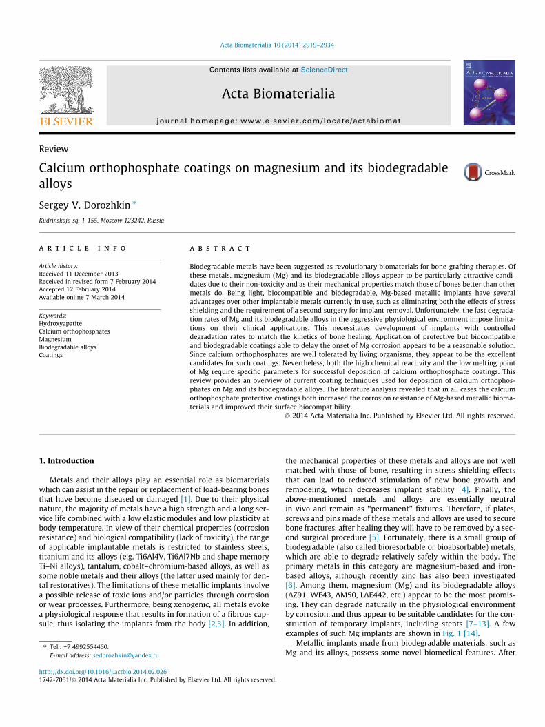
the mechanical properties of these metals and alloys are not wellmatched with those of bone, resulting in stress-shielding effectsthat can lead to reduced stimulation of new bone growth andremodeling, which decreases implant stability [4]. Finally, theabove-mentioned metals and alloys are essentially neutralin vivo and remain as ‘‘permanent’’ fixtures. Therefore, if plates,screws and pins made of these metals and alloys are used to securebone fractures, after healing they will have to be removed by a sec-ond surgical procedure [5]. Fortunately, there is a small group ofbiodegradable (also called bioresorbable or bioabsorbable) metals,which are able to degrade relatively safely within the body. Theprimary metals in this category are magnesium-based and iron-based alloys, although recently zinc has also been investigated[6]. Among them, magnesium (Mg) and its biodegradable alloys(AZ91, WE43, AM50, LAE442, etc.) appear to be the most promis-ing. They can degrade naturally in the physiological environmentby corrosion, and thus appear to be suitable candidates for the con-struction of temporary implants, including stents [7–13]. A fewexamples of such Mg implants are shown in Fig. 1 [14].
Metallic implants made from biodegradable materials, such asMg and its alloys, possess some novel biomedical features. After
2920 S.V. Dorozhkin / Acta Biomaterialia 10 (2014) 2919–2934
being implanted, they will slowly degrade, eliminating the neces-sity for subsequent surgeries to remove them, thereby acceleratingthe entire healing process with a simultaneous reduction in healthrisks, costs and scarring [15]. Nevertheless, to avoid various com-plications and undesired effects (Fig. 2A,B), a suitable degradationkinetics appears to be critical (Fig. 2C): any biodegradable implantmust continue to perform its function(s) until the damaged tissueshave been sufficiently recovered or healed [16]. Additionally, thedegradation (corrosion) products of such implants must be welltolerated by both the surrounding tissues and the organism as awhole. Fortunately, Mg2+ ions are the fourth most abundant cationin the human body and are stored mainly in bones. They are vital tometabolic processes, a cofactor in many enzymes and a key compo-
Fig. 1. Biodegradable orthopedic devices prepared from Mg and its alloys: (top)bone plates; (middle) screws for orthopedic fixation; (bottom) a porous scaffold forbone void filling. Scale bar = 10 mm. Reprinted from Ref. [14] with permission.
nent of the ribosomal machinery that translates the genetic infor-mation encoded by mRNA into polypeptide structures [17].Therefore, contrary to other implantable metals, the wear or corro-sion products of which can be potentially toxic or otherwise harm-ful to patients, those of Mg might be potentially beneficial topatients [18].
As can seen from the above, Mg, its biodegradable alloys andtheir corrosion products are well tolerated by the human body.However, in the vast majority of the cases, the in vivo corrosionkinetics of Mg and its alloys exceeds that of bone healing(Fig. 2A,B); therefore, it must be slowed down for implant applica-tions. Numerous investigations have shown that both the proper-ties and functional activity of any implantable biomaterial can beinfluenced by surface modifications, such as polishing, oxidation,passivation, coating deposition, ion-implantation, etc. [19,20]. Ofthese techniques, the application of synthetic calcium orthophos-phate coatings appears to be the most effective way of achievingsurface modification, and moreover improves the biocompatibilityand osteointegration of metallic implants.
2. A brief description of the two major constituents
2.1. Magnesium and its alloys
Mg is the eighth most abundant element on the surface of ourplanet, making up �1.93% by mass of the earth’s crust and�0.13% by mass of the oceans. Mg is an alkaline earth element,which are located in the second group of the periodic table. Allalkaline earth elements possess a very high chemical reactivityand form compounds with an oxidation number of +2. Therefore,they are not found free in nature. The first isolation of elementalMg was performed by Sir Humphry Davy in 1808 [21,22]. In1852, Robert Bunsen achieved viable commercial production ofMg by electrolysis, and Mg then began to be produced in smallquantities in America and Europe, initially for pyrotechnical useand as igniting bands or wires for flashlights of the nascent photo-graphic industry [23].
Mg and its biodegradable alloys are light in weight and low indensity (1.738 g cm�3 for pure Mg and 1.7–2.0 g cm�3 for thealloys—values that are similar to the density of bones: 1.8–2.1 g cm�3). Due to its relatively low melting point (650 �C), Mgis considered a fusible metals. The elastic modulus of Mg is�45 GPa [24], which is much closer to that of bone (trabecular/cancellous bones: 3–14.8 GPa, cortical bones: 18.6–27 GPa[25,26]) compared to compared to the moduli of other implantablemetals: Ti alloys, 110–117 GPa; stainless steels, 189–205 GPa;Co–Cr alloys, �230 GPa. In addition, the numerical values of thecompressive yield strength of bones and Mg are 130–180 and65–100 MPa, respectively, while those of fracture toughness are3–6 and 15–40 MPa m�2, respectively. Hence, by using Mg andits alloys for bone grafting the stress-shielding effect can bemitigated [7,10]. In addition, some antibacterial properties of Mghave been reported [27].
From the chemical point of view, its high reactivity (thestandard electrode potential of Mg2+
(aq) + 2e�M Mg(s) is �2.37 V, thatof Mg+
(aq) + e�M Mg(s) is �2.70 V and that of Mg(OH)2(s) + 2e�
M Mg(s) + 2OH� is �2.69 V [28]) makes Mg readily soluble in bodyfluids, which is the primary reason of its in vivo biodegradability.Therefore, when Mg is exposed to aqueous solutions, the followingoxidation reaction takes place on its surface [29,30]:
Mgþ 2H2O ¼MgðOHÞ2 # þH2 "
This provides a possibility to measure the corrosion kinetics ofMg and its biodegradable alloys by the release kinetics of hydrogen(Fig. 3) [31]. Unfortunately, the oxidized surface layers consisting

Fig. 2. A model explaining the improvements due to the presence of bioactive calcium orthophosphate coatings on Mg and its alloys. (A) A relatively rapid degradation rate ofMg might lead to formation of gaps at the interface. (B) A typical tetracycline label taken 14 weeks post-operation. (C) Protective calcium orthophosphate coatings can reducethe degradation rate and simultaneously ameliorate biocompatibility. (D) Corrosion-protective effects of calcium orthophosphate coatings measured via the H2 release rateand the change in pH value. Reprinted from Ref. [15] with permission.
Fig. 3. A schematic drawing of a H2 evolution collection set-up to follow thecorrosion kinetics of Mg and its alloys. Reprinted from Ref. [31] with permission.
S.V. Dorozhkin / Acta Biomaterialia 10 (2014) 2919–2934 2921
of hydrated forms of MgO and/or Mg(OH)2 are loose in nature andcannot provide sufficient protection to resist the corrosion encoun-tered in the physiological environment, which contains a high level(�104 mM) of chloride ions. The corrosion kinetics of Mg isstrongly accelerated in the presence of dissolved chloride ions,which are able to convert the insoluble MgO + Mg(OH)2 coatingsinto a soluble MgCl2, simultaneously decreasing the protected areaand promoting further dissolution of Mg [29,30]. Since further de-tails on the corrosion process of both Mg and its biodegradable al-loys are beyond the scope of this review, the readers interested inthis topic are directed to the specialist literature on this subject
[12,13,29–33] (the same sources also summarize the informationon available Mg alloys). For present purposes it is important simplyto recognize that, to be feasible for orthopedic applications, thecorrosion kinetics of Mg and biodegradable its alloys must be re-duced. Ideally, it should be slowed down to allow the mechanicalintegrity of the metal to remain intact during bone healing. Thiswould also minimize hydrogen production, which was observedas a disadvantageous by-product when using Mg [11,34].
There are a number of ways to improve the corrosion resistanceof pure Mg [12,32,33,35]. Briefly, they comprise the following ap-proaches: microstructure tailoring including both grain size[36,37] and texture [38], alloying [13,39–43], preparation of bio-composites [44–47], surface treatment [48–53] and deposition ofprotective coatings [43,47,54–65]. This review focuses on protec-tive calcium orthophosphate coatings on Mg and its biodegradablealloys. It should be stressed that, to improve corrosion resistance,the surface of Mg and its alloys can be coated with either calciumorthophosphates alone (the vast majority of publications cited inthis review) or calcium orthophosphate-based biocomposites[66–77]. As the use of implantable calcium orthophosphatesstarted in 1920 [78,79], while that of calcium orthophosphate-based biocomposites and hybrid biomaterials started only in1981 [80], one can conclude that the latter is just at the initialstages and many more publications are expected in the near future.Finally, it should be mentioned that Mg and its biodegradable al-loys can be also coated by calcium phosphate glass-ceramics[81,82]; however, that is another story.
2.2. Calcium orthophosphates
The main driving force behind the use of calcium orthophos-phates as bone substitute materials is their chemical similarity to
2922 S.V. Dorozhkin / Acta Biomaterialia 10 (2014) 2919–2934
the mineral component of mammalian bones and teeth [83–85]. Asa result, in addition to being non-toxic, they are biocompatible, notrecognized as foreign materials in the body and, most importantly,both exhibit bioactive behavior and integrate into living tissue bythe same processes that are active in remodeling healthy bone.This leads to an intimate physicochemical bond between the im-plants and bone, termed osteointegration [86]. More to the point,calcium orthophosphates are also known to support osteoblastadhesion and proliferation [87,88]. Even so, the major limitationsto the use of calcium orthophosphates as load-bearing biomaterialsare their mechanical properties: they are brittle with a poor fatigueresistance [89,90]. That is why, in biomedical applications, calciumorthophosphates are used primarily as fillers and coatings[85,86,91].
A complete list of known calcium orthophosphates, includingtheir standard abbreviations and major properties, is given inTable 1, while detailed information on calcium orthophosphates,their synthesis, structure, chemistry, other properties and biomed-ical application has been comprehensively reviewed recently [85].Even more thorough information on calcium orthophosphates canbe found in specialist books and monographs [92–97].
3. A brief description of the important pre- and post-depositionprocedures
Prior to be coated by calcium orthophosphates, in the vast majorityof the cases, the surface of Mg and its biodegradable alloys needs to beprepared. The preparation normally consists of cleaning and/or degre-asing to remove any sort of surface contamination arising from man-ufacturing. This procedure can be performed in acetone [31,56,57,63,64,66,69,70,73–75,98–119], ethanol [59,64,73,113–138], mixturesthereof [139], trichloroethylene [140], an aqueous solution of Na2CO3
[140], a mixture of 4% nitric acid + 96% ethylene glycol [129] ordistilled water [31,64,66,68,70,73,74,113,129,131,132, 140,141]. Inaddition, various types of physical modifications of the metallic
Table 1Existing calcium orthophosphates and their major properties [85].
Ca/P molarratio
Compound Formula
0.5 Monocalcium phosphate monohydrate(MCPM)
Ca(H2PO4)2�H2O
0.5 Monocalcium phosphate anhydrous (MCPAor MCP)
Ca(H2PO4)2
1.0 Dicalcium phosphate dihydrate (DCPD),mineral brushite
CaHPO4�2H2O
1.0 Dicalcium phosphate anhydrous (DCPA orDCP), mineral monetite
CaHPO4
1.33 Octacalcium phosphate (OCP) Ca8(HPO4)2(PO4)4�5H2O1.5 a-Tricalcium phosphate (a-TCP) a-Ca3(PO4)2
1.5 b-Tricalcium phosphate (b-TCP) b-Ca3(PO4)2
1.2–2.2 Amorphous calcium phosphates (ACP) CaxHy(PO4)z�nH2O, n = 3–15–20% H2O
1.5–1.67 Calcium-deficient hydroxyapatite (CDHA orCa-def HA)e
Ca10�x(HPO4)x(PO4)6�x(O(0 < x < 1)
1.67 Hydroxyapatite (HA, HAp or OHAp) Ca10(PO4)6(OH)2
1.67 Fluorapatite (FA or FAp) Ca10(PO4)6F2
1.67 Oxyapatite (OA, OAp or OXA)f, mineralvoelckerite
Ca10(PO4)6O
2.0 Tetracalcium phosphate (TTCP or TetCP),mineral hilgenstockite
Ca4(PO4)2O
a These compounds cannot be precipitated from aqueous solutions.b Cannot be measured precisely. However, the following values were found: 25.7 ± 0
dissolution in acidic buffer is: ACP� a-TCP� b-TCP > CDHA� HA > FA.c Stable at temperatures above 100 �C.d Always metastable.e Occasionally called ‘‘precipitated HA (PHA)’’.f The existence of OA remains questionable.
surface are used; examples include physical grinding and/orpolishing [31,56,59,63,64,66–75,98–147], drying [31,59,64,74,75,98–101,110,113,115,117,120,121,123,125,131,132,135–138], heattreatment [120,122,140] and/or autoclaving [114]. Furthermore, priorto deposition of calcium orthophosphates, the surface of Mg and itsalloys might be chemically treated (e.g. activated [67,99–101,125,145], alkaline treated [56,67,129,130,140], anodized [114,136],chemically polished [102–109], electrochemically polished [139],etched [64,75], passivated [59,119,134,148], pre-phosphatized[67,128,136], etc. [120]). More to the point, prior to deposition ofcalcium orthophosphates, the surface of Mg and its alloys can becoated with an interlayer of another compound, such as poly(e-caprolactone) [117], nicotinic acid [75], Mg(OH)2 [59], MgF2
[116,149], Ca(OH)2 [150] or titania [77] to enhance the corrosion resis-tance and coating flexibility. Pre-calcified coatings can also be applied[75,137]. All these types of treatment are usually performed by dip-ping, spraying, rinsing and/or soaking, depending on both the qualityrequirements and the limitations of the product to be coated. Finally,the surface of Mg and its biodegradable alloys can be sterilized priorto deposition of calcium orthophosphates [119]. As seen from thenumber of available references, grinding and/or polishing of Mg andits biodegradable alloys appears to be the most popular surface pre-treatment technique, followed by cleaning and/or degreasing.
In addition, after calcium orthophosphate coatings have beendeposited, various types of post-deposition treatment can be usedto improve their properties. For example, post-deposition heat-treatment (annealing) of calcium orthophosphates leads to conver-sion of the deposited amorphous (ACP) and non-apatite phases,such as dibasic calcium phosphate dihydrate (DCPD), into eitherhydroxyapatite (HA) [113] or Ca2P2O7 [130] (depending on the Ca/P ratio) with a simultaneous increase of coating crystallinity andcorrosion resistance, combined with a reduction in the residualstress. Furthermore, for the same purposes, chemical treatment ofthe coated samples in either aqueous alkaline solutions [69,99,129,134,142,144,148,151,152] or phosphate-buffered solution(PBS) [66,71,72] can be used (Fig. 4). In addition, the coated samples
Solubility at 25 �C,�log(Ks)
Solubility at25 �C, g l�1
pH stability range in aqueoussolutions at 25 �C
1.14 �18 0.0–2.0
1.14 �17 c
6.59 �0.088 2.0–6.0
6.90 �0.048 c
96.6 �0.0081 5.5–7.025.5 �0.0025 a
28.9 �0.0005 a
4.5; b b �5–12d
H)2�x �85 �0.0094 6.5–9.5
116.8 �0.0003 9.5–12120.0 �0.0002 7–12�69 �0.087 a
38–44 �0.0007 a
.1 (pH 7.40), 29.9 ± 0.1 (pH 6.00), 32.7 ± 0.1 (pH 5.28). The comparative extent of
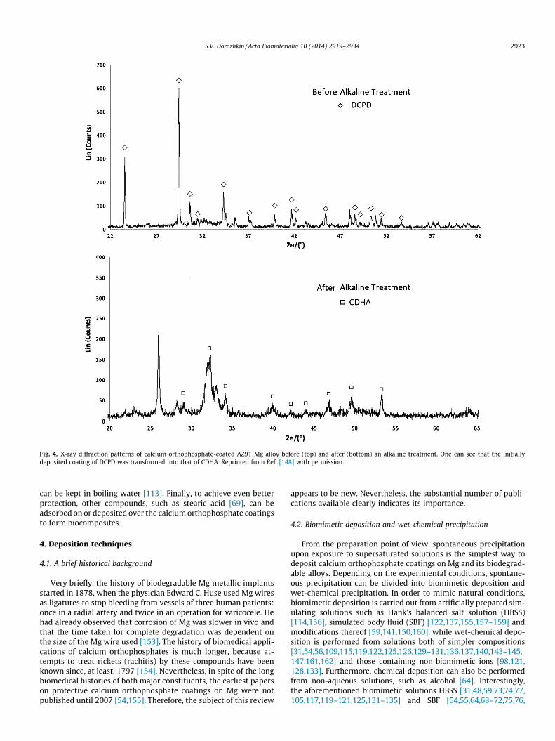
Fig. 4. X-ray diffraction patterns of calcium orthophosphate-coated AZ91 Mg alloy before (top) and after (bottom) an alkaline treatment. One can see that the initiallydeposited coating of DCPD was transformed into that of CDHA. Reprinted from Ref. [148] with permission.
S.V. Dorozhkin / Acta Biomaterialia 10 (2014) 2919–2934 2923
can be kept in boiling water [113]. Finally, to achieve even betterprotection, other compounds, such as stearic acid [69], can beadsorbed on or deposited over the calcium orthophosphate coatingsto form biocomposites.
4. Deposition techniques
4.1. A brief historical background
Very briefly, the history of biodegradable Mg metallic implantsstarted in 1878, when the physician Edward C. Huse used Mg wiresas ligatures to stop bleeding from vessels of three human patients:once in a radial artery and twice in an operation for varicocele. Hehad already observed that corrosion of Mg was slower in vivo andthat the time taken for complete degradation was dependent onthe size of the Mg wire used [153]. The history of biomedical appli-cations of calcium orthophosphates is much longer, because at-tempts to treat rickets (rachitis) by these compounds have beenknown since, at least, 1797 [154]. Nevertheless, in spite of the longbiomedical histories of both major constituents, the earliest paperson protective calcium orthophosphate coatings on Mg were notpublished until 2007 [54,155]. Therefore, the subject of this review
appears to be new. Nevertheless, the substantial number of publi-cations available clearly indicates its importance.
4.2. Biomimetic deposition and wet-chemical precipitation
From the preparation point of view, spontaneous precipitationupon exposure to supersaturated solutions is the simplest way todeposit calcium orthophosphate coatings on Mg and its biodegrad-able alloys. Depending on the experimental conditions, spontane-ous precipitation can be divided into biomimetic deposition andwet-chemical precipitation. In order to mimic natural conditions,biomimetic deposition is carried out from artificially prepared sim-ulating solutions such as Hank’s balanced salt solution (HBSS)[114,156], simulated body fluid (SBF) [122,137,155,157–159] andmodifications thereof [59,141,150,160], while wet-chemical depo-sition is performed from solutions both of simpler compositions[31,54,56,109,115,119,122,125,126,129–131,136,137,140,143–145,147,161,162] and those containing non-biomimetic ions [98,121,128,133]. Furthermore, chemical deposition can also be performedfrom non-aqueous solutions, such as alcohol [64]. Interestingly,the aforementioned biomimetic solutions HBSS [31,48,59,73,74,77,105,117,119–121,125,131–135] and SBF [54,55,64,68–72,75,76,
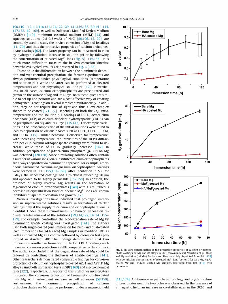
Fig. 5. In vitro determination of the protective properties of calcium orthophos-phate coatings on Mg and its alloys in SBF (corrosion tests). Variation of pH (top)and H2 evolution (middle) for bare and HA-coated Mg. Reprinted from Ref. [138]with permission. Concentration of released Mg2+ ions (bottom) for bare Mg, MgF2-coated Mg and HA/MgF2-coated Mg samples. Reprinted from Ref. [116] withpermission.
2924 S.V. Dorozhkin / Acta Biomaterialia 10 (2014) 2919–2934
100,110–112,116,118,121,124,127,129–131,136,138,139,141–144,147,152,162–169], as well as Dulbecco’s Modified Eagle’s Medium(DMEM) [119], minimum essential medium (MEM) [43] andaqueous solutions (0.8–3.5 wt.%) of NaCl [59,106,113,130], arecommonly used to study the in vitro corrosion of Mg and its alloys[11,170], and thus the protective properties of calcium orthophos-phate coatings [62]. The latter property can be measured in vitroby hydrogen evolution, increase in solution pH or by followingthe concentration of released Mg2+ ions (Fig. 5) [116,138]. It ismuch more difficult to measure the in vivo corrosion kinetics;nevertheless, typical results are presented in Fig. 6 [138].
To continue the differentiation between the biomimetic deposi-tion and wet-chemical precipitation, the former experiments arealways performed under physiological conditions (temperatureand solution pH), while the latter can be performed at elevatedtemperatures and non-physiological solution pH [120]. Neverthe-less, in all cases, calcium orthophosphates are precipitated andgrown on the surface of Mg and its alloys. Both techniques are sim-ple to set up and perform and are a cost-effective way of creatinghomogeneous coatings on several samples simultaneously. In addi-tion, they do not require line of sight and thus allow complexshapes to be coated [171,172]. Depending on both the Ca/P ratio,temperature and the solution pH, coatings of DCPD, octacalciumphosphate (OCP) or calcium-deficient hydroxyapatite (CDHA) canbe precipitated on Mg and its alloys [115,147]. For example, varia-tions in the ionic composition of the initial solutions were found tolead to deposition of various phases such as DCPD, DCPD + CDHA,and CDHA [115]. Similar behavior is observed for temperature:with increasing temperature, the intensities of the DCPD diffrac-tion peaks in calcium orthophosphate coatings were found to de-crease, while those of CDHA gradually increased [147]. Inaddition, precipitation of b-tricalcium phosphate (b-TCP) on Mgwas detected [120,128]. Since simulating solutions often containa number of various ions, ion-substituted calcium orthophosphatesare always deposited via biomimetic approach. For example, amor-phous carbonated calcium–magnesium orthophosphate coatingswere formed in SBF [155,157–159]. After incubation in SBF for5 days, the deposited coatings had a thickness exceeding 20 lmand appeared to be highly permeable [157,158]. In addition, thepresence of highly reactive Mg results in the formation ofMg-enriched calcium orthophosphates [140] with a simultaneousdecrease in crystallization kinetics because Mg2+ ions are knowninhibitors of apatite nucleation and growth [173].
Various investigations have indicated that prolonged immer-sion in supersaturated solutions results in formation of thickercoatings only if the supply of calcium and orthophosphate ions isplentiful. Under these circumstances, biomimetic deposition re-quires regular renewal of the solutions [59,114,122,137,141,155–159]. For example, controlling the biodegradation rate of Mg bybiomimetic apatite coating was investigated [141]. The authorsused both single-coated (one immersion for 24 h) and dual-coated(two immersions for 24 h each) Mg samples in modified SBF, aswell as uncoated Mg as a control, followed by corrosion tests per-formed in standard SBF. The findings demonstrated that twoimmersions resulted in formation of thicker CDHA coatings withincreased corrosion protection in SBF comparative to the controls.The authors concluded that the degradation rate of Mg could betailored by controlling the thickness of apatite coatings [141].Other researchers demonstrated comparable findings for corrosionprotection of calcium orthophosphate-coated AZ31 and AZ91D Mgalloys using both immersion tests in SBF [163] and electrochemicaltests [122], respectively. In support of this, still other investigatorsillustrated the corrosion protection of biomimetic CDHA-coatedpure Mg with subsequent increase in cell adhesion [58,159].Furthermore, the biomimetic precipitation of calciumorthophosphates on Mg can be performed under a magnetic field
[115,174]. A difference in particle morphology and crystal textureof precipitates near the two poles was observed. In the presence ofa magnetic field, an increase in crystallite sizes in the (020) and
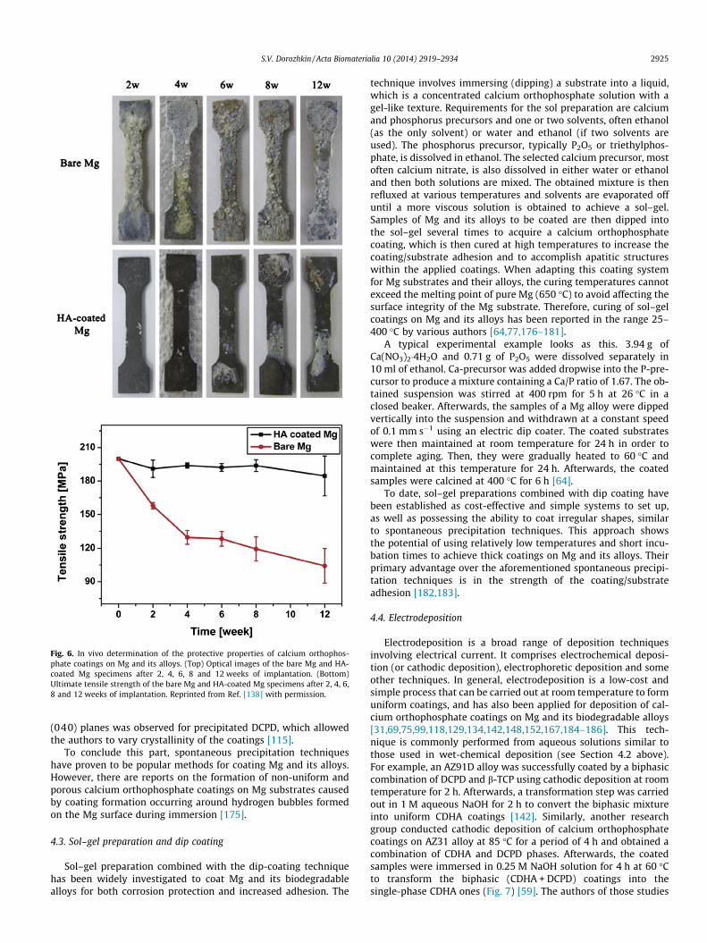
Fig. 6. In vivo determination of the protective properties of calcium orthophos-phate coatings on Mg and its alloys. (Top) Optical images of the bare Mg and HA-coated Mg specimens after 2, 4, 6, 8 and 12 weeks of implantation. (Bottom)Ultimate tensile strength of the bare Mg and HA-coated Mg specimens after 2, 4, 6,8 and 12 weeks of implantation. Reprinted from Ref. [138] with permission.
S.V. Dorozhkin / Acta Biomaterialia 10 (2014) 2919–2934 2925
(040) planes was observed for precipitated DCPD, which allowedthe authors to vary crystallinity of the coatings [115].
To conclude this part, spontaneous precipitation techniqueshave proven to be popular methods for coating Mg and its alloys.However, there are reports on the formation of non-uniform andporous calcium orthophosphate coatings on Mg substrates causedby coating formation occurring around hydrogen bubbles formedon the Mg surface during immersion [175].
4.3. Sol–gel preparation and dip coating
Sol–gel preparation combined with the dip-coating techniquehas been widely investigated to coat Mg and its biodegradablealloys for both corrosion protection and increased adhesion. The
technique involves immersing (dipping) a substrate into a liquid,which is a concentrated calcium orthophosphate solution with agel-like texture. Requirements for the sol preparation are calciumand phosphorus precursors and one or two solvents, often ethanol(as the only solvent) or water and ethanol (if two solvents areused). The phosphorus precursor, typically P2O5 or triethylphos-phate, is dissolved in ethanol. The selected calcium precursor, mostoften calcium nitrate, is also dissolved in either water or ethanoland then both solutions are mixed. The obtained mixture is thenrefluxed at various temperatures and solvents are evaporated offuntil a more viscous solution is obtained to achieve a sol–gel.Samples of Mg and its alloys to be coated are then dipped intothe sol–gel several times to acquire a calcium orthophosphatecoating, which is then cured at high temperatures to increase thecoating/substrate adhesion and to accomplish apatitic structureswithin the applied coatings. When adapting this coating systemfor Mg substrates and their alloys, the curing temperatures cannotexceed the melting point of pure Mg (650 �C) to avoid affecting thesurface integrity of the Mg substrate. Therefore, curing of sol–gelcoatings on Mg and its alloys has been reported in the range 25–400 �C by various authors [64,77,176–181].
A typical experimental example looks as this. 3.94 g ofCa(NO3)2�4H2O and 0.71 g of P2O5 were dissolved separately in10 ml of ethanol. Ca-precursor was added dropwise into the P-pre-cursor to produce a mixture containing a Ca/P ratio of 1.67. The ob-tained suspension was stirred at 400 rpm for 5 h at 26 �C in aclosed beaker. Afterwards, the samples of a Mg alloy were dippedvertically into the suspension and withdrawn at a constant speedof 0.1 mm s�1 using an electric dip coater. The coated substrateswere then maintained at room temperature for 24 h in order tocomplete aging. Then, they were gradually heated to 60 �C andmaintained at this temperature for 24 h. Afterwards, the coatedsamples were calcined at 400 �C for 6 h [64].
To date, sol–gel preparations combined with dip coating havebeen established as cost-effective and simple systems to set up,as well as possessing the ability to coat irregular shapes, similarto spontaneous precipitation techniques. This approach showsthe potential of using relatively low temperatures and short incu-bation times to achieve thick coatings on Mg and its alloys. Theirprimary advantage over the aforementioned spontaneous precipi-tation techniques is in the strength of the coating/substrateadhesion [182,183].
4.4. Electrodeposition
Electrodeposition is a broad range of deposition techniquesinvolving electrical current. It comprises electrochemical deposi-tion (or cathodic deposition), electrophoretic deposition and someother techniques. In general, electrodeposition is a low-cost andsimple process that can be carried out at room temperature to formuniform coatings, and has also been applied for deposition of cal-cium orthophosphate coatings on Mg and its biodegradable alloys[31,69,75,99,118,129,134,142,148,152,167,184–186]. This tech-nique is commonly performed from aqueous solutions similar tothose used in wet-chemical deposition (see Section 4.2 above).For example, an AZ91D alloy was successfully coated by a biphasiccombination of DCPD and b-TCP using cathodic deposition at roomtemperature for 2 h. Afterwards, a transformation step was carriedout in 1 M aqueous NaOH for 2 h to convert the biphasic mixtureinto uniform CDHA coatings [142]. Similarly, another researchgroup conducted cathodic deposition of calcium orthophosphatecoatings on AZ31 alloy at 85 �C for a period of 4 h and obtained acombination of CDHA and DCPD phases. Afterwards, the coatedsamples were immersed in 0.25 M NaOH solution for 4 h at 60 �Cto transform the biphasic (CDHA + DCPD) coatings into thesingle-phase CDHA ones (Fig. 7) [59]. The authors of those studies
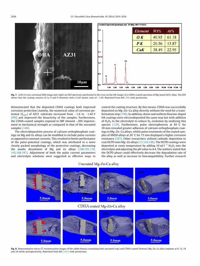
Fig. 7. (Left) A cross-sectional SEM image and (right) an EDS spectrum (performed in the cross on the left image) of a CDHA-coated specimen of Mg-based AZ31 alloy. The EDSshows that the coating consists of Ca, P and O elements with a Ca/P atomic ratio of �1.45. Reprinted from Ref. [99] with permission.
2926 S.V. Dorozhkin / Acta Biomaterialia 10 (2014) 2919–2934
demonstrated that the deposited CDHA coatings both improvedcorrosion protection (namely, the numerical value of corrosion po-tential (Ecorr) of AZ31 substrate increased from �1.6 to �1.42 V[99]) and improved the bioactivity of the samples. Furthermore,the CDHA-coated samples exposed to SBF showed �20% improve-ment in mechanical strength as compared to that of the uncoatedsamples [148].
The electrodeposition process of calcium orthophosphate coat-ings on Mg and its alloys can be modified to include pulse currentsas opposed to constant currents. This resulted in better performanceof the pulse-potential coatings, which was attributed to a moreclosely packed morphology of the protective coatings, decreasingthe anodic dissolution of Mg and its alloys [100,101,139,165,168,187]. Adjustment of both the pulse current parametersand electrolyte solutions were suggested as effective ways to
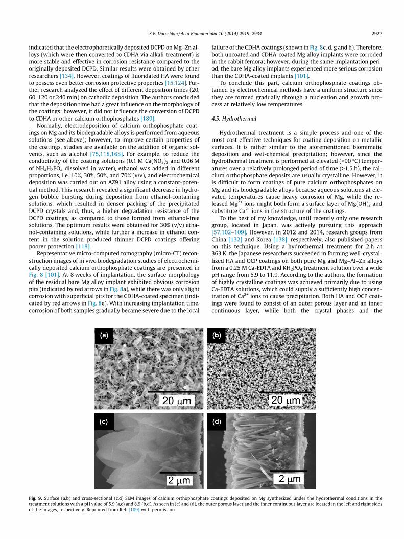
Fig. 8. Representative micro-CT reconstruction images of the rabbit femora containing boand 24 weeks postoperatively. Reprinted from Ref. [101] with permission.
control the coating structure. By this means, CDHA was successfullydeposited on Mg–Zn–Ca alloy directly without the need for a trans-formation step [100]. In addition, dense and uniform fluorine-dopedHA coatings were electrodeposited the same way but with additionof H2O2 to the electrolyte to reduce H2 evolution by oxidizing thisspecies [139]. Furthermore, pulse electrophoresis at 85 �C for30 min revealed greater adhesion of calcium orthophosphate coat-ings to Mg–Zn–Ca alloys, whilst pulse treatments of the coated sam-ples of AM50 alloys at 20 �C for 15 min displayed a higher corrosionresistance [187]. Other researchers utilized cathodic deposition tocoat DCPD onto Mg–Zn alloys [15,124,188]. The DCPD coatings weredeposited at room temperature by adding 10 ml l�1 H2O2 into theelectrolyte and adjusting the pH value to 4.4. The authors stated thatthe DCPD phase could effectively decrease the degradation rate ofthe alloy as well as increase its biocompatibility. Further research
th uncoated (top) and CDHA-coated (bottom) Mg–Zn–Ca alloy implants at 8, 12, 18
S.V. Dorozhkin / Acta Biomaterialia 10 (2014) 2919–2934 2927
indicated that the electrophoretically deposited DCPD on Mg–Zn al-loys (which were then converted to CDHA via alkali treatment) ismore stable and effective in corrosion resistance compared to theoriginally deposited DCPD. Similar results were obtained by otherresearchers [134]. However, coatings of fluoridated HA were foundto possess even better corrosion protective properties [15,124]. Fur-ther research analyzed the effect of different deposition times (20,60, 120 or 240 min) on cathodic deposition. The authors concludedthat the deposition time had a great influence on the morphology ofthe coatings; however, it did not influence the conversion of DCPDto CDHA or other calcium orthophosphates [189].
Normally, electrodeposition of calcium orthophosphate coat-ings on Mg and its biodegradable alloys is performed from aqueoussolutions (see above); however, to improve certain properties ofthe coatings, studies are available on the addition of organic sol-vents, such as alcohol [75,118,168]. For example, to reduce theconductivity of the coating solutions (0.1 M Ca(NO3)2 and 0.06 Mof NH4H2PO4 dissolved in water), ethanol was added in differentproportions, i.e. 10%, 30%, 50%, and 70% (v/v), and electrochemicaldeposition was carried out on AZ91 alloy using a constant-poten-tial method. This research revealed a significant decrease in hydro-gen bubble bursting during deposition from ethanol-containingsolutions, which resulted in denser packing of the precipitatedDCPD crystals and, thus, a higher degradation resistance of theDCPD coatings, as compared to those formed from ethanol-freesolutions. The optimum results were obtained for 30% (v/v) etha-nol-containing solutions, while further a increase in ethanol con-tent in the solution produced thinner DCPD coatings offeringpoorer protection [118].
Representative micro-computed tomography (micro-CT) recon-struction images of in vivo biodegradation studies of electrochemi-cally deposited calcium orthophosphate coatings are presented inFig. 8 [101]. At 8 weeks of implantation, the surface morphologyof the residual bare Mg alloy implant exhibited obvious corrosionpits (indicated by red arrows in Fig. 8a), while there was only slightcorrosion with superficial pits for the CDHA-coated specimen (indi-cated by red arrows in Fig. 8e). With increasing implantation time,corrosion of both samples gradually became severe due to the local
Fig. 9. Surface (a,b) and cross-sectional (c,d) SEM images of calcium orthophosphatetreatment solutions with a pH value of 5.9 (a,c) and 8.9 (b,d). As seen in (c) and (d), the ouof the images, respectively. Reprinted from Ref. [109] with permission.
failure of the CDHA coatings (shown in Fig. 8c, d, g and h). Therefore,both uncoated and CDHA-coated Mg alloy implants were corrodedin the rabbit femora; however, during the same implantation peri-od, the bare Mg alloy implants experienced more serious corrosionthan the CDHA-coated implants [101].
To conclude this part, calcium orthophosphate coatings ob-tained by electrochemical methods have a uniform structure sincethey are formed gradually through a nucleation and growth pro-cess at relatively low temperatures.
4.5. Hydrothermal
Hydrothermal treatment is a simple process and one of themost cost-effective techniques for coating deposition on metallicsurfaces. It is rather similar to the aforementioned biomimeticdeposition and wet-chemical precipitation; however, since thehydrothermal treatment is performed at elevated (>90 �C) temper-atures over a relatively prolonged period of time (>1.5 h), the cal-cium orthophosphate deposits are usually crystalline. However, itis difficult to form coatings of pure calcium orthophosphates onMg and its biodegradable alloys because aqueous solutions at ele-vated temperatures cause heavy corrosion of Mg, while the re-leased Mg2+ ions might both form a surface layer of Mg(OH)2 andsubstitute Ca2+ ions in the structure of the coatings.
To the best of my knowledge, until recently only one researchgroup, located in Japan, was actively pursuing this approach[57,102–109]. However, in 2012 and 2014, research groups fromChina [132] and Korea [138], respectively, also published paperson this technique. Using a hydrothermal treatment for 2 h at363 K, the Japanese researchers succeeded in forming well-crystal-lized HA and OCP coatings on both pure Mg and Mg–Al–Zn alloysfrom a 0.25 M Ca-EDTA and KH2PO4 treatment solution over a widepH range from 5.9 to 11.9. According to the authors, the formationof highly crystalline coatings was achieved primarily due to usingCa-EDTA solutions, which could supply a sufficiently high concen-tration of Ca2+ ions to cause precipitation. Both HA and OCP coat-ings were found to consist of an outer porous layer and an innercontinuous layer, while both the crystal phases and the
coatings deposited on Mg synthesized under the hydrothermal conditions in theter porous layer and the inner continuous layer are located in the left and right sides

Fig. 10. A schematic illustration of the formation and growth mechanism of HA coatings on Mg under hydrothermal conditions. Reprinted from Ref. [104] with permission.Another illustration of this mechanism is available in Ref. [57].
2928 S.V. Dorozhkin / Acta Biomaterialia 10 (2014) 2919–2934
microstructures of the coatings were found to vary with the pH ofthe treatment solutions. Namely, in weak acidic (pH 5.9) solutions,a dual-layer structure was formed: an outer coarse layer consistedof plate-like OCP crystals and an inner dense layer consisted pri-marily of HA crystals. In weak alkaline (pH 8.9) solutions, a dual-layer structure was also formed: an outer coarse layer consistedof rod-like HA crystals and an inner dense layer consisted ofwell-packed HA crystals (Fig. 9). In strong alkaline (pH 11.9) solu-tions, needle-like HA crystals were formed. Both layers were foundto grow with an increase in the treatment period. A thin Mg(OH)2
layer was also formed at the boundary between the calcium ortho-phosphate coatings and Mg substrates (Fig. 10). The HA and OCPcoatings were found to improve the corrosion resistance of bothpure Mg and Mg–Al–Zn alloys in both HBSS and a 3.5 wt.% NaClsolutions; however, the corrosion resistance of HA coatings was al-ways higher than that of OCP ones [57,102–109]. Similar resultswere obtained by other researchers [132,138]. In addition, suchcoatings showed good adhesive properties with slight plasticdeformation under cyclic stress below the fatigue limit. Neithercracks nor detachment was microscopically observed under 5% sta-tic elongation and under 3% cyclic elongation [108]. The authors re-vealed that the level of protection afforded by the calciumorthophosphate coatings could be varied by changing their crystalphase, microstructure and thickness. Thus, optimization of themicrostructure of the coatings is necessary to adjust the corrosionresistance of the coated Mg to the desired values.
4.6. Aerosol deposition
In addition, calcium orthophosphate coatings can be put downon Mg and its biodegradable alloys by an aerosol deposition tech-nique. This technique has been used to deposit HA onto the surfaceof both pure Mg and Mg previously covered by either poly(e-cap-rolactone) [117] or MgF2 [116]. To perform aerosol deposition,HA powder was sprayed onto Mg samples in a deposition chamberusing oxygen carrier gas at a flow rate of 5 � 10�4 m3 s�1 under apressure of 9.2 Torr. Scanning electron microscopy (SEM) observa-tions showed that when HA was deposited onto Mg with thepoly(e-caprolactone) interlayer, it was partially embedded intothis interlayer, forming composite-like structures [117]; however,when HA was deposited onto Mg with the MgF2 interlayer, nocomposite-like structures were observed [116]. Corrosion tests
performed in SBF revealed that such coatings had good corrosionresistance (Fig. 5, bottom). In addition, HA coatings on Mg withthe poly(e-caprolactone) interlayer were found to have better sta-bility during deformation compared to HA coatings on Mg withoutthe interlayer. These results revealed that coating Mg by an HA/poly(e-caprolactone) double layer might prove a promising ap-proach to reduce the corrosion rate of Mg and improve the coatingflexibility [117].
Furthermore, using aerosol deposition, Mg and its biodegrad-able alloys can be coated by calcium orthophosphate-based bio-composites. For example, HA/chitosan biocomposites have beendeposited on AZ31 alloy [68]. The authors employed a slit-typenozzle with a 10 � 0.5 mm2 rectangular opening and air as a car-rier gas with a flow rate of 30 l min�1. The 5 lm thick HA/chitosancoatings were deposited over the entire surface of the AZ31Mg al-loy substrates by scanning the substrates on the motorized X–Ystage for 1 min at a scanning speed of 1 mm s�1. The biocompositecoatings were found to exhibit high adhesion strengths rangingfrom 24.6 to 27.7 MPa and showed good corrosion resistance.Although addition of chitosan lowered the corrosion resistance ofthe HA coatings, their biocompatibility was improved [68].
4.7. Spin coating
Only two publications on spin coating could be found, and bothof these were devoted to deposition of calcium orthophosphate-based biocomposites on Mg alloys [73,76]. Initially, biocompositesof HA/collagen (HAC) [73] and HA/poly(lactic-co-glycolic acid)[76], respectively, were prepared. Afterwards, to deposit HAC ontothe surface of AZ31 alloy chips, a mixture of 2 g of poly(L-lactic acid)(PLLA) and HAC (at various PLLA/HAC ratios) was dissolved in 20 mlof dichloromethane and magnetically stirred for 30 min followed byultrasonic dispersion for 15 min. The prepared suspension was spincoated on pretreated AZ31 alloy chips for 30 s at a rotational speedof 2000 rpm. The coated surface was immediately dried by blowingat room temperature and, in order to obtain thick coatings, the pro-cedure was repeated five times. Corrosion studies performed inHBSS revealed that the biocomposite coatings suppressed the sharpincrease in pH value and Mg2+ release from the substrates, while thedegradation behavior of the alloy was correlated to the microstruc-ture of the coatings [73]. Both the deposition technique and theobtained corrosion results appeared to be similar for Mg alloys
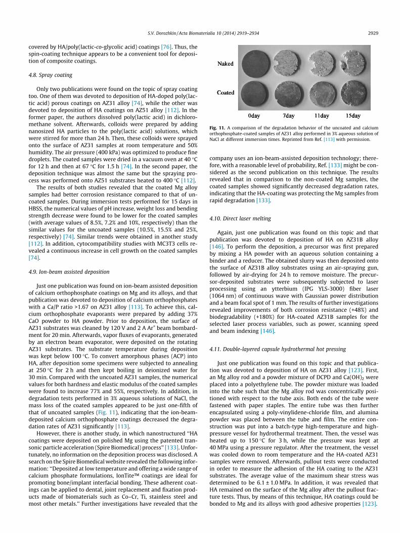
Fig. 11. A comparison of the degradation behavior of the uncoated and calciumorthophosphate-coated samples of AZ31 alloy performed in 3% aqueous solution ofNaCl at different immersion times. Reprinted from Ref. [113] with permission.
S.V. Dorozhkin / Acta Biomaterialia 10 (2014) 2919–2934 2929
covered by HA/poly(lactic-co-glycolic acid) coatings [76]. Thus, thespin-coating technique appears to be a convenient tool for deposi-tion of composite coatings.
4.8. Spray coating
Only two publications were found on the topic of spray coatingtoo. One of them was devoted to deposition of HA-doped poly(lac-tic acid) porous coatings on AZ31 alloy [74], while the other wasdevoted to deposition of HA coatings on AZ51 alloy [112]. In theformer paper, the authors dissolved poly(lactic acid) in dichloro-methane solvent. Afterwards, colloids were prepared by addingnanosized HA particles to the poly(lactic acid) solutions, whichwere stirred for more than 24 h. Then, these colloids were sprayedonto the surface of AZ31 samples at room temperature and 50%humidity. The air pressure (400 kPa) was optimized to produce finedroplets. The coated samples were dried in a vacuum oven at 40 �Cfor 12 h and then at 67 �C for 1.5 h [74]. In the second paper, thedeposition technique was almost the same but the spraying pro-cess was performed onto AZ51 substrates heated to 400 �C [112].
The results of both studies revealed that the coated Mg alloysamples had better corrosion resistance compared to that of un-coated samples. During immersion tests performed for 15 days inHBSS, the numerical values of pH increase, weight loss and bendingstrength decrease were found to be lower for the coated samples(with average values of 8.5%, 7.2% and 10%, respectively) than thesimilar values for the uncoated samples (10.5%, 15.5% and 25%,respectively) [74]. Similar trends were obtained in another study[112]. In addition, cytocompatibility studies with MC3T3 cells re-vealed a continuous increase in cell growth on the coated samples[74].
4.9. Ion-beam assisted deposition
Just one publication was found on ion-beam assisted depositionof calcium orthophosphate coatings on Mg and its alloys, and thatpublication was devoted to deposition of calcium orthophosphateswith a Ca/P ratio >1.67 on AZ31 alloy [113]. To achieve this, cal-cium orthophosphate evaporants were prepared by adding 37%CaO powder to HA powder. Prior to deposition, the surface ofAZ31 substrates was cleaned by 120 V and 2 A Ar+ beam bombard-ment for 20 min. Afterwards, vapor fluxes of evaporants, generatedby an electron beam evaporator, were deposited on the rotatingAZ31 substrates. The substrate temperature during depositionwas kept below 100 �C. To convert amorphous phases (ACP) intoHA, after deposition some specimens were subjected to annealingat 250 �C for 2 h and then kept boiling in deionized water for30 min. Compared with the uncoated AZ31 samples, the numericalvalues for both hardness and elastic modulus of the coated sampleswere found to increase 77% and 55%, respectively. In addition, indegradation tests performed in 3% aqueous solutions of NaCl, themass loss of the coated samples appeared to be just one-fifth ofthat of uncoated samples (Fig. 11), indicating that the ion-beam-deposited calcium orthophosphate coatings decreased the degra-dation rates of AZ31 significantly [113].
However, there is another study, in which nanostructured ‘‘HAcoatings were deposited on polished Mg using the patented tran-sonic particle acceleration (Spire Biomedical) process’’ [133]. Unfor-tunately, no information on the deposition process was disclosed. Asearch on the Spire Biomedical website revealed the following infor-mation: ‘‘Deposited at low temperature and offering a wide range ofcalcium phosphate formulations, IonTite™ coatings are ideal forpromoting bone/implant interfacial bonding. These adherent coat-ings can be applied to dental, joint replacement and fixation prod-ucts made of biomaterials such as Co–Cr, Ti, stainless steel andmost other metals.’’ Further investigations have revealed that the
company uses an ion-beam-assisted deposition technology; there-fore, with a reasonable level of probability, Ref. [133] might be con-sidered as the second publication on this technique. The resultsrevealed that in comparison to the non-coated Mg samples, thecoated samples showed significantly decreased degradation rates,indicating that the HA-coating was protecting the Mg samples fromrapid degradation [133].
4.10. Direct laser melting
Again, just one publication was found on this topic and thatpublication was devoted to deposition of HA on AZ31B alloy[146]. To perform the deposition, a precursor was first preparedby mixing a HA powder with an aqueous solution containing abinder and a reducer. The obtained slurry was then deposited ontothe surface of AZ31B alloy substrates using an air-spraying gun,followed by air-drying for 24 h to remove moisture. The precur-sor-deposited substrates were subsequently subjected to laserprocessing using an ytterbium (IPG YLS-3000) fiber laser(1064 nm) of continuous wave with Gaussian power distributionand a beam focal spot of 1 mm. The results of further investigationsrevealed improvements of both corrosion resistance (+48%) andbiodegradability (+180%) for HA-coated AZ31B samples for theselected laser process variables, such as power, scanning speedand beam indexing [146].
4.11. Double-layered capsule hydrothermal hot pressing
Just one publication was found on this topic and that publica-tion was devoted to deposition of HA on AZ31 alloy [123]. First,an Mg alloy rod and a powder mixture of DCPD and Ca(OH)2 wereplaced into a polyethylene tube. The powder mixture was loadedinto the tube such that the Mg alloy rod was concentrically posi-tioned with respect to the tube axis. Both ends of the tube werefastened with paper staples. The entire tube was then furtherencapsulated using a poly-vinylidene-chloride film, and aluminapowder was placed between the tube and film. The entire con-struction was put into a batch-type high-temperature and high-pressure vessel for hydrothermal treatment. Then, the vessel washeated up to 150 �C for 3 h, while the pressure was kept at40 MPa using a pressure regulator. After the treatment, the vesselwas cooled down to room temperature and the HA-coated AZ31samples were removed. Afterwards, pullout tests were conductedin order to measure the adhesion of the HA coating to the AZ31substrates. The average value of the maximum shear stress wasdetermined to be 6.1 ± 1.0 MPa. In addition, it was revealed thatHA remained on the surface of the Mg alloy after the pullout frac-ture tests. Thus, by means of this technique, HA coatings could bebonded to Mg and its alloys with good adhesive properties [123].
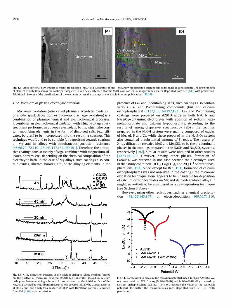
Fig. 12. Cross-sectional SEM images of micro-arc oxidized (MAO) Mg substrates: initial (left) and with deposited calcium orthophosphate coatings (right). The line scanningof element distribution across the coatings is depicted. It can be clearly seen that the MAO layer consists of magnesium silicates. Reprinted from Ref. [143] with permission.Additional pictures of the distributions of the elements across the coatings are available in other publications [57,169].
2930 S.V. Dorozhkin / Acta Biomaterialia 10 (2014) 2919–2934
4.12. Micro-arc or plasma electrolytic oxidation
Micro-arc oxidation (also called plasma electrolytic oxidation,or anodic spark deposition, or micro-arc discharge oxidation) is acombination of plasma-chemical and electrochemical processes.It combines an electrochemical oxidation with a high-voltage sparktreatment performed in aqueous electrolytic baths, which also con-tain modifying elements in the form of dissolved salts (e.g. sili-cates, borates) to be incorporated into the resulting coatings. Thistechnique was found to be suitable for depositing ceramic coatingson Mg and its alloys with simultaneous corrosion resistance[48,66,70–72,110,126,143,147,164,190,191]. Therefore, the protec-tive coatings consist mainly of MgO combined with magnesium sil-icates, borates, etc., depending on the chemical composition of theelectrolytic bath. In the case of Mg alloys, such coatings also con-tain oxides, silicates, borates, etc., of the alloying elements. In the
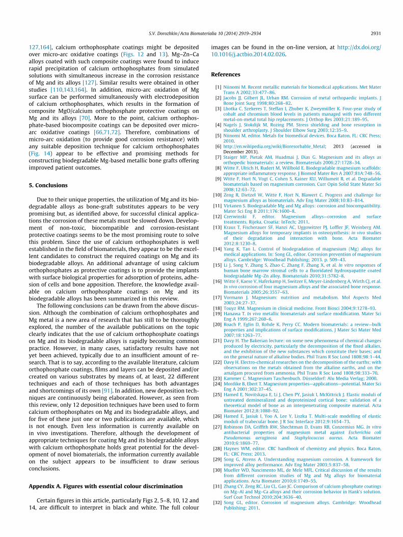
Fig. 13. X-ray diffraction patterns of the calcium orthophosphate coatings formedon the surface of micro-arc oxidized (MAO) Mg substrates soaked in calciumorthophosphate-containing solutions. It can be seen that the initial surface of theMAO Mg covered by MgO (bottom pattern) was covered initially by CDHA (patternsat 20–45 min) and finally by a mixture of CDHA with DCPD (top pattern). Reprintedfrom Ref. [126] with permission.
presence of Ca- and P-containing salts, such coatings also containvarious Ca- and P-containing compounds (but not calciumorthophosphates!) [127,135,169,192,193]. Ca- and P-containingcoatings were prepared on AZ91D alloy in both NaOH- andNa2SiO3-containing electrolytes with addition of sodium hexa-metaphosphate and calcium hypophosphite. According to theresults of energy-dispersive spectroscopy (EDS), the coatingsprepared in the NaOH system were mainly composed of oxidesof Mg, Al, P and Ca, while those prepared in the Na2SiO3 systemalso contained a substantial amount of Si oxide. The results ofX-ray diffraction revealed MgO and Mg2SiO4 to be the predominantphases in the coatings prepared in the NaOH and Na2SiO3 systems,respectively [192]. Similar results were obtained in other studies[127,135,169]. However, among other phases, formation ofCaNaPO4 was detected in one case because the electrolyte usedin that study contained CaCO3, Ca3(PO4)2 and 20 g l�1 of orthophos-phate ions [193]. Since, except for Ref. [193], formation of calciumorthophosphates was not observed in the coatings, the micro-arcoxidation technique alone appears to be unsuitable for depositionof calcium orthophosphates on Mg and its biodegradable alloys. Itmight, nevertheless, be considered as a pre-deposition technique(see Section 3 above).
However, using other techniques, such as chemical precipita-tion [72,126,143,147] or electrodeposition [66,70,71,110,
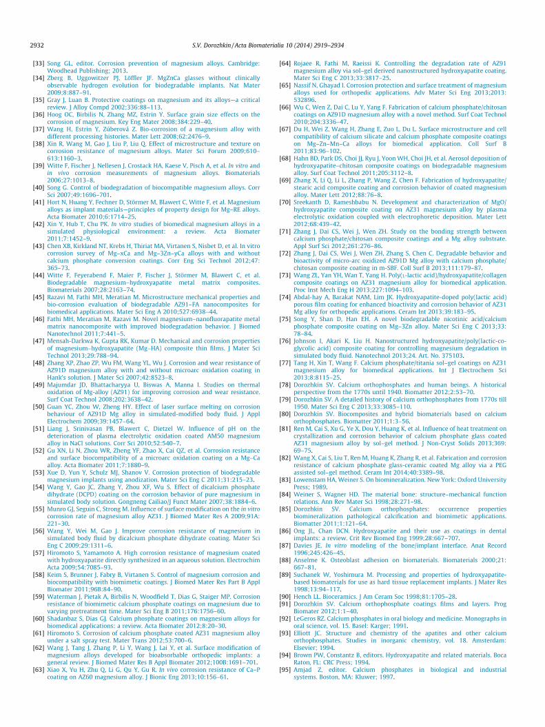
Fig. 14. Tafel curves to measure the corrosion potential in SBF for bare AZ91D alloy,micro-arc oxidized AZ91D alloy (MAO-AZ91D) and MAO-AZ91D alloy covered bycalcium orthophosphate coating. The more positive the value of the corrosionpotential, the better the corrosion resistance. Reprinted from Ref. [71] withpermission.
S.V. Dorozhkin / Acta Biomaterialia 10 (2014) 2919–2934 2931
127,164], calcium orthophosphate coatings might be depositedover micro-arc oxidative coatings (Figs. 12 and 13). Mg–Zn–Caalloys coated with such composite coatings were found to inducerapid precipitation of calcium orthophosphates from simulatedsolutions with simultaneous increase in the corrosion resistanceof Mg and its alloys [127]. Similar results were obtained in otherstudies [110,143,164]. In addition, micro-arc oxidation of Mgsurface can be performed simultaneously with electrodepositionof calcium orthophosphates, which results in the formation ofcomposite MgO/calcium orthophosphate protective coatings onMg and its alloys [70]. More to the point, calcium orthophos-phate-based biocomposite coatings can be deposited over micro-arc oxidative coatings [66,71,72]. Therefore, combinations ofmicro-arc oxidation (to provide good corrosion resistance) withany suitable deposition technique for calcium orthophosphates(Fig. 14) appear to be effective and promising methods forconstructing biodegradable Mg-based metallic bone grafts offeringimproved patient outcomes.
5. Conclusions
Due to their unique properties, the utilization of Mg and its bio-degradable alloys as bone-graft substitutes appears to be verypromising but, as identified above, for successful clinical applica-tions the corrosion of these metals must be slowed down. Develop-ment of non-toxic, biocompatible and corrosion-resistantprotective coatings seems to be the most promising route to solvethis problem. Since the use of calcium orthophosphates is wellestablished in the field of biomaterials, they appear to be the excel-lent candidates to construct the required coatings on Mg and itsbiodegradable alloys. An additional advantage of using calciumorthophosphates as protective coatings is to provide the implantswith surface biological properties for adsorption of proteins, adhe-sion of cells and bone apposition. Therefore, the knowledge avail-able on calcium orthophosphate coatings on Mg and itsbiodegradable alloys has been summarized in this review.
The following conclusions can be drawn from the above discus-sion. Although the combination of calcium orthophosphates andMg metal is a new area of research that has still to be thoroughlyexplored, the number of the available publications on the topicclearly indicates that the use of calcium orthophosphate coatingson Mg and its biodegradable alloys is rapidly becoming commonpractice. However, in many cases, satisfactory results have notyet been achieved, typically due to an insufficient amount of re-search. That is to say, according to the available literature, calciumorthophosphate coatings, films and layers can be deposited and/orcreated on various substrates by means of, at least, 22 differenttechniques and each of those techniques has both advantagesand shortcomings of its own [91]. In addition, new deposition tech-niques are continuously being elaborated. However, as seen fromthis review, only 12 deposition techniques have been used to formcalcium orthophosphates on Mg and its biodegradable alloys, andfor five of these just one or two publications are available, whichis not enough. Even less information is currently available onin vivo investigations. Therefore, although the development ofappropriate techniques for coating Mg and its biodegradable alloyswith calcium orthophosphate holds great potential for the devel-opment of novel biomaterials, the information currently availableon the subject appears to be insufficient to draw seriousconclusions.
Appendix A. Figures with essential colour discrimination
Certain figures in this article, particularly Figs 2, 5–8, 10, 12 and14, are difficult to interpret in black and white. The full colour
images can be found in the on-line version, at http://dx.doi.org/10.1016/j.actbio.2014.02.026.
References
[1] Niinomi M. Recent metallic materials for biomedical applications. Met MaterTrans A 2002;33:477–86.
[2] Jacobs JJ, Gilbert JL, Urban RM. Corrosion of metal orthopaedic implants. JBone Joint Surg 1998;80:268–82.
[3] Lhotka C, Szekeres T, Steffan I, Zhuber K, Zweymüller K. Four-year study ofcobalt and chromium blood levels in patients managed with two differentmetal-on-metal total hip replacements. J Orthop Res 2003;21:189–95.
[4] Nagels J, Stokdijk M, Rozing PM. Stress shielding and bone resorption inshoulder arthroplasty. J Shoulder Elbow Surg 2003;12:35–9.
[5] Niinomi M, editor. Metals for biomedical devices. Boca Raton, FL: CRC Press;2010.
[6] http://en.wikipedia.org/wiki/Bioresorbable_Metal; 2013 (accessed inDecember 2013).
[7] Staiger MP, Pietak AM, Huadmai J, Dias G. Magnesium and its alloys asorthopedic biomaterials: a review. Biomaterials 2006;27:1728–34.
[8] Witte F, Ulrich H, Rudert M, Willbold E. Biodegradable magnesium scaffolds:appropriate inflammatory response. J Biomed Mater Res A 2007;81A:748–56.
[9] Witte F, Hort N, Vogt C, Cohen S, Kainer KU, Willumeit R, et al. Degradablebiomaterials based on magnesium corrosion. Curr Opin Solid State Mater Sci2008;12:63–72.
[10] Zeng R, Dietzel W, Witte F, Hort N, Blawert C. Progress and challenge formagnesium alloys as biomaterials. Adv Eng Mater 2008;10:B3–B14.
[11] Virtanen S. Biodegradable Mg and Mg alloys: corrosion and biocompatibility.Mater Sci Eng B 2011;176:1600–8.
[12] Czerwinski F, editor. Magnesium alloys—corrosion and surfacetreatments. Rijeka, Croatia: InTech; 2011.
[13] Kraus T, Fischerauer SF, Hanzi AC, Uggowitzer PJ, Loffler JF, Weinberg AM.Magnesium alloys for temporary implants in osteosynthesis: in vivo studiesof their degradation and interaction with bone. Acta Biomater2012;8:1230–8.
[14] Yang K, Tan L. Control of biodegradation of magnesium (Mg) alloys formedical applications. In: Song GL, editor. Corrosion prevention of magnesiumalloys. Cambridge: Woodhead Publishing; 2013. p. 509–43.
[15] Li J, Song Y, Zhang S, Zhao C, Zhang F, Zhang X, et al. In vitro responses ofhuman bone marrow stromal cells to a fluoridated hydroxyapatite coatedbiodegradable Mg–Zn alloy. Biomaterials 2010;31:5782–8.
[16] Witte F, Kaese V, Haferkamp H, Switzer E, Meyer-Lindenberg A, Wirth CJ, et al.In vivo corrosion of four magnesium alloys and the associated bone response.Biomaterials 2005;26:3557–63.
[17] Vormann J. Magnesium: nutrition and metabolism. Mol Aspects Med2003;24:27–37.
[18] Touyz RM. Magnesium in clinical medicine. Front Biosci 2004;9:1278–93.[19] Hanawa T. In vivo metallic biomaterials and surface modification. Mater Sci
Eng A 1999;267:260–6.[20] Roach P, Eglin D, Rohde K, Perry CC. Modern biomaterials: a review—bulk
properties and implications of surface modifications. J Mater Sci Mater Med2007;18:1263–77.
[21] Davy H. The Bakerian lecture: on some new phenomena of chemical changesproduced by electricity, particularly the decomposition of the fixed alkalies,and the exhibition of the new substances which constitute their bases; andon the general nature of alkaline bodies. Phil Trans R Soc Lond 1808;98:1–44.
[22] Davy H. Electro-chemical researches on the decomposition of the earths; withobservations on the metals obtained from the alkaline earths, and on theamalgam procured from ammonia. Phil Trans R Soc Lond 1808;98:333–70.
[23] Kammer C. Magnesium Taschenbuch. Düsseldorf: Alu Media Verlag; 2000.[24] Mordike B, Ebert T. Magnesium properties—applications—potential. Mater Sci
Eng A 2001;302:37–45.[25] Hamed E, Novitskaya E, Li J, Chen PY, Jasiuk I, McKittrick J. Elastic moduli of
untreated demineralized and deproteinized cortical bone: validation of atheoretical model of bone as an interpenetrating composite material. ActaBiomater 2012;8:1080–92.
[26] Hamed E, Jasiuk I, Yoo A, Lee Y, Liszka T. Multi-scale modelling of elasticmoduli of trabecular bone. J R Soc Interface 2012;9:1654–73.
[27] Robinson DA, Griffith RW, Shechtman D, Evans RB, Conzemius MG. In vitroantibacterial properties of magnesium metal against Escherichia coliPseudomonas aeruginosa and Staphylococcus aureus. Acta Biomater2010;6:1869–77.
[28] Haynes WM, editor. CRC handbook of chemistry and physics. Boca Raton,FL: CRC Press; 2013.
[29] Song G, Atrens A. Understanding magnesium corrosion. A framework forimproved alloy performance. Adv Eng Mater 2003;5:837–58.
[30] Mueller WD, Nascimento ML, de Mele MFL. Critical discussion of the resultsfrom different corrosion studies of Mg and Mg alloys for biomaterialapplications. Acta Biomater 2010;6:1749–55.
[31] Zhang CY, Zeng RC, Liu CL, Gao JC. Comparison of calcium phosphate coatingson Mg–Al and Mg–Ca alloys and their corrosion behavior in Hank’s solution.Surf Coat Technol 2010;204:3636–40.
[32] Song GL, editor. Corrosion of magnesium alloys. Cambridge: WoodheadPublishing; 2011.
2932 S.V. Dorozhkin / Acta Biomaterialia 10 (2014) 2919–2934
[33] Song GL, editor. Corrosion prevention of magnesium alloys. Cambridge:Woodhead Publishing; 2013.
[34] Zberg B, Uggowitzer PJ, Löffler JF. MgZnCa glasses without clinicallyobservable hydrogen evolution for biodegradable implants. Nat Mater2009;8:887–91.
[35] Gray J, Luan B. Protective coatings on magnesium and its alloys—a criticalreview. J Alloy Compd 2002;336:88–113.
[36] Hoog OC, Birbilis N, Zhang MZ, Estrin Y. Surface grain size effects on thecorrosion of magnesium. Key Eng Mater 2008;384:229–40.
[37] Wang H, Estrin Y, Zúberová Z. Bio-corrosion of a magnesium alloy withdifferent processing histories. Mater Lett 2008;62:2476–9.
[38] Xin R, Wang M, Gao J, Liu P, Liu Q. Effect of microstructure and texture oncorrosion resistance of magnesium alloys. Mater Sci Forum 2009;610–613:1160–3.
[39] Witte F, Fischer J, Nellesen J, Crostack HA, Kaese V, Pisch A, et al. In vitro andin vivo corrosion measurements of magnesium alloys. Biomaterials2006;27:1013–8.
[40] Song G. Control of biodegradation of biocompatible magnesium alloys. CorrSci 2007;49:1696–701.
[41] Hort N, Huang Y, Fechner D, Störmer M, Blawert C, Witte F, et al. Magnesiumalloys as implant materials—principles of property design for Mg–RE alloys.Acta Biomater 2010;6:1714–25.
[42] Xin Y, Hub T, Chu PK. In vitro studies of biomedical magnesium alloys in asimulated physiological environment: a review. Acta Biomater2011;7:1452–9.
[43] Chen XB, Kirkland NT, Krebs H, Thiriat MA, Virtanen S, Nisbet D, et al. In vitrocorrosion survey of Mg–xCa and Mg–3Zn–yCa alloys with and withoutcalcium phosphate conversion coatings. Corr Eng Sci Technol 2012;47:365–73.
[44] Witte F, Feyerabend F, Maier P, Fischer J, Störmer M, Blawert C, et al.Biodegradable magnesium–hydroxyapatite metal matrix composites.Biomaterials 2007;28:2163–74.
[45] Razavi M, Fathi MH, Meratian M. Microstructure mechanical properties andbio-corrosion evaluation of biodegradable AZ91–FA nanocomposites forbiomedical applications. Mater Sci Eng A 2010;527:6938–44.
[46] Fathi MH, Meratian M, Razavi M. Novel magnesium–nanofluorapatite metalmatrix nanocomposite with improved biodegradation behavior. J BiomedNanotechnol 2011;7:441–5.
[47] Mensah-Darkwa K, Gupta RK, Kumar D. Mechanical and corrosion propertiesof magnesium–hydroxyapatite (Mg–HA) composite thin films. J Mater SciTechnol 2013;29:788–94.
[48] Zhang XP, Zhao ZP, Wu FM, Wang YL, Wu J. Corrosion and wear resistance ofAZ91D magnesium alloy with and without microarc oxidation coating inHank’s solution. J Mater Sci 2007;42:8523–8.
[49] Majumdar JD, Bhattacharyya U, Biswas A, Manna I. Studies on thermaloxidation of Mg-alloy (AZ91) for improving corrosion and wear resistance.Surf Coat Technol 2008;202:3638–42.
[50] Guan YC, Zhou W, Zheng HY. Effect of laser surface melting on corrosionbehaviour of AZ91D Mg alloy in simulated-modified body fluid. J ApplElectrochem 2009;39:1457–64.
[51] Liang J, Srinivasan PB, Blawert C, Dietzel W. Influence of pH on thedeterioration of plasma electrolytic oxidation coated AM50 magnesiumalloy in NaCl solutions. Corr Sci 2010;52:540–7.
[52] Gu XN, Li N, Zhou WR, Zheng YF, Zhao X, Cai QZ, et al. Corrosion resistanceand surface biocompatibility of a microarc oxidation coating on a Mg–Caalloy. Acta Biomater 2011;7:1880–9.
[53] Xue D, Yun Y, Schulz MJ, Shanov V. Corrosion protection of biodegradablemagnesium implants using anodization. Mater Sci Eng C 2011;31:215–23.
[54] Wang Y, Gao JC, Zhang Y, Zhou XF, Wu S. Effect of dicalcium phosphatedihydrate (DCPD) coating on the corrosion behavior of pure magnesium insimulated body solution. Gongneng Cailiao/J Funct Mater 2007;38:1884–6.
[55] Munro GJ, Seguin C, Strong M. Influence of surface modification on the in vitrocorrosion rate of magnesium alloy AZ31. J Biomed Mater Res A 2009;91A:221–30.
[56] Wang Y, Wei M, Gao J. Improve corrosion resistance of magnesium insimulated body fluid by dicalcium phosphate dihydrate coating. Mater SciEng C 2009;29:1311–6.
[57] Hiromoto S, Yamamoto A. High corrosion resistance of magnesium coatedwith hydroxyapatite directly synthesized in an aqueous solution. ElectrochimActa 2009;54:7085–93.
[58] Keim S, Brunner J, Fabry B, Virtanen S. Control of magnesium corrosion andbiocompatibility with biomimetic coatings. J Biomed Mater Res Part B ApplBiomater 2011;96B:84–90.
[59] Waterman J, Pietak A, Birbilis N, Woodfield T, Dias G, Staiger MP. Corrosionresistance of biomimetic calcium phosphate coatings on magnesium due tovarying pretreatment time. Mater Sci Eng B 2011;176:1756–60.
[60] Shadanbaz S, Dias GJ. Calcium phosphate coatings on magnesium alloys forbiomedical applications: a review. Acta Biomater 2012;8:20–30.
[61] Hiromoto S. Corrosion of calcium phosphate coated AZ31 magnesium alloyunder a salt spray test. Mater Trans 2012;53:700–6.
[62] Wang J, Tang J, Zhang P, Li Y, Wang J, Lai Y, et al. Surface modification ofmagnesium alloys developed for bioabsorbable orthopedic implants: ageneral review. J Biomed Mater Res B Appl Biomater 2012;100B:1691–701.
[63] Xiao X, Yu H, Zhu Q, Li G, Qu Y, Gu R. In vivo corrosion resistance of Ca–Pcoating on AZ60 magnesium alloy. J Bionic Eng 2013;10:156–61.
[64] Rojaee R, Fathi M, Raeissi K. Controlling the degradation rate of AZ91magnesium alloy via sol–gel derived nanostructured hydroxyapatite coating.Mater Sci Eng C 2013;33:3817–25.
[65] Nassif N, Ghayad I. Corrosion protection and surface treatment of magnesiumalloys used for orthopedic applications. Adv Mater Sci Eng 2013;2013:532896.
[66] Wu C, Wen Z, Dai C, Lu Y, Yang F. Fabrication of calcium phosphate/chitosancoatings on AZ91D magnesium alloy with a novel method. Surf Coat Technol2010;204:3336–47.
[67] Du H, Wei Z, Wang H, Zhang E, Zuo L, Du L. Surface microstructure and cellcompatibility of calcium silicate and calcium phosphate composite coatingson Mg–Zn–Mn–Ca alloys for biomedical application. Coll Surf B2011;83:96–102.
[68] Hahn BD, Park DS, Choi JJ, Ryu J, Yoon WH, Choi JH, et al. Aerosol deposition ofhydroxyapatite–chitosan composite coatings on biodegradable magnesiumalloy. Surf Coat Technol 2011;205:3112–8.
[69] Zhang X, Li Q, Li L, Zhang P, Wang Z, Chen F. Fabrication of hydroxyapatite/stearic acid composite coating and corrosion behavior of coated magnesiumalloy. Mater Lett 2012;88:76–8.
[70] Sreekanth D, Rameshbabu N. Development and characterization of MgO/hydroxyapatite composite coating on AZ31 magnesium alloy by plasmaelectrolytic oxidation coupled with electrophoretic deposition. Mater Lett2012;68:439–42.
[71] Zhang J, Dai CS, Wei J, Wen ZH. Study on the bonding strength betweencalcium phosphate/chitosan composite coatings and a Mg alloy substrate.Appl Surf Sci 2012;261:276–86.
[72] Zhang J, Dai CS, Wei J, Wen ZH, Zhang S, Chen C. Degradable behavior andbioactivity of micro-arc oxidized AZ91D Mg alloy with calcium phosphate/chitosan composite coating in m-SBF. Coll Surf B 2013;111:179–87.
[73] Wang ZL, Yan YH, Wan T, Yang H. Poly(L-lactic acid)/hydroxyapatite/collagencomposite coatings on AZ31 magnesium alloy for biomedical application.Proc Inst Mech Eng H 2013;227:1094–103.
[74] Abdal-hay A, Barakat NAM, Lim JK. Hydroxyapatite-doped poly(lactic acid)porous film coating for enhanced bioactivity and corrosion behavior of AZ31Mg alloy for orthopedic applications. Ceram Int 2013;39:183–95.
[75] Song Y, Shan D, Han EH. A novel biodegradable nicotinic acid/calciumphosphate composite coating on Mg–3Zn alloy. Mater Sci Eng C 2013;33:78–84.
[76] Johnson I, Akari K, Liu H. Nanostructured hydroxyapatite/poly(lactic-co-glycolic acid) composite coating for controlling magnesium degradation insimulated body fluid. Nanotechnol 2013;24. Art. No. 375103.
[77] Tang H, Xin T, Wang F. Calcium phosphate/titania sol–gel coatings on AZ31magnesium alloy for biomedical applications. Int J Electrochem Sci2013;8:8115–25.
[78] Dorozhkin SV. Calcium orthophosphates and human beings. A historicalperspective from the 1770s until 1940. Biomatter 2012;2:53–70.
[79] Dorozhkin SV. A detailed history of calcium orthophosphates from 1770s till1950. Mater Sci Eng C 2013;33:3085–110.
[80] Dorozhkin SV. Biocomposites and hybrid biomaterials based on calciumorthophosphates. Biomatter 2011;1:3–56.
[81] Ren M, Cai S, Xu G, Ye X, Dou Y, Huang K, et al. Influence of heat treatment oncrystallization and corrosion behavior of calcium phosphate glass coatedAZ31 magnesium alloy by sol–gel method. J Non-Cryst Solids 2013;369:69–75.
[82] Wang X, Cai S, Liu T, Ren M, Huang K, Zhang R, et al. Fabrication and corrosionresistance of calcium phosphate glass-ceramic coated Mg alloy via a PEGassisted sol–gel method. Ceram Int 2014;40:3389–98.
[83] Lowenstam HA, Weiner S. On biomineralization. New York: Oxford UniversityPress; 1989.
[84] Weiner S, Wagner HD. The material bone: structure–mechanical functionrelations. Ann Rev Mater Sci 1998;28:271–98.
[85] Dorozhkin SV. Calcium orthophosphates: occurrence propertiesbiomineralization pathological calcification and biomimetic applications.Biomatter 2011;1:121–64.
[86] Ong JL, Chan DCN. Hydroxyapatite and their use as coatings in dentalimplants: a review. Crit Rev Biomed Eng 1999;28:667–707.
[87] Davies JE. In vitro modeling of the bone/implant interface. Anat Record1996;245:426–45.
[88] Anselme K. Osteoblast adhesion on biomaterials. Biomaterials 2000;21:667–81.
[89] Suchanek W, Yoshimura M. Processing and properties of hydroxyapatite-based biomaterials for use as hard tissue replacement implants. J Mater Res1998;13:94–117.
[90] Hench LL. Bioceramics. J Am Ceram Soc 1998;81:1705–28.[91] Dorozhkin SV. Calcium orthophosphate coatings films and layers. Prog
Biomater 2012;1:1–40.[92] LeGeros RZ. Calcium phosphates in oral biology and medicine. Monographs in
oral science, vol. 15. Basel: Karger; 1991.[93] Elliott JC. Structure and chemistry of the apatites and other calcium
orthophosphates. Studies in inorganic chemistry, vol. 18. Amsterdam:Elsevier; 1994.
[94] Brown PW, Constantz B, editors. Hydroxyapatite and related materials. BocaRaton, FL: CRC Press; 1994.
[95] Amjad Z, editor. Calcium phosphates in biological and industrialsystems. Boston, MA: Kluwer; 1997.
S.V. Dorozhkin / Acta Biomaterialia 10 (2014) 2919–2934 2933
[96] Chow LC, Eanes ED, editors. Octacalcium phosphate. Monographs in oralscience, Vol. 18. Basel: Karger; 2001.
[97] Dorozhkin SV. Calcium orthophosphates: applications in nature biology andmedicine. Singapore: Pan Stanford; 2012.
[98] Xu L, Pan F, Yu G, Yang L, Zhang E, Yang K. In vitro and in vivo evaluation of thesurface bioactivity of a calcium phosphate coated magnesium alloy.Biomaterials 2009;30:1512–23.
[99] Wen C, Guan S, Peng L, Ren C, Wang X, Hu Z. Characterization anddegradation behavior of AZ31 alloy surface modified by bone-likehydroxyapatite for implant applications. Appl Surf Sci 2009;255:6433–8.
[100] Wang H, Guan S, Wang X, Ren C, Wang L. In vitro degradation and mechanicalintegrity of Mg–Zn–Ca alloy coated with Ca-deficient hydroxyapatite by thepulse electrodeposition process. Acta Biomater 2010;6:1743–8.
[101] Wang H, Guan S, Wang Y, Liu H, Wang H, Wang L, et al. In vivo degradationbehavior of Ca-deficient hydroxyapatite coated Mg–Zn–Ca alloy for boneimplant application. Coll Surf B 2011;88:254–9.
[102] Tomozawa M, Hiromoto S, Harada Y. Microstructure of hydroxyapatite-coated magnesium prepared in aqueous solution. Surf Coat Technol2010;204:3243–7.
[103] Hiromoto S, Tomozawa M. Corrosion behavior of magnesium withhydroxyapatite coatings formed by hydrothermal treatment. Mater Trans2010;51:2080–7.
[104] Tomozawa M, Hiromoto S. Growth mechanism of hydroxyapatite-coatingsformed on pure magnesium and corrosion behavior of the coatedmagnesium. Appl Surf Sci 2011;257:8253–7.
[105] Tomozawa M, Hiromoto S. Microstructure corrosion resistance and adhesivestrength of calcium phosphate coatings formed on pure magnesium by asimple immersion method. ESC Trans 2011;33:31–7.
[106] Hiromoto S, Tomozawa M. Hydroxyapatite coating of AZ31 magnesium alloyby a solution treatment and its corrosion behavior in NaCl solution. Surf CoatTechnol 2011;205:4711–9.
[107] Tomozawa M, Hiromoto S. Microstructure of hydroxyapatite- andoctacalcium phosphate-coatings formed on magnesium by a hydrothermaltreatment at various pH values. Acta Mater 2011;59:355–63.
[108] Hiromoto S, Tomozawa M, Maruyama N. Fatigue property of a bioabsorbablemagnesium alloy with a hydroxyapatite coating formed by a chemicalsolution deposition. J Mech Behav Biomed Mater 2013;25:1–10.
[109] Ohtsu N, Hiromoto S, Yamane M, Satoh K, Tomozawa M. Chemical andcrystallographic characterizations of hydroxyapatite- and octacalciumphosphate-coatings on magnesium synthesized by chemical solutiondeposition using XPS and XRD. Surf Coat Technol 2013;218:114–8.
[110] Rojaee R, Fathi M, Raeissi K. Electrophoretic deposition of nanostructuredhydroxyapatite coating on AZ91 magnesium alloy implants with differentsurface treatments. Appl Surf Sci 2013;285:664–73.
[111] Xiao X, Zhu QS, Su YC, Li GY. In vitro degradation and biocompatibility of Ca–Pcoated magnesium alloy. Chem Res Chin Univ 2013;29:285–9.
[112] Noorakma ACW, Zuhailawati H, Aishvarya V, Dhindaw BK. Hydroxyapatite-coated magnesium-based biodegradable alloy: cold spray deposition andsimulated body fluid studies. J Mater Eng Perform 2013;22:2997–3004.
[113] Yang JX, Jiao YP, Cui FZ, Lee IS, Yin QS, Zhang Y. Modification of degradationbehavior of magnesium alloy by IBAD coating of calcium phosphate. Surf CoatTechnol 2008;202:5733–6.
[114] Hiromoto S, Shishido T, Yamamoto A, Maruyama N, Somekawa H, Mukai T.Precipitation control of calcium phosphate on pure magnesium byanodization. Corr Sci 2008;50:2906–13.
[115] Yanovska A, Kuznetsov V, Stanislavov A, Danilchenko S, Sukhodub L.Calcium–phosphate coatings obtained biomimetically on magnesiumsubstrates under low magnetic field. Appl Surf Sci 2012;258:8577–84.
[116] Jo JH, Kang BG, Shin KS, Kim HE, Hahn BD, Park DS, et al. Hydroxyapatitecoating on magnesium with MgF2 interlayer for enhanced corrosionresistance and biocompatibility. J Mater Sci Mater Med 2011;22:2437–47.
[117] Jo JH, Li Y, Kim SM, Kim HE, Koh YH. Hydroxyapatite/poly(e-caprolactone)double coating on magnesium for enhanced corrosion resistance and coatingflexibility. J Biomater Appl 2013;28:617–25.
[118] Kannan MB. Improving the packing density of calcium phosphate coating ona magnesium alloy for enhanced degradation resistance. J Biomed Mater ResA 2013;101A:1248–54.
[119] Cui W, Beniash E, Gawalt E, Xu Z, Sfeir C. Biomimetic coating of magnesiumalloy for enhanced corrosion resistance and calcium phosphate deposition.Acta Biomater 2013;9:8650–9.
[120] Geng F, Tan LL, Jin XX, Yang JY, Yang K. The preparation cytocompatibility andin vitro biodegradation study of pure b-TCP on magnesium. J Mater Sci MaterMed 2009;20:1149–57.
[121] Xu L, Zhang E, Yang K. Phosphating treatment and corrosion properties ofMg–Mn–Zn alloy for biomedical application. J Mater Sci Mater Med2009;20:859–67.
[122] Hu J, Wang C, Ren W, Zhang S, Liu F. Microstructure evolution and corrosionmechanism of dicalcium phosphate dihydrate coating on magnesium alloy insimulated body fluid. Mater Chem Phys 2010;119:294–8.
[123] Onoki T, Yamamoto SY. Hydroxyapatite ceramics coating on magnesiumalloy via a double layered capsule hydrothermal hot-pressing. J Ceram SocJpn 2010;118:749–52.
[124] Song Y, Zhang S, Li J, Zhao C, Zhang X. Electrodeposition of Ca–P coatings onbiodegradable Mg alloy: in vitro biomineralization behavior. Acta Biomater2010;6:1736–42.
[125] Wang Q, Tan L, Xu W, Zhang B, Yang K. Dynamic behaviors of a Ca–P coatedAZ31B magnesium alloy during in vitro and in vivo degradations. Mater SciEng B 2011;176:1718–26.
[126] Liu GY, Hu J, Ding ZK, Wang C. Formation mechanism of calcium phosphatecoating on micro-arc oxidized magnesium. Mater Chem Phys 2011;130:1118–24.
[127] Gao JH, Guan SK, Chen J, Wang LG, Zhu SJ, Hu JH, et al. Fabrication andcharacterization of rod-like nano-hydroxyapatite on MAO coating supportedon Mg–Zn–Ca alloy. Appl Surf Sci 2011;257:2231–7.
[128] Chai H, Guo L, Wang X, Gao X, Liu K, Fu Y, et al. In vitro and in vivo evaluationson osteogenesis and biodegradability of a b-tricalcium phosphate coatedmagnesium alloy. J Biomed Mater Res A 2012;100A:293–304.
[129] Guan RG, Johnson I, Cui T, Zhao T, Zhao ZY, Li X, et al. Electrodeposition ofhydroxyapatite coating on Mg–4.0Zn–1.0Ca–0.6Zr alloy and in vitroevaluation of degradation hemolysis and cytotoxicity. J Biomed Mater ResA 2012;100A:999–1015.
[130] Xu LP, Zhang EL, Yang K. Biocorrosion property and cytocompatibility ofcalcium phosphate coated Mg alloy. Trans Nonferrous Met Soc China2012;22:2014–20.
[131] Li KK, Wang B, Yan B, Lu W. Microstructure in vitro corrosion and cytotoxicityof Ca–P coatings on ZK60 magnesium alloy prepared by simple chemicalconversion and heat treatment. J Biomater Appl 2012;28:375–84.
[132] Li KK, Wang B, Yan B, Lu W. Preparing Ca–P coating on biodegradablemagnesium alloy by hydrothermal method: in vitro degradation behavior.Chin Sci Bull 2012;57:2319–22.
[133] Iskandar ME, Aslani A, Liu H. The effects of nanostructured hydroxyapatitecoating on the biodegradation and cytocompatibility of magnesium implants.J Biomed Mater Res A 2013;101A:2340–54.
[134] Grubac Z, Metikoš-Hukovic M, Babic R. Electrocrystallization growth andcharacterization of calciumphosphate ceramics on magnesium alloys.Electrochim Acta 2013;109:694–700.
[135] Gan J, Tan L, Yang K, Hu Z, Zhang Q, Fan X, et al. Bioactive Ca–P coating withself-sealing structure on pure magnesium. J Mater Sci Mater Med2013;24:889–901.
[136] Zhang Y, Ma Y, Chen M, Wei J. Effects of anodizing biodegradable Mg–Zn–Zralloy on the deposition of Ca–P coating. Surf Coat Technol 2013;228:S111–5.
[137] Liu GY, Tang SW, Wang C, Hu J. Li DC Formation characteristic of Ca�Pcoatings on magnesium alloy surface. Trans Nonferrous Met Soc China2013;23:2294–9.
[138] Kim SM, Jo JH, Lee SM, Kang MH, Kim HE, Estrin Y, et al. Hydroxyapatite-coated magnesium implants with improved in vitro and in vivo biocorrosionbiocompatibility and bone response. J Biomed Mater Res A 2014;102A:429–41.
[139] Meng EC, Guan SK, Wang HX, Wang LG, Zhu SJ, Hu JH, et al. Effect ofelectrodeposition modes on surface characteristics and corrosion propertiesof fluorine-doped hydroxyapatite coatings on Mg–Zn–Ca alloy. Appl Surf Sci2011;257:4811–6.
[140] Munro JG, Strong M. The mechanism of deposition of calcium phosphatecoatings from solution onto magnesium alloy AZ31. J Biomed Mater Res A2009;90A:339–50.
[141] Zhang Y, Zhang G, Wei M. Controlling the biodegradation rate of magnesiumusing biomimetic apatite coating. J Biomed Mater Res B Appl Biomater2008;89B:408–14.
[142] Song Y, Shan D, Han E. Electrodeposition of hydroxyapatite coating on AZ91Dmagnesium alloy for biomaterial application. Mater Lett 2008;62:3276–9.
[143] Liu GY, Hu J, Ding ZK, Wang C. Bioactive calcium phosphate coating formedon micro-arc oxidized magnesium by chemical deposition. Appl Surf Sci2011;257:2051–7.
[144] Chen XB, Birbilis N, Abbott TB. A simple route towards a hydroxyapatite–Mg(OH)2 conversion coating for magnesium. Corr Sci 2011;53:2263–8.
[145] Chen XB, Birbilis N, Abbott TB. Effect of [Ca2+] and [PO43�] levels on the
formation of calcium phosphate conversion coatings on die-cast magnesiumalloy AZ91D. Corr Sci 2012;55:226–32.
[146] Santhanakrishnan S, Ho YH, Dahotre NB. Laser coating of hydroxyapatite onMg for enhanced physiological corrosion resistance and biodegradability.Mater Technol 2012;27:273–7.
[147] Liu G, Tang S, Li D, Hu J. Self-adjustment of calcium phosphate coating onmicro-arc oxidized magnesium and its influence on the corrosion behaviourin simulated body fluids. Corr Sci 2014;79:206–14.
[148] Kannan MB, Orr L. In vitro mechanical integrity of hydroxyapatite coatedmagnesium alloy. Biomed Mater 2011;6:045003.
[149] Lalk M, Reifenrath J, Angrisani N, Bondarenko A, Seitz JM, Mueller PP, et al.Fluoride and calcium-phosphate coated sponges of the magnesium alloyAX30 as bone grafts: a comparative study in rabbits. J Mater Sci Mater Med2013;24:417–36.
[150] Waterman J, Birbilis N, Dias GJ, Woodfield TBF, Staiger MP. Improving in vitrocorrosion resistance of biomimetic calcium phosphate coatings for Mgsubstrates using calcium hydroxide layer. Corr Eng Sci Technol2012;47:340–5.
[151] Su Y, Li G, Lian J. A chemical conversion hydroxyapatite coating on AZ60magnesium alloy and its electrochemical corrosion behavior. Int JElectrochem Sci 2012;7:11497–511.
[152] Jamesh M, Kumar S, Narayanan TSNS. Electrodeposition of hydroxyapatitecoating on magnesium for biomedical applications. J Coat Technol Res2012;9:495–502.
2934 S.V. Dorozhkin / Acta Biomaterialia 10 (2014) 2919–2934
[153] Huse EC. A new ligature? Chicago Med J Exam 1878;37:171–2.[154] Duncan A Sen, Duncan A Jun. Annals of medicine, for the year 1797.
Exhibiting a concise view of the latest and most important discoveries inmedicine and medical philosophy, vol. II. Edinburgh/London: G. Mudie & Son/G.G. & J. Robinson; 1991. p. 397–8.
[155] Cortés DA, López HY, Mantovani D. Spontaneous and biomimetic apatiteformation on pure magnesium. Mater Sci Forum 2007;539–543:589–94.
[156] Kuwahara H, Al-Abdullat Y, Ohta M, Tsutsumi S, Ikeuchi K, Mazaki N, et al.Surface reaction of magnesium in Hank’s solution. Mater Sci Forum2000;350:349–58.
[157] Rettig R, Virtanen S. Time-dependent electrochemical characterization of thecorrosion of a magnesium rare-earth alloy in simulated body fluids. J BiomedMater Res A 2008;85A:167–75.
[158] Rettig R, Virtanen S. Composition of corrosion layers on a magnesium rare-earth alloy in simulated body fluids. J Biomed Mater Res A 2009;88A:359–69.
[159] Lorenz C, Brunner JG, Kollmannsberger P, Jaafar L, Fabry B, Virtanen S. Effectof surface pre-treatments on biocompatibility of magnesium. Acta Biomater2009;5:2783–9.
[160] Yang JX, Jiao YP, Yin QS, Zhang Y, Zhang T. Calcium phosphate coating onmagnesium alloy by biomimetic method: investigation of morphologycomposition and formation process. Front Mater Sci Chin 2008;2:149–55.
[161] Yang JX, Cui FZ, Lee IS, Zhang Y, Yin QS, Xia H, et al. In vivo biocompatibilityand degradation behavior of Mg alloy coated by calcium phosphate in arabbit model. J Biomater Appl 2011;27:153–64.
[162] Kang MH, Jung HD, Kim SW, Lee SM, Kim HE, Estrin Y, et al. Production andbio-corrosion resistance of porous magnesium with hydroxyapatite coatingfor biomedical applications. Mater Lett 2013;108:122–4.
[163] Yang J, Cui F, Yin Q, Zhang Y, Zhang T, Wang X. Characterization anddegradation study of calcium phosphate coating on magnesium alloy boneimplant. IEEE Trans Plasma Sci 2009;37:1161–8.
[164] Shi Y, Qi M, Chen Y, Shi P. MAO–DCPD composite coating on Mg alloy fordegradable implant applications. Mater Lett 2011;65:2201–4.
[165] Zong Y, Niu JL, Mao L, Zhang XB, Yuan GY, Ding WJ. Improvement of corrosionperformance in simulated body fluid solution of Mg–Nd–Zn–Zr alloy coatedwith hydroxyapatite by pulse eletrodeposition. Corr Prot 2011;32:430–7.
[166] Smith MR, Atkinson P, White D, Piersma T, Gutierrez G, Rossini G,et al. Design and assessment of a wrapped cylindrical Ca–P AZ31 Mgalloy for critical-size ulna defect repair. J Biomed Mater Res B2012;100B:206–16.
[167] Choudhary L, Raman RKS, Nie JF. In vitro evaluation of degradation of acalcium phosphate coating on a Mg–Zn–Ca alloy in a physiologicalenvironment. Corrosion 2012;68:499–506.
[168] Kannan MB. Enhancing the performance of calcium phosphate coatingon a magnesium alloy for bioimplant applications. Mater Lett 2012;76:109–12.
[169] Pan YK, Chen CZ, Wang DG, Zhao TG. Effects of phosphates on microstructureand bioactivity of micro-arc oxidized calcium phosphate coatings on Mg–Zn–Zr magnesium alloy. Coll Surf B 2013;109:1–9.
[170] Xin Y, Hu T, Chu PK. Influence of test solutions on in vitro studies ofbiomedical magnesium alloys. J Electrochem Soc 2010;157:C238–43.
[171] Habibovic P, Barrere F, Blitterswijk C, Groot K, Layrolle P. Biomimetichydroxyapatite coating on metal implants. J Am Ceram Soc 2002;85:517–22.
[172] Liu Y, de Groot K, Hunziker E. Biomimetic mineral coatings in dental andorthopaedic implantology. Front Mater Sci Chin 2009;3:154–62.
[173] Bigi A, Falini G, Foresti E, Ripamonti A, Gazzano M, Roveri N. Magnesiuminfluence on hydroxyapatite crystallization. J Inorg Biochem 1993;49:69–78.
[174] Xu CY, Xu XL, Gao F, Chu FX, Wang Q, Guo QN. The crystal orientation ofhydroxyapatite coatings affected by the magnetic fields on magnesium alloy.Appl Mech Mater 2013;395–396:751–4.
[175] Cui F, Yang J, Jiao Y, Yin Q, Zhang Y, Lee I. Calcium phosphate coating onmagnesium alloy for modification of degradation behavior. Front Mater SciChin 2008;2:143–8.
[176] Galio A, Lamaka S, Zheludkevich M, Dick L, Mller I, Ferreira M. Inhibitor-doped sol–gel coatings for corrosion protection of magnesium alloy AZ31.Surf Coat Technol 2010;204:1479–86.
[177] Feil F, Frbeth W, Schtze M. Purely inorganic coatings based on nanoparticlesfor magnesium alloys. Electrochim Acta 2009;54:2478–86.
[178] Tan A, Soutar A, Annergren I, Liu Y. Multilayer sol–gel coatings for corrosionprotection of magnesium. Surf Coat Technol 2005;198:478–82.
[179] Hu J, Li Q, Zhong X, Zhang L, Chen B. Composite anticorrosion coatings forAZ91D magnesium alloy with molybdate conversion coating and silicon sol–gel coatings. Prog Org Coat 2009;66:199–205.
[180] Roy A, Singh SS, Datta MK, Lee B, Ohodnicki J, Kumta PN. Novel sol–gelderived calcium phosphate coatings on Mg4Y alloy. Mater Sci Eng B2011;176:1679–89.
[181] Tang H, Xin TZ, Luo Y, Wang FP. In vitro degradation of AZ31 magnesium alloycoated with hydroxyapatite by sol–gel method. Mater Sci Technol 2013;29:547–52.
[182] Lamaka S, Montemor M, Galio A, Zheludkevich M, Trindade C, Dick L, et al.Novel hybrid sol–gel coatings for corrosion protection of AZ31B magnesiumalloy. Electrochim Acta 2008;53:4773–83.
[183] Qu H, Wei M. Improvement of bonding strength between biomimetic apatitecoating and substrate. J Biomed Mater Res B Appl Biomater 2008;84B:436–43.
[184] Salman SA, Kuroda K, Okido M. Preparation and characterization ofhydroxyapatite coating on AZ31 Mg alloy for implant applications. BioinorgChem Appl 2013;2013:175756.
[185] Metikoš-Hukovic M, Babic R, Grubac Z, Petravic M, Peter R. Potential assistedformation and characterization of hydroxyapatite coatings on biodegradablemagnesium alloys. J Electrochem Soc 2013;160:H674–80.
[186] Lu W, Ou C, Zhan Z, Huang P, Yan B, Chen M. Microstructure and in vitrocorrosion properties of ZK60 magnesium alloy coated with calciumphosphate by electrodeposition at different temperatures. Int J ElectrochemSci 2013;8:10746–57.
[187] Srinivasan P, Liang J, Blawert C, Stormer M, Dietzel W. Characterization ofcalcium containing plasma electrolytic oxidation coatings on AM50magnesium alloy. Appl Surf Sci 2010;256:4017–22.
[188] Zhang S, Li J, Song Y, Zhao C, Zhang X. Influence of dicalcium phosphatedihydrate coating on the in vitro degradation of Mg–Zn alloy. Front Mater SciChin 2010;4:116–9.
[189] Zhang C. Preparation of calcium phosphate coatings on Mg–1.0 Ca alloy.Trans Nonferrous Met Soc China 2010;20:s655–9.
[190] Wang YM, Wang FH, Xu MJ, Zhao B, Guo LX, Ouyang JH. Microstructure andcorrosion behavior of coated AZ91 alloy by microarc oxidation for biomedicalapplication. Appl Surf Sci 2009;255:9124–31.
[191] Shi P, Ng WF, Wong MH, Cheng FT. Improvement of corrosion resistance ofpure magnesium in Hanks’ solution by microarc oxidation with sol–gel TiO2
sealing. J Alloys Compd 2009;469:286–92.[192] Yao Z, Li L, Jiang Z. Adjustment of the ratio of Ca/P in the ceramic coating on
Mg alloy by plasma electrolytic oxidation. Appl Surf Sci 2009;255:6724–8.[193] Ma C, Zhang X, Qu L, Li M. Calcium and phosphate biocoatings on magnesium
alloy fabricated by micro-arc oxidation. Adv Mater Res 2010;105–106:565–8.


