Embed Size (px)
Citation preview

Author contributions: F.P.: Conception and design, Collection and/or assembly of data, Data analysis and interpretation, Manuscript writing, Financial support; H.Y.: Collection and/or assembly of data, Data analysis and interpretation; A.J.: Collection and/or assembly of data; W.v.I.: Collection and/or assembly of data; F.G.: Financial support; M.R.: Conception and design, Data analysis and interpretation, Manuscript writing, Financial support., Formatted for Stem Cells; Price et al., (2012) 4. Correspondence should be addressed to M.A.R., Tel: (613) 739-6740, Fax: (613) 739-6294, E-mail: [email protected]; Received June 25, 2012; accepted for publication December 09, 2012; available online without subscription through the open access option. 1066-5099/2013/$30.00/0 doi: 10.1002/stem.1321 This article has been accepted for publication and undergone full peer review but has not been through the copyediting, typesetting, pagination and proofreading process which may lead to differences between this version and the Version of Record. Please cite this article as doi: 10.1002/stem.1321
STEM CELLS®
EMBRYONIC STEM CELLS/INDUCED PLURIPOTENT STEM CELLS Canonical Wnt Signaling Induces a Primitive Endoderm Metastable State in Mouse Embryonic Stem Cells Feodor D. Price1,2, Hang Yin1,2, Andrew Jones1,2, Wilfred van Ijcken3, Frank Grosveld3 and Michael A. Rudnicki1,2,4 1. Sprott Center For Stem Cell Research, Ottawa Hospital Research Institute, Regenerative Medicine Program, 501 Smyth Road, Ottawa, ON, Canada, K1H 8L6; 2. University of Ottawa, Cellular and Molecular Medicine, Faculty of Medicine, 501 Smyth Road, Ottawa, ON, Canada, K1H 8L6; 3. ERASMUS Center for Biomics, 3015GE Rotterdam, The Netherlands Key words. Canonical Wnt Signaling • Primitive Endoderm • Metastable • Mouse Embryonic Stem Cells ABSTRACT Activation of the canonical Wnt signaling pathway synergizes with leukemia inhibitory factor (LIF) to maintain pluripotency of mouse embryonic stem cells (mESCs). However, in the absence of LIF, Wnt signaling is unable to maintain ES cells in the undifferentiated state. To investigate the role of canonical Wnt signaling in pluripotency and lineage specification, we expressed Wnt3a in mESCs and characterized them in growth and differentiation. We found that activated canonical Wnt signaling induced the formation of a reversible metastable primitive endoderm state in mESC. Upon subsequent
differentiation, Wnt3a stimulated mESCs gave rise to large quantities of visceral endoderm. Furthermore, we determined that the ability of canonical Wnt signaling to induce a metastable primitive endoderm state was mediated by Tbx3. Our data demonstrates a specific role for canonical Wnt signaling in promoting pluripotency while at the same time priming cells for subsequent differentiation into the primitive endoderm lineage.
INTRODUCTION Embryonic stem cells (ESCs) are pluripotent stem cells derived from the inner cell mass (ICM) of a pre-implantation blastocyst and thus exhibit characteristic gene expression profiles and functional properties [1]. ESCs retain the capacity to repopulate an embryo by contributing to all somatic cell lineages, including the germline [2]. For these reasons ESCs are characterized as naive pluripotent cells.
Recent evidence implies ESCs are heterogeneous and consist of subpopulations with distinct functional and epigenetic states. These metastable subpopulations share similarities to three different transient cell populations in the mouse embryo: the ICM [3-5], the epiblast cells of post-implantation embryos [6] and the primitive endoderm [7]. Moreover, a particular metastable state is maintained in a dynamic equilibrium allowing for the inter-conversion between the ground state and a lineage primed

Wnt3a induces a metastable state in mES cells
2
state. The molecular mechanisms that direct the generation of specific ESC metastable states have not been defined. LIF/Stat-3 signaling is the predominant pathway involved in the maintenance of the pluripotent ground state in mESCs [8]. Currently the signaling pathways that regulate the epiblast and primitive endoderm metastable states remain largely unknown. The Wnt family of secreted glycoproteins plays diverse and critical roles in the proper specification and formation of organs and tissues [9, 10]. Previous experiments implicate Wnt signaling in the maintenance of potency for both embryonic and adult stem cells [9, 11-13]. Furthermore, numerous studies demonstrate that activating canonical Wnt signaling promotes pluripotency in both mouse and human ESCs [14, 15]. Under self-renewing conditions, Wnts synergize with LIF to maintain pluripotency [8, 16]. In addition, core pluripotency factors (Oct4, Sox2 and Nanog) are known to be regulated by canonical Wnt signaling [17-19]. In fact Tcf3 (Tcf7L1), a key downstream effector of β-catenin mediated canonical Wnt signaling, binds to the promoters of Oct4, Sox2, and Nanog and limits their expression [20, 21]. Consistent with the ability of Tcf3 to repress pluripotency, Tcf3-/-
mouse ESCs are delayed in their differentiation as embryoid bodies and are able to self-renew even upon LIF withdrawal [22]. The canonical Wnt pathway is regulated by the expression level and subcellular localization of β-catenin. Intracellular β-catenin is phosphorylated by GSK3-β when bound in a complex with Axin and adenomatous polyposis coli (APC). Phosphorylated β-catenin is subsequently degraded by the proteasome [11]. Upon binding of Wnt to Frizzled and Lrp5/6 cell surface receptors, an intracellular cascade in which Dishevelled (Dvl) inhibits the phosphorylation of β-catenin by GSK3-β is activated [23, 24]. Cytoplasmic β-catenin is stabilized and translocates into the nucleus to serve as a co-regulator for Tcf/Lef transcription
factors, which in turn bind to target genes to modulate their expression [25]. Numerous experiments have identified Wnt3a as a canonical Wnt and demonstrate that it can functionally affect pluripotency [26-28]. In embryonic development, the earliest activation of Wnt/β-catenin targets occurs in the visceral endoderm as evidenced by Tcf based reporters [29]. Many components of the Wnt signaling pathway are expressed in the blastocyst and in the uterine wall prior to implantation [30-32]. Although the AxinLacZ reporter is activated in the epiblast and primitive endoderm of the developing blastocyst [28, 33], opposing views persist regarding whether the Wnt/β-catenin pathway plays a functional role within the pre-implantation blastocyst [34-36]. The ability of Wnts to maintain pluripotency in mESCs is dependent on the LIF pathway as constitutively active β-catenin alone is unable to maintain self-renewal [16]. This implies that activated canonical Wnt signaling is insufficient to maintain the pluripotent state of embryonic stem cells. This further raises the issue of whether canonical Wnt signaling has other functions in undifferentiated mESCs. We investigated how activated canonical Wnt signaling regulates mESC pluripotency and cell fate commitment. We determined that activation of canonical Wnt signaling reinforces the undifferentiated state by promoting pluripotency genes while at the same time activating a subset of primitive endoderm genes. Notably, canonical Wnt signaling induces a metastable ESC state in which primitive endoderm genes are up regulated. This subtype of primitive endoderm ESC can interconvert between a Nanog-GFP high population as well as a Nanog-GFP low population. The induction of primitive endoderm genes in the undifferentiated state comes at the expense of the neuroectodermal and mesendodermal potential. Wnt3a-expressing ESCs display a predisposition for the primitive endoderm lineage and as a result display pronounced visceral endoderm potential

Wnt3a induces a metastable state in mES cells
3
following differentiation. We further found that induction of the primitive endoderm metastable state, following activation of canonical Wnt signaling, is mediated by the T-box transcription factor Tbx3. Together, our experiments reveal important insights into the early regulation of ESC pluripotency and commitment by canonical Wnt signaling.
MATERIALS AND METHODS Cell lines and Cell culture The following mouse ESC lines D3 Gl, J1, R1, and Millitrace Nanog GFP were maintained on a layer of irradiated MEFs (DR4) in GMEM high glucose medium (Wisent) supplemented with 15% fetal bovine serum (Hyclone), 1x non essential amino acids, 1x sodium pyruvate, 1x Glutamax, β-Mercaptoethanol, 1x Pen/Strep (Invitrogen), and 1000U ESGRO (Chemicon) at 37oC and 5% CO2. Wnt3a retroviral cell lines used throughout the differentiation time course were made from each of the three ES cell lines (D3, J1 and R1). Prior to differentiation mESCs were cultured for two passages on 0.2% gelatin. For embryoid body formation, mESCs were seeded in 20ul hanging drops at a density of 4x10⁴ cells/ml in ESC medium without LIF. Embryoid bodies were transferred to petri dishes on Day 3 of differentiation and cultured in suspension until Day 5 at which time they were transferred to gelatine coated culture dishes for the remainder of differentiation. Plat-E cells, a derivative of HEK293T cells were used to generate retrovirus. Plat-E cells were maintained in DMEM high glucose medium supplemented with 10% fetal bovine serum (Hyclone), 1x Pen/Strep, 1ug/ml Puromycin and 10ug/ml Blastocydin at 37oC and 5% CO2. 80% confluent Plat-E cells were transfected using Lipofectamine 2000 (Invitrogen) according to the manufacturer’s directions. Cells were fed with fresh DMEM 6hrs post transfection. 48hrs post tranfection retroviral supernatants were collected, centrifuged to remove cells and filtered (0.45uM) for purification. The proliferation assay was conducted by plating 200,000 cells per well of a 6-well dish.
Following each passage the cells were counted and re-plated at a density of 200,000 and the fold increase was multiplied to give rise to a total cell number amount. Recombinant Wnt3a was obtained from RnD systems (1324-WN). (2’Z,3’E)-6-Bromoindirubin-3′-oxime (BIO) reconstituted in DMSO at a concentration of 5mM (Sigma B1686). Immunostaining Embryoid bodies at 5 days of differentiation were briefly rinsed in PBS and subsequently frozen in a 2:1 ratio of Tissue-Tek OCT to 30% sucrose in a dry ice/ethanol bath. Serial cryosections 12um in thickness were cut using a cryostat (Leicia CM 1850). Sections were fixed in 4% PFA for 10min at room temperature followed by incubation in 100mM Glycine for 10min at room temperature. Fixed cells were permeabilized and blocked in 5% FBS, 0.2% Triton X-100 (Sigma) and 2% bovine serum albumin (Fisher Scientific) for 1hr at room temperature. Cells were incubated with primary antibodies (View Table SI) for 1 hour at room temperature or alternatively 4oC overnight. Secondary antibodies were diluted 1:1000 in PBS and incubated with fixed cells for 1hr at room temperature. Nuclei were counter-stained with Hoechst 33342 (Sigma). Images were taken on a Carl Zeiss Axioplan 2 Microscope with an AxioCam HRm b/w (Zeiss) camera using Axiovision v3.2 (Zeiss) acquisition software. Photoshop was used to adjust the baseline fluorescence. Western Blot Total protein was harvested in RIPA lysis buffer, subjected to SDSPAGE, and electroblotted onto Immobilon-P membranes (Millipore). Membranes were probed with primary antibody and HRP-conjugated secondary antibody in blocking solution. ECL (Amersham-Pharmacia) with Biomax XAR film (Kodak) was used to detect target proteins. Primary antibodies used for detection are found in Table SI. The secondary consisted of an HRP conjugated anti-mouse antibody (Biorad).

Wnt3a induces a metastable state in mES cells
4
siRNA knockdown of Tbx3 siRNA for Tbx3 was obtained from Ambion (s74777-4390771) and transfected into Nanog-GFP mES cellss using lipofectamine RNAiMax (Invitrogen) according to the manufacturer’s instructions. Fresh medium was added 6 hours post transfection and the cells were cultured an additional 48hrs prior to either RNA collection or FACS analysis. Fluorescent Activated Cell Sorting (FACS) Prior to FACSs sorting Nanog-GFP reporter ESCs were trypsinized, pelleted, and filtered through 30uM filters (Miltenyi Biotech). Cells were separated on a MoFlo cytometer (DakoCytomation) equipped with 3 lasers. Sorting gates were strictly defined based on both Side Scatter (SSC) vs GFP as well as PE vs GFP plots. Wild type J1 ESCs were used as a negative control for GFP. Gene expression analysis (qPCR) Total RNA was isolated and subjected to on column DNase digestion using an RNeasy mini Kit as per the manufacturer’s instructions (Qiagen). 1ug of RNA was reverse transcribed using iScript RT kit (Biorad) and subsequently diluted 1:5, with 1ul of template used per qPCR reaction. SYBR Green real-time PCR reactions (Biorad) were performed in duplicate using an MX3000p PCR machine (Stratagene, La Jolla, CA) and 40 cycles of amplification (95oC 10s; 60oC 30s; 72oC 30s). PCR primers for real-time PCR were designed using the online Primer3 software (http://frodo.wi.mit.edu/primer3/) with gene specific information obtained from Ensembl. All primer sequences are provided in Table SII. Relative fold change in expression was normalized to GAPDH and calculated using the ΔΔCT method (CT values < 30). Primer specificity was validated by denaturation curve analysis (55–94 °C) resulting in a single peak. Amplification-curve plotting and calculation of cycle threshold (Ct) values were performed using the MX3000p software (v3; Stratagene), with further calculations performed using Microsoft Excel. Each experiment was
performed independently on at least three occasions. Gene expression analysis (RNA-seq) Global gene expression was assessed in retroviral and recombinant Wnt3a cell lines relative to mock infected and bovine serum albumin (BSA) treated controls respectively. Total RNA was isolated and subjected to on column DNase digestion using an RNeasy mini Kit as per the manufacturer’s instructions (Qiagen). mRNA-Seq was performed at the Erasmus Center for Biomics (Rotterdam, The Netherlands) as per manufacturers instructions (Illumina). Raw Solexa reads were mapped to the mouse genome (mm9) by Tophat [37]. Gene expression values were calculated by Cufflinks with default parameters [38]. Differentially expressed genes between Wnt3a treated samples and corresponding mock or BSA controls were called by Cuffdiff [38-40]. Heatmaps were generated by GenePattern (http://www.broadinstitute.org/cancer/software/genepattern). Gene ontology of expression data was done using the functional annotation module of DAVID 6.7 (http://david.abcc.ncifcrf.gov) (Dennis et al., 2003; Huang da et al., 2009). Statistical analysis Statistical analysis was conducted using the Student’s t-test method of determining inference based on small samples. Two tailed paired t-tests were conducted between treated and control groups at a given day of differentiation or globally for all treated or control samples over the course of differentiation. * indicates a p value of < 0.05 while ** indicates a p value of < 0.01.
RESULTS Activated canonical Wnt signaling maintains pluripotency but primes cells for primitive endodermal commitment To investigate the regulation of ESC pluripotency and early lineage specification by canonical Wnt signaling, we generated mESCs

Wnt3a induces a metastable state in mES cells
5
lines that constitutively express Wnt3a. Mouse ESCs were infected with a self-inactivating retrovirus containing an EF1α promoter to drive the expression of Wnt3a followed by an IRES-Puro for selection. This approach ensures constant and maintained activation of canonical Wnt signaling in a cell-autonomous manner. Elevated levels of Wnt3a mRNA with respect to endogenous Wnt3a expression in control cell lines illustrate the effective and sustained over expression of Wnt3a in mESCs (Figure 1A). As previously described [41, 42], we observed a significant increase in the rate of proliferation, percentage of Ki67+ cells, along with a compacted colony morphology in Wnt3a-expressing mESCs (Figure 1B, Figure S1 and Figure 1C). To gain insight into how Wnt3a affects pluripotency we conducted RNA-seq from total mRNA isolated from cells either overexpressing Wnt3a or treated with 250ng/ml recombinant Wnt3a protein for 3 passages. We confirmed RNA-seq read quality and the distribution of reads prior to further gene analysis (Figure S2). In addition, the transcriptional activation of canonical Wnt targets was confirmed in recombinant and retroviral cell lines (Figure S3). To investigate the global transcriptional effects of canonical Wnt activation in mES cells we conducted an unbiased DAVID analysis of our RNA-seq data throughout a 10 day differentiation time course (Figure S4). Significant enrichment of GO terms representing Chordate embryonic development and embryonic development ending in birth or egg hatching was observed. To determine specifically which cellular lineages – either blastocyst or germ layer derived – were being affected by Wnt3a we choose to focus on panels of known markers for each lineage. Transcription factors (TFs) act to define cell-type specific lineage identity. Therefore, we compiled panels of known TFs along with additional gene markers specific for a given cellular lineage [43-45]. We focused on early
commitment and the lineages involved in the formation of the blastocyst (Trophoblast, Primitive Endoderm and Epiblast), and the primary germ layers (Ectoderm, Mesoderm, Endoderm). In this manner, we could determine the broad effects of Wnt3a on pluripotency and lineage induction. mRNA levels of the key pluripotency factor Nanog, a known target of canonical Wnt signaling in mESCs, and Klf4 were increased following Wnt3a induction [46]. By contrast, the majority of the other pluripotency markers remained either unchanged, or in the case of Dppa3 (Stella), were reduced (Figure 1D and 1E). Our RNA-seq and qPCR results for the trophectoderm panel of genes displayed mixed induction and repression upon Wnt3a over-expression in the undifferentiated state. Notably, expression levels of the core transcription factors that define the primitive endodermal lineage displayed a striking increase in the undifferentiated state both for RNA-seq and qPCR (Gata4 - 6 fold, Gata6 - 5 fold, Sox17 - 20 fold and Sox7 - 5 fold) (Figure 1D and 1E). Activation of the Wnt signaling pathway in mESC results in the reduced expression of neural ectoderm genes [47-51]. Consistent with these reports, our data shows that Pax6, an early marker for neural development, exhibited a 70% reduction in expression in Wnt3a treated mESC (Figure 1D and 1E). Furthermore, all 13 ectodermal markers examined by RNA-seq displayed reduced or unchanged expression levels in Wnt3a expressing cells. The majority of endodermal factors were similarly down regulated or unchanged (Figure 1D and E). Mesodermal genes displayed weak induction in undifferentiated cells. In concordance with our RNA-seq results, qPCR validation of RNA levels for core lineage markers followed similar patterns (Figure 1E). The effect of recombinant Wnt3a on mESC was similar yet subdued in comparison to retroviral over expression of Wnt3a. These changes include the induction of core pluripotency and

Wnt3a induces a metastable state in mES cells
6
primitive endoderm genes in the undifferentiated state and promotion of visceral endoderm following differentiation (Figure S5). To further support this claim we compared the number of genes per lineage panel that undergo an increase in expression following either recombinant or retroviral Wnt3a treatment. This analysis demonstrates that indeed recombinant Wnt3a treatment results in a similar pattern of gene induction during differentiation when compared to retroviral expression of Wnt3a for a given panel of genes (Figure S5B).Therefore, we conclude that Wnt3a alters the transcriptional program of ESCs to both promote pluripotency and prime cells for entry into the primitive endoderm lineage. Canonical Wnt signaling perturbs the pluripotent ground state of mESCs by inducing a primitive endoderm metastable state Our gene expression analysis indicated that activated canonical Wnt signaling could induce primitive endoderm genes in mESCs maintained in the undifferentiated state. Recent evidence suggests that pluripotent mESCs can exist in multiple metastable states that are characterized by distinct transcriptional profiles and functional characteristics akin to the epiblast, ICM and the primitive endoderm [3, 6, 7]. These metastable states are maintained in proliferating mESC cultures in the presence of LIF and can be distinguished based on the expression of Nanog [3], Rex1 [5], Stella [6], Hex [7] or Pecam1 [4]. The developmental mechanisms that direct mESCs to form a specific metastable state are currently unknown. Typically, mESCs display high and low Nanog-GFP populations in the undifferentiated state, which can interconvert and are defined as metastable states [3]. Therefore, to investigate whether canonical Wnt signaling induces a metastable mESC state, we analyzed the expression of GFP from a Nanog-GFP reporter cell line following retroviral infection of Wnt3a or empty vectors (Figure 2A). As noted previously, 93.1% of mESC infected with mock
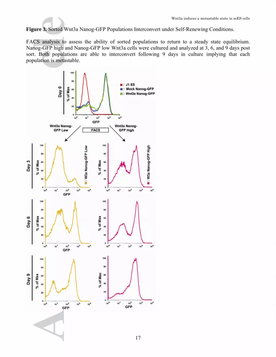
virus displayed high Nanog-GFP expression (Figure 2B). Importantly, following infection with Wnt3a virus we observed an almost 10-fold expansion in the size of the Nanog-GFP low population (Figure 2B). To characterize the identity of the different Nanog-GFP populations (high/low), they were FACS sorted and the expression of lineage markers analyzed by qPCR. The Nanog-GFP low population displayed markedly upregulated levels of primitive endoderm genes Gata4, Gata6, Sox17, Sox7, Cubn and Dab2 relative to the Nanog-GFP high population (Figure 2C). However, these same cells exhibited maintenance of expression of pluripotency gene such as Oct4 and Nanog. Therefore, the observed up regulation in primitive endoderm genes together with the maintenance of pluripotency gene expression suggests that these cells are in a lineage primed state [52]. Previous reports indicate that the activation of Sox17 in the primitive endoderm commits ESCs to terminal differentiation [53]. As Sox17 is up regulated in the Nanog-GFP low population, we investigated whether these cells were irreversibly committed to differentiation. Sorted Wnt3a Nanog-GFP high or low cells were cultured in the undifferentiated state for 9 days to determine their ability to interconvert. Nanog-GFP high cells maintain GFP expression while rapidly regenerating a GFP low population (Figure 3). We next tested whether the Wnt3a Nanog-GFP low cells could reactivate high levels of Nanog expression as evidenced by GFP. Two days post-sorting of Wnt3a induced Nanog-GFP low cells; we observed the formation of colonies exhibiting heterogeneous GFP expression (Figure S4). FACS analysis of cells after 3,6, and 9 days of culture revealed a return to the original distribution of Nanog-GFP expression (Figure 3 and Figure S4). These results indicate an equilibrium exists between the primitive endoderm and ICM metastable states and that this equilibrium can be modified but not exclusively restricted by Wnt3a. Taken together, these results strongly support the hypothesis that

Wnt3a induces a metastable state in mES cells
7
activated canonical Wnt signaling induces a primitive endoderm metastable state. Canonical Wnt signaling alters the differentiation potential of mESCs To investigate whether the induction of a metastable primitive endoderm state functionally altered the lineage potential of mESCs we conducted a differentiation time course spanning a mid differentiation time point (Day 5) and a late stage differentiation time point (Day 10) (Figure 4A). We observed a striking phenotype when Wnt3a-expressing mESCs were differentiated as embryoid bodies. These changes include an increase in embryoid body size relative to mock-infected control cells and the development of large cysts after 6 days of differentiation (Figure 4B and C). We further characterized the affect of Wnt3a on the differentiation ability of mESCs by conducting RNA-seq from cells isolated after 5 and 10 days of differentiation. Pluripotency markers increased following Wnt3a induced differentiation at Day 5 (8/10) and were subsequently strongly down regulated by Day 10 (8/10). Primitive endoderm markers displayed broad induction throughout the differentiation time course (Day 5 – 6/10 and Day 10 – 8/10). Trophectoderm markers demonstrated mixed induction and inhibition after 5 days of differentiation (4/11) and further up regulation by 10 days (8/11) of differentiation. The vast majority of ectodermal markers 8/13 (Day 5), and 11/13 (Day 10) displayed reduced or unchanged expression levels in Wnt3a expressing cells. Endoderm markers as a whole were all either down regulated or unchanged following differentiation (10/10 Day 5, 9/10 Day 10). Mesodermal genes displayed weak induction during early differentiation (4/10), while on day 10 core mesodermal genes T, Mixl1, and Nodal were all strongly inhibited (>3 fold down regulation). In concordance with our RNA-seq results, qPCR validation of RNA levels for core lineage markers followed similar patterns (Figure 4E).
Taken together, these results support the notion that canonical Wnt signaling in the absence of LIF promotes the formation of cystic embryoid bodies and alters the gene expression profile to match differentiation into the primitive endoderm lineage. Furthermore these results suggest that priming of primitive endoderm genes in the undifferentiated state functionally alters the differentiation potential of mESCs. Sustained activation of canonical Wnt signaling directs visceral endoderm commitment During blastocyst development the primitive endoderm is localized to a layer of cells that cover the epiblast and separate it from the blastocoel cavity. Following blastocyst implantation, the primitive endoderm will give rise to both the parietal endoderm to line the outer trophectoderm and the visceral endoderm to cover the epiblast [54]. Since activated canonical Wnt signaling induces up regulation of primitive endoderm genes and the formation of cystic EBs, we next tested whether Wnt3a promotes visceral endoderm. To assess visceral endoderm in our cultures, we examined the expression levels of genes enriched in visceral endoderm at the RNA and protein level. Primary visceral endoderm cell surface proteins Amn, Cubn and ApoE are all significantly up regulated based on our RNA-seq results (Figure 5A) as well as by qPCR (Figure 5B). Dab2 a cell membrane adapter protein along with the transcription factors Sox7, Hnf4a, Hnf1b, and Foxa2 are all up regulated upon Wnt3a induced differentiation. We confirmed the induction of visceral endoderm markers by immunofluorescence in differentiated embryoid bodies. Following 5 days of differentiation we observed an increase in the prevalence of Dab2, Gata4 and Sox17 proteins within EBs following Wnt3a over-expression (Figure 5C). This increase in the prevalence of EBs that express Gata4, Sox17 and Dab2 was quantified (Figure 5D) and further validated by western blot analysis (Figure 5E).

Wnt3a induces a metastable state in mES cells
8
Additional data demonstrating an increase in the prevalence of Cubn positive EBs is shown in Figure S8. Following continued Wnt3a expression the gene expression profile and protein complement of differentiating mESCs is enriched for primitive and visceral endoderm genes. Taken together these results indicate that upon differentiation the expression of Wnt3a directs commitment towards the visceral endoderm lineage. Activated canonical Wnt signaling acts through Tbx3 to induce a primitive endoderm metastable state Canonical Wnt signaling typically involves β-catenin binding with a Tcf/Lef family transcription factor on the promoter of Wnt target genes. To identify specific genes that are targeted by Wnt3a in undifferentiated mESCs we further analyzed our RNA-seq gene profiles obtained following retroviral and recombinant treatment. In this fashion we would gain mechanistic insight into how Wnt3a activates primitive endoderm genes. We identified genes that were both up regulated or down regulated in unison by either Wnt3a retroviral overexpression and recombinant Wnt3a treatment. Based on this, we identified three transcription factors Tbx3, Pou2f3, and Cdx1 that were significantly up regulated upon Wnt3a overexpression or recombinant Wnt3a treatment (Figure 6A). Up-regulation of these genes was subsequently confirmed by qPCR in ESCs overexpressing Wnt3a (Figure 6B). Of these three genes, Tbx3 has recently been shown to play a role in pluripotency and is also involved in inhibiting ectoderm and trophectoderm while promoting primitive endoderm formation [55]. Taking into account these findings we used siRNA directed against Tbx3 to determine whether it is required for Wnt3a induction of primitive endoderm genes and subsequently the Nanog-GFP low population. Indeed, sorted Wnt3a Nanog-GFP low cells that typically express enhanced Tbx3 demonstrate a reduction in Gata6 expression
levels following treatment with siTbx3 (Figure 6C). Furthermore, Nanog-GFP cells transfected with scrambled siRNA displayed a Nanog-GFP low population whereas the mock infected Nanog-GFP cells did not (Figure 6D). Upon knockdown of Tbx3, the percentage of Wnt3a Nanog-GFP low cells underwent a 25±2.7% decrease. Conversely when Tbx3 was knocked down we observed a 56±4.1% increase in the percentage of Nanog-GFP high cells (Figure 6E). Therefore, Tbx3 plays an important role in mediating Wnt3a induction of the primitive endoderm metastable state.
DISCUSSION Embryonic stem cells were initially viewed as a homogeneous pluripotent population that has the capacity to give rise to all cell types of the embryo proper. This simplistic view has recently been challenged following the identification of distinct metastable states that exist in equilibrium within the undifferentiated state. Metastability confers to ESCs the unique ability to interconvert between two distinct cellular states. The first state is fully pluripotent whereas the metastable state is lineage primed but reversible. Cells that reside within a given metastable state display a unique epigenetic signature and are primed for differentiation into a specific lineage [6]. However, the developmental signals and the molecular mechanisms that govern the formation and maintenance of metastable states have not been previously defined. Here we demonstrate that induction of the canonical Wnt signaling pathway in mESCs induces a pluripotent primitive endodermal metastable state. Moreover, we found that canonical Wnt signaling primes undifferentiated mESC for differentiation into the primitive endoderm lineage. Indeed, this early priming into the primitive endoderm culminates in a profound enrichment of visceral endoderm at the expense of other germ layer lineages in differentiating mESC cultures. Furthermore, we found that induction of a primitive endoderm

Wnt3a induces a metastable state in mES cells
9
metastable state following activation of the canonical Wnt signaling pathway is mediated by the T-box transcription factor Tbx3 (Figure 7). Thus, these results provide a mechanistic framework that explains how activated canonical Wnt signaling can promote visceral endoderm development. Three metastable states have been defined in mESC. The first is an ICM state characterized by cells that express Stella, Rex1, Nanog, and Pecam1. The second state is similar to the post-implantation epiblast (EpiSC) state and is defined by induction of Fgf5 and Brachyury. The third and most recently identified is a primitive endoderm-like metastable state characterised by the expression of the primitive endoderm marker Hex[7]. This state was first identified based on its low expression of Nanog and high expression of the primitive endoderm marker, Gata6 [46]. Further studies using Rex1, Hex and Sox17 support the idea for a primed primitive endoderm state in mESCs, yet differences exist regarding the ability of this population to reversibly convert back into the naive pluripotent state [5, 7, 53]. Our results demonstrate that activation of canonical Wnt signaling is capable of inducing a primed primitive endodermal metastable state in mESCs. Wnt3a primes mESCs for primitive endoderm specification (Figure 1D and E) and gives rise to a Nanog-GFP low population that up regulates expression of primitive endoderm genes (Figure 2B and C). Importantly, this population is capable of interconverting back to Nanog-GFP high cells following FACS isolation and in vitro culture (Figure 3A and B). These abilities support the characterization of this pluripotent primitive endoderm population as being metastable. Furthermore, lineage priming into the primitive endoderm is apparent as robust differentiation into visceral endoderm occurs in the absence of LIF (Figure 4B-E). Forced expression of Gata4 and Gata6 has been previously demonstrated to direct primitive endoderm differentiation in mESCs [56, 57].
Interestingly, we found induction of primitive endodermal genes such as Gata6, Gata4, Sox7 and Sox17 in both the undifferentiated and differentiated states. In addition, we observed a broad increase in primitive endoderm markers following Wnt3a induction of the canonical Wnt signaling pathway. Therefore, although Wnt3a treated mESCs remain pluripotent; they become primed for entry into the primitive endoderm lineage. This is an important distinction, as our experiments support the notion that Wnt3a promotes and simultaneously maintain two functionally discrete cells types. In embryonic development and in vitro differentiation of mESCs, canonical Wnts are associated with the induction of brachyury (T) and the mesendodermal lineage [27, 47, 58]. Interestingly, prior to gastrulation, the earliest activation of downstream target genes of canonical Wnt signaling occur in the nascent visceral endoderm [29]. Notably, mESCs expressing mutant activated β-catenin display an enlarged visceral endoderm and preferentially contribute to the visceral endoderm along with other extra-embryonic tissues [36]. Conversely, mice deficient for β-catenin lack appropriate organisation of the visceral endoderm resulting in defective anterior-posterior axis formation. In vitro, mESCs with an inactive form of adenomatous polyposis coli (APC) (ApcMin/Min), a key regulator of β-catenin degradation, display increased levels of visceral endoderm upon differentiation [15, 59]. Our experiments elucidate these results by demonstrating that activated canonical Wnt signaling induces a primitive endoderm metastable state in undifferentiated mESCs. The mechanisms involved in the induction and maintenance of metastable states remain poorly understood. Of several conserved genes up regulated upon recombinant Wnt3a treatment or retroviral overexpression, Tbx3 was chosen due to its expression in the undifferentiated state and for its role in specification of primitive endoderm. Recent studies have shown that Tbx3 along with Klf4 are able to promote pluripotency

Wnt3a induces a metastable state in mES cells
10
in the absence of LIF [60]. Tbx3 was previously identified as a downstream target of canonical Wnt signaling in murine and human liver tumours suggesting a direct mechanism exists for canonical Wnts to activate Tbx3 [61]. Tbx3 is required for extra-embryonic endoderm (ExEn) commitment and also functions to suppress trophectoderm and ectoderm differentiation in mESCs [55]. Overexpression of Tbx3 in mESCs maintained in the presence of LIF results in the induction of Gata6, Sox17, and Dab2 suggesting that the primary role of Tbx3 is to induce the primitive endoderm. Thus, our finding that Tbx3 is required for Wnt3a to induce the primitive endoderm state supports the hypothesis that Tbx3 is a canonical-Wnt target in mESCs.
SUMMARY In conclusion, our data reveals that activation of the canonical Wnt pathway by Wnt3a results in the induction of a metastable population of undifferentiated cells with characteristics of the primitive endoderm. Moreover, our results elucidate a mechanism mediated by Tbx3 in which activation of the canonical Wnt signaling pathway in undifferentiated mESC alters the gene expression profile and distribution of metastable states to promote a primitive endoderm metastable state. Indeed, stimulation of canonical Wnt signaling biases differentiation into the visceral endoderm lineage at the expense of ectoderm, mesoderm and endoderm. Thus, our
experiments indicate that induction of metastable states is subject to developmental control pathways acting during embryogenesis. Together, these findings provide important insight into the regulation of lineage specification and commitment during the development of the early embryo.
ACKNOWLEDGMENTS We thank Dr. James Ellis for the HSC1-eGFP and HSC1-eGFP IRES Puro vectors. David Wilson for help and technical advice regarding tissue sectioning. Paul Oleynik for cell sorting. M.A.R. holds the Canada Research Chair in Molecular Genetics and is an International Research Scholar of the Howard Hughes Medical Institute. This work was funded by the Canadian Institutes of Health Research grant 160266 and Ontario Research Fund grants to M.A.R., and by EuTRACC, a European Commission 6th Framework grant to F.G. F.D.P was supported by scholarships from the Canadian Stem Cell Network, the Muscular Dystrophy Society of Canada and a Doctoral Research Award from the Canadian Institutes of Health Research. Disclosure of Potential Conflicts of Interest The authors indicate no potential conflicts of interest.
REFERENCES 1. M.J. Evans, M.H. Kaufman, Establishment in culture of
pluripotential cells from mouse embryos, Nature, 292 (1981) 154-156.
2. A. Bradley, M. Evans, M.H. Kaufman, E. Robertson, Formation of germ-line chimaeras from embryo-derived teratocarcinoma cell lines, Nature, 309 (1984) 255-256.
3. I. Chambers, J. Silva, D. Colby, J. Nichols, B. Nijmeijer, M. Robertson, J. Vrana, K. Jones, L. Grotewold, A. Smith, Nanog safeguards pluripotency and mediates germline development, Nature, 450 (2007) 1230-1234.
4. T. Furusawa, K. Ohkoshi, C. Honda, S. Takahashi, T. Tokunaga, Embryonic stem cells expressing both platelet endothelial cell adhesion molecule-1 and stage-specific embryonic antigen-1 differentiate predominantly into epiblast cells in a chimeric embryo, Biol Reprod, 70 (2004) 1452-1457.
5. Y. Toyooka, D. Shimosato, K. Murakami, K. Takahashi, H. Niwa, Identification and characterization of subpopulations in undifferentiated ES cell culture, Development, 135 (2008) 909-918.
6. K. Hayashi, S.M. Lopes, F. Tang, M.A. Surani, Dynamic equilibrium and heterogeneity of mouse pluripotent stem cells with distinct functional and epigenetic states, Cell Stem Cell, 3 (2008) 391-401.
7. M.A. Canham, A.A. Sharov, M.S. Ko, J.M. Brickman, Functional heterogeneity of embryonic stem cells

Wnt3a induces a metastable state in mES cells
11
revealed through translational amplification of an early endodermal transcript, PLoS Biol, 8 (2010) e1000379.
8. A.G. Smith, Embryo-derived stem cells: of mice and men, Annu Rev Cell Dev Biol, 17 (2001) 435-462.
9. H. Clevers, Wnt/beta-catenin signaling in development and disease, Cell, 127 (2006) 469-480.
10. C.P. Petersen, P.W. Reddien, Wnt signaling and the polarity of the primary body axis, Cell, 139 (2009) 1056-1068.
11. C.Y. Logan, R. Nusse, The Wnt signaling pathway in development and disease, Annu Rev Cell Dev Biol, 20 (2004) 781-810.
12. T. Reya, H. Clevers, Wnt signalling in stem cells and cancer, Nature, 434 (2005) 843-850.
13. H.E. Fleming, V. Janzen, C. Lo Celso, J. Guo, K.M. Leahy, H.M. Kronenberg, D.T. Scadden, Wnt signaling in the niche enforces hematopoietic stem cell quiescence and is necessary to preserve self-renewal in vivo, Cell Stem Cell, 2 (2008) 274-283.
14. N. Sato, L. Meijer, L. Skaltsounis, P. Greengard, A.H. Brivanlou, Maintenance of pluripotency in human and mouse embryonic stem cells through activation of Wnt signaling by a pharmacological GSK-3-specific inhibitor, Nat Med, 10 (2004) 55-63.
15. M.F. Kielman, M. Rindapaa, C. Gaspar, N. van Poppel, C. Breukel, S. van Leeuwen, M.M. Taketo, S. Roberts, R. Smits, R. Fodde, Apc modulates embryonic stem-cell differentiation by controlling the dosage of beta-catenin signaling, Nat Genet, 32 (2002) 594-605.
16. K. Ogawa, R. Nishinakamura, Y. Iwamatsu, D. Shimosato, H. Niwa, Synergistic action of Wnt and LIF in maintaining pluripotency of mouse ES cells, Biochem Biophys Res Commun, 343 (2006) 159-166.
17. J. Hao, T.G. Li, X. Qi, D.F. Zhao, G.Q. Zhao, WNT/beta-catenin pathway up-regulates Stat3 and converges on LIF to prevent differentiation of mouse embryonic stem cells, Dev Biol, 290 (2006) 81-91.
18. M.P. Storm, H.K. Bone, C.G. Beck, P.Y. Bourillot, V. Schreiber, T. Damiano, A. Nelson, P. Savatier, M.J. Welham, Regulation of Nanog expression by phosphoinositide 3-kinase-dependent signaling in murine embryonic stem cells, J Biol Chem, 282 (2007) 6265-6273.
19. Y. Takao, T. Yokota, H. Koide, Beta-catenin up-regulates Nanog expression through interaction with Oct-3/4 in embryonic stem cells, Biochem Biophys Res Commun, 353 (2007) 699-705.
20. M.F. Cole, S.E. Johnstone, J.J. Newman, M.H. Kagey, R.A. Young, Tcf3 is an integral component of the core regulatory circuitry of embryonic stem cells, Genes Dev, 22 (2008) 746-755.
21. W.L. Tam, C.Y. Lim, J. Han, J. Zhang, Y.S. Ang, H.H. Ng, H. Yang, B. Lim, T-cell factor 3 regulates embryonic stem cell pluripotency and self-renewal by the transcriptional control of multiple lineage pathways, Stem Cells, 26 (2008) 2019-2031.
22. F. Yi, L. Pereira, B.J. Merrill, Tcf3 functions as a steady-state limiter of transcriptional programs of
mouse embryonic stem cell self-renewal, Stem Cells, 26 (2008) 1951-1960.
23. S. Angers, R.T. Moon, Proximal events in Wnt signal transduction, Nat Rev Mol Cell Biol, 10 (2009) 468-477.
24. C. Metcalfe, M. Bienz, Inhibition of GSK3 by Wnt signalling--two contrasting models, J Cell Sci, 124 (2011) 3537-3544.
25. B.T. MacDonald, K. Tamai, X. He, Wnt/beta-catenin signaling: components, mechanisms, and diseases, Dev Cell, 17 (2009) 9-26.
26. C. Kwon, J. Arnold, E.C. Hsiao, M.M. Taketo, B.R. Conklin, D. Srivastava, Canonical Wnt signaling is a positive regulator of mammalian cardiac progenitors, Proc Natl Acad Sci U S A, 104 (2007) 10894-10899.
27. T.P. Yamaguchi, S. Takada, Y. Yoshikawa, N. Wu, A.P. McMahon, T (Brachyury) is a direct target of Wnt3a during paraxial mesoderm specification, Genes Dev, 13 (1999) 3185-3190.
28. D.T. Berge, D. Kurek, T. Blauwkamp, W. Koole, A. Maas, E. Eroglu, R.K. Siu, R. Nusse, Embryonic stem cells require Wnt proteins to prevent differentiation to epiblast stem cells, Nat Cell Biol, 13 (2011) 1070-1075.
29. A. Ferrer-Vaquer, A. Piliszek, G. Tian, R.J. Aho, D. Dufort, A.K. Hadjantonakis, A sensitive and bright single-cell resolution live imaging reporter of Wnt/ss-catenin signaling in the mouse, BMC Dev Biol, 10 (2010) 121.
30. C. Kemp, E. Willems, S. Abdo, L. Lambiv, L. Leyns, Expression of all Wnt genes and their secreted antagonists during mouse blastocyst and postimplantation development, Dev Dyn, 233 (2005) 1064-1075.
31. S. Lloyd, T.P. Fleming, J.E. Collins, Expression of Wnt genes during mouse preimplantation development, Gene Expr Patterns, 3 (2003) 309-312.
32. O.A. Mohamed, D. Dufort, H.J. Clarke, Expression and estradiol regulation of Wnt genes in the mouse blastocyst identify a candidate pathway for embryo-maternal signaling at implantation, Biol Reprod, 71 (2004) 417-424.
33. O.A. Mohamed, H.J. Clarke, D. Dufort, Beta-catenin signaling marks the prospective site of primitive streak formation in the mouse embryo, Dev Dyn, 231 (2004) 416-424.
34. C. Chazaud, J. Rossant, Disruption of early proximodistal patterning and AVE formation in Apc mutants, Development, 133 (2006) 3379-3387.
35. H. Xie, S. Tranguch, X. Jia, H. Zhang, S.K. Das, S.K. Dey, C.J. Kuo, H. Wang, Inactivation of nuclear Wnt-beta-catenin signaling limits blastocyst competency for implantation, Development, 135 (2008) 717-727.
36. R. Kemler, A. Hierholzer, B. Kanzler, S. Kuppig, K. Hansen, M.M. Taketo, W.N. de Vries, B.B. Knowles, D. Solter, Stabilization of beta-catenin in the mouse zygote leads to premature epithelial-mesenchymal

Wnt3a induces a metastable state in mES cells
12
transition in the epiblast, Development, 131 (2004) 5817-5824.
37. C. Trapnell, L. Pachter, S.L. Salzberg, TopHat: discovering splice junctions with RNA-Seq, Bioinformatics, 25 (2009) 1105-1111.
38. C. Trapnell, B.A. Williams, G. Pertea, A. Mortazavi, G. Kwan, M.J. van Baren, S.L. Salzberg, B.J. Wold, L. Pachter, Transcript assembly and quantification by RNA-Seq reveals unannotated transcripts and isoform switching during cell differentiation, Nat Biotechnol, 28 (2010) 511-515.
39. A. Roberts, H. Pimentel, C. Trapnell, L. Pachter, Identification of novel transcripts in annotated genomes using RNA-Seq, Bioinformatics, 27 (2011) 2325-2329.
40. A. Roberts, C. Trapnell, J. Donaghey, J.L. Rinn, L. Pachter, Improving RNA-Seq expression estimates by correcting for fragment bias, Genome Biol, 12 (2011) R22.
41. S. Vijayakumar, G. Liu, I.A. Rus, S. Yao, Y. Chen, G. Akiri, L. Grumolato, S.A. Aaronson, High-frequency canonical Wnt activation in multiple sarcoma subtypes drives proliferation through a TCF/beta-catenin target gene, CDC25A, Cancer Cell, 19 (2011) 601-612.
42. M.S. Yun, S.E. Kim, S.H. Jeon, J.S. Lee, K.Y. Choi, Both ERK and Wnt/beta-catenin pathways are involved in Wnt3a-induced proliferation, J Cell Sci, 118 (2005) 313-322.
43. G. Guo, M. Huss, G.Q. Tong, C. Wang, L. Li Sun, N.D. Clarke, P. Robson, Resolution of cell fate decisions revealed by single-cell gene expression analysis from zygote to blastocyst, Dev Cell, 18 (2010) 675-685.
44. K. Kurimoto, Y. Yabuta, Y. Ohinata, Y. Ono, K.D. Uno, R.G. Yamada, H.R. Ueda, M. Saitou, An improved single-cell cDNA amplification method for efficient high-density oligonucleotide microarray analysis, Nucleic Acids Res, 34 (2006) e42.
45. C.A. Seguin, J.S. Draper, A. Nagy, J. Rossant, Establishment of endoderm progenitors by SOX transcription factor expression in human embryonic stem cells, Cell Stem Cell, 3 (2008) 182-195.
46. A.M. Singh, T. Hamazaki, K.E. Hankowski, N. Terada, A heterogeneous expression pattern for Nanog in embryonic stem cells, Stem Cells, 25 (2007) 2534-2542.
47. P. Gadue, T.L. Huber, P.J. Paddison, G.M. Keller, Wnt and TGF-beta signaling are required for the induction of an in vitro model of primitive streak formation using embryonic stem cells, Proc Natl Acad Sci U S A, 103 (2006) 16806-16811.
48. D. ten Berge, W. Koole, C. Fuerer, M. Fish, E. Eroglu, R. Nusse, Wnt signaling mediates self-organization and axis formation in embryoid bodies, Cell Stem Cell, 3 (2008) 508-518.
49. J. Aubert, H. Dunstan, I. Chambers, A. Smith, Functional gene screening in embryonic stem cells
implicates Wnt antagonism in neural differentiation, Nat Biotechnol, 20 (2002) 1240-1245.
50. L. Haegele, B. Ingold, H. Naumann, G. Tabatabai, B. Ledermann, S. Brandner, Wnt signalling inhibits neural differentiation of embryonic stem cells by controlling bone morphogenetic protein expression, Mol Cell Neurosci, 24 (2003) 696-708.
51. Y. Yoshikawa, T. Fujimori, A.P. McMahon, S. Takada, Evidence that absence of Wnt-3a signaling promotes neuralization instead of paraxial mesoderm development in the mouse, Dev Biol, 183 (1997) 234-242.
52. R. Mansson, A. Hultquist, S. Luc, L. Yang, K. Anderson, S. Kharazi, S. Al-Hashmi, K. Liuba, L. Thoren, J. Adolfsson, N. Buza-Vidas, H. Qian, S. Soneji, T. Enver, M. Sigvardsson, S.E. Jacobsen, Molecular evidence for hierarchical transcriptional lineage priming in fetal and adult stem cells and multipotent progenitors, Immunity, 26 (2007) 407-419.
53. K.K. Niakan, H. Ji, R. Maehr, S.A. Vokes, K.T. Rodolfa, R.I. Sherwood, M. Yamaki, J.T. Dimos, A.E. Chen, D.A. Melton, A.P. McMahon, K. Eggan, Sox17 promotes differentiation in mouse embryonic stem cells by directly regulating extraembryonic gene expression and indirectly antagonizing self-renewal, Genes Dev, 24 (2010) 312-326.
54. M. Bielinska, N. Narita, D.B. Wilson, Distinct roles for visceral endoderm during embryonic mouse development, Int J Dev Biol, 43 (1999) 183-205.
55. R. Lu, A. Yang, Y. Jin, Dual functions of T-box 3 (Tbx3) in the control of self-renewal and extraembryonic endoderm differentiation in mouse embryonic stem cells, J Biol Chem, 286 (2011) 8425-8436.
56. J. Fujikura, E. Yamato, S. Yonemura, K. Hosoda, S. Masui, K. Nakao, J. Miyazaki Ji, H. Niwa, Differentiation of embryonic stem cells is induced by GATA factors, Genes Dev, 16 (2002) 784-789.
57. D. Shimosato, M. Shiki, H. Niwa, Extra-embryonic endoderm cells derived from ES cells induced by GATA factors acquire the character of XEN cells, BMC Dev Biol, 7 (2007) 80.
58. S.J. Arnold, J. Stappert, A. Bauer, A. Kispert, B.G. Herrmann, R. Kemler, Brachyury is a target gene of the Wnt/beta-catenin signaling pathway, Mech Dev, 91 (2000) 249-258.
59. B. Rubinfeld, I. Albert, E. Porfiri, C. Fiol, S. Munemitsu, P. Polakis, Binding of GSK3beta to the APC-beta-catenin complex and regulation of complex assembly, Science, 272 (1996) 1023-1026.
60. H. Niwa, K. Ogawa, D. Shimosato, K. Adachi, A parallel circuit of LIF signalling pathways maintains pluripotency of mouse ES cells, Nature, 460 (2009) 118-122.
61. C.A. Renard, C. Labalette, C. Armengol, D. Cougot, Y. Wei, S. Cairo, P. Pineau, C. Neuveut, A. de Reynies, A. Dejean, C. Perret, M.A. Buendia, Tbx3 is a downstream target of the Wnt/beta-catenin pathway

Wnt3a induces a metastable state in mES cells
13
and a critical mediator of beta-catenin survival functions in liver cancer, Cancer Res, 67 (2007) 901-
910.
See www.StemCells.com for supporting information available online.

Wnt3a induces a metastable state in mES cells
14
Figure 1. Wnt3a Overexpression Elicits Increased Growth Rate and the Induction of Primitive Endoderm and Pluripotency Genes (A) Retrovirally infected mES cells expressing Wnt3a (black) or infected with a mock control (white). (B) Undifferentiated Wnt3a expressing cells proliferate to a greater degree in relation to a mock-infected control. Data are presented as the mean ± SEM from biological triplicate experiments in representative cell lines. (C) Bright field images of undifferentiated control (upper panel) and Wnt3a infected (lower panel) mouse ES cells grown on MEFs in the presence of LIF. 100x magnification, inset 50x magnification, scale bar denotes 100um. (D) Global ratio heat maps display an enrichment in Pluripotent and Primitive Endoderm lineage genes in undifferentiated ES cells overexpressing Wnt3a in relation to mock-infected controls. Genes in red were chosen as positive controls for each lineage. Yellow represents genes that increase in expression relative to the control while blue represents a decrease. (E) QPCR validation was conducted using undifferentiated mES cells expression Wnt3a (black) and Ctrl (white) for members from each lineage. Data are presented as the mean ± SEM of 3 biological triplicate samples in representative cell lines, normalized to GAPDH and relative to mock infected controls.

Wnt3a induces a metastable state in mES cells
15

Wnt3a induces a metastable state in mES cells
16
Figure 2. Wnt3a Alters the Metastable State of mES Cells by Promoting the Formation of Primitive Endoderm Cells. (A) Nanog-GFP cells infected with Wnt3a or a mock control retrovirus continue to express high levels of GFP and maintain an undifferentiated phenotype. Scale bar denotes 100um. (B) FACS analysis of Wnt3a Nanog-GFP cells identifies a new population of GFP-low cells. (C) QPCR analysis identifies a bias in the Wnt3a Nanog-GFP expressing cell fraction for genes specific to the primitive endoderm. Data are presented as the mean ± SEM of 3 biological triplicate samples in representative cell lines, normalized to GAPDH and relative to unsorted Nanog-GFP Wnt3a.
Wnt3a induces a metastable state in mES cells
17
Figure 3. Sorted Wnt3a Nanog-GFP Populations Interconvert under Self-Renewing Conditions. FACS analysis to assess the ability of sorted populations to return to a steady state equilibrium. Nanog-GFP high and Nanog-GFP low Wnt3a cells were cultured and analyzed at 3, 6, and 9 days post sort. Both populations are able to interconvert following 9 days in culture implying that each population is metastable.

Wnt3a induces a metastable state in mES cells
18
Figure 4. Wnt3a induces Primitive Endoderm and Pluripotency Genes during differentiation. (A) mESCs were differentiated in hanging drops for 3 days, cultured in suspension until day 5 and subsequently cultured as a monolayer until day 10. (B) EB morphology and size is altered upon continued Wnt3a expression during 9 days of differentiation in the absence of LIF. Scale bar represents 100um. (C) Larger EBs as a function of their perimeter result following Wnt3a overexpression during differentiation. Data are presented as the mean ± SEM from biological triplicate experiments in representative cell lines, normalized to mock infected controls. (D) RNAseq was conducted on total RNA collected from Days 5 and 10. Global ratio heat maps representing the difference in expression between Wnt3a overexpressed and mock-infected controls for blastocyst and germ layer lineages are shown. Genes in red were chosen as positive controls for each lineage. Yellow represents genes that increase in expression relative to the control while blue represents a decrease. (E) Induction of Pluripotency and Primitive Endoderm genes via QPCR results following Wnt3a overexpression. Data are presented as the mean ± SEM of 3 biological triplicate samples in representative cell lines, normalized to GAPDH and relative to mock infected controls.

Wnt3a induces a metastable state in mES cells
19

Wnt3a induces a metastable state in mES cells
20
Figure 5. Wnt3a induces formation of visceral endoderm in mESCs. (A) Global ratio heat map representing the difference in expression between Wnt3a overexpressed and mock-infected controls for biomarkers of visceral endoderm. Hnf1b was chosen as a positive control for visceral endoderm. Yellow represents genes that increase in expression relative to the control while blue represents a decrease. (B) Visceral Endoderm markers are enriched following Wnt3a overexpression. Data are presented as the mean ± SEM of 3 biological triplicate samples in representative cell lines, normalized to GAPDH and relative to mock infected controls. (C) Immunofluorescence analysis for biomarkers of primitive and visceral endoderm in mES cells following 5 days of differentiation as embryoid bodies. Scale bars denote 100um. (D) Wnt3a expressing EBs display enriched expression of Dab2, Gata4 or Sox17. Gata4 and Sox17 positive EBs must contain ≥2 positive nuclei. Data are presented as the mean ± SEM from two biological replicate experiments in representative cell lines. (E) Western blot analysis of total protein from cells (Mock/Wnt3a) collected at day 0 and day 5 of differentiation. Blots were probed with antibodies that recognize Gata4 or Dab2 where Tubulin was used as a loading control.

Wnt3a induces a metastable state in mES cells
21

Wnt3a induces a metastable state in mES cells
22
Figure 6. Tbx3 Mediates Wnt3a Induction of the Primitive Endoderm Metastable Population. (A) A Venn diagram compilation of statistically significant genes either up or down regulated between retroviral and recombinant treatment of Wnt3a in mES cells in the undifferentiated state. (B) Candidate transcription factors up regulated following Wnt3a induction were verified by QPCR. Data are presented as the mean ± SEM of 3 biological triplicate samples in representative cell lines, normalized to GAPDH and relative to mock infected controls. P = two-tailed Student’s t –test where * < 0.05 and ** < 0.01. (C) QPCR demonstrating a Tbx3 siRNA mediated reduction of primitive endoderm specific genes in Wnt3a Nanog-GFP high or low populations. (D) Following siRNA mediated knockdown of Tbx3 the % of Nanog-GFP low cells in the Wnt3a expressing cell line is reduced relative to the siScrambled control. (E) The % of Nanog-GFP high cells is increased upon Tbx3 mediated knockdown relative to the siScrambled Nanog-GFP Wnt3a control. Data are presented as the mean ± SEM of 3 biological triplicate samples in representative cell lines, normalized to GAPDH and relative to mock infected controls.

Wnt3a induces a metastable state in mES cells
23
Figure 7. Wnt3a Induces a Pluripotent Primitive Endoderm Metastable State that Affects the Differentiation Potential of mESCs. (A) Wnt3a induces the expression of Tbx3 which mediates the formation of a primitive endoderm metastable state in undifferentiated mES cells. (B) mESC differentiation in the absence (Left) and presence (Right) of Wnt3a.


