Embed Size (px)
Citation preview
Molecular Microbiology (2005)
58
(1), 244–256 doi:10.1111/j.1365-2958.2005.04820.xFirst published online 26 August 2005
© 2005 Blackwell Publishing Ltd
Blackwell Science, LtdOxford, UKMMIMolecular Microbiology0950-382XBlackwell Publishing Ltd, 2005
? 2005
58
1244256
Original Article
Carbohydrate engineering of streptococcal RPSY. Yoshida, S. Ganguly, C. A. Bush and J. O. Cisar
Accepted 7 June, 2005. *For correspondence. E-mail [email protected]; Tel. (
+
1) 301 496 1822; Fax (
+
1) 301 402 1064.
Carbohydrate engineering of the recognition motifs in streptococcal co-aggregation receptor polysaccharides
Yasuo Yoshida,
1
Soumya Ganguly,
2
C. Allen Bush
2
and John O. Cisar
1
*
1
Oral Infection and Immunity Branch, National Institute of Dental and Craniofacial Research, National Institutes of Health, Bethesda, MD 20892, USA.
2
Department of Chemistry and Biochemistry, University of Maryland Baltimore County, Baltimore, MD 21250, USA.
Summary
The cell wall polysaccharides of certain oral strepto-cocci function as receptors for the lectin-like surfaceadhesins on other members of the oral biofilm com-munity. Recognition of these receptor polysaccha-rides (RPS) depends on the presence of a host-likemotif, either GalNAc
bbbb
1-3Gal (Gn) or Gal
bbbb
1-3GalNAc(G), within the oligosaccharide repeating units ofdifferent RPS structural types. Type 2Gn RPS of
Streptococcus gordonii
38 and type 2G RPS of
Strep-tococcus oralis
J22 are composed of heptasaccha-ride repeats that are identical except for their host-likemotifs. In the current investigation, the genes for theglycosyltransferases that synthesize these motifswere identified by high-resolution nuclear magneticresonance (NMR) analysis of genetically alteredpolysaccharides. RPS production was switched fromtype 2Gn to 2G by replacing
wefC
and
wefD
in the type2Gn gene cluster of
S. gordonii
38 with
wefF
and
wefG
from the type 2G cluster of
S. oralis
J22. Disruptionof either
wefC
or
wefF
abolished cell surface RPSproduction. In contrast, disruption of
wefD
in the type2Gn cluster or
wefG
in the type 2G cluster eliminated
bbbb
-GalNAc from the Gn motif or
bbbb
-Gal from the G motif,resulting in mutant polysaccharides with hexa- ratherthan heptasaccharide subunits. The mutant polysac-charides reacted like wild-type RPS with rabbitantibodies against type 2Gn or 2G RPS but wereinactive as co-aggregation receptors. Additionalmutant polysaccharides with GalNAc
bbbb
1-3GalNAc orGal
bbbb
1-3Gal recognition motifs were engineered byreplacing
wefC
in the type 2Gn cluster with
wefF
or
wefF
in the type 2G cluster with
wefC
respectively. Thereactions of these genetically modified polysaccha-
rides as antigens and receptors provide furtherinsight into the structural basis of RPS function.
Introduction
Viridans group streptococci are primary colonizers of thehuman tooth surface (Nyvad and Kilian, 1987). Thesebacteria attach to salivary components that coat the min-eral surface and form a relatively simple biofilm commu-nity through growth and interactions with other bacteria.These interactions generally result from binding of galac-tose (Gal)- and
N
-acetylgalactosamine (GalNAc)-reactiveadhesins on species such as
Actinomyces naeslundii
tosurface receptors on the streptococci that initiate coloni-zation (Hsu
et al
., 1994; Palmer
et al
., 2003). Six differentstreptococcal receptor polysaccharides (RPS) have beenidentified from over 20
Streptococcus sanguis
,
S. gordo-nii
,
S. oralis
and
S. mitis
strains that co-aggregate with
A. naeslundii
(Cisar
et al
., 1997). Each structural type ofRPS is composed of a distinct hexa- or heptasacchariderepeating unit and each repeating unit contains a host-likemotif, either GalNAc
b
1-3Gal (Gn) or Gal
b
1-3GalNAc (G)(Cisar
et al
., 1995). Four structural types of RPS containGn motifs (i.e. RPS types 1Gn, 2Gn, 4Gn and 5Gn) andtwo types contain G motifs (i.e. RPS types 2G and 3G).These motifs are underlined in the closely related struc-tures of
S. gordonii
38 type 2Gn RPS (Reddy
et al
., 1994)and
S. oralis
J22 type 2G RPS (Abeygunawardana
et al
.,1990) given below.
Type 2Gn RPS:
[-6Gal
f
b
1-6GalNAc
b
1-3Gal
a1
-PO
4–
-6GalNAc
a1
-3Rha
b
1-4Glc
b
1-]
n
Rha
a1
-2
Type 2G RPS:
[-6Gal
f
b
1-6Gal
b
1-3GalNAc
a1
-PO
4–
-6GalNAc
a1
-3Rha
b
1-4Glc
b
1-]
n
Rha
a1
-2
Whereas both Gn- and G-types of RPS are receptorsof
A. naeslundii
type 2 fimbriae, only Gn-types of RPS arerecognized by the GalNAc-specific adhesins present onnon-RPS producing strains of
S. sanguis
and
S. gordonii
(Cisar
et al
., 1997). Recognition of the host-like featuresin these linear polysaccharides may depend on other fea-tures of these molecules such as the flexible
b
1-6 linkagefrom adjacent Gal
f
as well as the adjacent anionic phos-phodiester group (McIntire
et al
., 1988; Xu and Bush,1996). The host-like features of these polysaccharides,
Carbohydrate engineering of streptococcal RPS
245
© 2005 Blackwell Publishing Ltd,
Molecular Microbiology
,
58
, 244–256
although critical for interbacterial adhesion, contribute lit-tle to antigenicity. Instead, major immunological epitopesinclude features such as the common Rha branch regionof types 2Gn and 2G RPS (Reddy
et al
., 1994). Conse-quently, these polysaccharides, although distinguishableas receptors, react similarly as antigens.
The recent identification of the gene cluster for type 2GnRPS of
S. gordonii
38 (Xu
et al
., 2003) has providedinsight into the molecular basis of RPS structure. The firstfour genes in this cluster are homologues of the commonregulatory genes found in the capsular polysaccharide(CPS) gene clusters of
Streptococcus pneumoniae
(Jiang
et al
., 2001). The remaining 10 genes encode seven puta-tive glycosyltransferases, the number required for synthe-sis of a lipid-linked heptasaccharide repeating unit, arepeat unit transporter (Wzx), a polysaccharide poly-merase (Wzy) and galactofuranose mutase (Glf), theenzyme that supplies UDP-Gal
f
, one of five essential RPSprecursors. The first glycosyltransferase is a homologueWchA, the enzyme of
S. pneumoniae
that initiates CPSbiosynthesis by transferring Glc-1-PO
4
from UDP-Glc tocarrier lipid (Kolkman
et al
., 1997). The similar transfer ofGlc-1-PO
4
to carrier lipid in
S. gordonii
along with thepresence of one Glc unit per heptasaccharide repeatdefines the biosynthetic repeating unit of type 2Gn RPS.Synthesis of the recognition region of this repeating unit(i.e. the Gal
f
b
1-6GalNAc
b
1-3Gal
a
1-PO
4–
portion) maydepend on
wefC
,
wefD
and
wefE
. Thus, WefC may be aGal
a
1-PO
4
transferase based on its weak homology with
a putative ManNAc
a
1-PO
4
transferase of
Neisseria men-ingitidis
, WefD may form the
b
1-3 linkage between Gal-NAc to Gal based on its predicted inverting mechanism ofaction, and WefE, a putative Gal
f
transferase, may transferGal
f
to
b
-GalNAc to complete synthesis of the type 2Gnheptasaccharide repeat (Xu
et al
., 2003).The gene cluster of
S. oralis
J22 for biosynthesis of type2G RPS has now been identified thereby providing a basisfor comparative molecular studies to identify the genes forthe different host-like recognition motifs in type 2G and2Gn RPS. This was accomplished in the current study bythe structural characterization of genetically engineeredpolysaccharides. Moreover, the reactions of these poly-saccharides as receptors and antigens provide newinsight into molecular basis of RPS function.
Results
Comparison of the type 2G RPS gene cluster of
S. oralis
J22 with the polysaccharide gene clusters of other streptococci
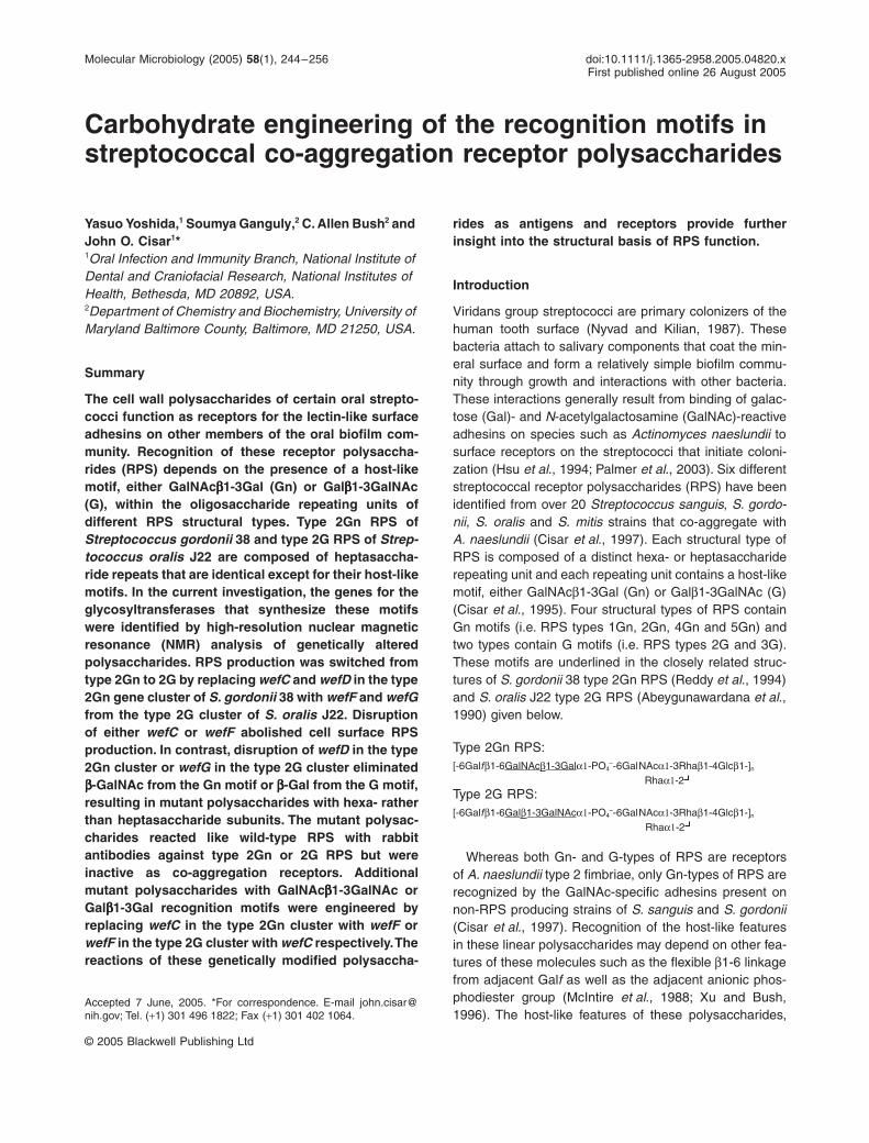
The RPS gene clusters of
S. gordonii
38 and
S. oralis
J22,although closely related, are located between differentgenes in the streptococcal chromosome (Fig. 1). The
S.gordonii
38 gene cluster is between
nrdG
and
orfO
whilethe
S. oralis
J22 cluster is between
dexB
and
aliA
, thesame genes that flank the CPS gene clusters of
S. pneu-moniae
(García
et al
., 2000). Insertion sequences
Fig. 1.
ORF diagrams of the type 2Gn RPS gene cluster of
S. gordonii
38 and type 2G RPS gene cluster of
S. oralis J22 indicating amino acid sequence identities of proteins encoded by corresponding genes and their putative roles in RPS biosynthesis. Each cluster contains four common regulatory genes ( ), seven genes that encode putative glycosyltransferases ( ) and additional genes for a putative polysaccharide polymerase ( ), a repeat unit transporter ( ) and enzymes for nucleotide sugar biosynthesis ( ). Flanking genes ( ) are also identified as are the positions of putative promoters ( ) and rho-independent transcriptional terminators ( ).
246 Y. Yoshida, S. Ganguly, C. A. Bush and J. O. Cisar
© 2005 Blackwell Publishing Ltd, Molecular Microbiology, 58, 244–256
commonly separate dexB and wzg (i.e. cpsA) in S.pneumoniae. However, these genes in S. oralis J22 areseparated by two aliB-like open reading frames (ORFs),similar to those recently identified in certain non-encap-sulated S. pneumoniae strains (Hathaway et al., 2004).The RPS and CPS gene clusters of these streptococci arealso distinguished by the extent of their association withthe four genes for dTDP-L-Rha biosynthesis. In Rha-containing serotypes of S. pneumoniae, these genesoccur in the order rmlA, rmlC, rmlB and rmlD at the 3¢-end of CPS gene clusters (Morona et al., 1999; Jianget al., 2001). However, in S. gordonii 38, rmlA, rmlC andrmlB are transcribed from a separate operon along withgalE2, the gene for a bifunctional epimerase that suppliesboth UDP-Gal and UDP-GalNAc. This operon is not asso-ciated with rmlD, which is transcribed independently (Xuet al., 2003). The arrangement of the rml genes in S. oralisJ22 is transitional between that seen in S. pneumoniaeand S. gordonii. Thus, rmlA, rmlC and rmlB are the lastthree genes in the S. oralis gene cluster and are followedby rmlD, which is transcribed in the opposite direction,presumably from a putative bidirectional promoterbetween this gene and aliA. Interestingly, the samearrangement of the rml genes and aliA has been noted ina strain of S. pneumoniae (Morona et al., 1999).
The first six genes in the RPS gene clusters of S.gordonii 38 and S. oralis J22 are homologues of those inthe CPS gene clusters of S. pneumoniae serotypes 18Cand 23F (Jiang et al., 2001). These include the four reg-ulatory genes at the 5¢-end of each cluster and the firsttwo genes for glycosyltransferases (i.e. wchA and wchF).The homology seen over this region is greater betweenS. pneumoniae and S. oralis J22 (i.e. from 73% to 87%identity at the level of predicted amino acid sequence)than between S. gordonii 38 and S. oralis J22 (Fig. 1).However, homology between the RPS gene clusters ofthese strains jumps from 61% identity for wchA to 87%identity for wchF and remains high (i.e. from 85% to 97%identity) for seven of the eight downstream genes. Theonly exception is wefD (57% identity) which, along withwefC, may account for synthesis of the Gn recognitionmotif in type 2Gn RPS. Consequently, the correspondinggenes in S. oralis J22 may not be wefC and wefD, butinstead, different genes associated with synthesis of theG recognition motif in type 2G RPS.
Identification of the genes for synthesis the recognition motifs in type 2Gn and 2G RPS
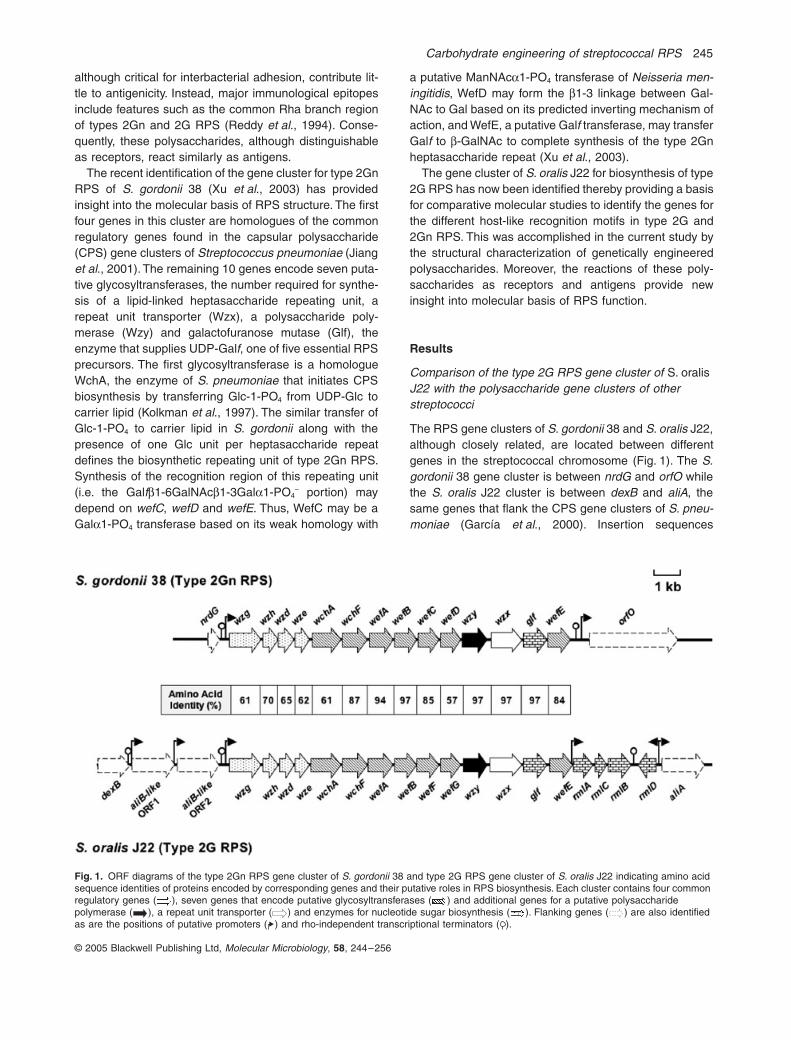
We replaced wefC and wefD in S. gordonii 38 with thecorresponding genes from S. oralis J22 to test the hypoth-esis that these genes direct synthesis of the distinct rec-ognition motifs in type 2Gn and 2G RPS. Initially, wefCand wefD were replaced by the ermAM cassette to obtain
S. gordonii GC15. This strain, which failed to bind RPS-specific immunoglobulin G (IgG) in dot immunoblotting(Fig. 2), was then transformed with a polymerase chainreaction (PCR) product that contained the correspondinggenes from S. oralis J22, which are now designated wefFand wefG, flanked by targeting sequences for S. gordonii38 wefB and wzy. Screening of approximately 10 000transformants by colony immunoblotting with RPS-specificIgG resulted in the identification of one immunoreactiveclone, designated S. gordonii GC16. DNA sequencing ofthis clone, in the region between wefB to wzy, establishedthe precise replacement of wefC and wefD in the type2Gn RPS gene cluster of S. gordonii 38 with wefF andwefG from S. oralis J22. Binding of RPS-specific IgG toS. gordonii GC16 was similar to that seen to wild-typeS. oralis J22 (Fig. 2).
The 1H-nuclear magnetic resonance (NMR) spectrumrecorded for the RPS isolated from S. gordonii GC16 wasidentical to that of previously characterized type 2G RPSof S. oralis J22 (Abeygunawardana et al., 1990; Abey-gunawardana and Bush, 1993). The structural reporterresonances in the spectra of these polysaccharides areidentical in both the anomeric region (5.5–4.5 p.p.m.) andmethyl region (2.2–1.2 p.p.m.). In addition, the same 1Hand 13C signals were seen in the heteronuclear single-quantum coherence (HSQC) spectra of the RPS from S.gordonii GC16 (results not shown) and S. oralis J22(Table S1). The RPS isolated from these two strains alsoreacted identically in immunodiffusion with rabbit antise-rum R49 against S. oralis J22 (results not shown), whichreadily distinguished type 2G from type 2Gn RPS (Fig. 3).Finally, the presence of a G-type RPS on S. gordoniiGC16 was indicated by differential binding of A. naeslundii12104 and S. sanguis SK1 in co-aggregation and bacteriaoverlay experiments (Fig. 4). In these experiments, A.naeslundii 12104, which recognizes Gn- and G-containingreceptors, bound type 2G RPS of S. oralis J22 and S.gordonii GC16 as well as type 2Gn RPS of S. gordonii 38while S. sanguis SK1, which has a Gn-specific adhesin,only bound type 2Gn RPS of S. gordonii 38. Thus, thereplacement of wefC and wefD in S. gordonii 38 with wefFand wefG from S. oralis J22 switched RPS productionfrom type 2Gn to 2G. This result not only identifies thegenes for the different recognition motifs in these polysac-charides, but also implies functional identity between theremaining five genes for glycosyltransferases in the type2Gn and 2G RPS gene clusters of S. gordonii 38 and S.oralis J22 respectively (Fig. 1).
Contributions of individual genes to RPS structure and function
Replacement of wefC in S. gordonii 38 or wefF in S.gordonii GC16 with ermAM abolished cell surface RPS
Carbohydrate engineering of streptococcal RPS 247
© 2005 Blackwell Publishing Ltd, Molecular Microbiology, 58, 244–256
production as shown by failure of RPS-specific IgG to bindeither resulting mutant (i.e. S. gordonii GC13 or S. gordo-nii GC18, respectively, in Fig. 2). The expression of down-stream genes in these mutants (i.e. wefD in S. gordoniiGC13 and wefG in S. gordonii GC18) was detected byreverse transcription polymerase chain reaction (RT-PCR)(results not shown), thereby suggesting that the loss ofcell surface RPS was not due to polar effects of theermAM insertions.
Surprisingly, ermAM replacement of wefD or wefG didnot abolish binding of RPS-specific IgG to the result-ing mutants (S. gordonii GC14 or S. gordonii GC28,respectively, in Fig. 2). The end points of these reactionswere, however, from three- to ninefold lower than those ofthe corresponding parental strains (i.e. S. gordonii 38 andS. gordonii GC16, respectively, in Fig. 2). The yields ofsoluble RPS isolated from mutanolysin digests of mutantcell walls were also low. A small sample of polysaccharidewas isolated from each mutant; however, the purity of thesample from S. gordonii GC28 was not suitable for struc-tural analysis. To circumvent this problem, an equivalentmutant was prepared by ermAM replacement of wefG inS. oralis J22, a parental strain that typically yields largeamounts of RPS (Cisar et al., 1997). The resulting mutant(S. oralis MC3) and S. gordonii GC28 gave comparable
reactions in RPS-specific dot immunoblotting (Fig. 2);however, the yield of mutant polysaccharide was greaterfrom the former strain, presumably because cell walls ofthis strain are more susceptible to mutanolysin digestion.Immunodiffusion experiments performed with the purifiedmutant polysaccharide of S. oralis MC3 revealed a singleantigen, identical to one of two components present in thepolysaccharide preparation of S. gordonii GC28 (resultsnot shown).
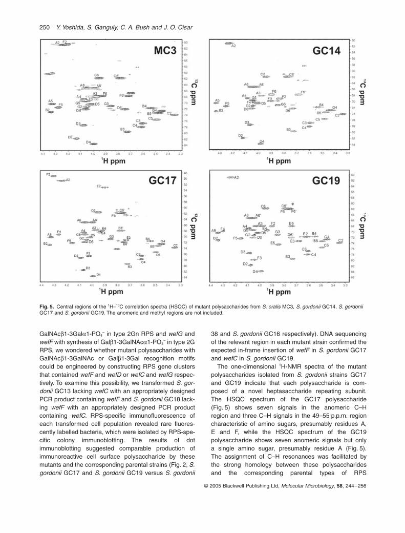
The presence of a novel hexasaccharide repeating sub-unit in the mutant polysaccharide of S. oralis MC3 wasinitially suggested by the appearance of six resonancesin the anomeric region of the 1H-NMR spectrum and con-firmed by the appearance of six C–H resonances in thecorresponding region of the HSQC spectrum (Fig. 5). Thesignals in the spin systems of each of the six sugar resi-dues were identified by 1H–1H correlation using homonu-clear coherence spectroscopy (COSY), homonuclear totalcorrelation spectroscopy (TOCSY) and nuclear Over-hauser effect spectroscopy (NOESY) with confirmationby heteronuclear multiple bond correlation (HMBC) andHSQC-TOCSY (data not shown) following standard meth-ods (Abeygunawardana and Bush, 1993). The completeassignment of all 1H and 13C resonances are given inTable S1. Positions of glycosidic linkages were identified
Fig. 2. Dot immunoblotting showing the reac-tion of RPS-specific IgG with decreasing num-bers of wild-type and mutant streptococci spotted on nitrocellulose. This antibody did not react with S. gordonii GC15, GC13 or GC18. The partial ORF diagram of the RPS gene clus-ter in each strain indicates the presence of genes from S. gordonii 38 ( ), S. oralis J22 ( ) or ermAM ( ).
248 Y. Yoshida, S. Ganguly, C. A. Bush and J. O. Cisar
© 2005 Blackwell Publishing Ltd, Molecular Microbiology, 58, 244–256
by HMBC and confirmed by NOESY as indicated inTable 1. The similarity in chemical shifts of the S. oralisMC3 polysaccharide with those previously assigned forthe polysaccharide of S. oralis J22 greatly facilitated theassignment as many could be proposed simply by chem-ical shift comparison then confirmed by correlation. Thereare some differences seen in the galactofuranoside resi-due, D, with larger differences in the a-GalNAc residue F.The shifts in the latter residue differ most as a-GalNAc issubstituted in the 3-position in S. oralis J22 and in the 6-position in S. oralis MC3 as is shown by HMBC, NOESYand the downfield 13C shift of C6. These findings indicatethe following structure for the mutant polysaccharide ofS. oralis MC3:
D F A B C[-6Galfb1-6GalNAca1-PO4
–-6GalNAca1-3Rhab1-4Glcb1-]nRhaa1-2
G
The presence of this polysaccharide on S. gordonii GC28was supported by NMR analysis of the partially purifiedpreparation obtained from this strain.
The relatively small quantity of purified polysaccharideisolated from S. gordonii GC14 rendered impractical themeasurement of long-range 1H–13C correlation spectrasuch as HMBC and HSQC-TOCSY. However, the overallsimilarity of this polysaccharide and the S. oralis MC3polysaccharide simplified the structure determination. Thetwo polysaccharides differ only in the presence of anamide function at C2 of residue F which is a-GalNAc inS. oralis MC3 and a-Gal in S. gordonii GC14. Thus, com-parison of the HSQC spectra of these polysaccharides
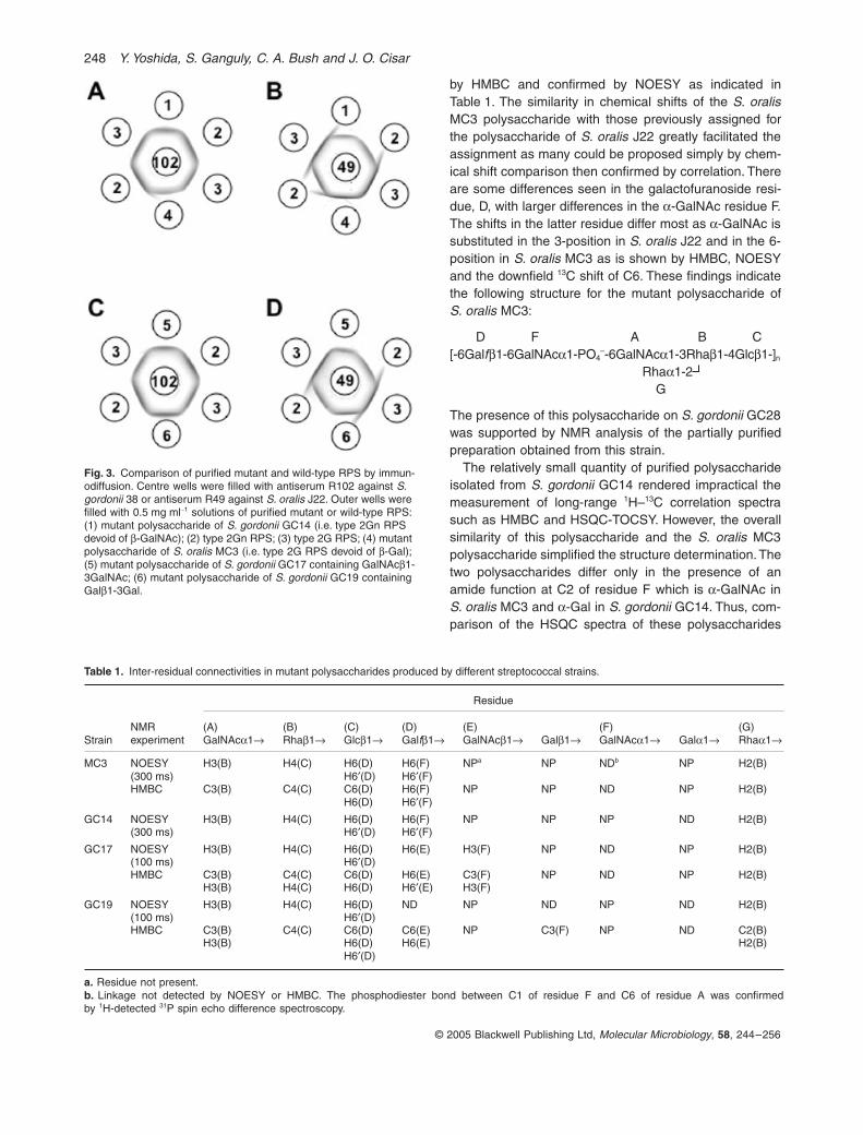
Fig. 3. Comparison of purified mutant and wild-type RPS by immun-odiffusion. Centre wells were filled with antiserum R102 against S. gordonii 38 or antiserum R49 against S. oralis J22. Outer wells were filled with 0.5 mg ml-1 solutions of purified mutant or wild-type RPS: (1) mutant polysaccharide of S. gordonii GC14 (i.e. type 2Gn RPS devoid of b-GalNAc); (2) type 2Gn RPS; (3) type 2G RPS; (4) mutant polysaccharide of S. oralis MC3 (i.e. type 2G RPS devoid of b-Gal); (5) mutant polysaccharide of S. gordonii GC17 containing GalNAcb1-3GalNAc; (6) mutant polysaccharide of S. gordonii GC19 containing Galb1-3Gal.
Table 1. Inter-residual connectivities in mutant polysaccharides produced by different streptococcal strains.
StrainNMR experiment
Residue
(A)GalNAca1Æ
(B)Rhab1Æ
(C)Glcb1Æ
(D)Galfb1Æ
(E)GalNAcb1Æ Galb1Æ
(F)GalNAca1Æ Gala1Æ
(G) Rhaa1Æ
MC3 NOESY H3(B) H4(C) H6(D) H6(F) NPa NP NDb NP H2(B)(300 ms) H6¢(D) H6¢(F)HMBC C3(B) C4(C) C6(D)
H6(D)H6(F)H6¢(F)
NP NP ND NP H2(B)
GC14 NOESY H3(B) H4(C) H6(D) H6(F) NP NP NP ND H2(B)(300 ms) H6¢(D) H6¢(F)
GC17 NOESY H3(B) H4(C) H6(D) H6(E) H3(F) NP ND NP H2(B)(100 ms) H6¢(D)HMBC C3(B) C4(C) C6(D) H6(E) C3(F) NP ND NP H2(B)
H3(B) H4(C) H6(D) H6¢(E) H3(F)
GC19 NOESY H3(B) H4(C) H6(D) ND NP ND NP ND H2(B)(100 ms) H6¢(D)HMBC C3(B) C4(C) C6(D) C6(E) NP C3(F) NP ND C2(B)
H3(B) H6(D)H6¢(D)
H6(E) H2(B)
a. Residue not present.b. Linkage not detected by NOESY or HMBC. The phosphodiester bond between C1 of residue F and C6 of residue A was confirmedby 1H-detected 31P spin echo difference spectroscopy.
Carbohydrate engineering of streptococcal RPS 249
© 2005 Blackwell Publishing Ltd, Molecular Microbiology, 58, 244–256
(Fig. 5) immediately suggested assignments for most ofthe signals (Table S1). A few small differences do, how-ever, exist in the chemical shifts for C–H pairs in residueD with more significant differences between the Gal orGalNAc residue F. The assignments for the S. gordoniiGC14 polysaccharide given in Table S1 were all confirmedby 1H correlation and NOESY spectra and the glycosidiclinkages were confirmed by comparison of cross-peaks inthe NOESY spectra of the polysaccharides of S. gordoniiGC14 and S. oralis MC3. These findings indicate thefollowing structure for the mutant polysaccharide of S.gordonii GC14:
D F A B C[-6Galfb1-6Gala1-PO4
–-6GalNAca1-3Rhab1-4Glcb1-]nRhaa1-2
Immunodiffusion performed with rabbit antisera againstS. gordonii 38 (Fig. 3A) or S. oralis J22 (Fig. 3B) revealedreactions of identity between the mutant polysaccharide
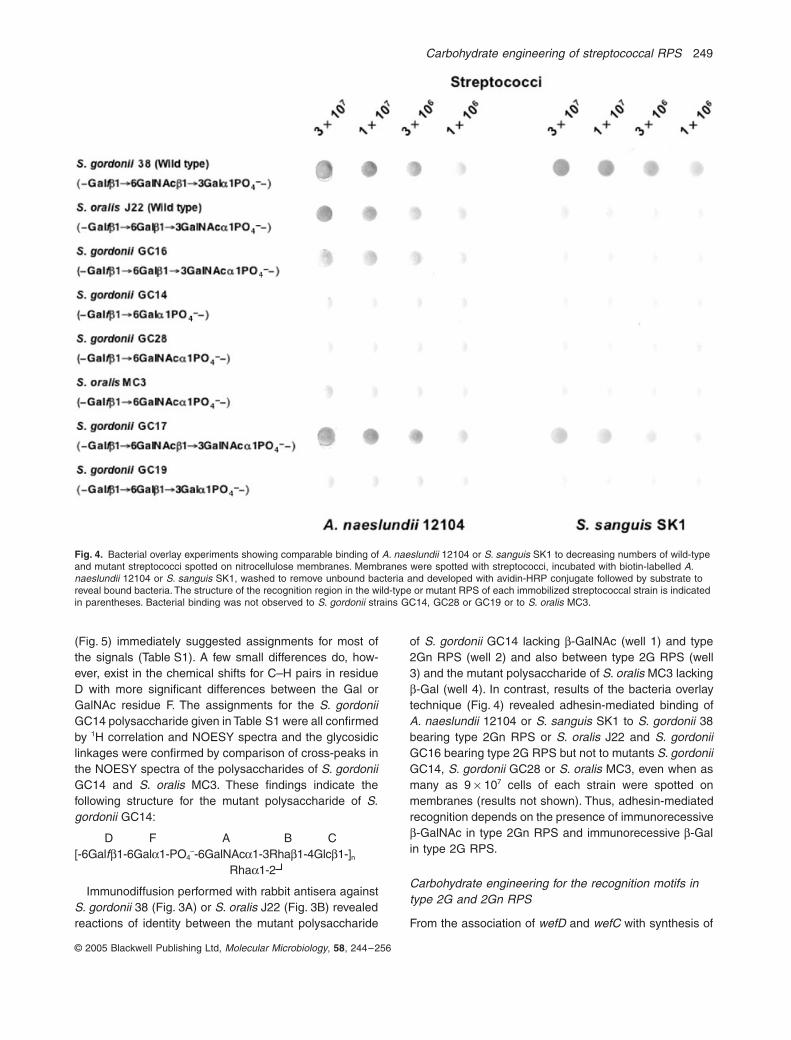
of S. gordonii GC14 lacking b-GalNAc (well 1) and type2Gn RPS (well 2) and also between type 2G RPS (well3) and the mutant polysaccharide of S. oralis MC3 lackingb-Gal (well 4). In contrast, results of the bacteria overlaytechnique (Fig. 4) revealed adhesin-mediated binding ofA. naeslundii 12104 or S. sanguis SK1 to S. gordonii 38bearing type 2Gn RPS or S. oralis J22 and S. gordoniiGC16 bearing type 2G RPS but not to mutants S. gordoniiGC14, S. gordonii GC28 or S. oralis MC3, even when asmany as 9 ¥ 107 cells of each strain were spotted onmembranes (results not shown). Thus, adhesin-mediatedrecognition depends on the presence of immunorecessiveb-GalNAc in type 2Gn RPS and immunorecessive b-Galin type 2G RPS.
Carbohydrate engineering for the recognition motifs in type 2G and 2Gn RPS
From the association of wefD and wefC with synthesis of
Fig. 4. Bacterial overlay experiments showing comparable binding of A. naeslundii 12104 or S. sanguis SK1 to decreasing numbers of wild-type and mutant streptococci spotted on nitrocellulose membranes. Membranes were spotted with streptococci, incubated with biotin-labelled A. naeslundii 12104 or S. sanguis SK1, washed to remove unbound bacteria and developed with avidin-HRP conjugate followed by substrate to reveal bound bacteria. The structure of the recognition region in the wild-type or mutant RPS of each immobilized streptococcal strain is indicated in parentheses. Bacterial binding was not observed to S. gordonii strains GC14, GC28 or GC19 or to S. oralis MC3.
250 Y. Yoshida, S. Ganguly, C. A. Bush and J. O. Cisar
© 2005 Blackwell Publishing Ltd, Molecular Microbiology, 58, 244–256
GalNAcb1-3Gala1-PO4– in type 2Gn RPS and wefG and
wefF with synthesis of Galb1-3GalNAca1-PO4– in type 2G
RPS, we wondered whether mutant polysaccharides withGalNAcb1-3GalNAc or Galb1-3Gal recognition motifscould be engineered by constructing RPS gene clustersthat contained wefF and wefD or wefC and wefG respec-tively. To examine this possibility, we transformed S. gor-donii GC13 lacking wefC with an appropriately designedPCR product containing wefF and S. gordonii GC18 lack-ing wefF with an appropriately designed PCR productcontaining wefC. RPS-specific immunofluorescence ofeach transformed cell population revealed rare fluores-cently labelled bacteria, which were isolated by RPS-spe-cific colony immunoblotting. The results of dotimmunoblotting suggested comparable production ofimmunoreactive cell surface polysaccharide by thesemutants and the corresponding parental strains (Fig. 2, S.gordonii GC17 and S. gordonii GC19 versus S. gordonii
38 and S. gordonii GC16 respectively). DNA sequencingof the relevant region in each mutant strain confirmed theexpected in-frame insertion of wefF in S. gordonii GC17and wefC in S. gordonii GC19.
The one-dimensional 1H-NMR spectra of the mutantpolysaccharides isolated from S. gordonii strains GC17and GC19 indicate that each polysaccharide is com-posed of a novel heptasaccharide repeating subunit.The HSQC spectrum of the GC17 polysaccharide(Fig. 5) shows seven signals in the anomeric C–Hregion and three C–H signals in the 49–55 p.p.m. regioncharacteristic of amino sugars, presumably residues A,E and F, while the HSQC spectrum of the GC19polysaccharide shows seven anomeric signals but onlya single amino sugar, presumably residue A (Fig. 5).The assignment of C–H resonances was facilitated bythe strong homology between these polysaccharidesand the corresponding parental types of RPS
Fig. 5. Central regions of the 1H–13C correlation spectra (HSQC) of mutant polysaccharides from S. oralis MC3, S. gordonii GC14, S. gordonii GC17 and S. gordonii GC19. The anomeric and methyl regions are not included.

Carbohydrate engineering of streptococcal RPS 251
© 2005 Blackwell Publishing Ltd, Molecular Microbiology, 58, 244–256
(Table S1). The quantities of RPS isolated from thesestrains were sufficient for 10 mg samples in NMRexperiments allowing confirmation of all assignments byboth homonuclear correlation (COSY, TOCSY, NOESY)and long-range heteronuclear correlation (HMBC,HSQC-TOCSY). In addition, all glycosidic linkages wereconfirmed by HMBC and NOESY spectra as docu-mented in Table 1. The results indicate the followingstructures for the mutant polysaccharides of S. gordoniiGC17 and S. gordonii GC19:
GC17 RPS:D E F A B C
[-6Galfb1-6GalNAcb1-3GalNAca1-PO4–-6GalNAca1-3Rhab1-4Glcb1-]n
Rhaa1-2G
GC19 RPS:D E F A B C
[-6Galfb1-6Galb1-3Gala1-PO4–-6GalNAca1-3Rhab1-4Glcb1-]n
Rhaa1-2G
Immunodiffusion performed with a rabbit antiserumagainst S. gordonii 38 (Fig. 3C) resulted in a reactionof antigenic identity between type 2Gn RPS (well 2)and type 2G RPS (well 3). In contrast, an obvious anti-genic difference between these polysaccharides wasevident when the same experiment was performed witha rabbit antiserum against S. oralis J22 (Fig. 3D). Inthis experiment, a comparable antigenic difference wasseen between type 2G RPS (well 3) and the mutantpolysaccharide of S. gordonii GC19 (well 6) but notbetween type 2G RPS (well 3) and the mutant polysac-charide of S. gordonii GC17 (well 5). Thus, the anti-genic difference detected between type 2G and 2GnRPS appears to involve the presence of GalNAca1-PO4
– in the former polysaccharide versus Gala1-PO4–
in the latter.The lectin-like interaction of A. naeslundii 12104 with
type 2Gn RPS of S. gordonii 38 or type 2G RPS of S.oralis J22 was readily demonstrated by visual assays forco-aggregation and also by the binding of biotinylated A.naeslundii 12104 to streptococci immobilized on nitrocel-luolose (Fig. 4). Interestingly, A. naeslundii 12104 boundto S. gordonii GC17 but failed to bind S. gordonii GC19thereby suggesting recognition of GalNAcb1-3GalNAc onthe former strain but not Galb1-3Gal of the latter. In similarexperiments, binding of S. sanguis SK1, which dependson a GalNAc-specific adhesin, was more pronounced toS. gordonii 38 than to S. gordonii GC17 suggesting apreference for GalNAcb1-3Gal in type 2Gn RPS overGalNAcb1-3GalNAc in the mutant polysaccharide of thelatter strain.
Discussion
Significant insights into the molecular basis of RPS struc-ture and function were gained in the present investigationfrom the production and characterization of differentgenetically modified polysaccharides. These polysaccha-rides were engineered by replacing specific genes in thetype 2Gn RPS gene cluster in S. gordonii 38 with theermAM cassette followed by replacement of this cassettewith complementary genes from the type 2G RPS genecluster of S. oralis J22. The first step abolished synthesisof cell surface RPS and the second step resulted in theproduction of a genetically modified polysaccharide thatwas detected by its reaction with an RPS-specific poly-clonal antibody and characterized by high-resolutionNMR. The NMR method used in the present study provedespecially useful for this purpose. The data of Table S1show that modest differences in structure lead to localizeddifferences in the C–H resonances. Thus, inspection of anHSQC spectrum readily suggested a tentative spectralassignment. For novel structures, the expected correla-tions were verified by homo- and heteronuclear correlationexperiments and the intersaccharide linkages determinedby long-range correlation. The combined results of molec-ular and structural studies show that deletion of wefDeliminated b-GalNAc from type 2Gn RPS, that deletion ofwefG eliminated b-Gal from type 2G RPS and that swap-ping wefC and wefF yielded mutant polysaccharides withGalNAcb1-3GalNAca1-PO4
– or Galb1-3Gala1-PO4– rec-
ognition motifs respectively. These findings clearly identifythe genes for the recognition motifs in types 2Gn and 2GRPS and also provide insight into the acceptor specifici-ties of the encoded glycosyltransferases.
The previously summarized results provide clear evi-dence that both GalNAca1-PO4
– and Gala1-PO4– are
acceptors for the WefD-mediated transfer of b-GalNAcand WefG-mediated transfer of b-Gal. The relaxed speci-ficity of WefD and WefG for these acceptors does not,however, exclude the possibility that each glycosyltras-ferase has a preference for one acceptor structure overthe other. Clearly, further enzymatic characterization ofthese proteins is needed to assess this possibility. In con-trast, with the relaxed acceptor specificity of WefD andWefG, the strict donor specificity of these and other glyc-osyltransferases involved in RPS biosynthesis is evidentfrom the NMR spectra of different wild-type or mutantpolysaccharides, which indicate structural homogeneity ofthe oligosaccharide repeating subunits in these mole-cules. Strict donor specificity of the GalNAc transferasesinvolved in type 2Gn RPS biosynthesis (i.e. WefA andWefD) is also evident from the absence of detectable cellsurface RPS on a previously described mutant of S. gor-donii 38, which synthesizes all necessary RPS precursorsexcept UDP-GalNAc (Xu et al., 2003).
252 Y. Yoshida, S. Ganguly, C. A. Bush and J. O. Cisar
© 2005 Blackwell Publishing Ltd, Molecular Microbiology, 58, 244–256
The production of type 2G RPS following the replace-ment of wefC and wefD in S. gordonii 38 with wefF andwefG from S. oralis J22 associates the putative Galf trans-ferase encoded by wefE with the synthesis of both Galfb1-6GalNAcb in type 2Gn RPS (Xu et al., 2003) and Galfb1-6Galb in type 2G RPS. Moreover, WefE transfers Galf toeither Gala1-PO4
– in S. gordonii GC14, which lacks wefD,or GalNAca1-PO4
– in S. oralis MC3, which lacks wefG,resulting in mutant polysaccharides with hexasacchariderepeating subunits. The presence of such repeats in type2Gn or 2G RPS is not evident from the NMR spectra ofthese wild-type polysaccharides. Thus, the efficient WefD-dependent transfer of b-GalNAc to Gala1-PO4
– or WefG-dependent transfer of b-Gal to GalNAca1-PO4
– in wild-type strains appears to prevent the WefE-dependenttransfer of Galf to these acceptors.
The presence of hexasaccharide repeats in the mutantpolysaccharides of S. gordonii GC14 and S. oralis MC3clearly indicates that the action of Wzy is not limited towild-type heptasaccharide repeating subunits. Similarly,Wzy encoded by a gene in the extracellular polysaccha-ride (EPS) gene cluster of Streptococcus thermophilusSfi6 was previously found to catalyse polymerization of abranched tetrasaccharide synthesized in S. thermophilusas well as a linear trisaccharide synthesized in Lactococ-cus lactis (Stingele et al., 1999). The yield of wild-typeEPS from S. thermophilus was, however, significantlygreater than that of mutant EPS from L. lactis. Likewise,the cell surface production of wild-type RPS by S. gordonii38 or S. oralis J22 was significantly greater than the pro-duction of mutant polysaccharide by S. gordonii GC14 orS. oralis MC3 respectively. Further studies are neededto determine whether the production of these mutantpolysaccharides is limited by the inefficient synthesis oflipid-linked hexasaccharide subunits or by the inefficientflipping or polymerization of these subunits from the actionof Wzx or Wzy respectively.
Molecular mimicry of host glycoconjugates by the sur-face carbohydrates of mucosal pathogens may contributeto evasion of the host response as well as to the produc-tion of anti-host antibody (Moran et al., 1996). The pro-duction of such antibodies in response to normal oralcolonization of RPS-bearing streptococci has not beendetected (J.O. Cisar, unpublished). We have, however,noted that the reactions of certain rabbit anti-streptococ-cal sera, most notably R49, distinguish type 2G from2Gn RPS (Cisar et al., 1997), thereby indicating detec-tion of an epitope(s) that is closely associated with thehost-like features of these polysaccharides. The presentfindings (Fig. 3) indicate that this epitope depends on thepresence of GalNAca1-PO4
– in type 2G RPS but not b-Gal. Thus, the antigenic region of this polysaccharideappears to extend to the edge of the host-like recognitionmotif.
The specificity of A. naeslundii type 2 fimbriae-mediatedadhesion has been assessed by the binding of bacteriato immobilized glycolipids (Brennan et al., 1987; Ström-berg and Karlsson, 1990) and neoglycoproteins (Ruhlet al., 1996) and also by saccharide inhibition of the co-aggregations observed between strains of A. naeslundiiand RPS-bearing streptococci (McIntire et al., 1982;1988; Cisar et al., 1997). The results of these studiessuggest that recognition of GalNAcb1-3Gal and Galb1-3GalNAc depends on the common features of these iso-meric structures. This interpretation is consistent withthe type 2 fimbriae-mediated recognition of GalNAcb1-3GalNAc, demonstrated previously by the binding of A.naeslundii 12104 to a glycolipid from human erythrocytes(Strömberg and Karlsson, 1990) and presently by thebinding of this strain to the mutant RPS of S. gordoniiGC17 (Fig. 4). However, A. naeslundii did not adhere tothe mutant RPS present on S. gordonii GC19, which hasGalb1-3Gal motifs, thereby clearly suggesting that recog-nition depends on the presence of N-acetyl groups. Thepossibility that the N-acetyl groups in GalNAcb1-3Gal-and Galb1-3GalNAc are accommodated at differentpositions in the binding site of the A. naeslundii adhesinprovides a simple explanation for strong binding ofA. naeslundii to these disaccharides and GalNAcb1-3GalNAc and the failure of this organism to bind Galb1-3Gal. Importantly, the current findings indicate that recog-nition of type 2Gn and 2G RPS involves both saccharideunits in the host-like motifs of these polysaccharides.
The genetic engineering of bacterial polysaccharidegene clusters has been suggested as a possible approachfor altering the rheological properties of EPS produced bylactic acid bacteria (Kleerebezem et al., 1999; Jolly et al.,2002; Welman and Maddox, 2003). The application of thisapproach has now been realized for the first time to ourknowledge by altering the recognition motifs in the RPSof oral viridans group streptococci. The ability to geneti-cally engineer bacterial surface carbohydrates has a widerange of potential applications as illustrated by the pre-vention of toxin-based enteric disease with a recombinantstrain of Escherichia coli engineered to surface express atoxin receptor mimic (Paton et al., 2000). The limits of thisemerging technology will ultimately be determined by thedonor and acceptor specificities of the glycosyltrans-ferases and polymerases encoded by available genes.The structural complexity of the surface polysaccharidespresent on oral viridans group streptococci suggest thatthese bacteria represent a rich source of such genes.Moreover, the likelihood that these genes can be used toengineer novel carbohydrate structures is increased bythe present finding that the acceptor specificity of certainglycosyltransferases involved in RPS biosynthesis ap-pears to be less strict than anticipated. Further studies ofstreptococcal RPS gene clusters are underway both to
Carbohydrate engineering of streptococcal RPS 253
© 2005 Blackwell Publishing Ltd, Molecular Microbiology, 58, 244–256
identify genetic markers for oral biofilm development andto explore the limits of carbohydrate engineering in thisexperimental system.
Experimental procedures
Bacterial strains and culture conditions
Table 2 lists the wild-type and mutant streptococci and acti-nomyces that were used in this study. The streptococcuspreviously identified as S. mitis J22 is now designated S.oralis J22 based on results from the recent sequencing ofhousekeeping genes in this strain (M. Kilian, pers. comm.).The bacteria listed were routinely grown at 37∞C in Todd–Hewitt broth (THB; Difco Laboratories). Erythromycin wasadded to a final concentration of 10 mg ml-1 for the cultivationof ermAM-containing strains. Chemically competent E. coliDH5a from Invitrogen was grown aerobically at 37∞C in LBbroth or agar, which was supplemented with 100 mg ml-1
ampicillin, 20 mg ml-1 chloramphenicol, 100 mg ml-1 kanamy-cin or 200 mg ml-1 erythromycin as required for the mainte-nance of plasmids.
Immunological methods
Rabbit antisera R102 against S. gordonii 38 and R49 againstS. oralis J22 have been described (Cisar et al., 1997). TheRPS-specific IgG used in this study was affinity-purified fromantiserum R49 by 4 M MgCl2 elution from a small column ofimmunoadsorbent containing coupled type 2G RPS as pre-viously described (Xu et al., 2003).
Dot immunoblotting was performed to detect the reactionof RPS-specific IgG with streptococci spotted on nitrocellu-lose membranes. Streptococci were harvested from station-ary-phase cultures, washed and suspended to a cell densityof approximately 2 ¥ 109 ml-1 in 0.02 M Tris-buffered saline(TBS) (pH 7.5) based on measurements of turbidity madewith a Klett-Summerson colorimeter. Nitrocellulose mem-branes were spotted with decreasing numbers of streptococciusing a Bio-Dot Microfiltration Apparatus (Bio-Rad) andblocked by incubation in TBS containing 0.1% Tween-20 and2% skim milk. Membranes were then incubated with RPS-specific IgG (50 ng ml-1) followed by horseradish peroxidase
(HRP)-conjugated goat anti-rabbit IgG (Bio-Rad) beforedevelopment with a Metal Enhanced DAB Substrate Kit(Pierce Biotechnology) to detect bound anti-RPS antibody.
Immunodiffusion experiments were performed in 1% aga-rose gel cast on Gel Bond Film (FMC BioProducts). Wellswere filled with undiluted rabbit antisera or 0.5 mg ml-1 puri-fied RPS and incubated overnight at 4∞C to allow immuno-precipitation. Gels were soaked 2 days in cold 0.5 M NaCl toremove soluble protein followed by water, dried and thenstained with Coomassie blue as previously described (Cisaret al., 1997).
Bacterial adhesion assays
Co-aggregations between A. naeslundii 12104 or S. sanguisSK1 and RPS-bearing streptococci were assessed visually(Cisar et al., 1979; Hsu et al., 1994). A previously describedbacterial overlay assay (Ruhl et al., 1996; 2004) was per-formed to compare the binding of biotin-labelled A. naeslundii12104 or S. sanguis SK1 to different immobilized wild-typeand mutant streptococcal strains. Decreasing numbers ofstreptococci were spotted on nitrocellulose membranes usinga Bio-Dot Microfiltration Apparatus as described above.Membranes were then blocked by overnight incubation at 4∞Cin TBS containing 5% bovine serum albumin, 0.1 mM CaCl2and 0.1 mM MgCl2 (blocking buffer). A. naeslundii 12104 andS. sanguis SK1 were harvested from overnight cultures andlabelled by incubation with 100 mg ml-1 sulphosuccinimidyl 6-(biotinamido) hexanoate-biotin (Pierce) for 1 h at room tem-perature. Pre-blocked nitrocellulose membranes were incu-bated with biotin-labelled bacteria (5 ¥ 108 per ml in blockingbuffer) for 3 h at 4∞C, washed in blocking buffer to removeunbound bacteria and incubated with 0.2 U of avidin-D-HRP(Bio-Rad) per ml followed by DAB substrate as describedabove to detect bound bacteria.
Cloning and sequencing of the gene cluster for type 2G RPS
The 27 833 bp DNA sequence of the S. oralis J22 RPS genecluster and flanking regions, which is available under Gen-Bank Accession No. AB181235, was assembled from thesequences of overlapping PCR fragments. The template for
Table 2. Bacterial strains used in this study.
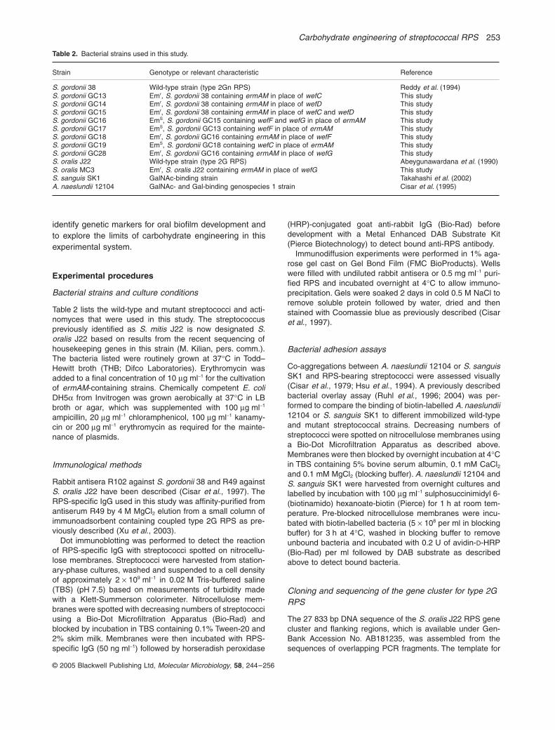
Strain Genotype or relevant characteristic Reference
S. gordonii 38 Wild-type strain (type 2Gn RPS) Reddy et al. (1994)S. gordonii GC13 Emr, S. gordonii 38 containing ermAM in place of wefC This studyS. gordonii GC14 Emr, S. gordonii 38 containing ermAM in place of wefD This studyS. gordonii GC15 Emr, S. gordonii 38 containing ermAM in place of wefC and wefD This studyS. gordonii GC16 EmS, S. gordonii GC15 containing wefF and wefG in place of ermAM This studyS. gordonii GC17 EmS, S. gordonii GC13 containing wefF in place of ermAM This studyS. gordonii GC18 Emr, S. gordonii GC16 containing ermAM in place of wefF This studyS. gordonii GC19 EmS, S. gordonii GC18 containing wefC in place of ermAM This studyS. gordonii GC28 Emr, S. gordonii GC16 containing ermAM in place of wefG This studyS. oralis J22 Wild-type strain (type 2G RPS) Abeygunawardana et al. (1990)S. oralis MC3 Emr, S. oralis J22 containing ermAM in place of wefG This studyS. sanguis SK1 GalNAc-binding strain Takahashi et al. (2002)A. naeslundii 12104 GalNAc- and Gal-binding genospecies 1 strain Cisar et al. (1995)
254 Y. Yoshida, S. Ganguly, C. A. Bush and J. O. Cisar
© 2005 Blackwell Publishing Ltd, Molecular Microbiology, 58, 244–256
initial PCR reactions was S. oralis J22 chromosomal DNA,which was prepared using a Wizard Genomic DNA purifica-tion kit (Promega). Reaction mixtures also contained KODHot Start DNA polymerase (Novagen) and primer pairsavailable from previous characterization of the type 2GnRPS gene cluster of S. gordonii 38 (Xu et al., 2003). Thesequences of the PCR fragments obtained were extendedwhen necessary by inverse PCR (Ochman et al., 1988).Briefly, genomic DNA from S. oralis J22 was digested withan appropriate restriction enzyme. After inactivation of thisenzyme, the digested DNA was circularized by self-ligationand used as template for PCR performed with appropriateprimers. PCR fragments were cloned in pBluescript (Strat-agene), pCR4Blunt-TOPO (Invitrogen) or pMCL200 (Nakanoet al., 1995) before DNA sequencing at Sequetech, MountainView, USA. Two independent amplicons of each cloned DNAfragment were sequenced and compared to insure the fidelityof PCR amplification. Sequences were assembled and anno-tated using Vector NTI software (Invitrogen). Nucleotide andpredicted amino acid sequence homologies with genes andproteins in the database were identified by BLAST (Altschulet al., 1990).
Construction of streptococcal mutant strains
The ermAM mutant strains listed in Table 2 were constructedby transformation of wild-type streptococci with DNA con-structs containing the ermAM cassette flanked by targetingsequences for the streptococcal gene of interest. The threeDNA sequences (i.e. upstream targeting sequence, ermAMcassette and downstream targeting sequence) were linkedby overlap extension PCR (Horton et al., 1989; Lee andMorrison, 1999) performed using KOD Hot Start DNA poly-merase. The primers used to amplify the ermAM cassettefrom pKSerm2 (Lunsford and London, 1996) and each tar-geting sequence (approximately 0.7 kb) from streptococcalgenomic DNA were designed to create nucleotide sequencecomplementarity between the 3¢-end of the upstream target-ing sequence and the 5¢-end of the ermAM cassette and alsobetween the 3¢-end of the ermAM cassette and the 5¢-end ofthe downstream targeting sequence. Overlap extension PCRwas then performed using the three DNA fragments as mixedtemplate and primers designed from the 5¢-end of theupstream targeting sequence and the 3¢-end of the down-stream targeting sequence. Transformation of S. gordonii 38and related mutant strains with overlap extension PCR prod-ucts was performed as previously described (Lunsford, 1995)except that the transformation medium contained 5% heat-inactivated horse serum (Sigma-Aldrich) in place of 0.15%filter sterilized bovine serum albumin. Selection of transfor-mants was by anerobic growth on plates of brain–heart infu-sion agar (BHI, Difco) containing 10 mg ml-1 erythromycin.The location of the ermAM cassette in each insertionalmutant strain (Table 2) was verified by the amplification ofspecific PCR products across the upstream and downstreamboundaries of the ermAM insertion, using primers for chro-mosomal sequences that were extraneous to those presentin the transforming DNA.
Streptococcus gordonii GC16, GC17 and GC19 wereobtained by replacement of the ermAM cassette in S. gordo-nii GC15, GC13 and GC18, respectively, with intact genes of
S. oralis J22. This was accomplished by transformation ofeach ermAM-containing strain with an overlap extensionPCR product that contained the S. oralis J22 gene(s) ofinterest flanked by targeting sequences for precise in-framereplacement of the ermAM cassette. Transformants wereidentified by their reaction with RPS-specific IgG in colonyimmunoblotting. Briefly, transformation reactions werediluted, spread on BHI plates and incubated anaerobically for1 day at 37∞C to allow growth of isolated colonies. A sterilenitrocellulose membrane was then placed on each plate incontact with the colonies. The membranes were carefullyremoved so as not to disturb the underlying colonies andprocessed by for binding of RPS-specific IgG (50 ng ml-1) toadsorbed bacteria by procedures that were essentially thesame as those described for dot immunoblotting.
Isolation of RPS
Streptococcal RPS was purified as previously described(Cisar et al., 1997) with minor modifications. Briefly, bacteriaharvested from 12–16 l of stationary-phase cultures weretreated with 0.1% Triton X-100 to disrupt membranes andthen digested with Ribonuclease A followed by Streptomycesgriseus Protease (both enzymes were from Sigma-Aldrich)to facilitate the removal cytoplasmic material. Surfacepolysaccharides were solubilized by mutanolysin (Sigma-Ald-rich) digestion of the resulting crude cell walls. Protein wasprecipitated from mutanolysin digests by the addition of coldtrichloroacetic acid to a final concentration of 5%. The solublefraction was neutralized and dialysed to remove salt beforepurification of RPS by gradient elution from a DEAE Sephacelanion exchange column. Purified polysaccharides weredetected by immunodiffusion and assays for total carbohy-drate in fractions that contained from 120 mM to 160 mMNaCl.
Nuclear magnetic resonance analysis
Nuclear magnetic resonance spectra were recorded on aBruker DRX 500 MHz spectrometer with a cryoprobe usingXWINNMR as the standard acquisition software. The NMRmeasurements were performed at 25∞C. Generally, a 10 mgsample of RPS (4 mg of the GC14 sample) was exchangedtwice with 3 ml of 99.96% D2O, lyophilized and dissolved in0.6 ml of 99.99% D2O. Chemical shifts were recorded relativeto internal acetone (1H, 2.225 p.p.m.; 13C, 31.05 p.p.m.). Allthe data were processed using NMRPIPE, NMRDRAW andNMRVIEW software. Double-quantum filtered COSY andTOCSY with a spin lock of 70 ms were carried out to assignthe scalar coupled proton of the same monosaccharide res-idue. For assignment of the 13C chemical shift, HSQC andHSQC-TOCSY with a spin lock of 70 ms were used. Inter-residual linkages were determined by NOESY with mixingtimes of 100 ms and 300 ms. HMBC with a delay of 50 msfor evolution of long-range coupling was used to further val-idate the linkages and for confirmation of the assignments.
Acknowledgements
This work was supported in part by the Intramural Research
Carbohydrate engineering of streptococcal RPS 255
© 2005 Blackwell Publishing Ltd, Molecular Microbiology, 58, 244–256
Program of the NIH, NIDCR, and by a fellowship from theJapanese Society for the Promotion of Science to Y.Y. and byGrant 02-12702 from the National Science Foundation toC.A.B. We thank Kelly Ten Hagen and John Thompsonfor their helpful suggestions during preparation of thismanuscript.
References
Abeygunawardana, C., and Bush, C.A. (1993) Determinationof the chemical structure of complex polysaccharides byheteronuclear NMR spectroscopy. Adv Biophy Chem 3:199–249.
Abeygunawardana, C., Bush, C.A., and Cisar, J.O. (1990)Complete structure of the polysaccharide from Streptococ-cus sanguis J22. Biochemistry 29: 234–248.
Altschul, S.F., Gish, W., Miller, W., Myers, E.W., and Lipman,D.J. (1990) Basic local alignment search tool. J Mol Biol215: 403–410.
Brennan, M.J., Joralmon, R.A., Cisar, J.O., and Sandberg,A.L. (1987) Binding of Actinomyces naeslundii to glycosph-ingolipids. Infect Immun 55: 487–489.
Cisar, J.O., Kolenbrander, P.E., and McIntire, F.C. (1979)Specificity of coaggregation reactions between human oralstreptococci and strains of Actinomyces viscosus or Acti-nomyces naeslundii. Infect Immun 24: 742–752.
Cisar, J.O., Sandberg, A.L., Abeygunawardana, C., Reddy,G.P., and Bush, C.A. (1995) Lectin recognition of host-likesaccharide motifs in streptococcal cell wall polysaccha-rides. Glycobiology 5: 655–662.
Cisar, J.O., Sandberg, A.L., Reddy, G.P., Abeygunawar-dana, C., and Bush, C.A. (1997) Structural and anti-genic types of cell wall polysaccharides from viridansgroup streptococci with receptors for oral actinomycesand streptococcal lectins. Infect Immun 65: 5035–5041.
García, E., Llull, D., Muñoz, R., Mollerach, M., and López, R.(2000) Current trends in capsular polysaccharide biosyn-thesis of Streptococcus pneumoniae. Res Microbiol 151:429–435.
Hathaway, L.J., Stutzmann Meier, P., Battig, P., Aebi, S., andMuhlemann, K. (2004) A homologue of aliB is found in thecapsule region of nonencapsulated Streptococcus pneu-moniae. J Bacteriol 186: 3721–3729.
Horton, R.M., Hunt, H.D., Ho, S.N., Pullen, J.K., and Pease,L.R. (1989) Engineering hybrid genes without the use ofrestriction enzymes: gene splicing by overlap extension.Gene 77: 61–68.
Hsu, S.D., Cisar, J.O., Sandberg, A.L., and Kilian, M. (1994)Adhesive properties of viridans group streptococcal spe-cies. Microb Ecol Health Dis 7: 125–137.
Jiang, S.M., Wang, L., and Reeves, P.R. (2001) Molecularcharacterization of Streptococcus pneumoniae type 4, 6B,8, and 18C capsular polysaccharide gene clusters. InfectImmun 69: 1244–1255.
Jolly, L., Vincent, S.J., Duboc, P., and Neeser, J.R. (2002)Exploiting exopolysaccharides from lactic acid bacteria.Antonie Van Leeuwenhoek 82: 367–374.
Kleerebezem, M., van Kranenburg, R., Tuinier, R., Boels, I.C.,Zoon, P., Looijesteijn, E., et al. (1999) Exopolysaccharidesproduced by Lactococcus lactis: from genetic engineering
to improved rheological properties? Antonie Van Leeuwen-hoek 76: 357–365.
Kolkman, M.A., van der Zeijst, B.A., and Nuijten, P.J. (1997)Functional analysis of glycosyltransferases encoded by thecapsular polysaccharide biosynthesis locus of Streptococ-cus pneumoniae serotype 14. J Biol Chem 272: 19502–19508.
Lee, M.S., and Morrison, D.A. (1999) Identification of a newregulator in Streptococcus pneumoniae linking quorumsensing to competence for genetic transformation. J Bac-teriol 181: 5004–5016.
Lunsford, R.D. (1995) A Tn4001 delivery system for Strepto-coccus gordonii (Challis). Plasmid 33: 153–157.
Lunsford, R.D., and London, J. (1996) Natural genetic trans-formation in Streptococcus gordonii: comX imparts spon-taneous competence on strain wicky. J Bacteriol 178:5831–5835.
McIntire, F.C., Crosby, L.K., and Vatter, A.E. (1982) Inhibitorsof coaggregation between Actinomyces viscosus T14V andStreptococcus sanguis 34: beta-galactosides, related sug-ars, and anionic amphipathic compounds. Infect Immun36: 371–378.
McIntire, F.C., Crosby, L.K., Vatter, A.E., Cisar, J.O., McNeil,M.R., Bush, C.A., et al. (1988) A polysaccharide fromStreptococcus sanguis 34 that inhibits coaggregation ofS. sanguis 34 with Actinomyces viscosus T14V. J Bacteriol170: 2229–2235.
Moran, A.P., Prendergast, M.M., and Appelmelk, B.J. (1996)Molecular mimicry of host structures by bacterial lipopoly-saccharides and its contribution to disease. FEMS Immu-nol Med Microbiol 16: 105–115.
Morona, J.K., Morona, R., and Paton, J.C. (1999) Compara-tive genetics of capsular polysaccharide biosynthesis inStreptococcus pneumoniae types belonging to serogroup19. J Bacteriol 181: 5355–5364.
Nakano, Y., Yoshida, Y., Yamashita, Y., and Koga, T. (1995)Construction of a series of pACYC-derived plasmid vec-tors. Gene 162: 157–158.
Nyvad, B., and Kilian, M. (1987) Microbiology of the earlycolonization of human enamel and root surfaces in vivo.Scand J Dent Res 95: 369–380.
Ochman, H., Gerber, A.S., and Hartl, D.L. (1988) Geneticapplications of an inverse polymerase chain reaction.Genetics 120: 621–623.
Palmer, R.J., Jr, Gordon, S.M., Cisar, J.O., and Kolen-brander, P.E. (2003) Coaggregation-mediated interactionsof streptococci and actinomyces detected in initial humandental plaque. J Bacteriol 185: 3400–3409.
Paton, A.W., Morona, R., and Paton, J.C. (2000) A newbiological agent for treatment of Shiga toxigenic Escheri-chia coli infections and dysentery in humans. Nat Med 6:265–270.
Reddy, G.P., Abeygunawardana, C., Bush, C.A., and Cisar,J.O. (1994) The cell wall polysaccharide of Streptococcusgordonii 38: structure and immunochemical comparisonwith the receptor polysaccharides of Streptococcus oralis34 and Streptococcus mitis J22. Glycobiology 4: 183–192.
Ruhl, S., Sandberg, A.L., Cole, M.F., and Cisar, J.O. (1996)Recognition of immunoglobulin A1 by oral actinomyces andstreptococcal lectins. Infect Immun 64: 5421–5424.
256 Y. Yoshida, S. Ganguly, C. A. Bush and J. O. Cisar
© 2005 Blackwell Publishing Ltd, Molecular Microbiology, 58, 244–256
Ruhl, S., Sandberg, A.L., and Cisar, J.O. (2004) Salivaryreceptors for the proline-rich protein-binding and lectin-likeadhesins of oral actinomyces and streptococci. J Dent Res83: 505–510.
Stingele, F., Vincent, S.J., Faber, E.J., Newell, J.W., Kamer-ling, J.P., and Neeser, J.R. (1999) Introduction of theexopolysaccharide gene cluster from Streptococcus ther-mophilus Sfi6 into Lactococcus lactis MG1363: productionand characterization of an altered polysaccharide. MolMicrobiol 32: 1287–1295.
Strömberg, N., and Karlsson, K.A. (1990) Characterization ofthe binding of Actinomyces naeslundii (ATCC 12104) andActinomyces viscosus (ATCC 19246) to glycosphingolip-ids, using a solid-phase overlay approach. J Biol Chem265: 11251–11258.
Takahashi, Y., Ruhl, S., Yoon, J.W., Sandberg, A.L., andCisar, J.O. (2002) Adhesion of viridans group streptococcito sialic acid-, galactose- and N-acetylgalactosamine-containing receptors. Oral Microbiol Immunol 17: 257–262.
Welman, A.D., and Maddox, I.S. (2003) Exopolysaccharidesfrom lactic acid bacteria: perspectives and challenges.Trends Biotechnol 21: 269–274.
Xu, Q., and Bush, C.A. (1996) Molecular modeling of theflexible cell wall polysaccharide of Streptococcus mitis J22on the basis of heteronuclear NMR coupling constants.Biochemistry 35: 14521–14529.
Xu, D.Q., Thompson, J., and Cisar, J.O. (2003) Geneticloci for coaggregation receptor polysaccharide biosynthe-sis in Streptococcus gordonii 38. J Bacteriol 185: 5419–5430.
Supplementary material
The following supplementary material is available for thisarticle online:Table S1. 1H and 13C chemical shifts in NMR spectra ofpolysaccharides from different streptococcal strains.


