Embed Size (px)
Citation preview

Polysaccharides Derived from Tragacanth as Biocompatible Polymersand Gels
Ali Fattahi,1,2,3# Paola Petrini,4# Fabiola Munarin,4 Yalda Shokoohinia,5 Mohammad Ali Golozar,1
Jaleh Varshosaz,3 Maria Cristina Tanzi41Department of Pharmaceutics, Faculty of Pharmacy, Kermanshah University of Medical Sciences, Kermanshah 6734667149, Iran2Department of Materials Engineering, Isfahan University of Technology, Isfahan 84146-83111, Iran3Isfahan Pharmaceutical Sciences Research Centre, Isfahan University of Medical Sciences, Isfahan 81745-359, Iran4Laboratorio di Biomateriali, Dipartimento di Chimica, Materiali e Ingegneria Chimica ’G. Natta’ and UdR INSTMMilano Politecnico,Politecnico di Milano, Piazza Leonardo da Vinci 32, 20133 Milano, Italy5Department of Pharmacognosy and Biotechnology, Kermanshah University of Medical Sciences, Kermanshah 6734667149, Iran#These authors contributed equally to this article.Correspondence to: A. Fattahi (E-mail: [email protected]) or P. Petrini (E-mail: [email protected])
ABSTRACT: Tragacanth gum (TG) is a natural gum whose biomedical applications are limited because of the low water solubility and
the possibility to form only weak water-insoluble gels. An innovative method to produce water-soluble tragacanth (WST) is assessed
in this work. WST structural characterization indicates a high-molecular weight polyuronic acid, which can undergo gelling by iono-
tropic complexation. Biological characterization shows no cytotoxicity on Hela, HepG2, and L929 cell lines. Furthermore, TG-based
and WST-based gel beads prepared by ionic crosslinking with ferric and zinc ions are studied. Ferric WST gels exerted better cell ad-
hesion with L929 cells than ferric alginate gels. These characteristics make WST a promising candidate for tissue engineering and
drug delivery applications. VC 2013 Wiley Periodicals, Inc. J. Appl. Polym. Sci. 129: 2092–2102, 2013
KEYWORDS: polysaccharides; polyelectrolytes; gels; biomaterials; rheology
Received 10 September 2012; accepted 14 December 2012; published online 7 January 2013DOI: 10.1002/app.38931
INTRODUCTION
Natural polysaccharides have attracted a rising attention in drug
delivery and tissue engineering as abundant, inexpensive, hydro-
philic, and biocompatible and degradable polymers. Diversity in
structures and molecular weights are responsible for their variety.
They can further undergo chemical and biochemical modifica-
tions.1,2 Compared to synthetic polymers, they are more similar
to the extracellular matrix, from a structural point of view and
due to their high hydrophilicity. They usually induce lower
inflammation reactions and cytotoxicity than synthetic materi-
als.3 Natural gums like gum Arabic, gum karaya, and TG are het-
eropolysaccharides produced by injury of the bark of plants as a
consequence of a defense mechanism to prevent infections and
dehydration. They have been the subject of intensive research in
food industry and traditional medicine because of their simplified
process of purification, low cost, availability, and their known
biocompatibility considering their long time application.4,5
Ionic polysaccharides present interesting properties related to
their polyelectrolytic nature. Positively charged polysaccharides
(e.g., chitosan) and negatively charged ones (e.g., hyaluronic
acid, alginate, pectin, chondroitin sulfate, heparin) can form
ionically crosslinked hydrogels under mild conditions, property
that is not a typical characteristic of synthetic hydrogels. The
possibility to produce hydrogels under conditions that do not
affect the activity of sensitive drugs, peptides, proteins, nucleic
acids, and cells1,6–8 holds a great applicative potential. Ionic
polysaccharides are thus widely used for drug delivery and tis-
sue engineering, in some cases exploiting the possibility to
obtain in situ gelation and as model extracellular matrices for
basic biological studies.2,8–14
Gum Tragacanth (TG), a complex mixture of different polysac-
charides, contains a branched, heterogeneous, and anionic poly-
saccharide structurally similar to pectins. It exudates from sev-
eral species of shrubs of the genus Astragalus mostly in certain
areas of Asia and in the semi-desert and mountainous regions
of Iran, Syria, Turkey.15–17 TG has been known and used for
thousands of years in texture, food, and pharmaceutical indus-
tries.5,18 Due to unique properties of tragacanth such as acid
Additional Supporting Information may be found in the online version of this article.
VC 2013 Wiley Periodicals, Inc.
2092 J. APPL. POLYM. SCI. 2013, DOI: 10.1002/APP.38931 WILEYONLINELIBRARY.COM/APP

and heat stability, surface activity, and emulsification ability
(both of water in oil and oil in water), it has been used exten-
sively as a stabilizer, emulsifier, and thickener in food, pharma-
ceutical, and cosmetic industries.18–20 It consists of two major
fractions: tragacanthin, a water-soluble fraction, and bassorin,
an insoluble but water-swellable fraction.21 The two portions of
the gum consist of both tragacanthic acid, partially esterified,
and arabinogalactan, but the percentage of each of them can
change depending on variation of species, seasonal and geo-
graphical composition as well as mode of harvesting. Arabino-
galactans, water-soluble polymers, are the major components of
tragacanthin, whereas the main portion of bassorin is esterified
tragacanthic acid.21
Despite some evidences of the use of tragacanth for drug deliv-
ery22–24 and wound healing,25 reproducibility, and low water
solubility of TG have been restricting its use as biocompatible
natural polymer in novel drug and cell delivery systems as well
as tissue engineering. Recently, tragacanth hydrogels with tuna-
ble properties were developed as membranes by covalent
crosslinking.26,27
The presence of fucose and galactose in the branches of TG
polysaccharides may improve cell adhesion and cell targeting,
considering that carbohydrate recognizing receptors are found
on the surface of the cell; as an example, hepatocytes, alveolar
macrophages, and L1210 mouse leukemia cell, are specifically
bound to galactose/N-acetyl b-galactosamine, a-mannose, and
a-fucose, respectively.28 Fucose-containing carbohydrates and
the fucosylated polysaccharides predominantly induce the differ-
entiation of normal human keratinocytes, whereas there is no
activity of these carbohydrates on cell proliferation and viabil-
ity.29 The increased elastic fiber density after topical application
of fucose-containing compound to the skin of rat suggests that
this is actually achieved by L-fucose and fucose-rich polysaccha-
rides.30 Polyanionic polysaccharides are particularly indicated to
obtain systems for gradual and controlled release of cells or
drugs31 to treat or repair damaged tissues.
In this study, besides proposing a modified extraction method
to produce water-soluble tragacanth (WST), the evaluation of
its structural and rheological properties was conducted to assess
the possibility of exploiting WST gels for such biomedical appli-
cations. Furthermore, cytotoxicity tests of WST were performed
on different types of cell lines, normal and cancerous (Hela,
HepG2, and L929 cell lines) with different surface receptors.
As cell or drug delivery systems recently proposed in the litera-
ture are usually in the physical form of beads, as microspheres
or nanospheres to be easily injected, inhaled, or swallowed,31
WST gel macroparticles were prepared in aqueous environment
under mild conditions, as a probe of WST gel formation capa-
bility. Fibroblast L929 line was chosen for a first adhesion test
in view of a possible use of tragacanth as injectable material.
EXPERIMENTAL
Preparation of WST
The procedure to obtain WST from insoluble tragacanth was
modified from methods previously described in the litera-
ture.19,21 Briefly, 1 g of tragacanth (ribbon type from Astragalus
gossypinus, purchased from local market of Isfahan, Iran) was
dispersed in 100 mL of distilled water, and the suspension was
stirred overnight. NaOH was added to obtain a 0.5 M NaOH
solution, and the mixture was stirred for 6 h at 4�C. Then the
alkaline solution was neutralized by 1 M HCl to achieve a final
pH of 7.5. The solution was centrifuged for 10 min at 6000
rpm and then filtered in sinter funnel to remove any insoluble
residue. The filtered solution was concentrated by rotary evapo-
rator at 60�C under vacuum. Absolute ethanol was added to the
concentrated solution to achieve 70% v/v ethanol. The precipi-
tate was separated by sinter funnel and it was washed by 70%
ethanol and absolute ethanol three times. The final product was
dried and it will be further referred as WST.
Molecular Weight Analyses
The intrinsic viscosity of WST solutions was measured using an
Ostwald viscometer. The measurements were made at a temper-
ature of 24 6 1�C. WST (100 mg) was dissolved in 50 mL of
0.1 M NaCl solution at room temperature.32 Then, 5 mL of the
solution was added into the capillary viscometer, and the efflux
time was measured. After each viscosity measurement, WST so-
lution was diluted by 5 mL of 0.1 M NaCl. Intrinsic viscosity
[g] was determined by eq. (1).
½g� ¼c!0
limðg� gsÞ
gsC(1)
where g is the solution viscosity, gs is the solvent viscosity, and
C is the solution concentration.
Size exclusion chromatography (SEC) was performed in aque-
ous 0.1 M NaNO3 solution at 25�C (0.8 mL/min) using a
Waters system (Waters, Milano, Italy 1515 pump) equipped
with a precolumn (Ultrahydrogel Guard, 60 � 40 mm, Waters,
Milano, Italy), three Ultrahydrogel columns (Ultrahydrogel 250,
500, 1000; 7.8 � 300 mm, Waters, Milano, Italy) and a refrac-
tive index detector (Waters 2414). The weight-average molecular
weight (Mw) and molecular weight distribution (Mw/Mn) of
WST were calculated on the basis of a pullulan calibration (Sho-
dex Showa Denko pullulan standards, range 708–5.9 kDa,
Waters, Milano, Italy). Raw TG could not be analyzed being a
suspension and not a homogeneous solution.
Rheological Characterization
Rheological characterization was performed with AR 1500ex
rheometer (TA Instruments, Milano, Italy), equipped with a
cone-plate geometry (diameter ¼ 2 cm, truncation ¼ 32 mm,
working gap ¼ 32 mm). Dispersions and solutions (1%, w/v) of
TG and WST, respectively, were analyzed at 25�C, with flow and
oscillatory assays.
Flow ramp tests were conducted to define the linear viscoelastic
region, in which the stress generated varies linearly with the
applied strain. Strain ramps were performed at F ¼ 1 Hz
frequency over an oscillation strain of 0.1–300%.
The flow behavior was determined with shear rates in the range
of 0.1–1000 s�1, and viscosity curves were fitted using the non-
Newtonian Carreau (eq. (2)) and Cross (eq. (3)) viscosity
models.
ARTICLE
WWW.MATERIALSVIEWS.COM WILEYONLINELIBRARY.COM/APP J. APPL. POLYM. SCI. 2013, DOI: 10.1002/APP.38931 2093
ga ¼ g1 þ ðg0 � g1Þ=½1 þ ðsCA _cÞ2�M (2)
ga ¼ g1 þ ðg0 � g1Þ=½1 þ ðsCR _cÞm� (3)
where ga is the apparent viscosity, g0 is the zero shear rate vis-
cosity, g1 the infinite shear rate viscosity, t is the time constant,
while m and M are dimensionless parameters of the model.
Shear sweep tests were assessed to measure the yield stress, vary-
ing the shear from 100 s�1 to 0.01 s�1 at T ¼ 25�C.
Dynamic properties were investigated with oscillatory assays,
measuring the storage (G0) and loss (G00) moduli in the linear
viscoelastic range (LVR) at constant temperature (T ¼ 25 �C),
varying the angular frequency from 1 to 100 rad/s.
Determination of Galacturonic Acid and Degree of De-
Esterification
The methylester and galacturonic acid content of TG and WST
were determined according to the titrimetric method developed
for pectins2 of Food Chemical Codex (FCC) IV and US Phar-
macopeia (USP) XXVI. Dried samples of TG or WST (500 mg)
were transferred to a 250 mL flask and put in 100 mL of dis-
tilled water. After the samples were completely dispersed (in
case of TG) or dissolved (in case of WST), five drops of phenol-
phthalein (Sigma Aldrich, Milano, Italy, 31923-6, 0.5% w/v so-
lution in 50% w/v ethyl alcohol) was added, and the sample
was titrated with 0.5 M sodium hydroxide. The result was
recorded as the initial titer (IT). Then, 10 mL of 0.5 M sodium
hydroxide was added, the sample was shaken vigorously and
allowed to stand for 15 min; 10 mL of 0.5 M hydrochloric acid
was added, and the sample was shaken until the pink color dis-
appeared. The solution was titrated with 0.5 M sodium hydrox-
ide to a faint pink color that persisted after vigorous shaking
(end point). This volume of titration was recorded as the sa-
ponification titer (final titer, FT).33 Degree of esterification
(DE) was calculated from eq. (4).
%DE ¼ 100 � ½FT=ðIT=FTÞ � 100� (4)
Fourier Transformed Infrared (FTIR) Spectroscopy
Spectroscopy
FTIR spectra were acquired in transmission mode from TG and
WST powders and dried WST beads. The spectra were recorded
with a Nicolet 6700 spectrometer (Thermo Electron Corpora-
tion, Italy), in the spectral range of 4000–400 cm�1 at a resolu-
tion of 4 cm�1.
Evaluation of Gelling Behavior of TG and WST
To evaluate the possibility to prepare beads, TG and WST in dif-
ferent concentrations (1–4%, w/v) were dropped into the cross-
linking solution (calcium chloride, ferric chloride, and zinc chlo-
ride (Merck, Germany) in different concentrations. The beads
were left in the curing solution at a slow stirring to avoid agglom-
eration for 20–30 min. Then prepared beads were collected by ny-
lon filter and extensively washed in water under slow stirring.
Morphological Studies
The surface and internal morphology of the crosslinked beads
were examined using a scanning electron microscope (SEM,
SERON technology, AIS2100). Prior to examination, the sam-
ples were lyophilized, fixed on a brass stub, and coated with a
gold-palladium layer under argon atmosphere using a gold
sputter module in a high vacuum evaporator. For internal mor-
phology study, lyophilized beads were cut in sections by the
sharp cutter.
Cell Culture
HepG2 (human hepatocellular carcinoma cell line), Hela
(human epithelial cervical cancer cell line), and L9 29 (murine
fibroblast-like cell line) were obtained from Pasteur Institute,
Iran. Cells were maintained in RPMI-1640 supplemented with
10% (v/v) fetal bovine serum (FBS) and penicillin/streptomycin
(50 IU ml�1, 500 lg ml�1) at 37�C in a humidified atmosphere
containing 5% CO2. Cells were subcultured regularly using
trypsin/Ethylenediaminetetraacetic acid (EDTA).
In Vitro Cytotoxicity
To assess the cell viability of WST, a test with 3-(4,5- dime-
thylthiazole-2-yl)-2,5-diphenyl tetrazolium bromide (MTT,
Sigma Aldrich, Italy) was performed. The MTT enters in the
mitochondria of cells, and it is reduced by viable cells to forma-
zan crystals, that produce a dark-purple color. When adding
DMSO, formazan crystals dissolve and can be measured by
spectrophotometry.
The cytotoxicity of WST was performed against HepG2, Hela,
and L929 cell lines. Briefly, HepG2, Hela, and L929 cells were
plated in 96-well plates and grown for 24 h. The cells were
exposed to a serial of concentrations of WST (1–500 lg ml�1),
at 37�C for another 72 h. At the end of incubation time, 20 lL
of MTT solution with the concentration of 5 mg/mL was added
and incubated for further 3 h at 37�C. The medium containing
unreacted MTT was removed, and each well was washed with
50 lL of PBS. Then 150 lL of dimethyl sulphoxide (DMSO)
was added to each well to dissolve the formazan crystals. Finally,
the absorbance of dissolved formazan was measured at 540 nm
in an Enzyme-linked immunosorbent assay (ELSIA) reader
(Bio-Rad, Model 680).34,35 All the experiments were performed
in triplicate. Statistical analyses were achieved using the SPSS
software package (v.17). One-way ANOVA tests and Tukey Post
Hoc test were used with a confidence level of a � 0.05 to assess
of statistically significant homogeneous subsets.
In Vitro Cell Adhesion Study
As substrates for cell adhesion tests, films of crosslinked WST
were prepared by adding 1 mL of 1% w/v WST solution into
the cell culture plate (24 wells). The solutions were dried at
60�C overnight then 2 mL of 0.25% ferric chloride was added
into each well and incubated at room temperature for 6 h to
crosslink WST film. To remove free ferric ions, crosslinked films
were washed several times with deionized water. Alginate films
were prepared as control, following the same procedure.
The plates coated by WST or alginate film were sterilized by
70% ethanol and UV light under laminar flow hood. Briefly, 2
mL of 70% ethanol was added into each well and incubated
under laminar hood for 1 h and then the plate was exposed at
UV light for 10 min. Finally, ethanol solution was discarded,
ARTICLE
2094 J. APPL. POLYM. SCI. 2013, DOI: 10.1002/APP.38931 WILEYONLINELIBRARY.COM/APP

and wells were washed by sterile PBS for several times to
remove any ethanol residue.
1 mL (5 � 104 cells/ml) of L929 cell suspension was added into
each well, and plate was incubated in 37�C for 36 h. After that
the morphology of the cells under the surface was evaluated by
inverted optical microscopy and the average of the adherent
cells over the total number of cells, counted in at least three
images of different well plates, gave the mean percentage of cell
adhesion for each sample.
RESULTS AND DISCUSSION
Preparation and Characterization of WST
Although tragacanth contains an anionic polysaccharide, traga-
canthic acid, it shows only weak interactions with cationic me-
tallic ions, therefore the formation of water-insoluble gels is pre-
vented. Its low solubility restricts its application as biomedical
polymer, as solubility is needed for many different aspects,
including its modification to prepare water-insoluble gels, coat-
ings, scaffolds, and reactions to functionalize the polymer for
targeting. Whole tragacanth or the soluble part, including also
neutral polysaccharides as arabinogalactan, is generally consid-
ered in food industry.5 The insoluble part, mainly composed of
cellulose, arabinogalactan, and esterified tragacanthic acid, is in
some cases discarded. The extraction method proposed in this
work relates to the retrieval of the esterified tragacanthic acid
from the insoluble part of tragacanth in addition to the traga-
canthic acid in the soluble part. The possibility to obtain
COOH-rich polysaccharides is the premise for the obtainment
of ionic crosslinked, water-insoluble gels.
Tragacanth ribbons [Figure 1(a)] were dispersed in water [Fig-
ure 1(b)], and the water suspension was treated with NaOH.
This was aimed both to separate cellulose, not soluble in alka-
line solution, and to promote the dissolution of tragacanthic
esters through de-esterification by alkaline saponification.
Finally, the selective precipitation in 70% (v/v) ethanol was per-
formed to separate arabinogalactan from tragacanthic acid as
WST [Figure 1(c)].
On the basis of the hypothesis of structural similarity, the
chemical titration method of the methyl ester content of pectins
was applied to TG and WST. Results are consistent with a low
degree of esterification (94.3 6 1.3% of de-esterified galactu-
ronic acid) in WST comparing to TG (63.1 6 2.8% of de-esteri-
fied galacturonic acid). Intrinsic viscosity is related to the mo-
lecular weight of the polymers by Mark-Houwink equation ([g]
¼ KMa). For tragacanth from A. gossypinus, ribbon type, K and
a were evaluated as 9.077 � 10�5 and 0.87, respectively.19 For
the isolated WST, an intrinsic viscosity of 11.95 dL/g was
obtained, corresponding to an indicative molecular weight of
770 kDa. However, due to the different variables, strictly de-
pendent on the structure of the polymers, such as interchain
interactions, polydispersity, polymer/solvent interactions, and
the presence of charges on the macromolecules,32 a systematic
study on this specific type of tragacanth has to be performed.
SEC results indicate a chromatographic peak centered at 22 min
with a polydispersity typical of low disperse polymers (Mw/Mn
¼ 1.9). The peak is slightly asymmetric being broader toward
higher elution times. Mw and Mn, relative to pullulan standards,
were calculated as 300 and 157 kDa,, respectively.
FTIR of raw tragacanth (Figure 2) shows typical bands of poly-
saccharides. Asymmetric stretching of the many hydroxyl groups
in TG is a broad and intense band in the spectral range of 3680–
3000 cm�1. Symmetric and asymmetric stretching of the differ-
ent CH bonds is found in the range of 3000–2800 cm�1 while
CH deformations are at 1437 cm�1. The specific spectral features
of other branched polysaccharides containing polyuronic acids,
such as pectins,36 can also be found in TG spectrum. Consider-
ing this similarity, we propose the attribution of the infrared
bands (Table I). Several bands are related to the vibrational
modes of COOH in galacturonic acid and its salts and esters,
namely asymmetric stretching (1740–1600 cm�1), symmetric
stretching of carboxylate groups and methyl groups in methyl
esters of galacturonic acid (1417 and 1368 cm�1, respectively).
The mid-infrared region at 1300–1000 cm�1 [Figure 2(b)] con-
tains ring vibrations overlapped with stretching vibrations of
(CAOH) side groups and the (CAOAC) glycosidic bond vibra-
tion. Polygalacturonic acids have distinctive absorption band
maxima in this region, with very strong absorptions at 1100
and 1017 cm�1,36 maximum absorption bands at 1070 and
1043 cm�1 indicating the presence of galactose containing poly-
saccharides such as arabinogalactans and galactans. In this
region, raw tragacanth shows the typical spectral pattern of ara-
binogalactans although the presence of the polygalacturonic
bands is detectable (Figure 2). The presence of bands at 834
and 898 cm�1 indicates the presence of both a-anomer and b-
anomer, respectively.
Two different structural changes can be hypothesized to explain
the differences between WST and raw TG spectra (Figure 3): the
basic pH of the solution in the extraction process promotes the
de-esterification process of the ester groups and induces salifica-
tion of the free COOH groups. Infrared spectroscopy indicates
a sharp decrease of the band of C¼¼O of COOH and COOR
groups (masym C¼¼O at 1739 cm�1) following the alkaline treat-
ment, associated to a steep increase of intensity of carboxylate
groups (masym C¼¼O at 1616 cm�1 þ msym C¼¼O at 1417 cm�1).
As the disappearance of 1368 cm�1 peak, associated to CH3
deformations in OCH3 from ester groups, is observable, a
Figure 1. Ribbon type tragacanth (a), tragacanth suspension (25 mg/mL,
pH 5.2) (b), and WST (25 mg/mL, pH 4.5) (c). [Color figure can be
ARTICLE
WWW.MATERIALSVIEWS.COM WILEYONLINELIBRARY.COM/APP J. APPL. POLYM. SCI. 2013, DOI: 10.1002/APP.38931 2095

contribution to the discussed changes in C¼¼O stretching bands
has to be found in a partial de-esterification of methyl esters of
galacturonic acids. This result is consistent with the extremely
high degree of de-esterification obtained from the chemical ti-
tration of WST (94.28 6 1.31%). The slight shift toward higher
frequency of masym OH (3680–3000 cm�1 for TG to 3700–3030
Figure 2. FTIR spectra of raw tragacanth (TG), (a) region 4000–500 cm�1 and (b) enlargement for the mid-infrared region at 1250–750 cm�1. [Color
figure can be viewed in the online issue, which is available at wileyonlinelibrary.com.]
Table I. FTIR Band Attribution for Tragacanth, WST, and Tragacanth Gels (Fe/WST)
Raw TG (cm�1) WST (cm�1) Fe/WST (cm�1) Vibrational mode
3377 3384 3384 Asymmetric strectching of OH group (freeþH-bonded)
2925 2935 2936, 2977 Asymmetric stretching of CH group
2872 2872 2872 Shoulder, symmetric stretching of CH group
1739 1739 1739 Asymmetric stretching of C¼¼O in galacturonic acid þ its esters
1616 1610 1630 Asymmetric stretching of carboxylate group
1437 – 1429 Deformations of CH group
1417 1416 Symmetric stretching of carboxylate group
1368 – 1375, 1359 Symmetric deformation of OCH3
1273 – 1254 CH2 twist and rock
1148 1144 1152 Stretching CAC in homogalacturonan
1110 – – Shoulder, CAO stretching of homogalacturonan
1077 1078, 1088 1075, 1090 Galactose containing segments (arabinogalactan, b-galactan or others)
– 1039 1042 Galactose containing segments (arabinogalactan, b-galactan or others)
1020 1021 – Stretching CAC in homogalacturonan
953 962 972 C1-H, ring
898 898 899 b anomer
830 833 833 a anomer
ARTICLE
2096 J. APPL. POLYM. SCI. 2013, DOI: 10.1002/APP.38931 WILEYONLINELIBRARY.COM/APP

cm�1 for WST) indicates an increase in OH groups involved in
hydrogen bonding versus free OH, which can be related to the
higher ionization of the polysaccharide.
Alkaline treatments typically produce unsaturated residues in
case of high degree of methylation. They were not observable in
our condition of analyses. The reasons can be related either to
the limited extent of occurrence of b-elimination or to the over-
lapping of the contribution of the typical bands of unsaturated
bonds with the intense bands in the same spectral areas.
Rheological Characterization
The flow and dynamic curves of TG and WST 1% (w/v) sus-
pension and solutions are presented in Figures 4–6. From the
oscillation amplitude test, it was possible to discriminate two
different regions, namely a linear viscoelastic region (below 18%
oscillation strain, corresponding to shear stress of 8.02 Pa and
0.18 Pa for TG and WST, respectively) in which G0 and G00 were
constant, and a nonlinear one (above 18% oscillation strain) in
which G0 and G00 started to decrease with the increase of the os-
cillatory strain (Supporting Information Figure A). Accordingly,
flow and oscillatory tests were then performed within the linear
viscoelastic domain.
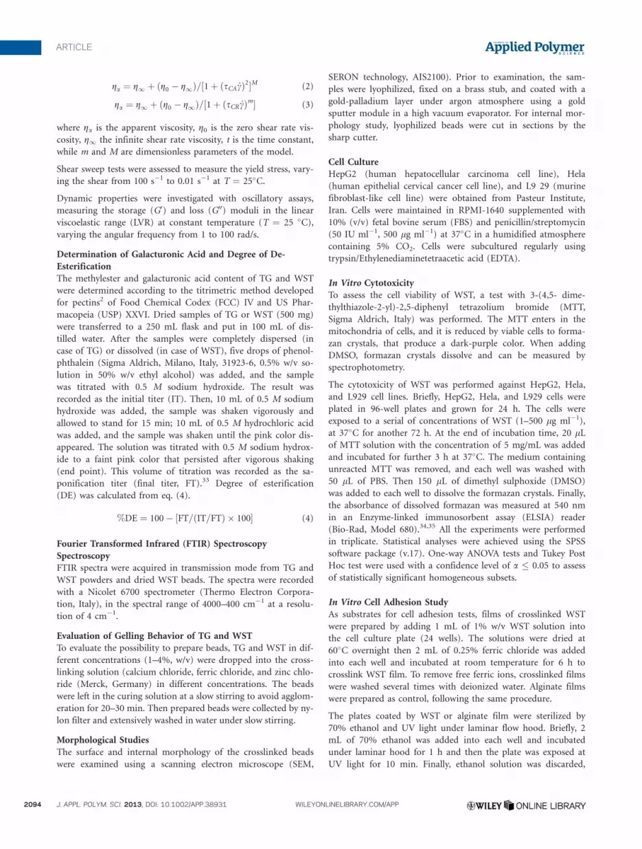
Viscosity and stress of TG solutions resulted higher than the
ones of WST (Figure 5) and comparable to the values found in
the literature for tragacanthin solutions.19,37 The differences
observed in the flow curves can be attributed to the different
physical form of the samples: TG is a suspension of insoluble
microgels or nanogels, while WST is an aqueous solution. Both
TG and WST exhibited a shear-thinning behavior, where the
shear viscosity decreases as the applied shear rates increases.
Viscosity data were fitted with the Carreau and Cross models
for non-Newtonian fluids (Table II), showing optimal fitting for
Carreau model.
To support the presence of shear-thinning behavior, the yield
stress was investigated with shear sweep measurements (Figure
5). For non-Newtonian solutions, the viscosity displays a slope
near �1 at low shear rates, which indicates the predominance
of yield stress effects, and the value of the yield stress remains
almost constant. The measured yield stress was 5.64 Pa for TG
and 0.06 Pa for WS, in both cases comparable with the stress
values at the end of the LVR.
The dynamic behavior of TG and WST is shown in Figure 6.
The procedures followed to extract WST from TG caused G0
and G00 values to decrease over the whole frequency range. The
obtained results are in correspondence with the trend reported
by other authors working in similar concentration ranges of A.
gossypinus tragacanth.38,39
The storage (G0) and loss (G00) moduli of TG slightly increased
with frequency, with G0 greater than G00 at almost all the frequency
range covered, indicating a gel-like behavior of TG (Figure 6). As
Figure 3. FTIR of raw tragancanth (bottom line, blue), WST (medium line, red), and Fe/WST (top line, green), (a) region 4000–500 cm�1 and (b) and
enlargement for the mid-infrared region at 1250–750 cm�1. Dashed lines indicate the differences in the bands. [Color figure can be viewed in the online
issue, which is available at wileyonlinelibrary.com.]
ARTICLE
WWW.MATERIALSVIEWS.COM WILEYONLINELIBRARY.COM/APP J. APPL. POLYM. SCI. 2013, DOI: 10.1002/APP.38931 2097
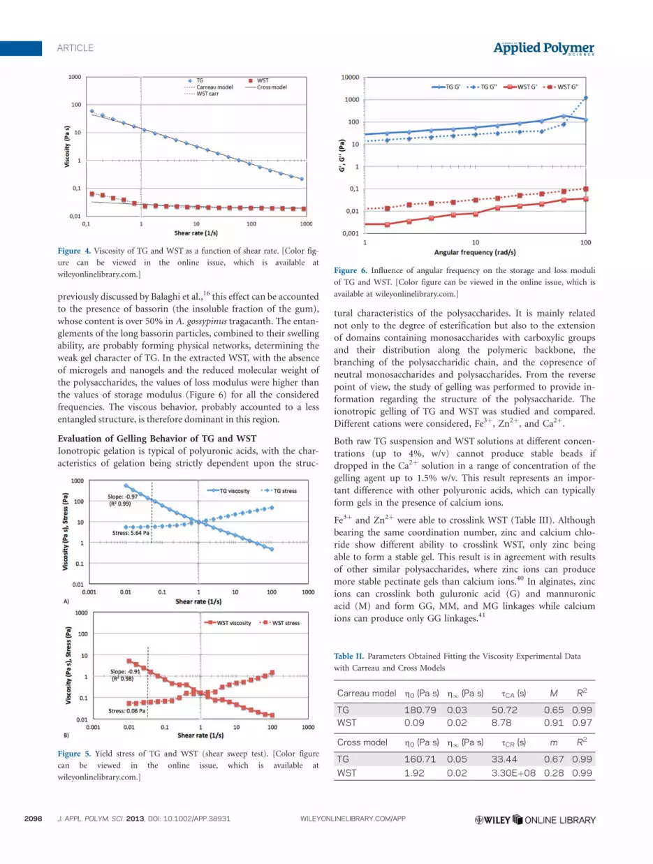
previously discussed by Balaghi et al.,16 this effect can be accounted
to the presence of bassorin (the insoluble fraction of the gum),
whose content is over 50% in A. gossypinus tragacanth. The entan-
glements of the long bassorin particles, combined to their swelling
ability, are probably forming physical networks, determining the
weak gel character of TG. In the extracted WST, with the absence
of microgels and nanogels and the reduced molecular weight of
the polysaccharides, the values of loss modulus were higher than
the values of storage modulus (Figure 6) for all the considered
frequencies. The viscous behavior, probably accounted to a less
entangled structure, is therefore dominant in this region.
Evaluation of Gelling Behavior of TG and WST
Ionotropic gelation is typical of polyuronic acids, with the char-
acteristics of gelation being strictly dependent upon the struc-
tural characteristics of the polysaccharides. It is mainly related
not only to the degree of esterification but also to the extension
of domains containing monosaccharides with carboxylic groups
and their distribution along the polymeric backbone, the
branching of the polysaccharidic chain, and the copresence of
neutral monosaccharides and polysaccharides. From the reverse
point of view, the study of gelling was performed to provide in-
formation regarding the structure of the polysaccharide. The
ionotropic gelling of TG and WST was studied and compared.
Different cations were considered, Fe3þ, Zn2þ, and Ca2þ.
Both raw TG suspension and WST solutions at different concen-
trations (up to 4%, w/v) cannot produce stable beads if
dropped in the Ca2þ solution in a range of concentration of the
gelling agent up to 1.5% w/v. This result represents an impor-
tant difference with other polyuronic acids, which can typically
form gels in the presence of calcium ions.
Fe3þ and Zn2þ were able to crosslink WST (Table III). Although
bearing the same coordination number, zinc and calcium chlo-
ride show different ability to crosslink WST, only zinc being
able to form a stable gel. This result is in agreement with results
of other similar polysaccharides, where zinc ions can produce
more stable pectinate gels than calcium ions.40 In alginates, zinc
ions can crosslink both guluronic acid (G) and mannuronic
acid (M) and form GG, MM, and MG linkages while calcium
ions can produce only GG linkages.41
Figure 4. Viscosity of TG and WST as a function of shear rate. [Color fig-
ure can be viewed in the online issue, which is available at
wileyonlinelibrary.com.]
Figure 5. Yield stress of TG and WST (shear sweep test). [Color figure
can be viewed in the online issue, which is available at
wileyonlinelibrary.com.]
Figure 6. Influence of angular frequency on the storage and loss moduli
of TG and WST. [Color figure can be viewed in the online issue, which is
available at wileyonlinelibrary.com.]
Table II. Parameters Obtained Fitting the Viscosity Experimental Data
with Carreau and Cross Models
Carreau model g0 (Pa s) g1 (Pa s) sCA (s) M R2
TG 180.79 0.03 50.72 0.65 0.99WST 0.09 0.02 8.78 0.91 0.97
Cross model g0 (Pa s) g1 (Pa s) sCR (s) m R2
TG 160.71 0.05 33.44 0.67 0.99
WST 1.92 0.02 3.30Eþ08 0.28 0.99
ARTICLE
2098 J. APPL. POLYM. SCI. 2013, DOI: 10.1002/APP.38931 WILEYONLINELIBRARY.COM/APP

Spherical beads were observed when 1% WST solution was
dropped into Fe3þ solution over a critical concentration
(�0.125% v/w FeCl3). Zinc was less effective than ferric ion as
crosslinking agents at higher concentration (�1% v/w) and lon-
ger curing times are necessary to produce stable macrospheres.
Moreover, increasing the Zn2þ concentration to 2–4% w/v
resulted in beads with outer thin shells and liquid cores that
tended to be disrupted during the washing steps. As reported in
literature, Zn2þ crosslinked pectins42,43 possess lower water con-
tent if compared to pectin gels obtained from other cations.
The obtained results can be consistent with an outer shell char-
acterized by reduced water content if compared to Fe3þ cross-
linked beads, thus slowing or preventing the penetration of the
ions in the core of the beads. The increase of concentration was
only effective in a faster production of a Zn crosslinked surface
of the bead. According to these observations, only Fe-WST was
further studied.
Structural Characterization of the Beads
As ferric ions replace sodium ions in the WST sodium salt, the
charge density, radius, and atomic weight of the cation are
changed, creating a new environment around the carboxyl
group.44–46 Accordingly, results, reported in Table I and Figure
3(a,b), indicate peak shifts and modifications mainly in the
bands relative to carboxyl group, namely asymmetric and sym-
metric stretching of carboxylate group and twisting band of
CH2. Other minor structural modifications are observed in the
FTIR spectrum of Fe/WST, for example, symmetric deformation
of AOACH3 groups in methyl esters shows a doublet at 1375
and 1359 cm�1, that was a single band for TG at 1368 cm�1.
Morphology of the Beads
Figure 7(a) shows spherical, narrow distribution of the size of
the beads obtained by a digital camera. SEM images of lyophi-
lized beads, showing surface and internal morphology are
reported in Figure 7b–d.
Table III. Gelling Behavior of TG and WST in the Presence of Metallic
Ions
Polymerconcentration
Gellingagent Results
(w/v) Type (w/v) mM
1% FeCl3 0.062% 3.9 Unstable spherical bead
0.125% 7.7 Stable spherical bead
0.250% 15.4 Stable spherical bead
0.500% 30.8 Stable spherical bead
1.000% 61.6 Stable spherical bead
1–2% ZnCl2 1.000% 73.3 Stable spherical bead
2.000% 146.6 Unstable spherical bead
3.000% 220.0 Unstable bead
4.000% 293.2 Unstable bead
Figure 7. (a) Optical image of beads, (b) SEM image of bead, (c) SEM image of surface of bead, (d) SEM image of internal structure of bead. Beads
were prepared by 1% WST and 0.5% ferric chloride. [Color figure can be viewed in the online issue, which is available at wileyonlinelibrary.com.]
ARTICLE
WWW.MATERIALSVIEWS.COM WILEYONLINELIBRARY.COM/APP J. APPL. POLYM. SCI. 2013, DOI: 10.1002/APP.38931 2099

Lyophilization may have induced changes in the morphology, in
the intermolecular bonds and in the porosity. The samples ana-
lyzed by SEM are indeed different from the swollen samples,
but still information can be gained from the comparison of the
micrographs of the beads, lyophilized in the same conditions, as
the sublimation of water is leaving a porosity which is somehow
related to the different water distribution in the wet samples.
This is giving, indirectly, information about the different char-
acteristics of the gels that can be obtained from WST.
No microporosity can be observed on the surface (Figure 7c),
indicating a low water content prior lyophilization of the beads.
The internal part of beads is highly porous (Figure 7d). As
shown in EDX analysis, the amount of ferric ions in internal
part is almost two times more concentrated than in the interior
part (Figure 8). These observations are pursuant to a high cross-
linking degree on the surface of the beads resulting in low water
content, while a less crosslinked gel, that contains higher amount
of water in the interior part, could account the higher porosity
resulting from lyophilization. Some cracks are observable on the
surface (Figure 7b) suggesting again the different water content,
which may be connected with a higher volume expansion during
the freezing process in comparison to the surface of the bead.
The inhomogeneous water content of the inner and outer layer
can be considered an interesting characteristic to be further
investigated to control the thickness of the outer layer affecting
drug loading and release profiles. Higher water content in the
inner part can also be positively considered for cell encapsula-
tion. Data regarding Zn-crosslinked beads indicates that the
effect of the cation concentration can modify this characteristic.
Cytotoxicity and Cell Adhesion
Viability data of L929, Hela, and HepG2 cells (cultivated with
RPMI-1640) at 72 h are shown in Figure 9. To investigate cell
viability, WST cytotoxicity on two cancerous cell lines of Hela
Figure 8. EDX analysis of ferric crosslinked WST beads: (a) surface of beads, (b) internal part. [Color figure can be viewed in the online issue, which is
available at wileyonlinelibrary.com.]
Figure 9. Relative cell viability of WST solution on L929 cells, Hela cells,
and HepG2 cells. The relative cell viability read for the control (tissue cul-
ture polystyrene from culture plates) after 72 h of incubation was taken as
the reference (100%). Significance was calculated by ANOVA (*p � 0).
ARTICLE
2100 J. APPL. POLYM. SCI. 2013, DOI: 10.1002/APP.38931 WILEYONLINELIBRARY.COM/APP

and HepG2, and a fibroblast cell line of L929 has been assayed.
WST has shown no toxicity either in Hela and HepG2 as can-
cerous cell lines or in L929 normal cell line. Cell viability of
L929 cells was slightly improved in concentration of 1–100 lg/
ml, and there are significant differences between viability of
L929 and Hela cell lines in 1 and 10 lg/ml. By comparing via-
bility of HepG2 with Hela cells no significant differences can be
detected between HepG2 which expose asialoglycoprotein-recep-
tors (ASGP-R) and galactose receptor and Hela, which does not
express ASGP-R. Therefore, cells owing different characteristics
are compatible with WST.
One of the main disadvantages of anionic polysaccharides in
cell and tissue engineering is poor cell adhesion. L929 adhered
on ferric crosslinked WST films with morphology similar to the
culture plate. Ferric crosslinked alginate films, as control,
showed a round morphology indicating poor cell adhesion (Fig-
ure 10). Considering spherical cells as nonadhered cells, the
mean percentage of adhered cells was 10% and 50% for alginate
gel and WST gel, respectively. The better cell adhesion of WST
can be attributed to different interactions of cells with the spe-
cific side chain sugars of tragacanth.
CONCLUSIONS
In this study, a procedure was set up to prepare WST by de-
esterification of TG, while retaining a high molecular weight
and the main structural characteristics of raw TG and overcom-
ing some of its typical limitations such as poor solubility and a
limited ability to form water stable gels. Evaluation of WST cy-
totoxicity on Hela, HepG2, and L929 indicated lack of toxicity
on these cell lines even at relatively high concentrations (500
lg/mL). WST was able to gel, and its gelling behavior is strictly
dependent of the counter ion type. Among ferric, zinc, and cal-
cium cations, ferric could interact with WST strongly thus mak-
ing a stable hydrogel. Unlike pectin and alginate, well-known
anionic polysaccharides, WST was not able to form hydrogels
with calcium ions. This different behavior toward the different
counter ions to promote ionotropic gelation could be related to
the content and distribution, along the high molecular weight
chains, of carboxylic acids available for ionic crosslinking and a
specific branching, which can hinder the accessibility of these
groups. Cell adhesion study proved cell adhesion ability of WST
crosslinked with ferric ions. These properties of WST all to-
gether make this material a potential candidate for cell/tissue
engineering, and future studies might highlight its applications
also in the field of controlled drug delivery.
ACKNOWLEDGMENTS
The authors gratefully acknowledge Monica Moscatelli for GPC
analyses and Marco Coletti for the support in the interpretation of
the rheological analysis.
REFERENCES
1. Liu, Z.; Jiao, J.; Wang, Y.; Zhou, C.; Zhang, Z. Adv. Drug
Deliv. Rev. 2008, 60, 1650.
2. Munarin, F.; Petrini, P.; Tanzi, M. C.; Barbosa, M. A.;
Granja, P. L. Soft Matter 2012, 8, 4731.
3. Mano, J.; Silva, G.; Azevedo, H.; Malafaya, P.; Sousa, R.;
Silva, S.; Boesel, L.; Oliveira, J.; Santos, T.; Marques, A. J. R.
Soc. Interface 2007, 4, 999.
4. Rana, V.; Rai, P.; Tiwary, A. K.; Singh, R. S.; Kennedy, J. F.;
Knill, C. J. Carbohydr. Polym. 2011, 83, 1031.
5. Verbeken, D.; Dierckx, S.; Dewettinck, K. Appl. Microbiol.
Biotechnol. 2003, 63, 10.
6. Thimma, R. T.; Tammishetti, S. J. Appl. Polym. Sci. 2001,
82, 3084.
Figure 10. Cell adhesion of L929 on (a) cell culture plate, (b) ferric algi-
nate film, (c) ferric crosslinked WST film. Images were acquired by reverse
phase microscope (Olympus IX70). [Color figure can be viewed in the
online issue, which is available at wileyonlinelibrary.com.]
ARTICLE
WWW.MATERIALSVIEWS.COM WILEYONLINELIBRARY.COM/APP J. APPL. POLYM. SCI. 2013, DOI: 10.1002/APP.38931 2101

7. Munarin, F.; Petrini, P.; Fare, S.; Tanzi, M. C. J. Mater. Sci.:
Mater. Med. 2010, 21, 365.
8. Munarin, F.; Guerreiro, S.; Grellier, M.; Tanzi, M. C.; Bar-
bosa, M. A.; Petrini, P.; Granja, P. L. Biomacromolecule
2011, 12, 568.
9. Munarin, F.; Tanzi, M. C.; Petrini, P. Int. J. Biol. Macromol.
2012, 51, 681.
10. Gazori, T.; Khoshayand, M.; Azizi, E.; Yazdizade, P.;
Nomani, A.; Haririan, I. Carbohydr. Polym. 2009, 77, 599.
11. Augst, A. D.; Kong, H. J.; Mooney, D. J. Macromol. Biosci.
2006, 6, 623.
12. El-Sherbiny, I. M. Carbohydr. Polym. 2010, 80, 1125.
13. Mura, C.; Manconi, M.; Valenti, D.; Manca, M. L.; Dk�ez-
Sales, O.; Loy, G.; Fadda, A. M. Carbohydr. Polym. 2011, 85,
578.
14. Oliveira, G. F.; Ferrari, P. C.; Carvalho, L. Q.; Evangelista,
R. C. Carbohydr. Polym. 2010, 82, 1004.
15. Khajavi, R.; Pourgharbi, S.; Rashidi, A.; Kiumarsi, A. Int. J.
Eng. 2004, 17, 201.
16. Balaghi, S.; Mohammadifar, M.; Zargaraan, A. Food Biophys.
2010, 5, 59.
17. Mirhosseini, H.; Amid, B. T. Food Res. Int. 2012, 46, 387.
18. Phillips, G. O.; Williams, P. A. Handbook of Hydrocolloids;
CRC Press LLC: Boca Raton, FL, 2000, p 231.
19. Mohammadifar, M. A.; Musavi, S. M.; Kiumarsi, A.; Wil-
liams, P. A. Int. J. Biol. Macromol. 2006, 38, 31.
20. Seaman, J.; Davidson, R. Handbook of Water Soluble Gums
and Resins; Mc Graw-Hill Book Company: New York, 1980.
21. Aspinall, G.; Baillie, J. J. Chem. Soc. 1963, 1963, 1702.
22. Kaffashi, B.; Zandieh, A.; Khadiv-Parsi, P. Macromol. Symp.
2006, 239, 120.
23. Siahi, M. R.; Barzegar-Jalali, M.; Monajjemzadeh, F.; Ghaf-
fari, F.; Azarmi, S. AAPS Pharm. Sci. Tech. 2005, 6, 626.
24. Cevik, A. G. S J. Microencaps. 2000, 17, 565.
25. Moghbel, A.; Naji, M. Sci. Med. J. 2008, 7, 274.
26. Kiani, A.; Shahbazi, M.; Asempour, H. J. Appl. Polym. Sci.
2012, 1, 99.
27. Kiani, A.; Asempour, H. J. Appl. Polym. Sci. 2012, 126,
E478.
28. Cho, C.; Seo, S.; Park, I.; Kim, S.; Kim, T.; Hoshiba, T.;
Harada, I.; Akaike, T. Biomaterials 2006, 27, 576.
29. Deters, A. M.; Lengsfeld, C.; Hensel, A. J. Ethnopharmacol.
2005, 102, 391.
30. Robert, L.; Fodil-Bourahla, I.; Bizbiz, L.; Robert, A. Biomed.
Pharmacother. 2004, 58, 123.
31. Munarin, F.; Petrini, P.; Bozzini, S.; Tanzi, M. C. J. Appl.
Biomater. Function. Mater. 2012, 10, 67.
32. Yoo, S-H.; Fishman, M. L.; Hotchkiss, A. T., Jr.; Lee, H. G.
Food Hydrocoll. 2006, 20, 62.
33. Ghaffari, A.; Navaee, K.; Oskoui, M.; Bayati, K.; Rafiee-Teh-
rani, M. Eur. J. Pharm. Biopharm. 2007, 67, 175.
34. Yoo, H.; Park, T. J. Control Release 2001, 70, 63.
35. Yokoyama, M.; Okano, T.; Sakurai, Y.; Suwa, S.; Kataoka, K.
J. Control Release 1996, 39, 351.
36. Kacurakova, M.; Capek, P.; Sasinkova, V.; Wellner, N.;
Ebringerova, A. Carbohydr. Polym. 2000, 43, 195.
37. Chenlo, F.; Moreira, R.; Silva, C. J. Food Eng. 2010, 96, 107.
38. Balaghi, S.; Mohammadifar, M. A.; Zargaraan, A. Food Bio-
phys. 2010, 5, 59.
39. Balaghi, S.; Mohammadifar, M. A.; Zargaraan, A.; Gavlighi,
H. A.; Mohammadi, M. Food Hydrocolloid 2011, 25, 1775.
40. Das, S.; Chaudhury, A.; Ng, K. Y. Int. J. Pharm. 2011, 406,
11.
41. Jay, S. M.; Saltzman, W. M. J. Control Release 2009, 134, 26.
42. Kawadkar, J.; Meenakshi, K. C.; Ram, A. DARU J. Pharm.
Sci. 2010, 18, 211.
43. Goh, C. H.; Heng, P. W. S.; Chan, L. W. Carbohydr. Polym.
2012, 88, 1.
44. Pathak, T.; Yun, J.; Lee, J.; Paeng, K. Carbohydr. Polym.
2010, 81, 633.
45. Assifaoui, A.; Loupiac, C.; Chambin, O.; Cayot, P. Carbo-
hydr. Res. 2010, 345, 929.
46. Papageorgiou, S.; Kouvelos, E.; Favvas, E.; Sapalidis, A.;
Romanos, G.; Katsaros, F. Carbohydr. Res. 2010, 345, 469.
ARTICLE
2102 J. APPL. POLYM. SCI. 2013, DOI: 10.1002/APP.38931 WILEYONLINELIBRARY.COM/APP


