Embed Size (px)
Citation preview
Carbohydrates
by Assist. Prof. Görke Gürel Peközer
Yıldız Technical University Biomedical Engineering Department
Fall 2020
Carbohydrates Carbohydrates (also called saccharides) are—on the basis of mass—the most
abundant class of biological molecules on Earth.
Although all organisms can synthesize carbohydrates from available monomers, photosynthetic organisms, including bacteria, algae, and plants convert solar energy to chemical energy that is then used to make carbohydrate from carbon dioxide.
Carbohydrates play several crucial roles in living organisms. In animals and plants, carbohydrate polymers act as energy storage molecules. Animals can ingest carbohydrates that can then be oxidized to yield energy for metabolic processes.
Polymeric carbohydrates are also found in cell walls and in the protective coatings of many organisms.
Other carbohydrate polymers are marker molecules that allow one type of cell to recognize and interact with another type.
Carbohydrate derivatives are found in a number of biological molecules, including some coenzymes and the nucleic acids.
Glycoconjugates are carbohydrate derivatives in which one or more carbohydrate chains are linked covalently to a peptide, protein, or lipid. These derivatives include proteoglycans, peptidoglycans, glycoproteins, and glycolipids.
Carbohydrates can be described by the number of monomeric units they contain.
Monosaccharides are the smallest units of carbohydrate structure.
Oligosaccharides are polymers of two to about 20 monosaccharide residues. The most common oligosaccharides are disaccharides, which consist of two linked monosaccharide residues.
Polysaccharides are polymers that contain many (usually more than 20) monosaccharide residues.
Monosaccharides
The simplest of the carbohydrates, monosaccharides, are water-soluble, white, crystalline solids that have a sweet taste.
Glucose, fructose and galactose are some of them.
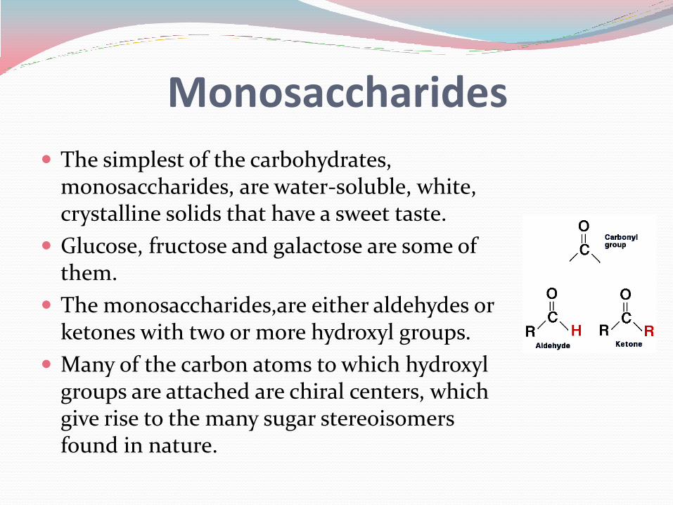
The monosaccharides,are either aldehydes or ketones with two or more hydroxyl groups.
Many of the carbon atoms to which hydroxyl groups are attached are chiral centers, which give rise to the many sugar stereoisomers found in nature.
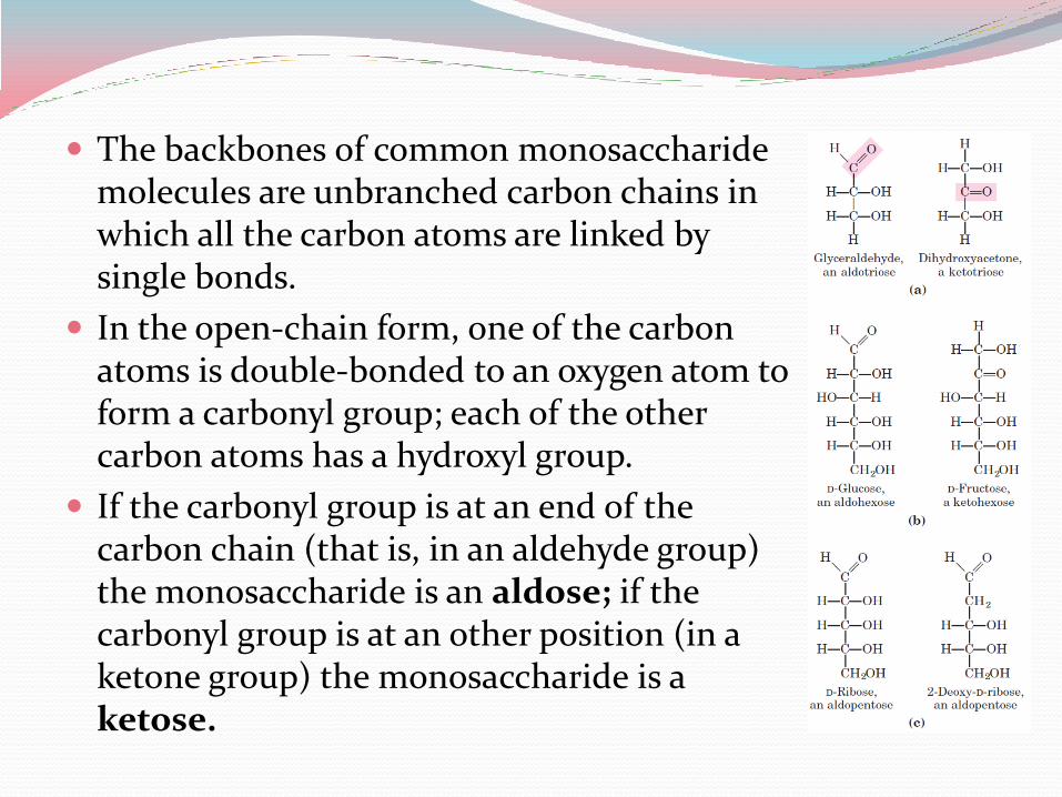
The backbones of common monosaccharide molecules are unbranched carbon chains in which all the carbon atoms are linked by single bonds.
In the open-chain form, one of the carbon atoms is double-bonded to an oxygen atom to form a carbonyl group; each of the other carbon atoms has a hydroxyl group.
If the carbonyl group is at an end of the carbon chain (that is, in an aldehyde group) the monosaccharide is an aldose; if the carbonyl group is at an other position (in a ketone group) the monosaccharide is a ketose.
Monosaccharides with four, five, six, and seven carbon atoms in their backbones are called, respectively, tetroses, pentoses, hexoses, and heptoses.
There are aldoses and ketoses of each of these chain lengths: aldotetroses and ketotetroses, aldopentoses and ketopentoses.
The hexoses, which include the aldohexose D-glucose and the ketohexose D-fructose, are the most common monosaccharides in nature.
The aldopentoses D-ribose and 2-deoxy-D-ribose are components of nucleotides and nucleic acids.
Monosaccharides
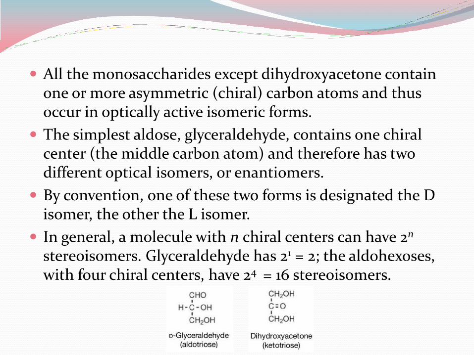
All the monosaccharides except dihydroxyacetone contain one or more asymmetric (chiral) carbon atoms and thus occur in optically active isomeric forms.
The simplest aldose, glyceraldehyde, contains one chiral center (the middle carbon atom) and therefore has two different optical isomers, or enantiomers.
By convention, one of these two forms is designated the D isomer, the other the L isomer.
In general, a molecule with n chiral centers can have 2n
stereoisomers. Glyceraldehyde has 21 = 2; the aldohexoses, with four chiral centers, have 24 = 16 stereoisomers.
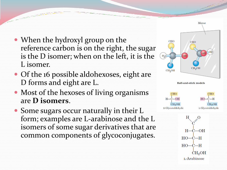
When the hydroxyl group on the reference carbon is on the right, the sugar is the D isomer; when on the left, it is the L isomer.
Of the 16 possible aldohexoses, eight are D forms and eight are L.
Most of the hexoses of living organisms are D isomers.
Some sugars occur naturally in their L form; examples are L-arabinose and the L isomers of some sugar derivatives that are common components of glycoconjugates.
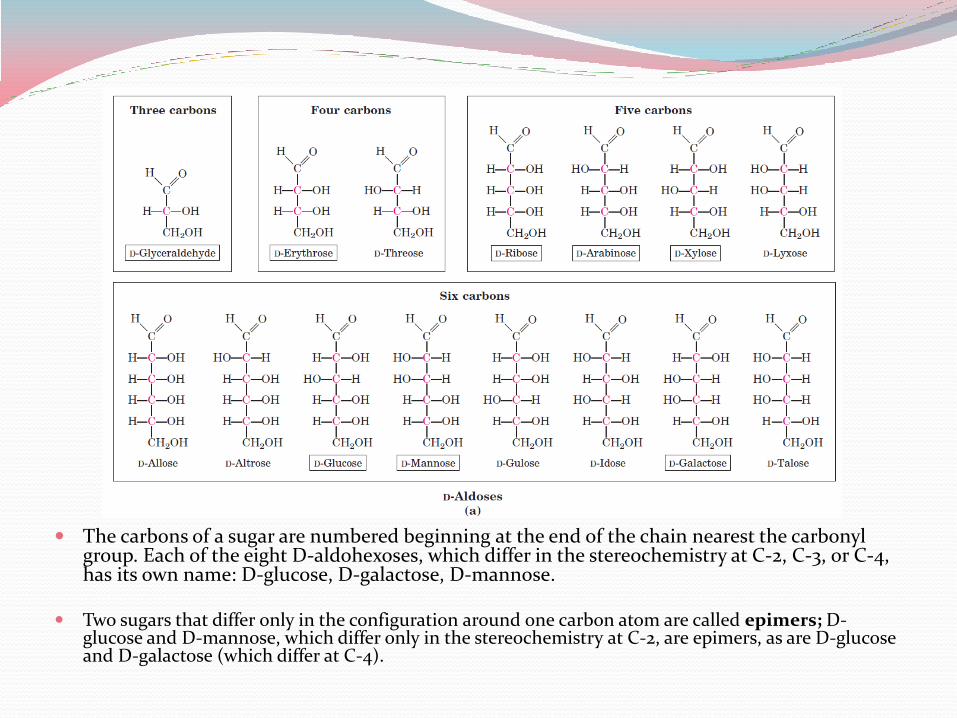
The carbons of a sugar are numbered beginning at the end of the chain nearest the carbonyl group. Each of the eight D-aldohexoses, which differ in the stereochemistry at C-2, C-3, or C-4, has its own name: D-glucose, D-galactose, D-mannose.
Two sugars that differ only in the configuration around one carbon atom are called epimers; D-glucose and D-mannose, which differ only in the stereochemistry at C-2, are epimers, as are D-glucose and D-galactose (which differ at C-4).
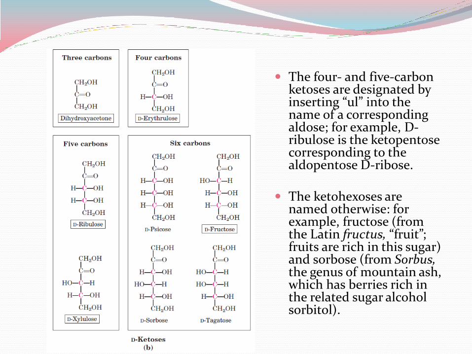
The four- and five-carbon ketoses are designated by inserting “ul” into the name of a corresponding aldose; for example, D-ribulose is the ketopentose corresponding to the aldopentose D-ribose.
The ketohexoses are named otherwise: for example, fructose (from the Latin fructus, “fruit”; fruits are rich in this sugar) and sorbose (from Sorbus, the genus of mountain ash, which has berries rich in the related sugar alcohol sorbitol).

In fact, in aqueous solution, aldotetroses and all monosaccharides with five or more carbon atoms in the backbone occur predominantly as cyclic (ring) structures in which the carbonyl group has formed a covalent bond with the oxygen of a hydroxyl group along the chain.
The formation of these ring structures is the result of a general reaction between alcohols and aldehydes or ketones to form derivatives called hemiacetals or hemiketals, which contain an additional asymmetric carbon atom and thus can exist in two stereoisomeric forms.
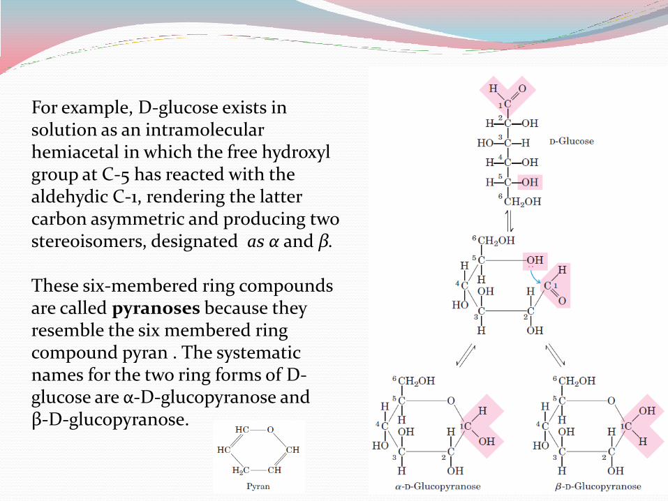
For example, D-glucose exists in solution as an intramolecular hemiacetal in which the free hydroxyl group at C-5 has reacted with the aldehydic C-1, rendering the latter carbon asymmetric and producing two stereoisomers, designated as α and β. These six-membered ring compounds are called pyranoses because they resemble the six membered ring compound pyran . The systematic names for the two ring forms of D-glucose are α-D-glucopyranose and β-D-glucopyranose.
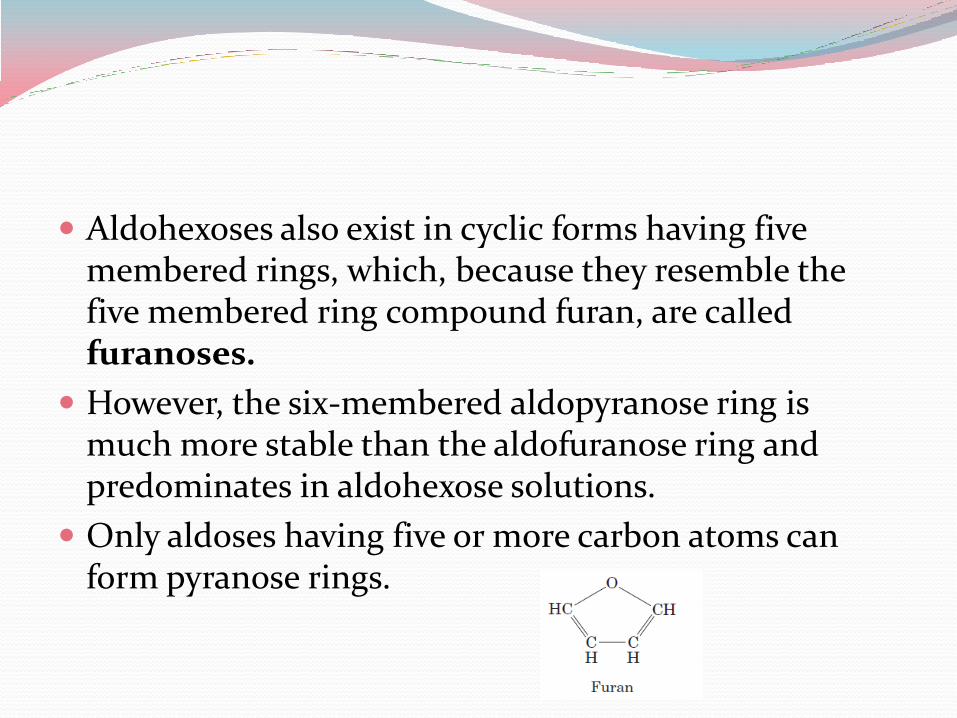
Aldohexoses also exist in cyclic forms having five membered rings, which, because they resemble the five membered ring compound furan, are called furanoses.
However, the six-membered aldopyranose ring is much more stable than the aldofuranose ring and predominates in aldohexose solutions.
Only aldoses having five or more carbon atoms can form pyranose rings.
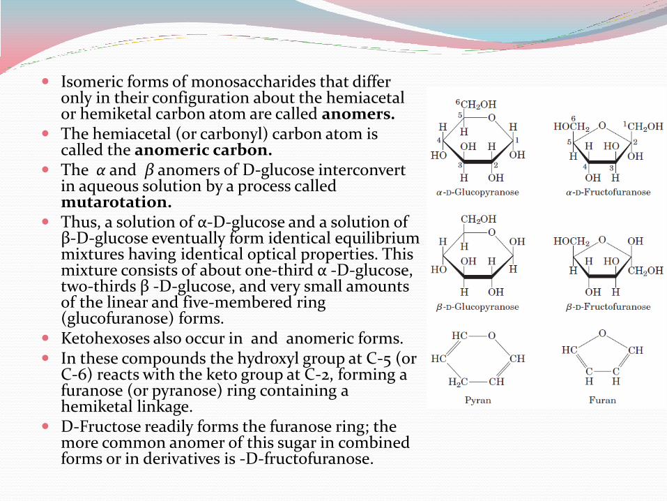
Isomeric forms of monosaccharides that differ only in their configuration about the hemiacetal or hemiketal carbon atom are called anomers.
The hemiacetal (or carbonyl) carbon atom is called the anomeric carbon.
The α and β anomers of D-glucose interconvert in aqueous solution by a process called mutarotation.
Thus, a solution of α-D-glucose and a solution of β-D-glucose eventually form identical equilibrium mixtures having identical optical properties. This mixture consists of about one-third α -D-glucose, two-thirds β -D-glucose, and very small amounts of the linear and five-membered ring (glucofuranose) forms.
Ketohexoses also occur in and anomeric forms. In these compounds the hydroxyl group at C-5 (or
C-6) reacts with the keto group at C-2, forming a furanose (or pyranose) ring containing a hemiketal linkage.
D-Fructose readily forms the furanose ring; the more common anomer of this sugar in combined forms or in derivatives is -D-fructofuranose.
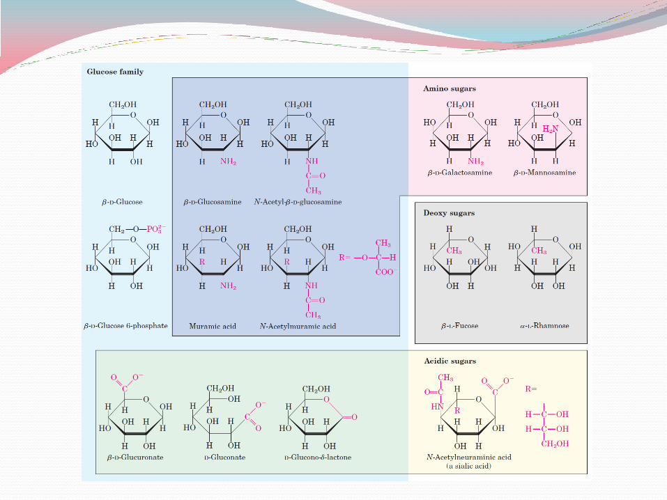
Sugar Derivatives There are a number of sugar derivatives in which a hydroxyl group in the parent
compound is replaced with another substituent, or a carbon atom is oxidized to a carboxyl group.
Sugar phophates: Monosaccharides are often converted to phosphate esters which are intermediates in
carbohydrate metabolism. One effect of sugar phosphorylation within cells is to trap the sugar inside the cell; most cells do not have plasma membrane transporters for phosphorylated sugars. Phosphorylation also activates sugars for subsequent chemical transformation.
Deoxy sugars: In these derivatives, a hydrogen atom replaces one of the hydroxyl groups in the parent
monosaccharide. 2-Deoxy-D-ribose is an important building block for DNA.
Aminosugars: In glucosamine, galactosamine, and mannosamine, the hydroxyl at C-2 of the parent
compound is replaced with an amino group. The amino group is nearly always condensed with acetic acid, as in N-acetylglucosamine. This glucosamine derivative is part of many structural polymers, including those of the bacterial cell wall.
Bacterial cell walls also contain a derivative of glucosamine, N-acetylmuramic acid, in which lactic acid (a three-carbon carboxylic acid) is ether-linked to the oxygen at C-3 of N-acetylglucosamine. The substitution of a hydrogen for the hydroxyl group at C-6 of L-galactose or L-mannose produces L-fucose or L-rhamnose, respectively; these deoxy sugars are found in plant polysaccharides and in the complex oligosaccharide components of glycoproteins and glycolipids.
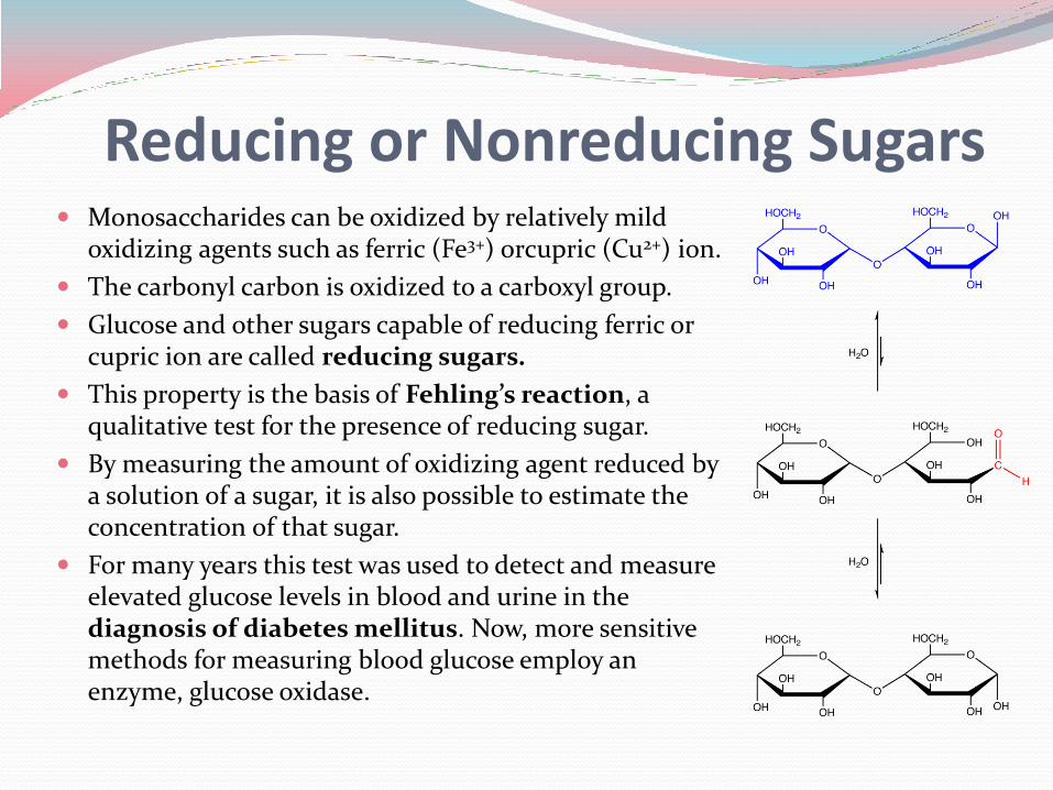
Reducing or Nonreducing Sugars Monosaccharides can be oxidized by relatively mild
oxidizing agents such as ferric (Fe3+) orcupric (Cu2+) ion.
The carbonyl carbon is oxidized to a carboxyl group.
Glucose and other sugars capable of reducing ferric or cupric ion are called reducing sugars.
This property is the basis of Fehling’s reaction, a qualitative test for the presence of reducing sugar.
By measuring the amount of oxidizing agent reduced by a solution of a sugar, it is also possible to estimate the concentration of that sugar.
For many years this test was used to detect and measure elevated glucose levels in blood and urine in the diagnosis of diabetes mellitus. Now, more sensitive methods for measuring blood glucose employ an enzyme, glucose oxidase.
The reducing ability of a sugar polymer is of more than analytical interest. The polymeric chains of oligosaccharides and polysaccharides show directionality based on their reducing and nonreducing ends.
There is usually one reducing end (the residue containing the free anomeric carbon) and one nonreducing end in a linear polymer.
All the internal glycosidic bonds of a polysaccharide involve acetals. The internal residues are not in equilibrium with open-chain forms and thus cannot reduce metal ions.
A branched polysaccharide has a number of nonreducing ends but only one reducing end.
Disaccharides Disaccharides (such as maltose, lactose, and sucrose)
consist of two monosaccharides joined covalently by an O-glycosidic bond, which is formed when a hydroxyl group of one sugar reacts with the anomeric carbon of the other.
This reaction represents the formation of an acetal from a hemiacetal (such as glucopyranose) and an alcohol (a hydroxyl group of the second sugar molecule).
Glycosidic bonds are readily hydrolyzed by acid but resist cleavage by base.
Thus disaccharides can be hydrolyzed to yield their free monosaccharide components by boiling with dilute acid.
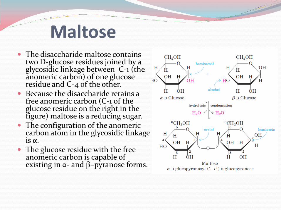
The disaccharide maltose contains two D-glucose residues joined by a glycosidic linkage between C-1 (the anomeric carbon) of one glucose residue and C-4 of the other.
Because the disaccharide retains a free anomeric carbon (C-1 of the glucose residue on the right in the figure) maltose is a reducing sugar.
The configuration of the anomeric carbon atom in the glycosidic linkage is α.
The glucose residue with the free anomeric carbon is capable of existing in α- and β–pyranose forms.
Maltose
To name reducing disaccharides such as maltose unambiguously, and especially to name more complex oligosaccharides, several rules are followed. By convention, the name describes the compound with its nonreducing end to the left, and we can “build up” the name in the following order. 1. Give the configuration (α or β) at the anomeric carbon joining the
first monosaccharide unit (on the left) to the second. 2. Name the nonreducing residue; to distinguish five- and six-
membered ring structures, insert “furano” or “pyrano” into the name. 3. Indicate in parentheses the two carbon atoms joined by the
glycosidic bond, with an arrow connecting the two numbers; for example, (14) shows that C-1 of the first-named sugar residue is joined to C-4 of the second.
4. Name the second residue. 5. If there is a third residue, describe the second glycosidic bond by the
same conventions.
Following this convention for naming oligosaccharides, maltose is α-D-glucopyranosyl-(14)-D-glucopyranose.
Giving the configuration of the anomeric carbon and naming the carbons joined by the glycosidic bond nomenclature can be abbreviated, such as for maltose it is Glc(α14)Glc.
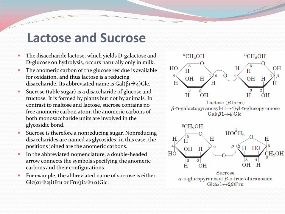
Lactose and Sucrose The disaccharide lactose, which yields D-galactose and
D-glucose on hydrolysis, occurs naturally only in milk.
The anomeric carbon of the glucose residue is available for oxidation, and thus lactose is a reducing disaccharide. Its abbreviated name is Gal(β14)Glc.
Sucrose (table sugar) is a disaccharide of glucose and fructose. It is formed by plants but not by animals. In contrast to maltose and lactose, sucrose contains no free anomeric carbon atom; the anomeric carbons of both monosaccharide units are involved in the glycosidic bond.
Sucrose is therefore a nonreducing sugar. Nonreducing disaccharides are named as glycosides; in this case, the positions joined are the anomeric carbons.
In the abbreviated nomenclature, a double-headed arrow connects the symbols specifying the anomeric carbons and their configurations.
For example, the abbreviated name of sucrose is either Glc(α12β)Fru or Fru(β21 α)Glc.
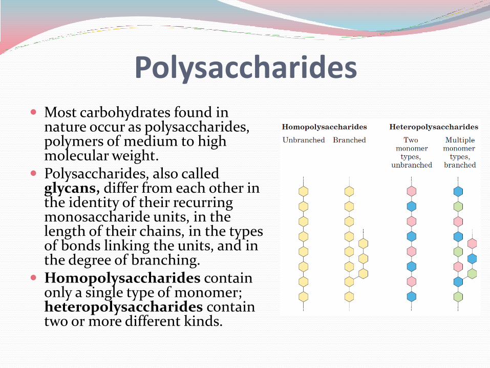
Polysaccharides Most carbohydrates found in
nature occur as polysaccharides, polymers of medium to high molecular weight.
Polysaccharides, also called glycans, differ from each other in the identity of their recurring monosaccharide units, in the length of their chains, in the types of bonds linking the units, and in the degree of branching.
Homopolysaccharides contain only a single type of monomer; heteropolysaccharides contain two or more different kinds.
Some homopolysaccharides serve as storage forms of monosaccharides that are used as fuels; starch and glycogen are homopolysaccharides of this type.
Other homopolysaccharides (cellulose and chitin, for example) serve as structural elements in plant cell walls and animal exoskeletons.
Heteropolysaccharides provide extracellular support for organisms of all kingdoms.
For example, the rigid layer of the bacterial cell envelope (the peptidoglycan) is composed in part of a heteropolysaccharide built from two alternating monosaccharide units.
In animal tissues, the extracellular space is occupied by several types of heteropolysaccharides, which form a matrix that holds individual cells together and provides protection, shape, and support to cells, tissues, and organs.
Unlike proteins, polysaccharides generally do not have definite molecular weights. This difference is a consequence of the mechanisms of assembly of the two types of polymers.
Proteins are synthesized on a template (messenger RNA) of defined sequence and length, by enzymes that follow the template exactly.
For polysaccharide synthesis there is no template; rather, the program for polysaccharide synthesis is intrinsic to the enzymes that catalyze the polymerization of the monomeric units, and there is no specific stopping point in the synthetic process.
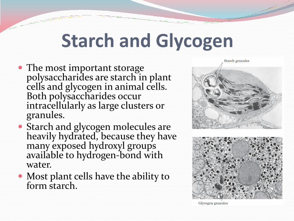
Starch and Glycogen The most important storage
polysaccharides are starch in plant cells and glycogen in animal cells. Both polysaccharides occur intracellularly as large clusters or granules.
Starch and glycogen molecules are heavily hydrated, because they have many exposed hydroxyl groups available to hydrogen-bond with water.
Most plant cells have the ability to form starch.
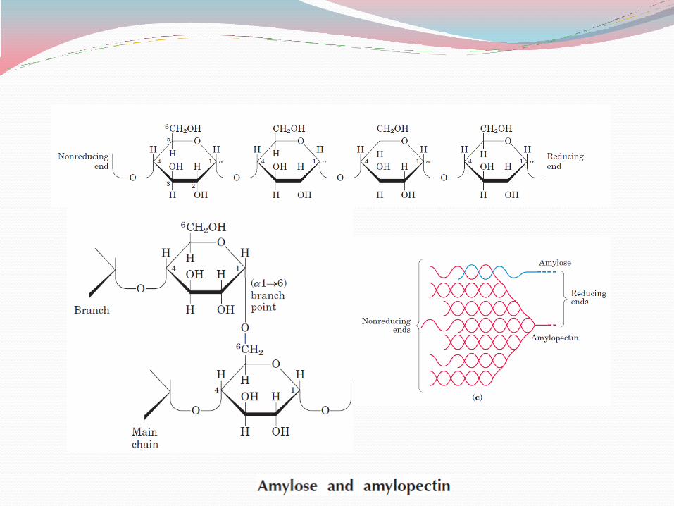
STARCH Starch contains two types of glucose polymer, amylose and
amylopectin.
Amylose consists of long, unbranched chains of D-glucose residues connected by (α14) linkages. Such chains vary in molecular weight from a few thousand to more than a million.
Amylopectin also has a high molecular weight (up to 100 million) but unlike amylose is highly branched.
The glycosidic linkages joining successive glucose residues in amylopectin chains are (α14); the branch points (occurring every 24 to 30 residues) are (α16) linkages.
GLYCOGEN Glycogen is the main storage polysaccharide of animal cells.
Like amylopectin, glycogen is a polymer of (1n4)-linked subunits of glucose, with (1n6)-linked branches, but glycogen is more extensively branched (on average, every 8 to 12 residues) and more compact than starch.
Glycogen is especially abundant in the liver, where it may constitute as much as 7% of the wet weight; it is also present in skeletal muscle.
In hepatocytes glycogen is found in large granules, which are themselves clusters of smaller granules composed of single, highly branched glycogen molecules with an average molecular weight of several million.
Such glycogen granules also contain, in tightly bound form, the enzymes responsible for the synthesis and degradation of glycogen.
Glycogen has a similar structure with starch but is more highly branched and more compact.
Because each branch in glycogen ends with a nonreducing sugar unit, a glycogen molecule has as many nonreducing ends as it has branches, but only one reducing end.
When glycogen is used as an energy source, glucose units are removed one at a time from the nonreducing ends.
Degradative enzymes that act only at nonreducing ends can work simultaneously on the many branches, speeding the conversion of the polymer to monosaccharides.
CELLULOSE Cellulose, a fibrous, tough, water-insoluble substance, is found in the cell walls of
plants, particularly in stalks, stems, trunks, and all the woody portions of the plant body.
Cellulose constitutes much of the mass of wood, and cotton is almost pure cellulose.
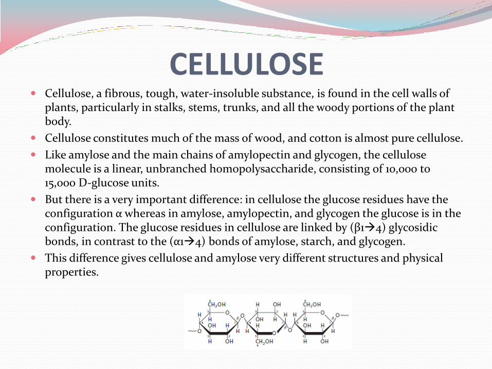
Like amylose and the main chains of amylopectin and glycogen, the cellulose molecule is a linear, unbranched homopolysaccharide, consisting of 10,000 to 15,000 D-glucose units.
But there is a very important difference: in cellulose the glucose residues have the configuration α whereas in amylose, amylopectin, and glycogen the glucose is in the configuration. The glucose residues in cellulose are linked by (β14) glycosidic bonds, in contrast to the (α14) bonds of amylose, starch, and glycogen.
This difference gives cellulose and amylose very different structures and physical properties.
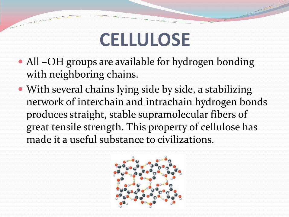
CELLULOSE All –OH groups are available for hydrogen bonding
with neighboring chains.
With several chains lying side by side, a stabilizing network of interchain and intrachain hydrogen bonds produces straight, stable supramolecular fibers of great tensile strength. This property of cellulose has made it a useful substance to civilizations.
Glycogen and starch ingested in the diet are hydrolyzed by α-amylases, enzymes in saliva and intestinal secretions that break (α14) glycosidic bonds between glucose units.
Most animals cannot use cellulose as a fuel source, because they lack an enzyme to hydrolyze the (β14) linkages.
Some microorganisms and fungi can digest cellulose since they have cellulase enzyme. Bacteria in some organisms’ intestinal tract can help that organism to digest cellulose.
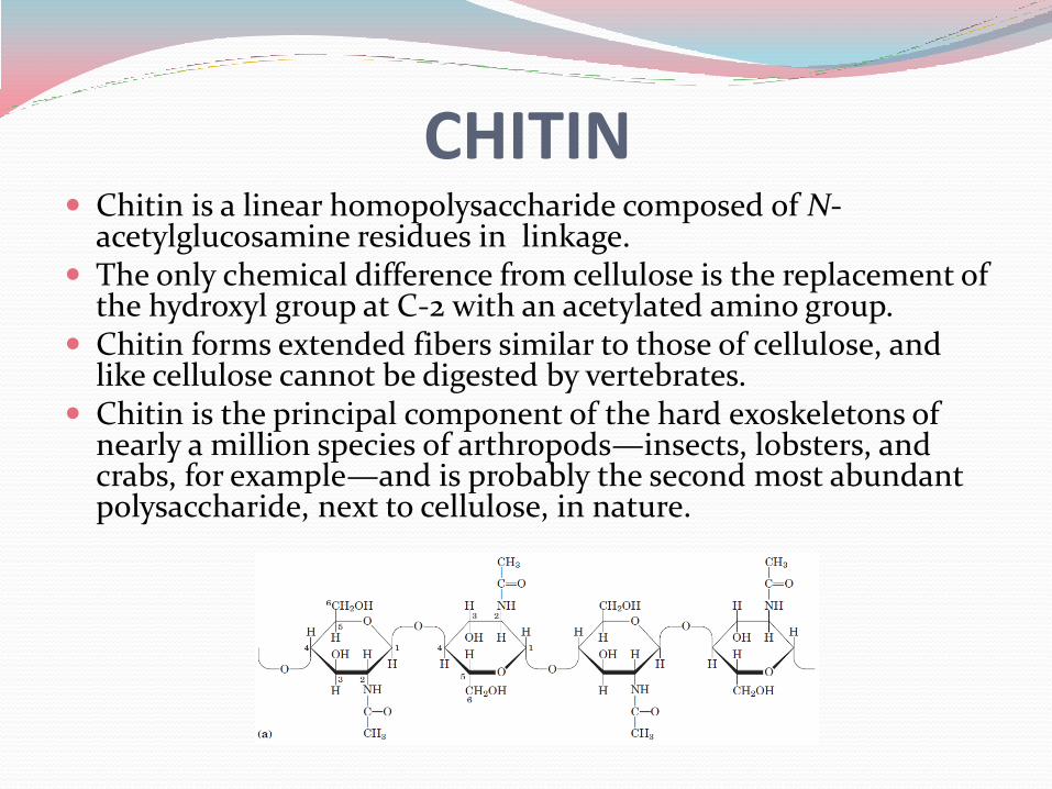
CHITIN Chitin is a linear homopolysaccharide composed of N-
acetylglucosamine residues in linkage. The only chemical difference from cellulose is the replacement of
the hydroxyl group at C-2 with an acetylated amino group. Chitin forms extended fibers similar to those of cellulose, and
like cellulose cannot be digested by vertebrates. Chitin is the principal component of the hard exoskeletons of
nearly a million species of arthropods—insects, lobsters, and crabs, for example—and is probably the second most abundant polysaccharide, next to cellulose, in nature.
3D Structure of Polysaccharides The folding of polysaccharides in three dimensions
follows the same principles as those governing polypeptide structure: Rotation around the covalent bonds which are stabilized
by weak interactions within or between molecules.
Hydrogen bonds, hydrophobic, and van der Waals interactions, and, for polymers with charged subunits, electrostatic interactions play role.
Because polysaccharides have so many hydroxyl groups, hydrogen bonding has an especially important influence on their structure.
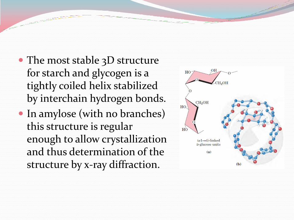
The most stable 3D structure for starch and glycogen is a tightly coiled helix stabilized by interchain hydrogen bonds.
In amylose (with no branches) this structure is regular enough to allow crystallization and thus determination of the structure by x-ray diffraction.
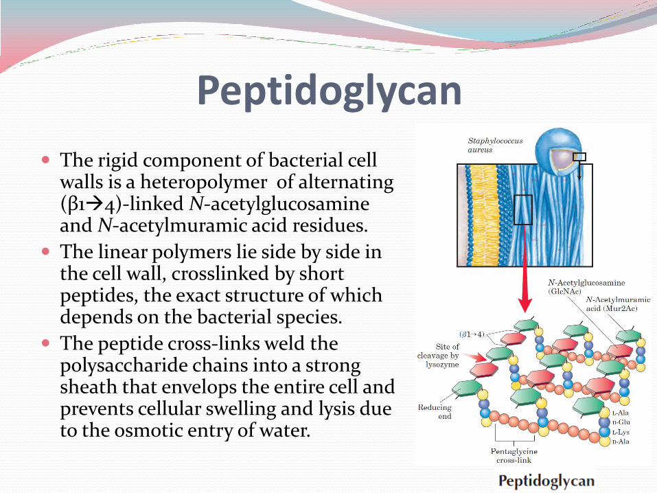
The rigid component of bacterial cell walls is a heteropolymer of alternating (β14)-linked N-acetylglucosamine and N-acetylmuramic acid residues.
The linear polymers lie side by side in the cell wall, crosslinked by short peptides, the exact structure of which depends on the bacterial species.
The peptide cross-links weld the polysaccharide chains into a strong sheath that envelops the entire cell and prevents cellular swelling and lysis due to the osmotic entry of water.
Peptidoglycan
The extracellular space in the tissues of multicellular animals is filled with a gel-like material, the extracellular matrix, also called ground substance, which holds the cells together and provides a porous pathway for the diffusion of nutrients and oxygen to individual cells.
The extracellular matrix is composed of an interlocking meshwork of heteropolysaccharides and fibrous proteins such as collagen, elastin, fibronectin, and laminin.
These heteropolysaccharides, the glycosaminoglycans, are a family of linear polymers composed of repeating disaccharide units.
These polymers provide viscosity, adhesiveness, and tensile strength to the extracellular matrix.
Hyaluronic acid, a type of glycosaminoglycan, form clear, highly viscous solutions that serve as lubricants in the synovial fluid of joints and give the vertebrate eye its jelly like consistency.
Hyaluronate is also an essential component of the extracellular matrix of cartilage and tendons, to which it contributes tensile strength and elasticity as a result of its strong interactions with other components of the matrix.
Glycoconjugates In addition to their important roles as stored fuels (starch,
glycogen, dextran) and as structural materials (cellulose, chitin, peptidoglycans), polysaccharides and oligosaccharides are information carriers: they serve as destination labels for some proteins and as mediators of specific cell-cell interactions and interactions between cells and the extracellular matrix.
Specific carbohydrate containing molecules act in cell-cell recognition and adhesion, cell migration during development, blood clotting, the immune response, and wound healing are a few of their many roles.
In most of these cases, the informational carbohydrate is covalently joined to a protein or a lipid to form a glycoconjugate, which is the biologically active molecule: proteoglycans, glycoproteins, glycolipids.
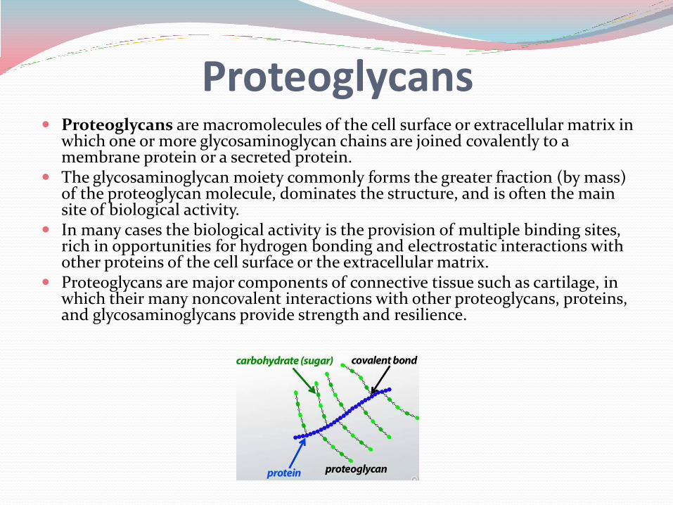
Proteoglycans Proteoglycans are macromolecules of the cell surface or extracellular matrix in
which one or more glycosaminoglycan chains are joined covalently to a membrane protein or a secreted protein.
The glycosaminoglycan moiety commonly forms the greater fraction (by mass) of the proteoglycan molecule, dominates the structure, and is often the main site of biological activity.
In many cases the biological activity is the provision of multiple binding sites, rich in opportunities for hydrogen bonding and electrostatic interactions with other proteins of the cell surface or the extracellular matrix.
Proteoglycans are major components of connective tissue such as cartilage, in which their many noncovalent interactions with other proteoglycans, proteins, and glycosaminoglycans provide strength and resilience.
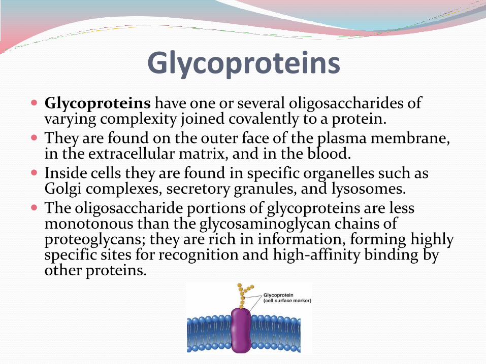
Glycoproteins Glycoproteins have one or several oligosaccharides of
varying complexity joined covalently to a protein. They are found on the outer face of the plasma membrane,
in the extracellular matrix, and in the blood. Inside cells they are found in specific organelles such as
Golgi complexes, secretory granules, and lysosomes. The oligosaccharide portions of glycoproteins are less
monotonous than the glycosaminoglycan chains of proteoglycans; they are rich in information, forming highly specific sites for recognition and high-affinity binding by other proteins.
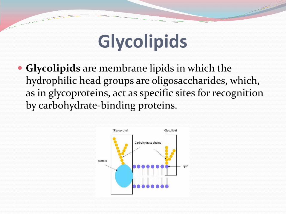
Glycolipids Glycolipids are membrane lipids in which the
hydrophilic head groups are oligosaccharides, which, as in glycoproteins, act as specific sites for recognition by carbohydrate-binding proteins.