Embed Size (px)
Citation preview
Case ReportCanine Bilateral Conjunctivo-Palpebral Dermoid:Description of Two Clinical Cases and Discussion ofthe Relevance of the Terminology
O. Balland,1 I. Raymond,2 I. Mathieson,3 P. F. Isard,4
Emilie Vidémont-Drevon,4 and T. Dulaurent4
1Centre Hospitalier Veterinaire, 95 rue des Mazurots, 54710 Ludres, France2Department of Clinical Sciences, National Veterinary School, 23 chemin des Capelles, BP 87614, 31076 Toulouse Cedex 3, France3Eyevet Referrals, 41-43 Halton Station Road, Sutton Weaver, Cheshire WA7 3DN, UK4Centre Hospitalier Veterinaire, 275 route Imperiale, 74370 Saint-Martin Bellevue, France
Correspondence should be addressed to O. Balland; [email protected]
Received 15 January 2015; Accepted 10 March 2015
Academic Editor: Paola Roccabianca
Copyright © 2015 O. Balland et al. This is an open access article distributed under the Creative Commons Attribution License,which permits unrestricted use, distribution, and reproduction in any medium, provided the original work is properly cited.
Two young dogs were presented for the evaluation of an abnormally haired appearance of both eyes since adoption. In one dog,the lesions were symmetrical and appeared as disorganized skin tissue located on the cutaneous aspect of the lateral portion ofboth lower eyelids, and continuing to the palpebral and the bulbar conjunctiva, thus forming continuous lesions. In the otherdog, a similar lesion was present in the right eye (OD), but the lesion of the left eye (OS) was of discontinuous, disorganizedskin tissue located midway on the lower eyelid and on the lateral bulbar conjunctiva. The lesions were surgically removed androutinely processed for histopathological analysis. Definitive diagnosis was conjunctivo-palpebral dermoids for each dog.Dermoidsare usually considered to be choristoma (normal tissue in an abnormal location) when they are located on the ocular surface (corneaand/or conjunctiva) and as hamartoma when located on the palpebral skin. The lesion presentation in these two dogs reveals thatnames of “choristoma” alone or “hamartoma” alone are not accurate to depict the continuous, composite, conjunctivo-palpebraldermoids. These cases suggest that choristoma and hamartoma might develop subsequently from the same abnormal event duringthe embryonic development, which means that the lesion location might be the only difference between the two terms.
1. Introduction
Dermoids result from the formation of histologically normalcutaneous tissue in an abnormal location during embryonicdevelopment [1–3]. These developmental anomalies are usu-ally classified in the choristoma group [1].The dermoid tissuehas all of the characteristics of skin: an epidermis, dermis, fattissue, sebaceous glands, hair follicles, and hairs [1]. Numer-ous types of ocular surface dermoid have been described indogs [4–7], cats [8, 9], horses [10–12], and anecdotally invarious other species [13–19]: corneal dermoid [4–7, 15, 19,20], conjunctivo-corneal dermoid [9, 10, 16, 17], conjunctivo-palpebral dermoid [3, 20], and dermoid of the nictitating
membrane [10]. Human ocular dermoids have also beendescribed. They can be isolated lesions or a component ofGoldenhar syndrome, which also affects other organs [21–24]. In dogs, the most frequent form is the conjunctivo-cor-neal dermoid [3]. However, the conjunctival-palpebral der-moid, for which a genetic predisposition has been identified,is a rare finding [3]. The predisposed breeds are GermanShepherd, Dalmatian, and Saint-Bernard dogs [3]. In thelatter breed, the presence of a dermoid is thought to be asso-ciated with other developmental anomalies such as eyelidcoloboma [25]. The small number of cases described in theliterature does not allow any particular sexual predispositionto be identified.
Hindawi Publishing CorporationCase Reports in Veterinary MedicineVolume 2015, Article ID 876141, 6 pageshttp://dx.doi.org/10.1155/2015/876141
2 Case Reports in Veterinary Medicine
2. Clinical Cases
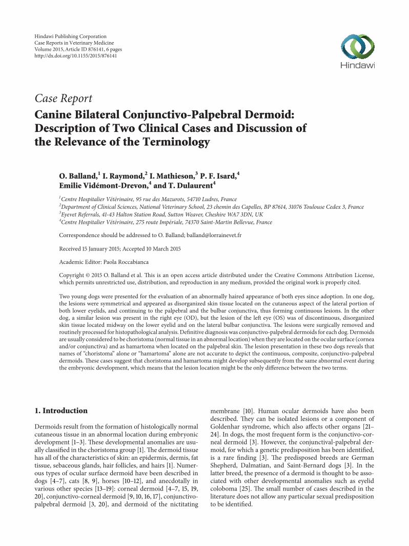
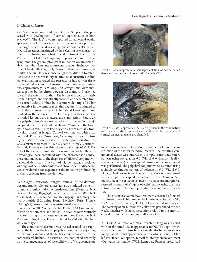
2.1. Case 1. A 4-month-old male German Shepherd dog pre-sented with development of corneal pigmentation in botheyes (OU). The dog’s owners reported an abnormal ocularappearance in OU, associated with a copious mucopurulentdischarge, since the dog’s adoption several weeks earlier.Medical treatment instituted by the referring veterinarian, oftopical administration of fusidic acid ointment (FucithalmicVet, Leo) BID led to a temporary improvement in the dog’ssymptoms.The general physical examination was unremark-able. An abundant mucopurulent ocular discharge waspresent bilaterally (Figure 1). Vision testing gave unreliableresults. The pupillary response to light was difficult to estab-lish due to the poor visibility of intraocular structures. Adne-xal examination revealed the presence of haired skin tissuein the lateral conjunctival fornix. These hairs were numer-ous, approximately 3 cm long, and straight and were mat-ted together by the chronic ocular discharge and orientedtowards the external canthus. The lesion was approximately8mm in length, and was slightly elevated and separated fromthe corneo-scleral limbus by a 2mm wide strip of bulbarconjunctiva in the temporal canthal region. It continued toreach the cutaneous aspect of the lateral lower eyelid andresulted in the absence of the lid margin in this area. Theidentified lesions were bilateral and symmetrical (Figure 2).The palpebral lengthwasmeasuredwith calipers (Castroviejocompass): the upper eyelid length was 29mm and the lowereyelid was 28mm (4mm laterally and 24mm medially fromthe skin tissue) in length. Corneal examination with a slitlamp (SL 15, Kowa, Dusseldorf, Germany) revealed diffusepigmentation of low density in the temporal quadrant ofOU. Schirmer’s tear test (STT,MSD Sante Animal, Clermont-Ferrand, France) was within the normal range of OU. Therest of the ocular examination was unremarkable. The epi-demiological data, combined with the characteristic clinicalpresentation, led us to the diagnosis of bilateral conjunctivo-palpebral dermoids. The corneal pigmentation, associatedwith signs of ocular discomfort and chronic ocular discharge,was considered a consequence of the irritation produced bythe hairs growing from the dermoid.
2.1.1. Surgical Procedure. Surgical removal of the dermoidwas undertaken. General anaesthesia was induced using int-ravenous administration of medetomidine (Domitor ND,Sogeval, Laval, 40 𝜇g/kg), ketamine (Imalgene 1000 ND,Merial SAS, Villeurbanne, France, 5mg/kg), and morphinehydrochloride (Morphine 10mg, Lavoisier, Paris, France,0.05mg/kg). Anaesthesia was maintained using inhaled iso-flurane (IsofloND,Axience, Pantin, France, 1.8%) and oxygenfollowing endotracheal intubation.The eyelids were routinelyprepared using a povidone-iodine solution (Vetedine ND,Vetoquinol SA, Lures, France, diluted to 2%) after the hairwas carefully cut.
The conjunctival dermoid was incised around its periph-ery, at the limit of the lateral palpebral conjunctiva adjoiningthe external canthus and the bulbar conjunctiva close to thecorneoscleral limbus. The excision was continued ventrallyon the cutaneous aspect of the eyelid with a V-shape incision,
Figure 1: Case 1 appearance at initial presentation. Abnormal hairedtissue and copious mucoid ocular discharge in OU.
Figure 2: Case 1 appearance OS. Hair is present in the conjunctivalfornix and extends beyond the lateral canthus. Ocular discharge andcorneal pigmentation are also identified.
in order to achieve full excision of the dermoid and recon-struction of the lower palpebral margin. The resulting con-junctival defect was sutured in a simple continuous suturepattern using polyglactin 6-0 (Vicryl 6-0, Elanco, Neuilly-sur-Seine, France). A two-layered closure of the lower eyelidwas performed. The palpebral conjunctiva was sutured usinga simple continuous pattern of polyglactin 6-0 (Vicryl 6-0,Elanco, Neuilly-sur-Seine, France).The skinwas then suturedwith a simple interrupted pattern of nylon 5-0 (Prolene 5-0,Elanco, Neuilly-sur-Seine, France).The palpebralmargin wasrestored bymeans of a “figure-of eight” suture, using the samesuture material. The same procedure was followed on eachside.
The postoperative medical treatment consisted of topicaladministration of chloramphenicol ointment (Ophtalon ND,TVM, Lempdes, France) TID OU for a period of 2 weeks.The wearing of an Elizabethan collar was prescribed for oneweek, together with strict precautions related to the animal’sconvalescence (short sanitary walks on a leash).
2.2. Case 2. A 1-year-old male French bulldog was referredwith an abnormal ocular appearance inOU.The dog’s ownersreported chronic profuse bilateral ocular discharge, an abnor-mally haired eyelid appearance, and a whitish lesion of theleft eye since his adoption. Topical chloramphenicol ointment(Ophtalon pommade, TVM, Lempdes, France) prescribed
Case Reports in Veterinary Medicine 3
by the referring veterinarian for 2 weeks resulted in a slightreduction of the discharge.
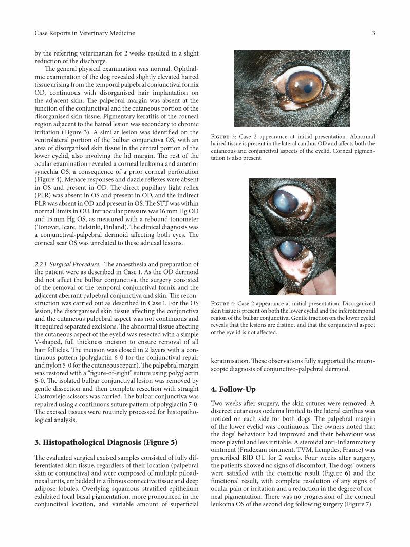
The general physical examination was normal. Ophthal-mic examination of the dog revealed slightly elevated hairedtissue arising from the temporal palpebral conjunctival fornixOD, continuous with disorganised hair implantation onthe adjacent skin. The palpebral margin was absent at thejunction of the conjunctival and the cutaneous portion of thedisorganised skin tissue. Pigmentary keratitis of the cornealregion adjacent to the haired lesion was secondary to chronicirritation (Figure 3). A similar lesion was identified on theventrolateral portion of the bulbar conjunctiva OS, with anarea of disorganised skin tissue in the central portion of thelower eyelid, also involving the lid margin. The rest of theocular examination revealed a corneal leukoma and anteriorsynechia OS, a consequence of a prior corneal perforation(Figure 4). Menace responses and dazzle reflexes were absentin OS and present in OD. The direct pupillary light reflex(PLR) was absent in OS and present in OD, and the indirectPLRwas absent inOD and present inOS.The STTwaswithinnormal limits in OU. Intraocular pressure was 16mmHgODand 15mm Hg OS, as measured with a rebound tonometer(Tonovet, Icare, Helsinki, Finland).The clinical diagnosis wasa conjunctival-palpebral dermoid affecting both eyes. Thecorneal scar OS was unrelated to these adnexal lesions.
2.2.1. Surgical Procedure. The anaesthesia and preparation ofthe patient were as described in Case 1. As the OD dermoiddid not affect the bulbar conjunctiva, the surgery consistedof the removal of the temporal conjunctival fornix and theadjacent aberrant palpebral conjunctiva and skin.The recon-struction was carried out as described in Case 1. For the OSlesion, the disorganised skin tissue affecting the conjunctivaand the cutaneous palpebral aspect was not continuous andit required separated excisions.The abnormal tissue affectingthe cutaneous aspect of the eyelid was resected with a simpleV-shaped, full thickness incision to ensure removal of allhair follicles. The incision was closed in 2 layers with a con-tinuous pattern (polyglactin 6-0 for the conjunctival repairand nylon 5-0 for the cutaneous repair).Thepalpebralmarginwas restored with a “figure-of-eight” suture using polyglactin6-0. The isolated bulbar conjunctival lesion was removed bygentle dissection and then complete resection with straightCastroviejo scissors was carried. The bulbar conjunctiva wasrepaired using a continuous suture pattern of polyglactin 7-0.The excised tissues were routinely processed for histopatho-logical analysis.
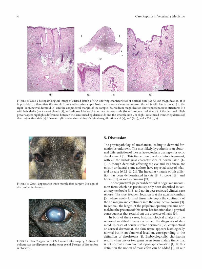
3. Histopathological Diagnosis (Figure 5)
The evaluated surgical excised samples consisted of fully dif-ferentiated skin tissue, regardless of their location (palpebralskin or conjunctiva) and were composed of multiple piload-nexal units, embedded in a fibrous connective tissue and deepadipose lobules. Overlying squamous stratified epitheliumexhibited focal basal pigmentation, more pronounced in theconjunctival location, and variable amount of superficial
Figure 3: Case 2 appearance at initial presentation. Abnormalhaired tissue is present in the lateral canthus OD and affects both thecutaneous and conjunctival aspects of the eyelid. Corneal pigmen-tation is also present.
Figure 4: Case 2 appearance at initial presentation. Disorganizedskin tissue is present on both the lower eyelid and the inferotemporalregion of the bulbar conjunctiva. Gentle traction on the lower eyelidreveals that the lesions are distinct and that the conjunctival aspectof the eyelid is not affected.
keratinisation.These observations fully supported themicro-scopic diagnosis of conjunctivo-palpebral dermoid.
4. Follow-Up
Two weeks after surgery, the skin sutures were removed. Adiscreet cutaneous oedema limited to the lateral canthus wasnoticed on each side for both dogs. The palpebral marginof the lower eyelid was continuous. The owners noted thatthe dogs’ behaviour had improved and their behaviour wasmore playful and less irritable. A steroidal anti-inflammatoryointment (Fradexam ointment, TVM, Lempdes, France) wasprescribed BID OU for 2 weeks. Four weeks after surgery,the patients showed no signs of discomfort.The dogs’ ownerswere satisfied with the cosmetic result (Figure 6) and thefunctional result, with complete resolution of any signs ofocular pain or irritation and a reduction in the degree of cor-neal pigmentation. There was no progression of the cornealleukoma OS of the second dog following surgery (Figure 7).
4 Case Reports in Veterinary Medicine
∗
∗
∗
L R
#
A
A
A
S
S
S
S
(a)
(b) (c)(d) (e)
Figure 5: Case 2 histopathological image of excised lesion of OD, showing characteristics of normal skin. (a) At low magnification, it isimpossible to differentiate the sample from another skin sample. Note the anatomical continuum from the left (eyelid hamartoma, L) to theright (conjunctival dermoid, R) and the conjunctival margin of the sample (#). Medium magnification shows pilosebaceous structures (∗)with hair shafts (→ ), sweat glands (S), and adipose lobules (A) on the cutaneous side (b) and conjunctival side (c) of the dermoid. Highpower aspect highlights differences between the keratinized epidermis (d) and the smooth, non-, or slight-keratinized thinner epidermis ofthe conjunctival side (e). Haematoxylin and eosin staining. Original magnification ×10 (a), ×40 (b, c), and ×200 (d, e).
Figure 6: Case 1 appearance three month after surgery. No sign ofdiscomfort is observed.
Figure 7: Case 2 appearance OS, 1 month after surgery. A discreetoblique scar is still present on the lower eyelid. No sign of discomfortis observed.
5. Discussion
The physiopathological mechanism leading to dermoid for-mation is unknown. The most likely hypothesis is an abnor-mal differentiation of the surface ectodermduring embryonicdevelopment [1]. This tissue then develops into a tegument,with all the histological characteristics of normal skin [1–3]. Although dermoids affecting the eye and its adnexa aremostly unilateral, some authors have reported cases of bilat-eral disease [6, 12–16, 21]. The hereditary nature of this afflic-tion has been demonstrated in cats [8, 9], cows [14], andhorses [11], as well as humans [24].
The conjunctival-palpebral dermoid in dogs is an uncom-mon form which has previously only been described in vet-erinary textbooks [1, 3] and not in peer reviewed clinical casereports. The most frequent location is at the external canthus[3], where newly formed tissue interrupts the continuity ofthe lid margin and continues into the conjunctival fornix [3].In general, the length of the palpebral opening remains nor-mal, but the presence of this tissue has functional and physicalconsequences that result from the presence of hairs [3].
In both of these cases, histopathological analysis of theremoved modified tissues confirmed the diagnosis of der-moid. In cases of ocular surface dermoids (i.e., conjunctivalor corneal dermoids), the skin tissue appears histologicallynormal but in an abnormal location, corresponding to thedefinition of choristoma [1]. Embryologically, choristomaresults when one or two germ layers form mature tissue thatis not normally found in that topographic location [1]. To thisdefinition the notion of mass effect can be added [1]. In our
Case Reports in Veterinary Medicine 5
cases, the only difference between the conjunctival portion ofthe dermoid and normal skin is that the keratinization of theepidermis disappeared or significantly decreased, probablydue to the lacrimal fluid that permanently bathed the skin.In cases of cutaneous palpebral dermoids, the location isnormal, but the development of the skin is abnormal, witha larger number and size of skin components as compared tonormal skin, corresponding to the definition of hamartoma[1, 26]. According to Dubielzig et al., hamartoma is an “exces-sive amount of mature tissue (hypertrophy and/or hyper-plasia) occurring in a location in which tissue is usuallyfound” [1]. The mass effect is also reported as a componentof the definition of hamartoma [1].
Both eyes of Case 1 and OD of Case 2 presented withcontinuous conjunctivo-palpebral dermoid, thus formingcomposite lesions, whereas OS of Case 2 presented with twodistinct lesions affecting the cutaneous portion of the eyelidand the conjunctiva.The abnormal skin island located on thelower eyelid of OS of Case 2 corresponds to the definitionof hamartoma. The abnormal skin island located on theconjunctiva of the same eye corresponds to the definition ofchoristoma. However, we believe that the current acceptedterminology inadequately describes the entire compositelesion (OUofCase 1 andODofCase 2). Indeed, the cutaneousportion of the lesion occurs in a normal location, excludingthe term “choristoma.” In comparison, the term “hamartoma”does not fit with the conjunctival portion of the lesion,because the anomaly occurs in a location in which skin tissueis usually not found. To the best of our knowledge, there is nospecific or accurate term to define the entire cutaneoconjunc-tival lesion. Both parts of the lesion are probably secondaryto the same abnormal event during embryonic development,affecting two adjacent areas of different nature (e.g., the skinand the conjunctival mucosa). The coexistence of distinctseparated lesions on OS of Case 2 and continuous cuta-neoconjunctival lesion on OD of the same patient make ushypothesize that choristomas and hamartomasmay be due tothe same developmental process, meaning that the notion oflocation might be the only difference between the two terms.
The use of the term “dermoid” also seems controversial.For some authors, dermoids are strictly defined as choris-tomas [1, 2, 4] and cannot be applied for a skin location,whereas for some others [3], by usage and extension, the term“dermoid” can be used for either hamartomas (when occur-ring on the eyelid) or choristomas (when occurring on the eyesurface). In our opinion, as illustrated in these two cases withcontinuous cutaneoconjunctival lesions, the developmentalabnormality and biologic mechanism causing the lesion areprobably the same. This is the reason why the term “der-moid” remains accurate for the three possible entities: strictlypalpebral lesions, strictly conjunctival (or corneal) lesions,or composite conjunctivo-palpebral lesions with a physicalcontinuum. For this last entity, as the nature of the lesion(dermoid) cannot be associated with “hamartoma” alone or“choristoma” alone, we propose to describe it as a “choristo-hamartoma.”
Independently from the terminology, the clinical presen-tation of such a lesion is comparable with that observed incases of conjunctival of corneal dermoid. Hairs chronically
traumatize the ocular surface, leading to superficial inflam-mation characterised by conjunctivitis, corneal neovascular-isation, and pigmentation [3]. This abrasion is painful forthe patient and provokes a chronic ocular discharge as wellas an intense blepharospasm. The patient’s abnormal ocularappearance and pain are usually the primary reasons forconsultation [3].
The treatment is always surgical and obviously requiresresection of the malformed area, sometimes associated withgraft procedures [3, 27–29]. During the surgery, particularcare must be taken in reconstruction of the palpebral marginin order to conserve palpebral function and remove any riskof iatrogenic ocular irritation. When the palpebral margindeficit is small following resection, a simple edge-to-edgesuture can be used, with continuity of the margin beingensured by means of a “figure-of-eight” suture [3]. If theresection leads to a more significant deficit, a more complexoculoplastic procedure is indicated [3]. When the develop-mental anomaly is severe and has serious consequences (ocu-lar pigmentation or even ulceration and chronic ocular pain),early surgery is recommended [3]. In the case ofmore discreteconditions having less significant physical and functionalconsequences, surgery can be carried out at the age of threemonths [3]. In Case 1, the condition was serious and causedconsiderable pain as well as intense corneal pigmentation,but surgery was performed at the age of 4 months, followingthe referring veterinarian’s unsuccessful medical treatment.In both cases, themalformationwas characteristically locatedin the outer canthal region except in OS of Case 2. Althoughit covered a significant portion of the conjunctival fornix,alteration of the palpebralmarginwasminimal, and so simpleresection and classical edge-to-edge reconstruction were suf-ficient to permanently remove the source of irritation, restorenormal palpebral function, and result in the rapid resolutionof discomfort and recovery of the dog’s normal temperament.
In conclusion, despite the simple but uncommon clinicalpresentation of these two cases, they illustrate the limitationof the terminology to describe development of abnormalitiesaffecting the ocular surface and adnexa.
Conflict of Interests
The authors declare that there is no conflict of interestsregarding the publication of this paper.
References
[1] R. R. Dubielzig, K. L. Ketring, G. J. McLellan, and D. M.Albert, “Diseases of eyelids and conjunctiva,” in VeterinaryOcular Pathology: A Comparative Review, pp. 143–199, SaundersElsevier, London, UK, 1st edition, 2010.
[2] F. C. Stades and A. van der Woerdt, “Diseases and surgery ofthe canine eyelid,” in Veterinary Ophthalmology, K. N. Gelatt,B. C. Gilger, and T. J. Kern, Eds., pp. 832–893, Wiley-Blackwell,Ames, Iowa, USA, 5th edition, 2013.
[3] C. S. Cook, “Ocular embryology and congenital malforma-tions,” in Veterinary Ophthalmology, K. N. Gelatt, B. C. Gilger,and T. J. Kern, Eds., pp. 3–38, Wiley-Blackwell, Ames, Iowa,USA, 5th edition, 2013.
6 Case Reports in Veterinary Medicine
[4] D. K. Brudenall, M. E. Bernays, and R. L. Peiffer Jr., “Centralcorneal dermoid in a Labrador retriever puppy,” Journal of SmallAnimal Practice, vol. 48, no. 10, pp. 588–590, 2007.
[5] K. Horikiri, K. Ozaki, H. Maeda, and I. Narama, “Cornealdermoid in two laboratory beagle dogs,” Jikken Dobutsu, vol. 43,no. 3, pp. 417–420, 1994.
[6] K. N. Gelatt, “Bilateral corneal dermoids and distichiasis in adog,”VeterinaryMedicine and Small Animals Clinics, vol. 66, no.7, pp. 658–659, 1971.
[7] S. Minamide and K. Suzuki, “Corneal choristoma in a beagledog,” Australian Veterinary Journal, vol. 75, no. 2, pp. 93–94,1997.
[8] M. B. Glaze, “Congenital and hereditary ocular abnormalities incats,”Clinical Techniques in Small Animal Practice, vol. 20, no. 2,pp. 74–82, 2005.
[9] P. M. Hendy-Ibbs, “Familial feline epibulbar dermoids,” Veteri-nary Record, vol. 116, no. 1, pp. 13–14, 1985.
[10] S. M. Greenberg, C. E. Plummer, D. E. Brooks, S. L. Craft, andJ. A. Conway, “Third eyelid dermoid in a horse,” VeterinaryOphthalmology, vol. 15, no. 5, pp. 351–354, 2012.
[11] J. R. Joyce, J. E. Martin, R. W. Storts, and L. Skow, “Iridialhypoplasia (aniridia) accompanied by limbic dermoids andcataracts in a group of related quarterhorses,” Equine VeterinaryJournal Supplement, no. 10, pp. 26–28, 1990.
[12] S. A. McLaughlin and A. H. Brightman, “Bilateral oculardermoids in a colt,” Equine Practice, vol. 5, pp. 10–14, 1983.
[13] K. N. Gelatt, “Corneo-conjunctival dermoid cyst in a calf,”Veterinary Medicine and Small Animals Clinics, vol. 67, no. 11,article 1217, 1972.
[14] S. D. Barkyoumb and H. W. Leipold, “Nature and causeof bilateral ocular dermoids in Hereford cattle,” VeterinaryPathology, vol. 21, no. 3, pp. 316–324, 1984.
[15] J. E. Croshaw, “Bilateral corneal dermoid in a calf: a case report,”Journal of the American VeterinaryMedical Association, vol. 135,pp. 216–218, 1959.
[16] D. K. Brudenall, D. A. Ward, L. A. Kerr, and S. J. Newman,“Bilateral corneoconjunctival dermoids and nasal choristomasin a calf,” Veterinary Ophthalmology, vol. 11, no. 3, pp. 202–206,2008.
[17] E. E. B. LaDouceur, J. Ernst, and M. K. Keel, “Unilateralcorneoscleral choristomas (corneal dermoids) in a white-taileddeer (Odocoileus virginianus),” Journal of Wildlife Diseases, vol.48, no. 3, pp. 826–828, 2012.
[18] C. P. Moore, J. B. Shaner, R. M. Halenda, C. S. Rosenfeld, andW.K. Suedmeyer, “Congenital ocular anomalies and ventricularseptal defect in a dromedary camel (Camelus dromedarius),”Journal of Zoo andWildlife Medicine, vol. 30, no. 3, pp. 423–430,1999.
[19] C. W. Nichols and M. Yanoff, “Dermoid of a rat cornea,”Pathologia Veterinaria, vol. 6, no. 3, pp. 214–216, 1969.
[20] D. D. Lawson, “Corneal dermoids in animals,” VeterinaryRecord, vol. 97, no. 23, pp. 449–450, 1975.
[21] P. Henkind, G. Marinoff, A. Manas, and A. Friedman, “Bilateralcorneal dermoids,” American Journal of Ophthalmology, vol. 76,no. 6, pp. 972–977, 1973.
[22] A. Mahdavi Fard and L. Pourafkari, “Images in clinical medi-cine. The hairy eyeball—limbal dermoid,” The New EnglandJournal of Medicine, vol. 368, no. 1, p. 64, 2013.
[23] M. D. R. A. Gonzalez, A. Navas, A. Haber, T. Ramırez-Luquın,and E. O. Graue-Hernandez, “Ocular dermoids: 116 consecutivecases,” Eye and Contact Lens, vol. 39, no. 2, pp. 188–191, 2013.
[24] J. Zhu, H.-B. Cheng, N. Fan et al., “Studies of a pedigree withlimbal dermoid cyst,” International Journal of Ophthalmology,vol. 5, no. 5, pp. 641–643, 2012.
[25] H. Brandsch and V. Schmidt, “Erbanalytische Untersuchungenzum Dermoid des Auges beim Hund,” Monatshefte fur Vet-erinarmedizin, vol. 37, pp. 305–306, 1982.
[26] T. L. Gross, P. J. Ihrke, E. J. Walder, and V. K. Affolter, Skin Dis-eases of the Dog and Cat. Clinical and Histopathologic Diagnosis,Blackwell Publishing, Ames, Iowa, USA, 2nd edition, 2005.
[27] M. Kalpravidh, P. Tuntivanich, S. Vongsakul, and S. Siri-vaidyapong, “Canine amniotic membrane transplantation forcorneal reconstruction after the excision of dermoids in dogs,”Veterinary Research Communications, vol. 33, no. 8, pp. 1003–1012, 2009.
[28] J.-I. Lee, M.-J. Kim, I.-H. Kim, Y.-B. Kim, and M.-C. Kim,“Surgical correction of corneal dermoid in a dog,” Journal ofVeterinary Science, vol. 6, no. 4, pp. 369–370, 2005.
[29] A. Pirouzian, “Management of pediatric corneal limbal der-moids,” Clinical Ophthalmology, vol. 7, pp. 607–614, 2013.

Submit your manuscripts athttp://www.hindawi.com
Veterinary MedicineJournal of
Hindawi Publishing Corporationhttp://www.hindawi.com Volume 2014
Veterinary Medicine International
Hindawi Publishing Corporationhttp://www.hindawi.com Volume 2014
Hindawi Publishing Corporationhttp://www.hindawi.com Volume 2014
International Journal of
Microbiology
Hindawi Publishing Corporationhttp://www.hindawi.com Volume 2014
AnimalsJournal of
EcologyInternational Journal of
Hindawi Publishing Corporationhttp://www.hindawi.com Volume 2014
PsycheHindawi Publishing Corporationhttp://www.hindawi.com Volume 2014
Evolutionary BiologyInternational Journal of
Hindawi Publishing Corporationhttp://www.hindawi.com Volume 2014
Hindawi Publishing Corporationhttp://www.hindawi.com
Applied &EnvironmentalSoil Science
Volume 2014
Biotechnology Research International
Hindawi Publishing Corporationhttp://www.hindawi.com Volume 2014
Agronomy
Hindawi Publishing Corporationhttp://www.hindawi.com Volume 2014
International Journal of
Hindawi Publishing Corporationhttp://www.hindawi.com Volume 2014
Journal of Parasitology Research
Hindawi Publishing Corporation http://www.hindawi.com
International Journal of
Volume 2014
Zoology
GenomicsInternational Journal of
Hindawi Publishing Corporationhttp://www.hindawi.com Volume 2014
InsectsJournal of
Hindawi Publishing Corporationhttp://www.hindawi.com Volume 2014
The Scientific World JournalHindawi Publishing Corporation http://www.hindawi.com Volume 2014
Hindawi Publishing Corporationhttp://www.hindawi.com Volume 2014
VirusesJournal of
ScientificaHindawi Publishing Corporationhttp://www.hindawi.com Volume 2014
Cell BiologyInternational Journal of
Hindawi Publishing Corporationhttp://www.hindawi.com Volume 2014
Hindawi Publishing Corporationhttp://www.hindawi.com Volume 2014
Case Reports in Veterinary Medicine


