Embed Size (px)
Citation preview
�-Catenin Expression Pattern in Stage I and IIOvarian Carcinomas
Relationship with �-Catenin Gene Mutations,Clinicopathological Features, and Clinical Outcome
Carlos Gamallo,* Jose Palacios,* Gema Moreno,*Jorge Calvo de Mora,* Asuncion Suarez,* andAlvaro Armas†
From Departamento de Anatomıa Patologica * and
Departamento de Ginecologıa y Obstetricia,† Hospital La Paz,
Madrid, Spain
The immunohistochemical expression pattern of�-catenin has been correlated with �-catenin genemutations, clinicopathological features, and diseaseoutcome in 69 stage I and II ovarian carcinomas.�-Catenin expression was localized in the nuclei, inaddition to the cytoplasm and membrane, in 11 tu-mors (16%): nine endometrioid carcinomas withwidespread nuclear expression and two serous carci-nomas with focal nuclear expression. The remaining58 carcinomas (84%) only had membranous �-cate-nin expression. All but one of the endometrioid car-cinomas with nuclear �-catenin expression had con-siderable squamous metaplasia, and five of thesecases had large areas of endometrioid tumor of lowmalignant potential. In addition, �-catenin nuclearexpression was observed in atypical epithelial cells inendometriotic glands adjacent to an endometrioidcarcinoma. Sequencing was performed on 25 tumorsand corresponding normal tissue: all 13 endometri-oid tumors as well as 12 carcinomas of other histo-logical types (four serous, two clear cell , two muci-nous, and two mixed). There were oncogenicmutations in the phosphorylation sequence forGSK-3� in exon 3 of the �-catenin gene in sevenendometrioid carcinomas with �-catenin nuclear ex-pression. Three mutations affected codon 32 (D32G,D32Y, and D32Y), one affected codon 33 (S33C), twoaffected codon 37 (S37C and S37F), and one affectedcodon 41 (T41A). No mutations were observed in theother 18 carcinomas analyzed, comprising two endo-metrioid and two serous carcinomas with �-cateninnuclear expression, and 14 carcinomas of differenthistological types with only membranous expression.In the univariate and multivariate survival analyses,�-catenin nuclear expression was selected as an indi-
cator of good prognosis, because no patient whosetumor expressed �-catenin in the nuclei showed re-lapses or died, in contrast to the 19 relapses anddeaths among patients with tumors that only had�-catenin membranous expression, including threeof the four patients with endometrioid carcinomas.Oncogenic �-catenin mutation is characteristic of agroup of endometrioid carcinomas with a good prog-nosis, most of which originate from previous benignor borderline lesions. Endometrioid carcinomas withexclusively membranous expression of �-cateninseem to represent a different subgroup of carcinomasthat probably have a worse prognosis. In early-stageovarian cancer, determination of the �-catenin ex-pression pattern could prove to be a useful marker forselecting low-risk patients. (Am J Pathol 1999,155:527–536)
Ovarian cancer is a highly agressive gynecological ma-lignancy affecting approximately 27,000 women per yearin the United Sates and producing almost 15,000 deathsyearly.1 Although the prognosis of early-stage ovariancancer is considerably better than in advanced disease,30–40% of early-stage patients will eventually die as aresult of their tumors.2 High-grade histology, tumor cellsin ascites or peritoneal washings, a ruptured capsule, orthe presence of a tumor on the external surface havebeen described as unfavorable prognostic factors in ear-ly-stage ovarian cancer,3,4 and adjuvant chemotherapyis strongly recommended.2
Relatively little is known about the molecular eventsthat lead to the development of ovarian cancer, and nomolecular markers are generally accepted as prognosticindicators. Although most cases of epithelial tumors of
Supported by research grants 98/0151 from the Fondo de Investigacio-nes Sanitarias de la Seguridad Social, CAM 08.1/0020/1997 from theComunidad Autonoma de Madrid, and SAF 98-0085-C03-03 from theMinisterio de Educacion y Cultura, Spain.
Accepted for publication April 11, 1999.
Address reprint requests to Dr. Carlos Gamallo, Departamento deAnatomıa Patologica, Hospital La Paz, Paseo de la Castellana 261, 28046Madrid, Spain. E-mail: [email protected].
American Journal of Pathology, Vol. 155, No. 2, August 1999
Copyright © American Society for Investigative Pathology
527
the ovary seem to have a common origin in the surfaceepithelium covering the ovary, different pathways andgenetic alterations have been implicated in the develop-ment of cystadenomas, borderline tumors, and carcino-mas of different histological subtypes and stages of pro-gression. For example, loss of heterozygocity and acomplete loss of chromosome 17 is characteristic of theadvanced stages of serous carcinomas,5 whereas rasmutation participates in the development of tumors withmucinous differentiation, but not in other histologicaltypes.6 Recently, we have reported data that stronglysuggest that �-catenin may function as an oncogen in theinitiation of some ovarian endometrioid carcinomas inwhich �-catenin is mainly expressed in the nucleus andcytoplasm.7 However, the clinical implications of this find-ing remain to be established.
�-Catenin is a multifunctional protein involved in atleast two important biological processes: cell-cell adhe-sion and signal transduction (transcriptional activation).8
The role of �-catenin in adhesion is well established. Inthe adherens junctions of epithelial cells, the cytoplasmicdomain of E-cadherin organizes a peripheral proteincomplex, including �-catenin, �-catenin, and �-catenin(plakoglobin), that is necessary for adhesion to occur.9
The role of �-catenin in the linkage between �-cateninand E-cadherin in this complex is probably regulated byepidermal growth factor-mediated tyrosine phosphoryla-tion of the �-catenin.8,9 Molecules of the cadherin/catenincomplex have been implicated in differentiation and tu-mor progression, because their reduced expression isnot only frequent in poorly differentiated tumors, but alsomight produce loss of adhesiveness and increased inva-sive and metastatic potential.10–13
In addition, �-catenin participates in the transductionof signals and activates transcription by forming com-plexes with DNA binding proteins in the T-cell factor-lymphoid enhancer factor (TcF-lef) family.14 The free (cy-toplasmic) �-catenin level is low in normal cells becausethe protein is targeted for destruction in the ubiquitin-proteasome system15 by adenomatous polyposis coli(APC) protein together with glycogen synthetase ki-nase-3� (GSK-3�)16 and other molecules such as axin orconductin8; thus wild-type APC plays an essential role inthe clearance of unnecessary �-catenin from the cyto-plasm. Disruption of the APC-mediated regulation of the�-catenin-Tcf pathway was first implicated in the devel-opment of colon carcinomas and melanoma.17,18 Muta-tions that inactivate APC increase cytoplasmic levels offree �-catenin,19 which may then act as an oncoproteinthrough constitutive �-catenin-Tcf-regulated transcrip-tion.14 In the same manner, activating �-catenin muta-tions render this pathway insensitive to the effect of WTAPC.17,18 To date, oncogenic �-catenin mutations havebeen described in several malignancies, such as carci-nomas of the colon,17,20–23 ovary,7 prostate,24 liver,25
and endometrium26 and medulloblastomas.27
This study analyzes the expression pattern of �-cate-nin in a series of 69 stage I and II ovarian carcinomas andtheir relationships with �-catenin gene mutation, clinico-pathological features, and survival to better understandthe biological and clinical relevance of �-catenin pathway
activation in ovarian cancer. Some of the clinicopatholog-ical and genetic features of 30 cases have previouslybeen reported.7
Materials and Methods
Patients
This study was conducted on 69 primary ovarian carci-nomas diagnosed at the Department of Pathology of LaPaz Hospital, Madrid. All cases were in stage I or II andhad a minimum follow-up period of 5 years. Staging wasdone following the International Federation of Gynecol-ogy and Obstetrics (FIGO) system. Retrospective re-views of patient files were performed to obtain all perti-nent data on the primary tumor, type of surgery, adjuvanttreatment, recurrence, and survival. If necessary, pa-tients were retrospectively assigned a FIGO stage on thebasis of their clinical history. The tumors were routinelyfixed in 10% formalin and embedded in paraffin. Histo-logical typing was performed according to the WorldHealth Organization classification. Histological, immuno-histochemical, and DNA studies were performed on for-malin-fixed, paraffin-embedded tissue samples.
Immunohistochemistry
Immunohistochemistry for �-catenin was performed bythe avidin-biotin-alkaline phosphatase method, as previ-ously reported.7 A heat-induced antigen retrieval step(deparaffinized sections were immersed in 0.01 mol/Lsodium citrate buffer (pH 6.0) and incubated in a pres-sure cooker for 3 minutes) was performed. The mouseanti-human �-catenin monoclonal antibody (TransductionLaboratories, Lexington, KY) was applied at a dilution of1:200. In negative controls, the primary antibody wasomitted or replaced with an irrelevant antibody.
Evaluation of Immunohistochemical Staining
Two patterns of �-catenin expression were considered:membranous, if �-catenin was localized in the cell mem-brane only; and nuclear, when �-catenin was expressedin the nucleus, irrespective of the percentage of stainednuclei or simultaneous expression of �-catenin in mem-brane and cytoplasm.
A semiquantitative estimation of membranous �-cate-nin expression was made, using a composite score ob-tained by adding the values of the immunoreaction inten-sity and relative abundance of the �-cateninimmunoreactive cells, as previously reported for cad-herins.28,29 Briefly, the intensity was graded from 0(equivalent to background staining of the acellularstroma) to �3 (intense stain equivalent to normal surfaceepithelium). The abundance of �-catenin-positive cellswas graded from 0 to 4 (0 � less than 5% of positivecells; 1 � 5–25%; 2 � 26–50%; 3 � 51–75%; 4 �76–100%). �-Catenin expression was considered pre-served when the composite score was 6 or 7. Cases with
528 Gamallo et alAJP August 1999, Vol. 155, No. 2
scores between 0 and 5 were considered to be tumorswith reduced �-catenin expression.
DNA Analysis
Polymerase chain reaction (PCR) was performed as pre-viously reported7 on 25 tumors and corresponding nor-mal tissue: all 13 endometrioid tumors as well as 12carcinomas of other histological types (four serous, twoclear cell, two mucinous, and two mixed). DNA was ex-tracted from paraffin blocks containing a large proportionof epithelial tumor cells (�75%), but no microdissectionof selected areas was performed. Normal tissue in eachcase consisted of samples from fallopian tube or myo-metrium.
The primers used were 240F (ATG GAA CCA GACAGA AAA GC) and 439F (GCT ACT TGT TCT GAG TGAAG), which amplified a 200-bp fragment of exon 3 of the�-catenin gene, encompassing the sequence for GSK-3�phosphorylation. After purification, PCR products weresequenced in a 373 Automated Sequencer or a 310Automater Sequencer (Applied Biosystems, Foster City,CA), using the Abi Prism dRodhamine Terminator CycleSequencing Ready Reaction Kit (Applied Biosystems).PCR primers were used as the primers for sequencingthe two strands of the amplified products. Cases withabnomal DNA sequences were subjected to DNA extrac-tion from additional tissue sections, PCR, and sequenc-ing, to confirm the presence of true mutations.
Statistical Methods
The �2 test was used to analyze the statistical signifi-cance of the relationship between �-catenin expressionand the clinicopathological and immunohistochemicalvariables. Relapse-free survival and overall survival, de-fined as the time from diagnosis to relapse or death,respectively, were used as a measure of prognosis. Uni-variate survival curves were estimated using the Kaplanand Meier method and compared using the log rank test.Multivariate analysis was done using Cox’s proportionalhazards regression model. Analyses were carried outusing JMP (Version 3.0.1; SAS Institute, Cary, NC).
Results
Clinicopathological Features
Some clinicopathological, inmunohistochemical, and ge-netic features of 30 cases have previously been report-ed.7 The mean age of the 69 patients was 55 years(range 19–77). There were 19 (27.5%) stage Ia, 4 (6%)stage Ib, 28 (40.5%) stage Ic, 5 (7%) stage IIa, 9 (13%)stage IIb, and 4 (6%) stage IIc tumors. There were 27(39%) grade I carcinomas, 28 (41%) grade II, and 14(20%) grade III. No patient showed macroscopic residualdisease after surgery. All patients received postoperativetherapy: 17 (24.6%) radiotherapy and 52 (75.4%) chemo-therapy: 33 melphalan and adryamicine, 15 melphalanalone, and 4 cisplatin-based chemotherapy.
�-Catenin Expression and Correlations withClinicopathological Features
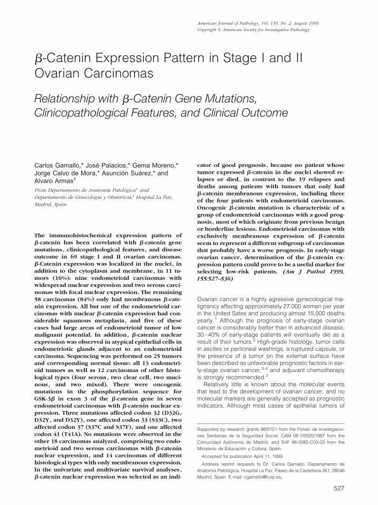
All analyzed tumors showed membrane �-catenin ex-pression with variable extension and intensity, but simul-taneous nuclear �-catenin expression (nuclear pattern)was observed in only 11 (16%) ovarian carcinomas (Fig-ure 1A); the remaining 58 tumors (84%) only had mem-brane expression (membranous pattern) (Figure 1B). Theexpression pattern (nuclear versus membranous) corre-lated significantly with the histological type (Table 1),inasmuch as 9 cases with nuclear expression were en-dometrioid carcinomas. Nuclear expression was also ob-served in two serous carcinomas. In endometrioid le-sions, the percentage of stained nuclei ranged between25% and 90% of the neoplastic cells. In contrast, inserous carcinomas the percentage of immunoreactivenuclei did not exceed 5%. The different �-catenin expres-sion patterns did not correlate with tumor grade andstage. In addition, postoperative therapy was similar intumors with a nuclear or membranous �-catenin expres-sion pattern (Table 1).
All but one endometrioid carcinoma with nuclear�-catenin expression also had evident squamous meta-plasia, and five of these cases had large areas of endo-metrioid tumor of low malignant potential with an adeno-fibromatous, papillary, or mixed growth pattern (Table 2).Endometrioid carcinomas with �-catenin membranousexpression only did not have squamous metaplasia, andtwo cases had a growth pattern resembling that of sex-cord stromal tumors. Four mixed carcinomas with anendometrioid component showed only �-catenin mem-branous expression. Nuclear �-catenin expression wasobserved in epithelial cells of endometriotic cyst (Figure1C) adjacent to an endometrioid carcinoma with �-cate-nin nuclear expression.
As previously noted, �-catenin expression was ob-served in the membrane in all of the 69 carcinomas, butonly 15 cases (22%) had preserved membrane expres-sion (score 6–7), and the remaining 54 carcinomas hadreduced membrane expression (score 3–5). The status ofmembrane �-catenin expression (preserved versus re-duced) did not correlate to the histological type, grade, orstage of the patient’s tumor (Table 3).
�-Catenin Gene Mutations
Direct sequence analysis of the PCR amplification productsshowed heterozygous substitution mutations of �-cateningene exon 3 in seven endometrioid carcinomas with nu-clear �-catenin expression: three at codon 32 (Figure 1Eand 1F), one at codon 33 (Figure 1F), two at codon 37, andone at codon 41. At codon 32, two GAC to TAC (D32Y)changes and a GAC to GGC (D32G) change were ob-served; at codon 33, a TCT to TGT change (S33C) wasobserved; at codon 37, a TCT to TTT change (S37F) anda TCT to TGT change (S37C) were observed; and finally,an ACC to GCC change at codon 41 (T41A) was ob-served. To further confirm sequencing data in the caseswith mutation in S37, PCR products were digested
�-Catenin in Ovarian Cancer 529AJP August 1999, Vol. 155, No. 2
Figure 1. A: Nuclear �-catenin expression pattern in an endometrioid carcinoma. B: Membranous �-catenin expression pattern in an endometrioid carcinoma. C:Nuclear �-catenin expression in some nuclei of epithelial cells of an endometriotic cyst. D: Normal sequence of codons 32 and 33 in exon 3 of �-catenin gene.E: GAC to GGC change in codon 32 (D32G) in tumor 1. F: GAC to TAC change in codon 32 (D32Y) in tumor 3. G: TCT to TGT change in codon 33 (S33C) intumor 4. H: Analysis with HinfI restriction endonuclease of PCR products from cases 3 and 4. In normal tissue (N) digestion produces three fragments of 133, 60, and7 bp. In tumor tissues, heterozygous mutations in codons 32 and 33 eliminate a restriction site in the mutated allele and generate an additional fragment of 67 bp.
530 Gamallo et alAJP August 1999, Vol. 155, No. 2
with XmnI restriction endonuclease. Mutations affectingcodons 32 and 33 were confirmed by HinfI restrictionanalysis (Figure 1H). All detected mutations were alsoconfirmed by resequencing.
No �-catenin gene mutations were observed in twoendometrioid carcinomas with nuclear �-catenin expres-sion pattern, four endometrioid carcinomas with a mem-branous �-catenin pattern (Table 3), or 12 non-endomte-rioid carcinomas, two serous carcinomas with focalnuclear �-catenin expression, and 10 carcinomas withmembranous �-catenin pattern.
Outcome Analysis
The mean follow-up time of the study was 8.9 � 6.8 years(range 1–24 years; median follow-up period 7 years).Among the 69 patients, 19 (33.3%) showed a relapse,
which occurred between 4 and 46 months after surgery.All patients who relapsed subsequently died (13–48months after surgery) of the disease.
The variables analyzed were the type of postoperativetherapy (radiotherapy, melphalan, melphalan and adry-amicine, and cisplatinun- based chemotherapy), histo-logical type, histological grade, stage (Ia-b versus Ic-II),�-catenin expression pattern (nuclear versus membra-nous), and membrane �-catenin expression (preservedversus reduced).
Relapse-free survival was significantly related to histo-logical grade (�2 � 26.61; P � 0.0000), stage (�2 � 9.61;P � 0.0019), and �-catenin expression pattern (�2 �5.82; P � 0.016), but was unrelated to the type of post-operative therapy (�2 � 5.11; P � 0.163), membrane�-catenin expression (�2 � 1.93; P � 0.164), and histo-logical type (�2 � 6.88; P � 0.153) in the univariate
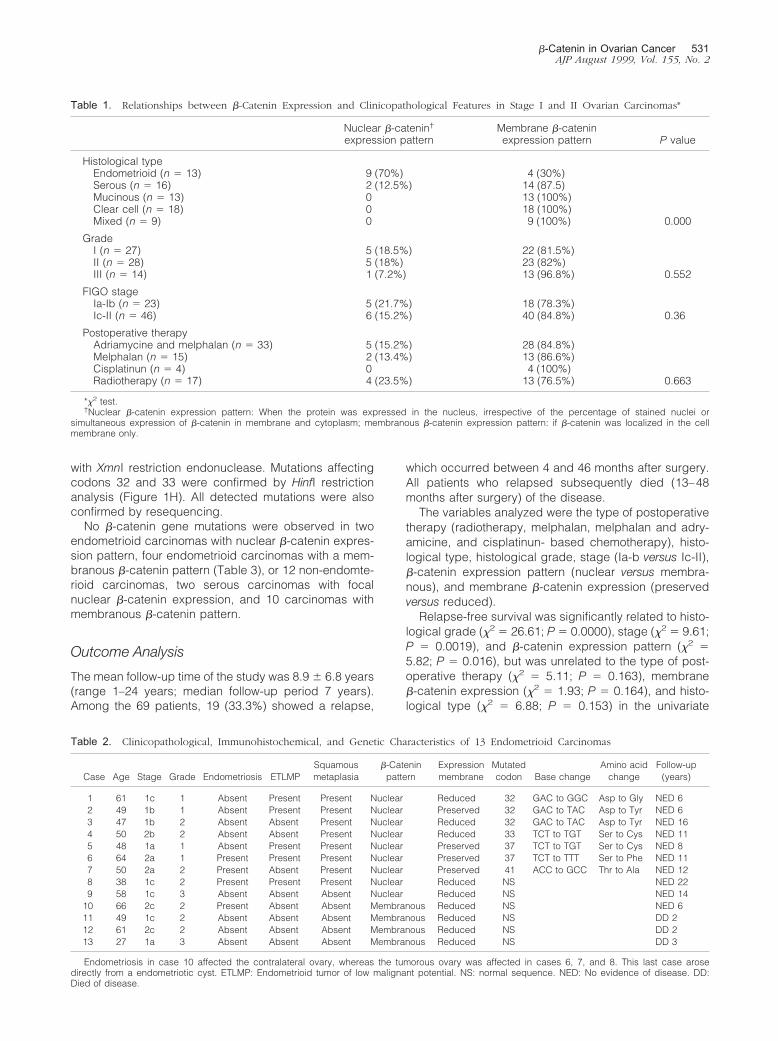
Table 1. Relationships between �-Catenin Expression and Clinicopathological Features in Stage I and II Ovarian Carcinomas*
Nuclear �-catenin†
expression patternMembrane �-cateninexpression pattern P value
Histological typeEndometrioid (n � 13) 9 (70%) 4 (30%)Serous (n � 16) 2 (12.5%) 14 (87.5)Mucinous (n � 13) 0 13 (100%)Clear cell (n � 18) 0 18 (100%)Mixed (n � 9) 0 9 (100%) 0.000
GradeI (n � 27) 5 (18.5%) 22 (81.5%)II (n � 28) 5 (18%) 23 (82%)III (n � 14) 1 (7.2%) 13 (96.8%) 0.552
FIGO stageIa-Ib (n � 23) 5 (21.7%) 18 (78.3%)Ic-II (n � 46) 6 (15.2%) 40 (84.8%) 0.36
Postoperative therapyAdriamycine and melphalan (n � 33) 5 (15.2%) 28 (84.8%)Melphalan (n � 15) 2 (13.4%) 13 (86.6%)Cisplatinun (n � 4) 0 4 (100%)Radiotherapy (n � 17) 4 (23.5%) 13 (76.5%) 0.663
*�2 test.†Nuclear �-catenin expression pattern: When the protein was expressed in the nucleus, irrespective of the percentage of stained nuclei or
simultaneous expression of �-catenin in membrane and cytoplasm; membranous �-catenin expression pattern: if �-catenin was localized in the cellmembrane only.
Table 2. Clinicopathological, Immunohistochemical, and Genetic Characteristics of 13 Endometrioid Carcinomas
Case Age Stage Grade Endometriosis ETLMPSquamousmetaplasia
�-Cateninpattern
Expressionmembrane
Mutatedcodon Base change
Amino acidchange
Follow-up(years)
1 61 1c 1 Absent Present Present Nuclear Reduced 32 GAC to GGC Asp to Gly NED 62 49 1b 1 Absent Present Present Nuclear Preserved 32 GAC to TAC Asp to Tyr NED 63 47 1b 2 Absent Absent Present Nuclear Reduced 32 GAC to TAC Asp to Tyr NED 164 50 2b 2 Absent Absent Present Nuclear Reduced 33 TCT to TGT Ser to Cys NED 115 48 1a 1 Absent Present Present Nuclear Preserved 37 TCT to TGT Ser to Cys NED 86 64 2a 1 Present Present Present Nuclear Preserved 37 TCT to TTT Ser to Phe NED 117 50 2a 2 Present Absent Present Nuclear Preserved 41 ACC to GCC Thr to Ala NED 128 38 1c 2 Present Present Present Nuclear Reduced NS NED 229 58 1c 3 Absent Absent Absent Nuclear Reduced NS NED 14
10 66 2c 2 Present Absent Absent Membranous Reduced NS NED 611 49 1c 2 Absent Absent Absent Membranous Reduced NS DD 212 61 2c 2 Absent Absent Absent Membranous Reduced NS DD 213 27 1a 3 Absent Absent Absent Membranous Reduced NS DD 3
Endometriosis in case 10 affected the contralateral ovary, whereas the tumorous ovary was affected in cases 6, 7, and 8. This last case arosedirectly from a endometriotic cyst. ETLMP: Endometrioid tumor of low malignant potential. NS: normal sequence. NED: No evidence of disease. DD:Died of disease.
�-Catenin in Ovarian Cancer 531AJP August 1999, Vol. 155, No. 2
analysis. Multivariate analysis also selected histologicalgrade (�2 � 16.82; P � 0.0022; risk ratio � 4.63; 95%confidence interval � 1.40–20.82), stage (�2 � 7.96; P �0.0048; risk ratio � 5.81; 95% confidence interval� 1.61–37.18), and �-catenin pattern (�2 � 1028; P �0.003; risk ratio � 0.000005) as independent predictorsof relapse-free survival.
Overall survival was also significantly related to histo-logical grade (�2 � 21.12; P � 0.0000), stage (�2 � 9.92;P � 0.0016), and �-catenin expression pattern (�2 �6.74; P � 0.0094), but was unrelated to the type ofpostoperative therapy (�2 � 3.39; P � 0.334), membrane�-catenin expression (�2 � 1.38; P � 0.51), and histo-logical type (�2 � 6.37; P � 0.17) in the univariate anal-ysis. Multivariate analysis also selected histologicalgrade (�2 � 12.25; P � 0.0022; risk ratio � 4.13; 95%confidence interval � 1.25–18.54), stage (�2 � 7.54; P �0.0060; risk ratio � 5.54; 95% confidence interval� 1.54–35.41), and �-catenin pattern (�2 � 8.91; P �0.0028; risk ratio � 0.000005), as independent predictorsof OS. The nuclear pattern of �-catenin expression wasan indicator of good prognosis, inasmuch as not onerecurrence or death occurred among these patients.
DiscussionThis study confirms our previous observation7 in a re-duced number of cases that �-catenin expression inovarian cancer has two patterns: nucleocytoplasmic andmembranous, which reflect the two main known functionsof �-catenin: signal transduction and cell adhesion.Widespread �-catenin nuclear expression is mainly ob-served in endometrioid carcinoma, suggesting that acti-vation of the �-catenin-Tcf signaling pathway mediatesdevelopment in a group of cases of this subtype of ovar-ian cancer. Although the activation of this signaling path-way could hypothetically be achieved by different molec-ular events, this study demonstrated that most ovarianendometrioid carcinomas with nucleocytoplasmic ex-pression of �-catenin have �-catenin-gene mutations.
Ovarian carcinomas may arise de novo from surfaceovarian epithelium (or epithelial inclusion cysts) or frombenign or borderline lesions.30 It has been suggestedthat most serous carcinomas arise de novo, whereasmost mucinous carcinomas originate from benign or bor-derline tumors.31 A proportion of endometrioid carcino-mas may arise from previous benign lesions such asendometriosis and benign tumors of the same histologi-cal type.32,33 Endometrioid tumors of the ovary are eitherbenign, usually taking the form of an adenofibroma orcystoadenofibroma, or overtly malignant. Between thesetwo well-established categories, there is a spectrum oflesions that have received different names in the literature(proliferating endometrioid adenofibromas,32 atypicaland borderline endometriod adenofibromas,33 prolifera-tive endometrioid adenofibromas, and endometrioid tu-mors of low malignant potential34,35). Proliferating, bor-derline, or low-malignant-potential endometrioid tumorsare characterized by adenocystadenofibromatous orpapillary growth, epithelial proliferation, and atypia with-out stromal invasion.32–35 In addition, they frequentlyshow squamous metaplasia and have an excellent prog-nosis, although carcinomas may arise in them.32–35 Wepreviously reported �-catenin mutation in one borderlineor low-malignant-potential endometrioid tumor. In thisstudy we observed �-catenin nuclear expression in fivecarcinomas with large areas of endometrioid tumor of lowmalignant potential, four of which had �-catenin muta-tions. Although we have not studied benign endometrioidadenofibromas, these data indicate that �-catenin muta-tion would be implicated in the transformation processleading to the development of a subgroup of endometri-oid carcinomas that have borderline or low-malignant-potential endometrioid tumors as their precursor. In ad-dition, the observation of �-catenin nuclear expression inendometriotic glands adjacent to one carcinoma with�-catenin gene mutation suggests that �-catenin couldbe implicated in the malignant transformation of somecases of endometriosis.
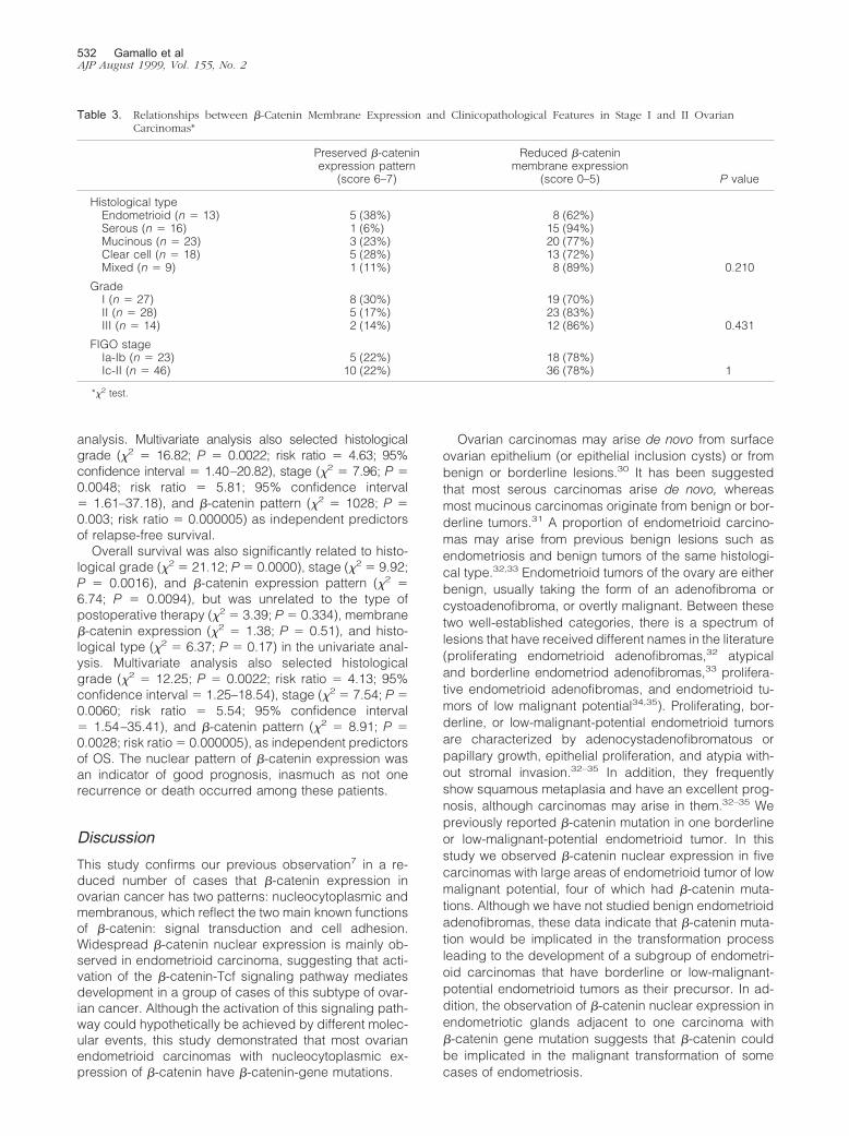
Table 3. Relationships between �-Catenin Membrane Expression and Clinicopathological Features in Stage I and II OvarianCarcinomas*
Preserved �-cateninexpression pattern
(score 6–7)
Reduced �-cateninmembrane expression
(score 0–5) P value
Histological typeEndometrioid (n � 13) 5 (38%) 8 (62%)Serous (n � 16) 1 (6%) 15 (94%)Mucinous (n � 23) 3 (23%) 20 (77%)Clear cell (n � 18) 5 (28%) 13 (72%)Mixed (n � 9) 1 (11%) 8 (89%) 0.210
GradeI (n � 27) 8 (30%) 19 (70%)II (n � 28) 5 (17%) 23 (83%)III (n � 14) 2 (14%) 12 (86%) 0.431
FIGO stageIa-Ib (n � 23) 5 (22%) 18 (78%)Ic-II (n � 46) 10 (22%) 36 (78%) 1
*�2 test.
532 Gamallo et alAJP August 1999, Vol. 155, No. 2
We also observed �-catenin mutations in three endo-metrioid carcinomas without associated preinvasive ar-eas but with evident squamous metaplasia. In contrast,none of the four endometrioid tumors with only membra-nous expression of �-catenin and without �-catenin mu-tations had squamous metaplasia. This finding may indi-cate that �-catenin mutations could be implicated in thegenesis of so-called adenoacanthomas, a suggestionthat must be confirmed in a larger series of cases.
Since the original description of oncogenic �-cateninmutation, several tumor types have been analyzed.�-Catenin point mutations have been reported in 2%, 5%,13%, and 19% of medulloblastomas,27 prostate carcino-mas,24 endometrial carcinomas,26 and hepatocellularcarcinomas,25 respectively. Candidus et al36 have notfound evidence for mutation in this gene in gastric (dif-fuse and intestinal subtypes) and breast (ductal andlobular subtypes) carcinomas, although their sample wassmall. In ovarian carcinomas, we have observed �-cate-nin mutations in only 10% of all stage I and II tumorsanalyzed, but in 50% of the endometrioid carcinomas.This is the highest frequency of �-catenin mutation so farreported in any histological type of human carcinoma andis probably due to the characteristics of the sample an-alyzed (low stages and a high proportion of cases origi-nated in preinvasive lesions).
Most studies on �-catenin mutations have only studiedthe consensus sequence for GSK-3� phosphorylation inexon 3. Mutations affecting this sequence, mainly in theamino acids implicated in the down-regulation of �-cate-nin through phosphorylation by this serine/threonine ki-nase (Ser,33 Ser,37, Thr41, and Ser45),17 probably rendera fraction of cellular �-catenin insensitive to APC-medi-ated down-regulation and are responsible for the up-regulation of cytoplasmic �-catenin and its distribution inthe nuclei of tumor cells, as was immunohistochemicallydetected in the present series. Mutation at codon 32changing a highly conserved aspartic acid was observedin three ovarian carcinomas. Mutations have been re-ported at this codon in hepatocellular25 and prostate25
carcinomas, and it has been suggested that the alterationmay change the protein structure and inhibit phosphory-lation.
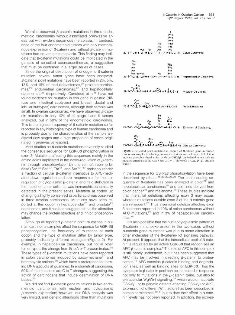
Although all reported �-catenin point mutations in hu-man carcinoma samples affect the sequence for GSK-3�phosphorylation, the frequency of mutations at eachcodon and the type of mutation differ by tumor type,probably indicating different etiologies (Figure 2). Forexample, in hepatocellular carcinoma, but not in othertumor types, the change from G to A or T predominates.25
These types of �-catenin mutations have been reportedin colon carcinomas induced by azoxymethane37 andheterocyclic amines,38 which have a preference for form-ing DNA adducts at guanines. In endometrial carcinoma,50% of the mutations are C to T changes, suggesting theaction of carcinogens that induce deamination of DNAbases.26
We did not find �-catenin gene mutations in two endo-metrioid carcinomas with nuclear and cytoplasmic�-catenin expression; however, our DNA analysis wasvery limited, and genetic alterations other than mutations
in the sequence for GSK-3� phosphorylation have beendescribed by others.18,20,22,23,25 The entire coding se-quence of �-catenin has been analyzed in colon22 andhepatocellular carcinomas25 and cell lines derived fromcolon cancer20 and melanoma.18 These studies indicatethat interstitial deletions affecting exon 3 may occur,whereas mutations outside exon 3 of the �-catenin geneare infrequent.20 Thus insertional deletion affecting exon3 has been reported in 10% of colon carcinomas withoutAPC mutations,22 and in 3% of hepatocellular carcino-mas.25
It is also possible that the nucleocytoplasmic pattern of�-catenin immunoexpression in the two cases without�-catenin gene mutations was due to some alteration inother molecules of the �-catenin-Tcf signaling pathway.At present, it appears that the intracellular pool of �-cate-nin is regulated by an active GSK-3� that recognizes anAPC-�-catenin complex.8 The role of APC in this complexis still poorly understood, but it has been suggested thatAPC may be involved in directing �-catenin to protea-somes.15 APC contains �-catenin binding and degrada-tion sites, as well as binding sites for GSK-3�. Thus thecytoplasmic �-catenin pool can be increased in responsenot only to mutations in the �-catenin gene, but also toextracellular Wg/Wnt signaling,39 which would inactivateGSK-3�, or to genetic defects affecting GSK-3� or APC.Expression of different Wnt factors has been described inhuman carcinomas,40,41 but to date their effect in �-cate-nin levels has not been reported. In addition, the expres-
Figure 2. Reported point mutation in exon 3 of �-catenin gene in humancarcinoma samples (excluding preinvasive lesions and cell lines). Bold lettersindicate phosphorylated amino acids by GSK-3�. Underlined letters indicatemutated amino acids (D Asp; S Ser; G Gly; T Thr) (refs. 17, 21, 23–27, and thisseries).
�-Catenin in Ovarian Cancer 533AJP August 1999, Vol. 155, No. 2
sion pattern for these factors in ovarian cancer remains tobe established. The role of GSK-3� in human oncogen-esis is not well known. Recent studies do not report anyalterations of GSK-3� in medulloblastomas and coloncarcinomas, some of which have mutations of the �-cate-nin gene.23,27 Increased �-catenin cytoplasmic and nu-clear levels can also be observed in cases of APC inac-tivation (ie, mutations of APC in colorectal polyps andcarcinomas19,42 and other conditions, such as sporadicaggressive fibromatosis43). Loss of heterozygosity hasbeen reported at the APC locus in sporadic ovary carci-nomas with a frequency of 50% in a series that includedonly four endometrioid carcinomas44; nevertheless, thisseems to be a late event in tumor progression and doesnot correlate with mutation in the APC gene.44
We did not observe �-catenin mutations in serous,mucinous clear cell, or mixed carcinomas, including fourmixed tumors with an endometrioid component and twoserous carcinomas with focal nuclear �-catenin immuno-expression. In these two cases we did not perform mi-crodissection of the nuclear immunostained areas, andwe could not exclude mutations in these cases. �-Cate-nin mutations have been reported as occurring focally insome prostate cancers, suggesting that these mutationsmay occur during tumor progression.24 Interestingly, twoof the colon cancers reported by Kitaeva et al21 and twocolon cancer cell lines (HCT 116 and SW48), which have�-catenin gene mutations, also show a replication error-positive phenotype, suggesting that the activating muta-tions in �-catenin that occur in some tumors may beassociated with an underlying defect in DNA mismatchrepair.
In the present series, the significant prognostic valueof the traditional clinicopathological variables (grade andstage) as predictors of relapse-free and overall survival at5 years in early-stage ovarian cancer2–4 was confirmed.In addition, the �-catenin expression pattern was se-lected as an independent prognostic marker, indicatingthat cases with nuclear expression have an excellentprognosis, with no relapses or deaths among them. Ob-viously, these results must be taken as preliminary be-cause of the small number of cases, but they do suggest�-catenin as a potential prognostic marker that could beanalyzed in larger series from different institutions. Al-though we did not observe any deaths among endometri-oid carcinomas with a nuclear �-catenin expression pat-tern, three of the four patients with endometrioidcarcinomas with only membranous expression died.These findings suggest two oncogenic pathways in en-dometrioid carcinomas with different clinical implications.Identification of molecular defects in the subgroup ofpatients without nuclear �-catenin expression and withapparently poor prognosis would be helpful in selecting aspecific therapy for each patient.
In addition to the role of �-catenin in signal transduc-tion, this molecule has a pivotal role in cell adhesionthrough its participation in the cadherin/catenin complexof adherens junctions. Changes in the expression andstructure of �-catenin lead to loss of adhesiveness andmay promote invasiveness and metastasis in tumorcells.9,12,13 Mutations of �-catenin disrupting the interac-
tion between E-cadherin and �-catenin have been de-scribed in tumor cell lines that grow as loose aggregatesor single cells.45,46 In human tumors, the expression ofmembranous �-catenin has been analyzed in head andneck,47 esophageal,48,49 gastric,50 colon,51 breast,52
and ovarian53,54 carcinomas. It has been observed that88% of head and neck squamous cell carcinomas, 70%of esophageal carcinomas, 50% of gastric carcinomas,and 30% of infiltrating ductal carcinomas of the breastshow reduced membranous �-catenin expression. Insome reports, reduced expression of �-catenin has beencorrelated with poorly differentiated tumors, advancedstage, and poor survival.48,50
Davis et al54 observed reduced immunohistochemical�-catenin expression in 21% of a series of 34 ovariancarcinomas, but Fujimoto et al53 did not find differencesin mRNA �-catenin expression between normal ovarytissue and ovarian carcinomas. We observed reducedmembranous �-catenin expression in 78% of our ovariancarcinomas. �-Catenin expression did not correlate withhistological type, tumor differentiation, progression(stage), or survival. However, it should be rememberedthat other molecules of the cadherin/catenin complexmay be more important than �-catenin in modulatingadhesion in ovarian carcinomas. For example, �-cateninwas reduced in approximately 60% of cases in a largeseries of stage I and II ovarian carcinomas, and severereduction of �-catenin expression (�30%) in stage I ovar-ian carcinomas was a predictor of poor prognosis in theunivariate and multivariate analysis.55
In summary, the pattern of �-catenin expression differsbetween ovarian carcinomas of differing histologicaltypes. APC-�-catenin-Tcf signaling pathway activationsecondary to oncogenic �-catenin mutation is character-istic of a group of endometrioid carcinomas with nuclear�-catenin expression and a good prognosis, most ofwhich originate in previous benign or borderline lesions.Endometrioid carcinomas with only membrane expres-sion of �-catenin seem to represent a different subgroupof carcinomas that is not associated with the �-cateninsignaling pathway and which, probably, has a worseprognosis. Determination of the �-catenin expressionpattern in early-stage ovarian cancer might prove to be auseful marker for selecting low-risk patients.
AcknowledgmentsThe authors thank Vicente Sanchez, Petra Rubio, andInmaculada Briones for technical assistance with the im-munohistochemical study.
References
1. Parker SL, Tong T, Bolden S, Wingo PA: Cancer statistics, 1997. CACancer J Clin 1997, 47:5–27
2. Canistra SA: Cancer of the ovary. N Engl J Med 1993, 329:1550–15593. Dembo AJ, Davy M, Stenwig AE, Berle EJ, Bush RS, Kjorstad K:
Prognostic factors in patients with stage I epithelial ovarian cancer.Obstet Gynecol 1990, 75:263–273
4. Vergote IB, Kaern J, Abeler VM, Pettersen EO, De Vos LN, Trope CG:
534 Gamallo et alAJP August 1999, Vol. 155, No. 2
Analysis of prognostic factors in stage I epithelial ovarian carcinoma:importance of degree of differentiation and deoxyribonucleic acidploidy in predicting relapse. Am J Obstet Gynecol 1993, 169:40–52
5. Pieretti M, Powell DE, Gallion HH, Case EA, Conway PS, Turker MS:Genetic alterations on chromosome 17 distinguish different types ofepithelial ovarian tumors. Hum Pathol 1995, 26:393–397
6. Cuatrecasas M, Villanueva A, Matıas-Guiu X, Prat J: K-ras mutationsin mucinous ovarian tumors: a clinicopathological and molecularstudy of 95 cases. Cancer 1997, 79:1581–1586
7. Palacios, Gamallo C: Mutations in the �-catenin gene (CTNNB1) inendometrioid ovarian carcinomas. Cancer Res 1998, 58:1344–1347
8. Bath AIM, Natke IS, Nelson WJ: Cadherins, catenins, and APCprotein: interplay between cytoeskeletal complexes and signallingpatways. Curr Opin Cell Biol 1997, 9:683–690
9. Ilyas M, Tomlison IPM: The interaction of APC, E-cadherin and �-cate-nin in tumor development and progression. J Pathol 1997, 182:128–137
10. Pignatelli M: E-cadherin: a biological marker of tumour differentiation.J Pathol 1993, 171:81–82
11. Takeichi M: Cadherins in cancer: implications for invasion and me-tastasis. Curr Opin Cell Biol 1993, 5:806–811
12. Shiozaki H, Oka H, Inoue M, Tamura S, Monden M: E-cadherinmediated adhesion system in cancer cells. Cancer 1996, 77:1605–1613
13. Hirohashi S: Inactivation of E-cadherin mediated cell adhesion sys-tem in human cancers. Am J Pathol 1998, 153:333–339
14. Korinek V, Barker N, Morin PJ, Van Wichen D, de Weger R, KinzlerKW, Vogelstein B, Clevers H: Constitutive transcriptional activation bya �-catenin-Tcf complex in APC�/� colon carcinoma. Science 1997,275:1784–1787
15. Aberle H, Bauer A, Stappert J, Kispert A, Kemler R: �-Catenin is atarget for the ubiquitin-proteasome pathway. EMBO J 1997, 16:3797–3804
16. Rubinfeld B, Albert I, Porfiri E, Fiol C, Munemitsu S, Polakis P: Bindingof GSK3� to the APC-�-catenin complex and regulation of complexassembly. Science 1996, 272:1023–1026
17. Morin PJ, Sparks AB, Korinek V, Barker N, Clevers H, Vogelstein B,Kinzler KW: Activation of �-catenin-Tcf signalling in colon cancer bymutations in �-catenin or APC. Science 1997, 275:1787–1790
18. Rubinfeld B, Robbins P, El-Gamil M, Albert I, Porfiri E, Polakis P:Stabilization of �-catenin by genetic defects in melanoma cell lines.Science 1997, 275:1790–1792
19. Inomata M, Ochiai A, Akimoto S, Kitano S, Hirohashi S: Alteration of�-catenin expression in colonic epithelial cells of familial adenoma-tous polyposis patients. Cancer Res 1996, 56:2213–2217
20. Ilyas M, Tomlison IPM, Rowan A, Pignatelli M, Bodmer WF: �-cateninmutations in cell lines established from human colorectal cancers.Proc Natl Acad Sci USA 1997, 94:10330–10334
21. Kitaeva MN, Grogan L, Williams JP, Dimond E, Nakahara K, HausnerP, DeNobile JW, Soballe PW, Kirsch IR: Mutations in �-catenin areuncommon in colorectal cancer occurring in occasional replicationerror-positive tumors. Cancer Res 1997, 4478–4481
22. Iwao K, Nakamori S, Kameyama M, Imaoka S, Kinoshita M, Fukui T,Ishiguro S, Nakamura Y, Miyoshi Y: Activation of the �-catenin geneby interstitial deletion involving exon 3 in primary colorectal carcino-mas without adenomatous polyposis coli mutations. Cancer Res1998, 58:1021–1026
23. Spark AB, Morin PJ, Volgestein B, Kinzler KW: Mutational analysis ofAPC/�-catenin/Tcf patway in colorectal cancer. Cancer Res 1998,58:1130–1134
24. Voeller HJ, Truica CI, Gelman EP: �-catenin mutations in prostatecancer. Cancer Res 1998, 2520–2523
25. Miyoshi Y, Iwao K, Nagasawa Y, Aihara T, Sasaki Y, Imaoka S, MurataM, Shimano T, Nakamura Y: Activation of �-catenin gene in primaryhepatocellular carcinomas by somatic alteration involving exon 3.Cancer Res 1998, 58:2524–2527
26. Fukuchi T, Sakamoto M, Tsuda H, Maruyama K, Nozawa S, HiroashiS: �-catenin mutations in carcinoma of the uterine endometrium.Cancer Res 1998, 58:3526–3528
27. Zurawel RH, Chiappa SA, Allen C, Raffel C: Sporadic medulloblasto-mas contain oncogenic �-catenin mutations. Cancer Res 1998, 58:896–899
28. Gamallo C, Palacios J, Suarez A, Pizarro A, Navarro P, Quintanilla M,and Cano A: Correlation of E-cadherin expression with differentiation
grade and histological type in breast carcinoma. Am J Pathol 1993,142:987–993
29. Palacios J, Benito N, Pizarro A, Suarez A, Espada J, Cano A, GamalloC: Anomalous expression of P-cadherin in breast carcinoma. Corre-lation with E-cadherin expression, and pathological features. Am JPathol 1995, 146:605–612
30. Bell DA, Scully RE: Early de novo ovarian cancer: a study of fourteencases. Cancer 1994, 73:1859–1864
31. Puls LE, Powel DE, DePriest PD, Gallion HH, Hunter JE, Kryscio RJ,van Nagell JR Jr: Transition from benign to malignant epithelium inmucinous and serous ovarian cystadenocarcinoma. Gynecol Oncol1992, 47:53–57
32. Roth LM, Czernobilsky B, Langley FA: Ovarian endometrioid adeno-fibromatous and cystadenofibromatous tumors: benign, proliferatingand malignant. Cancer 1981, 48:1838–1845
33. Bell DA, Scully RE: Atypical and borderline endometrioid adenofibro-mas of the ovary. A report of 27 cases. Am J Surg Pathol 1985,9:205–214
34. Snyder RR, Norris HJ, Tavassoli F: Proliferative endometrioid tumorsand endometrioid tumors of low malignant potential of the ovary. Aclinicopathological study of 46 cases. Am J Surg Pathol 1989, 12:661–671
35. Norris HJ: Proliferative endometrioid tumors and endometrioid tumorsof low malignant potential of the ovary. Int J Gynecol Pathol 1993,12:134–140
36. Candidus S, Bischoff P, Becker KF, Hofler H: No evidence for muta-tions in the �- and �-catenin genes in human gastric and breastcancer. Cancer Res 1996, 56:49–36
37. Takahashi M, Fukuda K, Sugimura T, Wakabayashi K: �-Catenin isfrequently mutated and demonstrates altered cellular location inazoxymethane-induced rat colon tumors. Cancer Res 1998, 58:42–46
38. Dashwood RH, Suzui M, Nakagama H, Sugimura T, Nagao M: Highfrequency of �-catenin (Ctnnb1) mutation in colon tumors induced bytwo heterocyclic amines in the F334 rat. Cancer Res 1998, 58:1127–1128
39. Peifer M, Pai LM, Casey M: Phosphorylation of the Drosophila adhe-rens junction protein Armadillo: roles of Wingless signal and Zeste-whit 3 kinase. Dev Biol 1994, 543–546
40. Huget EL, McMahon JA, McMahon AP, Bicknell R, Harris AL: Differ-ential expression of human Wnt genes 2, 3, 4, and 7b in human breastcell lines and normal and disease state of human breast tissue.Cancer Res 1994, 54:2615–2621
41. Iozzo RV, Eichteiter I, Danielson KG: Aberrant expression of thegrowth factor Wnt 5A in human malignancy. Cancer Res 1995, 55:1495–3499
42. Valizadeh A, Karayiannakis AJ, El-Hariry I, Kmiot W, Pignatelli M:Expression of E-cadherin associated molecules (�, �, and �-cateninsand p120) in colorectal polyps. Am J Pathol 1997, 150:1977–1984
43. Alman BA, Li C,Pajerski ME, Dıaz-Cano S, Wolfe HJ: Increased�-catenin protein, and somatic APC mutations in sporadic aggressivefibromatoses (desmoid tumors). Am J Pathol 1997, 151:324–329
44. Allan GJ, Cottrell S, Trowsdale J, Foulkes WD: Loss of heterozygosityon chromosome 5 in sporadic ovarian carcinoma is a late event andis not associated with mutations in APC at 5q21–22. Hum Mutat 1994,3:283–291
45. Oyama T, Kanai Y, Ochiai A, Akimoto S, Oda T, Yanagihara K,Nagafuchi A, Tsukit S, Shibamoto S, Ito F, Takeichi M, Matsuda H,Hiroashi S: A truncated �-catenin disrupts the interaction betweenE-cadherin and �-catenin. A cause of loss of intercellular adhesive-ness in human cancer cell lines. Cancer Res 1994, 54:6282–6287
46. Kawanishi J, Kato J, Sasaki K, Fujii S, Watanabe N, Niitsu Y: Loss ofE-cadherin-dependent cell-adhesion due to mutation of �-cateningene in a human cancer cell line, HSC-39. Mol Cell Biol 1995, 15:1175–1181
47. Andrews NA, Jones AS, Heliwell TR, Kinsella AR: Expression ofE-cadherin-catenin cell adhesion complex in primary squamous cellcarcinomas of the head and neck and their nodal metastases. Br JCancer 1997, 75:1474–1480
48. Krishnadath K, Tilanus HW, Van Blankestein M, Hop WCJ, KremerED, Dinjens WND, Bosman FT: Reduced expression of the cadherin/catenin complex in oesophageal adenocarcinomas correlates withpoor prognosis. J Pathol 1997, 182:331–338
�-Catenin in Ovarian Cancer 535AJP August 1999, Vol. 155, No. 2

49. Nakanishi Y, Ochiai A, Akimoto S, Kato H, Watanabe H, Tachimori Y,Yamamoto S, Hirohashi S: Expression of E-cadherin, �-catenin,�-catenin and plakoglobin in esophageal carcinoma and its prognos-tic significance. Oncology 1997, 54:158–165
50. Jawhari A, Jordan S, Poole S, Browne P, Pignatelli M, Farthing M:Abnormal immunoreactivity of E-cadherin-catenin complex in gastriccarcinoma: relationship with patient survival. Gastroenterology 1997,112:46–54
51. Hao X, Tomlinson I, Ilyas M, Palazzo JP, Talbot IC: Reciprocity be-tween membranous and nuclear expression of b-catenin in colorectaltumors. Virchows Arch 1997, 431:167–172
52. Hashizume R, Koizumi H, Ihara A, Ohta T, Uchikoshi T: Expression of
�-catenin in normal breast carcinoma: a comparative study withepithelial cadherin and �-catenin. Histopathology 1996, 29:139–146
53. Fujimoto J, Ichigo S, Hirose R, Sakaguchi H, and Tamaya T: Expres-sion of E-cadherin and �- and �-catenin mRNAs in ovarian cancers.Cancer Lett 1997, 115:207–212
54. Davies BR, Worsley SD, Ponder BAJ: Expression of E-cadherin,�-catenin and �-catenin in normal ovarian surface epithelium andepithelial ovarian cancer. Histopathology 1998, 32:69–80
55. Anttila M, Kosma V-M, Ji H, Wei-Ling X, Puolakka J, Juhola M,Saarikoski S, Syrjanen K: Clinical significance of �-catenin, collagenIV, and Ki-67 expression in epithelial ovarian cancer. J Clin Oncol1998, 16:2591–2600
536 Gamallo et alAJP August 1999, Vol. 155, No. 2


![[Third National Ovarian Consensus]](https://img.pdfslide.net/doc/110x75/6355648cf4b7d3d11c0c9f9a/third-national-ovarian-consensus.jpg)


