Embed Size (px)
Citation preview

CD74 deficiency amelioratesPseudomonas aeruginosa-inducedocular infectionTanweer Zaidi1, Thomas Reidy1, Samantha D’Ortona1, Raina Fichorova2, Gerald Pier1 & Mihaela Gadjeva1
1Department of Medicine, Channing Laboratory, Brigham and Women’s Hospital, Harvard Medical School, Boston MA 02115,2Laboratory of Genital Tract Biology, Department of Obstetrics, Gynecology and Reproductive Biology, Brigham and Women’sHospital, Harvard Medical School, Boston, MA 02115.
Eye trauma and contact lens wear are the main factors that predispose to the development of infectiouskeratitis. The existing therapies fail to control the inflammation-driven tissue damage that occurs duringPseudomonas aeruginosa infection. Antibiotic treatment reduces bacterial burdens, but betterinterventions are needed to alleviate tissue damage resulting from local inflammation. We have previouslydocumented that inhibition of macrophage migration inhibitory factor (MIF) reduces the bacterial levelsand the inflammatory damage during keratitis. Here, we report that mice deficient for CD74, the putativeMIF receptor, developed milder Pseudomonas aeruginosa-induced disease, characterized by decreasedproinflammatory mediators and reduced bacterial presence in the cornea. However, topical inhibition ofMIF using antibodies applied to the cornea further promoted recovery from disease, suggesting that inaddition to MIF-dependent signaling events, MIF-triggered CD74-independent signaling pathways regulatesensitization to P. aeruginosa-induced infection.
Eye trauma and contact lens wear are the main factors that predispose to the development of infectiouskeratitis. In the United States, one in 2,500 daily contact lens wearers and one in 500 overnight wearersdevelop bacterial keratitis each year. Corneal disease ranges from self-limiting to sight threatening, the latter
requiring rapid diagnosis and treatment to prevent vision loss1. The organism most often isolated from contactlens associated corneal ulcers is Pseudomonas aeruginosa2. Pseudomonas keratitis is a rapidly developing diseasethat can progress to involve the entire cornea within 48 hours, which may lead to corneal perforation. Theexisting therapies often fail to control the excessive tissue damage that is induced during P. aeruginosa infection3.While antibiotic treatment can reduce the bacterial burden, tissue damage occurs as a result of exaggerated localinflammation. Thus, a combined therapy including agents that control local inflammatory responses could behighly beneficial.
As in any human infectious disease, inflammatory responses in microbial keratitis are both beneficial andharmful. Initially inflammatory mediators are needed to recruit polymorphonuclear neutrophils (PMNs) andeffectively resolve infection4. However, if innate immunity elicits an overly-robust response, inflammatorymediators become a part of a hypersensitivity response, which contributes to tissue damage and increasespathology5,6. Using model of bacterial keratitis in mice induced by scratch injury, typical of corneal trauma,we previously demonstrated that deficiency in macrophage migration inhibitory factor (MIF) appears protectiveduring acute infection induced by P. aeruginosa as evidenced by reduced corneal pathology and increasedbacterial clearance when a small molecular inhibitor of MIF was used for treatment during infection7. Hence,a better understanding of MIF-driven processes in response to infection will pinpoint key molecular targets fordevelopment of innovative therapeutic strategies.
MIF is an integral component of inflammatory responses8. MIF directly, or indirectly sustains expression of alarge panel of pro-inflammatory cytokines such as TNF̃-a, IFN-c, IL-1b, IL-2, IL-6, IL-8, MIP-2, NO, COX2,products of the arachidonic acid pathway, matrix metalloproteinases, etc9–12. The majority of these processes mostlikely depend on the interaction of MIF with a receptor complex composed of CD74/CD44, the former com-ponent being primarily studied as the major histocompatibility complex (MHC) class II invariant chain and thelater component noted for its ability to bind hyaluronic acid and other matrix metalloproteinases13. Binding ofMIF to CD74/CD44 results in activation of Mitogen-Activated Protein Kinase (MAPK), production of PGE214
and further induction of inflammatory mediators14–16. About 8–10% of total cellular CD74 is expressed on the cell
SUBJECT AREAS:MEDICAL RESEARCH
INNATE IMMUNITY
BACTERIA
BIOLOGICAL SCIENCES
Received26 April 2011
Accepted11 July 2011
Published8 August 2011
Correspondence andrequests for materials
should be addressed toM.G. (mgadjeva@rics.
bwh.harvard.edu)
SCIENTIFIC REPORTS | 1 : 58 | DOI: 10.1038/srep00058 1

surface complexed with CD4416, suggesting that CD74 may haveimportant non-chaperone –related functions when complexed withCD44. Consistently, blockade of CD74 reduces MIF-dependentmonocyte arrest ex vivo, chemokine expression, and neutrophilrecruitment17.
To examine the role of CD74 in P. aeruginosa-induced ocularkeratitis, CD74 KO and C57BL6 control mice were infected withdifferent laboratory strains or clinical isolates of P. aeruginosa anddisease pathology examined. As expected, CD74 KO mice had milderdisease progression with decreased proinflammatory mediatorsreleased such as IL-1b and TNF-a, and reduced bacterial presencein the cornea. However, topical inhibition of MIF by application ofspecific antibody onto the cornea promoted further recovery fromthe disease, suggesting that in addition to previously described MIF-induced CD74-dependent pathways, sensitization to disease occursvia MIF-triggered CD74-independent pathways.
ResultsBacterial burdens after Pseudomonas aeruginosa eye infection areelevated in WT mice compared with CD74 KO mice. CD74 KO andC57BL6 WT mice were infected with different doses of the invasiveP. aeruginosa strain 6294 with corneal pathology and bacterialburdens determined at different times post-infection (Figure 1).Forty-eight h after infection with 1 3 105 cfu/eye of P. aeruginosastrain 6294 the differences in bacterial loads between the CD74 KOand C57BL6 mice were significant (Figure 1A): there were about1000-fold fewer bacteria recovered from the corneal tissue of theinfected CD74 KO mice compared to the infected C57BL6 mice.Consistent with the reduced bacterial levels, the CD74 KO miceinfected with P. aeruginosa strain 6294 had a significant decreasein corneal pathology (P50.01) as evident by haematoxylin-eosinstaining of tissue sections obtained from the eyes of the infectedanimals (Figure 1B). To determine whether the reduced pathologyin the CD74 KO mice was evident following a higher bacterialchallenge dose, separate cohorts of CD74 KO and C57BL6 WTmice were infected with a fifty-fold higher dose of P. aeruginosa6294 (5 3 106 cfu/ mouse eye; Figure 1C). Under these conditions,the infected CD74 KO mice also exhibited milder disease withsignificantly fewer bacteria recovered from the infected corneas(Figure 1C). As expected, the pathology was not as radicallyreduced as when the lower bacterial inoculum was used to induceinfection. As is known from published data18, if the infection is left toproceed for a longer period, perforation of the cornea will occurwithin 5 days in the C57BL6 mouse strain that is highly sensitiveto P. aeruginosa keratitis. To determine whether the infected CD74KO were capable of maintaining the milder phenotype, a separatecohort of mice were infected with P,aeruginosa 6294 and cornealpathology monitored. At 72 h post-infection, the sensitive C57BL6presented with significant corneal opacity, whereas the infectedCD74 KO mice appeared to control the infection better(Figure 1D). Consistently, lower bacterial counts were recoveredfrom the infected corneas of the CD74 KO mice.
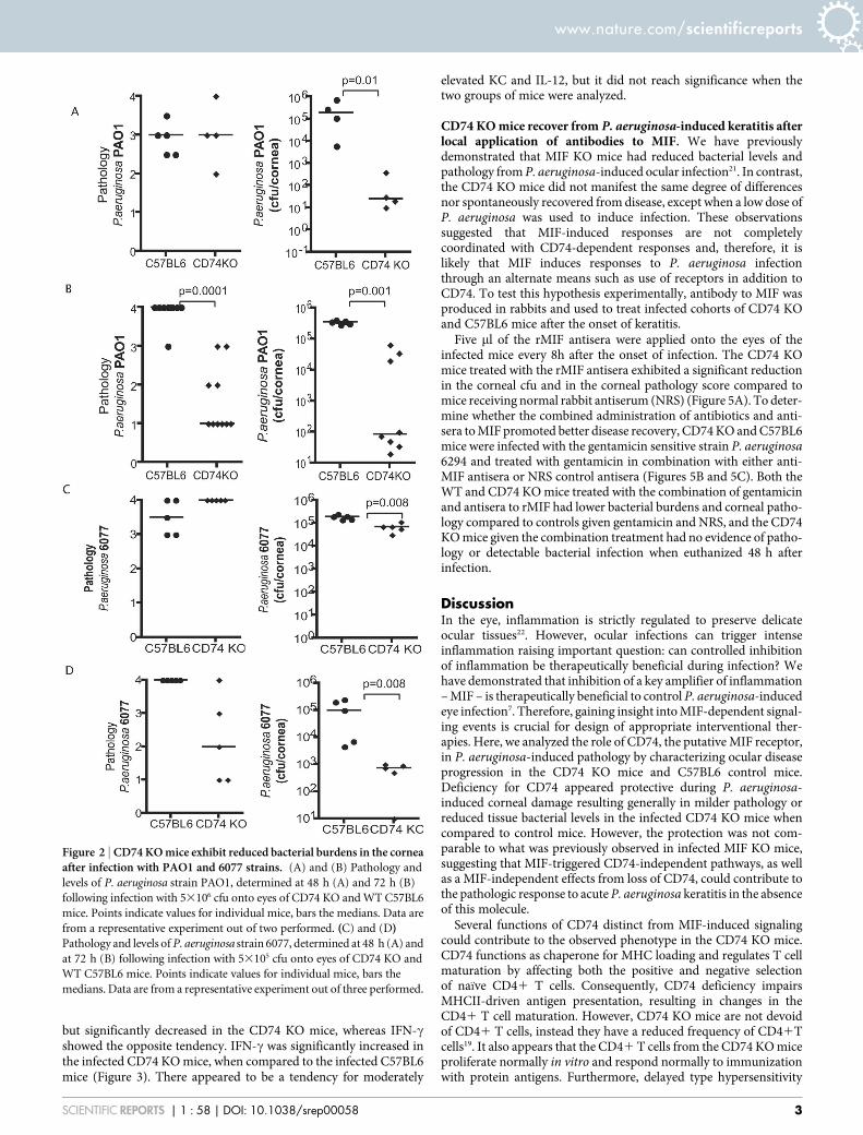
To determine whether the reduced pathology in CD74 KO micewas specific to the P. aeruginosa 6294 strain, similar experimentswere performed using another invasive P. aeruginosa strain, PAO1(Figure 2A and B), as well as a cytotoxic strain, 6077 (Figure 2Cand D). In both cases, the bacterial levels recovered from the cornealtissue were lower in the infected CD74 KO when compared to theinfected C57BL6 mice as early as 48 h post-infection. Significantreduction in the pathology scores in the CD74 knockout mice wasevident at 72 h post-infection, consistent with the observations forthe P. aeruginosa strain 6294.
CD74 regulates inflammatory responses to P. aeruginosa. SinceCD74 deficiency impairs MHCII-driven antigen presentation, it ispossible that the activation of T-cell responses to P. aeruginosa could
be altered in the CD74 KO mice19,20. Therefore, the observed lowerbacterial burdens and, in some cases, milder disease in the CD74 KOmay be due to alterations of the T-cell activation profile in addition toMIF-dependent activities. To examine the inflammatory cytokineprofiles in the infected CD74 KO and C57BL6 control mice,cohorts of mice were inoculated with P. aeruginosa 6294 and amultiplex cytokine assay performed on corneal samples at 48 hand 72 h post-infection. At 48 h after infection there were nosignificant differences in the levels of TNF-a, KC, IL-1b, or IFN-cbetween C57BL6 and CD74 KO mice (Figure 3). In addition, nodifferences were measured in IL-2, IL-4, IL-5, and IL-10 (Figure 4).However, at 72 h post-infection TNF-a, and IL-1b were modestly,
Figure 1 | CD74 KO mice have milder P. aeruginosa-induced ocularkeratitis. (A) Pathology and levels of P. aeruginosa strain 6294, determined
48 h following infection with 13105 cfu onto eyes of CD74 KO and WT
C57BL6 mice. Points indicate values for individual mice, bars the medians.
Data are from a representative experiment out of two performed. (B)
Histopathology images of corneal sections from C57BL6 and CD74 KO mice.
(C) and (D) Levels and pathology of P. aeruginosa strain 6294, determined at
72 h following infection with 53106 cfu onto eyes of CD74 KO and WT
C57BL6 mice. Points indicate values for individual mice, bars the medians.
Data are from a representative experiment out of three performed.
www.nature.com/scientificreports
SCIENTIFIC REPORTS | 1 : 58 | DOI: 10.1038/srep00058 2
but significantly decreased in the CD74 KO mice, whereas IFN-cshowed the opposite tendency. IFN-c was significantly increased inthe infected CD74 KO mice, when compared to the infected C57BL6mice (Figure 3). There appeared to be a tendency for moderately
elevated KC and IL-12, but it did not reach significance when thetwo groups of mice were analyzed.
CD74 KO mice recover from P. aeruginosa-induced keratitis afterlocal application of antibodies to MIF. We have previouslydemonstrated that MIF KO mice had reduced bacterial levels andpathology from P. aeruginosa-induced ocular infection21. In contrast,the CD74 KO mice did not manifest the same degree of differencesnor spontaneously recovered from disease, except when a low dose ofP. aeruginosa was used to induce infection. These observationssuggested that MIF-induced responses are not completelycoordinated with CD74-dependent responses and, therefore, it islikely that MIF induces responses to P. aeruginosa infectionthrough an alternate means such as use of receptors in addition toCD74. To test this hypothesis experimentally, antibody to MIF wasproduced in rabbits and used to treat infected cohorts of CD74 KOand C57BL6 mice after the onset of keratitis.
Five ml of the rMIF antisera were applied onto the eyes of theinfected mice every 8h after the onset of infection. The CD74 KOmice treated with the rMIF antisera exhibited a significant reductionin the corneal cfu and in the corneal pathology score compared tomice receiving normal rabbit antiserum (NRS) (Figure 5A). To deter-mine whether the combined administration of antibiotics and anti-sera to MIF promoted better disease recovery, CD74 KO and C57BL6mice were infected with the gentamicin sensitive strain P. aeruginosa6294 and treated with gentamicin in combination with either anti-MIF antisera or NRS control antisera (Figures 5B and 5C). Both theWT and CD74 KO mice treated with the combination of gentamicinand antisera to rMIF had lower bacterial burdens and corneal patho-logy compared to controls given gentamicin and NRS, and the CD74KO mice given the combination treatment had no evidence of patho-logy or detectable bacterial infection when euthanized 48 h afterinfection.
DiscussionIn the eye, inflammation is strictly regulated to preserve delicateocular tissues22. However, ocular infections can trigger intenseinflammation raising important question: can controlled inhibitionof inflammation be therapeutically beneficial during infection? Wehave demonstrated that inhibition of a key amplifier of inflammation– MIF – is therapeutically beneficial to control P. aeruginosa-inducedeye infection7. Therefore, gaining insight into MIF-dependent signal-ing events is crucial for design of appropriate interventional ther-apies. Here, we analyzed the role of CD74, the putative MIF receptor,in P. aeruginosa-induced pathology by characterizing ocular diseaseprogression in the CD74 KO mice and C57BL6 control mice.Deficiency for CD74 appeared protective during P. aeruginosa-induced corneal damage resulting generally in milder pathology orreduced tissue bacterial levels in the infected CD74 KO mice whencompared to control mice. However, the protection was not com-parable to what was previously observed in infected MIF KO mice,suggesting that MIF-triggered CD74-independent pathways, as wellas a MIF-independent effects from loss of CD74, could contribute tothe pathologic response to acute P. aeruginosa keratitis in the absenceof this molecule.
Several functions of CD74 distinct from MIF-induced signalingcould contribute to the observed phenotype in the CD74 KO mice.CD74 functions as chaperone for MHC loading and regulates T cellmaturation by affecting both the positive and negative selectionof naı̈ve CD41 T cells. Consequently, CD74 deficiency impairsMHCII-driven antigen presentation, resulting in changes in theCD41 T cell maturation. However, CD74 KO mice are not devoidof CD41 T cells, instead they have a reduced frequency of CD41Tcells19. It also appears that the CD41 T cells from the CD74 KO miceproliferate normally in vitro and respond normally to immunizationwith protein antigens. Furthermore, delayed type hypersensitivity
Figure 2 | CD74 KO mice exhibit reduced bacterial burdens in the corneaafter infection with PAO1 and 6077 strains. (A) and (B) Pathology and
levels of P. aeruginosa strain PAO1, determined at 48 h (A) and 72 h (B)
following infection with 53106 cfu onto eyes of CD74 KO and WT C57BL6
mice. Points indicate values for individual mice, bars the medians. Data are
from a representative experiment out of two performed. (C) and (D)Pathology and levels of P. aeruginosa strain 6077, determined at 48 h (A) and
at 72 h (B) following infection with 53105 cfu onto eyes of CD74 KO and
WT C57BL6 mice. Points indicate values for individual mice, bars the
medians. Data are from a representative experiment out of three performed.
www.nature.com/scientificreports
SCIENTIFIC REPORTS | 1 : 58 | DOI: 10.1038/srep00058 3

(DTH) responses appear comparable with that of the C57BL6 con-trol mice, suggesting that Th1 skewing is occurring20. The ability ofthe naı̈ve CD41 T cells in the CD74 KO mouse environment todevelop into Th1 or, possibly Th17, subsets was verified in a colitismodel wherein disease was induced by administration of 2,4,6-trini-trobenzenesulfonic acid (TNBS). TNBS challenged CD74 KOand C57BL6 mice had comparable inflammatory responses20.Interestingly, Topilski et al reported that the CD74 KO micemounted slightly elevated IFN-c responses after antigenic challenge,suggestive of elevated Th1 responses20. Consistent with these find-ings, we do not find defects in Th1 inflammatory cytokine synthesis
in response to P. aeruginosa corneal challenge. The Th1 versus Th2profiling was examined based on relative presence of IL-2, IL-4, IL-5,IL-10, IL-12, IFN-c and TNF-a in the corneal tissues. While the IL-2,IL-12 levels were comparable in the infected CD74 KO and C57BL6mice, the IFN-c levels were slightly elevated in the CD74 KO, whencompared to the P. aeruginosa-infected C57BL6 mice. Therefore, weruled out that the milder phenotype observed in the CD74 KO wasdue to significant decrease in disease-mediating Th1 skewing23.
The genetic background of the mice influences disease severity inocular keratitis24. While C57BL6 mice appear sensitive, BALB C micehave milder responses to P. aeruginosa-induced keratitis. Therefore,
Figure 3 | CD74 regulates proinflammatory responses to P. aeruginosa. Groups of 5 individual CD74 KO mice and 5 C57Bl6 mice were infected with
5 3 106 cfu placed onto scratch-injured eyes. Mouse corneas were harvested at 48 h (A) and 72 h (B) after infection. The levels of mouse cytokines
in corneal lysates were simultaneously measured using a MSD multiplex 7-spot electrochemiluminescence (ECL) assay. The levels of cytokines measured
in the different groups were compared with the Mann-Whitney U test.
www.nature.com/scientificreports
SCIENTIFIC REPORTS | 1 : 58 | DOI: 10.1038/srep00058 4

a possible defect in Th2 skewing in the CD74 KO mice could affectdisease progression in these animals. To address this issue, Th2 sub-set specific cytokines were quantified and no significant changes inthe IL-4 and IL-5 levels were observed, suggesting no major altera-tions in the CD41 Th2 subset skewing (Figure 4). These data areconsistent with previously reported observations that the CD74 KOmice develop normal Th2 responses25.
Previous reports have highlighted the importance of MIF-inducedCD74 regulated inflammatory responses during autoimmuneinflammation14,16. MIF binding to the CD74/CD44 complex resultsin elevated MAPK activation and sustained cytokine production bymacrophages and/or epithelial cells7,12,26,27. Our previous data sug-gested that protection against P. aeruginosa-induced acute keratitiscorrelated with decreased TNF-a, IL-6, and IL-1b recovered from theinfected corneas7. Consistently, we observed lower levels of TNF-aand IL-1b at 72h post-infection, but not at 48h post-infection, inCD74 KO mice, suggesting that a reduction in the sustained inflam-matory responses may contribute to some of the lessened diseaseobserved in these mice. However, as these differences were not mani-fest early in the infection they likely reflect the effects from reducedbacterial levels recovered from the CD74 KO mice, but these reduc-tions probably came too late to have an impact on the initial develop-ment of inflammation-induced corneal damage during P. aeruginosainfection.
The conclusion that there are CD74-independent cellular res-ponse pathways that are induced by MIF supported by the obser-vation that when the CD74 KO mice were therapeutically treatedwith antisera to MIF after the onset of disease there were furtherreductions in corneal pathology and bacterial burdens, consistentwith previously published data21 that inhibiting MIF biologicallyreduced disease pathology in the infected C57BL6 mice. Futurestudies should establish whether these functions of MIF could beattributed to its ability to serve as a non-cognate ligand forCXCR228 or relate to cell-intrinsic functions of MIF mediated byMIF-interacting intracellular proteins29.
In conclusion, infection can negatively affect host fitness throughtwo distinct mechanisms. Firstly, P. aeruginosa can directly causetissue damage through a variety of virulence activities. Secondly,tissue damage can be caused by the inflammatory responses to thepathogen. Thus, treatment of infection must come in two forms toenable recovery from the two sources of tissue damage. Our datashows that combined with antibiotic treatment, the antibody toMIF markedly reduced pathology and bacterial levels in WTC57BL6 mice, but essentially resulted in complete resolution ofpathology and bacterial infection in the CD74 KO mice. These find-ings suggest that a combination of antibiotics, inhibition of MIF andinhibition of CD74-dependent signaling could provide an optimaltherapeutic strategy for treating P. aeruginosa infections.
MethodsMice. All studies were performed in accordance with the Harvard Medical SchoolAnimal Care and Use Committee guidelines. Breeding pairs of CD74 knockout (KO)mice were a kind gift from Dr. F. Vascotto (Harvard Medical School). Control mice(C57BL6) were obtained from Taconic Farms. Mice were housed and bred in theChanning Laboratory Animal Care Facilities
Bacterial strains and inocula. Invasive P. aeruginosa strains PAO1, 6294 and ExotoxinU-producing cytotoxic strain 6077 were used throughout these experiments. Effectsfrom low dose (1 3 105 cfu/eye) and high dose (5 3 106 cfu/eye) inocula were initiallyevaluated to determine the doses to use in the in vivo infection studies. Generally, thebacterial strains were grown overnight at 37uC on tryptic soy broth agar plates andbacterial cells suspended to a concentration of 1 3 109 cfu/ml in saline solution.
Purification of rMIF. The human MIF cDNA containing IMAGE clone (ATCC, VA)was amplified by PCR, sequenced and subcloned into E. coli BL21 expression pet11bplasmid as in30. Recombinant protein was expressed in E. coli BL21 cells grownovernight at 37uC on lysogeny (L) agar plates containing 50 mg carbenicillin /mL(Sigma-Aldrich, MO). Individual colonies were selected and grown in 2 ml LB mediacontaining 50 ug/ml carbenicillin with shaking for 2 h at 37 C. 1– 2 mL of thebacterial cultures were transferred to 50 mL conical tubes each containing 25 mL LBwith 50 mg/ml carbinicillin (Sigma-Aldrich, MO). Individual colonies were selectedand grown with shaking for 2 h at 37uC in 2 ml lysogeny broth (LB) media containing50 mg carbenicillin /ml. One to two ml of the bacterial cultures were transferred to50 ml conical tubes each containing 25 ml LB with 50 mg carbenicillin /ml. The
Figure 4 | CD74 deficiency does not radically inhibit Th2 cytokines present in the cornea after P. aeruginosa infection. Groups of 5 CD74 KO mice and
5 C57Bl6 mice were infected with 5 3 106 cfu placed onto scratch-injured eyes. Mouse corneas were harvested at 48 h (A) and 72 h (B) after infection. The
levels of mouse cytokines in corneal lysates were simultaneously measured using a MSD multiplex 7-spot electrochemiluminescence (ECL) assay.
The levels of cytokines measured in the different groups were compared with the Mann-Whitney U test.
www.nature.com/scientificreports
SCIENTIFIC REPORTS | 1 : 58 | DOI: 10.1038/srep00058 5

cultures were shaken for several hours untill the bacterial cultures reached anOD600 . 0.8. About 10 ml of bacterial culture was transferred to Erlenmeyer flaskscontaining 500 mL LB with 50 mg carbenicillin/ml and the bacterial culture densityadjusted to an OD600 0.1–0.2. Cultures were incubated with shaking at 37uC for1.5 hrs, the OD600 was measured every 30 min. When the cultures reached anOD600 ,0.9, 1mM isopropyl b-D-1-thiogalactopyranoside (IPTG) (Invitrogen, MD)was added to induce transcription of the cloned MIF gene and the E. coli cellsgrown for additional 3–3.5 hrs with shaking at 37uC. Bacteria were harvested bycentrifugation at 3000 rpm for 15 min using Beckman ultracentrifuge. The bacterialpellets were stored at 220uC.
E.coli pellets were lysed with glass beads (Sigma-Aldrich, MO) in 25 mM HEPES,1% Triton X-100, 1 mM EDTA, pH 7.5 buffer. The lysate was centrifuged in aBeckman refrigerated (4C) centrifuge at 10000 g for 20 minutes and passed through47 mm 5 micron filters, followed by 0.45u filter using a vacuum filtration setup.Approximately 175 mL of clarified rMIF E. coli lysate, harvested from 500 ml ofbacterial cultures, was loaded onto a 42 mL Fractogel DEAE anion exchange columnin 25 mM HEPES, 0.1% Tween 20, pH 7.5 buffer at 13 ml/minute. The effluent andthe wash were pooled. The pH of the pooled material was adjusted with 30 mL of0.5 M MES, pH 5.5. The resultant solution had a pH between 5.5 and 6.0. Thismaterial was loaded onto a CM Sepharose Cation Exchange column and elutedwith a step gradient at 0.5 M MES, 500 mM NaCl, pH 5.5. Purified rMIF wasdialyzed against 25mM HEPES, 150 mM NaCl pH7.4. and stored frozen at 220C.
Generation of the rabbit polyclonal anti-MIF antisera. rMIF containing liposomeswere prepared for immunization purposes. The liposome composition was25 mg/mL lipids, 0.25 mg/mL CpG, 1 mg/mL rMIF. The lipids were formulatedas a blend of 40% phosphatidyletanolamine (1550) (Avanti Polar Lipids, AL;663 g/mole), 40% phopshatidylcholine (1550) (Avanti Polar Lipids, AL; 705 g/mole),10% Cholesterol (Avanti Polar Lipids, AL; 386 g/mole), 10% DC-Cholesterol (AvantiPolar Lipids, AL; 537 g/mole). Rabbits were immunized in the popliteal lymph nodewith 100 mg rMIF/ liposome mixture, rested for three weeks, and then re-challengedagain in the popliteal lymph node. After 4 weeks of rest, rabbits were immunizedintramuscularly with 100 mg rMIF/liposomes and sera were harvested a week later.The immunizations were carried out at Lampire Biological Laboratories and end-point binding titers of 12, 800 and 25, 600 obtained in two different rabbits.
Infection model. Infections were carried out as described previously31. Briefly, micewere anesthetized with injections of 100 mg ketamine/Kg and 10 mg xylazine/Kg.Three 0.5 cm scratches were made on the cornea and an inoculum of P. aeruginosadelivered in 5 ml onto the eye. Mice remained sedated for about 30 min. Forevaluation of corneal pathology, daily scores are recorded by an observer unaware ofthe experimental status of the animals based on a scoring system using a graded scaleof 0 to 4 as follows: 0, eye macroscopically identical to the uninfected contra-lateralcontrol eye; 1, faint opacity partially covering the pupil; 2, dense opacity covering thepupil; 3, dense opacity covering the entire anterior segment; and 4, perforation of the
Figure 5 | Treatment with anti-MIF antisera promotes further recovery from P. aeruginosa-induced corneal infection and pathology in CD74 KOmice. (A) Groups of CD74 KO mice were treated with either rabbit control antisera or anti-MIF antisera every 8 h after the induction of infection for the
duration of 2 days after which infected corneas were harvested, homogenized, and bacteria quantified. Points indicate values from individual mice, bars
the medians. (B) Groups of C57BL6 and CD74 KO mice were treated with either gentamicin, non-immune rabbit antisera, or gentamicin and anti-MIF
antisera every 8 hours after infection for 2 days after which infected corneas were harvested, and bacteria quantified. (C) Pathology scores plotted. Points
indicate values from individual mice, bars the medians. Data analysis performed with 1way Anova with Dunn’s Multiple Comparison Test (Prizm 4.0).
Each experiment was repeated twice.
www.nature.com/scientificreports
SCIENTIFIC REPORTS | 1 : 58 | DOI: 10.1038/srep00058 6

cornea, phthisis bulbi, shrinkage of the globe after inflammatory disease, or both. Todetermine the levels of bacteria in the cornea 48 or 72 h after infection, mice weresacrificed, eyes enucleated and corneas dissected from the ocular surface. To quantifythe levels of P. aeruginosa, corneas were excised and suspended in a tryptic soybroth, 0.5% Triton X-100 solution, vortexed, serial dilutions made and plated onP. aeruginosa selective cetrimide plates.
For treatment, groups of mice were infected with P. aeruginosa strain 6294, thentreated topically for 2 days with 5 ml of a solution of 100 mg gentamicin/ml(Invitrogen, MD), along with a rabbit polyclonal antisera to MIF. Anti-sera andgentamycin treatments were administered every 8 hours after the initiation ofinfection. Bacteria in the cornea were quantified 2 days after infection by homo-genizing and plating the corneal tissue. Separate cohorts of CD74 KO mice weretreated with either the antiserum to MIF or a pre-immune control antiserum in theabsence of gentamicin. All antisera were pre-adsorbed with P. aeruginosa to eliminateany natural anti-bacterial reactivity.
Histopathology examinations. Eyes were enucleated from euthanized mice andfixed in 4% paraformaldehyde then embedded in paraffin. Four mm sections werecut, and stained with hematoxylin-eosin to visualize tissue morphology followingpreviously used techniques32.
Preparation of corneal lysates. Groups of 7 MIF KO mice and 7 C57BL6 mice wereinfected with 1 3 106 P. aeruginosa strain 6294 cfu placed onto scratch-injured eyes.Mouse corneas were harvested at 48 h and 72 h after infection, washed in F12 media,then each cornea was homogenized in 500 ml PBS containing a mix of proteaseinhibitors (Complete, Roche) and supplemented with 0.5% Triton to disrupt plasmamembranes using an OMNI International tissue master homogenizer 125. Solutionswere clarified by 5 min centrifugation at 4C and maximum rpm in a table-topEppendorf 5415R centrifuge. The supernatants were stored frozen at 280uC untilprocessed for cytokine analysis.
Cytokine analysis. Mouse cytokines in corneal lysates were measured using a MesoScale Discovery (MSD) multiplex 7-spot electrochemiluminescence (ECL) assay andoutputs measured by an ultra low noise charge-coupled device (CCD) Imager 2400(Meso Scale Discovery, Gaithersburg, MD, USA). The cytokines measured includedinterleukin (IL-1, TNF-a, IL-2, IL-4, IL-5, IL-10, IFN-c to establish Th1 versus Th2profiling in the tissues. The MSD ECL platform has been previously validated againstcytokine standards recommended by WHO and U.K. National Institute for BiologicalStandards and Control (NIBSC) and by comparison to traditional ELISA33. Cytokineconcentrations were adjusted to total protein levels measured in the corneal extract bythe BCA assay (Thermo Scientific, Rockford, IL) using a multi-label Victor 2 counterto measure colorimetric reactions (Perkin Elmer, Boston, MA).
Statistical analysis. Statistical analysis of the differences in the cytokine levels wereperformed by Mann- Whitney U tests and differences were considered significant ifthe p value was ,0.05 (Prism 4.0 for Macintosh). Statistical analysis of the cornealpathology scores was either by use of the Mann-Whitney U test for pair-wisecomparisons or the Kruskal-Wallis non-parametric ANOVA with Dunn’s Procedurefor Multigroup comparisons and individual 2-group comparisons (Prism 4.0 forMacintosh).
1. Robertson, D. M., Petroll, W. M., Jester, J. V., & Cavanagh, H. D. Currentconcepts: contact lens related Pseudomonas keratitis. Cont Lens Anterior Eye 30,94–107 (2007).
2. Fleiszig, S. M. & Evans, D. J. The pathogenesis of bacterial keratitis: studies withPseudomonas aeruginosa. Clin Exp Optom 85, 271–278 (2002).
3. Hazlett, L. D. & Hendricks, R. L. Reviews for immune privilege in the year 2010:immune privilege and infection. Ocul Immunol Inflamm 18, 237–243.
4. Sun, Y. et al. TLR4 and TLR5 on corneal macrophages regulate Pseudomonasaeruginosa keratitis by signaling through MyD88-dependent and -independentpathways. J Immunol 185, 4272–4283.
5. Thakur, A., Barrett, R. P., Hobden, J. A., & Hazlett, L. D. Caspase-1 inhibitorreduces severity of pseudomonas aeruginosa keratitis in mice. Investigativeophthalmology & visual science 45, 3177–3184 (2004).
6. Medzhitov, R. Damage control in host-pathogen interactions. Proceedings of theNational Academy of Sciences of the United States of America 106, 15525–15526(2009).
7. Gadjeva, M., Nagashima, J., Zaidi, T., Mitchell, R. A., & Pier, G. B. Inhibition ofmacrophage migration inhibitory factor ameliorates ocular Pseudomonasaeruginosa-induced keratitis. PLoS Pathog 6, e1000826.
8. Flaster, H., Bernhagen, J., Calandra, T., & Bucala, R. The macrophage migrationinhibitory factor-glucocorticoid dyad: regulation of inflammation and immunity.Molecular endocrinology (Baltimore, Md) 21, 1267–1280 (2007).
9. Bernhagen, J. et al. MIF is a pituitary-derived cytokine that potentiates lethalendotoxaemia. Nature 365, 756–759 (1993).
10. Bernhagen, J., Calandra, T., & Bucala, R. The emerging role of MIF in septic shockand infection. Biotherapy 8, 123–127 (1994).
11. Calandra, T., Bernhagen, J., Mitchell, R. A., & Bucala, R. The macrophage is animportant and previously unrecognized source of macrophage migrationinhibitory factor. The Journal of experimental medicine 179, 1895–1902 (1994).
12. Calandra, T. et al. MIF as a glucocorticoid-induced modulator of cytokineproduction. Nature 377, 68–71 (1995).
13. Shachar, I. & Haran, M. The secret second life of an innocent chaperone: the storyof CD74 and B cell/chronic lymphocytic leukemia cell survival. Leuk Lymphoma.
14. Leng, L. et al. MIF signal transduction initiated by binding to CD74. The Journal ofexperimental medicine 197, 1467–1476 (2003).
15. Lue, H. et al. Rapid and transient activation of the ERK MAPK signalling pathwayby macrophage migration inhibitory factor (MIF) and dependence on JAB1/CSN5 and Src kinase activity. Cell Signal 18, 688–703 (2006).
16. Shi, X. et al. CD44 is the signaling component of the macrophage migrationinhibitory factor-CD74 receptor complex. Immunity 25, 595–606 (2006).
17. Schwartz, V. et al. A functional heteromeric MIF receptor formed by CD74 andCXCR4. FEBS letters 583, 2749–2757 (2009).
18. Hazlett, L. D. Inflammatory response to Pseudomonas aeruginosa keratitis. Theocular surface 3 (4 Suppl), S139–141 (2005).
19. Elliott, E. A. et al. The invariant chain is required for intracellular transport andfunction of major histocompatibility complex class II molecules. The Journal ofexperimental medicine 179, 681–694 (1994).
20. Topilski, I., Harmelin, A., Flavell, R. A., Levo, Y., & Shachar, I. Preferential Th1immune response in invariant chain-deficient mice. J Immunol 168, 1610–1617(2002).
21. Gadjeva, M., Nagashima, J., Zaidi, T., Mitchell, R. A., & Pier, G. B. Inhibition ofmacrophage migration inhibitory factor ameliorates ocular Pseudomonasaeruginosa-induced keratitis. PLoS Pathog 6, e1000826 (2010).
22. Niederkorn, J. Y. See no evil, hear no evil, do no evil: the lessons of immuneprivilege. Nature immunology 7, 354–359 (2006).
23. Kwon, B. & Hazlett, L. D. Association of CD41 T cell-dependent keratitis withgenetic susceptibility to Pseudomonas aeruginosa ocular infection. J Immunol159, 6283–6290 (1997).
24. Hazlett, L. D., McClellan, S., Kwon, B., & Barrett, R. Increased severity ofPseudomonas aeruginosa corneal infection in strains of mice designated as Th1versus Th2 responsive. Investigative ophthalmology & visual science 41, 805–810(2000).
25. Brown, D. R. et al. T helper subset differentiation in the absence of invariant chain.J Exp Med 185, 31–41 (1997).
26. Beswick, E. J. et al. Helicobacter pylori-induced IL-8 production by gastricepithelial cells up-regulates CD74 expression. J Immunol 175, 171–176 (2005).
27. Beswick, E. J. et al. Helicobacter pylori binds to CD74 on gastric epithelial cells andstimulates interleukin-8 production. Infection and immunity 73, 2736–2743(2005).
28. Weber, C. et al. Structural determinants of MIF functions in CXCR2-mediatedinflammatory and atherogenic leukocyte recruitment. Proc Natl Acad Sci U S A105, 16278–16283 (2008).
29. Merk, M. et al. The Golgi-associated protein p115 mediates the secretion ofmacrophage migration inhibitory factor. J Immunol 182, 6896–6906 (2009).
30. Bernhagen, J. et al. Purification, bioactivity, and secondary structure analysis ofmouse and human macrophage migration inhibitory factor (MIF). Biochemistry33, 14144–14155 (1994).
31. Preston, M. J. et al. Rapid and sensitive method for evaluating Pseudomonasaeruginosa virulence factors during corneal infections in mice. Infect Immun 63,3497–3501 (1995).
32. Gadjeva, M., Wang, Y., & Horwitz, B. H. NF-kappaB p50 and p65 subunits controlintestinal homeostasis. European journal of immunology 37, 2509–2517 (2007).
33. Fichorova, R. N. et al. Biological and technical variables affecting immunoassayrecovery of cytokines from human serum and simulated vaginal fluid:a multicenter study. Anal Chem 80 (12), 4741–4751 (2008).
AcknowledgementsThis work was supported by Public Health Service grant 5R21EY019944 - 02 (MG) andEY016144 (GP) from the National Eye Institute.
Author contributionsTZ and MG prepared figures 1,2,5; RF and MG prepared 3 and 4. RF analyzed the MSDcytokine data; TR purified rMIF and prepared the liposomes; SD maintained the mousecolony and provided help with experiments; MG designed experiments, analyzed data andwrote the manuscript text. All authors (MG, SD, TR, RF, JP, TZ) reviewed the manuscript.
Additional informationCompeting financial interests: The authors declare no competing financial interests.
License: This work is licensed under a Creative CommonsAttribution-NonCommercial-NoDerivative Works 3.0 Unported License. To view a copyof this license, visit http://creativecommons.org/licenses/by-nc-nd/3.0/
How to cite this article: Zaidi, T. et al. CD74 deficiency ameliorates Pseudomonasaeruginosa-induced ocular infection. Sci. Rep. 1, 58; DOI:10.1038/srep00058 (2011).
www.nature.com/scientificreports
SCIENTIFIC REPORTS | 1 : 58 | DOI: 10.1038/srep00058 7


