Embed Size (px)
Citation preview
Emerging Infectious DiseasesEmerging Infectious Diseases is published monthly by the
National Center for Infectious Diseases, Centers for DiseaseControl and Prevention (CDC), 1600 Clifton Road, Mailstop D61,Atlanta, GA 30333, USA. Telephone 404-371-5329, fax 404-371-5449, email [email protected].
The opinions expressed by authors contributing to this journaldo not necessarily reflect the opinions of the Centers for DiseaseControl and Prevention or the institutions with which the authorsare affiliated.
All material published in Emerging Infectious Diseases is inthe public domain and may be used and reprinted without specialpermission; proper citation, however, is required.
Use of trade names is for identification only and does notimply endorsement by the Public Health Service or by the U.S.Department of Health and Human Services.
∞ Emerging Infectious Diseases is printed on acid-free paper that meetsthe requirements of ANSI/NISO 239.48-1992 (Permanence of Paper)
EDITORIAL STAFF
Founding EditorJoseph E. McDade, Rome, Georgia, USAManaging Senior EditorPolyxeni Potter, Atlanta, Georgia, USAAssociate EditorsCharles B. Beard, Ft. Collins, Colorado, USADavid Bell, Atlanta, Georgia, USAPatrice Courvalin, Paris, FranceStephanie James, Bethesda, Maryland, USABrian W.J. Mahy, Atlanta, Georgia, USATakeshi Kurata, Tokyo, JapanMartin I. Meltzer, Atlanta, Georgia, USADavid Morens, Washington, D.C., USATanja Popovic, Atlanta, Georgia, USAPatricia M. Quinlisk, Des Moines, Iowa, USAGabriel Rabinovich, Buenos Aires, ArgentinaDidier Raoult, Marseilles, FrancePierre Rollin, Atlanta, Georgia, USAMario Raviglione, Geneva, Switzerland Copy EditorsCarol Snarey, Anne Mather, Cathy Young,Mary Anne CastranioProductionReginald Tucker, Ann KitchenEditorial AssistantCarolyn Collins
EDITORIAL BOARD
Dennis Alexander, Addlestone Surrey, United KingdomBan Allos, Nashville, Tennessee, USAMichael Apicella, Iowa City, Iowa, USABarry J. Beaty, Ft. Collins, Colorado, USAMartin J. Blaser, New York, New York, USADavid Brandling-Bennet, Washington, D.C., USADonald S. Burke, Baltimore, Maryland, USACharles H. Calisher, Ft. Collins, Colorado, USAArturo Casadevall, New York, New York, USAThomas Cleary, Houston, Texas, USAAnne DeGroot, Providence, Rhode Island, USAVincent Deubel, Providence, Rhode Island, USAEd Eitzen, Washington, D.C., USADuane J. Gubler, Ft. Collins, Colorado, USAScott Halstead, Arlington, Virginia, USADavid L. Heymann, Geneva, SwitzerlandSakae Inouye, Tokyo, JapanCharles King, Cleveland, Ohio, USAKeith Klugman, Atlanta, Georgia, USAS.K. Lam, Kuala Lumpur, MalaysiaBruce R. Levin, Atlanta, Georgia, USAMyron Levine, Baltimore, Maryland, USAStuart Levy, Boston, Massachusetts, USAJohn S. MacKenzie, Brisbane, AustraliaTom Marrie, Edmonton, Alberta, CanadaJohn E. McGowan, Jr., Atlanta, Georgia, USAStephen S. Morse, New York, New York, USAPhilip P. Mortimer, London, United KingdomFred A. Murphy, Davis, California, USABarbara E. Murray, Houston, Texas, USAP. Keith Murray, Ames, Iowa, USAStephen Ostroff, Atlanta, Georgia, USARosanna W. Peeling, Geneva, SwitzerlandDavid H. Persing, Seattle, Washington, USAGianfranco Pezzino, Topeka, Kansas, USARichard Platt, Boston, Massachusetts, USALeslie Real, Atlanta, Georgia, USADavid Relman, Palo Alto, California, USANancy Rosenstein, Atlanta, Georgia, USAConnie Schmaljohn, Frederick, Maryland, USATom Schwan, Hamilton, Montana, USAIra Schwartz, Valhalla, New York, USATom Shinnick, Atlanta, Georgia, USARobert Shope, Galveston, Texas, USABonnie Smoak, Bethesda, Maryland, USARosemary Soave, New York, New York, USAP. Frederick Sparling, Chapel Hill, North Carolina, USAJan Svoboda, Prague, Czech RepublicBala Swaminathan, Atlanta, Georgia, USARobert Swanepoel, Johannesburg, South AfricaPhillip Tarr, Seattle, Washington, USATimothy Tucker, Cape Town, South AfricaElaine Tuomanen, Memphis, Tennessee, USADavid Walker, Galveston, Texas, USAMary E. Wilson, Cambridge, Massachusetts, USA
Emerging Infectious Diseases • Vol. 9, No. 10, October 2003
A Peer-Reviewed Journal Tracking and Analyzing Disease Trends pages 1197–1362
EDITOR-IN-CHIEFD. Peter Drotman
www.cdc.gov/eid
PerspectiveSyndromic Surveillance and Bioterrorism-related Epidemics . . . . . . . . . . . . . . . . . .1197J.W. Buehler et al.
ResearchIllness in Intensive Care Staff after Brief Exposure to Severe Acute Respiratory Syndrome . . . . . . . . . . . . . . . . . . . . . . . .1205D.C. Scales et al.
Superantigens and Streptococcal Toxic Shock Syndrome . . . . . . . . . . . . . . . . . . . . . . . .1211T. Proft et al.
Hazards of Healthy Living: Bottled Water and Salad Vegetables as Risk Factors for Campylobacter Infection . . . . . . . . . . . . . . . . . . . .1219M.R. Evans et al.
Escherichia coli O157 Exposure in Wyoming and Seattle: Serologic Evidence of Rural Risk . . . . . . . . . . . . . . . . . . . . . . . .1226J.P. Haack et al.
Characterization of Waterborne Outbreak–associated Campylobacter jejuni,Walkerton, Ontario . . . . . . . . . . . . . . . . . . . . . . . . . . .1232C.G. Clark et al.
Cultural Contexts of Ebola in Northern Uganda . . . . .1242B.S. Hewlett and R.P. Amola
1918 Influenza Pandemic Caused by Highly Conserved Viruses with Two Receptor-Binding Variants . . . . . . . . . . . . . . . . . . . . .1249A.H. Reid et al.
Cephamycin Resistance in Clinical Isolates and Laboratory-derived Strains of Escherichia coli, Nova Scotia, Canada . . . . . . . . . . .1254B. Clarke et al.
Mass Antibiotic Treatment for Group AStreptococcus Outbreaks in Two Long-Term Care Facilities . . . . . . . . . . . . . . . . . . . . . .1260A. Smith et al.
Anthelmintic Baiting of Foxes against UrbanContamination with Echinococcus multilocularis . . . .1266D. Hegglin et al.
Cephalosporin-resistant Escherichia coliamong Summer Camp Attendees with Salmonellosis . . . . . . . . . . . . . . . . . . . . . . . . . . .1273G. Prats et al.
Multijurisdictional Approach to Biosurveillance, Kansas City . . . . . . . . . . . . . . . . . . .1281M.A. Hoffman et al.
Environmental Risk and Meningitis Epidemics in Africa . . . . . . . . . . . . . . . . . . . . . . . . . . .1287A.M. Molesworth et al.
DispatchesSevere Acute Respiratory Syndrome: Lessons from Singapore . . . . . . . . . . . . . . . . . . . . . . .1294K. Singh et al.
A Peer-Reviewed Journal Tracking and Analyzing Disease Trends Vol.9, No.10, October 2003
On the Cover:Jacques-Louis David (1748-1825). Coronation of Empress Josephine by Napoleon I at Notre Dame deParis, 2 December 1804 (1806-1807) Oil on canvas, 6.1 m x 9.31 m. Photo: Peter WilliRéunion des Musées Nationaux/Art Resource, NY. Chateaux deVersailles et de Trianon, Versailles, France
About the Cover, pg. 1361
All material published in Emerging Infectious Diseases is in the publicdomain and may be used and reprinted without special permission; propercitation, however, is appreciated.

West Nile Virus Transmission in Resident Birds, Dominican Republic . . . . . . . . . . . . .1299O. Komar et al.
West Nile Virus Encephalitis and Myocarditis in Wolf and Dog . . . . . . . . . . . . . . . . . . . .1303C.A. Lichtensteiger et al.
Weissella confusa Infection in Primate (Cercopithecus mona) . . . . . . . . . . . . . . . . . .1307A.I. Vela et al.
Mycobacterium tuberculosis Beijing Genotype, the Netherlands . . . . . . . . . . . . . . . . . . . . .1310M.W. Borgdorff et al.
Saliva and Meningococcal Transmission . . . . . . . . . .1314H.J. Orr et al.
Small Colony Variants of Staphylococcus aureus and Pacemaker-related Infection . . . . . . . . . .1316H. Seifert et al.
West Nile Virus Detection in American Crows . . . . . .1319S.A. Yaremych et al.
Severe Histoplasmosis in Travelers to Nicaragua . . .1322M. Weinberg et al.
Mayaro Virus in Wild Mammals, French Guiana . . . .1326B. de Thoisy et al.
The European Commission’s Task Force on Bioterrorism . . . . . . . . . . . . . . . . . . . . . . . . .1330A. Tegnell et al.
Wild-type Measles Virus in Brain Tissue of Children with Subacute Sclerosing Panencephalitis, Argentina . . . . . . . . . . . . . . . . . . . . .1333P.R. Barrero et al.
Cat or Dog Ownership and Seroprevalence ofEhrlichiosis, Q Fever, and Cat-Scratch Disease . . . .1337M. Skerget et al.
Flying Squirrel–associated Typhus, United States . . . . . . . . . . . . . . . . . . . . . . . . . . . . . . .1341M.G. Reynolds et al.
Chlamydia trachomatis Infections in Female Soldiers, Israel . . . . . . . . . . . . . . . . . . . . . . . .1344E.S. Bamberger et al.
LettersClostridium tertium in Necrotizing Fascitis and Gangrene . . . . . . . . . . . . . . . . . . . . . . . .1347P. Ray et al.
Dengue Hemorrhagic Fever, Uttaradit, Thailand . . . .1348J. Patumanond et al.
Antimicrobial Drug–resistant Salmonella Typhimurium (Reply to Helms) . . . . . . . . . . . . . . . . . .1350J. Dahl
Antimicrobial Drug–resistant Salmonella Typhimurium (Reply to Dahl) . . . . . . . . . . . . . . . . . . .1350M. Helms
Serogroup A Neisseria meningitidis Outside Meningitis Belt in Southwest Cameroon . . . . . . . . . . .1351P. Cunin et al.
West Nile Virus Meningitis in Patient with Common Variable Immunodeficiency . . . . . . . . .1353A.M. Alonto et al.
Isolation of Enterobacter sakazakii from Midgut of Stomoxys calcitrans . . . . . . . . . . . . . . . . . .1355J.V. Hamilton et al.
Book ReviewExotic Viral Diseases: A Global Guide . . . . . . . . . . . .1357M. Bell
News & NotesConference SummaryDrug-resistant Streptococcus pneumoniaeand Methicillin-resistant Staphylococcus aureus Surveillance . . . . . . . . . . . . . . . . . . . . . . . . . .1358L.A. Hawley et al.
About the Cover . . . . . . . . . . . . . . . . . . . . . . . . . . . . .1360P. Potter
A Peer-Reviewed Journal Tracking and Analyzing Disease Trends Vol.9, No.10, October 2003
To facilitate rapid detection of a future bioterroristattack, an increasing number of public health departmentsare investing in new surveillance systems that target theearly manifestations of bioterrorism-related disease.Whether this approach is likely to detect an epidemic soon-er than reporting by alert clinicians remains unknown. Thedetection of a bioterrorism-related epidemic will depend onpopulation characteristics, availability and use of healthservices, the nature of an attack, epidemiologic features ofindividual diseases, surveillance methods, and the capaci-ty of health departments to respond to alerts. Predictinghow these factors will combine in a bioterrorism attack maybe impossible. Nevertheless, understanding their likelyeffect on epidemic detection should help define the useful-ness of syndromic surveillance and identify approaches toincreasing the likelihood that clinicians recognize andreport an epidemic.
Because of heightened concerns about the possibility ofbioterrorist attacks, public health agencies are testing
new methods of surveillance intended to detect the earlymanifestations of illness that may occur during a bioterror-ism-related epidemic. Broadly labeled “syndromic surveil-lance,” these efforts encompass a spectrum of activitiesthat include monitoring illness syndromes or events, suchas medication purchases, that reflect the prodromes ofbioterrorism-related diseases (1–9). The Centers forDisease Control and Prevention (CDC) estimates that, asof May 2003, health departments in the United States haveinitiated syndromic surveillance systems in approximately100 sites throughout the country (T. Treadwell, CDC, pers.comm.). The goal of these systems is to enable earlierdetection of epidemics and a more timely public healthresponse, hours or days before disease clusters are recog-nized clinically, or before specific diagnoses are made andreported to public health authorities. Whether this goal isachievable remains unproved (4,5,10).
Establishing a diagnosis is critical to the public healthresponse to a bioterrorism-related epidemic, since thediagnosis will guide the use of vaccinations, medications,and other interventions. Absent a bioterrorism attack, pre-dicting whether syndromic surveillance will trigger aninvestigation that yields a diagnosis before clinicians makeand report a diagnosis is not possible. Our objective is toconsider the mix of hypothetical factors that may affect thedetection of epidemics attributable to CDC category Abioterrorism agents (11).
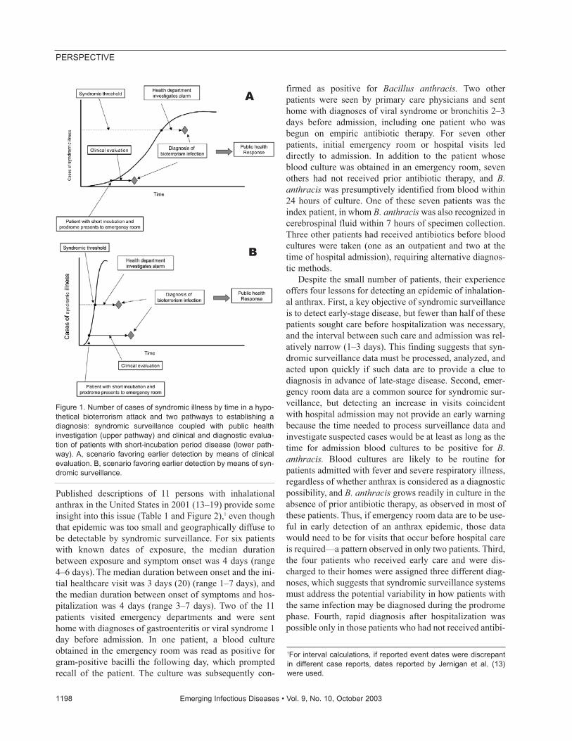
Establishing a DiagnosisTwo pathways to establishing a diagnosis are described
by the scenarios below and in Figure 1, using a single, clan-destine dissemination of an anthrax aerosol as an example.
Detection through Syndromic SurveillanceThe early signs of inhalational anthrax include nonspe-
cific symptoms that may persist for several days before theonset of more severe disease (12). Patients with prodromalillnesses seek outpatient care and are assigned nonspecificdiagnoses such as “viral syndrome.” Data on patients fit-ting various syndromic criteria are transferred to the healthdepartment and tested for aberrant trends. This process“flags” that a statistical detection threshold has beenexceeded. Epidemiologists conclude that a preliminaryinvestigation is warranted and collect blood for culturefrom several patients. Within 18 hours, one culture yieldsa presumptive diagnosis of anthrax, prompting a full-scaleresponse.
Detection through Clinician ReportingSome persons in whom inhalational anthrax develops
will have short incubation periods and prodromes (12).Respiratory distress occurs in one such person, and he ishospitalized. Routine admission procedures include bloodcultures. Within 18 hours, a presumptive diagnosis ofanthrax is made. The patient’s physician informs the localhealth department, prompting a full-scale response.
In practice, how a bioterrorism attack might be detect-ed and diagnosed will probably be more complex.
Emerging Infectious Diseases • Vol. 9, No. 10, October 2003 1197
PERSPECTIVE
Syndromic Surveillance andBioterrorism-related Epidemics
James W. Buehler,* Ruth L. Berkelman,* David M. Hartley,† and Clarence J. Peters‡
*Emory University Rollins School of Public Health, Atlanta,Georgia, USA; †University of Maryland School of Medicine,Baltimore, Maryland, USA; and ‡University of Texas MedicalBranch, Galveston, Texas, USA
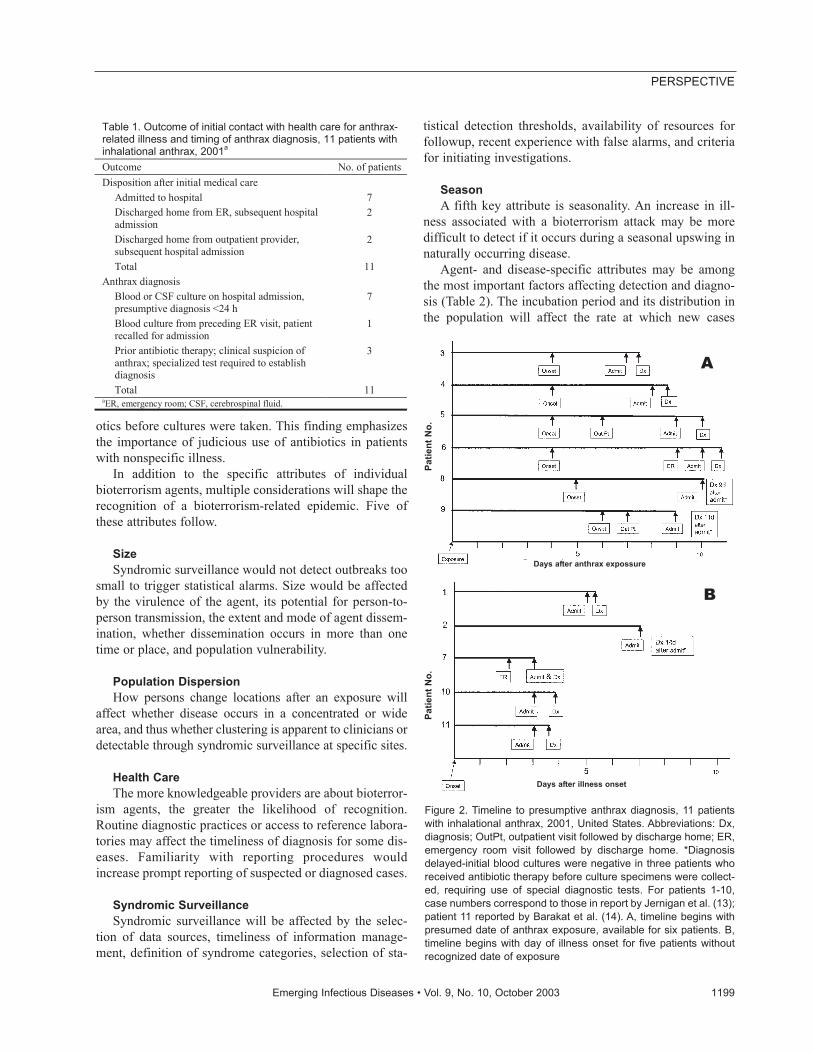
Published descriptions of 11 persons with inhalationalanthrax in the United States in 2001 (13–19) provide someinsight into this issue (Table 1 and Figure 2),1 even thoughthat epidemic was too small and geographically diffuse tobe detectable by syndromic surveillance. For six patientswith known dates of exposure, the median durationbetween exposure and symptom onset was 4 days (range4–6 days). The median duration between onset and the ini-tial healthcare visit was 3 days (20) (range 1–7 days), andthe median duration between onset of symptoms and hos-pitalization was 4 days (range 3–7 days). Two of the 11patients visited emergency departments and were senthome with diagnoses of gastroenteritis or viral syndrome 1day before admission. In one patient, a blood cultureobtained in the emergency room was read as positive forgram-positive bacilli the following day, which promptedrecall of the patient. The culture was subsequently con-
firmed as positive for Bacillus anthracis. Two otherpatients were seen by primary care physicians and senthome with diagnoses of viral syndrome or bronchitis 2–3days before admission, including one patient who wasbegun on empiric antibiotic therapy. For seven otherpatients, initial emergency room or hospital visits leddirectly to admission. In addition to the patient whoseblood culture was obtained in an emergency room, sevenothers had not received prior antibiotic therapy, and B.anthracis was presumptively identified from blood within24 hours of culture. One of these seven patients was theindex patient, in whom B. anthracis was also recognized incerebrospinal fluid within 7 hours of specimen collection.Three other patients had received antibiotics before bloodcultures were taken (one as an outpatient and two at thetime of hospital admission), requiring alternative diagnos-tic methods.
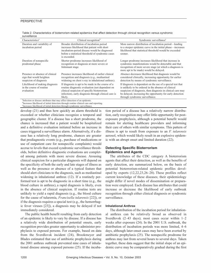
Despite the small number of patients, their experienceoffers four lessons for detecting an epidemic of inhalation-al anthrax. First, a key objective of syndromic surveillanceis to detect early-stage disease, but fewer than half of thesepatients sought care before hospitalization was necessary,and the interval between such care and admission was rel-atively narrow (1–3 days). This finding suggests that syn-dromic surveillance data must be processed, analyzed, andacted upon quickly if such data are to provide a clue todiagnosis in advance of late-stage disease. Second, emer-gency room data are a common source for syndromic sur-veillance, but detecting an increase in visits coincidentwith hospital admission may not provide an early warningbecause the time needed to process surveillance data andinvestigate suspected cases would be at least as long as thetime for admission blood cultures to be positive for B.anthracis. Blood cultures are likely to be routine forpatients admitted with fever and severe respiratory illness,regardless of whether anthrax is considered as a diagnosticpossibility, and B. anthracis grows readily in culture in theabsence of prior antibiotic therapy, as observed in most ofthese patients. Thus, if emergency room data are to be use-ful in early detection of an anthrax epidemic, those datawould need to be for visits that occur before hospital careis required—a pattern observed in only two patients. Third,the four patients who received early care and were dis-charged to their homes were assigned three different diag-noses, which suggests that syndromic surveillance systemsmust address the potential variability in how patients withthe same infection may be diagnosed during the prodromephase. Fourth, rapid diagnosis after hospitalization waspossible only in those patients who had not received antibi-
1198 Emerging Infectious Diseases • Vol. 9, No. 10, October 2003
PERSPECTIVE
1For interval calculations, if reported event dates were discrepantin different case reports, dates reported by Jernigan et al. (13)were used.
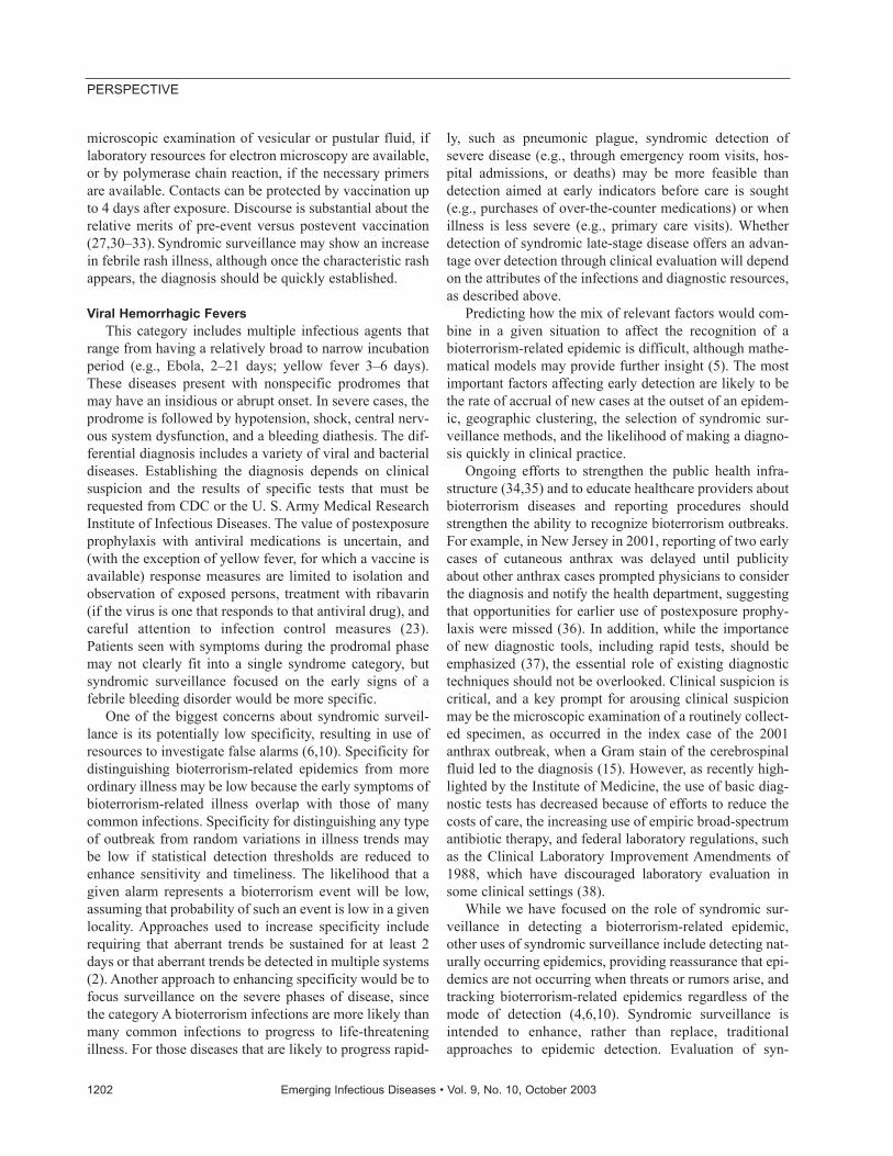
Figure 1. Number of cases of syndromic illness by time in a hypo-thetical bioterrorism attack and two pathways to establishing adiagnosis: syndromic surveillance coupled with public healthinvestigation (upper pathway) and clinical and diagnostic evalua-tion of patients with short-incubation period disease (lower path-way). A, scenario favoring earlier detection by means of clinicalevaluation. B, scenario favoring earlier detection by means of syn-dromic surveillance.
A
B
otics before cultures were taken. This finding emphasizesthe importance of judicious use of antibiotics in patientswith nonspecific illness.
In addition to the specific attributes of individualbioterrorism agents, multiple considerations will shape therecognition of a bioterrorism-related epidemic. Five ofthese attributes follow.
SizeSyndromic surveillance would not detect outbreaks too
small to trigger statistical alarms. Size would be affectedby the virulence of the agent, its potential for person-to-person transmission, the extent and mode of agent dissem-ination, whether dissemination occurs in more than onetime or place, and population vulnerability.
Population DispersionHow persons change locations after an exposure will
affect whether disease occurs in a concentrated or widearea, and thus whether clustering is apparent to clinicians ordetectable through syndromic surveillance at specific sites.
Health CareThe more knowledgeable providers are about bioterror-
ism agents, the greater the likelihood of recognition.Routine diagnostic practices or access to reference labora-tories may affect the timeliness of diagnosis for some dis-eases. Familiarity with reporting procedures wouldincrease prompt reporting of suspected or diagnosed cases.
Syndromic SurveillanceSyndromic surveillance will be affected by the selec-
tion of data sources, timeliness of information manage-ment, definition of syndrome categories, selection of sta-
tistical detection thresholds, availability of resources forfollowup, recent experience with false alarms, and criteriafor initiating investigations.
SeasonA fifth key attribute is seasonality. An increase in ill-
ness associated with a bioterrorism attack may be moredifficult to detect if it occurs during a seasonal upswing innaturally occurring disease.
Agent- and disease-specific attributes may be amongthe most important factors affecting detection and diagno-sis (Table 2). The incubation period and its distribution inthe population will affect the rate at which new cases
Emerging Infectious Diseases • Vol. 9, No. 10, October 2003 1199
PERSPECTIVE
Table 1. Outcome of initial contact with health care for anthrax-related illness and timing of anthrax diagnosis, 11 patients with inhalational anthrax, 2001a Outcome No. of patients Disposition after initial medical care
Admitted to hospital 7 Discharged home from ER, subsequent hospital admission
2
Discharged home from outpatient provider, subsequent hospital admission
2
Total 11 Anthrax diagnosis
Blood or CSF culture on hospital admission, presumptive diagnosis <24 h
7
Blood culture from preceding ER visit, patient recalled for admission
1
Prior antibiotic therapy; clinical suspicion of anthrax; specialized test required to establish diagnosis
3
Total 11 aER, emergency room; CSF, cerebrospinal fluid.
A
B
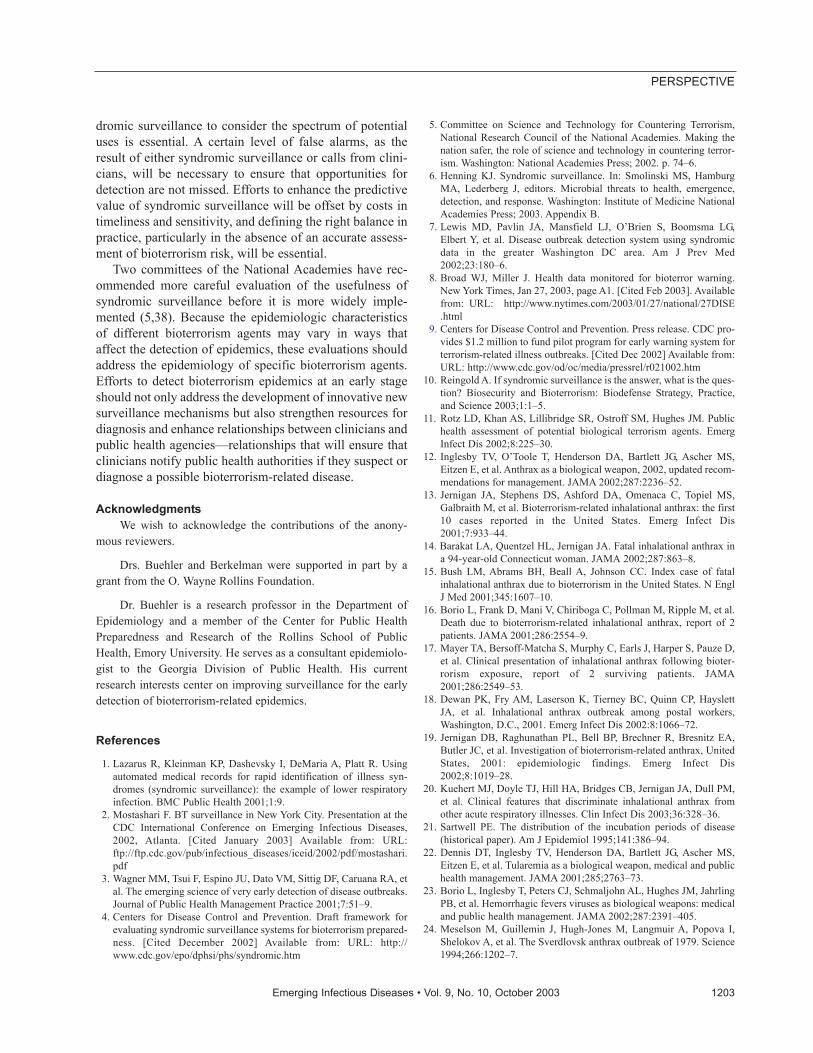
Figure 2. Timeline to presumptive anthrax diagnosis, 11 patientswith inhalational anthrax, 2001, United States. Abbreviations: Dx,diagnosis; OutPt, outpatient visit followed by discharge home; ER,emergency room visit followed by discharge home. *Diagnosisdelayed-initial blood cultures were negative in three patients whoreceived antibiotic therapy before culture specimens were collect-ed, requiring use of special diagnostic tests. For patients 1-10,case numbers correspond to those in report by Jernigan et al. (13);patient 11 reported by Barakat et al. (14). A, timeline begins withpresumed date of anthrax exposure, available for six patients. B,timeline begins with day of illness onset for five patients withoutrecognized date of exposure
Days after illness onset
Patie
nt N
o.Pa
tient
No.
Days after anthrax expossure
develop (21) and thus how quickly an alarm threshold isexceeded or whether clinicians recognize a temporal andgeographic cluster. If a disease has a short prodrome, thechance is increased that a patient would be hospitalizedand a definitive evaluation initiated before an increase incases triggered a surveillance alarm. Alternatively, if a dis-ease has a relatively long prodrome, chances are greaterthat prediagnostic events (e.g., purchase of medications oruse of outpatient care for nonspecific complaints) wouldaccrue to levels that exceed syndromic surveillance thresh-olds, before definitive diagnostic evaluations are complet-ed among patients with more severe disease. Arousingclinical suspicion for a particular diagnosis will depend onthe specificity of both the early and late stages of illness aswell as the presence or absence of a typical feature thatshould alert clinicians to the diagnosis, such as mediastinalwidening in inhalational anthrax (12). If a routinely per-formed test is apt to be diagnostic in a short time (e.g., theblood culture in anthrax), a rapid diagnosis is likely, evenin the absence of clinical suspicion. If routine tests areunlikely to yield a rapid diagnosis (e.g., the blood culturefor the cause of tularemia, Francisella tularensis [22]), orif the diagnosis requires a special test (e.g., the hemorrhag-ic fever viruses [23]), a diagnosis may be delayed if notimmediately considered.
The public health benefit resulting from early detectionof an epidemic is likely to vary by disease. If a disease hasa relatively wide distribution of potential onsets, earlyrecognition provides greater opportunity to administer pro-phylaxis to exposed persons. For example, based on datafrom the Sverdlovsk incident (24), Brookmeyer andBlades estimated that use of antibiotic prophylaxis duringthe 2001 anthrax outbreak prevented nine cases of inhala-tional disease among exposed persons (25). If the incuba-
tion period of a disease has a relatively narrow distribu-tion, early recognition may offer little opportunity for post-exposure prophylaxis, although a potential benefit wouldremain for alerting healthcare providers and informingtheir care of others with similar symptoms. This pattern ofillness is apt to result from exposure to an F. tularensisaerosol, which would likely result in an explosive epidem-ic with an abrupt onset and limited duration (22).
Detecting Specific Bioterrorism Epidemics and Agents
The attributes of the CDC category A bioterrorismagents that affect their detection, as well as the benefits ofearly detection, are summarized below, on the basis ofpotential bioterrorism-related epidemic profiles devel-oped by experts (12,22,23,26–28). These profiles reflectcurrent knowledge of these diseases; their epidemiologymight differ if novel modes of dissemination or prepara-tion were employed. Each disease has attributes that couldincrease or decrease the likelihood of early outbreakrecognition through either clinical diagnosis or syndromicsurveillance.
Inhalational AnthraxThe distribution of the incubation period for inhalation-
al anthrax can be relatively broad as observed inSverdlovsk (2–43 days); most cases occur within 1–2weeks after exposure (24). In the 2001 U.S. outbreak, thedistribution of incubation periods was more limited, 4–6days, although later-onset cases may have been averted byantibiotic prophylaxis (25). The nonspecific prodrome foranthrax may last from several hours to several days. Takentogether, these data suggest that the initial slope of an epi-demic curve may be comparatively gradual during the first
1200 Emerging Infectious Diseases • Vol. 9, No. 10, October 2003
PERSPECTIVE
Table 2. Characteristics of bioterrorism-related epidemics that affect detection through clinical recognition versus syndromic surveillance Characteristicsa Clinical recognitionb Syndromic surveillancec Duration and variability of incubation period
Broader distribution of incubation period increases likelihood that patient with short incubation-period disease would be diagnosed before a statistical threshold of syndromic cases is exceeded.
More narrow distribution of incubation period—leading to a steeper epidemic curve in the initial phase—increases likelihood that statistical threshold would be exceeded sooner.
Duration of nonspecific prodromal phase
Shorter prodrome increases likelihood of recognition or diagnosis at more severe or fulminant stage.
Longer prodrome increases likelihood that increase in syndromic manifestations would be detectable and that recognition of more severe stage (at which a diagnosis is more apt to be made) would be delayed.
Presence or absence of clinical sign that would heighten suspicion of diagnosis
Presence increases likelihood of earlier clinical recognition and diagnosis (e.g., mediastinal widening on chest x-ray in inhalational anthrax).
Absence decreases likelihood that diagnosis would be considered clinically, increasing opportunity for earlier detection by means of syndromic surveillance.
Likelihood of making diagnosis in the course of routine evaluation
If diagnosis is apt to be made in the course of a routine diagnostic evaluation (not dependent on clinical suspicion of specific bioterrorism infection), early diagnosis through clinical care is likely.
If diagnosis is dependent on the use of a special test that is unlikely to be ordered in the absence of clinical suspicion of diagnosis, then diagnosis in clinical care may be delayed, increasing the opportunity for early detection through syndromic surveillance.
aInfection or disease attributes that may affect detection of an epidemic. bIncreases likelihood of initial detection through routine clinical care and reporting. cIncreases likelihood of initial detection through syndromic surveillance.
week, leading to slower recognition through syndromicsurveillance than for other infections caused by bioterror-ist agents with pulmonary manifestations, such astularemia or pneumonic plague (22,28). In contrast, medi-astinal widening on chest x-ray or computed tomographicscan or Gram stain of cerebrospinal or pleural fluid shouldlead an alert and knowledgeable physician to consider thediagnosis of anthrax, even though these tests may not beconducted until relatively late in the clinical course. B.anthracis is likely to be detected quickly in cultures, favor-ing clinical recognition. Retrospective analysis of datafrom 2001 showed that inhalational anthrax can be distin-guished from influenzalike illness or community-acquiredpneumonia by using an algorithm that combines clinicaland laboratory findings (20), although the practical utilityof this approach is untested. In addition to permittingantibiotic use among ill persons, early recognition wouldenable postexposure antibiotic prophylaxis (12,25).
Tularemia The typical incubation period for tularemia is relatively
narrow after a person is exposed to aerosolized F. tularen-sis, with abrupt onset of nonspecific febrile illness, with orwithout respiratory symptoms, in 3–5 days (range 1–14days), followed by rapid progression to life-threateningpneumonitis (22). This relatively narrow incubation periodfor most patients and rapid progression to severe diseasewould lead to a rapid increase in cases after a large andacute exposure. Finding a number of such cases in a shortinterval should trigger both syndromic surveillance alarmsand clinical suspicion. F. tularensis is a slow-growing andfastidious organism and may take up to 5 days after inoc-ulation to be detectable, if it is detected at all, in a routine-ly processed blood culture. The use of special laboratorytechniques may be required, delaying the likelihood ofdetection in the absence of clinical suspicion. After an epi-demic is recognized, specific antibiotic therapy is recom-mended for exposed persons in whom a febrile illnessdevelops (22).
Pneumonic Plague Exposure to aerosolized Yersinia pestis results in pneu-
monic plague, which has a typical incubation period of 2to 4 days (range 1–6 days). The disease has a relativelyshort prodrome, followed by rapidly progressive pneumo-nia (28), which would lead to a rapid increase in cases atthe onset of an epidemic. Standard clinical laboratory find-ings are nonspecific, which alone might not prompt clini-cal suspicion, but microscopic examination of a sputumsmear may show characteristic findings, which shouldprompt consideration of the diagnosis. Cultures of blood orsputum are apt to show growth within 24 to 48 hours, butroutine procedures may misidentify Y. pestis unless the
diagnosis is suspected and special attention is given tospecimen processing. Confirming the diagnosis dependson special tests available through reference laboratories.Treatment the first day of symptoms is generally consid-ered necessary to prevent death in pneumonic plague, soearly recognition of an aerosol plague attack would enablelife-saving use of antibiotics in febrile patients and prophy-laxis of contacts (28).
Botulism Foodborne botulism typically has a relatively narrow
incubation period (12–72 hours), which may vary from 2hours to 8 days, depending on the inoculum. For the threeknown cases of inhalational botulism attributed to a rela-tively low exposure to aerosolized toxin, the incubationperiod was approximately 72 hours (26). The characteris-tic clinical picture of descending paralysis should promptconsideration of botulism, and this unique pattern amongbioterrorism agents lends itself to a specific syndrome cat-egory. However, the illness may be misdiagnosed, asobserved in a large foodborne outbreak of botulism in1985; 28 persons who had eaten at a particular restaurantand in whom botulism had developed were assigned otherdiagnoses before the geographically dispersed outbreakwas recognized and publicized in the media (26,29).Symptoms of inhalational botulism, with choking, dyspha-gia, and dysarthria dominating the clinical picture, maydiffer from those associated with ingestion of toxin andcomplicate recognition of the disease. Specialized testingfor botulinum toxin is available at a limited number ofstate laboratories and CDC. Postexposure prophylaxis islimited by the scarcity of, and potential for, allergic reac-tions to botulinum antitoxin, leading to recommendationsthat exposed persons be observed carefully for early signsof botulism, which should prompt antitoxin use (26).Antitoxin should be given as early as possible, another factthat highlights the importance of early detection.Depending on the level of exposure and the geographicdispersion of affected persons, syndromic surveillance forcharacteristic neurologic symptoms could aid outbreakdetection, or the occurrence of an epidemic might be obvi-ous to clinicians.
Smallpox The incubation period of smallpox is usually 12–14
days but may range from 7 to 17 days. The early sympto-matic phase includes a severe febrile illness and appear-ance of a nonspecific macular rash over a 2- to 4-day peri-od, followed by evolution to a vesicular and then pustularrash over the next 4 to 5 days (27). Thus, the initial phaseof smallpox may lend itself to detection through surveil-lance of a febrile rash illness syndrome. Once smallpox issuspected, the virus can be rapidly detected by electron
Emerging Infectious Diseases • Vol. 9, No. 10, October 2003 1201
PERSPECTIVE
microscopic examination of vesicular or pustular fluid, iflaboratory resources for electron microscopy are available,or by polymerase chain reaction, if the necessary primersare available. Contacts can be protected by vaccination upto 4 days after exposure. Discourse is substantial about therelative merits of pre-event versus postevent vaccination(27,30–33). Syndromic surveillance may show an increasein febrile rash illness, although once the characteristic rashappears, the diagnosis should be quickly established.
Viral Hemorrhagic FeversThis category includes multiple infectious agents that
range from having a relatively broad to narrow incubationperiod (e.g., Ebola, 2–21 days; yellow fever 3–6 days).These diseases present with nonspecific prodromes thatmay have an insidious or abrupt onset. In severe cases, theprodrome is followed by hypotension, shock, central nerv-ous system dysfunction, and a bleeding diathesis. The dif-ferential diagnosis includes a variety of viral and bacterialdiseases. Establishing the diagnosis depends on clinicalsuspicion and the results of specific tests that must berequested from CDC or the U. S. Army Medical ResearchInstitute of Infectious Diseases. The value of postexposureprophylaxis with antiviral medications is uncertain, and(with the exception of yellow fever, for which a vaccine isavailable) response measures are limited to isolation andobservation of exposed persons, treatment with ribavarin(if the virus is one that responds to that antiviral drug), andcareful attention to infection control measures (23).Patients seen with symptoms during the prodromal phasemay not clearly fit into a single syndrome category, butsyndromic surveillance focused on the early signs of afebrile bleeding disorder would be more specific.
One of the biggest concerns about syndromic surveil-lance is its potentially low specificity, resulting in use ofresources to investigate false alarms (6,10). Specificity fordistinguishing bioterrorism-related epidemics from moreordinary illness may be low because the early symptoms ofbioterrorism-related illness overlap with those of manycommon infections. Specificity for distinguishing any typeof outbreak from random variations in illness trends maybe low if statistical detection thresholds are reduced toenhance sensitivity and timeliness. The likelihood that agiven alarm represents a bioterrorism event will be low,assuming that probability of such an event is low in a givenlocality. Approaches used to increase specificity includerequiring that aberrant trends be sustained for at least 2days or that aberrant trends be detected in multiple systems(2). Another approach to enhancing specificity would be tofocus surveillance on the severe phases of disease, sincethe category A bioterrorism infections are more likely thanmany common infections to progress to life-threateningillness. For those diseases that are likely to progress rapid-
ly, such as pneumonic plague, syndromic detection ofsevere disease (e.g., through emergency room visits, hos-pital admissions, or deaths) may be more feasible thandetection aimed at early indicators before care is sought(e.g., purchases of over-the-counter medications) or whenillness is less severe (e.g., primary care visits). Whetherdetection of syndromic late-stage disease offers an advan-tage over detection through clinical evaluation will dependon the attributes of the infections and diagnostic resources,as described above.
Predicting how the mix of relevant factors would com-bine in a given situation to affect the recognition of abioterrorism-related epidemic is difficult, although mathe-matical models may provide further insight (5). The mostimportant factors affecting early detection are likely to bethe rate of accrual of new cases at the outset of an epidem-ic, geographic clustering, the selection of syndromic sur-veillance methods, and the likelihood of making a diagno-sis quickly in clinical practice.
Ongoing efforts to strengthen the public health infra-structure (34,35) and to educate healthcare providers aboutbioterrorism diseases and reporting procedures shouldstrengthen the ability to recognize bioterrorism outbreaks.For example, in New Jersey in 2001, reporting of two earlycases of cutaneous anthrax was delayed until publicityabout other anthrax cases prompted physicians to considerthe diagnosis and notify the health department, suggestingthat opportunities for earlier use of postexposure prophy-laxis were missed (36). In addition, while the importanceof new diagnostic tools, including rapid tests, should beemphasized (37), the essential role of existing diagnostictechniques should not be overlooked. Clinical suspicion iscritical, and a key prompt for arousing clinical suspicionmay be the microscopic examination of a routinely collect-ed specimen, as occurred in the index case of the 2001anthrax outbreak, when a Gram stain of the cerebrospinalfluid led to the diagnosis (15). However, as recently high-lighted by the Institute of Medicine, the use of basic diag-nostic tests has decreased because of efforts to reduce thecosts of care, the increasing use of empiric broad-spectrumantibiotic therapy, and federal laboratory regulations, suchas the Clinical Laboratory Improvement Amendments of1988, which have discouraged laboratory evaluation insome clinical settings (38).
While we have focused on the role of syndromic sur-veillance in detecting a bioterrorism-related epidemic,other uses of syndromic surveillance include detecting nat-urally occurring epidemics, providing reassurance that epi-demics are not occurring when threats or rumors arise, andtracking bioterrorism-related epidemics regardless of themode of detection (4,6,10). Syndromic surveillance isintended to enhance, rather than replace, traditionalapproaches to epidemic detection. Evaluation of syn-
1202 Emerging Infectious Diseases • Vol. 9, No. 10, October 2003
PERSPECTIVE
dromic surveillance to consider the spectrum of potentialuses is essential. A certain level of false alarms, as theresult of either syndromic surveillance or calls from clini-cians, will be necessary to ensure that opportunities fordetection are not missed. Efforts to enhance the predictivevalue of syndromic surveillance will be offset by costs intimeliness and sensitivity, and defining the right balance inpractice, particularly in the absence of an accurate assess-ment of bioterrorism risk, will be essential.
Two committees of the National Academies have rec-ommended more careful evaluation of the usefulness ofsyndromic surveillance before it is more widely imple-mented (5,38). Because the epidemiologic characteristicsof different bioterrorism agents may vary in ways thataffect the detection of epidemics, these evaluations shouldaddress the epidemiology of specific bioterrorism agents.Efforts to detect bioterrorism epidemics at an early stageshould not only address the development of innovative newsurveillance mechanisms but also strengthen resources fordiagnosis and enhance relationships between clinicians andpublic health agencies—relationships that will ensure thatclinicians notify public health authorities if they suspect ordiagnose a possible bioterrorism-related disease.
AcknowledgmentsWe wish to acknowledge the contributions of the anony-
mous reviewers.
Drs. Buehler and Berkelman were supported in part by agrant from the O. Wayne Rollins Foundation.
Dr. Buehler is a research professor in the Department ofEpidemiology and a member of the Center for Public HealthPreparedness and Research of the Rollins School of PublicHealth, Emory University. He serves as a consultant epidemiolo-gist to the Georgia Division of Public Health. His currentresearch interests center on improving surveillance for the earlydetection of bioterrorism-related epidemics.
References
1. Lazarus R, Kleinman KP, Dashevsky I, DeMaria A, Platt R. Usingautomated medical records for rapid identification of illness syn-dromes (syndromic surveillance): the example of lower respiratoryinfection. BMC Public Health 2001;1:9.
2. Mostashari F. BT surveillance in New York City. Presentation at theCDC International Conference on Emerging Infectious Diseases,2002, Atlanta. [Cited January 2003] Available from: URL:ftp://ftp.cdc.gov/pub/infectious_diseases/iceid/2002/pdf/mostashari.pdf
3. Wagner MM, Tsui F, Espino JU, Dato VM, Sittig DF, Caruana RA, etal. The emerging science of very early detection of disease outbreaks.Journal of Public Health Management Practice 2001;7:51–9.
4. Centers for Disease Control and Prevention. Draft framework forevaluating syndromic surveillance systems for bioterrorism prepared-ness. [Cited December 2002] Available from: URL: http://www.cdc.gov/epo/dphsi/phs/syndromic.htm
5. Committee on Science and Technology for Countering Terrorism,National Research Council of the National Academies. Making thenation safer, the role of science and technology in countering terror-ism. Washington: National Academies Press; 2002. p. 74–6.
6. Henning KJ. Syndromic surveillance. In: Smolinski MS, HamburgMA, Lederberg J, editors. Microbial threats to health, emergence,detection, and response. Washington: Institute of Medicine NationalAcademies Press; 2003. Appendix B.
7. Lewis MD, Pavlin JA, Mansfield LJ, O’Brien S, Boomsma LG,Elbert Y, et al. Disease outbreak detection system using syndromicdata in the greater Washington DC area. Am J Prev Med2002;23:180–6.
8. Broad WJ, Miller J. Health data monitored for bioterror warning.New York Times, Jan 27, 2003, page A1. [Cited Feb 2003]. Availablefrom: URL: http://www.nytimes.com/2003/01/27/national/27DISE.html
9. Centers for Disease Control and Prevention. Press release. CDC pro-vides $1.2 million to fund pilot program for early warning system forterrorism-related illness outbreaks. [Cited Dec 2002] Available from:URL: http://www.cdc.gov/od/oc/media/pressrel/r021002.htm
10. Reingold A. If syndromic surveillance is the answer, what is the ques-tion? Biosecurity and Bioterrorism: Biodefense Strategy, Practice,and Science 2003;1:1–5.
11. Rotz LD, Khan AS, Lillibridge SR, Ostroff SM, Hughes JM. Publichealth assessment of potential biological terrorism agents. EmergInfect Dis 2002;8:225–30.
12. Inglesby TV, O’Toole T, Henderson DA, Bartlett JG, Ascher MS,Eitzen E, et al. Anthrax as a biological weapon, 2002, updated recom-mendations for management. JAMA 2002;287:2236–52.
13. Jernigan JA, Stephens DS, Ashford DA, Omenaca C, Topiel MS,Galbraith M, et al. Bioterrorism-related inhalational anthrax: the first10 cases reported in the United States. Emerg Infect Dis2001;7:933–44.
14. Barakat LA, Quentzel HL, Jernigan JA. Fatal inhalational anthrax ina 94-year-old Connecticut woman. JAMA 2002;287:863–8.
15. Bush LM, Abrams BH, Beall A, Johnson CC. Index case of fatalinhalational anthrax due to bioterrorism in the United States. N EnglJ Med 2001;345:1607–10.
16. Borio L, Frank D, Mani V, Chiriboga C, Pollman M, Ripple M, et al.Death due to bioterrorism-related inhalational anthrax, report of 2patients. JAMA 2001;286:2554–9.
17. Mayer TA, Bersoff-Matcha S, Murphy C, Earls J, Harper S, Pauze D,et al. Clinical presentation of inhalational anthrax following bioter-rorism exposure, report of 2 surviving patients. JAMA2001;286:2549–53.
18. Dewan PK, Fry AM, Laserson K, Tierney BC, Quinn CP, HayslettJA, et al. Inhalational anthrax outbreak among postal workers,Washington, D.C., 2001. Emerg Infect Dis 2002:8:1066–72.
19. Jernigan DB, Raghunathan PL, Bell BP, Brechner R, Bresnitz EA,Butler JC, et al. Investigation of bioterrorism-related anthrax, UnitedStates, 2001: epidemiologic findings. Emerg Infect Dis2002;8:1019–28.
20. Kuehert MJ, Doyle TJ, Hill HA, Bridges CB, Jernigan JA, Dull PM,et al. Clinical features that discriminate inhalational anthrax fromother acute respiratory illnesses. Clin Infect Dis 2003;36:328–36.
21. Sartwell PE. The distribution of the incubation periods of disease(historical paper). Am J Epidemiol 1995;141:386–94.
22. Dennis DT, Inglesby TV, Henderson DA, Bartlett JG, Ascher MS,Eitzen E, et al. Tularemia as a biological weapon, medical and publichealth management. JAMA 2001;285;2763–73.
23. Borio L, Inglesby T, Peters CJ, Schmaljohn AL, Hughes JM, JahrlingPB, et al. Hemorrhagic fevers viruses as biological weapons: medicaland public health management. JAMA 2002;287:2391–405.
24. Meselson M, Guillemin J, Hugh-Jones M, Langmuir A, Popova I,Shelokov A, et al. The Sverdlovsk anthrax outbreak of 1979. Science1994;266:1202–7.
Emerging Infectious Diseases • Vol. 9, No. 10, October 2003 1203
PERSPECTIVE
25. Brookmeyer R, Blades N. Prevention of inhalational anthrax in theUS outbreak. Science 2002;295:1861.
26. Arnon SS, Schechter R, Inglesby TV, Henderson DA, Bartlett JG,Ascher MS, et al. Botulinum toxin as a biological weapon: medicaland public health management. JAMA 2001;285:1059–70.
27. Henderson DA, Inglesby TV, Bartlett JG, Ascher MS, Eitzen E,Jahrling PB, et al. Smallpox as a biological weapon: medical and pub-lic health management. JAMA 1999;281:2127–37.
28. Inglesby TV, Dennis DT, Henderson DA, Bartlett JG, Ascher MS,Eitzen E, et al. Plague as a biological weapon: medical and publichealth management. JAMA 2000;283:2281–90.
29. St Louis ME, Peck SH, Bowering D, Morgan GB, Blatherwick J,Banerjee S, et al. Botulism from chopped garlic: delayed recognitionof a major outbreak. Ann Intern Med 1988;108:363–8.
30. Meltzer MI, Damon I, LeDuc JW, Millar JD. Modeling potentialresponses to smallpox as a bioterrorist weapon. Emerg Infect Dis2001;7:959–69.
31. Gani R, Leach S. Transmission potential of smallpox in contempo-rary populations. Nature 2001;414:748–51.
32. Kaplan EH, Craft DL, Wein LM. Emergency response to a smallpoxattack: the case for mass vaccination. PNAS 2002:99:10935–40.
33. Halloran ME, Longini IM, Nizam A, Yang Y. Containing bioterroristsmallpox. Science 2002;298:1428–32.
34. United States Department of Health and Human Services. HHSannounces $1.1 billion in funding to states for bioterrorism prepared-ness. Press release. Jan 31, 2002 [Cited March 2003]. Available from:URL: http://www.hhs.gov/news/press/2002pres/20020131b.html
35. United States Department of Health and Human Services. HHSannounces bioterrorism aid for states, including special opportunityfor advance fund. Press release. Mar 20, 2003 [Cited March 2003].Available from: URL: http://www.hhs.gov/news/press/2003pres/20030320.html
36. Bresnitz EA, DiFerdinando GT. Lessons from the anthrax attacks of2001, the New Jersey experience. Clinics in Occupational andEnvironmental Medicine 2003;2:227–52.
37. National Institute of Allergy and Infectious Diseases Office ofCommunications and Public Liaison. HHS accelerates bioterrorismresearch: new programs expedite ideas from concerned scientists.Press release. Dec 6, 2001 [Cited March 2003]. Available from: URL:http://www.niaid.nih.gov/newsroom/releases/accelbio.htm
38. Smolinski MS, Hamburg MA, Lederberg JA, editors. Microbialthreats to health, emergence, detection, and response. Washington:Institute of Medicine National Academies Press; 2003. p. 183–94.
Address for correspondence: James Buehler, Rollins School of PublicHealth, Rm. 416, Emory University, 1518 Clifton Rd., NE, Atlanta, GA30322, USA; fax: 404-712-8345; email: [email protected]
1204 Emerging Infectious Diseases • Vol. 9, No. 10, October 2003
PERSPECTIVE
Use of trade names is for identification only and does not implyendorsement by the Public Health Service or by the U.S.Department of Health and Human Services.
Severe acute respiratory syndrome (SARS) is a threatto healthcare workers. After a brief, unexpected exposureto a patient with SARS, 69 intensive-care staff at risk forSARS were interviewed to evaluate risk factors. SARSdeveloped in seven healthcare workers a median of 5 days(range 3–8) after last exposure. SARS developed in 6 of 31persons who entered the patient’s room, including 3 whowere present in the room >4 hours. SARS occurred in threeof five persons present during the endotracheal intubation,including one who wore gloves, gown, and N-95 mask. Thesyndrome also occurred in one person with no apparentdirect exposure to the index patient. In most, but not allcases, developing SARS was associated with factors typi-cal of droplet transmission. Providing appropriate quaran-tine and preventing illness in healthcare providers substan-tially affects delivery of health care.
Severe acute respiratory syndrome (SARS) is a diseasethat consists of fever and respiratory symptoms that
can progress to respiratory failure and death (1). SARS ismost likely to develop in healthcare workers and house-hold or family contacts of infected persons (2–4).Unprotected exposure to SARS in hospitals has severalpotential consequences, which include the following: ill-ness in persons and healthcare workers; transmission ofSARS from ill healthcare workers and patients to visitorsand household contacts; and reduced ability of the health-care system to deliver care because of illness in or quaran-tine of healthcare workers. In addition, the psychologicalimpact of isolation and quarantine can be substantial (5).As a result, understanding factors associated with SARStransmission after exposure to SARS patients is importantand would assist with formulating appropriate quarantineprocedures. We describe our experience with a large num-ber of healthcare workers who were exposed to a patient inan intensive-care unit (ICU) with undiagnosed SARS.
Index PatientOn March 23, 2003, a 74-year-old immunocompro-
mised man was transferred to our ICU from a hospitalwhere the original cluster of Toronto’s SARS casesoccurred (2). The patient originally had signs and symp-toms consistent with a presumptive diagnosis of communi-ty-acquired pneumonia. Before transfer, SARS wasexcluded from the differential diagnosis because thepatient had not traveled, had never left the emergencydepartment of the referring hospital, and had only had asingle recent outpatient visit to an area of the original hos-pital in which SARS had not been identified. Upon arrivalin our ICU, the patient was placed in precautions formethicillin-resistant Staphylococcus aureus (MRSA)pending admission screening results (6). Therapy withbroad-spectrum antimicrobial drugs was initiated.Humidified high-flow oxygen was administered for thefirst 5 h, noninvasive positive pressure ventilation byoronasal mask for the next 18.25 h, and invasive mechan-ical ventilation for the subsequent time (7.5 h).Endotracheal intubation required fiber-optic placement.That the extent of the outbreak at the referring institutionwas larger than originally appreciated became apparent atthis time; therefore, the patient was transferred to anotherfacility for placement in negative pressure isolation forpossible exposure to SARS. Subsequently, his familymembers became ill, and the SARS-associated coronaviruswas identified in the patient’s respiratory secretions (poly-merase chain reaction testing of bronchoalveolar lavageconfirmed the diagnosis of SARS).
QuarantineOnce the risk for SARS was identified, all patients in
the ICU were considered to have been potentially exposed.To prevent spread of SARS, we closed the ICU to admis-sions and discharges and implemented strict respiratoryand contact precautions for all remaining patients. Wequarantined 69 healthcare workers who were considered tobe at high risk for developing SARS. On the basis of our
Emerging Infectious Diseases • Vol. 9, No. 10, October 2003 1205
RESEARCH
Illness in Intensive Care Staff afterBrief Exposure to Severe Acute
Respiratory SyndromeDamon C. Scales,* Karen Green,* Adrienne K. Chan,* Susan M. Poutanen,* Donna Foster,*
Kylie Nowak,* Janet M. Raboud,† Refik Saskin,* Stephen E. Lapinsky,* and Thomas E. Stewart*†
*Mount Sinai Hospital, Toronto, Ontario, Canada; and †UniversityHealth Network, Toronto, Ontario, Canada
understanding of disease transmission, we arbitrarilydecided that persons at high risk included anyone who hadentered the index patient’s room or who had been in theICU for >4 hours during the patient’s 30.75-h stay.
MethodsAfter research ethics board approval and informed con-
sent, two researchers used a structured questionnaire tointerview quarantined healthcare workers. The question-naire elicited demographic information, details abouthealth, and information about exposure to the indexpatient. Time of exposure was categorized as follows: <1min, 1–10 min, 11–30 min, 31–60 min, 1–4 h, or >4 h.Exposure proximity, procedures performed, and infection-control precautions were documented. Each healthcareworker was asked about symptoms suggestive of SARSthat developed during or after the quarantine period.
For healthcare workers in whom suspected or probableSARS developed, additional data were collected about thenature and course of their illness. Suspected and probableSARS were defined according to the definitions issued bythe World Health Organization (WHO) (7). Symptoms ofsuspected SARS were a fever >38°C, respiratory symp-toms, and an epidemiologic link with a SARS patient; allquarantined healthcare workers were considered to havean epidemiologic link on the basis of contact with theindex patient. Probable SARS was defined as suspectedSARS with radiographic lung infiltrates.
StatisticsAll data were entered into an Access (Microsoft Corp.,
Redman, WA) database by using double data entry tech-nique and analyzed by using SAS version 8.0 (SASInstitute, Inc., Cary, NC). For comparisons of characteris-tics of healthcare workers with SARS to those of health-care workers without SARS, we used the two-sample t testfor normally distributed variables, Wilcoxon rank sum testfor ordinal and skewed continuous variables, and Fisherexact test for categorical variables. Two-sided tests wereused for all comparisons. A p value of <0.05 was consid-ered to be statistically significant. Classification andregression tree methods were used to identify predictors ofdeveloping SARS (8). The healthcare workers were divid-ed into two groups by examining all possible cutpoints ofall predictor variables to find the cutpoint of a predictorvariable that resulted in the largest difference in the prob-abilities of developing SARS between the two resultingsubgroups. This procedure was performed repeatedly foreach resulting subgroup until all members of the subgrouphad the same SARS status or the subgroup was too smallto warrant further splitting.
Results Of the 69 quarantined patients, 63 were interviewed.
Five declined, and one could not be contacted. SARS didnot develop in healthcare workers who were not quaran-tined and patients who had been in the unit at the time ofthe exposure.
SARS DevelopmentSARS developed in 7 of the 69 quarantined healthcare
workers (6 probable, 1 suspected; Table 1). One healthcareworker had a history of type II diabetes mellitus; all otherhealthcare workers were previously healthy. The mediantime from exposure to the index patient to onset of symp-toms was 5 days (range 3–8 days). All probable case-patients were hospitalized and required oxygen but did notrequire ICU care. Treatment with levofloxacin (500 mgonce a day for 7 days) and ribavirin (2,000–2,200 mg load-ing dose followed by 1,200 mg every 6 h for 4 days andsubsequent tapering off) was administered to all admittedcase-patients, and all but one received systemic corticos-teroids (1 mg/kg prednisone or equivalent once a day for 5days with subsequent tapering off). The median hospitalstay was 19.5 days (range 13–25 days). All case-patientswere discharged. However, 28–32 days after discharge, allreported continued dyspnea with exercise.
Room VisitationThirty-one healthcare workers had entered the index
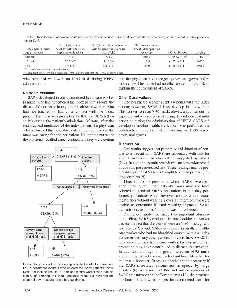
patient’s room; SARS developed in 6 (19%). The contactcharacteristics and infection control precautions used bythe healthcare workers who entered the patient’s room areshown in Table 2. All six healthcare workers in whomSARS developed and who entered the patient’s roomreported being present >11 min; three were in the room for>4 hours. SARS attack rates were higher among healthcareworkers who spent more time in the index patient’s room;in addition, a dose-response effect occurred between dura-tion of exposure and risk of developing SARS (Table 3).
Contact with Index PatientAll six healthcare workers with SARS who entered the
index patient’s room also touched the patient, and allreported performing a procedure that involved contactwith the patient’s mucous membranes or respiratory secre-tions (Table 2). Three of the six healthcare workers report-ed wearing gloves during this contact. In contrast, of the 13healthcare workers without SARS, 12 (92%) used gloveswhen touching the patient (odds ratio [OR] 0.08, 95% con-fidence interval [CI] 0.01 to 1.11, p=0.07). Selected con-tact characteristics predictive of the development of SARSin healthcare workers who entered the patient’s roomappear in the Figure.
1206 Emerging Infectious Diseases • Vol. 9, No. 10, October 2003
RESEARCH
SARS developed in three of the five persons presentduring the endotracheal intubation of the patient. Duringthis procedure, the patient’s respiratory secretions weresplashed onto the uncovered cheek of one of the healthcareworkers. No other healthcare worker reported direct skinexposure to the patient’s bodily secretions at any time dur-ing his admission. Two of the three persons in whomSARS developed after the endotracheal intubation wore agown, surgical mask, and gloves; one healthcare workerwore a gown, gloves, and N-95 mask. Of the two health-care workers present during endotracheal intubation inwhom SARS did not develop, one was a postgraduatemedical trainee who assisted with manual ventilation (bag-valve-mask ventilation using a Laerdal bag) and was posi-tioned to the side of the patient rather than directly over thepatient’s head. This healthcare worker wore gown, gloves,
and surgical mask during the procedure. The second work-er was a respiratory therapist who helped prepare the nec-essary equipment while wearing gown, gloves, and an N-95 mask.
Of the healthcare workers who entered the indexpatient’s room, 22 were present at some time during theadministration of noninvasive positive-pressure ventilation(NPPV), and SARS developed in 4 (18%). Each of these 4healthcare workers, but only 1 of the 18 healthcare work-ers who remained well, reported being present in the roomfor >31 minutes during the administration of NPPV (OR105, 95% CI 3 to 3,035, p <0.001). The one worker inwhom SARS did not develop despite being present duringNPPV therapy for >31 minutes wore a surgical mask,gown, and gloves. One of the 4 healthcare workers inwhom SARS developed and 4 of the 18 healthcare workers
Emerging Infectious Diseases • Vol. 9, No. 10, October 2003 1207
RESEARCH
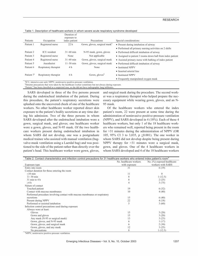
Table 1. Description of healthcare workers in whom severe acute respiratory syndrome developed
Patients Occupation
Duration of exposure to
index patient Precautions Special considerations Patient 1 Registered nurse 22 h Gown, gloves, surgical maskb • Present during intubation of airway
• Performed all primary nursing activities on 2 shifts Patient 2 ICU resident 31–60 min N-95 mask, gown, gloves • Performed difficult intubation of airway Patient 3 Registered nurse None Not applicable • Assigned to patient 3 rooms down hall from index patient Patient 4 Registered nurse 31–60 min Gown, gloves, surgical mask • Assisted primary nurse with bathing of index patient Patient 5 Anesthetist 11–30 min Gown, gloves, surgical mask • Performed difficult intubation of airway Patient 6 Respiratory therapist 4 h None • Instituted NPPV
• Inserted arterial line Patient 7c Respiratory therapist 6 h Gown, glovesb • Instituted NPPV
• Frequently manipulated oxygen mask aICU, intensive-care unit; NPPV, noninvasive positive-pressure ventilation. bDenotes precautions that were taken by the healthcare worker sometimes but not always during exposure. cPatient 7 has been classified as a suspected case, as she did not have radiographic lung infiltrates.
Table 2. Contact characteristics and infection control precautions for 31 healthcare workers who entered index patient’s rooma
Exposure type No. healthcare workers
with exposure No. (%) exposed healthcare
workers with SARS Entry into room 31 6 (19) Contact duration for those entering the room
<10 min 11 0
11–30 min 8 1 (12.5) 31 min to 4 h 8 2 (25) >4 h 4 3 (75)
Nature of contact Touched patient 19 6 (32) Contact with mucous membranes 10 4 (40) Performed procedure involving contact with mucous membranes or respiratory secretions 15 6 (40) Present during NPPV 22 4 (18) Performed or assisted intubation 5 3 (60)
Infection control precautions used during exposure Always wore at least:
Gloves 15 3 (20) Gown and gloves 15 3 (20) Any mask (N-95 or surgical mask) 13 3 (23) Gown, gloves, and N-95 mask 6 1 (17) Gown, gloves, and surgical mask 6 2 (30) Gown, gloves, and any mask 12 3 (25) No precautions 8 1 (12.5)
aNPPV, noninvasive positive-pressure ventilation.
who remained well wore an N-95 mask during NPPVadministration.
No Room VisitationSARS developed in one quarantined healthcare worker
(a nurse) who had not entered the index patient’s room; thedisease did not occur in any other healthcare workers whohad not touched or had close contact with the indexpatient. The nurse was present in the ICU for 18.75 h (twoshifts) during the patient’s admission. Of note, after theendotracheal intubation of the index patient, the physicianwho performed this procedure entered the room where thenurse was caring for another patient. Neither the nurse northe physician recalled direct contact, and they were certain
that the physician had changed gloves and gown beforeroom entry. This nurse had no other epidemiologic risk toexplain the development of SARS.
Other ObservationsOne healthcare worker spent >4 hours with the index
patient; however, SARS did not develop in this worker.This worker wore an N-95 mask, gloves, and gown duringexposure and was not present during the endotracheal intu-bation or during the administration of NPPV. SARS diddevelop in another healthcare worker who performed theendotracheal intubation while wearing an N-95 mask,gown, and gloves.
DiscussionOur results suggest that proximity and duration of con-
tact to a patient with SARS are associated with risk forviral transmission, an observation suggested by others(2–4). In addition, certain procedures, such as endotrachealintubation, pose increased risk. These findings may be pre-dictable given that SARS is thought to spread primarily bylarge droplets (9).
Three of the six persons in whom SARS developedafter entering the index patient’s room may not haveadhered to standard MRSA precautions in that they per-formed procedures which involved contact with mucousmembranes without wearing gloves. Furthermore, we wereunable to determine if hand washing impacted SARStransmission, as this information was not collected.
During our study, we made two important observa-tions. First, SARS developed in one healthcare workerdespite the fact that the worker wore an N-95 mask, gown,and gloves. Second, SARS developed in another health-care worker who had no identified contact with the indexpatient or with any other persons known to have SARS. Inthe case of the first healthcare worker, the absence of eyeprotection may have contributed to disease transmission.In addition, although this person wore an N-95 maskwhile in the patient’s room, he had not been fit-tested forthis mask; however, fit-testing should not be necessary ifthe SARS-associated coronavirus is spread by largedroplets (6). As a result of this and similar episodes ofSARS transmission in the Toronto area (10), the provinceof Ontario has now made specific recommendations for
1208 Emerging Infectious Diseases • Vol. 9, No. 10, October 2003
RESEARCH
Table 3. Development of severe acute respiratory syndrome (SARS) in healthcare workers, depending on time spent in index patient’s room (N=31)a
Time spent in index patient’s room
No. (%) healthcare workers with specified exposure with SARS
No. (%) healthcare workers without specified exposure
with SARS
Odds of developing SARS after specified
exposure 95% CI for OR p value <10 min 0/11 6/20 (30) 0.097b (0.005 to 1.91)b 0.07 >31 min 5/12 (42) 1/19 (5) 12.9 (1.27 to 131) 0.022 >4 h 3/4 (75) 3/27 (11) 24.0 (1.85 to 311) 0.016 aCI, confidence interval; OR, odds ratio. bThese logit estimators use a correction of 0.5 in every cell of the table that contains a zero.
Figure. Regression tree describing selected contact characteris-tics in healthcare workers who entered the index patient’s room.Does not include results for one healthcare worker who had nohistory of entering the index patient’s room but neverthelessacquired severe acute respiratory syndrome.
healthcare workers performing intubation that involveincreased protection (available from: URL: www.sars.medtau.org) (11), and protective eye wear is currentlymandated for patient encounters. In the second case,transmission could have occurred in a number of possibleroutes. The nurse may have come within sufficient rangeof the SARS patient to be exposed to large droplets.Recent reports indicate that the virus may survive for sev-eral hours on fomites or in body secretions (12) and raisethe possibility of transmission by indirect contact withcontaminated objects or of inadvertent carriage andspread by another healthcare worker. Fecal transmission isunlikely as the patient did not have a bowel movementduring his stay. True airborne spread may also haveoccurred. Although evidence does not support this routeof transmission for the SARS-associated coronavirus,existing literature suggests that other coronaviruses maybe spread by an airborne route in certain circumstances(13).
Given our lack of knowledge about the transmissibilityof SARS at the time this exposure occurred, we made aconservative decision to quarantine for 10 days all personswho were in the unit for at least 4 h or who had a historyof entry into the affected patient’s room. In addition, weclosed the ICU to admissions and discharges for a 10-dayperiod, markedly affecting our institution’s ability to deliv-er health care. In fact, during the Toronto outbreak, sever-al of the city’s ICUs were closed as a result of quarantineand illness in staff with similar consequences (14); byinfecting healthcare workers, SARS has an impact on thehealth of an entire community. A less aggressive quaran-tine approach may have been as effective in controllingtransmission and allowed more staff to be available forwork. For instance, only persons who have had direct con-tact with the patient (i.e., entered the patient’s room) couldhave been quarantined. If we had taken this approach, thequarantine would have excluded six persons with SARSfrom the workplace but only removed 25 of the 62 personswho remained well. However, this approach would havemissed one healthcare worker in whom SARS developed.Another approach might be to monitor staff closely forSARS-related symptoms while they continue their usualactivities and quarantine only those in whom symptomsoccur. This approach would require evidence that SARScannot be transmitted before symptom onset, confidence inthe facility’s ability to identify symptomatic staff, and reli-ability of healthcare workers in reporting symptoms. Wethink that our quarantine approach prevented secondaryspread of illness to other persons who may have come incontact with the workers in whom SARS developed.
Our study involved a small number of cases, and defin-itive conclusions cannot be drawn from a report of thissize. For example, although SARS developed in our staff
within the 10-day quarantine period, others have demon-strated that the time period from infection to onset ofsymptoms may be >10 days (15). One of the strengths ofour study is that the exposure occurred during a definedperiod in a contained unit, and as such, there is less poten-tial for confounding caused by the exposure of healthcareworkers to multiple SARS patients.
Our observations emphasize the consequences of miss-ing the diagnosis of SARS for even a relatively brief peri-od. In our experience, we would make the following rec-ommendations. First, the possibility of unexpected expo-sure of healthcare workers to patients with SARS shouldbe anticipated, and once such exposure is recognized,those deemed to be at risk for SARS transmission shouldbe promptly quarantined. Second, vigilant surveillance forsymptoms of SARS must be maintained by all healthcareworkers who work in institutions with SARS patients;SARS may develop in healthcare workers even when theydo not have direct exposure to patients with SARS. Inaddition, protocols for managing patients with SARSshould include not only contact and respiratory precau-tions but also procedures that minimize patient contactsince duration and proximity of contact increase the riskfor transmission of SARS. Finally, additional precautionsshould be taken when performing high-risk procedures,such as endotracheal intubation (11).
Though many of the healthcare workers in our ICUwere exposed to the patient with SARS, our experiencesuggests that the greatest risk for SARS transmissionoccurs in those healthcare workers with prolonged expo-sure or direct physical contact with the patient. Use ofgowns, gloves, and masks as barriers appears to reduce therisk for SARS transmission in most but not all situations.Additional information will be needed to determine ifmodes of transmission beyond droplet spread are impor-tant. We think this information will be helpful to institu-tions dealing with similar exposures to patients with SARSand developing quarantine protocols.
AcknowledgmentsWe thank Patrick Cheng, Margaret McArthur, and Agron
Plebneshi for their assistance with data entry; Ron Heslegrave forhis advice regarding the consent form; Farida Hasin-Shakoor foradministrative assistance; and Allan S. Detsky and Arthur S.Slutsky for critically reading the manuscript.
Two of our investigators (K.G., R.S.) were supported in partby a grant from the Ontario Ministry of Health and Long-TermCare.
Dr. Scales is an internist, intensivist, and a postgraduate stu-dent in the Clinical Epidemiology Program at the University ofToronto. His current areas of research are the epidemiology andmanagement of respiratory infections in the intensive-care unit,
Emerging Infectious Diseases • Vol. 9, No. 10, October 2003 1209
RESEARCH
including ventilator-associated pneumonia and critical-caredelivery models.
References
1. Severe acute respiratory syndrome (SARS). Wkly Epidemiol Rec2003;78:81–3.
2. Poutanen SH, Low DE, Henry B, Finkelstein S, Rose D, Green K, etal. Identification of severe acute respiratory syndrome in Canada. NEngl J Med 2003;348:1995–2005.
3. Tsang KW, Pak LH, Gaik CO, Yee WK, Wang T, Chan-Yeung M, etal. A cluster of cases of severe acute respiratory syndrome in HongKong. N Engl J Med 2003;348:1977–85.
4. Lee N, Hui D, Wu A, Chan P, Cameron P, Joynt GM, et al. A majoroutbreak of severe acute respiratory syndrome in Hong Kong. N EnglJ Med 2003;348:1986–94.
5. Maunder R, Hunter J, Vincent L, Bennett J, Peladeau N, Leszcz M, etal. The immediate psychological and occupational impact of the 2003SARS outbreak in a teaching hospital. CMAJ 2003;168:1245–51.
6. Health Canada. Routine practices and additional precautions for pre-venting the transmission of infection in health care. CanadaCommunicable Disease Report July 1999;25S4. [Accessed May 23,2003]. Available from: URL: http://www.hc-sc.gc.ca/pphb-dgspsp/publicat/ccdr-rmtc/99vol25/25s4/index.html
7. World Health Organization. Case definitions for surveillance ofsevere acute respiratory syndrome (SARS) (revised May 1, 2003).[Accessed May 23, 2003]. Available from: URL:http://www.who.int/csr/sars/casedefinition/en/
8. Breiman L, Friedman JH, Olshen RA, Stone CJ. Classification andregression trees. Belmont (CA): Wadsworth Inc.; 1984.
9. Seto WH, Tsang D, Yung RWH, Ching TY, Ng TK, Ho LM, et al.Effectiveness of precautions against droplets and contact in preven-tion of nosocomial transmission of severe acute respiratory syndrome(SARS). Lancet 2003;361:1519–20.
10. Centers for Disease Control and Prevention. Cluster of severe acuterespiratory syndrome cases among protected health-care workers—Toronto, Canada, April 2003. MMWR Morb Mortal Wkly Rep2003;52:433–6.
11. Young JG, D’Cunha C, the SARS Provincial Operations Centre.SARS—directive to all Ontario acute care hospitals for high-risk pro-cedures. Ontario Ministry of Health and Long Term Care. Directive03-11, June 16, 2003 [Accessed June 17, 2003]. Available from:URL: http://www.oma.org/phealth/sars.htm
12. World Health Organization. Studies of SARS virus survival, situationin China—Update 47. [Accessed May 21, 2003]. Available from:URL: http://www.who.int/csr/sarsarchive/2003_05_05/en/
13. Ijaz MK, Brunner AH, Sattar SA, Nair RC, Johnson-Lussenburg CM.Survival characteristics of airborne human coronavirus 229E. J GenVirol 1985;66:2743–8.
14. Fowler RA, Lapinsky SE, Hallett D, Detsky AS, Sibbald WJ, SlutskyAS, et al. Critically ill patients with severe acute respiratory syn-drome (SARS). JAMA 2003;290:367–73.
15. Donnelly CA, Ghani AC, Leung GM, Hedley AJ, Fraser C, Riley S,et al. Epidemiologic determinants of spread of causal agent of severeacute respiratory syndrome in Hong Kong. Lancet 2003;361:1761–6.
Address for correspondence: Tom Stewart, Mount Sinai Hospital andUniversity Health Network, 600 University Avenue, Suite 1818, Toronto,Ontario M5G 1X5, Canada; fax: 416-586-5981; email: [email protected]
1210 Emerging Infectious Diseases • Vol. 9, No. 10, October 2003
RESEARCH
The print journal is available at no charge to public health professionals
YES, I would like to receive Emerging Infectious Diseases.
Please print your name and businessaddress in the box and return by fax to404-371-5449 or mail to
EID EditorCDC/NCID/MS D611600 Clifton Road, NEAtlanta, GA 30333
Moving? Please give us your new address (in the box) and print the number of your oldmailing label here_________________________________________
�������������������� ���������������
Superantigens produced by Streptococcus pyogeneshave been implicated with streptococcal toxic shock syn-drome (STSS). We analyzed 19 acute-phase serum sam-ples for mitogenic activity from patients with severe strepto-coccal disease. The serum samples from two patients inthe acute phase of STSS showed strong proliferative activ-ity. Streptococcal mitogenic exotoxin (SME) Z-1 and strep-tococcal pyrogenic exotoxin (SPE)-J were identified in onepatient with peritonitis who recovered after 2 weeks inintensive care. SMEZ-16 was found in a second patientwho died on the day of admission. Sequential serum sam-ples taken on day 3 after admission from patient 1 showedclearance of mitogenic activity but absence of neutralizinganti-SMEZ antibodies. Serum samples taken on day 9 fromthis patient showed evidence of seroconversion with highlevels of anti-SMEZ antibodies that neutralized SMEZ-1and 12 other SMEZ-variants. These results imply that ahigh level of SMEZ production by group A streptococcus isa causative event in the onset and subsequent severity ofSTSS.
Since the 1980s, a marked increase has occurred in high-ly invasive group A streptococcal (GAS) infections, in
particular streptococcal toxic shock syndrome (STSS)associated with necrotizing fasciitis or myositis (1–4). Theclassical case definition for STSS is similar to staphylo-coccal toxic shock, caused by Staphylococcus aureus, butthe outcome is more serious in STSS, with a reported deathrate of 30% to 70% (2,5,6).
The multiorgan involvement in STSS suggests that atoxin produced by GAS might be involved in pathogene-sis. Prime candidates are the streptococcal superantigens(SAgs), a family of highly mitogenic proteins secretedindividually or in certain combinations by manyStreptococcus pyogenes strains (7–10), although other vir-ulence factors, such as streptolysin O and various cell wallantigens can also cause toxic shock (11). Superantigenssimultaneously bind to major histocompatibility complexclass II molecules and T-cell receptor molecules bearing a
particular V-β region. This binding results in the activationof a large proportion of antigen-presenting cells and Tcells, with subsequent release of high systemic levels ofcytokines (12–15).
Several lines of evidence support the hypothesis of SAginvolvement in STSS. Toxic shock syndrome (TSS) toxin,produced by S. aureus, has been associated with mostmenstrual TSS cases (7). TSS toxin is a typical SAg that isfunctionally and structurally related to the staphylococcaland streptococcal SAgs (16). Moreover, animal modelshave shown that TSS toxin and other SAgs induce TSS-like symptoms in rabbits and rodents (17,18). The lack ofneutralizing anti-SAg antibodies appears to be a key riskfactor for the development of staphylococcal and strepto-coccal toxic shock (19,20).
The major cytokines released from antigen-presentingcells and T cells after activation by SAgs are tumor necro-sis factor alpha (TNF-α), tumor necrosis factor beta (TNF-β), interleukin (IL)-1, and IL-2 (11–14). TNF-α is theprime mediator of shock; anti–TNF-α has been shown toinhibit the progression of SAg-driven shock in mice andbaboons (17,18,21).
In contrast to TSS toxin and staphylococcal TSS, theassociation of individual streptococcal SAgs to STSS ismuch less understood. Several studies described the poten-tial involvement of streptococcal pyrogenic exotoxin(SPE) A in invasive streptococcal disease (2,19,22,23),while others reported an association with SPE-C (24,25).In addition, some cases of STSS are not associated withSPE-A or SPE-C (26). Notably, these studies were per-formed without knowledge of other streptococcal SAgsthat are now known to exist.
Superantigen activity found in acute-phase serum sam-ples from streptococcal disease patients has been reported.(In this article, the term “acute-phase serum” refers toserum taken on the day of admission). Sriskandan et al.published a study of seven patients with severe streptococ-cal infections: SPE-A was detected in serum samples fromfour patients (27). Recently, Norby-Teglund and Berdalreported a strong proliferative response in an acute-phaseserum sample collected from a patient with STSS, indicat-
Emerging Infectious Diseases • Vol. 9, No. 10, October 2003 1211
RESEARCH
Superantigens and StreptococcalToxic Shock Syndrome
Thomas Proft,* Shiranee Sriskandan,† Lily Yang,* and John D. Fraser*
*University of Auckland, Auckland, New Zealand; and †ImperialCollege, London, United Kingdom
ing that the sample contained an unknown SAg (28).Mitogenic activity was also detected in serum samplesfrom mice infected with a SAg-producing S. pyogenesstrain (29).
Since these reports, several novel streptococcal SAgshave been identified, including streptococcal mitogenicexotoxin Z (SMEZ; 30), SMEZ-2, SPE-G, SPE-H (31),SPE-J, and SPE-I (32), all possessing typical SAg featuresand are highly mitogenic on human T cells. In addition,several variants of SMEZ showed significant antigenicvariation (33). These findings suggest that, in addition toSPE-A and SPE-C, one or more of these novel toxinsmight be involved in STSS.
All known streptococcal SAgs (with the exception ofSMEZ, SPE-G, and SPE-J) are localized on mobile DNAelements (34). As a consequence, each GAS isolate usual-ly carries the genes for SMEZ, SPE-G, and SPE-J, plus acertain combination of other sag genes. Not much isknown about the control of sag gene expression, but arecent study indicates that an unknown host factor isinvolved in the control of SPE-C expression (35)
We analyzed serum samples from 19 patients withsevere streptococcal infections for mitogenic activity toidentify bioactive SAgs and find a correlation betweenSAg activity and disease severity. In addition, we geno-typed the matching streptococcal isolates from thesepatients for all known streptococcal sag genes and testedthem for their ability to produce SAg protein in vitro.
Material and Methods
Patient Serum Samples and Streptococcal IsolatesWe included serum samples from all 21 patients
referred to the Hammersmith Hospital’s InfectiousDiseases service from November 1994 to November 2000who had microbiologically confirmed invasive GAS dis-ease and required hospital admission. Two patients whoused intravenous drugs were subsequently excluded toreduce the risk for bloodborne viruses. Aliquots of serum(residual to serum required for clinical purposes) were sep-arated from blood drawn for clinical purposes and frozenimmediately at –70°C before testing for mitogens or anti-bodies. Samples were obtained at the point of admission tohospital (at initiation of antibiotic therapy) and then onsequential days during treatment up to a maximum of 10days. Streptococcal isolates were cultured directly fromblood or tissue, identified by the hospital diagnostic labo-ratory, and then cultured once in Todd Hewitt broth beforeimmediate freezing in 15% glycerol and before growth forSAg analysis. All 19 patients had invasive streptococcaldisease; patients with STSS were identified by using stan-dard criteria (1–3) (Table). The study was approved by theHammersmith Hospital Research Ethics Committee.
Toxin Proliferation AssayHuman peripheral blood lymphocytes (PBLs) were
purified from blood of a healthy donor by usingHistopaque Ficoll (Sigma Chemical Co., St. Louis, MO)fractionation. PBLs were incubated in 96-well, round-bot-tom microtiter plates at 105 cells per well with RPMI-10(RPMI with 10% fetal calf serum [FCS]) containing vary-ing dilutions of recombinant toxins. After 3 days, 0.1 µCi[3H] thymidine was added to each well and cells wereincubated for another 24 h. Cells were harvested andcounted on a scintillation counter.
Jurkat cells (a human T-cell line) and LG-2 cells (ahuman B-lymphoblastoid cell line, homozygous for HLA-DR1) were harvested in log phase and resuspended inRPMI-10. One hundred microliters of the cell suspension,containing 1x10⁵ Jurkat cells and 2x10⁴ LG-2 cells wasmixed with 100 µL of S. pyogenes culture supernatant(undiluted, 1:10, 1:100) on 96-well plates. After incubatingovernight at 37°C, 100-µL aliquots were transferred onto afresh plate and 100 µL (1x10⁴) of SeI cells (IL-2 depend-ent murine T-cell line) per well was added. After incubat-ing for 24 h, 0.1 µCi [3H] thymidine was added to eachwell, and cells were incubated for another 24 h. Cells wereharvested and counted on a scintillation counter. As a con-trol, a dilution series of IL-2 was incubated with SeI cells.
PBLs were obtained and stimulated as described undertoxin proliferation assay above, with the exception that the10% FCS was replaced by 5% FCS plus 5% patient serum.All recombinant toxins were used at subsaturating concen-trations, which were 0.05 ng/mL (SMEZ-2), 0.1 ng/mL (allother SMEZ variants, SPE-C, SPE-I, SPE-J, streptococcalsuperantigen [SSA]), 1 ng/mL (SPE-G), 2 ng/mL (SPE-A),and 10 ng/mL (SPE-H).
PBLs from a single donor were used for all tests. Wedetermined the neutralizing response by comparing the T-cell proliferation with a control test using 10% FCS insteadof 5% patient serum plus 5% FCS. The relative inhibitionwas calculated as 1-[cpm (patient serum) per cpm(FCS)]x100.
S. pyogenes isolates were grown overnight in 10 mL ofbrain heart infusion (BHI) medium (Difco Laboratories,Detroit, MI) at 37°C in 15-mL Falcon tubes without agita-tion. The cells were spun down and washed, and thegenomic DNA was extracted as described previously (33).The purified DNA was resuspended in 50 µL of Tris-EDTAbuffer and used for polymerase chain reaction (PCR) withspecific primers for the speA, speC, speG, speH, speI, speJ,ssa, and smez genes as described previously (31–33). Inaddition, a primer pair specific to a DNA region encodingthe 23S rRNA (33) was used as a positive control.
Recombinant forms of SPE-A, SPE-C, SPE-G, SPE-H,SPE-I, SPE-J, SSA, SMEZ-1, and SMEZ-2 were producedin Escherichia coli by using the pGEX-2T expression sys-
1212 Emerging Infectious Diseases • Vol. 9, No. 10, October 2003
RESEARCH
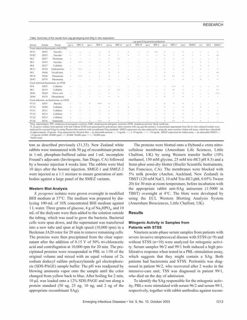
tem as described previously (31,33). New Zealand whiterabbits were immunized with 50 µg of recombinant proteinin 1-mL phosphate-buffered saline and 1-mL incompleteFreund’s adjuvants (Invitrogene, San Diego, CA) followedby a booster injection 4 weeks later. The rabbits were bled10 days after the booster injection. SMEZ-1 and SMEZ-2were injected as a 1:1 mixture to ensure generation of anti-bodies against a large panel of the SMEZ variants.
Western Blot AnalysisS. pyogenes isolates were grown overnight in modified
BHI medium at 37°C. The medium was prepared by dia-lyzing 100-mL of 10X concentrated BHI medium against1 L water. Three grams of glucose, 4 g of Na2HPO4, and 10mL of the dialysate were then added to the solution outsidethe tubing, which was used to grow the bacteria. Bacterialcells were spun down, and the supernatant was transferredinto a new tube and spun at high speed (10,000 rpm) in aBeckman JA20 rotor for 20 min to remove remaining cells.The proteins were then precipitated from the clear super-natant after the addition of 0.15 V of 50% tri-chloraceticacid and centrifugation at 10,000 rpm for 20 min. The pre-cipitated proteins were resuspended in PBL to 1/50 of theoriginal volume and mixed with an equal volume of 2xsodium dodecyl sulfate–polyacrylamide gel electrophore-sis (SDS-PAGE) sample buffer. The pH was readjusted byblowing ammonia vapor onto the sample until the colorchanged from yellow back to blue. After boiling for 2 min,10 µL was loaded onto a 12% SDS-PAGE and run along aprotein standard (50 ng, 25 ng, 10 ng, and 2 ng of theappropriate recombinant SAg).
The proteins were blotted onto a Hybond-c extra nitro-cellulose membrane (Amersham Life Sciences, LittleChalfont, UK) by using Western transfer buffer (10%methanol, 150 mM glycine, 25 mM tris-HCl pH 8.5) and aSemi-phor semi-dry blotter (Hoefer Scientific Instruments,San Francisco, CA). The membranes were blocked with5% milk powder (Anchor, Auckland, New Zealand) inTBST (120 mM NaCl, 10 mM Tris-HCl ph8, 0.05% Tween20) for 30 min at room temperature, before incubation withthe appropriate rabbit anti-SAg antiserum (1:5000 inTBST) overnight at 4°C. The blots were developed byusing the ECL Western Blotting Analysis System(Amersham Biosciences, Little Chalfont, UK).
Results
Mitogenic Activity in Samples from Patients with STSS
Nineteen acute-phase serum samples from patients withsevere invasive streptococcal disease with STSS (n=9) andwithout STSS (n=10) were analyzed for mitogenic activi-ty. Serum samples 96/2 and 99/1 both induced a high pro-liferative response when tested in a PBL-stimulation assay,which suggests that they might contain a SAg. Bothpatients had bacteremia and STSS. Peritonitis was diag-nosed in patient 96/2, who recovered after 2 weeks in theintensive-care unit; TSS was diagnosed in patient 99/1,who died on the day of admission.
To identify the SAg responsible for the mitogenic activ-ity, PBLs were stimulated with serum 96/2 and serum 99/1,respectively, together with rabbit antibodies against recom-
Emerging Infectious Diseases • Vol. 9, No. 10, October 2003 1213
RESEARCH
Table. Summary of the results from sag genotyping and SAg in vitro expression sag gene/SAg protein production
Serum Isolate Focus spe-a SPE-A spe-c SPE-C spe-g SPE-G spe-h SPE-H spe-i SPE-I spe-j SPE-J smez SMEZ ssa SSA SMEZc Focal infection/bacteremia with STSS 94/31 H292 Fasciitis - - - - + - + - + ++ + - + - - - + 95/02 H293 Fasciitis - - - - + - + - - - + - + - - - ++ 96/2 H297 Peritonitis + - - - + ++ - - - - + + + - - - + 98/5 H325 Fasciitis + +++ - - + - - - - - + - + - + - + 98/8 H327 Cellulitis - - - - + + - - - - + - + _ + + - 98/11 H330 Endometritis + +++ - - + - - - - - + + + - + - + 99/1 H360 Occult bact. - - + + + + - - - - + - + - - - ++ 99/18 H366 Pneumonia + +++ - - + - - - - - + ++ + - - - + 20/07 H378 Pneumonia - - - - + - + - - - + - + - + - + Focal infection/bacteremia, no STSS 95/8 H295 Cellulitis - - - - + - - - - - + +++ + - - - ++ 98/1 H319 Cellulitis - - - - + ++ - - - - + ++ + - - - ++ 20/01 H369 Pelvic clot - - - - + - - - - - + + + - - - +++ 20/04 H370 Thromobosis - - - - + + - - - - + - + - - - - Focal infection, no bacteremia, no STSS 97/15 H307 Bursitis - - + +++ + - - - - - + + + - - - + 97/18 H308 Cellulitis + - - - + - + - - - + - + - + - - 97/21 H311 Cellulitis - - - - + - - - - - + - + - - - + 97/23 H314 Cellulitis + - - - + + + - + - + + + - - - ++ 97/24 H315 Cellulitis + - - - + - - - - - + + + - - - - 97/26 H316 Amnionitis - - + +++ + + - - - - + ++ + - + - - aSAg, superantigen; SPE, streptococcal pyrogenic exotoxin; SME, streptococcal mitogenic exotoxin; STSS, streptococcal toxic shock syndrome. bS. pyogenes isolates from patients with and without STSS were genotyped by polymerase chain reaction with sag specific primers. Concentrated supernatant from the in vitro cultured isolates were analyzed for secreted SAgs by using Western blot analysis with recombinant SAg standards. SMEZ expression was also analyzed by using the more sensitive Jurkat cell assay, which has a threshold of approximately 10 pg/mL. SAg expression by Western blot: -, no detectable protein; +, <2 ng/mL; ++, 2–10 ng/mL; +++, >10 ng/mL. SMEZ expression by Jurkat assay: -, no detectable SMEZ; +, >10 pg/ml (10,000–20,000 cpm); ++, 20,000–30,000 cpm; +++, >30,000 cpm. cJurkat assay.
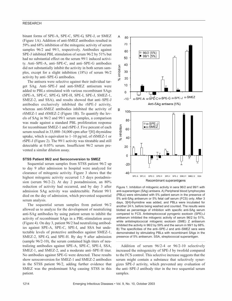
binant forms of SPE-A, SPE-C, SPE-G, SPE-J, or SMEZ(Figure 1A). Addition of anti-SMEZ antibodies resulted in59% and 68% inhibition of the mitogenic activity of serumsamples 96/2 and 99/1, respectively. Antibodies againstSPE-J inhibited PBL stimulation of serum 96/2 by 51% buthad no substantial effect on the serum 99/1 induced activi-ty. Anti–SPE-A, anti–SPE-C, and anti–SPE-G antibodiesdid not substantially inhibit the activity in both serum sam-ples, except for a slight inhibition (18%) of serum 96/2activity by anti–SPE-G antibodies.
The antisera were selective against their individual tar-get SAg. Anti–SPE-J and anti-SMEZ antiserum wereadded to PBLs stimulated with various recombinant SAgs(SPE-A, SPE-C, SPE-G, SPE-H, SPE-I, SPE-J, SMEZ-1,SMEZ-2, and SSA), and results showed that anti–SPE-Jantibodies exclusively inhibited the rSPE-J activity,whereas anti-SMEZ antibodies inhibited the activity ofrSMEZ-1 and rSMEZ-2 (Figure 1B). To quantify the lev-els of SAg in 96/2 and 99/1 serum samples, a comparisonwas made against a standard PBL proliferation responsefor recombinant SMEZ-1 and rSPE-J. Five percent of eachserum resulted in 33,000–34,000 cpm after 3[H]-thymidineuptake, which is equivalent to 1–10 pg/mL of rSMEZ-1 orrSPE-J (Figure 2). The 99/1 activity was titratable and stilldetectable at 0.05% serum. Insufficient 96/2 serum pre-vented a similar dilution assay.
STSS Patient 96/2 and Seroconversion to SMEZSequential serum samples from STSS patient 96/2 up
to day 9 after admission to hospital were analyzed forclearance of mitogenic activity. Figure 3 shows that thehighest mitogenic activity occurred 1.5 days postadmis-sion (serum 96/2-2). At day 2 postadmission, an 80%reduction of activity had occurred, and by day 3 afteradmission SAg activity was undetectable. Patient 99/1died on the day of admission, which prevented sequentialserum analysis.
The sequential serum samples from patient 96/2allowed us to analyze for the development of neutralizinganti-SAg antibodies by using patient serum to inhibit theactivity of recombinant SAgs in a PBL-stimulation assay(Figure 4). On day 3, patient 96/2 had neutralizing antibod-ies against SPE-A, SPE-C, SPE-I, and SSA but unde-tectable levels of protective antibodies against SMEZ-1,SMEZ-2, SPE-G, and SPE-H. By day 9 after admission(sample 96/2-10), the serum contained high titers of neu-tralizing antibodies against SPE-A, SPE-C, SPE-I, SSA,SMEZ-1, and SMEZ-2, and a moderate anti–SPE-H titer.No antibodies against SPE-G were detected. These resultsshow seroconversion for SMEZ-1 and SMEZ-2 antibodiesin the STSS patient 96/2, adding further evidence thatSMEZ was the predominant SAg causing STSS in thispatient.
Addition of serum 96/2-4 or 96/2-10 selectivelyincreased the mitogenicity of SPE-J by twofold comparedto the FCS control. This selective increase suggests that theserum might contain a substance that selectively syner-gizes SPE-J activity, which prevented any evaluation ofthe anti–SPE-J antibody titer in the two sequential serumsamples.
1214 Emerging Infectious Diseases • Vol. 9, No. 10, October 2003
RESEARCH
Figure 1. Inhibition of mitogenic activity in sera 96/2 and 99/1 withanti-superantigen (SAg) antisera. A) Peripheral blood lymphocytes(PBLs) were stimulated with 5% patient serum in the presence of5% anti-SAg antiserum or 5% fetal calf serum (FCS) only. After 3days, 3[H]-thymidine was added, and PBLs were incubated foranother 24 h, before being washed and counted. The results wereblotted as percentage of inhibition with specific anti-SAg serumcompared to FCS. Antistreptococcal pyrogenic exotoxin (SPE)-Jantiserum inhibited the mitogenic activity of serum 96/2 by 51%,while antistreptococcal mitogenic exotoxin (SME) Z antiseruminhibited the activity in 96/2 by 59% and the serum in 99/1 by 68%.B) The specificities of the anti–SPE-J and anti–SMEZ sera weredemonstrated by stimulating PBLs with recombinant SAgs in thepresence of 5% antiserum. SSA, streptococcal superantigen.
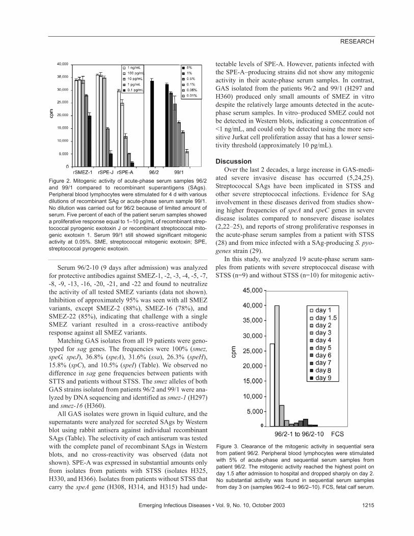
Serum 96/2-10 (9 days after admission) was analyzedfor protective antibodies against SMEZ-1, -2, -3, -4, -5, -7,-8, -9, -13, -16, -20, -21, and -22 and found to neutralizethe activity of all tested SMEZ variants (data not shown).Inhibition of approximately 95% was seen with all SMEZvariants, except SMEZ-2 (88%), SMEZ-16 (78%), andSMEZ-22 (85%), indicating that challenge with a singleSMEZ variant resulted in a cross-reactive antibodyresponse against all SMEZ variants.
Matching GAS isolates from all 19 patients were geno-typed for sag genes. The frequencies were 100% (smez,speG, speJ), 36.8% (speA), 31.6% (ssa), 26.3% (speH),15.8% (spC), and 10.5% (speI) (Table). We observed nodifference in sag gene frequencies between patients withSTTS and patients without STSS. The smez alleles of bothGAS strains isolated from patients 96/2 and 99/1 were ana-lyzed by DNA sequencing and identified as smez-1 (H297)and smez-16 (H360).
All GAS isolates were grown in liquid culture, and thesupernatants were analyzed for secreted SAgs by Westernblot using rabbit antisera against individual recombinantSAgs (Table). The selectivity of each antiserum was testedwith the complete panel of recombinant SAgs in Westernblots, and no cross-reactivity was observed (data notshown). SPE-A was expressed in substantial amounts onlyfrom isolates from patients with STSS (isolates H325,H330, and H366). Isolates from patients without STSS thatcarry the speA gene (H308, H314, and H315) had unde-
tectable levels of SPE-A. However, patients infected withthe SPE-A–producing strains did not show any mitogenicactivity in their acute-phase serum samples. In contrast,GAS isolated from the patients 96/2 and 99/1 (H297 andH360) produced only small amounts of SMEZ in vitrodespite the relatively large amounts detected in the acute-phase serum samples. In vitro–produced SMEZ could notbe detected in Western blots, indicating a concentration of<1 ng/mL, and could only be detected using the more sen-sitive Jurkat cell proliferation assay that has a lower sensi-tivity threshold (approximately 10 pg/mL).
DiscussionOver the last 2 decades, a large increase in GAS-medi-
ated severe invasive disease has occurred (5,24,25).Streptococcal SAgs have been implicated in STSS andother severe streptococcal infections. Evidence for SAginvolvement in these diseases derived from studies show-ing higher frequencies of speA and speC genes in severedisease isolates compared to nonsevere disease isolates(2,22–25), and reports of strong proliferative responses inthe acute-phase serum samples from a patient with STSS(28) and from mice infected with a SAg-producing S. pyo-genes strain (29).
In this study, we analyzed 19 acute-phase serum sam-ples from patients with severe streptococcal disease withSTSS (n=9) and without STSS (n=10) for mitogenic activ-
Emerging Infectious Diseases • Vol. 9, No. 10, October 2003 1215
RESEARCH
Figure 2. Mitogenic activity of acute-phase serum samples 96/2and 99/1 compared to recombinant superantigens (SAgs).Peripheral blood lymphocytes were stimulated for 4 d with variousdilutions of recombinant SAg or acute-phase serum sample 99/1.No dilution was carried out for 96/2 because of limited amount ofserum. Five percent of each of the patient serum samples showeda proliferative response equal to 1–10 pg/mL of recombinant strep-tococcal pyrogenic exotoxin J or recombinant streptococcal mito-genic exotoxin 1. Serum 99/1 still showed significant mitogenicactivity at 0.05%. SME, streptococcal mitogenic exotoxin; SPE,streptococcal pyrogenic exotoxin.
Figure 3. Clearance of the mitogenic activity in sequential serafrom patient 96/2. Peripheral blood lymphocytes were stimulatedwith 5% of acute-phase and sequential serum samples frompatient 96/2. The mitogenic activity reached the highest point onday 1.5 after admission to hospital and dropped sharply on day 2.No substantial activity was found in sequential serum samplesfrom day 3 on (samples 96/2–4 to 96/2–10). FCS, fetal calf serum.
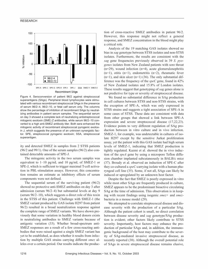
ity and detected SMEZ in samples from 2 STSS patients(96/2 and 99/1). One of the serum samples (96/2) also con-tained detectable amounts of SPE-J.
The mitogenic activity in the two serum samples wasequivalent to 1–10 pg/mL and 10 pg/mL of SMEZ-1 orSPE-J, which is sufficient to trigger maximal T-cell activa-tion in PBL-stimulation assays. However, this concentra-tion remains an estimate as inhibitory effects of serumcomponents were not defined.
The sequential serum of the surviving patient (96/2)showed no protective anti-SMEZ antibodies on day 3 afteradmission (serum 96/2–4) but substantial levels at day 9(serum 96/2–10), which suggests a direct role of this toxinin the STSS of this patient. Challenge with SMEZ-1 (theSMEZ variant produced by GAS isolate H297 from patient96/2) resulted in a broad neutralization response againstthe complete range of SMEZ variants. We have shown pre-viously that some variation in healthy blood donors existsin neutralizing antibodies to SMEZ variants because ofantigenic variation (33). Whether broad-spectrum anti-SMEZ responses are a result of a few cross-reacting anti-bodies that were raised against a single SMEZ variant hasyet to be established, as does whether it results from infec-tion by multiple GAS strains carrying different smez al-leles over a certain period. Our results indicate the produc-
tion of cross-reactive SMEZ antibodies in patient 96/2.However, this response might not reflect a generalresponse, and SMEZ concentration in the blood might playa critical role.
Analysis of the 19 matching GAS isolates showed nobias in sag genotype between STSS isolates and non-STSSisolates. Furthermore, the results are consistent with thesag gene frequencies previously observed in 39 S. pyo-genes isolates from New Zealand patients with sore throat(n=29), wound infection (n=4), acute glomerulonephritis(n=1), otitis (n=1), endometritis (n=2), rheumatic fever(n=1), and skin ulcer (n=1) (36). The only substantial dif-ference was the frequency of the speC gene, found in 42%of New Zealand isolates and 15.8% of London isolates.These results suggest that genotyping of sag genes alone isnot predictive for type or severity of streptococcal disease.
We found no substantial difference in SAg productionin cell cultures between STSS and non-STSS strains, withthe exception of SPE-A, which was only expressed inSTSS strains and suggests a tight association of SPE-A insome cases of STSS. These data are consistent with datafrom other groups that showed a link between SPE-Aexpression and severe streptococcal disease (17,22,23).Evidence points to very different regulation of SAg pro-duction between in vitro culture and in vivo infection.SMEZ-1, for example, was undetectable in cultures of iso-late H297 except by the sensitive Jurkat proliferationassay, yet the patient with this GAS isolate had high serumlevels of SMEZ-1, indicating that SMEZ production istightly regulated. Kazmi et al. showed the in vivo induc-tion of the speA gene by using a micropore Teflon diffu-sion chamber implanted subcutaneously in BALB/c mice(37). Broudy et al. observed an induction of SPE-C afterthey co-cultured a speC-carrying isolate with a human pha-ryngeal cell line (35). Some, if not all, SAgs can likely beinduced or upregulated by an unknown host factor.
Despite the fact that SMEZ is poorly expressed in vitrowhile most other SAgs are frequently produced in culture,SMEZ appears to be the predominant bioactive circulatingSAg at the time of admission. This observation is in keep-ing with recent findings using isogenic smez+ and smez-bacteria in a mouse model (29).
We attempted to correlate streptococcal disease and dis-ease severity with the production of a particular SAg.Although the patient cohort is small, no direct correlationbetween disease severity and sag genotype/SAg produc-tion is evident; other factors likely contribute to STSSseverity. Importantly, host factors may enhance the pro-duction of particular SAgs and, in addition, the immuno-genic background of the host may contribute to the sever-ity of SAg-mediated invasive streptococcal disease, asrecently reported (38). Although the overall potential roleof SAgs in severe streptococcal disease remains elusive,
1216 Emerging Infectious Diseases • Vol. 9, No. 10, October 2003
RESEARCH
Figure 4. Seroconversion of patient 96/2 against streptococcalsuperantigens (SAgs). Peripheral blood lymphocytes were stimu-lated with various recombinant streptococcal SAgs in the presenceof serum 96/2–4, 96/2–10, or fetal calf serum only. The columnsshow the percentage of inhibition of recombinant SAgs by neutral-izing antibodies in patient serum samples. The sequential serumon day 3 showed a complete lack of neutralizing antistreptococcalmitogenic exotoxin (SME) Z antibodies, while serum 96/2–10 con-verted to a high anti-SMEZ antibody titer. Both sera enhanced themitogenic activity of recombinant streptococcal pyrogenic exotox-in J, which suggests the presence of an unknown synergistic fac-tor. SPE, streptococcal pyrogenic exotoxin; SSA, streptococcalsuperantigen.
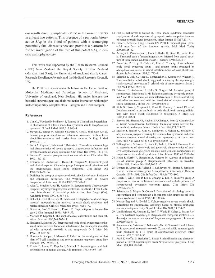
our results directly implicate SMEZ in the onset of STSSin at least two patients. This presence of a particular biore-active SAg in the blood of patients with a reemergingpotentially fatal disease is new and provides a platform forfurther investigation of the role of this potent SAg in dis-ease pathophysiology.
This work was supported by the Health Research Council(HRC) New Zealand, the Royal Society of New Zealand(Marsden Fast Start), the University of Auckland (Early CareerResearch Excellence Award), and the Medical Research Council,UK.
Dr. Proft is a senior research fellow in the Department ofMolecular Medicine and Pathology, School of Medicine,University of Auckland, New Zealand. His major interests arebacterial superantigens and their molecular interaction with majorhistocompatibility complex class II antigen and T-cell receptor.
References
1. Cone L, Woodard P, Schlievert P, Tomory G. Clinical and bacteriolog-ic observations of a toxic shock-like syndrome due to Streptococcuspyogenes. N Engl J Med 1987;317:146–9.
2. Stevens D, Tanner M, Winship J, Swarts R, Ries K, Schlievert P, et al.Severe group A streptococcal infections associated with a toxicshock-like syndrome and scarlet fever toxin A. N Engl J Med1989;321:1–7.
3. Forni A, Kaplan E, Schlievert P, Roberts R. Clinical and microbiolog-ical characteristics of severe group A streptococcus infections andstreptococcal toxic shock syndrome. Clin Infect Dis 1995;21:333–40.
4. Stevens D. Invasive group A streptococcus infections. Clin Infect Dis1992;14:2–13.
5. Eriksson BK, Andersson J, Holm SE, Norgren M. Epidemiologicaland clinical aspects of invasive group A streptococcal infections andthe streptococcal toxic shock syndrome. Clin Infect Dis1998;27:1428–36.
6. Defining the group A streptococcal toxic shock syndrome. Rationaleand consensus definition. The Working Group on SevereStreptococcal Infections. JAMA 1993;269:390–1.
7. Alouf J, Mueller-Alouf H, Koehler W. Superantigenic Streptococcuspyogenes erythrogenic/pyrogenic exotoxins. In: Alouf J, Freer J, edi-tors. Sourcebook of bacterial protein toxins. San Diego (CA):Academic Press; 1999. p. 567–88.
8. Bohach G, Fast D, Nelson R, Schlievert P. Staphylococcal and strep-tococcal pyrogenic toxins involved in toxic shock syndrome andrelated illnesses. Crit Rev Microbiol 1990;17:251–72.
9. Bernal A, Proft T, Fraser J, Posnett D. Superantigens in human dis-ease. J Clin Immunol 1999;19:149–57.
10. Marrack P, Kappler J. The staphylococcal enterotoxins and their rel-atives. Science 1990;248:705–11.
11. Hackett SP, Stevens DL. Streptococcal toxic shock syndrome: synthe-sis of tumor necrosis factor and interleukin-1 by monocytes stimulat-ed with pyrogenic exotoxin A and streptolysin O. J Infect Dis1992;165:879–85.
12. Herman A, Kappler J, Marrack P, Pullen A. Superantigens: mecha-nism of T-cell stimulation and role in immune responses. Annu RevImmunol 1991;9:745–72.
13. Kotzin B, Leung D, Kappler J, Marrack P. Superantigens and theirpotential role in human disease. Adv Immunol 1993;54:99–166.
14. Fast D, Schlievert P, Nelson R. Toxic shock syndrome–associatedstaphylococcal and streptococcal pyrogenic toxins are potent inducersof tumor necrosis factor production. Infect Immun 1989;57:291–4.
15. Fraser J, Arcus V, Kong P, Baker EN, Proft T. Superantigens—pow-erful modifiers of the immune system. Mol Med Today2000;6:125–32.
16. Acharya K, Passalacqua E, Jones E, Harlos K, Stuart D, Brehm R, etal. Structural basis of superantigen action inferred from crystal struc-ture of toxic-shock syndrome toxin-1. Nature 1994;367:94–7.
17. Bonventre P, Heeg H, Cullen C, Lian C. Toxicity of recombinanttoxic shock syndrome toxin 1 and mutant toxins produced byStaphylococcus aureus in rabbit infection model of toxic shock syn-drome. Infect Immun 1993;61:793–9.
18. Miethke T, Wahl C, Heeg K, Echtenacher B, Krammer P, Wagner H.T cell-mediated lethal shock triggered in mice by the superantigenstaphylococcal enterotoxin B: critical role of tumor necrosis factor. JExp Med 1992;175:91–8.
19. Eriksson B, Andersson J, Holm S, Norgren M. Invasive group Astreptococcal infections: T1M1 isolates expressing pyrogenic exotox-ins A and B in combination with selective lack of toxin-neutralizingantibodies are associated with increased risk of streptococcal toxicshock syndrome. J Infect Dis 1999;180:410–8.
20. Stolz S, Davis J, Vergeront J, Crass B, Chesney P, Wand PJ, et al.Development of serum antibody to toxic shock toxin among individ-uals with toxic shock syndrome in Wisconsin. J Infect Dis1985;151:883–9.
21. Stevens DL, Bryant AE, Hackett SP, Chang A, Peer G, Kosanke S, etal. Group A streptococcal bacteremia: the role of tumor necrosis fac-tor in shock and organ failure. J Infect Dis 1996;173:619–26.
22. Musser J, Hauser A, Kim M, Schlievert P, Nelson K, Selander R.Streptococcus pyogenes causing toxic-shock-like syndrome and otherinvasive diseases: clonal diversity and pyrogenic exotoxin expres-sion. Proc Natl Acad Sci U S A 1991;88:2668–72.
23. Talkington D, Schwartz B, Black C, Todd J, Elliott J, Breiman R, etal. Association of phenotypic and genotypic characteristics of inva-sive Streptococcus pyogenes isolates with clinical components ofstreptococcal toxic shock syndrome. Infect Immun 1993;61:3369–74.
24. Holm S, Norrby A, Bergholm A, Norgren M. Aspects of pathogene-sis of serious group A streptococccal infections in Sweden,1988–1989. J Infect Dis 1992;166:31–7.
25. Demers B, Simor AE, Vellend H, Schlievert PM, Byrne S, JamiesonF, et al. Severe invasive group A streptococcal infections in Ontario,Canada: 1987–1991. Clin Infect Dis 1993;16:792–800.
26. Hsueh P, Wu J, Tsai P, Liu J, Chuang Y, Luh K. Invasive group Astreptococcal disease in Taiwan is not associated with the presence ofstreptococcal pyrogenic exotoxin genes. Clin Infect Dis1998;26:584–9.
27. Sriskandan S, Moyes D, Cohen J. Detection of circulating bacterialsuperantigen and lymphotoxin-a in patients with streptococcal toxicshock syndrome. Lancet 1996;348:1315–6.
28. Norrby-Teglund A, Berdal J. Culture-negative severe septic shock:indications for streptococcal aetiology based on plasma antibodiesand superantigen activity. Scand J Infect Dis 2001;33:634–7.
29. Unnikrishnan M, Altmann D, Proft T, Wahid F, Cohen J, Fraser J, etal. The bacterial superantigen streptococcal mitogenic exotoxin Z isthe major immunoactive agent of Streptococcus pyogenes. J Immunol2002;169:2561–9.
30. Kamezawa Y, Nakahara T, Nakano S, Abe Y, Nozaki-Renard J, IsonoT. Streptococcal mitogenic exotoxin Z, a novel acidic superantigenictoxin produced by a T1 strain of Streptococcus pyogenes. InfectImmun 1997;65:3828–33.
31. Proft T, Moffatt S, Berkahn C, Fraser J. Identification and character-ization of novel superantigens from Streptococcus pyogenes. J ExpMed 1999;189:89–101.
Emerging Infectious Diseases • Vol. 9, No. 10, October 2003 1217
RESEARCH
32. Proft T, Arcus V, Handley V, Baker E, Fraser J. Immunological andbiochemical characterization of streptococcal pyrogenic exotoxins Iand J (SPE-I and SPE-J) from Streptococcus pyogenes. J Immun2001;166:6711–9.
33. Proft T, Moffatt S, Weller K, Paterson A, Martin D, Fraser J. Thestreptococcal superantigen SMEZ exhibits wide allelic variation,mosaic structure, and significant antigenic variation. J Exp Med2000;191:1765–76.
34. Ferretti JJ, McShan WM, Ajdic D, Savic DJ, Savic G, Lyon K, et al.Complete genome sequence of an M1 strain of Streptococcus pyo-genes. Proc Natl Acad Sci U S A 2001;98:4658–63.
35. Broudy T, Pancholi V, Fischetti V. Induction of lysogenic bacterio-phage and phage-associated toxin from group A streptococci duringcoculture with human pharyngeal cells. Infect Immun2001;69:1440–3.
36. Proft T, Webb PD, Handley V, Fraser JD. Two novel superantigensfound in both group A and group C streptococcus. Infect Immun2003;71:1361–9.
37. Kazmi S, Kansal R, Aziz R, Hooshdaran M, Norrby-Teglund A, LowD, et al. Reciprocal, temporal expression of SpeA and SpeB by inva-sive M1T1 group A streptococcal isolates in vivo. Infect Immun2001;69:4988–95.
38. Kotb M, Norrby-Teglund A, McGeer A, El-Sherbini H, Dorak M,Khurshid A, et al. An immunogenetic and molecular basis for differ-ences in outcomes of invasive group A streptococcal infections. NatMed 2002;8:1398–404.
Address for correspondence: John D. Fraser, Department of MolecularMedicine and Pathology, Faculty of Medical and Health Sciences,University of Auckland, Private Bag 92019, Auckland, New Zealand; fax:+64-9-373-7492; email: [email protected]
1218 Emerging Infectious Diseases • Vol. 9, No. 10, October 2003
RESEARCH
All material published in Emerging Infectious Diseases is in thepublic domain and may be used and reprinted without special per-mission; proper citation, however, is appreciated.
Search ppast iissues oof EEID aat wwww.cdc.gov/eid
Campylobacter is the most common cause of bacteri-al gastroenteritis worldwide, yet the etiology of this infectionremains only partly explained. In a retrospective cohortstudy, we compared 213 sporadic campylobacter case-patients with 1,144 patients with negative fecal samples.Information was obtained on food history, animal contact,foreign travel, leisure activities, medical conditions, andmedication use. Eating chicken, eating food from a friedchicken outlet, eating salad vegetables, drinking bottledwater, and direct contact with cows or calves were all inde-pendently associated with infection. The population-attrib-utable fractions for these risk factors explained nearly 70%of sporadic campylobacter infections. Eating chicken is awell-established risk factor, but consuming salad and bot-tled water are not. The association with salad may beexplained by cross-contamination of food within the home,but the possibility that natural mineral water is a risk factorfor campylobacter infection could have wide public healthimplications.
Campylobacter is the most commonly reported bacteri-al cause of foodborne infection in the Western world
and affects more than 2 million people in the United Stateseach year (1). In England and Wales, over 50,000 campy-lobacter cases are reported each year and show no signs ofa decline in incidence (2). For every case reported to labo-ratory surveillance, another seven cases are estimated tooccur in the community, suggesting that from 0.5% to1.0% of the United Kingdom’s population is infectedannually (3). Although the infection usually causes a mild,self-limiting illness, serious sequelae, including Guillain-Barré syndrome and death, occur in approximately 1 in1,000 and 1 in 20,000 infections, respectively (1). Manynational food safety agencies, such as the Food StandardsAgency in the United Kingdom, have set goals of reducingfood poisoning. To achieve these goals, a much clearer
understanding of the etiology of campylobacter infectionwill be necessary.
In spite of the frequency of campylobacter infections,the cause has proved elusive. Recognized outbreaks arerare and are usually caused by contaminated water, milk,or poultry (4,5). However, these food products explainonly a small proportion of sporadic cases, and the sourceof infection is unaccounted for in >60% of U.K. campy-lobacter cases (6,7). Several case-control studies of riskfactors for sporadic campylobacter infection have beenperformed in the United Kingdom (6–10), but many unan-swered questions remain. We conducted a retrospectivecohort study that involved mailing a questionnaire to thepatient at the time the fecal specimen was received by thelaboratory to investigate the cause of sporadic campy-lobacter infection in the community.
MethodsThe study population included all persons living in the
Cardiff area who consulted their general practitioner forgastrointestinal symptoms and subsequently submitted adiagnostic fecal sample for microbiologic testing fromJanuary 1 through December 31, 2001. Cardiff PublicHealth Laboratory is the sole laboratory providing a diag-nostic microbiology service for the area. All specimenswere cultured for Campylobacter spp., Salmonella spp,Shigella spp., and Escherichia coli O157 and examined forova and parasites, by standard methods. Follow-up speci-mens from the same patient (<4 weeks after the previousspecimen submission date); specimens received from hos-pital wards and other sites were excluded from study. Thestudy was approved by the local research ethics committee.
Immediately upon receipt of the specimen at the labo-ratory (next working day), a questionnaire, together withan explanatory letter and a postage-paid envelope, wasmailed to the patient. Patients who had not respondedwithin 1 week were sent a reminder letter and providedwith another questionnaire on request. The questionnaireasked about basic personal details, including age, sex,
Emerging Infectious Diseases • Vol. 9, No. 10, October 2003 1219
RESEARCH
Hazards of Healthy Living: BottledWater and Salad Vegetables as RiskFactors for Campylobacter Infection
Meirion R. Evans,* C. Donald Ribeiro,† and Roland L. Salmon‡
*University of Wales College of Medicine, Cardiff, United Kingdom;†Cardiff Public Health Laboratory, Cardiff, United Kingdom; and‡Public Health Laboratory Service Communicable DiseaseSurveillance Centre (Wales), Cardiff, United Kingdom
employment status, occupation, details of illness, anddetails of household contacts. It included sections on for-eign travel, food and drink eaten, animal contact (pets andfarm animals), outdoor leisure activities (gardening, walk-ing, visits to parks or farms, fishing, swimming, andsports), and questions on specific medical conditions andmedication (antacids, H2 antagonists, and antibiotics). Thefood history covered meat and fish, poultry and eggs, veg-etables (raw vegetables, leaf vegetables [e.g., lettuce],salad vegetables [e.g., tomato], and prepared salads [e.g.,coleslaw]), fruit, milk and dairy products, drinking water(tap water, bottled water, and other sources), and eating out(type of restaurant or takeaway). Participants were askedto respond yes or no and, to the question of exposure fortap water, to indicate the number of glasses drunk per day.All questions related to exposure in the 7 days before theonset of symptoms, except for those on antibiotics, whichconcerned the month before illness onset.
Case-patients were defined as any patient, not associat-ed with an outbreak, who submitted a fecal sample thatwas positive for Campylobacter spp. on microbiologic cul-ture. Case-patients were compared with patients whosesamples were negative on culture and microscopic exami-nation. Patients with an alternative microbiologic diagno-sis were excluded (unless they had dual infection withcampylobacter).
Initial univariate analysis was performed with Epi Infosoftware (v. 6.04; Centers for Disease Control andPrevention) to calculate maximum likelihood estimates forMantel-Haenzel odds ratios (OR) with exact 95% mid-pconfidence limits. Continuous variables were analyzedusing the t test or Mann-Whitney U test, as appropriate. Allreported p values are two sided. Multiple logistic regres-sion models were constructed with Stata software (v. 6,Stata Corporation, College Station, TX). Risk factors wereselected a priori on biologic grounds and grouped into fourexposure categories: food and drink consumption; animalcontact; leisure activities, including foreign travel; andmedical history. Logistic regression models were first con-structed for risk factors within each exposure category(adjustment A). We then fitted a model that combined allthe independent risk factors (for which the Wald test pvalue for the adjusted OR was <0.10) from the four expo-sure categories (adjustment B). Finally, to detect any resid-ual confounding, we fitted all personal factors with a pvalue of <0.10 (age group, presence of a child <5 years ofage in the household, and employment status). Of these,only age group interacted significantly with the other termsas tested by goodness of fit and was therefore included inthe final model (adjustment C). The population-attributa-ble fraction for each risk factor associated with campy-lobacter infection was calculated by using methodsdescribed by Greenland and Drescher (11).
ResultsQuestionnaires were sent to 2,694 eligible patients;
fecal samples from 346 (12.8%) were positive forCampylobacter spp. (including 4 dual infections: 3 withsalmonella infection, 1 with giardiasis). No campylobacteroutbreak occurred during the study period. Ninety-onepatients (3.4%) were positive for other organisms (42Salmonella spp., 20 Giardia lamblia, 12 Cryptosporidiumsp., 7 Clostridium difficile, 2 Shigella sp., 2 E. coli O157,1 amoebic dysentery, and 5 other parasites); these wereexcluded from further analysis. Responses were receivedfrom 213 (61.6%) of 346 persons with campylobacterinfection and 1,144 (50.7%) of 2,257 persons with nega-tive specimen results. Median delay in response (from datequestionnaire sent to date questionnaire returned) was 6days (range 2–73 days) for case-patients and 7 days (range1–77 days) for non-case-patients (Kruskal-Wallis H 1.81,p=0.18).
Personal Factors and SymptomsCase-patients (median 43 years of age, range 0–80
years of age) were significantly older than non-case-patients (median 36 years of age, range 0–100 years ofage) (Kruskal-Wallis H 5.31, p=0.02) (Table 1). Non-case-patients were also more likely to come from a householdthat included a child <5 years of age (even after adjustingfor the age of the respondent), although not more likely toreport prior diarrheal illness in a household contact. Case-patients were more likely to report symptoms than non-case-patients, particularly fever (OR 3.19; 95% confidenceinterval [CI] 2.36 to 4.31), muscle aches (OR 3.13; 95% CI2.32 to 4.22), and abdominal pain (OR 3.40; 95% CI 2.32to 5.12. Nearly all case-patients and most non-case-patients had diarrhea, but case-patients (18.3%) were morelikely than non-case-patients (11.8%) to report blood in thestool (OR 1.67; 95% CI 1.12 to 2.46).
Food and Drink ConsumedCase-patients were more likely to report eating meat,
including beef, pork, and ham; poultry products, includingchicken and eggs; and a variety of uncooked vegetablesand fruit, including lettuce, other salad vegetables (cucum-ber, tomatoes, etc.), preprepared salad (coleslaw, etc.), andfresh or frozen berries. An association existed with drink-ing bottled water (OR 1.98; 95% CI 1.48 to 2.67) andbetween infection and drinking cold tap water (OR 1.51;95% CI 1.06 to 2.18) but not with drinking cold milk.Neither tap water nor milk consumption showed a doseresponse relationship. Case-patients were more likely tohave eaten out in the 7 days before illness onset, particu-larly at a fried chicken outlet, sandwich bar, or otherunspecified restaurant.
1220 Emerging Infectious Diseases • Vol. 9, No. 10, October 2003
RESEARCH
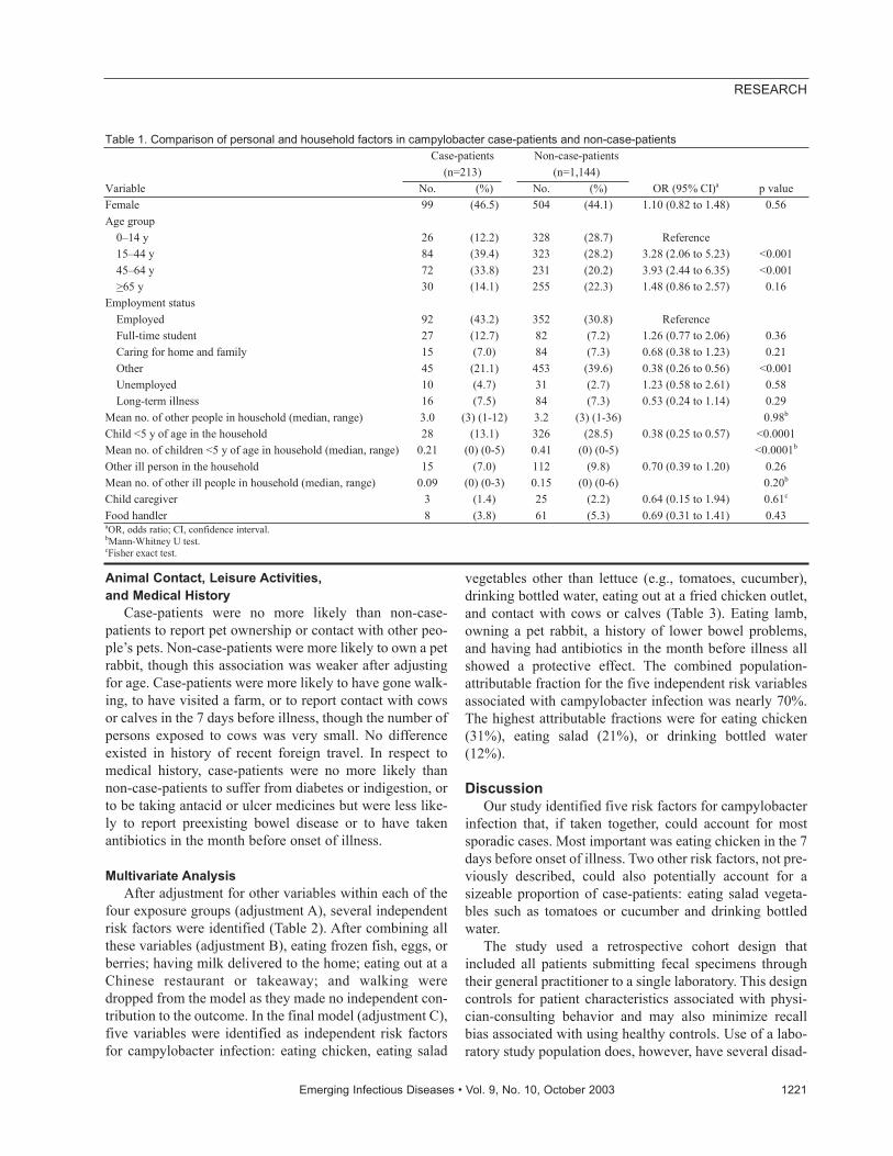
Animal Contact, Leisure Activities, and Medical History
Case-patients were no more likely than non-case-patients to report pet ownership or contact with other peo-ple’s pets. Non-case-patients were more likely to own a petrabbit, though this association was weaker after adjustingfor age. Case-patients were more likely to have gone walk-ing, to have visited a farm, or to report contact with cowsor calves in the 7 days before illness, though the number ofpersons exposed to cows was very small. No differenceexisted in history of recent foreign travel. In respect tomedical history, case-patients were no more likely thannon-case-patients to suffer from diabetes or indigestion, orto be taking antacid or ulcer medicines but were less like-ly to report preexisting bowel disease or to have takenantibiotics in the month before onset of illness.
Multivariate AnalysisAfter adjustment for other variables within each of the
four exposure groups (adjustment A), several independentrisk factors were identified (Table 2). After combining allthese variables (adjustment B), eating frozen fish, eggs, orberries; having milk delivered to the home; eating out at aChinese restaurant or takeaway; and walking weredropped from the model as they made no independent con-tribution to the outcome. In the final model (adjustment C),five variables were identified as independent risk factorsfor campylobacter infection: eating chicken, eating salad
vegetables other than lettuce (e.g., tomatoes, cucumber),drinking bottled water, eating out at a fried chicken outlet,and contact with cows or calves (Table 3). Eating lamb,owning a pet rabbit, a history of lower bowel problems,and having had antibiotics in the month before illness allshowed a protective effect. The combined population-attributable fraction for the five independent risk variablesassociated with campylobacter infection was nearly 70%.The highest attributable fractions were for eating chicken(31%), eating salad (21%), or drinking bottled water(12%).
DiscussionOur study identified five risk factors for campylobacter
infection that, if taken together, could account for mostsporadic cases. Most important was eating chicken in the 7days before onset of illness. Two other risk factors, not pre-viously described, could also potentially account for asizeable proportion of case-patients: eating salad vegeta-bles such as tomatoes or cucumber and drinking bottledwater.
The study used a retrospective cohort design thatincluded all patients submitting fecal specimens throughtheir general practitioner to a single laboratory. This designcontrols for patient characteristics associated with physi-cian-consulting behavior and may also minimize recallbias associated with using healthy controls. Use of a labo-ratory study population does, however, have several disad-
Emerging Infectious Diseases • Vol. 9, No. 10, October 2003 1221
RESEARCH
Table 1. Comparison of personal and household factors in campylobacter case-patients and non-case-patients Case-patients
(n=213) Non-case-patients
(n=1,144) Variable No. (%) No. (%) OR (95% CI)a
p value
Female 99 (46.5) 504 (44.1) 1.10 (0.82 to 1.48) 0.56 Age group
0–14 y 26 (12.2) 328 (28.7) Reference 15–44 y 84 (39.4) 323 (28.2) 3.28 (2.06 to 5.23) <0.001 45–64 y 72 (33.8) 231 (20.2) 3.93 (2.44 to 6.35) <0.001 >65 y 30 (14.1) 255 (22.3) 1.48 (0.86 to 2.57) 0.16
Employment status Employed 92 (43.2) 352 (30.8) Reference Full-time student 27 (12.7) 82 (7.2) 1.26 (0.77 to 2.06) 0.36 Caring for home and family 15 (7.0) 84 (7.3) 0.68 (0.38 to 1.23) 0.21 Other 45 (21.1) 453 (39.6) 0.38 (0.26 to 0.56) <0.001 Unemployed 10 (4.7) 31 (2.7) 1.23 (0.58 to 2.61) 0.58 Long-term illness 16 (7.5) 84 (7.3) 0.53 (0.24 to 1.14) 0.29
Mean no. of other people in household (median, range) 3.0 (3) (1-12) 3.2 (3) (1-36) 0.98b
Child <5 y of age in the household 28 (13.1) 326 (28.5) 0.38 (0.25 to 0.57) <0.0001 Mean no. of children <5 y of age in household (median, range) 0.21 (0) (0-5) 0.41 (0) (0-5) <0.0001b
Other ill person in the household 15 (7.0) 112 (9.8) 0.70 (0.39 to 1.20) 0.26 Mean no. of other ill people in household (median, range) 0.09 (0) (0-3) 0.15 (0) (0-6) 0.20b
Child caregiver 3 (1.4) 25 (2.2) 0.64 (0.15 to 1.94) 0.61c Food handler 8 (3.8) 61 (5.3) 0.69 (0.31 to 1.41) 0.43 aOR, odds ratio; CI, confidence interval. bMann-Whitney U test. cFisher exact test.
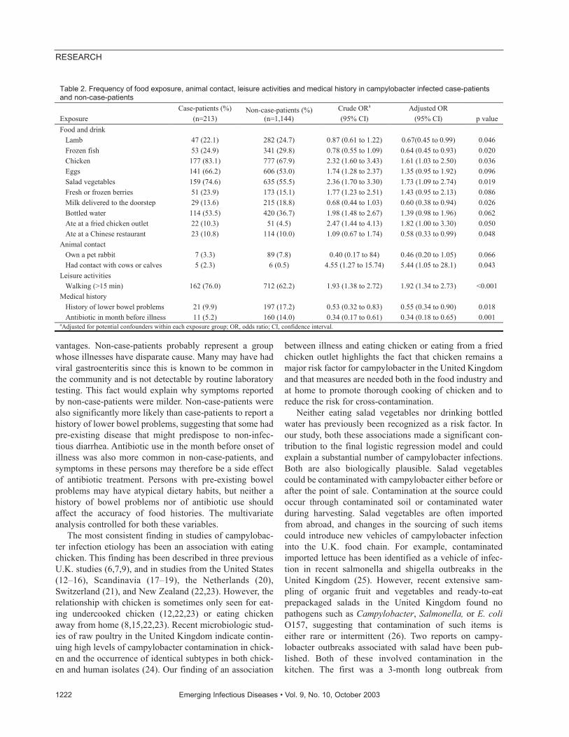
vantages. Non-case-patients probably represent a groupwhose illnesses have disparate cause. Many may have hadviral gastroenteritis since this is known to be common inthe community and is not detectable by routine laboratorytesting. This fact would explain why symptoms reportedby non-case-patients were milder. Non-case-patients werealso significantly more likely than case-patients to report ahistory of lower bowel problems, suggesting that some hadpre-existing disease that might predispose to non-infec-tious diarrhea. Antibiotic use in the month before onset ofillness was also more common in non-case-patients, andsymptoms in these persons may therefore be a side effectof antibiotic treatment. Persons with pre-existing bowelproblems may have atypical dietary habits, but neither ahistory of bowel problems nor of antibiotic use shouldaffect the accuracy of food histories. The multivariateanalysis controlled for both these variables.
The most consistent finding in studies of campylobac-ter infection etiology has been an association with eatingchicken. This finding has been described in three previousU.K. studies (6,7,9), and in studies from the United States(12–16), Scandinavia (17–19), the Netherlands (20),Switzerland (21), and New Zealand (22,23). However, therelationship with chicken is sometimes only seen for eat-ing undercooked chicken (12,22,23) or eating chickenaway from home (8,15,22,23). Recent microbiologic stud-ies of raw poultry in the United Kingdom indicate contin-uing high levels of campylobacter contamination in chick-en and the occurrence of identical subtypes in both chick-en and human isolates (24). Our finding of an association
between illness and eating chicken or eating from a friedchicken outlet highlights the fact that chicken remains amajor risk factor for campylobacter in the United Kingdomand that measures are needed both in the food industry andat home to promote thorough cooking of chicken and toreduce the risk for cross-contamination.
Neither eating salad vegetables nor drinking bottledwater has previously been recognized as a risk factor. Inour study, both these associations made a significant con-tribution to the final logistic regression model and couldexplain a substantial number of campylobacter infections.Both are also biologically plausible. Salad vegetablescould be contaminated with campylobacter either before orafter the point of sale. Contamination at the source couldoccur through contaminated soil or contaminated waterduring harvesting. Salad vegetables are often importedfrom abroad, and changes in the sourcing of such itemscould introduce new vehicles of campylobacter infectioninto the U.K. food chain. For example, contaminatedimported lettuce has been identified as a vehicle of infec-tion in recent salmonella and shigella outbreaks in theUnited Kingdom (25). However, recent extensive sam-pling of organic fruit and vegetables and ready-to-eatprepackaged salads in the United Kingdom found nopathogens such as Campylobacter, Salmonella, or E. coliO157, suggesting that contamination of such items iseither rare or intermittent (26). Two reports on campy-lobacter outbreaks associated with salad have been pub-lished. Both of these involved contamination in thekitchen. The first was a 3-month long outbreak from
1222 Emerging Infectious Diseases • Vol. 9, No. 10, October 2003
RESEARCH
Table 2. Frequency of food exposure, animal contact, leisure activities and medical history in campylobacter infected case-patients and non-case-patients
Exposure
Case-patients (%) (n=213)
Non-case-patients (%) (n=1,144)
Crude ORa (95% CI)
Adjusted OR (95% CI)
p value
Food and drink Lamb 47 (22.1) 282 (24.7) 0.87 (0.61 to 1.22) 0.67(0.45 to 0.99) 0.046 Frozen fish 53 (24.9) 341 (29.8) 0.78 (0.55 to 1.09) 0.64 (0.45 to 0.93) 0.020 Chicken 177 (83.1) 777 (67.9) 2.32 (1.60 to 3.43) 1.61 (1.03 to 2.50) 0.036 Eggs 141 (66.2) 606 (53.0) 1.74 (1.28 to 2.37) 1.35 (0.95 to 1.92) 0.096 Salad vegetables 159 (74.6) 635 (55.5) 2.36 (1.70 to 3.30) 1.73 (1.09 to 2.74) 0.019 Fresh or frozen berries 51 (23.9) 173 (15.1) 1.77 (1.23 to 2.51) 1.43 (0.95 to 2.13) 0.086 Milk delivered to the doorstep 29 (13.6) 215 (18.8) 0.68 (0.44 to 1.03) 0.60 (0.38 to 0.94) 0.026 Bottled water 114 (53.5) 420 (36.7) 1.98 (1.48 to 2.67) 1.39 (0.98 to 1.96) 0.062 Ate at a fried chicken outlet 22 (10.3) 51 (4.5) 2.47 (1.44 to 4.13) 1.82 (1.00 to 3.30) 0.050 Ate at a Chinese restaurant 23 (10.8) 114 (10.0) 1.09 (0.67 to 1.74) 0.58 (0.33 to 0.99) 0.048
Animal contact Own a pet rabbit 7 (3.3) 89 (7.8) 0.40 (0.17 to 84) 0.46 (0.20 to 1.05) 0.066 Had contact with cows or calves 5 (2.3) 6 (0.5) 4.55 (1.27 to 15.74) 5.44 (1.05 to 28.1) 0.043
Leisure activities Walking (>15 min) 162 (76.0) 712 (62.2) 1.93 (1.38 to 2.72) 1.92 (1.34 to 2.73) <0.001
Medical history History of lower bowel problems 21 (9.9) 197 (17.2) 0.53 (0.32 to 0.83) 0.55 (0.34 to 0.90) 0.018 Antibiotic in month before illness 11 (5.2) 160 (14.0) 0.34 (0.17 to 0.61) 0.34 (0.18 to 0.65) 0.001
aAdjusted for potential confounders within each exposure group; OR, odds ratio; CI, confidence interval.
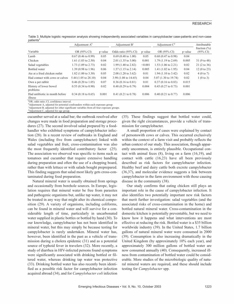
cucumber served at a salad bar; the outbreak resolved afterchanges were made in food preparation and storage proce-dures (27). The second involved salad prepared by a food-handler who exhibited symptoms of campylobacter infec-tion (28). In a recent review of outbreaks in England andWales (including five from campylobacter) linked withsalad vegetables and fruit, cross-contamination was alsothe most frequently identified contributory factor (25).The association we observed was specific to items such astomatoes and cucumber that require extensive handlingduring preparation and often the use of a chopping board,rather than with lettuce or with salads bought preprepared.This finding suggests that salad most likely gets cross-con-taminated during food preparation.
Natural mineral water is usually obtained from springsand occasionally from borehole sources. In Europe, legis-lation requires that mineral water be free from parasitesand pathogenic organisms but, unlike tap water, it may notbe treated in any way that might alter its chemical compo-sition (29). A variety of organisms, including coliforms,can be found in mineral water and will survive for a con-siderable length of time, particularly in uncarbonatedwater supplied in plastic bottles or bottled by hand (30). Toour knowledge, campylobacter has not been identified inmineral water, but this may simply be because testing forcampylobacter is rarely undertaken. Mineral water has,however, been identified in the past as a vehicle of trans-mission during a cholera epidemic (31) and as a potentialsource of typhoid fever in travelers (32). More recently, astudy of diarrhea in HIV-infected persons found symptomswere significantly associated with drinking bottled or fil-tered water, whereas drinking tap water was protective(33). Drinking bottled water has also recently been identi-fied as a possible risk factor for campylobacter infectionacquired abroad (34), and for Campylobacter coli infection
(35). These findings suggest that bottled water could,given the right circumstances, provide a vehicle of trans-mission for campylobacter.
A small proportion of cases were explained by contactof personswith cows or calves. This occurred exclusivelywithin the context of a farm visit and probably reflects theurban context of our study. This association, though appar-ently uncommon, is entirely plausible. Occupational con-tact with animal feces (8), living on a farm (16,19), andcontact with cattle (16,23) have all been previouslydescribed as risk factors for campylobacter infection.Healthy beef and dairy cattle both excrete campylobacter(36,37), and molecular evidence suggests a link betweencampylobacter in the farm environment with those causingdisease in the community (38).
Our study confirms that eating chicken still plays animportant role in the cause of campylobacter infection. Italso identifies two potentially important new risk factorsthat merit further investigation: salad vegetables (and theassociated risks of cross-contamination in the home) andbottled natural mineral water. Cross-contamination in thedomestic kitchen is potentially preventable, but we need toknow how it happens and what interventions are mosteffective at reducing the risk. Bottled water is a $35 billionworldwide industry (39). In the United States, 1.7 billiongallons of natural mineral water were consumed in 2000(39). Consumption is also increasing dramatically in theUnited Kingdom (by approximately 10% each year), andapproximately 300 million gallons of bottled water arenow consumed annually (40). Consequently, increased ill-ness from contamination of bottled water could be consid-erable. More studies of the microbiologic quality of natu-ral mineral waters are required, and these should includetesting for Campylobacter spp.
Emerging Infectious Diseases • Vol. 9, No. 10, October 2003 1223
RESEARCH
Table 3. Multiple logistic regression analysis showing independently associated variables in campylobacter case-patients and non-case-patientsa
Adjustment Ab Adjustment Bc Adjustment Cd
Variable OR (95% CI) p value Odds ratio (95% CI) p value OR (95% CI) p value
Attributable fraction (%)
(95% CI) Lamb 0.67 (0.46 to 0.99) 0.05 0.69 (0.48 to 1.00) 0.05 0.68 (0.47 to 0.98) 0.04 Chicken 1.61 (1.03 to 2.50) 0.04 2.01 (1.35 to 3.00) 0.001 1.79 (1.19 to 2.69) 0.005 31 (9 to 48) Salad vegetables 1.73 (1.09 to 2.73) 0.02 1.99 (1.40 to 2.82) <0.001 1.53 (1.06 to 2.21) 0.02 21 (2 to 36) Bottled water 1.39 (0.98 to 1.96) 0.06 1.57 (1.15 to 2.14) 0.005 1.41 (1.02 to 1.95) 0.04 12 (0 to 23) Ate at a fried chicken outlet 1.82 (1.00 to 3.30) 0.05 2.08 (1.20 to 3.62) 0.01 1.94 (1.10 to 3.42) 0.02 4 (0 to 7) Had contact with cows or calves 5.44 (1.05 to 28.10) 0.04 3.98 (1.08 to 14.65) 0.04 5.07 (1.30 to 19.74) 0.02 1 (0 to 3) Own a pet rabbit 0.46 (0.20 to 1.05) 0.07 0.36 (0.16 to 0.81) 0.01 0.37 (0.16 to 0.83) 0.015 History of lower bowel problems
0.55 (0.34 to 0.90) 0.02 0.48 (0.29 to 0.79) 0.004 0.45 (0.27 to 0.73) 0.001
Had antibiotic in month before illness
0.34 (0.18 to 0.65) 0.001 0.41 (0.21 to 0.78) 0.006 0.40 (0.21 to 0.77) 0.006
aOR; odds ratio; CI, confidence interval. bAdjustment A, adjusted for potential confounders within each exposure group. cAdjustment B, adjusted for other significant variables from all four exposure groups. dAdjustment C, adjusted for age group.
AcknowledgmentsWe thank clerical staff at the Public Health Laboratory
Service Communicable Disease Surveillance Centre (Wales) andthe Cardiff Public Health Laboratory for their assistance with thestudy, and all the patients who participated.
This work was funded by a grant from the Public HealthLaboratory Service Small Scientific Initiative Fund, grant no.2001042.
Dr. Evans is a regional epidemiologist working with thePublic Health Laboratory Service Communicable DiseaseSurveillance Centre (Wales). After training as a public healthphysician, he worked for many years as a field epidemiologist.His current research interests include the epidemiology of food-borne disease and traveler’s diarrhea, hepatitis C, and sexuallytransmitted infections.
References
1. Allos BM. Campylobacter jejuni infections: update on emergingissues and trends. Clin Infect Dis 2001;32:1201–6.
2. Public Health Laboratory Service. Laboratory reports ofCampylobacter spp., England and Wales 1986–2001. [accessed July16, 2003] Available from: URL: http://www.phls.org.uk/topics_az/campylo/data_faecal_ew.htm
3. Wheeler JG, Sethi D, Cowden JM, Wall PG, Rodrigues LC, TompkinsDS, et al. on behalf of the Infections Intestinal Disease StudyExecutive. Study of infectious intestinal disease in England: rates inthe community, presenting to general practice, and reported to nation-al surveillance. BMJ 1999;318:1046–50.
4. Pebody RG, Ryan MJ, Wall PG. Outbreaks of campylobacter infec-tion: rare events for a common pathogen. Commun Dis Rep CDRRev 1997;7:R33–7.
5. Frost JA, Gillespie IA, O’Brien SJ. Public health implications ofcampylobacter outbreaks in England and Wales, 1995–9: epidemio-logical and microbiological investigations. Epidemiol Infect2002;128:111–8.
6. Neal KR, Slack RC. The autumn peak in campylobacter gastroenteri-tis. Are the risk factors the same for travel- and UK-acquired campy-lobacter infections? J Public Health Med 1995;17:98–102.
7. Rodrigues LC, Cowen JM, Wheeler JG, Sethi D, Wall PG,Cumberland P, et al. The study of infectious intestinal disease inEngland: risk factors for cases of infectious intestinal disease withCampylobacter jejuni infection. Epidemiol Infect 2001;127:185–93.
8. Adak GK, Cowden JM, Nicholas S, Evans HS. The Public HealthLaboratory Service national case-control study of primary indigenouscampylobacter gastroenteritis: case-control study. Epidemiol Infect1995;115:15–22.
9. Neal KR, Slack RC. Diabetes mellitus, anti-secretory drugs and otherrisk factors for campylobacter gastro-enteritis in adults: a case-con-trol study. Epidemiol Infect 1997:119:307–11.
10. Lighton LL, Kasczmarski EB, Jones DM. A study of risk factors forcampylobacter infection in late spring. Public Health1991;105:199–203.
11. Greenland S, Drescher K. Maximum likelihood estimates of attribut-able fraction from logistic models. Biometrics 1993;49:865–72.
12. Hopkins RS, Olmsted R, Istre GR. Endemic Campylobacter jejuni inColorado: identified risk factors. Am J Public Health 1984;74:249–50.
13. Harris NV, Weiss NS, Nolan CM. The role of poultry and meats in theetiology of Campylobacter jejuni/coli enteritis. Am J Public Health1986;76:407–11.
14. Deming MS, Tauxe RV, Blake PA, Dixon SE, Fowler BS, Jones TS,et al. Campylobacter enteritis at a university: transmission from eat-ing chicken and from cats. Am J Epidemiol 1987;126:526–34.
15. Effler P, Ieong M-C, Kimura A, Nakata M, Burr R, Cremer E, et al.Sporadic Campylobacter jejuni infections in Hawaii: associationswith prior antibiotic use and commercially prepared chicken. J InfectDis 2001;183:1152–5.
16. Friedman C, Reddy S, Samuel M, Marcus R, Bender J, Desai S, et al.Risk factors for sporadic Campylobacter infections in the UnitedStates: a case-control study on FoodNet sites. In: Abstracts of the 2ndInternational Conference on Emerging Infectious Diseases. Atlanta,GA, July 2000 [accessed 24 May 2002]. Available from: URL:http://www.cdc.gov/foodnet/pub/iceid/2000/friedman_c.htm
17. Norkrans G, Svedhem A. Epidemiological aspects of Campylobacterjejuni enteritis. J Hyg (Lond) 1982;89:163–70.
18. Kapperud G, Skjerve E, Bean NH, Ostroff SM, Lassen J. Risk factorsfor sporadic campylobacter infections: results of a case-control studyin southeastern Norway. J Clin Microbiol 1992;30:3117–21.
19. Studahl A, Andersson Y. Risk factors for indigenous campylobacterinfection: a Swedish case-control study. Epidemiol Infect2000;125:269–75.
20. Oosterom J, den Uyl CH, Banffer JR, Huisman J. Epidemiologicalinvestigations on Campylobacter jejuni in households with a primaryinfection. J Hyg (Lond) 1984;93:325–32.
21. Schorr D, Schmid H, Rieder HL, Baumgartner A, Vorkauf H, BurnensA. Risk factors for Campylobacter enteritis in Switzerland. ZentralblHyg Umweltmed 1994;196:327–37.
22. Ikram R, Chambers S, Mitchell P, Brieseman MA, Ikam OH. A casecontrol study to determine risk factors for campylobacter infection inChristchurch in the summer of 1992–3. N Z Med J 1994;107:430–2.
23. Eberhart-Phillips J, Walker N, Garrett N, Bell D, Sinclair D, RaingerW, et al. Campylobacteriosis in New Zealand: results of a case-con-trol study. J Epidemiol Community Health 1997;51:686–91.
24. Kramer JM, Frost JA, Bolton FJ, Wareing DR. Campylobacter con-tamination of raw meat and poultry at retail sale: identification ofmultiple types and comparison with isolates from human infection. JFood Prot 2000;63:1654–9.
25. Long SM, Adak GK, O’Brien SJ, Gillespie IA. General outbreaks ofinfectious intestinal disease linked with salad vegetables and fruit,England and Wales, 1992–2000. Commun Dis Public Health2002;5:101–5.
26. Mitchell RT. The microbiological status of ready to eat fruit and veg-etables. In: Abstracts of the 3rd International Conference onEmerging Infectious Diseases; Atlanta, Georgia; 2002 Mar 20–24;Abstract 67. Washington: American Society for Microbiology; 2002.
27. Kirk M, Waddell R, Dalton C, Creaser A, Rose N. A prolonged out-break of campylobacter infection at a training facility. Commun DisIntell 1997;21:57–61.
28. Blaser MJ, Checko P, Bopp C, Bruce A, Hughes JM. Campylobacterenteritis associated with foodborne transmission. Am J Epidemiol1982;116:886–94.
29. Barrell RAE, Hunter PR, Nichols G. Microbiological standards forwater and their relationship to health risk. Commun Dis Public Health2000;3:8–13.
30. Hunter PR. The microbiology of bottled natural mineral waters. JAppl Bacteriol 1993;74:345–53.
31. Blake PA, Rosenberg ML, Florencia J, Costa KB, do Prado QuintinoL, Gangarosa EJ. Cholera in Portugal, 1974. II. Transmission by bot-tled water. Am J Epidemiol 1977;105:344–8.
32. Harris JR. Are bottled beverages safe for travelers? Am J PublicHealth 1982;72:787–8.
33. Eisenberg JNS, Wade TJ, Charles S, Vu M, Hubbard A, Wright CC,et al. Risk factors in HIV-associated diarrheal disease: the role ofdrinking water, medication and immune status. Epidemiol Infect2002;128:73–81.
1224 Emerging Infectious Diseases • Vol. 9, No. 10, October 2003
RESEARCH
34. Gillespie IA, O’Brien SJ, Frost JA. The acquisition of ciprofloxacinresistance in travel-associated and home-acquired Campylobacterjejuni infection: a case-case comparison. In: Abstracts of the 3rdInternational Conference on Emerging Infectious Diseases; Atlanta,Georgia; 2002 Mar 20-24; Session 48. Washington: American Societyfor Microbiology; 2002.
35. Gillespie IA, O’Brien SJ, Frost JA, Adak GK, Horby P, Swan AV, etal. A case-case comparison of Campylobacter coli and Camylobacterjejuni infection: a tool for generating hypotheses. Emerg Infect Dis2002;8:937–42.
36. Wesley IV, Wells SJ, Harmon KM, Green A, Schroeder-Tucker L,Glover M, et al. Fecal shedding of Campylobacter and Arcobacterspp. in dairy cattle. Appl Environ Microbiol 2000;66:1994–2000.
37. Hoar BR, Atwill ER, Elmi C, Farver TB. An examination of risk fac-tors associated with beef cattle shedding pathogens of potentialzoonotic concern. Epidemiol Infect 2001;127:147–55.
38. Fitzgerald C, Stanley K, Andrew S, Jones K. Use of pulsed-field gelelectrophoresis and flagellin gene typing in identifying clonal groupsof Campylobacter jejuni and Campylobacter coli in farm and clinicalenvironments. Appl Environ Microbiol 2001;67:1429–36.
39. Bottled Water Web. Portal for the bottled water industry. [accessed 7August 2002]. Available from: URL: http://www.bottledwaterweb.com/
40. Memorandum by The British Soft Drinks Association, Ltd. (DWB14). In: House of Commons. Ninth report of the EnvironmentTransport and Regional Affairs Select Committee. London: TheStationery Office; 2001.
Address for correspondence: M.R. Evans, Department of Epidemiology,Statistics and Public Health, University of Wales College of Medicine,Cardiff CF14 4XN, UK; fax: +44 2920 521987; email: [email protected]
Emerging Infectious Diseases • Vol. 9, No. 10, October 2003 1225
RESEARCH
Search ppast iissues oof EEID aat wwww.cdc.gov/eid
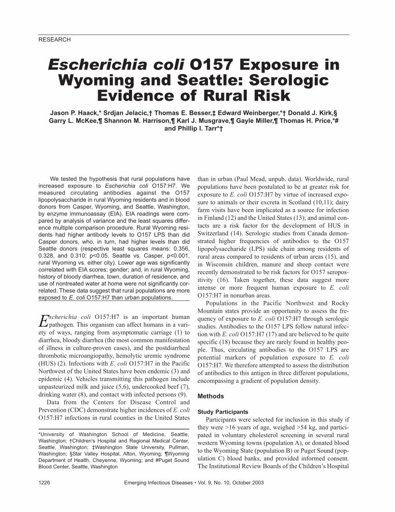
We tested the hypothesis that rural populations haveincreased exposure to Escherichia coli O157:H7. Wemeasured circulating antibodies against the O157lipopolysaccharide in rural Wyoming residents and in blooddonors from Casper, Wyoming, and Seattle, Washington,by enzyme immunoassay (EIA). EIA readings were com-pared by analysis of variance and the least squares differ-ence multiple comparison procedure. Rural Wyoming resi-dents had higher antibody levels to O157 LPS than didCasper donors, who, in turn, had higher levels than didSeattle donors (respective least squares means: 0.356,0.328, and 0.310; p<0.05, Seattle vs. Casper, p<0.001,rural Wyoming vs. either city). Lower age was significantlycorrelated with EIA scores; gender; and, in rural Wyoming,history of bloody diarrhea, town, duration of residence, anduse of nontreated water at home were not significantly cor-related. These data suggest that rural populations are moreexposed to E. coli O157:H7 than urban populations.
Escherichia coli O157:H7 is an important humanpathogen. This organism can affect humans in a vari-
ety of ways, ranging from asymptomatic carriage (1) todiarrhea, bloody diarrhea (the most common manifestationof illness in culture-proven cases), and the postdiarrhealthrombotic microangiopathy, hemolytic uremic syndrome(HUS) (2). Infections with E. coli O157:H7 in the PacificNorthwest of the United States have been endemic (3) andepidemic (4). Vehicles transmitting this pathogen includeunpasteurized milk and juice (5,6), undercooked beef (7),drinking water (8), and contact with infected persons (9).
Data from the Centers for Disease Control andPrevention (CDC) demonstrate higher incidences of E. coliO157:H7 infections in rural counties in the United States
than in urban (Paul Mead, unpub. data). Worldwide, ruralpopulations have been postulated to be at greater risk forexposure to E. coli O157:H7 by virtue of increased expo-sure to animals or their excreta in Scotland (10,11); dairyfarm visits have been implicated as a source for infectionin Finland (12) and the United States (13); and animal con-tacts are a risk factor for the development of HUS inSwitzerland (14). Serologic studies from Canada demon-strated higher frequencies of antibodies to the O157lipopolysaccharide (LPS) side chain among residents ofrural areas compared to residents of urban areas (15), andin Wisconsin children, manure and sheep contact wererecently demonstrated to be risk factors for O157 seropos-itivity (16). Taken together, these data suggest moreintense or more frequent human exposure to E. coliO157:H7 in nonurban areas.
Populations in the Pacific Northwest and RockyMountain states provide an opportunity to assess the fre-quency of exposure to E. coli O157:H7 through serologicstudies. Antibodies to the O157 LPS follow natural infec-tion with E. coli O157:H7 (17) and are believed to be quitespecific (18) because they are rarely found in healthy peo-ple. Thus, circulating antibodies to the O157 LPS arepotential markers of population exposure to E. coliO157:H7. We therefore attempted to assess the distributionof antibodies to this antigen in three different populations,encompassing a gradient of population density.
Methods
Study ParticipantsParticipants were selected for inclusion in this study if
they were >16 years of age, weighed >54 kg, and partici-pated in voluntary cholesterol screening in several ruralwestern Wyoming towns (population A), or donated bloodto the Wyoming State (population B) or Puget Sound (pop-ulation C) blood banks, and provided informed consent.The Institutional Review Boards of the Children’s Hospital
1226 Emerging Infectious Diseases • Vol. 9, No. 10, October 2003
RESEARCH
Escherichia coli O157 Exposure inWyoming and Seattle: Serologic
Evidence of Rural Risk Jason P. Haack,* Srdjan Jelacic,† Thomas E. Besser,‡ Edward Weinberger,*† Donald J. Kirk,§ Garry L. McKee,¶ Shannon M. Harrison,¶ Karl J. Musgrave,¶ Gayle Miller,¶ Thomas H. Price,*#
and Phillip I. Tarr*†
*University of Washington School of Medicine, Seattle,Washington; †Children’s Hospital and Regional Medical Center,Seattle, Washington; ‡Washington State University, Pullman,Washington; §Star Valley Hospital, Afton, Wyoming; ¶WyomingDepartment of Health, Cheyenne, Wyoming; and #Puget SoundBlood Center, Seattle, Washington
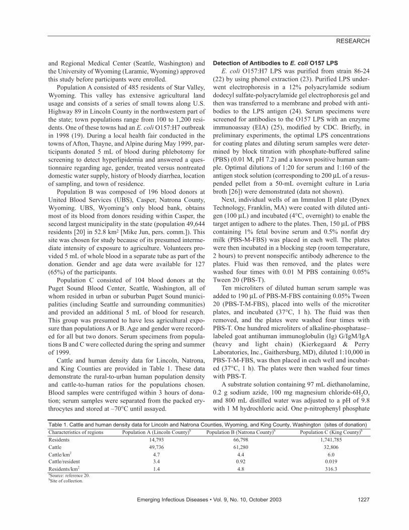
and Regional Medical Center (Seattle, Washington) andthe University of Wyoming (Laramie, Wyoming) approvedthis study before participants were enrolled.
Population A consisted of 485 residents of Star Valley,Wyoming. This valley has extensive agricultural landusage and consists of a series of small towns along U.S.Highway 89 in Lincoln County in the northwestern part ofthe state; town populations range from 100 to 1,200 resi-dents. One of these towns had an E. coli O157:H7 outbreakin 1998 (19). During a local health fair conducted in thetowns of Afton, Thayne, and Alpine during May 1999, par-ticipants donated 5 mL of blood during phlebotomy forscreening to detect hyperlipidemia and answered a ques-tionnaire regarding age, gender, treated versus nontreateddomestic water supply, history of bloody diarrhea, locationof sampling, and town of residence.
Population B was composed of 196 blood donors atUnited Blood Services (UBS), Casper, Natrona County,Wyoming. UBS, Wyoming’s only blood bank, obtainsmost of its blood from donors residing within Casper, thesecond largest municipality in the state (population 49,644residents [20] in 52.8 km2 [Mike Jun, pers. comm.]). Thissite was chosen for study because of its presumed interme-diate intensity of exposure to agriculture. Volunteers pro-vided 5 mL of whole blood in a separate tube as part of thedonation. Gender and age data were available for 127(65%) of the participants.
Population C consisted of 104 blood donors at thePuget Sound Blood Center, Seattle, Washington, all ofwhom resided in urban or suburban Puget Sound munici-palities (including Seattle and surrounding communities)and provided an additional 5 mL of blood for research.This group was presumed to have less agricultural expo-sure than populations A or B. Age and gender were record-ed for all but two donors. Serum specimens from popula-tions B and C were collected during the spring and summerof 1999.
Cattle and human density data for Lincoln, Natrona,and King Counties are provided in Table 1. These datademonstrate the rural-to-urban human population densityand cattle-to-human ratios for the populations chosen.Blood samples were centrifuged within 3 hours of dona-tion; serum samples were separated from the packed ery-throcytes and stored at –70°C until assayed.
Detection of Antibodies to E. coli O157 LPSE. coli O157:H7 LPS was purified from strain 86-24
(22) by using phenol extraction (23). Purified LPS under-went electrophoresis in a 12% polyacrylamide sodiumdodecyl sulfate-polyacrylamide gel electrophoresis gel andthen was transferred to a membrane and probed with anti-bodies to the LPS antigen (24). Serum specimens werescreened for antibodies to the O157 LPS with an enzymeimmunoassay (EIA) (25), modified by CDC. Briefly, inpreliminary experiments, the optimal LPS concentrationsfor coating plates and diluting serum samples were deter-mined by block titration with phosphate-buffered saline(PBS) (0.01 M, pH 7.2) and a known positive human sam-ple. Optimal dilutions of 1:20 for serum and 1:160 of theantigen stock solution (corresponding to 200 µL of a resus-pended pellet from a 50-mL overnight culture in Luriabroth [26]) were demonstrated (data not shown).
Next, individual wells of an Immulon II plate (DynexTechnology, Franklin, MA) were coated with diluted anti-gen (100 µL) and incubated (4°C, overnight) to enable thetarget antigen to adhere to the plates. Then, 150 µL of PBScontaining 1% fetal bovine serum and 0.5% nonfat drymilk (PBS-M-FBS) was placed in each well. The plateswere then incubated in a blocking step (room temperature,2 hours) to prevent nonspecific antibody adherence to theplates. Fluid was then removed, and the plates werewashed four times with 0.01 M PBS containing 0.05%Tween 20 (PBS-T).
Ten microliters of diluted human serum sample wasadded to 190 µL of PBS-M-FBS containing 0.05% Tween20 (PBS-T-M-FBS), placed into wells of the microtiterplates, and incubated (37°C, 1 h). The fluid was thenremoved, and the plates were washed four times withPBS-T. One hundred microliters of alkaline-phosphatase–labeled goat antihuman immunoglobulin (Ig) G/IgM/IgA(heavy and light chain) (Kierkegaard & PerryLaboratories, Inc., Gaithersburg, MD), diluted 1:10,000 inPBS-T-M-FBS, was then placed in each well and incubat-ed (37°C, 1 h). The plates were then washed four timeswith PBS-T.
A substrate solution containing 97 mL diethanolamine,0.2 g sodium azide, 100 mg magnesium chloride-6H2O,and 800 mL distilled water was adjusted to a pH of 9.8with 1 M hydrochloric acid. One p-nitrophenyl phosphate
Emerging Infectious Diseases • Vol. 9, No. 10, October 2003 1227
RESEARCH
Table 1. Cattle and human density data for Lincoln and Natrona Counties, Wyoming, and King County, Washington (sites of donation) Characteristics of regions Population A (Lincoln County)b Population B (Natrona County)b Population C (King County)b
Residents 14,793 66,798 1,741,785 Cattle 49,736 61,280 32,806 Cattle/km2
Cattle/resident 4.7 3.4
4.4 0.92
6.0 0.019
Residents/km2 1.4 4.8 316.3 aSource: reference 20. bSite of collection.
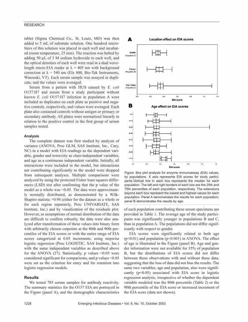
tablet (Sigma Chemical Co., St. Louis, MO) was thenadded to 5 mL of substrate solution. One hundred micro-liters of this solution was placed in each well and incubat-ed (room temperature, 25 min). The reaction was halted byadding 50 µL of 3 M sodium hydroxide to each well, andthe optical densities of each well were read in a dual wave-length micro-EIA reader at λ = 405 nm with backgroundcorrection at λ = 540 nm (Elx 800, Bio-Tek Instruments,Winooski, VT). Each serum sample was assayed in dupli-cate, and the values were averaged.
Serum from a patient with HUS caused by E. coliO157:H7 and serum from a study participant withoutknown E. coli O157:H7 infection in population A wereincluded as duplicates on each plate as positive and nega-tive controls, respectively, and values were averaged. Eachplate also contained controls without antigen or primary orsecondary antibody. All plates were normalized linearly inrelation to the positive control in the first group of serumsamples tested.
AnalysisThe complete dataset was first studied by analysis of
variance (ANOVA, Proc GLM, SAS Institute, Inc., Cary,NC) in a model with EIA readings as the dependent vari-able, gender and town/city as class-independent variables,and age as a continuous independent variable. Initially, allinteractions were included in the model, but interactionsnot contributing significantly to the model were droppedfrom subsequent analyses. Multiple comparisons wereanalyzed by using the protected Fisher least squares differ-ences (LSD) test after confirming that the p value of themodel as a whole was <0.05. The data were approximate-ly normally distributed, as demonstrated by a Wilk-Shapiro statistic >0.98 (either for the dataset as a whole orfor each region separately, Proc UNIVARIATE, SASInstitute, Inc.) and by visualization of the residuals plot.However, as assumptions of normal distribution of the dataare difficult to confirm robustly, the data were also ana-lyzed after transformation of these values into binary formwith arbitrarily chosen cutpoints at the 80th and 90th per-centiles of the EIA scores or with the entire range of EIAscores categorized at 0.05 increments, using stepwiselogistic regression (Proc LOGISTIC, SAS Institute, Inc.)with the same independent variables as described abovefor the ANOVA (27). Statistically, p values <0.05 wereconsidered significant for comparisons, and p values <0.05were set as the criterion for entry and for retention intologistic regression models.
ResultsWe tested 785 serum samples for antibody reactivity.
The summary statistics for the O157 EIA are portrayed inthe Figure (panel A), and the demographic characteristics
of each population contributing these serum specimens areprovided in Table 1. The average age of the study partici-pants was significantly younger in populations B and C,than in population A. The populations did not differ signif-icantly with respect to gender.
EIA scores were significantly related to both age(p<0.01) and population (p<0.001) in ANOVA. The effectof age is illustrated in the Figure (panel B). Age and gen-der information were not available for 35% of populationB, but the distributions of EIA scores did not differbetween those observations with and without these data,suggesting that the loss of data did not bias the results. Thesame two variables, age and population, also were signifi-cantly (p<0.05) associated with EIA score in logisticregression analysis, irrespective of whether the dependentvariable modeled was the 80th percentile (Table 2) or the90th percentile of the EIA score or increased increment ofthe EIA score (data not shown).
1228 Emerging Infectious Diseases • Vol. 9, No. 10, October 2003
RESEARCH
Figure. Box plot analysis for enzyme immunoassay (EIA) values,by populations. X axis represents EIA scores for study partici-pants.Vertical line in each box represents the median for eachpopulation. The left and right borders of each box are the 25th and75th percentiles of each population, respectively. The extensionsbeyond each box represent the lowest and highest values for eachpopulation. Panel A demonstrates the results for each population;panel B demonstrates the results by age.
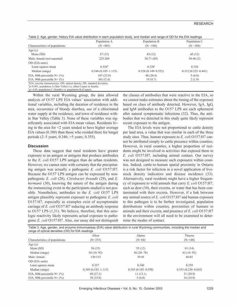
Within the rural Wyoming group, the data allowedanalysis of O157 LPS EIA values’ association with addi-tional variables, including the duration of residence in thearea, occurrence of bloody diarrhea, use of a chlorinatedwater supply at the residence, and town of residence with-in Star Valley (Table 3). None of these variables was sig-nificantly associated with EIA mean values. Residents liv-ing in the area for <2 years tended to have higher averageEIA values (0.380) than those who resided there for longerperiods (2–5 years, 0.346; >5 years, 0.355).
DiscussionThese data suggest that rural residents have greater
exposure to an antigen or antigens that produce antibodiesto the E. coli O157 LPS antigen than do urban residents.However, we cannot state with certainty that the precipitat-ing antigen was actually a pathogenic E. coli O157:H7.Because the O157 LPS antigen can be expressed by non-pathogenic E. coli (28), Citrobacter freundii (29), and E.hermanii (30), knowing the nature of the antigen duringthe immunizing event in the participants studied is not pos-sible. Nonetheless, antibodies to the E. coli O157 LPSantigen plausibly represent exposure to pathogenic E. coliO157:H7, especially as examples exist of asymptomaticcarriage of E. coli O157:H7 inducing an antibody responseto O157 LPS (1,31). We believe, therefore, that this sero-logic reactivity likely represents actual exposure to patho-genic E. coli O157:H7. Also, our assay did not distinguish
the classes of antibodies that were reactive in the EIA, sowe cannot make estimates about the timing of the exposurebased on class of antibody detected. However, IgA, IgG,and IgM antibodies to the O157 LPS are each ephemeralafter natural symptomatic infections (32). Thus, the anti-bodies that we detected in this study quite likely representrecent exposure to the antigen.
The EIA levels were not proportional to cattle densityper land area, a value that was similar in each of the threestudy sites. Thus, human exposure to E. coli O157:H7 can-not be attributed simply to cattle presence within counties.However, in rural counties, a higher proportion of resi-dents might be involved in activities that exposed them toE. coli O157:H7, including animal contact. Our surveywas not designed to measure such exposures within coun-ties. Indeed, cattle-to-human spatial proximity in Ontariois a risk factor for infection in a novel application of live-stock density indicators and disease incidence (33).Alternatively, rural residents might have a higher frequen-cy of exposure to wild animals that carry E. coli O157:H7,such as deer (34), their excreta, or water that has been con-taminated with their excreta. However, if a link betweenany animal source of E. coli O157:H7 and human exposureto this pathogen is to be further investigated, populationdistributions within counties, proximities of humans toanimals and their excreta, and presence of E. coli O157:H7in the environment will all need to be examined to deter-mine the modes of contact.
Emerging Infectious Diseases • Vol. 9, No. 10, October 2003 1229
RESEARCH
Table 2. Age, gender, history EIA value distribution in each population study, and median and range of OD for the EIA readings Population A Population B Population C
Characteristics of populations (N =485) (N =196) (N =104) Age (y)
Mean (SD) 57 (15) 45 (12) 45 (12) Male: female (not reported) 225:260 56:71 (69) 56:46 (2) OD (EIA units)
Least squares mean 0.356b 0.328c 0.310 Median (range) 0.348 (0.185–1.115) 0.328 (0.149–0.522) 0.312 (0.222–0.441)
EIA, 80th percentile N+ (%) EIA, 90th percentile N+ (%)
107 (22.0) 60 (12.4)
40 (20.4) 19 (9.7)
5 (4.8) 2 (1.9)
aEIA, enzyme immunoassay; OD, optical density; SD, standard deviation. bp<0.001, population A (Star Valley) vs. either Casper or Seattle. cp< 0.05, population C (Seattle) vs. population B (Casper).
Table 3. Age, gender, and enzyme immunoassay (EIA) value distribution in rural Wyoming communities, including the median and range of optical densities (OD) for EIA readings
Afton Alpine Thayne Characteristics of populations (N=253) (N=84) (N=148) Age (y) Mean (SD) 56 (15) 55 (12) 61 (14) Median (range) 56 (16–92) 56 (20–78) 63 (16–92) Male: female 120:133 39:45 66:82 OD (EIA units) Least squares mean 0.357 0.340 0.359 Median (range) 0.349 (0.192–1.115) 0.343 (0.185–0.550) 0.353 (0.230–0.643) EIA, 80th percentile N+ (%) EIA, 90th percentile N+ (%)
69 (27.3) 38 (15.0)
11 (13.1) 5 (6.0)
31 (20.9) 16 (10.8)
Our data differ from results of previous studies in termsof statistical analysis and also because we included a gra-dient of population density. Specifically, our principalanalysis did not require assignment of persons to seropos-itive or seronegative status categories. While some relationmight exist between percentage of persons who are desig-nated as having reactive or nonreactive status on the basisof cutoffs and the comparative distribution of serologicreactivities in populations, we believe that, when examin-ing continuous variables, a test that compares values ascontinuous measurements (such as analysis of variance)provides more information and is less arbitrary than theassignment of categorical positives and negatives. We did,however, also examine proportions above two cutpoints,and the same trends were noted. For unknown reasons, weidentified higher EIA scores in younger study participants,whereas in Ontario, IgG antibodies to the O157 LPS werehighest in participants in their fifth decade of life (15).Comparing our age-related EIA scores to those reportedrecently from Wisconsin, where older children had higherseropositive rates, is not possible because the latter studyfocused on a child population and did not evaluate olderWisconsin residents (16).
We caution against interpreting our data to mean thatrural populations are immune to, and thereby protectedfrom, E. coli O157:H7 infections. Many study participantshad antibody levels that were probably too low to conferprotection, even if one assumes that antibodies to this anti-gen protect against infection. In this regard, an E. coliO157:H7 infection, which almost always induces a briskand high-titer humoral immune response to the O157 LPSantigen, may not confer a permanently protectiveresponse, as evidenced by documentations of recurrentinfections (35,36). However, in a recent E. coli O157:H7outbreak in the Star Valley, a higher frequency of antibodylevels to the O157 LPS among the resident population wasproposed as the reason for a lower attack rate among resi-dents than among visitors (19). We also urge against gen-eralizing the trend observed in this study, which is derivedfrom the analysis of only three populations, to all ruralpopulations, without additional, more widespread, studiesbecause the populations studied might not be representa-tive. Nonetheless, our data are consistent with the hypoth-esis that rural residence carries with it a greater risk forexposure to E. coli O157:H7 than does urban residence.
In summary, we have identified an age-dependent, gen-der-independent, risk for probable exposure to E. coliO157:H7 in persons living in rural communities. Thisexposure frequency is plausibly environmental, rather thanfoodborne, in origin because food is distributed widelythroughout North America. However, the possibility existsthat particular food consumption practices, such as drink-ing raw milk in rural communities, as has been noted in the
United Kingdom (37,38), might have been responsible forthis exposure. We also cannot exclude the possibility thatthe differences observed relate to the nature of the partici-pants studied. That is, donors to blood banks might havedifferent exposures than participants in lipid screenings athealth fairs. Future studies should attempt to identify thepoints of exposure to this antigen, confirm that E. coliO157:H7 is, indeed, the source of the inciting antigen, and,if it is, minimize human contact with this pathogen.
AcknowledgmentsWe thank Jennifer L. Falkenhagen for assistance in manu-
script preparation, Paul Mead for sharing unpublished data withus, and William Bibb for advice concerning enzyme immunoas-say protocols.
The Escherichia coli Gift Fund at the Children’s Hospitaland Regional Medical Center and NIH grant DK52081 supportthis project.
Dr. Haack is a resident at the Mayo Clinic, Rochester,Minnesota. This work was performed as a thesis project at theUniversity of Washington School of Medicine.
References
1. Wilson JB, Clarke RC, Renwick SA, Rahn K, Johnson RP, KarmaliMA, et al. Vero cytotoxigenic Escherichia coli infection in dairy farmfamilies. J Infect Dis 1996;174:1021–7.
2. Tarr PI. Escherichia coli O157:H7: clinical, diagnostic, and epidemi-ological aspects of human infection. Clin Infect Dis 1995;20:1–8.
3. MacDonald KL, O’Leary MJ, Cohen ML, Norris P, Wells JG, Noll E,et al. Escherichia coli O157:H7, an emerging gastrointestinalpathogen. Results of a one-year, prospective, population-based study.JAMA 1988;259:3567–70.
4. Bell BP, Goldoft M, Griffin PM, Davis MA, Gordon DC, Tarr PI, etal. A multistate outbreak of Escherichia coli O157:H7–associatedbloody diarrhea and hemolytic uremic syndrome from hamburgers.The Washington experience. JAMA 1994;272:1349–53.
5. Keene WE, Hedberg K, Herriott DE, Hancock DD, McKay RW,Barrett TJ, et al. A prolonged outbreak of Escherichia coli O157:H7infections caused by commercially distributed raw milk. J Infect Dis1997;176:815–8.
6. Besser RE, Lett SM, Weber JT, Doyle MP, Barrett TJ, Wells JG, et al.An outbreak of diarrhea and hemolytic uremic syndrome fromEscherichia coli O157:H7 in fresh-pressed apple cider. JAMA1993;269:2217–20.
7. Riley LW, Remis RS, Helgerson SD, McGee HB, Wells JG, DavisBR, et al. Hemorrhagic colitis associated with a rare Escherichia coliserotype. N Engl J Med 1983;308:681–5.
8. Swerdlow DL, Woodruff BA, Brady RC, Greiffin PM, Tippen S,Donnell HD Jr, et al. A waterborne outbreak in Missouri ofEscherichia coli O157:H7 associated with bloody diarrhea and death.Ann Intern Med 1992;117:812–9.
9. Belongia EA, Osterholm MT, Soler JT, Ammend DA, Braun JE,MacDonald KL. Transmission of Escherichia coli 0157:H7 infectionin Minnesota child day-care facilities. JAMA 1993;269:883–8.
10. Locking ME, O’Brien SJ, Reilly WJ, Wright EM, Campbell DM,Coia JE, et al. Risk factors for sporadic cases of Escherichia coliO157 infection: the importance of contact with animal excreta.Epidemiol Infect 2001;127:215–20.
1230 Emerging Infectious Diseases • Vol. 9, No. 10, October 2003
RESEARCH
Emerging Infectious Diseases • Vol. 9, No. 10, October 2003 1231
RESEARCH
11. O’Brien SJ, Adak GK, Gilham C. Contact with farming environmentas a major risk factor for Shiga toxin (Vero cytotoxin–producingEscherichia coli O157 infection in humans. Emerg Infect Dis2001;7:1049–51.
12. Lahti E, Eklund M, Ruutu P, Siitonen A, Rantala L, Nuorti P, et al.Use of phenotyping and genotyping to verify transmission ofEscherichia coli O157:H7 from dairy farms. Eur J Clin MicrobiolInfect Dis 2002;21:189–95.
13. Crump JA, Sulka AC, Langer AJ, Schaben C, Crielly AS, Gage R, etal. An outbreak of Escherichia coli O157:H7 infections among visi-tors to a dairy farm. N Engl J Med 2002;347:555–60.
14. Kernland KH, Laux-End R, Truttmann AC, Reymond D, BianchettiMG. [How is hemolytic-uremic syndrome in childhood acquired inSwitzerland?]. [In German] Schweiz Med Wochenschr1997;127:1229–33.
15. Reymond D, Johnson RP, Karmali MA, Petric M, Winkler M,Johnson S, et al. Neutralizing antibodies to Escherichia coli Verocytotoxin 1 and antibodies to O157 lipopolysaccharide in healthyfarm family members and urban residents. J Clin Microbiol1996;34:2053–7.
16. Belongia EA, Chyou PH, Greenlee RT, Perez-Perez G, Bibb WF, DeVries EO. Diarrhea incidence and farm-related risk factors forEscherichia coli 0157:H7 and Campylobacter jejuni antibodiesamong rural children. J Infect Dis 2003;187:1460–8.
17. Banatvala N, Griffin PM, Greene KD, Barrett TJ, Bibb WF, GreenJH, et al. The United States National Prospective Hemolytic UremicSyndrome Study: microbiologic, serologic, clinical, and epidemio-logic findings. J Infect Dis 2001;183:1063–70.
18. Reida P, Wolff M, Pohls HW, Kuhlmann W, Lehmacher A, Aleksic S,et al. An outbreak due to enterohaemorrhagic Escherichia coliO157:H7 in a children day care centre characterized by person-to-person transmission and environmental contamination. ZentralblBakteriol 1994;281:534–43.
19. Olsen SJ, Miller G, Kennedy M, Higgins C, Walford J, McKee G, etal. A waterborne outbreak of Escherichia coli O157:H7 infectionsand hemolytic uremic syndrome: implications for rural water sys-tems. Emerg Infect Dis 2002;8:370–5.
20. U.S. Census Bureau and County Quickfacts. Available from: URL:http://quickfacts.census.gov/qfd/index.html
21. U.S. Department of Agriculture National Agricultural StatisticsService Census of Agriculture. Available from: URL: www.nass.usda.gov/census/
22. Tarr PI, Neill MA, Clausen CR, Newland JW, Neill RJ, Moseley SL.Genotypic variation in pathogenic Escherichia coli O157:H7 isolatedfrom patients in Washington, 1984–1987. J Infect Dis1989;159:344–7.
23. Inzana TJ, Pichichero ME. Lipopolysaccharide subtypes ofHaemophilus influenzae type b from an outbreak of invasive disease.J Clin Microbiol 1984;20:145–50.
24. Bilge SS, Vary JC Jr, Dowell SF, Tarr PI. Role of the Escherichia coliO157:H7 O side chain in adherence and analysis of an rfb locus.Infect Immun 1996;64:4795–801.
25. Toth I, Barrett TJ, Cohen ML, Rumschlag HS, Green JH, WachsmuthIK. Enzyme-linked immunosorbent assay for products of the 60-megadalton plasmid of Escherichia coli serotype O157:H7. J ClinMicrobiol 1991;29:1016–9.
26. Sambrook J, Fritsch EF, Maniatis T. Molecular cloning: a laboratorymanual. 2nd ed. Cold Spring Harbor (NY): Cold Spring HarborLaboratory Press; 1989.
27. Glantz SS, Glantz SA. Primer of biostatistics. 4th ed. New York:McGraw-Hill Health Professions Division; 1997.
28. Tarr PI, Schoening LM, Yea YL, Ward TR, Jelacic S, Whittam TS.Acquisition of the rfb-gnd cluster in evolution of Escherichia coliO55 and O157. J Bacteriol 2000;182:6183–91.
29. Bettelheim KA, Evangelidis H, Pearce JL, Sowers E, Strockbine NA.Isolation of a Citrobacter freundii strain which carries theEscherichia coli O157 antigen. J Clin Microbiol 1993;31:760–1.
30. Chapman PA. Evaluation of commercial latex slide test for identify-ing Escherichia coli O157. J Clin Pathol 1989;42:1109–10.
31. Bielaszewska M, Janda J, Blahova K, Minarikova H, Jokova E,Karmali MA, et al. Human Escherichia coli O157:H7 infection asso-ciated with the consumption of unpasteurized goat’s milk. EpidemiolInfect 1997;119:299–305.
32. Ludwig K, Bitzan M, Bobrowski C, Muller-Wiefel DE. Escherichiacoli O157 fails to induce a long-lasting lipopolysaccharide-specific,measurable humoral immune response in children with hemolytic-uremic syndrome. J Infect Dis 2002;186:566–9.
33. Valcour JE, Michel P, McEwen SA, Wilson JB. Associations betweenindicators of livestock farming intensity and incidence of humanshiga toxin–producing Escherichia coli infection. Emerg Infect Dis2002;8:252–7.
34. Rice DH, Hancock DD, Besser TE. Verotoxigenic E. coli O157colonisation of wild deer and range cattle. Vet Rec 1995;137:524.
35. Siegler RL, Griffin PM, Barrett TJ, Strockbine NA. Recurrenthemolytic uremic syndrome secondary to Escherichia coli O157:H7infection. Pediatrics 1993;91:666–8.
36. Robson WL, Leung AK, Miller-Hughes DJ. Recurrent hemorrhagiccolitis caused by Escherichia coli O157:H7. Pediatr Infect Dis J1993;12:699–701.
37. Sibbald CJ, Sharp JC. Campylobacter infection in urban and ruralpopulations in Scotland. J Hyg (Lond) 1985;95:87–93.
38. Barrett NJ. Communicable disease associated with milk and dairyproducts in England and Wales: 1983–1984. J Infect 1986;12:265–72.
Address for correspondence: P. I. Tarr, Edward Mallinckrodt Departmentof Pediatrics and Department of Molecular Microbiology, WashingtonUniversity School of Medicine, and the Division of Gastroenterology, St.Louis Children’s Hospital, St. Louis, MO 63110, USA; fax: 314-286-2911; email: [email protected]
The Walkerton, Canada, waterborne outbreak of 2000resulted from entry of Escherichia coli O157:H7 andCampylobacter spp. from neighboring farms into the townwater supply. Isolates of Campylobacter jejuni andCampylobacter coli obtained from outbreak investigationswere characterized by phenotypic and genotypic methods,including heat-stable and heat-labile serotyping, phage typ-ing, biotyping, fla–restriction fragment length polymorphism(RFLP) typing, and pulsed-field gel electrophoresis. Twomain outbreak strains were identified on the basis of heat-stable serotyping and fla-RFLP typing. These strains pro-duced a limited number of types when tested by othermethods. Isolates with types indistinguishable from, or sim-ilar to, the outbreak types were found only on one farm nearthe town of Walkerton, whereas cattle from other farms car-ried a variety of Campylobacter strains with different typecharacteristics. Results of these analyses confirmed resultsfrom epidemiologic studies and the utility of using severaldifferent typing and subtyping methods for completely char-acterizing bacterial populations.
An outbreak of Campylobacter jejuni in a farming com-munity in southern Ontario, Canada, in 1985 resulted
from contamination of well water caused by spring run-offand heavy rains (1). In May 2000, a second waterborneoutbreak of Escherichia coli O157:H7 and Campylobacteroccurred in Bruce County, Ontario. Well water serving thetown of Walkerton was contaminated by surface water car-rying livestock waste immediately after heavy rains (2,3).A detailed microbiologic and epidemiologic analysis of themost recent outbreak may provide insights that could helpmake this type of outbreak less frequent.
Most sporadic cases of campylobacteriosis are associat-ed with preparation or consumption of poultry products
(4). Outbreaks have been associated with consumption ofunpasteurized milk or unchlorinated water (5). An estimat-ed 20% of cases of illness caused by C. jejuni are due tovehicles of infection other than food, including water (6).Waterborne outbreaks of Campylobacter tend to occur inspring or early fall, an association attributed to seasonalityof surface water contamination and infection in cattleherds (5). Contaminated water sources have been implicat-ed in outbreaks involving E. coli O157:H7 andCampylobacter together in Scotland (7) and in New YorkState (8,9). The former outbreak resulted from sewagecontamination of the water supply of a small village inFife, Scotland. The latter outbreak was associated withcontamination of wells at a state fair (10). Excrement frombirds and animals, including cattle, has been shown to con-taminate surface water supplies used by humans infectedwith Campylobacter (9).
Campylobacter spp. have been found to cause water-borne outbreaks worldwide; such outbreaks are a particu-lar problem in Scandinavian countries where many peopledrink untreated water from streams and other sources (11).Untreated surface water has also been implicated inCampylobacter outbreaks in New Zealand (12,13),Finland (14), England, Wales (15,16), Australia (17), andthe United States (18). In Canada, outbreaks have beenrarely detected and have been associated with contamina-tion of surface water (19,20) and consumption of unpas-teurized milk (21).
In the United States, disease caused by C. jejuni or C.coli has been estimated to affect 7 million people annual-ly, causing 110–511 deaths and costing $1.2–$6 billion(22). These organisms are responsible for 17% of all hos-pitalizations related to foodborne illness in the UnitedStates, and although associated with a much lower case-fatality rate than Salmonella spp. and E. coli O157:H7,they account for 5% of food-related deaths (6). Although
1232 Emerging Infectious Diseases • Vol. 9, No. 10, October 2003
RESEARCH
Characterization of WaterborneOutbreak–associatedCampylobacter jejuni,
Walkerton, Ontario Clifford G. Clark,* Lawrence Price,* Rafiq Ahmed,* David L. Woodward,* Pasquale L. Melito,*
Frank G. Rodgers,* Frances Jamieson,† Bruce Ciebin,† Aimin Li,† and Andrea Ellis‡
*Health Canada, Winnipeg, Manitoba, Canada; †Ontario Ministryof Health, Toronto, Ontario, Canada; and ‡Health Canada,Guelph, Ontario, Canada
the incidence of Campylobacter infections generallyappears to be higher in industrialized than in developingnations, some evidence exists that campylobacteriosis maybe important from a social and economic point of view(23).
Epidemiologic and microbiologic analyses were under-taken to better understand the circumstances leading to theWalkerton outbreak. C. jejuni was isolated from patientsassociated with the outbreak, and C. jejuni and C. coliwere isolated from animals and animal manure on farmslocated near the town wells. This work summarizes thephenotypic and genotypic typing results for isolates asso-ciated with the outbreak.
Materials and Methods
Epidemiologic Investigations Identification of the outbreak, definition of cases, and
the results of epidemiologic descriptive and cross-section-al studies have been described (2,3). Isolates from personswho did not meet all requirements for the case definition,but who resided in southwestern Ontario and became illduring the period of the outbreak, were also sent to theNational Laboratory for Enteric Pathogens (NLEP),Winnipeg, Manitoba, for further analysis. A detaileddescription of the epidemiologic investigations is in prepa-ration.
Environmental Specimens Environmental studies related to the outbreak have
been described previously (2,3). Initial investigations iden-tified 13 livestock farms within a 4-km radius of the threewells serving the town of Walkerton. From May 30 to June13, 2000, a minimum of five manure samples per farmwere obtained and tested for human enteric pathogens.Bovine rectal swabs and manure were collected from asubset of these farms in follow-up studies on June 13. Allspecimens were screened for Campylobacter spp., and iso-lates were forwarded to NLEP for further testing.
Processing of Specimens Patient stool specimens were collected into Cary-Blair
transport medium and sent to the Central Public HealthLaboratory, Ministry of Health and Long-Term Care,Toronto, Ontario. Specimens from animal manure were col-lected aseptically in sterile bags and forwarded to the samelaboratory. Stools (approximately 1 g) from both sourceswere added into liquid enrichment medium (LEM) ordirectly onto charcoal-selective medium (CSM) and incu-bated at 42°C in a microaerobic atmosphere (5% O2, 10%CO2, 85% N2) for 24 h and 48 h. Cultures in LEM weresubcultured to CSM and incubated as indicated above.Isolates submitted to the NLEP were routinely cultured on
Mueller-Hinton agar (Oxoid Ltd., London, England) con-taining 10% sheep blood and stored frozen at –70°C inglycerol peptone water. Isolates were routinely incubated ateither 37°C or 42°C in a microaerobic atmosphere.
Identification of IsolatesColonies suspected of being Campylobacter were
Gram stained and tested for oxidase, catalase, and hippu-rate hydrolysis. Presumptive identification of C. coli wasachieved by the indoxyl acetate test and by determiningsusceptibility to nalidixic acid (30-µg disk) and cephaloth-in (30-µg disk). Biotyping was performed as described byLior (24). In addition to biotyping, the polymerase chainreaction–restriction fragment length polymorphism (PCR-RFLP) identification scheme described by Marshall et al.(25) was used to confirm species identification. Primersspecific to C. jejuni (25) and to C. coli (26) were used toconfirm the identity of any “hippurate-negative” C. jejuni.Any isolates that were hippurate-negative in the tube testbut positive by PCR for the hippuricase gene and negativeby PCR for the aspartokinase gene associated with C. coliwere confirmed by retesting by both methods.
Strain SubtypingHeat-labile (HL) serotyping was performed by the
method of Lior et al. (27). HS serotyping, in which passivehemagglutination was used to detect heat-stable antigens,was performed by the method of Penner and Hennessy(28). Phage typing of isolates was performed as describedby Frost et al. (29). Fla-RFLP typing was performed by themethod of Nachamkin et al. (30). Numerical type designa-tions from 1–101 were assigned at the NLEP. PFGE wasdone according to the method of Ribot et al. (31) withSmaI and KpnI. The isolates tested by PFGE were the firsthuman and animal isolates to be sent to the NLEP, and test-ing continued until type characteristics of outbreak strainswere identified and the epidemiologic designations ofpatients involved in the outbreak were confirmed microbi-ologically. After this, only biotyping, serotyping, andphage typing were used to characterize outbreak strains.Fla-RFLP typing was implemented some time after theoutbreak in an attempt to determine the effectiveness ofthis method for subtyping outbreak strains. All isolatestested by PFGE, and a random selection of isolates nottested by PFGE, were subject to fla-RFLP analysis.
ResultsA detailed description of the epidemiologic and envi-
ronmental investigations is the subject of a manuscript inpreparation (A. Ellis, pers. comm.). A total of 532 humanstool specimens were tested for Campylobacter spp. Stoolsfrom 116 persons were positive for the organism, and 11 ofthese were also positive for E. coli O157:H7. Of these 116
Emerging Infectious Diseases • Vol. 9, No. 10, October 2003 1233
RESEARCH
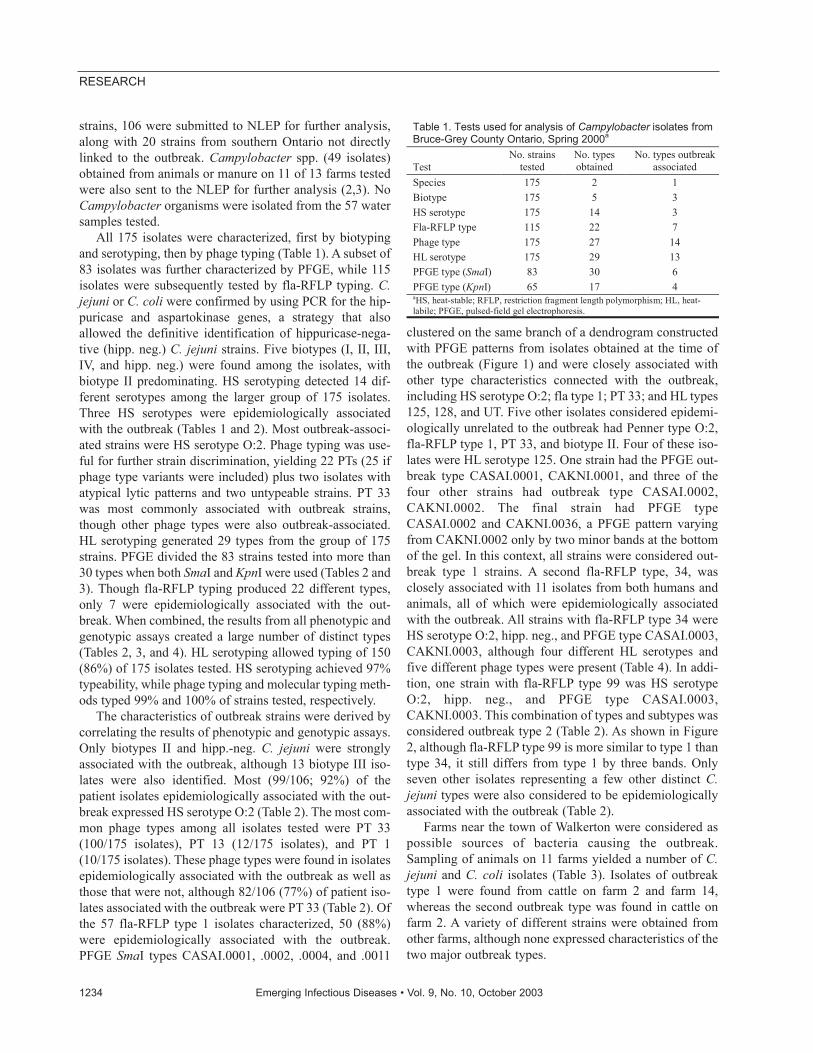
strains, 106 were submitted to NLEP for further analysis,along with 20 strains from southern Ontario not directlylinked to the outbreak. Campylobacter spp. (49 isolates)obtained from animals or manure on 11 of 13 farms testedwere also sent to the NLEP for further analysis (2,3). NoCampylobacter organisms were isolated from the 57 watersamples tested.
All 175 isolates were characterized, first by biotypingand serotyping, then by phage typing (Table 1). A subset of83 isolates was further characterized by PFGE, while 115isolates were subsequently tested by fla-RFLP typing. C.jejuni or C. coli were confirmed by using PCR for the hip-puricase and aspartokinase genes, a strategy that alsoallowed the definitive identification of hippuricase-nega-tive (hipp. neg.) C. jejuni strains. Five biotypes (I, II, III,IV, and hipp. neg.) were found among the isolates, withbiotype II predominating. HS serotyping detected 14 dif-ferent serotypes among the larger group of 175 isolates.Three HS serotypes were epidemiologically associatedwith the outbreak (Tables 1 and 2). Most outbreak-associ-ated strains were HS serotype O:2. Phage typing was use-ful for further strain discrimination, yielding 22 PTs (25 ifphage type variants were included) plus two isolates withatypical lytic patterns and two untypeable strains. PT 33was most commonly associated with outbreak strains,though other phage types were also outbreak-associated.HL serotyping generated 29 types from the group of 175strains. PFGE divided the 83 strains tested into more than30 types when both SmaI and KpnI were used (Tables 2 and3). Though fla-RFLP typing produced 22 different types,only 7 were epidemiologically associated with the out-break. When combined, the results from all phenotypic andgenotypic assays created a large number of distinct types(Tables 2, 3, and 4). HL serotyping allowed typing of 150(86%) of 175 isolates tested. HS serotyping achieved 97%typeability, while phage typing and molecular typing meth-ods typed 99% and 100% of strains tested, respectively.
The characteristics of outbreak strains were derived bycorrelating the results of phenotypic and genotypic assays.Only biotypes II and hipp.-neg. C. jejuni were stronglyassociated with the outbreak, although 13 biotype III iso-lates were also identified. Most (99/106; 92%) of thepatient isolates epidemiologically associated with the out-break expressed HS serotype O:2 (Table 2). The most com-mon phage types among all isolates tested were PT 33(100/175 isolates), PT 13 (12/175 isolates), and PT 1(10/175 isolates). These phage types were found in isolatesepidemiologically associated with the outbreak as well asthose that were not, although 82/106 (77%) of patient iso-lates associated with the outbreak were PT 33 (Table 2). Ofthe 57 fla-RFLP type 1 isolates characterized, 50 (88%)were epidemiologically associated with the outbreak.PFGE SmaI types CASAI.0001, .0002, .0004, and .0011
clustered on the same branch of a dendrogram constructedwith PFGE patterns from isolates obtained at the time ofthe outbreak (Figure 1) and were closely associated withother type characteristics connected with the outbreak,including HS serotype O:2; fla type 1; PT 33; and HL types125, 128, and UT. Five other isolates considered epidemi-ologically unrelated to the outbreak had Penner type O:2,fla-RFLP type 1, PT 33, and biotype II. Four of these iso-lates were HL serotype 125. One strain had the PFGE out-break type CASAI.0001, CAKNI.0001, and three of thefour other strains had outbreak type CASAI.0002,CAKNI.0002. The final strain had PFGE typeCASAI.0002 and CAKNI.0036, a PFGE pattern varyingfrom CAKNI.0002 only by two minor bands at the bottomof the gel. In this context, all strains were considered out-break type 1 strains. A second fla-RFLP type, 34, wasclosely associated with 11 isolates from both humans andanimals, all of which were epidemiologically associatedwith the outbreak. All strains with fla-RFLP type 34 wereHS serotype O:2, hipp. neg., and PFGE type CASAI.0003,CAKNI.0003, although four different HL serotypes andfive different phage types were present (Table 4). In addi-tion, one strain with fla-RFLP type 99 was HS serotypeO:2, hipp. neg., and PFGE type CASAI.0003,CAKNI.0003. This combination of types and subtypes wasconsidered outbreak type 2 (Table 2). As shown in Figure2, although fla-RFLP type 99 is more similar to type 1 thantype 34, it still differs from type 1 by three bands. Onlyseven other isolates representing a few other distinct C.jejuni types were also considered to be epidemiologicallyassociated with the outbreak (Table 2).
Farms near the town of Walkerton were considered aspossible sources of bacteria causing the outbreak.Sampling of animals on 11 farms yielded a number of C.jejuni and C. coli isolates (Table 3). Isolates of outbreaktype 1 were found from cattle on farm 2 and farm 14,whereas the second outbreak type was found in cattle onfarm 2. A variety of different strains were obtained fromother farms, although none expressed characteristics of thetwo major outbreak types.
1234 Emerging Infectious Diseases • Vol. 9, No. 10, October 2003
RESEARCH
Table 1. Tests used for analysis of Campylobacter isolates from Bruce-Grey County Ontario, Spring 2000a
Test No. strains
tested No. types obtained
No. types outbreak associated
Species 175 2 1 Biotype 175 5 3 HS serotype 175 14 3 Fla-RFLP type 115 22 7 Phage type 175 27 14 HL serotype 175 29 13 PFGE type (SmaI) 83 30 6 PFGE type (KpnI) 65 17 4 aHS, heat-stable; RFLP, restriction fragment length polymorphism; HL, heat-labile; PFGE, pulsed-field gel electrophoresis.
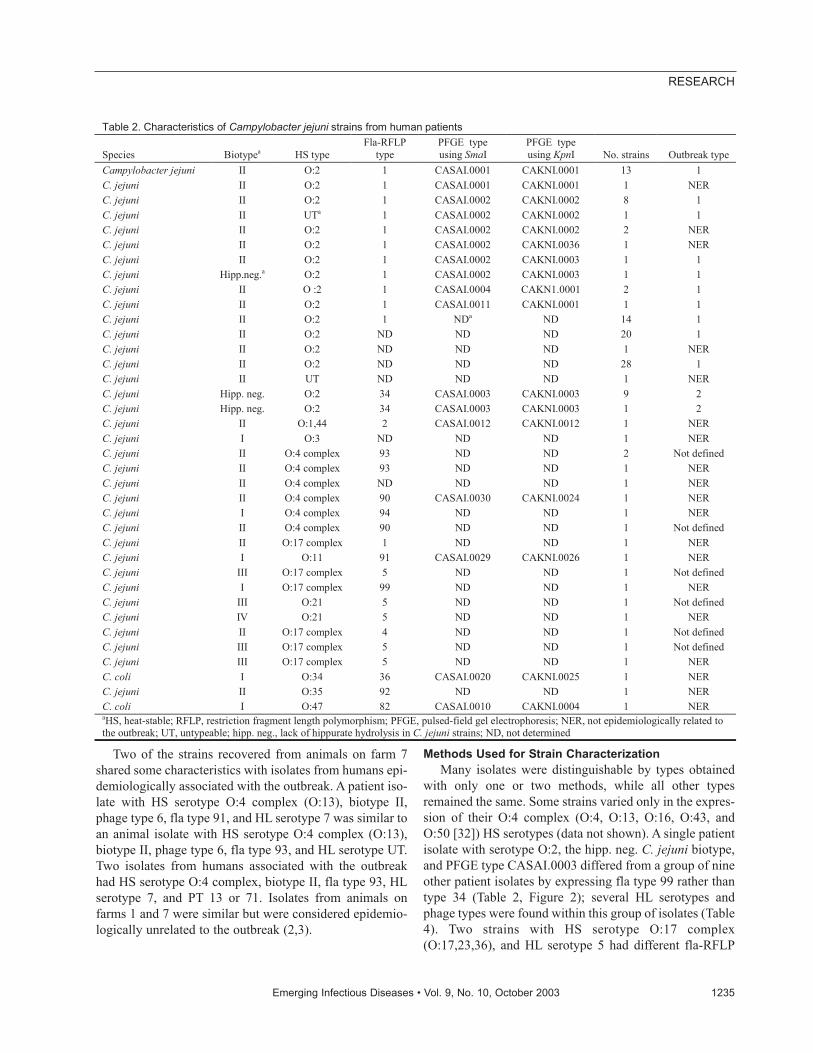
Two of the strains recovered from animals on farm 7shared some characteristics with isolates from humans epi-demiologically associated with the outbreak. A patient iso-late with HS serotype O:4 complex (O:13), biotype II,phage type 6, fla type 91, and HL serotype 7 was similar toan animal isolate with HS serotype O:4 complex (O:13),biotype II, phage type 6, fla type 93, and HL serotype UT.Two isolates from humans associated with the outbreakhad HS serotype O:4 complex, biotype II, fla type 93, HLserotype 7, and PT 13 or 71. Isolates from animals onfarms 1 and 7 were similar but were considered epidemio-logically unrelated to the outbreak (2,3).
Methods Used for Strain Characterization Many isolates were distinguishable by types obtained
with only one or two methods, while all other typesremained the same. Some strains varied only in the expres-sion of their O:4 complex (O:4, O:13, O:16, O:43, andO:50 [32]) HS serotypes (data not shown). A single patientisolate with serotype O:2, the hipp. neg. C. jejuni biotype,and PFGE type CASAI.0003 differed from a group of nineother patient isolates by expressing fla type 99 rather thantype 34 (Table 2, Figure 2); several HL serotypes andphage types were found within this group of isolates (Table4). Two strains with HS serotype O:17 complex(O:17,23,36), and HL serotype 5 had different fla-RFLP
Emerging Infectious Diseases • Vol. 9, No. 10, October 2003 1235
RESEARCH
Table 2. Characteristics of Campylobacter jejuni strains from human patients
Species Biotypea HS type Fla-RFLP
type PFGE type using SmaI
PFGE type using KpnI No. strains Outbreak type
Campylobacter jejuni II O:2 1 CASAI.0001 CAKNI.0001 13 1 C. jejuni II O:2 1 CASAI.0001 CAKNI.0001 1 NER C. jejuni II O:2 1 CASAI.0002 CAKNI.0002 8 1 C. jejuni II UTa 1 CASAI.0002 CAKNI.0002 1 1 C. jejuni II O:2 1 CASAI.0002 CAKNI.0002 2 NER C. jejuni II O:2 1 CASAI.0002 CAKNI.0036 1 NER C. jejuni II O:2 1 CASAI.0002 CAKNI.0003 1 1 C. jejuni Hipp.neg.a O:2 1 CASAI.0002 CAKNI.0003 1 1 C. jejuni II O :2 1 CASAI.0004 CAKN1.0001 2 1 C. jejuni II O:2 1 CASAI.0011 CAKNI.0001 1 1 C. jejuni II O:2 1 NDa ND 14 1 C. jejuni II O:2 ND ND ND 20 1 C. jejuni II O:2 ND ND ND 1 NER C. jejuni II O:2 ND ND ND 28 1 C. jejuni II UT ND ND ND 1 NER C. jejuni Hipp. neg. O:2 34 CASAI.0003 CAKNI.0003 9 2 C. jejuni Hipp. neg. O:2 34 CASAI.0003 CAKNI.0003 1 2 C. jejuni II O:1,44 2 CASAI.0012 CAKNI.0012 1 NER C. jejuni I O:3 ND ND ND 1 NER C. jejuni II O:4 complex 93 ND ND 2 Not defined C. jejuni II O:4 complex 93 ND ND 1 NER C. jejuni II O:4 complex ND ND ND 1 NER C. jejuni II O:4 complex 90 CASAI.0030 CAKNI.0024 1 NER C. jejuni I O:4 complex 94 ND ND 1 NER C. jejuni II O:4 complex 90 ND ND 1 Not defined C. jejuni II O:17 complex 1 ND ND 1 NER C. jejuni I O:11 91 CASAI.0029 CAKNI.0026 1 NER C. jejuni III O:17 complex 5 ND ND 1 Not defined C. jejuni I O:17 complex 99 ND ND 1 NER C. jejuni III O:21 5 ND ND 1 Not defined C. jejuni IV O:21 5 ND ND 1 NER C. jejuni II O:17 complex 4 ND ND 1 Not defined C. jejuni III O:17 complex 5 ND ND 1 Not defined C. jejuni III O:17 complex 5 ND ND 1 NER C. coli I O:34 36 CASAI.0020 CAKNI.0025 1 NER C. jejuni II O:35 92 ND ND 1 NER C. coli I O:47 82 CASAI.0010 CAKNI.0004 1 NER aHS, heat-stable; RFLP, restriction fragment length polymorphism; PFGE, pulsed-field gel electrophoresis; NER, not epidemiologically related to the outbreak; UT, untypeable; hipp. neg., lack of hippurate hydrolysis in C. jejuni strains; ND, not determined
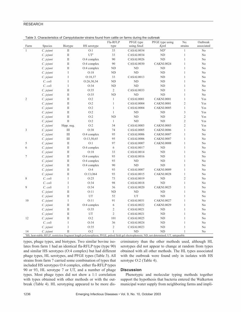
types, phage types, and biotypes. Two similar bovine iso-lates from farm 1 had an identical fla-RFLP type (type 90)and similar HS serotypes (O:4 complex) but had differentphage types, HL serotypes, and PFGE types (Table 3). Allstrains from farm 7 carried some combination of types thatincluded HS serotypes O:4 complex, either fla-RFLP types90 or 93, HL serotype 7 or UT, and a number of phagetypes. Most phage types did not show a 1:1 correlationwith types obtained with other methods or with the out-break (Table 4). HL serotyping appeared to be more dis-
criminatory than the other methods used, although HLserotypes did not appear to change at random from typesobtained with all other methods. The HL types associatedwith the outbreak were found only in isolates with HSserotype O:2 (Table 4).
DiscussionPhenotypic and molecular typing methods together
support the hypothesis that bacteria entered the Walkertonmunicipal water supply from neighboring farms and impli-
1236 Emerging Infectious Diseases • Vol. 9, No. 10, October 2003
RESEARCH
Table 3. Characteristics of Campylobacter strains found from cattle on farms during the outbreak
Farm Species Biotype HS serotype Fla-RFLP
type PFGE type using SmaI
PFGE type using KpnI
No. strains
Outbreak associated
1 C. jejuni II O:1 33 CASAI.0034 NDa 1 No C. jejuni II UTa 33 CASAI.0034 ND 1 No C. jejuni II O:4 complex 90 CASAI.0026 ND 1 No C. jejuni II O:4 complex 90 CASAI.0030 CAKNI.0024 1 No C. jejuni II O:4 complex ND ND ND 1 No C. jejuni I O:18 ND ND ND 1 No C. jejuni I O:18,37 33 CASAI.0013 ND 1 No C. coli I O:26,30,34 ND ND ND 1 No C. coli I O:34 ND ND ND 1 No C. jejuni II O:35 2 CASAI.0033 ND 1 No C. jejuni II O:35 ND ND ND 1 No 2 C. jejuni II O:2 1 CASAI.0001 CAKNI.0001 1 Yes C. jejuni II O:2 1 CASAI.0004 CAKNI.0001 2 Yes C. jejuni II O:2 1 CASAI.0004 CAKNI.0005 1 Yes C. jejuni II O:2 1 ND ND 3 Yes C. jejuni II O:2 ND ND ND 2 Yes C. jejuni II O:2 1 ND ND 2 Yes C. jejuni Hipp. neg. O:2 34 CASAI.0003 CAKNI.0003 2 Yes 3 C. jejuni III O:38 74 CASAI.0005 CAKNI.0006 1 No C. jejuni III O:4 complex 95 CASAI.0006 CAKNI.0007 1 No C. jejuni III O:13,50,65 95 CASAI.0006 CAKNI.0007 1 No 5 C. jejuni II O:1 97 CASAI.0007 CAKNI.0008 1 No 6 C. jejuni II O:4 complex 6 CASAI.0017 ND 1 No C. jejuni II O:18 33 CASAI.0014 ND 1 No 7 C. jejuni II O:4 complex 93 CASAI.0016 ND 1 No C. jejuni II O:4 complex 93 ND ND 1 No C. jejuni II O:4 complex 90 ND ND 1 No C. jejuni II O:4 93 CASAI.0007 CAKNI.0009 1 No C. jejuni II O:13,O64 93 CASAI.0015 CAKNI.0028 1 No 8 C. coli I O:25 73 CASAI.0019 ND 2 No C. coli I O:34 98 CASAI.0018 ND 1 No 9 C. coli I O:34 36 CASAI.0020 CAKNI.0025 1 No C. jejuni II O:11 ND ND ND 1 No C. jejuni II UT 52 UT ND 1 No 10 C. jejuni I O:11 91 CASAI.0031 CAKNI.0027 1 No C. jejuni II O:4 complex 6 CASAI.0022 CAKNI.0029 1 No C. jejuni II O:35 2 CASAI.0021 ND 1 No C. jejuni II UT 2 CASAI.0021 ND 1 No 12 C. jejuni II O:2 101 CASAI.0025 ND 1 No C. coli I O:34 36 CASAI.0024 ND 1 No C. jejuni I O:35 2 CASAI.0023 ND 1 No 14 C. jejuni II O:2 1 ND ND 1 No aHS, heat-stable; RFLP, restriction fragment length polymorphism; PFGE, pulsed field gel electrophoresis; ND, not determined; UT, untypeable.
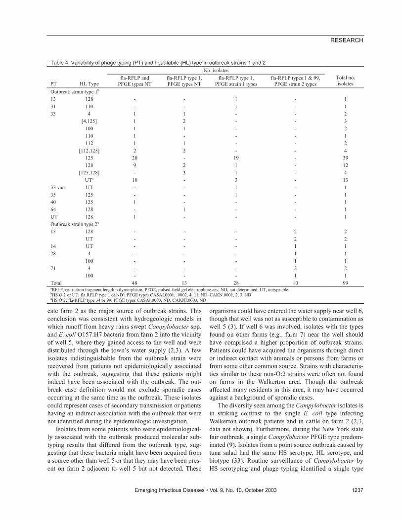
cate farm 2 as the major source of outbreak strains. Thisconclusion was consistent with hydrogeologic models inwhich runoff from heavy rains swept Campylobacter spp.and E. coli O157:H7 bacteria from farm 2 into the vicinityof well 5, where they gained access to the well and weredistributed through the town’s water supply (2,3). A fewisolates indistinguishable from the outbreak strain wererecovered from patients not epidemiologically associatedwith the outbreak, suggesting that these patients mightindeed have been associated with the outbreak. The out-break case definition would not exclude sporadic casesoccurring at the same time as the outbreak. These isolatescould represent cases of secondary transmission or patientshaving an indirect association with the outbreak that werenot identified during the epidemiologic investigation.
Isolates from some patients who were epidemiological-ly associated with the outbreak produced molecular sub-typing results that differed from the outbreak type, sug-gesting that these bacteria might have been acquired froma source other than well 5 or that they may have been pres-ent on farm 2 adjacent to well 5 but not detected. These
organisms could have entered the water supply near well 6,though that well was not as susceptible to contamination aswell 5 (3). If well 6 was involved, isolates with the typesfound on other farms (e.g., farm 7) near the well shouldhave comprised a higher proportion of outbreak strains.Patients could have acquired the organisms through director indirect contact with animals or persons from farms orfrom some other common source. Strains with characteris-tics similar to these non-O:2 strains were often not foundon farms in the Walkerton area. Though the outbreakaffected many residents in this area, it may have occurredagainst a background of sporadic cases.
The diversity seen among the Campylobacter isolates isin striking contrast to the single E. coli type infectingWalkerton outbreak patients and in cattle on farm 2 (2,3,data not shown). Furthermore, during the New York statefair outbreak, a single Campylobacter PFGE type predom-inated (9). Isolates from a point source outbreak caused bytuna salad had the same HS serotype, HL serotype, andbiotype (33). Routine surveillance of Campylobacter byHS serotyping and phage typing identified a single type
Emerging Infectious Diseases • Vol. 9, No. 10, October 2003 1237
RESEARCH
Table 4. Variability of phage typing (PT) and heat-labile (HL) type in outbreak strains 1 and 2 No. isolates
PT HL Type fla-RFLP and
PFGE types NT fla-RFLP type 1, PFGE types NT
fla-RFLP type 1, PFGE strain 1 types
fla-RFLP types 1 & 99, PFGE strain 2 types
Total no. isolates
Outbreak strain type 1b 13 128 - - 1 - 1 31 110 - - 1 - 1 33 4 1 1 - - 2 [4,125] 1 2 - - 3 100 1 1 - - 2 110 1 - - - 1 112 1 1 - - 2 [112,125] 2 2 - - 4 125 20 - 19 - 39 128 9 2 1 - 12 [125,128] - 3 1 - 4 UTa 10 - 3 - 13 33 var. UT - - 1 - 1 35 125 - - 1 - 1 40 125 1 - - - 1 64 128 - 1 - - 1 UT 128 1 - - - 1 Outbreak strain type 2c 13 128 - - - 2 2 UT - - - 2 2 14 UT - - - 1 1 28 4 - - - 1 1 100 - - - 1 1 71 4 - - - 2 2 100 - - - 1 1 Total 48 13 28 10 99 aRFLP, restriction fragment length polymorphism; PFGE, pulsed-field gel electrophoresies; ND, not determined; UT, untypeable. bHS O:2 or UT; fla RFLP type 1 or NDa; PFGE types CASAI.0001, .0002, 4, 11, ND, CAKN.0001, 2, 3, ND cHS O:2; fla-RFLP type 34 or 99; PFGE types CASAI.0003, ND, CAKNI.0003, ND
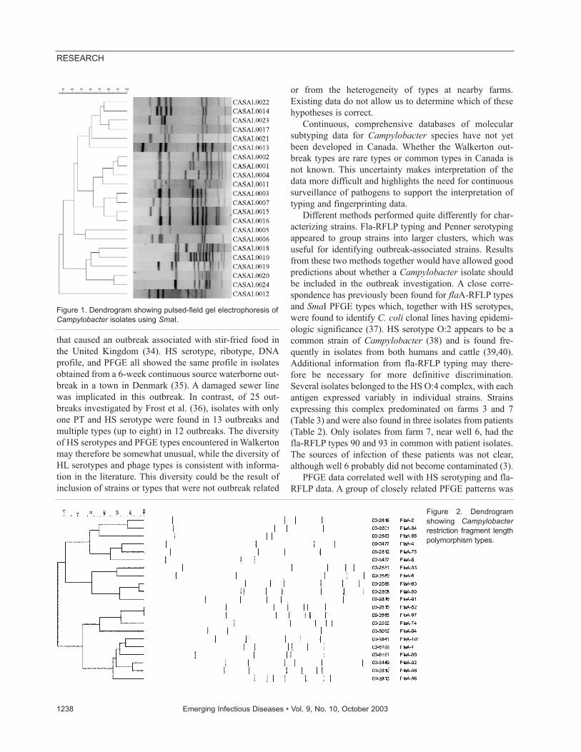
that caused an outbreak associated with stir-fried food inthe United Kingdom (34). HS serotype, ribotype, DNAprofile, and PFGE all showed the same profile in isolatesobtained from a 6-week continuous source waterborne out-break in a town in Denmark (35). A damaged sewer linewas implicated in this outbreak. In contrast, of 25 out-breaks investigated by Frost et al. (36), isolates with onlyone PT and HS serotype were found in 13 outbreaks andmultiple types (up to eight) in 12 outbreaks. The diversityof HS serotypes and PFGE types encountered in Walkertonmay therefore be somewhat unusual, while the diversity ofHL serotypes and phage types is consistent with informa-tion in the literature. This diversity could be the result ofinclusion of strains or types that were not outbreak related
or from the heterogeneity of types at nearby farms.Existing data do not allow us to determine which of thesehypotheses is correct.
Continuous, comprehensive databases of molecularsubtyping data for Campylobacter species have not yetbeen developed in Canada. Whether the Walkerton out-break types are rare types or common types in Canada isnot known. This uncertainty makes interpretation of thedata more difficult and highlights the need for continuoussurveillance of pathogens to support the interpretation oftyping and fingerprinting data.
Different methods performed quite differently for char-acterizing strains. Fla-RFLP typing and Penner serotypingappeared to group strains into larger clusters, which wasuseful for identifying outbreak-associated strains. Resultsfrom these two methods together would have allowed goodpredictions about whether a Campylobacter isolate shouldbe included in the outbreak investigation. A close corre-spondence has previously been found for flaA-RFLP typesand SmaI PFGE types which, together with HS serotypes,were found to identify C. coli clonal lines having epidemi-ologic significance (37). HS serotype O:2 appears to be acommon strain of Campylobacter (38) and is found fre-quently in isolates from both humans and cattle (39,40).Additional information from fla-RFLP typing may there-fore be necessary for more definitive discrimination.Several isolates belonged to the HS O:4 complex, with eachantigen expressed variably in individual strains. Strainsexpressing this complex predominated on farms 3 and 7(Table 3) and were also found in three isolates from patients(Table 2). Only isolates from farm 7, near well 6, had thefla-RFLP types 90 and 93 in common with patient isolates.The sources of infection of these patients was not clear,although well 6 probably did not become contaminated (3).
PFGE data correlated well with HS serotyping and fla-RFLP data. A group of closely related PFGE patterns was
1238 Emerging Infectious Diseases • Vol. 9, No. 10, October 2003
RESEARCH
Figure 1. Dendrogram showing pulsed-field gel electrophoresis ofCampylobacter isolates using SmaI.
Figure 2. Dendrogramshowing Campylobacterrestriction fragment lengthpolymorphism types.
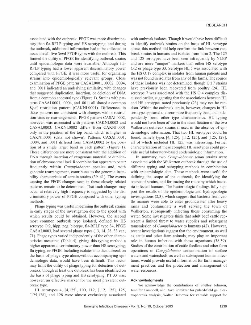
associated with the outbreak. PFGE was more discrimina-tory than fla-RFLP typing and HS serotyping, and duringthe outbreak, additional information had to be collected toassociate all five SmaI PFGE types with the outbreak. Thislimited the utility of PFGE for identifying outbreak strainsuntil epidemiologic data were available. Although fla-RFLP typing had a lower apparent discriminatory powercompared with PFGE, it was more useful for organizingstrains into epidemiologically relevant groups. Closeexamination of PFGE patterns CASAI.0001, .0002, .0004,and .0011 indicated an underlying similarity, with changesthat suggested duplication, insertion, or deletion of DNAfrom a common ancestral type (Figure 1). Strains with pat-terns CASAI.0001, .0004, and .0011 all shared a commonKpnI restriction pattern (CAKNI.0001). Differences inthese patterns are consistent with changes within restric-tion sites or rearrangements. PFGE pattern CASAI.0002,however, was associated with patterns CAKNI.0002 andCASAI.0003. CAKNI.0002 differs from CAKNI.0001only in the position of the top band, which is higher inCAKNI.0001 (data not shown). Patterns CASAI.0001,.0004, and .0011 differed from CASAI.0002 by the posi-tion of a single larger band in each pattern (Figure 1).These differences are more consistent with the addition ofDNA through insertion of exogenous material or duplica-tion of chromosomal loci. Recombination appears to occurfrequently within Campylobacter species and, withgenomic rearrangement, contributes to the genomic insta-bility characteristic of certain strains (39–41). The eventscausing the PFGE changes seen in these closely relatedpatterns remain to be determined. That such changes mayoccur at relatively high frequency is suggested by the dis-criminatory power of PFGE compared with other typingmethods.
Phage typing was useful in defining the outbreak strainsin early stages of the investigation due to the speed withwhich results could be obtained. However, the secondmost common outbreak type isolated, defined by HSserotype O:2, hipp. neg. biotype, fla-RFLP type 34, PFGECASAI.0003, had several phage types (13, 14, 28, 33 var.,71). Phage types varied independently of the other charac-teristics measured (Table 4), giving this typing method ahigher apparent discriminatory power than HS serotyping,fla typing, or PFGE. Including isolates into the outbreak onthe basis of phage type alone,without accompanying epi-demiologic data, would have been difficult. This factormay limit the utility of phage typing for detection of out-breaks, though at least one outbreak has been identified onthe basis of phage typing and HS serotyping. PT 33 was,however, an effective marker for the most prevalent out-break type.
HL serotypes 4, [4,125], 100, 112, [112, 125], 125,[125,128], and 128 were almost exclusively associated
with outbreak isolates. Though it would have been difficultto identify outbreak strains on the basis of HL serotypealone, this method did help confirm the link between out-break strains in humans and isolates from farm 2. The 125and 128 serotypes have been seen infrequently by NLEPand are more “unique” markers than either HS serotypeO:2 or phage type 33. Serotype HL 5 was associated withthe HS O:17 complex in isolates from human patients andwas not found in isolates from any of the farms. The sourceof these isolates was not determined, though O:17 strainshave previously been recovered from poultry (24). HLserotype 7 was associated with the HS O:4 complex dis-cussed earlier, suggesting that the associations between HLand HS serotypes noted previously (23) may not be ran-dom. Within the outbreak strain, however, changes in HLserotype appeared to occur more frequently than, and inde-pendently from, other type characteristics. HL typingwould not have been of use in the identification of the twoWalkerton outbreak strains if used in the absence of epi-demiologic information. That two HL serotypes could befound, namely types [4, 125], [112, 125], and [125, 128],all of which included HL 125, was interesting. Furthercharacterization of these complex HL serotypes could pro-vide useful laboratory-based epidemiologic information.
In summary, two Campylobacter jejuni strains wereassociated with the Walkerton outbreak through the use ofdifferent typing and subtyping methods in combinationwith epidemiologic data. These methods were useful fordefining the scope of the outbreak, for identifying thesource of strains, and for tracing the route by which bacte-ria infected humans. The bacteriologic findings fully sup-port the results of the epidemiologic and hydrogeologicinvestigations (2,3), which suggest that bacteria from cat-tle manure were able to enter groundwater after heavyrains and contaminate a well serving the town ofWalkerton, subsequently infecting those consuming thewater. Some investigators think that adult beef cattle rep-resent a limited threat to water supplies and subsequenttransmission of Campylobacter to humans (42). However,recent investigations suggest that the environment, as wellas cattle and other farm animals, may play an importantrole in human infection with these organisms (38,39).Studies of the contribution of cattle feedlots and other farmoperations to Campylobacter contamination of surfacewaters and watersheds, as well as subsequent human infec-tions, would provide useful information for farm manage-ment practices and the protection and management ofwater resources.
AcknowledgmentsWe acknowledge the contributions of Shelley Johnson,
Jennifer Campbell, and Dave Spreitzer for pulsed-field gel elec-trophoresis analysis; Walter Demczuk for valuable support for
Emerging Infectious Diseases • Vol. 9, No. 10, October 2003 1239
RESEARCH
database management, laboratory surveillance, communicationactivities during the outbreak, and phage typing; and LouisBryden for critiquing the manuscript and providing advice.
Dr. Clark is currently a research scientist at the NationalLaboratory for Enteric Pathogens, National MicrobiologyLaboratory (Winnipeg, Manitoba), Population and Public HealthBranch, Health Canada. His current research interests includepathogenesis, population structure, and genetic exchange withinenteric bacteria.
References
1. Millson M, Bokhout M, Carlson J, Spielberg L, Aldis R, Borczyk A,et al. An outbreak of Campylobacter jejuni gastroenteritis linked tomeltwater contamination of a municipal well. Can J Public Health1991;82:27–31.
2. Bruce-Grey-Owen Sound Health Unit. Waterborne outbreak of gas-troenteritis associated with a contaminated municipal water supply,Walkerton, Ontario, May–June 2000. Can Comm Dis Rep2000;26–20:170–3.
3. McQuigge, M. The investigative report on the Walkerton outbreak ofwaterborne gastroenteritis: May–June 2000. Bruce-Grey-OwenSound Health Unit; 2000.
4. Altekreuse SF, Cohen ML, Swerdlow DL. Emerging foodbornediseases. Emerg Infect Dis 1997;3:285–93.
5. Tauxe RV. Epidemiology of Campylobacter jejuni infections in theUnited States and other industrialized nations. In: Nachamkin I,Tomkins S, Blaser M, editors. Campylobacter jejuni: current statusand future trends. Washington: American Society for Microbiology;1992. p. 9–19.
6. Mead PS, Slutsker L, Dietz V, McCaig LF, Bresee JS, Shapiro C, etal. Food-related illness and death in the United States. Emerg InfectDis 1999;5:607–25.
7. Jones IG, Roworth M. An outbreak of Escherichia coli O157 andcampylobacteriosis associated with contamination of a drinkingwater supply. Public Health 1996;110: 277–82.
8. Centers for Disease Control and Prevention. Outbreak of Escherichiacoli O157:H7 and Campylobacter among attendees of theWashington county fair–New York, 1999. MMWR Morb MortalWkly Rep 1999;48:803–4.
9. Bopp DJ, Sauders BD, Waring AL, Ackelsberg J, Dumas N, Braun-Howland E, et al. Detection, isolation, and molecular subtyping ofEscherichia coli O157:H7 and Campylobacter jejuni associated witha large waterborne outbreak. J Clin Microbiol 2003;41:174–80.
10. Jones K. Campylobacters in water, sewage, and the environment. JAppl Microbiol 2001;90:68S–79S.
11. Brieseman MA. Town water supply as the cause of an outbreak ofcampylobacter infection. NZ Med J 1987;100:212–3.
12. Stehr-Green J, Nicholls C, McEwan S, Payne A, Mitchell P.Waterborne outbreak of Campylobacter jejuni in Christchurch: theimportance of a combined epidemiologic and microbiologic investi-gation. NZ Med J 1991;104:356–8.
13. Miettinen IT, Zacheus O, von Bonsdorff C-H, Vartiainen T.Waterborne epidemics in Finland in 1998–1999. Water Sci Tech2001;43:67–71.
14. Furtado C, Adak GK, Stuart JM, Wall PG, Evans HS, Casemore DP.Outbreaks of waterborne infectious intestinal disease in England andWales, 1992–5. Epidemiol Infect 1998;121:109–19.
15. Duke LA, Breathnach AS, Jenking DR, Harkis BA, Codd AW. Amixed outbreak of cryptosporidium and campylobacter infectionassociated with a private water supply. Epidemiol Infect1996;116:303–8.
16. Merritt A, Miles R, Bates J. An outbreak of Campylobacter enteritison an island resort, north Queensland. Commun Dis Intell1999;23:215–9.
17. Kramer MH, Herwaldt BL, Craun GF, Calderon RL, Juranek DD.Surveillance for waterborne-disease outbreaks—United States,1993–1994. MMWR Morb Mortal Wkly Rep Surveillance Summary1996;45:1–33.
18. Vogt RL, Sours HE, Barrett T, Feldman RA, Dickinson RJ, WitherellL. Campylobacter enteritis associated with contaminated water. AnnIntern Med 1982;96:292–6.
19. Alary M, Nadeau D. An outbreak of Campylobacter enteritis associ-ated with a community water supply. Can J Public Health1990;81:268–71.
20. Glover D, Ross A, Lugsdin J. Gastroenteritis outbreak at an industri-al camp—British Columbia. Can Commun Dis Rep 1992;18:66–8.
21. Taylor DE, Salama MS, Tabor H, Richter M. Pulsed-field gelelectrophoresis for epidemiologic studies of Campylobacterhyointestinalis isolates. J Clin Microbiol 1992;30:1982–4.
22. Buzby JC, Roberts T. Economic and trade impacts of microbial food-borne illness. World Health Statistical Quarterly 1997;50:57–66.
23. Todd ECD. Epidemiology of foodborne diseases: a worldwidereview. World Health Statistical Quarterly 1997;50:30–50.
24. Lior H. New, extended biotyping scheme for Campylobacter jejuni,Campylobacter coli, and “Campylobacter laridis.” J Clin Microbiol1984;20:636–40.
25. Marshall SM, Melito PL, Woodward DL, Johnson WM, Rodgers FG,Mulvey MR. Rapid identification of Campylobacter, Arcobacter, andHelicobacter isolates by PCR-restriction fragment lengthpolymorphism analysis of the 16S rRNA gene. J Clin Microbiol1999;37:4158–60.
26. Linton D, Lawson AJ, Owen RJ, Stanley J. PCR detection,identification to species level, and fingerprinting of Campylobacterjejuni and Campylobacter coli direct from diarrheic samples. J ClinMicrobiol 1997;35:2568–72.
27. Lior H, Woodward DL, Edgar JA, Laroche LJ, Gill P. Serotyping ofCampylobacter jejuni by slide agglutination based on heat-labileantigenic factors. J Clin Microbiol 1982;15:761–8.
28. Penner JL, Hennessy JN. Passive hemagglutination technique forserotyping Campylobacter fetus subsp. jejuni on the basis of solubleheat-stable antigens. J Clin Microbiol 1980;12:732–7.
29. Frost JA, Kramer JM, Gillanders SA. Phage typing of Campylobacterjejuni and Campylobacter coli and its use as an adjunct to serotyping.Epidemiol Infect 1999;123:47–55.
30. Nachamkin I, Bohachick K, Patton CM. Flagellin gene typing ofCampylobacter jejuni by restriction fragment length polymorphismanalysis. J Clin Microbiol 1993;31:1531–6.
31. Ribot EM, Fitzgerald C, Kubota K, Swaminathan B, Barrett TJ.Rapid pulsed-field gel electrophoresis protocol for subtyping ofCampylobacter jejuni. J Clin Microbiol 2001;39:1889–94.
32. Jackson CJ, Fox AJ, Jones DM, Wareing DRA, Hutchinson DN.Associations between heat stable (O) and heat-labile (HL) serogroupantigens of Campylobacter jejuni: evidence for interstrainrelationships within three O/HL serovars. J Clin Microbiol1998;36:2223–8.
33. Roels TH, Wickus B, Kazmierczak JJ, Nicholson MA, Kurzynski TA,Davis JP. A foodborne outbreak of Campylobacter jejuni (O:33)infection associated with tuna salad: a rare strain in an unusualvehicle. Epidemiol Infect 121:281–7.
34. Evans M, Lane W, Frost JA, Nylen G. A campylobacter outbreakassociated with stir-fried food. Epidemiol Infect 1998;121:275–9.
35. Engberg J, Gerner-Smidt P, Scheutz F, Nielsen EM, On SLW, MølbakK. Water-borne Campylobacter jejuni infection in a Danish town—a6-week continuous source outbreak. Clin Microbiol Infect1998;4:648–56.
1240 Emerging Infectious Diseases • Vol. 9, No. 10, October 2003
RESEARCH
36. Frost JA, Gillespie IA, O’Brien SJ. Public health implications ofcampylobacter outbreaks in England and Wales, 1995–9:epidemiological and microbiological investigations. Epidemiol Infect2002;128:111–8.
37. Stanley J, Linton D, Sutherland K, Jones C, Owen RJ. High-resolution genotyping of Campylobacter coli identifies clones ofepidemiologic and evolutionary significance. J Infect Dis1995;172:1130–4.
38. Woodward DL, Rodgers FG. Identification of Campylobacter heat-stable and heat-labile antigens by combining the Penner and Liorserotyping schemes. J Clin Microbiol 2002;40:741–5.
39. Fitzgerald C, Stanley K, Andrew S, Jones K. Use of pulsed-field gelelectrophoresis and flagellin gene typing in identifying clonal groupsof Campylobacter jejuni and Campylobacter coli in farm and clinicalenvironments. Appl Environ Microbiol 2001;67:1429–36.
40. Nielsen EM, Enberg J, Fussing V, Petersen L, Brogren C-H, On SLW.Evaluation of phenotypic and genotypic methods for subtypingCampylobacter jejuni isolates from humans, poultry, and cattle. JClin Microbiol 2000;38:3800–10.
41. Wassenaar TM, Geilhausen B, Newell DG. Evidence of genomeinstability of Campylobacter jejuni isolated from poultry. ApplEnviron Microbiol 1998;64:1816–21.
42. Hoar BR, Atwill ER, Elmi C, Farver TB. An examination of risk fac-tors associated with beef cattle shedding pathogens of human con-cern. Epidemiol Infect 2001;127:147–55.
Address for correspondence: Clifford Clark, National Laboratory forEnteric Pathogens, Canadian Science Centre for Human and AnimalHealth, 1015 Arlington St., Winnipeg, Manitoba, R3E 3R2, Canada; fax:(204) 789-2142; email: [email protected]
Emerging Infectious Diseases • Vol. 9, No. 10, October 2003 1241
RESEARCH
Search past issues of EID at www.cdc.gov/eid
Technical guidelines for the control of Ebola hemor-rhagic fever (EHF) indicate that understanding local viewsand responses to an outbreak is essential. However, fewstudies with such information exist. Thus, we used qualita-tive and quantitative methods to determine how local resi-dents of Gulu, Uganda, viewed and responded to the2000–2001 outbreak of EHF. Results indicated that Acholipeople used at least three explanatory models to explainand respond to the outbreak; indigenous epidemic controlmeasures were often implemented and were consistent withthose being promoted by healthcare workers; and some cul-tural practices amplified the outbreak (e.g., burial practices).However, most persons were willing to modify and work withnational and international healthcare workers.
Many emerging disease specialists are sensitive to andacknowledge the potential importance of social sci-
ence in disease control, but seldom is this perspective con-sidered when organizing response efforts. In part, this sit-uation exists because so little research in this area hasbeen conducted. The special issue on Ebola in The Journalof Infectious Diseases (1) does not include any articles onthe behavioral aspects of the disease. However, WorldHealth Organization (WHO) technical guidelines forresponding to Ebola hemorrhagic fever (EHF) state that,in conducting epidemiologic surveillance, “Special atten-tion must be given to the actual perception of the outbreakby the community. In particular, specific cultural elementsand local beliefs must be taken into account to ensureproper messages, confidence, and close cooperation of thecommunity” (2).
We describe the first systematic sociocultural study ofan outbreak of EHF. The outbreak occurred in several loca-tions in northern Uganda in 2000 to 2001. We conductedthis research in villages and neighborhoods in and aroundGulu during the last month of the outbreak. The field studyaimed to: 1) describe local explanatory models of EHF; 2)provide understanding of topics of concern to WHO (i.e.,burial practices, patients’ fear of going to the hospital, therole of traditional healers in disease transmission); and 3)
identify local and international beliefs and practices thatenhanced or were detrimental to the control of EHF. An“explanatory model” refers to a person’s or culture’s expla-nations and predictions regarding a particular illness.Some of the questions asked when trying to understand anexplanatory model include: How do persons refer to the ill-ness? How do they explain it (i.e., cause)? What do theysee as appropriate treatments? What do they do to preventthe illness? Patients, physicians, healthcare workers, andlocal residents in different parts of the world each haveexplanatory models for different illnesses. Providing careand treatment for a particular disease is often based onnegotiating these different models.
Background The 2000–2001 Uganda outbreak was one of the largest
EHF outbreaks to date, with 425 presumptive cases and224 deaths (case-fatality rate 53%). Most patients werewomen (269 or 63%). The earliest reported presumptivecase-patient had disease onset on August 30, 2000, and thelast case began on January 9, 2001 (3).
The Gulu EHF outbreak was relatively unusual in com-parison to other recent EHF outbreaks (e.g., DemocraticRepublic of Congo [DRC], Gabon) in that the diseaseaffected primarily one ethnic group, the Acholi, and mostof the district medical staff and decision makers (e.g., dis-trict medical officer, director of health education) werealso from this ethnic group. Also, most (60%) EHF casesoccurred in the urban area of Gulu town. Gulu District hasapproximately 470,000 people, primarily Acholi, and 60%of the population live in protected villages because of rebelactivity.
Most Acholi are agro-pastoralists and have a socialorganization strongly influenced by patrilineal descent andpatrilocal postmarital residence (4). Other researchers havewritten about Acholi and other Nilotic peoples’ healthbeliefs (5–7), but none has described cultural responses toepidemic diseases.
MethodsQualitative and quantitative methods were used. The
first 2 weeks of the research emphasized open-ended and
1242 Emerging Infectious Diseases • Vol. 9, No. 10, October 2003
RESEARCH
Cultural Contexts of Ebola inNorthern Uganda
Barry S. Hewlett* and Richard P. Amola†
*Washington State University, Vancouver, Washington, USA; and†Ministry of Health, Adjumani, Uganda
focus group interviews as well as document review (e.g.,health education materials, reports). The last few daysemphasized the development of systematic questionnaires.Open-ended interviews were conducted with the follow-ing: 1) 10 persons and four focus groups in villages orneighborhoods with large numbers of early cases of EHF;2) 8 persons and one focus group with survivors of EHF(both healthcare workers and community members); 3)four focus groups with male and female elders (two meet-ings with each gender); 4) 3 persons and two focus groupsof children; 5) 4 persons and two focus groups with health-care workers responsible for the isolation unit and counsel-ing survivors; and 6) 4 persons and one focus group withtraditional healers. Focus group meetings usually had 5–8participants, with the exception of the survivor focus groupmeeting, which had 35 participants.
Questionnaires were administered to 85 Gulu HighSchool students 15–21 years of age (all members of threesenior classes; 33 men, 52 women). Precoded question-naires were administered to 49 adults in Gulu (25 women,24 men; an adult from every third house from two random-ly selected Gulu neighborhoods) and 60 EHF survivors (22men, 38 women; all survivors were located through a sur-vivors’ organization). We also examined existing docu-ments, such as field reports and health education materialsused in the outbreak (e.g., posters, brochures, music cas-settes, videos).
Results
Explanatory ModelsTable 1 summarizes three primary explanatory models
identified by the Acholi. The third model is biomedical.Biomedical models have existed in the area for >100 years,and all Acholi know these models well and use them often.In the early phases of the outbreak, many families, think-ing the disease was a bacterial infection or malaria, turnedto tetracycline or chloroquine. Most early case-patientswent to the hospital seeking biomedical treatment. Thebiomedical model for EHF was introduced in late Octoberby the Ugandan Ministry of Health. The health educationprogram was multidimensional (e.g., posters, radio shows,videos, brochures) and transmitted this model effectively.However, by the time the EHF biomedical model wasintroduced, local people had already used two otherindigenous explanatory models.
Both of these models require an understanding of theconcept of jok, which is common to many Nilotic-speakingpeoples, including the Acholi. Jok are spirits or gods (8).Many different types of jok exist; they have names andreportedly are often found near bodies of water, moun-tains, and natural salt licks for cattle. Jok are generallybenevolent, as they provide and control resources, but they
can also cause harm if they are not respected. Deference toand respect for others are central values in Acholi life, andspiritual life reflects and reinforces these values. Thesespirits are like elders in the community; the Acholi listen towhat they have to say, do what they say without question,and give them gifts to show respect.
Traditional healers (ajwaka), who are primarilywomen, obtain their powers to heal from specific jok thatthey have acquired through time. Most healers acquire asmany as 10 such spirits during their lifetime. Each spirithas a name and a specific kind of knowledge (e.g., treat-ments for mental confusion or infertility).
At first, many persons treated the symptoms of EHF asa regular illness and sought a variety of both biomedical(i.e., malarial drugs or antibiotics) and indigenous cures(i.e., herbs, traditional healers). In late September 2000,the heads of families in neighborhoods with many deathsasked a traditional healer to locate poisons (yat) in andaround the lineage household that might be causing the ill-ness and death (Table 1, first explanatory model). Thehealers used their jok and special spears to locate poisonedobjects (e.g., roots, bones) in the neighborhood. The heal-er’s jok was also called on to communicate with the spiritassociated with a particular poison and to determine ifburning the object and sacrificing goats and sheep wasnecessary to demonstrate respect. Once the poison wasremoved and respect demonstrated, the healers said thedeaths would stop. But the deaths continued. Yat removalis not cheap; each EHF-affected family paid 150,000Ugandan shillings (U.S.$88), four to five goats or sheep,and one chicken (about half the annual income for a ruralAcholi family). The families pointed out that in addition tothe enormous loss of loved ones, an incredible amount offamily assets was lost in trying to treat the ill.
In early October, residents began to realize that this out-break was more than a regular kind of illness and began toclassify it as two gemo (two [illness] gemo [epidemic]), thesecond explanatory model in Table 1. Gemo is a bad spirit(type of jok that comes suddenly and causes a mysteriousillness and death in many people within a very short peri-od of time). Gemo reportedly comes like the wind in that itcomes rapidly from a particular direction and affects manypeople, but the wind itself does not necessarily bring it.Acholi have experienced other types of gemo (e.g.,measles and smallpox). Forty-nine of 50 adults inter-viewed indicated a belief that Ebola was a type of gemo.The term two gemo was also used in health educationposters and music.
Gemo is said to be mysterious in that it just comes onits own, but several people indicated that it comes becauseof lack of respect and honor for the gods. Elders indicatedthat in the past, lack of respect for jok of tura (hills, moun-tains, bodies of water) was the major cause of gemo.
Emerging Infectious Diseases • Vol. 9, No. 10, October 2003 1243
RESEARCH
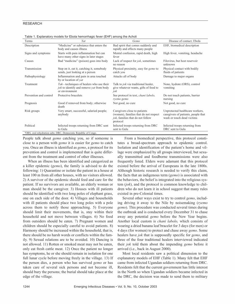
People talk about gemo catching you, so if someone isclose to a person with gemo it is easier for gemo to catchyou. Once an illness is identified as gemo, a protocol for itsprevention and control is implemented that is quite differ-ent from the treatment and control of other illnesses.
When an illness has been identified and categorized asa killer epidemic (gemo), the family is advised to do thefollowing: 1) Quarantine or isolate the patient in a house atleast 100 m from all other houses, with no visitors allowed.2) A survivor of the epidemic should feed and care for thepatient. If no survivors are available, an elderly woman orman should be the caregiver. 3) Houses with ill patientsshould be identified with two long poles of elephant grass,one on each side of the door. 4) Villages and householdswith ill patients should place two long poles with a poleacross them to notify those approaching. 5) Everyoneshould limit their movements, that is, stay within theirhousehold and not move between villages. 6) No foodfrom outsiders should be eaten. 7) Pregnant women andchildren should be especially careful to avoid patients. 8)Harmony should be increased within the household, that is,there should be no harsh words or conflicts within the fam-ily. 9) Sexual relations are to be avoided. 10) Dancing isnot allowed. 11) Rotten or smoked meat may not be eaten,only eat fresh cattle meat. 12) Once the patient no longerhas symptoms, he or she should remain in isolation for onefull lunar cycle before moving freely in the village. 13) Ifthe person dies, a person who has survived gemo or hastaken care of several sick persons and not become ill,should bury the persons; the burial should take place at theedge of the village.
From a biomedical perspective, this protocol consti-tutes a broad-spectrum approach to epidemic control.Isolation and identification of the patient’s home and vil-lage were emphasized by all groups interviewed, but sexu-ally transmitted and foodborne transmissions were alsofrequently listed. Elders were adamant that this protocolexisted before the arrival of Europeans in the late 1800s.Although historic research is needed to verify this claim,the facts that an indigenous term (gemo) is associated withthe behaviors, the belief is integrated into the religious sys-tem (jok), and the protocol is common knowledge to chil-dren who do not learn it in school suggest that many rulesexisted in pre-Colonial times.
Several other ways exist to try to control gemo, includ-ing driving it away to the Nile by noisemaking (ryemogemo). This procedure was conducted several times duringthe outbreak and is conducted every December 31 to chaseaway any potential gemo before the New Year begins.Another local custom is chani labolo, which consists ofwearing a dried banana leaf bracelet for 3 days (for men) or4 days (for women) to protect and chase away gemo. Somehealers have jok that is supposedly specific for gemo, andthree of the four traditional healers interviewed indicatedtheir jok told them about the impending gemo before itarrived (i.e., back in August 2000).
Most local residents saw a political dimension to theexplanatory models of EHF (Table 1). Many felt that EHFcame from infected Ugandan soldiers returning from DRC.Residents felt that the current government has little interestin the North so when Ugandan soldiers became infected inthe DRC, the decision was made to send them to military
1244 Emerging Infectious Diseases • Vol. 9, No. 10, October 2003
RESEARCH
Table 1. Explanatory models for Ebola hemorrhagic fever (EHF) among the Acholi Terms Yat Gemo Disease of contact; Ebola Description “Medicine” or substance that enters the
body and causes illness Bad spirit that comes suddenly and rapidly and effects many people
EHF, biomedical description
Signs and symptoms Starts with pain inflammation but can have many other signs in later stages
Mental confusion, rapid death, high fever
High fever, vomiting, headache
Causes Bad “medicine” (poison) goes into body Lack of respect for jok, sometimes no reason
Filovirus, but host reservoir unknown
Transmission Step on it, eat it, catching it, somebody sends, just looking at a person
Physical proximity, easy for gemo to catch you
Physical contact with bodily fluids of patients
Pathophysiology Inflammation and pain in area touched by or location of yat
Attacks all of body Damage to major organs
Treatment Tak—techniques of healers who use their jok to identify and remove yat from body or environment
Talk to jok via traditional healer, give whatever wants, gifts of food to jok
None, hydrate (ORS), control vomiting
Prevention and control Protective bracelets See protocol in text, chani labolo, ryemo gemo
Do not touch patients, barrier nursing
Prognosis Good if removed from body; otherwise death
Not good, no cure Not good, no cure
Risk groups Very smart, successful, salaried people; anybody
Caregivers close to patients (women), families that do not respect jok, families that do not follow protocol
Unprotected healthcare workers, caregivers of patients, people that wash or touch dead victims
Political Infected troops returning from DRC sent to Gulu
Infected troops returning from DRC sent to Gulu
Infected troops returning from DRC sent to Gulu
aORS, oral rehydration salts; DRC, Democratic Republic of Congo.
bases in Gulu. Although some of the first female victims ofEHF had relationships with men in the military, existingepidemiologic evidence does not support the DRC originhypothesis. The origin of this outbreak is not known.Political dimensions to disease, killer epidemics, in partic-ular, are common. The first author’s visit to Gabon duringthe 1996 EHF outbreak indicated that local people believedthat the French military, which had military exercises in thearea just before the outbreak, were partially responsible forintroducing the disease. In addition, the 2003 Congo out-break was linked, in part, to activities of Euro-Americansconducting research in a national park there.
Many informants and healthcare workers indicated thatfleeing the village or neighborhood was common, in par-ticular in locations with the highest number of infectedpersons. Fleeing is not an explicit part of the explanatorymodels but makes some sense because the gemo or bio-medical models indicate the illness is a rapid killer trans-mitted by close contact with infected persons.
Most persons involved in this outbreak were familiarwith all three explanatory models and did not necessarilysee them as contradictory. Healthcare workers emphasizedthe biomedical model, but many Acholi healthcare workersparticipated in ryemo gemo when it passed through thecommunity. Some persons and villages turned to the yatand gemo explanatory models but did not hesitate to pur-chase tetracycline and other medicines to treat cases ofEHF. The first two explanatory models may seem strange tointernational health workers, but they reflect a holistic andsocial view of illness common to many people in the world.Acholi are aware of the biomedical model but view illnessas having social, spiritual, and biological dimensions. Theepidemic control protocol is a good example. Family mem-bers refrain from sex and quarreling to show respect to jok(spiritual) and increase family harmony and peace (social).
Issues of Concern
Funerals and Burials National and international healthcare workers were
concerned that burial practices contributed to the amplifi-cation of EHF. A brief study indicated that once a persondied, his or her paternal aunt (father’s sister) was called towash and prepare the body for burial. If the father did nothave a sister, an older woman in the victim’s patriline wasasked to prepare the body. Generally, the woman removedthe clothes from the body, washed the body, and dressedthe deceased in a favorite outfit. At the funeral, all familymembers ritually washed their hands in a common bowl,and during open casket all were welcome to come up todeceased person and give a final touch on the face or else-where (called a love touch). The body was then wrapped ina white cloth or sheet and buried. The person was buried
next to or near their household. This practice is the normalsystem of burial.
However, when disease is classified as gemo, burialpractices change. The body is not touched and is buriedoutside or at the edge of the village. The designated care-giver, someone who has survived the outbreak or an olderwoman, is responsible for washing and preparing the bodyfor burial.
Various activities associated with burial practices con-tributed to transmission of EHF (Table 2). Washing thebody was a possible means of infection for women only,while a touch was a more common means of infectionamong men. The fact that 63% of the survivors in thisstudy had their first symptoms in October implies that theyprobably became infected before laboratory tests con-firmed EHF and before the disease was designated as atype of gemo in many communities. Caregiving, especial-ly by women, contributed substantially to many cases,which explains, in part, why 63% of all presumptive EHFcases in Uganda were in women.
WHO was also concerned that local persons were notcoming to the hospital when symptoms first emerged.Healthcare workers theorized that patients were afraid ofbeing buried at the airfield if they died. Persons were run-ning and hiding when the ambulance arrived to take themto hospital. Later interviews indicated, however, that theairfield burial was not the problem. As described in the pro-tocol, once an illness is identified as a killer epidemic, bur-ial at the edge of the village is expected. Rather, sourcesindicated, many persons ran from the ambulance and didnot seek treatment quickly because they feared they wouldnever see their family once they were admitted to the hos-pital. A fear of Euro-Americans buying and selling body-parts is common in many parts of central Africa but wasespecially pronounced in Gulu hospitals because bodieswere placed in body bags and taken to the airfield to beburied without relatives being notified. Relatives were notalways around at the time of death, and healthcare workerswere required to dispose of the body as quickly as possible.The anger and bad feelings about not being informed weredirected toward healthcare workers in the isolation unit.This fear could have been averted by allowing familymembers to see the body in the bag and allowing familymembers to escort the body to the burial ground.
Emerging Infectious Diseases • Vol. 9, No. 10, October 2003 1245
RESEARCH
Table 2. How survivors thought they contracted Ebola hemorrhagic fever (EHF)a
% How survivors felt they acquired Ebola Men (n=22) Women (n=38) Washing body of EHF victim 0 21 Love touch 32 11 Transporting EHF patient to hospital 5 16 Caregiving of EHF patient 27 53 aMore than one response possible per informant.
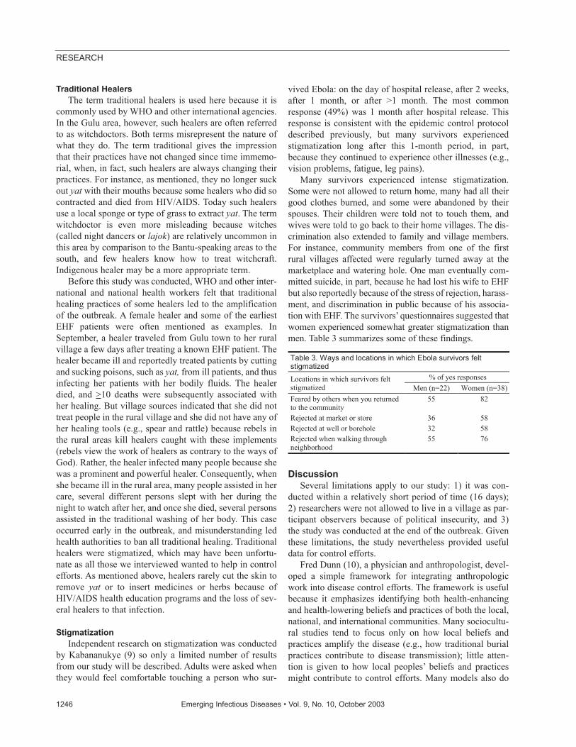
Traditional HealersThe term traditional healers is used here because it is
commonly used by WHO and other international agencies.In the Gulu area, however, such healers are often referredto as witchdoctors. Both terms misrepresent the nature ofwhat they do. The term traditional gives the impressionthat their practices have not changed since time immemo-rial, when, in fact, such healers are always changing theirpractices. For instance, as mentioned, they no longer suckout yat with their mouths because some healers who did socontracted and died from HIV/AIDS. Today such healersuse a local sponge or type of grass to extract yat. The termwitchdoctor is even more misleading because witches(called night dancers or lajok) are relatively uncommon inthis area by comparison to the Bantu-speaking areas to thesouth, and few healers know how to treat witchcraft.Indigenous healer may be a more appropriate term.
Before this study was conducted, WHO and other inter-national and national health workers felt that traditionalhealing practices of some healers led to the amplificationof the outbreak. A female healer and some of the earliestEHF patients were often mentioned as examples. InSeptember, a healer traveled from Gulu town to her ruralvillage a few days after treating a known EHF patient. Thehealer became ill and reportedly treated patients by cuttingand sucking poisons, such as yat, from ill patients, and thusinfecting her patients with her bodily fluids. The healerdied, and >10 deaths were subsequently associated withher healing. But village sources indicated that she did nottreat people in the rural village and she did not have any ofher healing tools (e.g., spear and rattle) because rebels inthe rural areas kill healers caught with these implements(rebels view the work of healers as contrary to the ways ofGod). Rather, the healer infected many people because shewas a prominent and powerful healer. Consequently, whenshe became ill in the rural area, many people assisted in hercare, several different persons slept with her during thenight to watch after her, and once she died, several personsassisted in the traditional washing of her body. This caseoccurred early in the outbreak, and misunderstanding ledhealth authorities to ban all traditional healing. Traditionalhealers were stigmatized, which may have been unfortu-nate as all those we interviewed wanted to help in controlefforts. As mentioned above, healers rarely cut the skin toremove yat or to insert medicines or herbs because ofHIV/AIDS health education programs and the loss of sev-eral healers to that infection.
StigmatizationIndependent research on stigmatization was conducted
by Kabananukye (9) so only a limited number of resultsfrom our study will be described. Adults were asked whenthey would feel comfortable touching a person who sur-
vived Ebola: on the day of hospital release, after 2 weeks,after 1 month, or after >1 month. The most commonresponse (49%) was 1 month after hospital release. Thisresponse is consistent with the epidemic control protocoldescribed previously, but many survivors experiencedstigmatization long after this 1-month period, in part,because they continued to experience other illnesses (e.g.,vision problems, fatigue, leg pains).
Many survivors experienced intense stigmatization.Some were not allowed to return home, many had all theirgood clothes burned, and some were abandoned by theirspouses. Their children were told not to touch them, andwives were told to go back to their home villages. The dis-crimination also extended to family and village members.For instance, community members from one of the firstrural villages affected were regularly turned away at themarketplace and watering hole. One man eventually com-mitted suicide, in part, because he had lost his wife to EHFbut also reportedly because of the stress of rejection, harass-ment, and discrimination in public because of his associa-tion with EHF. The survivors’ questionnaires suggested thatwomen experienced somewhat greater stigmatization thanmen. Table 3 summarizes some of these findings.
DiscussionSeveral limitations apply to our study: 1) it was con-
ducted within a relatively short period of time (16 days);2) researchers were not allowed to live in a village as par-ticipant observers because of political insecurity, and 3)the study was conducted at the end of the outbreak. Giventhese limitations, the study nevertheless provided usefuldata for control efforts.
Fred Dunn (10), a physician and anthropologist, devel-oped a simple framework for integrating anthropologicwork into disease control efforts. The framework is usefulbecause it emphasizes identifying both health-enhancingand health-lowering beliefs and practices of both the local,national, and international communities. Many sociocultu-ral studies tend to focus only on how local beliefs andpractices amplify the disease (e.g., how traditional burialpractices contribute to disease transmission); little atten-tion is given to how local peoples’ beliefs and practicesmight contribute to control efforts. Many models also do
1246 Emerging Infectious Diseases • Vol. 9, No. 10, October 2003
RESEARCH
Table 3. Ways and locations in which Ebola survivors felt stigmatized
% of yes responses Locations in which survivors felt stigmatized Men (n=22) Women (n=38) Feared by others when you returned to the community
55 82
Rejected at market or store 36 58 Rejected at well or borehole 32 58 Rejected when walking through neighborhood
55 76
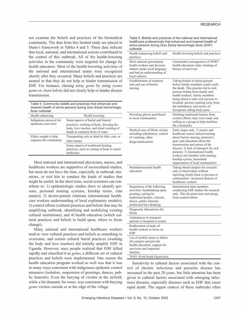
not examine the beliefs and practices of the biomedicalcommunity. The data from this limited study are placed inDunn’s framework in Tables 4 and 5. These data indicatethat local, national, and international actions contributed tothe control of this outbreak. All of the health-loweringactivities in the community were targeted for change byhealth educators. Most of the health-lowering activities ofthe national and international teams were recognizedshortly after they occurred. Many beliefs and practices areneutral in that they do not help or hinder transmission ofEHF. For instance, chasing away gemo by using ryemogemo or chani labolo did not clearly help or hinder diseasetransmission.
Most national and international physicians, nurses, andhealthcare workers are supportive of sociocultural studies,but most do not have the time, especially in outbreak situ-ations, or tool kits to conduct the kinds of studies thatmight be useful. In the short term, social scientists can con-tribute to: 1) epidemiologic studies (how to identify per-sons, personal naming systems, kinship terms, clannames); 2) doctor-patient relations (international health-care workers understanding of local explanatory models);3) control efforts (cultural practices and beliefs that may beamplifying outbreak, identifying and mobilizing existingcultural institutions); and 4) health education (which cul-tural practices and beliefs to build upon, where to focuschange).
Many national and international healthcare workerstend to view cultural practices and beliefs as something toovercome, and certain cultural burial practices (washingthe body and love touches) did initially amplify EHF inUganda. However, once people realized that EHF killedrapidly and classified it as gemo, a different set of culturalpractices and beliefs were implemented. One reason thehealth education program worked so well was that it wasin many ways consistent with indigenous epidemic controlmeasures (isolation, suspension of greetings, dances, pub-lic funerals). Even the burying of victims at the airfield,while a bit dramatic for some, was consistent with buryinggemo victims outside or at the edge of the village
Sensitivity to cultural factors associated with the con-trol of chronic infectious and parasitic disease hasincreased in the past 20 years, but little attention has beengiven to cultural factors associated with emerging infec-tious diseases, especially diseases such as EHF that causerapid death. The urgent context of these outbreaks often
Emerging Infectious Diseases • Vol. 9, No. 10, October 2003 1247
RESEARCH
Table 4. Community beliefs and practices that enhanced and lowered health of some persons during Gulu Ebola hemorrhagic fever outbreak Health enhancing Health lowering Indigenous protocol for epidemics (see text)
Some aspects of burial and funeral practices: washing of body, dressing the body, love touches, and ritual washing of hands in common bowl of water Transporting sick or dead by bike, cart, or other means
Elders sought to help organize the community
Some aspects of traditional healing practices, such as cutting of body to insert medicines
Table 5. Beliefs and practices of the national and international healthcare professionals that enhanced and lowered health of some persons during Gulu Ebola hemorrhagic fever (EHF) outbreak Health-enhancing beliefs and practices
Health-lowering beliefs and practices
Most national government health workers and decision makers spoke local language and had an understanding of local cultures
Unintended consequences of WHOa health education video: burning of houses of survivors
Establishment of isolation unit and use of barrier nursing
Taking bodies to burial ground before family members could verify the death. This practice led to sick persons hiding from family and health workers; family members being afraid to take sick persons to hospital; persons running away from the ambulance; and stories of Europeans selling body parts
Providing gloves and bleach to local communities
Omitting traditional healers from control efforts; they were ready and willing as a group to help mobilize the community
Medical care of Ebola victims including rehydration, control of vomiting, other drugs/medications
Early stages only: 1) nurses and healthcare nurses lacked training about barrier nursing, protective gear, and education about the transmission and nature of the disease; 2) lack of transport for sick patients; 3) international health workers not familiar with naming, kinship system, household organization of local communities
Multidimensional health education
Taking blood samples for research only or blood taken without reporting results back to persons or communities’ increased distrust of healthcare workers
Suspension of the following activities: handshaking upon greeting, cutting by traditional healers, schools, discos, public funerals, traditional beer drinking
International team members conducting EHF studies for research only. This diverted time and energy from control efforts
Diagnostic laboratories for Ebola
Ambulances to transport patients to hospital to isolate
Reallocation of tasks of health workers to focus on EHF
Use of mobile teams to follow all contacts and provide health education, support for survivors and impacted families
aWHO, World Health Organization.
leads to the neglect of local people’s feelings and knowl-edge. The general impression is that, without Westernintervention, the epidemic would kill hundreds and spreadto all parts of the world; local practices and beliefs are per-ceived only as amplifying the outbreaks. Our study was thefirst systematic sociocultural study of EHF. It showed thatsome cultural practices did indeed amplify the outbreak.However, an important finding was that local people havebeliefs and practices in place that can be useful to controlrapid epidemics, such as EHF, with high fatalities. Becauselocal people have lived with high mortality rates and seri-ous epidemics for some time, their knowledge may be use-ful to national and international teams in their efforts tocontrol emerging diseases.
AcknowledgmentsWe thank the families and persons who openly shared their
Ebola experiences; Cathy Roth, Thomas Oyok, Paul Onek, OkotLokach, Claudio Blé, Dan Bausch, and Chris Lane for their com-ments and support; Karl Johnson and Barney Cline for their com-ments on earlier drafts of this paper; and Kitza Francis for assis-tance in administering the survivor questionnaire.
Dr. Hewlett is a cultural anthropologist with interests in thecultural contexts of infectious and parasitic disease, evolutionarytheory, and child development. He has conducted research inCentral Africa for 30 years, mostly with Aka hunter-gatherers.
Mr. Amola is a medical officer with the Ugandan Ministryof Health. His research interests are primary healthcare andhealth education.
References
1. Special issue on Ebola hemorrhagic fever. J Infect Dis1999;179(Suppl 1).
2. World Health Organization. WHO recommended guidelines for epi-demic preparedness and response: Ebola haemorrhagic fever (EHF).Geneva: the Organization; 1997.
3. World Health Organization. Outbreak of Ebola haemorrhagic fever,Uganda, August 2000–January 2001. Wkly Epidemiol Rec2001;76:41–8.
4. Atkinson RR. Acholi. In: Middleton J, Rassam A, editors.Encyclopedia of world cultures: Africa and the Middle East. Boston:GK Hall; 1995.
5. Geissler PW. Worms are out life. Part I: understandings of worms andthe body among the Luo of Western Kenya. Anthropology andMedicine 1998;5:63–79.
6. Geissler PW. Worms are our life. Part II: Luo children’s thoughtsabout worms and illness. Anthropology and Medicine1998;5:133–44.
7. Cohen J. Deep denial. Sciences 2001;41:20–5.8. Evans Pritchard EE. The Nuer. Oxford (UK): Clarendon; 1956.9. Kabananukye KIB. Denial, discrimination and stigmatisation: the
case of Ebola epidemic in some districts, Uganda: Ugandan Ministryof Health, National Ebola Task Force; 2001
10. Dunn FL. Social determinants in tropical disease. In: Warren KS,Mahmoud ADF, editors. Tropical and geographical medicine. NewYork: McGraw-Hill; 1985.
Address for correspondence: Barry S. Hewlett, Department ofAnthropology, Washington State University, Vancouver, 14204 NESalmon Creek Ave., Vancouver, Washington, 98686 USA; fax: 360-546-9036; email: [email protected]
1248 Emerging Infectious Diseases • Vol. 9, No. 10, October 2003
RESEARCH
OPPORTUNITIES FOR PEER REVIEWERS
The editors of Emerging Infectious Diseases seek toincrease the roster of reviewers for manuscripts submitted byauthors all over the world for publication in the journal. If youare interested in reviewing articles on emerging infectious dis-ease topics, please e-mail your name, address, curriculumvitae, and areas of expertise to [email protected]
At Emerging Infectious Diseases, we always requestreviewers’ consent before sending manuscripts, limit reviewrequests to three or four per year, and allow 2-4 weeks forcompletion of reviews. We consider reviewers invaluable inthe process of selecting and publishing high-quality scientificarticles and acknowledge their contributions in the journalonce a year.
Even though it brings no financial compensation, participa-tion in the peer-review process is not without rewards.Manuscript review provides scientists at all stages of theircareer opportunities for professional growth by familiarizingthem with research trends and the latest work in the field ofinfectious diseases and by improving their own skills for pre-senting scientific information through constructive criticismof those of their peers. To view the spectrum of articles wepublish, information for authors, and our extensive styleguide, visit the journal web site at www.cdc.gov/eid.
For more information on participating in the peer-reviewprocess of Emerging Infectious Diseases, [email protected] or call the journal office at 404-371-5329.
All material published in Emerging Infectious Diseases is in thepublic domain and may be used and reprinted without special per-mission; proper citation, however, is appreciated.
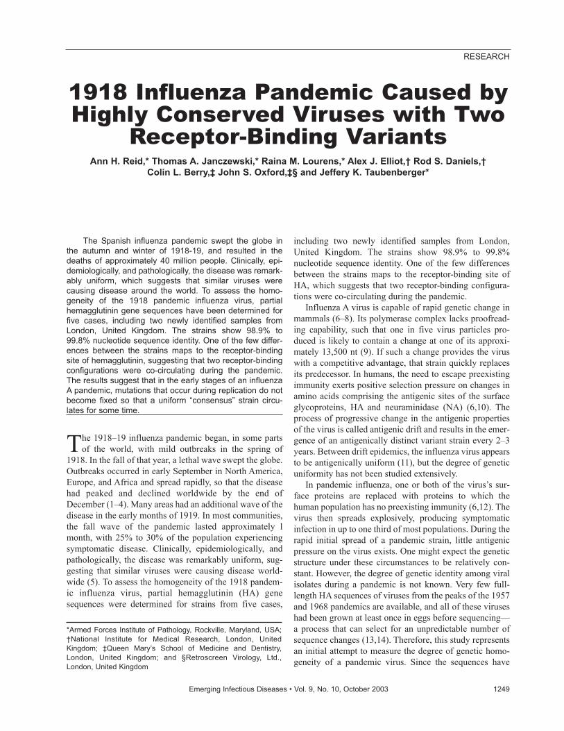
The Spanish influenza pandemic swept the globe inthe autumn and winter of 1918-19, and resulted in thedeaths of approximately 40 million people. Clinically, epi-demiologically, and pathologically, the disease was remark-ably uniform, which suggests that similar viruses werecausing disease around the world. To assess the homo-geneity of the 1918 pandemic influenza virus, partialhemagglutinin gene sequences have been determined forfive cases, including two newly identified samples fromLondon, United Kingdom. The strains show 98.9% to99.8% nucleotide sequence identity. One of the few differ-ences between the strains maps to the receptor-bindingsite of hemagglutinin, suggesting that two receptor-bindingconfigurations were co-circulating during the pandemic.The results suggest that in the early stages of an influenzaA pandemic, mutations that occur during replication do notbecome fixed so that a uniform “consensus” strain circu-lates for some time.
The 1918–19 influenza pandemic began, in some partsof the world, with mild outbreaks in the spring of
1918. In the fall of that year, a lethal wave swept the globe.Outbreaks occurred in early September in North America,Europe, and Africa and spread rapidly, so that the diseasehad peaked and declined worldwide by the end ofDecember (1–4). Many areas had an additional wave of thedisease in the early months of 1919. In most communities,the fall wave of the pandemic lasted approximately lmonth, with 25% to 30% of the population experiencingsymptomatic disease. Clinically, epidemiologically, andpathologically, the disease was remarkably uniform, sug-gesting that similar viruses were causing disease world-wide (5). To assess the homogeneity of the 1918 pandem-ic influenza virus, partial hemagglutinin (HA) genesequences were determined for strains from five cases,
including two newly identified samples from London,United Kingdom. The strains show 98.9% to 99.8%nucleotide sequence identity. One of the few differencesbetween the strains maps to the receptor-binding site ofHA, which suggests that two receptor-binding configura-tions were co-circulating during the pandemic.
Influenza A virus is capable of rapid genetic change inmammals (6–8). Its polymerase complex lacks proofread-ing capability, such that one in five virus particles pro-duced is likely to contain a change at one of its approxi-mately 13,500 nt (9). If such a change provides the viruswith a competitive advantage, that strain quickly replacesits predecessor. In humans, the need to escape preexistingimmunity exerts positive selection pressure on changes inamino acids comprising the antigenic sites of the surfaceglycoproteins, HA and neuraminidase (NA) (6,10). Theprocess of progressive change in the antigenic propertiesof the virus is called antigenic drift and results in the emer-gence of an antigenically distinct variant strain every 2–3years. Between drift epidemics, the influenza virus appearsto be antigenically uniform (11), but the degree of geneticuniformity has not been studied extensively.
In pandemic influenza, one or both of the virus’s sur-face proteins are replaced with proteins to which thehuman population has no preexisting immunity (6,12). Thevirus then spreads explosively, producing symptomaticinfection in up to one third of most populations. During therapid initial spread of a pandemic strain, little antigenicpressure on the virus exists. One might expect the geneticstructure under these circumstances to be relatively con-stant. However, the degree of genetic identity among viralisolates during a pandemic is not known. Very few full-length HA sequences of viruses from the peaks of the 1957and 1968 pandemics are available, and all of these viruseshad been grown at least once in eggs before sequencing—a process that can select for an unpredictable number ofsequence changes (13,14). Therefore, this study representsan initial attempt to measure the degree of genetic homo-geneity of a pandemic virus. Since the sequences have
Emerging Infectious Diseases • Vol. 9, No. 10, October 2003 1249
RESEARCH
1918 Influenza Pandemic Caused byHighly Conserved Viruses with Two
Receptor-Binding Variants Ann H. Reid,* Thomas A. Janczewski,* Raina M. Lourens,* Alex J. Elliot,† Rod S. Daniels,†
Colin L. Berry,‡ John S. Oxford,‡§ and Jeffery K. Taubenberger*
*Armed Forces Institute of Pathology, Rockville, Maryland, USA;†National Institute for Medical Research, London, UnitedKingdom; ‡Queen Mary’s School of Medicine and Dentistry,London, United Kingdom; and §Retroscreen Virology, Ltd.,London, United Kingdom
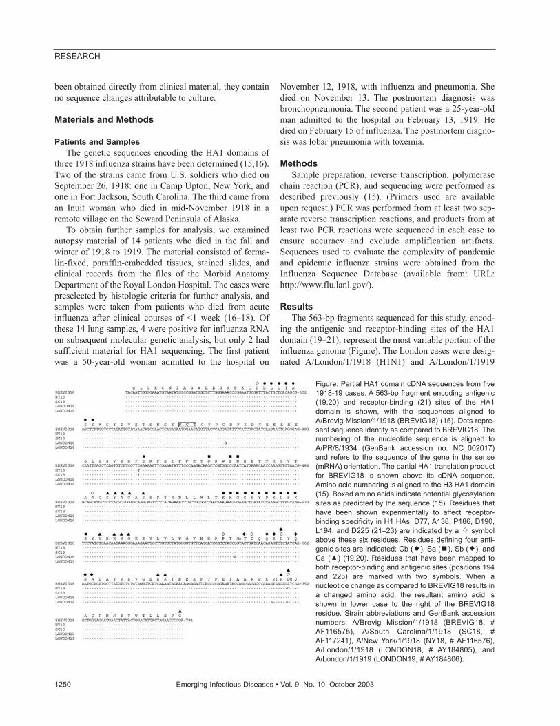
been obtained directly from clinical material, they containno sequence changes attributable to culture.
Materials and Methods
Patients and SamplesThe genetic sequences encoding the HA1 domains of
three 1918 influenza strains have been determined (15,16).Two of the strains came from U.S. soldiers who died onSeptember 26, 1918: one in Camp Upton, New York, andone in Fort Jackson, South Carolina. The third came froman Inuit woman who died in mid-November 1918 in aremote village on the Seward Peninsula of Alaska.
To obtain further samples for analysis, we examinedautopsy material of 14 patients who died in the fall andwinter of 1918 to 1919. The material consisted of forma-lin-fixed, paraffin-embedded tissues, stained slides, andclinical records from the files of the Morbid AnatomyDepartment of the Royal London Hospital. The cases werepreselected by histologic criteria for further analysis, andsamples were taken from patients who died from acuteinfluenza after clinical courses of <1 week (16–18). Ofthese 14 lung samples, 4 were positive for influenza RNAon subsequent molecular genetic analysis, but only 2 hadsufficient material for HA1 sequencing. The first patientwas a 50-year-old woman admitted to the hospital on
November 12, 1918, with influenza and pneumonia. Shedied on November 13. The postmortem diagnosis wasbronchopneumonia. The second patient was a 25-year-oldman admitted to the hospital on February 13, 1919. Hedied on February 15 of influenza. The postmortem diagno-sis was lobar pneumonia with toxemia.
MethodsSample preparation, reverse transcription, polymerase
chain reaction (PCR), and sequencing were performed asdescribed previously (15). (Primers used are availableupon request.) PCR was performed from at least two sep-arate reverse transcription reactions, and products from atleast two PCR reactions were sequenced in each case toensure accuracy and exclude amplification artifacts.Sequences used to evaluate the complexity of pandemicand epidemic influenza strains were obtained from theInfluenza Sequence Database (available from: URL:http://www.flu.lanl.gov/).
ResultsThe 563-bp fragments sequenced for this study, encod-
ing the antigenic and receptor-binding sites of the HA1domain (19–21), represent the most variable portion of theinfluenza genome (Figure). The London cases were desig-nated A/London/1/1918 (H1N1) and A/London/1/1919
1250 Emerging Infectious Diseases • Vol. 9, No. 10, October 2003
RESEARCH
Figure. Partial HA1 domain cDNA sequences from five1918-19 cases. A 563-bp fragment encoding antigenic(19,20) and receptor-binding (21) sites of the HA1domain is shown, with the sequences aligned toA/Brevig Mission/1/1918 (BREVIG18) (15). Dots repre-sent sequence identity as compared to BREVIG18. Thenumbering of the nucleotide sequence is aligned toA/PR/8/1934 (GenBank accession no. NC_002017)and refers to the sequence of the gene in the sense(mRNA) orientation. The partial HA1 translation productfor BREVIG18 is shown above its cDNA sequence.Amino acid numbering is aligned to the H3 HA1 domain(15). Boxed amino acids indicate potential glycosylationsites as predicted by the sequence (15). Residues thathave been shown experimentally to affect receptor-binding specificity in H1 HAs, D77, A138, P186, D190,L194, and D225 (21–23) are indicated by a ( symbolabove these six residues. Residues defining four anti-genic sites are indicated: Cb (#), Sa (!), Sb ("), andCa (%) (19,20). Residues that have been mapped toboth receptor-binding and antigenic sites (positions 194and 225) are marked with two symbols. When anucleotide change as compared to BREVIG18 results ina changed amino acid, the resultant amino acid isshown in lower case to the right of the BREVIG18residue. Strain abbreviations and GenBank accessionnumbers: A/Brevig Mission/1/1918 (BREVIG18, #AF116575), A/South Carolina/1/1918 (SC18, #AF117241), A/New York/1/1918 (NY18, # AF116576),A/London/1/1918 (LONDON18, # AY184805), andA/London/1/1919 (LONDON19, # AY184806).
(H1N1). These two sequences, when compared to the threepreviously sequenced North American strains (15), differfrom each other by 1 nt to 3 nt, showing a sequence iden-tity of 98.9% to 99.8%.
A/London/1/1918 shows 2 nt differences, compared toA/Brevig Mission/1/1918, one of which would change theamino acid at codon 188 from G to S (amino acid number-ing is aligned to the H3 influenza HA). This residue is nearseveral of the residues that have been shown experimental-ly to affect receptor-binding specificity of H1 HAs (21–23)and next to one of the mapped Sb antigenic site residues(19,20). A/London/1/1919 shows 3 nt differences fromA/Brevig Mission/1/1918, 2 of which are nonsynonymous,resulting in changes of V223I and D225G. The V223Ichange is near Ca antigenic site residues, and the D225Gchange is at a residue that functions both in receptor-bind-ing and as a Ca antigenic site residue. Amino acid 225 alsovaries among North American strains; A/NewYork/1/1918, like A/London/1/1919, has a glycine at posi-tion 225, as do most avian influenza strains. A/SouthCarolina/1/1918 and A/Brevig Mission/1/1918, likeA/London/1/1918 and most subsequent human H1 strains,have aspartic acid at this position (Figure) (15). The rela-tive genetic homogeneity of the 1918–19 isolates encour-aged us to analyze sequences from the 1957 and 1968 pan-demics.
GenBank contains complete HA1 domain–encodingsequences for eight 1957 H2N2 strains. As noted in previ-ous studies of receptor-binding specificity (22,24), the1957 strains have undergone varying passage histories, butall have been passed at least once. Three of the strains havebeen sequenced more than once and differ by as many as 8nt within the same strain. Between sequences, the numberof nucleotide differences ranges from only 1 nt differencebetween A/Chile/6/1957 and A/Davis/1/1957 to 12 differ-ences between one of the A/Japan/305/1957 sequences andone of the A/Singapore/1/1957 sequences. Overall, thesequences show 98.9% to 99.9% identity at the nucleotidelevel, and 98.5% to 100% identity at the amino acid level.
More limited sequence data are available for the 1968H3N2 pandemic strains. The complete HA1 domainsequence is available for only three strains, two of whichhave been sequenced twice each. The twoA/NT/60/68/29C sequences differ by 4 nt. The most diver-gent sequences differ by 24 nt (A/NT/60/68/29C vs.A/Hong Kong/1/68), thus showing 97.6% to 100% identi-ty between sequences at the nucleotide level, and 96.0% to100% identity at the amino acid level.
Studies from epidemic years have yielded similarresults. A 2001 study (25) examined variation in the HAgene of human H3N2 viruses in Spain from 1996 to 2000.During this time, strains antigenically similar toA/Wuhan/359/1995 were replaced by strains similar to
A/Sydney/5/1997 and then by strains similar toA/Panama/2007/1999. Within the groups of virusesbelonging to each antigenic group, sequence variation wasminimal. For example, among the viruses that reacted anti-genically with Sydney, but not Panama and Wuhan, 2–10nt differences occurred over the 591 nt sequenced (98.3%to 99.7% identity).
An unpublished study provides sequences of the HA1domain of the H3-subtype HA of 16 A/Sydney/05/1997-like (H3N2) influenza virus isolates circulating in Canadaduring the 1997/98 influenza epidemic season (GenBankno. AF087700–AF087702, AF087707, AF087708,AF096306–AF096316) (26). Two of the isolates had iden-tical sequences, while the others varied by 1 nt to 14 ntover 981 nt (98.6% to 100% identity).
DiscussionThe three North American 1918 influenza strains
sequenced previously were isolated from patients separat-ed by nearly 2 months in time and almost 4,000 miles indistance (27). Two nucleotide differences were foundamong these three strains, one of which resulted in anamino acid substitution in the receptor-binding site (15).All three cases likely derived from the initial introductionof the fall wave into the United States, believed to haveoccurred in Boston in early September 1918. The virusthen spread rapidly from Camp Devens, Massachusetts,the first U.S. army base to experience the epidemic, whichthen reached army bases throughout the eastern UnitedStates within 2 weeks (2). Influenza probably reachedBrevig Mission, Alaska, via Seattle, Washington. The pan-demic reached Camp Lewis, Washington, in mid-September, following the arrival of a troop ship fromPhiladelphia, Pennsylvania (1,2), and spread to Seattle bylate September. After careful screening to exclude sickpassengers, a ship left Seattle for Nome, Alaska, in mid-October, but days after its arrival local residents beganfalling ill (1). An account of the pandemic as it occurred inBrevig Mission reports that visitors from Nome broughtthe disease to the village in November (28). This chain ofevents suggests that the Alaskan outbreak was not theresult of a separate introduction of the 1918 influenza fromAsia to the West Coast of the United States.
The spring wave of the 1918 epidemic was widespreadin France and Spain during April and May but did notreach England until June. The fall wave also arrived some-what later in England than in continental Europe and theUnited States; peak mortality in London occurred duringthe first 2 weeks of November (2). A second peak occurredin the third week of February 1919. One strain from eachof these peaks was sequenced for this study.
Our results show that strains separated by over 7,500miles (Brevig Mission, Alaska, to London, United
Emerging Infectious Diseases • Vol. 9, No. 10, October 2003 1251
RESEARCH
Kingdom) and several months (September 26, 1918, toFebruary 15, 1919) share a sequence identity of 99%. Thislevel of genetic homogeneity is slightly higher than thatseen for the available 1957 and 1968 pandemic strains, butthe 1957 and 1968 strains were not sequenced directlyfrom clinical material. Sequences from different passagesof the same strain were sometimes as different from eachother as they were from other strains (29), suggesting thatsequence heterogeneity observed was the result of cultureadaptation, making it impossible to determine how homo-geneous the pandemic viruses actually were. Even so, the1957 and 1968 pandemic strains show >97% identitybetween strains. Similar levels of genetic homogeneitywere seen in strains from case-patients isolated from a driftepidemic in 1997. Thus, influenza viruses circulating dur-ing a single outbreak, whether epidemic or pandemic,show levels of sequence identity consistent with the uni-formity of the 1918 cases.
Despite the uniformity of the 1918 strains, one of thevariable sites is an amino acid known to be important inreceptor binding (21). At a subset of amino acids criticalfor receptor binding, avian strains differ from swine H1s atonly one amino acid, E190D (15). At these amino acids,two of the cases (A/New York/1/1918 andA/London/1/1919) are identical to that of A/sw/Iowa/1976/31 (a classical swine strain). The other 1918cases have an additional change from the avian consensusat amino acid 225. Since swine viruses with the samereceptor site as A/sw/Iowa/1976/31 bind both SAα2,3Galand SAα2,6Gal (14), A/New York/1/1918 andA/London/1/1919 probably also had the capacity to bindboth receptors. Because two of five 1918-19 analyzed fallwave strains from case-patients have the swine-like recep-tor-binding pattern, the E190D change alone is apparentlysufficient to allow viral replication in the human respirato-ry tract. However, the existence of three strains with theadditional G225D change shows that both receptor-bind-ing variants were co-circulating throughout the pandemic.The current evidence does not suggest progression fromone receptor-binding pattern to the other during the pan-demic, since the two variants are present, on both conti-nents, both early and late in the pandemic. Since residue225 has also been identified as part of the Ca antigenic site(19), the co-circulating strains possibly differed in anti-genic reactivity as well as receptor-binding characteristics.
This study is the first to examine the genetic homogene-ity of a pandemic influenza virus directly from clinicalmaterial. The results suggest that in the early stages of apandemic, mutations that occur during replication do notbecome fixed so that a uniform consensus strain circulatesfor some time. Studies of influenza strains circulating after1919 should provide insight into how pandemic virusesevolve after the initial waves through immunologically
vulnerable populations. In terms of pandemic planning,our results indicate that a specific antiviral drug or vaccinewould have a uniform effect during the important andoften lethal first wave of a pandemic (30,31).
This work was supported by a grant from the NationalInstitute of Allergy and Infectious Diseases (R01 AI50619-01) toJ.K.T. and by the intramural funds of the Armed Forces Instituteof Pathology. J.S.O., C.L.B., and R.S.D. gratefully acknowledgefinancial support from the Wellcome Trust and the Ian Heap fund.
Ms. Reid is a research biologist in the Molecular PathologyDivision at the Armed Forces Institute of Pathology. Her princi-pal research interest is pandemic influenza.
References
1. Crosby A. America’s forgotten pandemic. Cambridge: CambridgeUniversity Press; 1989.
2. Jordan E. Epidemic influenza: a survey. Chicago: American MedicalAssociation; 1927.
3. Reid AH, Taubenberger JK, Fanning TG. The 1918 Spanish influen-za: integrating history and biology. Microbes Infect 2001;3:81–7.
4. Taubenberger JK, Reid AH, Janczewski TA, Fanning TG. Integratinghistorical, clinical and molecular genetic data in order to explain theorigin and virulence of the 1918 Spanish influenza virus. Philos TransR Soc Lond B Biol Sci 2001;356:1829–39.
5. Patterson KD, Pyle GF. The geography and mortality of the 1918influenza pandemic. Bull Hist Med 1991;65:4–21.
6. Wright PE, Webster RG. Orthomyxoviruses. In: Knipe DM, HowleyPM, editors. Fields virology. Vol 1. Philadelphia: Lippincott Williamsand Wilkins; 2001:1533–79.
7. Webster RG, Bean WJ, Gorman OT, Chambers TM, Kawaoka Y.Evolution and ecology of influenza A viruses. Microbiol Rev1992;56:152–79.
8. Lamb RA, Takeda M. Death by influenza virus protein. Nat Med2001;7:1286–8.
9. Parvin JD, Smith FI, Palese P. Rapid RNA sequencing using double-stranded template DNA, SP6 polymerase, and 3'-deoxynucleotidetriphosphates. DNA. 1986;5:167–71.
10. Hay AJ, Gregory V, Douglas AR, Lin YP. The evolution of humaninfluenza viruses. Philos Trans R Soc Lond B Biol Sci2001;356:1861–70.
11. Centers for Disease Control and Prevention. Influenza summaryupdate: 2001–2 influenza season summary. June 10, 2002. [AccessedSeptember 23, 2002]. Available from: URL: http://www.cdc.gov/nci-dod/diseases/flu/weeklyarchives/01-02summary.htm
12. Kilbourne E. Influenza pandemics in perspective. JAMA1977;237:1225–8.
13. Schild GC, Oxford JS, de Jong JC, Webster RG. Evidence for host-cell selection of influenza virus antigenic variants. Nature1983;303:706–9.
14. Gambaryan A, Tuzikov A, Piskarev V, Yamnikova SS, Lvov DK,Robertson JS, et al. Specification of receptor-binding phenotypes ofinfluenza virus isolates from different hosts using synthetic sialylgly-copolymers: non-egg-adapted human H1 and H3 influenza A andinfluenza B viruses share a common high binding affinity for 6'-sia-lyl(N-acetyllactosamine). Virology 1997;232:345–50.
15. Reid AH, Fanning TG, Hultin JV, Taubenberger JK. Origin and evo-lution of the 1918 “Spanish” influenza virus hemagglutinin gene.Proc Natl Acad Sci U S A 1999;96:1651–6.
1252 Emerging Infectious Diseases • Vol. 9, No. 10, October 2003
RESEARCH
16. Taubenberger JK, Reid AH, Krafft AE, Bijwaard KE, Fanning TG.Initial genetic characterization of the 1918 “Spanish” influenza virus[see comments]. Science. 1997;275:1793–6.
17. Winternitz MC, Wason IM, McNamara FP. The pathology of influen-za. New Haven (CT): Yale University Press; 1920.
18. Wolbach SB. Comments on the pathology and bacteriology of fatalinfluenza cases, as observed at Camp Devens, Mass. Johns HopkinsHospital Bulletin 1919;30:104.
19. Caton AJ, Brownlee GG, Yewdell JW, Gerhard W. The antigenicstructure of the influenza virus A/PR/8/34 hemagglutinin (H1 sub-type). Cell 1982;31:417–27.
20. Raymond F, Caton A, Cox N, Kendal AP, Brownlee GG. The anti-genicity and evolution of influenza H1 haemagglutinin, from1950–57 and 1977–1983: two pathways from one gene. Virology1986;148:275–87.
21. Matrosovich M, Gambaryan A, Teneberg S, Piskarev VE, YamnikovaSS, Lvov DK, et al. Avian influenza A viruses differ from humanviruses by recognition of sialyloigosaccharides and gangliosides andby a higher conservation of the HA receptor-binding site. Virology1997;233:224–34.
22. Rogers G, D’Souza B. Receptor binding properties of human and ani-mal H1 influenza virus isolates. Virology 1989;173:317–22.
23. Matrosovich MN, Gambaryan AS, Tuzikov AB, Byramova NE,Mochalova LV, Golbraikh AA, et al. Probing of the receptor-bindingsites of the H1 and H3 influenza A and influenza B virus hemagglu-tinins by synthetic and natural sialosides. Virology 1993;196:111–21.
24. Matrosovich M, Tuzikov A, Bovin N, Gambaryan A, Klimov A,Castrucci MR, et al. Early alterations of the receptor-binding proper-ties of H1, H2, and H3 avian influenza virus hemagglutinins aftertheir introduction into mammals. J Virol 2000;74:8502–12.
25. Coiras MT, Aguilar JC, Galiano M, Carlos S, Gregory V, Lin YP, etal. Rapid molecular analysis of the haemagglutinin gene of humaninfluenza A H3N2 viruses isolated in Spain from 1996 to 2000. ArchVirol 2001;146:2133–47.
26. Macken C, Lu H, Goodman J, Boykin L. The value of a database insurveillance and vaccine selection. In: Osterhaus A, Cox N, HampsonA, editors. Options for the control of influenza IV. Amsterdam:Excerpta Medica; 2001. p. 103–6.
27. Reid AH, Taubenberger JK. The 1918 flu and other influenza pan-demics: “over there” and back again. Lab Invest 1999;79:95–101.
28. Fosso C. Alone with death on the Tundra. In: Hedin R, Holthaus G,editors. Alaska: Reflections on land and spirit. Tucscon (AZ):University of Arizona Press; 1989. p. 215–22.
29. Connor R, Kawaoka Y, Webster R, Paulson J. Receptor specificity inhuman, avian, and equine H2 and H3 influenza virus isolates.Virology 1994;205:17–23.
30. Gensheimer KF, Fukuda K, Brammer L, Cox N, Patriarca PA, StrikesRA. Preparing for pandemic influenza: the need for enhanced surveil-lance. Vaccine 2002;20(Suppl 2):S63–5.
31. Hayden FG. Perspectives on antiviral use during pandemic influenza.Philos Trans R Soc Lond B Biol Sci 2001;356:1877–84.
Address for correspondence: Jeffery K. Taubenberger, Armed ForcesInstitute of Pathology, Department of Cellular Pathology and Genetics,1413 Research Blvd., Building 101, Room 1057, Rockville, MD 20850-3125, USA; fax: 1-301-295-9507; email: [email protected]
Emerging Infectious Diseases • Vol. 9, No. 10, October 2003 1253
RESEARCH
AmpC β-lactamase, altered porins, or both are usuallyresponsible for cefoxitin resistance in Escherichia coli. Weexamined the relative importance of each. We studied 18strains of clinical isolates with reduced cefoxitin susceptibil-ity and 10 initially-susceptible strains passaged throughcefoxitin-gradient plates. Of 18 wild-resistant strains, 9 hadidentical promoter mutations (including creation of a con-sensus 17-bp spacer) and related pulsed-field gel elec-trophoresis patterns; the other 9 strains were unrelated.Nine strains had attenuator mutations; two strains did notexpress OmpC or OmpF. After serial passage, 8 of 10strains developed cefoxitin resistance, none developedpromoter or attenuator mutations, 6 lost both the OmpCand OmpF porin proteins, and 1 showed decreased pro-duction of both. One strain had neither porin alteration orincreased AmpC production. Porin mutants may occurmore commonly and be less fit and less inclined to spreador cause disease than strains with increased β-lactamaseexpression.
The development of antibiotic resistance in Escherichiacoli has important clinical implications. E. coli is
among the most frequently isolated bacterium in a varietyof clinical settings. The development of resistance to olderagents such as ampicillin and trimethoprim-sulfamethoxa-zole, as well as the emerging problem of fluoroquinoloneresistance, may substantially limit our antibiotic choices(1,2).
Although cephamycin-resistant E. coli is relativelyuncommon, widespread use of β-lactam antiboties maycontribute to the development and spread of these strains.In 1999, Sahm et al. reported that 0.16% of E. coli wereresistant to cephamycins (3). At a local level, unpublisheddata from the Queen Elizabeth II Health Science Centre inHalifax, Nova Scotia indicated that, of the 5,767 strains of
E. coli processed from urine samples, 0.4% werecephamycin resistant.
All strains of E. coli possess a gene that encodes anAmpC β-lactamase. Usually, almost no β-lactamase is pro-duced because the gene is preceded by a weak promoterand a strong attenuator (4). Surveys of resistance mecha-nisms in cephamycin-resistant strains have most oftenidentified promoter or attenuator mutations, which resultsin an up-regulation of AmpC β-lactamase production(5–7). Occasionally, cephamycin-resistant strains bearmobilized β-lactamases derived from bacteria such asCitrobacter feundii (8). In addition, mutation or alteredexpression of outer membrane proteins constituting porinscan also contribute to cephamycin resistance. To ourknowledge, no investigators have concurrently looked foralterations in porins in addition to promoter-attenuatormutations. Porin alterations might work together to pro-duce a higher level of resistance. In addition, porin alter-ations may protect E. coli and allow subsequent selectionfor promoter and attenuator mutants.
We examined E. coli strains collected at our hospital todetermine the basis for resistance. In addition, we createdcephamycin-resistant strains of E. coli by serial passage oncefoxitin-containing medium to determine which of thesetwo resistance mechanisms was predominant and if ourfindings were representative of those seen in clinical iso-lates.
Materials and Methods
Bacterial StrainsWe collected strains of E. coli from midstream urine
from inpatients and from patients in the community.Eighteen strains with reduced susceptibility (MIC >8mg/L) to cefoxitin were included in the analysis, whichrepresented all resistant strains collected during a 6-monthperiod in 2001. For the in vitro development of resistance,
1254 Emerging Infectious Diseases • Vol. 9, No. 10, October 2003
RESEARCH
Cephamycin Resistance in ClinicalIsolates and Laboratory-derived
Strains of Escherichia coli,Nova Scotia, Canada
Brian Clarke,* Margot Hiltz,* Heather Musgrave,* and Kevin R. Forward*
*Dalhousie University and the Queen Elizabeth II Health SciencesCentre, Halifax, Nova Scotia, Canada
we selected 10 clinical isolates from urine that were fullysusceptible to β-lactam antibiotics. In both cases, weexcluded duplicate strains from the same patient. E. coliisolates were identified with conventional biochemicalreactions. Organisms were identified by spot indole and β-glucuronidase assays and confirmed by automated Vitekby using GNI+ cards, and antibiotic susceptibilities wereperformed by using GNS 606 cards (bioMerieux CanadaInc., St. Laurent, Quebec).
Analysis of Promoter and Attenuator MutationsE. coli chromosomal DNA was isolated with a QIAmp
DNA Mini Kit (Qiagen Inc., Valencia, CA), according tothe manufacturer’s instructions. Using standard methods,we performed polymerase chain reaction (PCR) with a pre-viously published primer set and protocol which amplifiesthe region of DNA including the –35 box of the AmpC pro-moter and the 3′ end of the attenuator, producing a 271-bpamplicon (5). Amplification was performed in a PTC-200Peltier Thermal Cycler (MJ Research, Boston, MA). Theamplicons were resolved by 2% agarose gel electrophore-sis and visualized after staining with ethidium bromide.The amplicons were purified by using the QIAquick PCRPurification Kit (Qiagen Inc.) and sequenced directly inboth directions by using the dideoxy chain terminationprocedure of Sanger et al. on an ABI Prism automatedsequencer at York University Core Molecular Laboratory,Toronto, Ontario, Canada.
Molecular FingerprintingStrains sharing similar promoter or attenuator muta-
tions were fingerprinted by pulsed field gel electrophoresis(PFGE) by using a modification of the method of Gautom(9). In brief, a standardized suspension of E. coli was pre-pared from overnight cultures and treated with lysozymeand proteinase K. Plugs were prepared in low-meltagarose. Solidified plugs were deproteinated with sodiumlauryl sarcosine and proteinase K, and then washed repeat-edly. Two millimeter slices of plug were digested withXbal at 37°C for 3 h in the recommended buffer. Plugswere loaded onto a 1% agarose gel and resolved with aCHEF-Mapper system (Bio-Rad Laboratories,Mississauga, Ontario).
Outer Membrane ProfilesBacteria were grown overnight in Luria-Burtani (LB)
broth with or without 4 mg/L of cefoxitin. To study Ompexpression, 30 mL of LB broth was injected with 300 µLof a bacterial cell suspension from an overnight culture.Cultures were incubated at 37°C in a shaking water bath at250 rpm to an optical density at 600 nm of 1.0. Cell mem-branes were disrupted with a sonicator for 2 min with 30-sec cycles intermittent on ice. Cell debris was removed by
centrifugation at 10,000 g for 10 min at 4°C. Cytoplasmicmembrane proteins were differentially solubilized for 20min at room temperature with 1.7% sodium-lauryl-sarcosi-nate in 100 mM Tris, pH 8.0. The suspension was then cen-trifuged at 100,000 g for 20 min at 4°C, and the pellet con-taining the outer membrane proteins was resuspended in100 µL sterile distilled water. Omp preparations were ana-lyzed by urea-sodium dodecyl sulfate-polyacrylamide gelelectrophoresis at 30 mA in gels prepared with 11% acry-lamide, 0.3% bisacrylamide, 8 M urea, and 0.1% SDSusing the discontinuous buffer system of Laemmli (10).The gels were stained with Coomassie brilliant blue. Thepositions of OmpC and OmpF on the Omp profiles wereascertained by comparing the profiles of Omp preparationsfrom the E. coli reference strains MH760 (ompR472OmpC- OmpF+) and MH1461 (envz11 OmpC+ OmpF-)(11).
Development of Cefoxitin-resistant StrainsCefoxitin-resistant mutants were obtained by serially
passaging the wild type strains on 9 cm x 9 cm2 gradientplates containing a maximum of 32 mg/L cefoxitin in MHagar, as previously described (12). Plates were incubated atroom temperature overnight before use to ensure properdiffusion of the antibiotic. Streaked plates were incubatedovernight at 37°C and the colony that grew furthest up thecefoxitin gradient was selected and replated on a fresh gra-dient plate the following day. A total of 12 passages wereperformed for each strain at a maximum concentration of32 mg/L cefoxitin and an additional 15 passages at a max-imum of 128 mg/L cefoxitin. Isoelectric focusing was per-formed by using a modification of the method (13).
ResultsEach of the 18 strains with reduced susceptibility to
cefoxitin was also resistant to ampicillin, cephalothin, andamoxicillin/clavulanate acid. All were imipenem suscepti-ble. Isoelectric focusing demonstrated chromosomalAmpC in all strains; no other β-lactamases were identified.A summary of promoter and attenuator mutations, as wellas alterations in outer membrane profiles, is shown inTable 1 and Figures 1 and 2. Strains QE1–QE9 were iden-tical or closely related by PFGE. Each strain had a 1-bpinsertion in the spacer region between the –35 and –10boxes. This insertion created a consensus 17-bp spacer. Inaddition to this mutation, these strains had additionalmutations at –73, +6, and +81. Strain QE7 also had a dele-tion in the loop of the attenuator. None of these strains hadchanges in their outer membrane protein profiles.
The other nine strains (QE10–QE18) had differentPFGE patterns and came from diverse locations (differenthospitals and from both outpatients and inpatients). StrainQE10 had a C to T mutation at –42 and a G to A at –18,
Emerging Infectious Diseases • Vol. 9, No. 10, October 2003 1255
RESEARCH
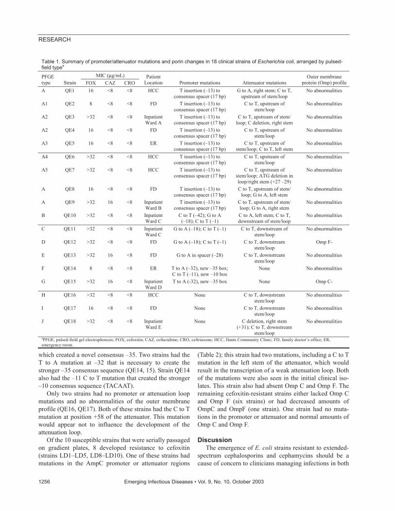
which created a novel consensus –35. Two strains had theT to A mutation at –32 that is necessary to create thestronger –35 consensus sequence (QE14, 15). Strain QE14also had the –11 C to T mutation that created the stronger–10 consensus sequence (TACAAT).
Only two strains had no promoter or attenuation loopmutations and no abnormalities of the outer membraneprofile (QE16, QE17). Both of these strains had the C to Tmutation at position +58 of the attenuator. This mutationwould appear not to influence the development of theattenuation loop.
Of the 10 susceptible strains that were serially passagedon gradient plates, 8 developed resistance to cefoxitin(strains LD1–LD5, LD8–LD10). One of these strains hadmutations in the AmpC promoter or attenuator regions
(Table 2); this strain had two mutations, including a C to Tmutation in the left stem of the attenuator, which wouldresult in the transcription of a weak attenuation loop. Bothof the mutations were also seen in the initial clinical iso-lates. This strain also had absent Omp C and Omp F. Theremaining cefoxitin-resistant strains either lacked Omp Cand Omp F (six strains) or had decreased amounts ofOmpC and OmpF (one strain). One strain had no muta-tions in the promoter or attenuator and normal amounts ofOmp C and Omp F.
DiscussionThe emergence of E. coli strains resistant to extended-
spectrum cephalosporins and cephamycins should be acause of concern to clinicians managing infections in both
1256 Emerging Infectious Diseases • Vol. 9, No. 10, October 2003
RESEARCH
Table 1. Summary of promoter/attenuator mutations and porin changes in 18 clinical strains of Escherichia coli, arranged by pulsed-field typea
MIC (µg/mL) PFGE type Strain FOX CAZ CRO
Patient Location Promoter mutations Attenuator mutations
Outer membrane protein (Omp) profile
A QE1 16 <8 <8 HCC T insertion (–13) to consensus spacer (17 bp)
G to A, right stem; C to T, upstream of stem/loop
No abnormalities
A1 QE2 8 <8 <8 FD T insertion (–13) to consensus spacer (17 bp)
C to T, upstream of stem/loop
No abnormalities
A2 QE3 >32 <8 <8 Inpatient Ward A
T insertion (–13) to consensus spacer (17 bp)
C to T, upstream of stem/ loop; C deletion, right stem
No abnormalities
A2 QE4 16 <8 <8 FD T insertion (–13) to consensus spacer (17 bp)
C to T, upstream of stem/loop
No abnormalities
A3 QE5 16 <8 <8 ER T insertion (–13) to consensus spacer (17 bp)
C to T, upstream of stem/loop; C to T, left stem
No abnormalities
A4 QE6 >32 <8 <8 HCC T insertion (–13) to consensus spacer (17 bp)
C to T, upstream of stem/loop
No abnormalities
A5 QE7 >32 <8 <8 HCC T insertion (–13) to consensus spacer (17 bp)
C to T, upstream of stem/loop; ATG deletion in loop/right stem (+27 –29)
No abnormalities
A QE8 16 <8 <8 FD T insertion (–13) to consensus spacer (17 bp)
C to T, upstream of stem/ loop; G to A, left stem
No abnormalities
A QE9 >32 16 <8 Inpatient Ward B
T insertion (–13) to consensus spacer (17 bp)
C to T, upstream of stem/ loop; G to A, right stem
No abnormalities
B QE10 >32 <8 <8 Inpatient Ward C
C to T (–42); G to A (–18); C to T (–1)
C to A, left stem; C to T, downstream of stem/loop
No abnormalities
C QE11 >32 <8 <8 Inpatient Ward C
G to A (–18); C to T (–1) C to T, downstream of stem/loop
No abnormalities
D QE12 >32 <8 <8 FD G to A (–18); C to T (–1) C to T, downstream stem/loop
Omp F-
E QE13 >32 16 <8 FD G to A in spacer (–28) C to T, downstream stem/loop
No abnormalities
F QE14 8 <8 <8 ER T to A (–32), new –35 box; C to T (–11), new –10 box
None No abnormalities
G QE15 >32 16 <8 Inpatient Ward D
T to A (-32), new –35 box None Omp C-
H QE16 >32 <8 <8 HCC None C to T, downstream stem/loop
No abnormalities
I QE17 16 <8 <8 FD None C to T, downstream stem/loop
No abnormalities
J QE18 >32 <8 <8 Inpatient Ward E
None C deletion, right stem (+31); C to T, downstream
stem/loop
No abnormalities
aPFGE, pulsed-field gel electrophoresis; FOX, cefoxitin; CAZ, ceftazidime; CRO, ceftriaxone; HCC, Hants Community Clinic; FD, family doctor’s office; ER, emergency room.
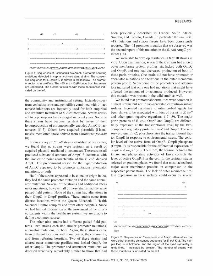
the community and institutional setting. Extended-spec-trum cephalosporins and penicillins combined with β- lac-tamase inhibitors are frequently used for both empiricaland definitive treatment of E. coli infections. Strains resist-ant to cephamycins have emerged in recent years. Some ofthese strains have become resistant by virtue of theirhyperproduction of chromosomally encoded AmpC β-lac-tamases (5–7). Others have acquired plasmidic β-lacta-mases; most often those derived from Citrobacter freundii(8).
In our survey of E. coli strains identified at our center,we found that no strains were resistant as a result ofacquired-plasmid–mediated β-lactamases. These strains allproduced substantial amounts of AmpC β-lactamase withthe isoelectric point characteristic of the E. coli–derivedAmpC. The predominant reason for the hyperproductionof AmpC appeared to be promoter mutations, attenuatormutations, or both.
Half of the strains appeared to be clonal in origin in thatthey had the same promoter mutation and the same attenu-ator mutations. Several of the strains had additional atten-uator mutations; however, all of these strains had the samepulsed-field pattern. None of the strains had alterations intheir OmpC or OmpF profiles. These strains came fromdiverse locations within the Queen Elizabeth II HealthSciences Centre complex and from other hospitals. Sincewe had limited information on the movement of the infect-ed patients within the healthcare system, we are unable todefine a common source.
The other nine strains had different pulsed-field pat-terns. Two strains each had similar promoter mutations,attenuator mutations, or both. Again, these strains camefrom different locations within our center, the community,and from referring hospitals. Two of these strains hadaltered outer membrane profiles; one lacked OmpF, theother OmpC. The promoter and attenuator mutations wedetected were very remarkably similar to those that had
been previously described in France, South Africa,Sweden, and Toronto, Canada. In particular the –42, –31,–18 mutations and spacer inserts have been consistentlyreported. The –11 promoter mutation that we observed wasthe second report of this mutation in the E. coli AmpC pro-moter (14).
We were able to develop resistance in 8 of 10 strains invitro. Upon examination, seven of these strains had alteredouter membrane protein profiles; six lacked both OmpCand OmpF, and one had decreased production of both ofthese porin proteins. One strain did not have promoter orattenuator mutations or alterations in the outer membraneprotein profile. Sequencing of the promoters and attenua-tors indicated that only one had mutations that might haveaffected the amount of β-lactamase produced. However,this mutation was present in the wild strain as well.
We found that promoter abnormalities were common inclinical strains but not in lab-generated cefoxitin-resistantisolates. Increased resistance to antimicrobial agents hasbeen shown to be associated with loss of porins in E. coliand other gram-negative organisms (15–19). The majorporin proteins of E. coli, OmpF and OmpC, are differen-tially expressed at the transcriptional level by the two-component regulatory proteins, EnvZ and OmpR. The sen-sory protein, EnvZ, phosphorylates the transcriptional fac-tor OmpR in response to environmental stress. The cellu-lar level of the active form of OmpR, OmpR-phosphate(OmpR-P), is responsible for the differential expression ofompF and ompC (20). Therefore, the tension between thekinase and phosphatase activities of EnvZ controls thelevel of active OmpR-P in the cell. In the resistant strainsselected on gradient plates, we found that most lacked bothmajor outer membrane proteins in comparison to therespective parent strain. The lack of outer membrane pro-tein expression in these isolates could occur by several
Emerging Infectious Diseases • Vol. 9, No. 10, October 2003 1257
RESEARCH
Figure 1. Sequences of Escherichia coli AmpC promoters showingmutations detected in cephamycin-resistant strains. The consen-sus sequence for E. coli K12 is shown in the last row. The promot-er region is in boldface. The –35 and –10 (Pribnow box) hexamersare underlined. The number of strains with these mutations is indi-cated on the left.
Figure 2. Sequences of Escherichia coli AmpC attenuators thatwere other than the consensus sequence for E. coli K12. The hair-pin loop is in boldface, and the region of the dyad symmetry isunderlined. * indicates bp deletion. The number of strains withthese mutations is indicated on the left.
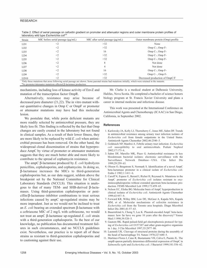
mechanisms, including loss of kinase activity of EnvZ andmutation of the transcription factor OmpR.
Alternatively, resistance may arise because ofdecreased pore diameter (21,22). The in vitro mutant with-out quantitative changes in Omp C or OmpF or promoteror attenuator mutations may have had this molecularlesion.
We postulate that, while porin deficient mutants aremore readily selected by antimicrobial pressure, they arelikely less fit. This finding is reflected by the fact that Ompchanges are easily created in the laboratory but not foundin clinical samples. As a result of their lower fitness, theyare more likely to be replaced by wild E. coli when antimi-crobial pressure has been removed. On the other hand, thewidespread clonal dissemination of strains that hyperpro-duce AmpC by virtue of promoter or attenuator mutationssuggests that they are much better able and more likely tocontribute to the spread of cephamycin resistance.
The ampC β-lactamase produced by E. coli hydrolyzespenicillins, cephalosporins, and cephamycins. In doing so,β-lactamase increases the MICs to third-generationcephalosporins but, as our data suggest, seldom above thebreakpoint set by the National Committee for ClinicalLaboratory Standards (NCCLS). This situation is analo-gous to that of many TEM- and SHB-derived β-lacta-mases. Using third-generation cephalosporins or peni-cillin/β-lactamase–inhibitor combinations to treat seriousinfections caused by ampC up-regulated strains may bemore imprudent. Just as we would not be inclined to treatan E. coli bearing an extended spectrum β-lactamase witha ceftriaxone MIC of 2 mg/L with ceftriaxone, we wouldnot treat an ampC β-lactamase up-regulated E. coli strainwith a third-generation cephalosporin. To the best of ourknowledge, no publication has documented treatment fail-ures in such circumstances, and no NCCLS guidelinesexist. Nevertheless, our practice is to report all of thesestrains as resistant to third-generation cephalosporins andto cautioning against their use.
Mr. Clarke is a medical student at Dalhousie University,Halifax, Nova Scotia. He completed a bachelor of science honorsbiology program at St. Francis Xavier University and plans acareer in internal medicine and infectious disease.
This work was presented at the International Conference onAntimicrobial Agents and Chemotherapy (ICAAC) in San Diego,California, in September 2002.
References
1. Karlowsky JA, Kelly LJ, Thornsberry C, Jones ME, Sahm DF. Trendsin antimicrobial resistance among urinary tract infection isolates ofEscherichia coli from female outpatients in the United States.Antimicrob Agents Chemother 2002;46:2540–5.
2. Goldraich NP, Manfroi A. Febrile urinary tract infection: Escherichiacoli susceptibility to oral antimicrobials. Pediatr Nephrol2002;17:173–6.
3. Sahm DF, Marsilio MK, Piazz G. Antimicrobial resistance in keybloodstream bacterial isolates: electronic surveillance with theSurveillance Network Database—USA. Clin Infect Dis1999;29:259–63.
4. Olsson O, Bergstrom S, Normark S. Identification of a novel AmpCbeta-lactamase promoter in a clinical isolate of Escherichia coli.Embo J 1982;1:1411–6.
5. Caroff N, Espaze E, Berard I, Richet H, Reynaud A. Mutations in theAmpC promoter of Escherichia coli isolates resistant to oxy-iminocephalosporins without extended spectrum beta-lactamase pro-duction. FEMS Microbiol Lett 1999;173:459–65.
6. Nelson EC, Elisha BG. Molecular basis of AmpC hyperproduction inclinical isolates of Escherichia coli. Antimicrob Agents Chemother1999;43:957–9.
7. Forward KR, Willey BM, Low DE, McGeer A, Kapala MA, KapalaMM, et al. Molecular mechanisms of cefoxitin resistance inEscherichia coli from the Toronto area hospitals. Diagn MicrobiolInfect Dis 2001;41:57–63.
8. Bauernfeind A, Chong Y, Lee K. Plasmid-encoded AmpC beta-lacta-mases: how far have we gone 10 years after the discovery? YonseiMed J 1998;39:520–5.
9. Gautom RK. Rapid pulsed-field gel electrophoresis protocol for typ-ing of Escherichia coli O157:H7 and other gram-negative organismsin 1 day. J Clin Microbiol 1997;35:2977–80.
10. Laemmli UK. Cleavage of structural proteins during the assembly ofthe head of bacteriophage T4. Nature 1970;227:680–5.
11. Martinez-Flores I, Cano R, Bustamante VH, Calva E, Puente JL. TheompB operon partially determines differential expression of OmpC inSalmonella typhi and Escherichia coli. J Bacteriol 1999;181:556–62.
1258 Emerging Infectious Diseases • Vol. 9, No. 10, October 2003
RESEARCH
Table 2. Effect of serial passage on cefoxitin gradient on promoter and attenuator regions and outer membrane protein profiles of laboratory wild type Escherichia colia,b Strain MIC before serial passage (µg/mL) MIC after serial passage (µg/mL) Outer membrane protein (Omp) profile
LD1 <2 >32 None LD2 <2 >32 Omp C-, Omp F- LD3 <2 16 Omp C-, Omp F- LD4 <2 >32 Omp C-, Omp F- LD5 <2 >32 Omp C-, Omp F- LD6 <2 8 Not done LD7 <2 4 Not done LD8 <2 16 Omp C-, Omp F- LD9 <2 >32 Omp C- , Omp F- LD10 <2 >32 Decreased production of OmpC/F aOnly those mutations that arose following serial passage are shown. Some parental strains had mutations initially, which were retained in the mutants. bNo promoter/attenuator mutations affected β-lactamase production.
12. Rice LB, Bonomo RA. Genetic and biochemical mechanisms of anti-bacterial resistance to antimicrobial agents. In: Lorian V, editor.Antibiotics in laboratory medicine. 4th ed. Baltimore: Williams andWilkins; 1996. p. 282.
13. Matthew M, Harris AM. Identification of beta-lactamases by analyti-cal isoelectric focusing: correlation with bacterial taxonomy. J GenMicrobiol 1976;94:55–67.
14. Corvec SCN, Espaze E, Marraillac J, Reynaud A. Analysis of theeffect of the –11 AmpC promoter mutation in clinical strains ofEscherichia coli. In: Abstracts of the 41st Interscience Conference onAntimicrobial Agents and Chemotherapy. Sep 2001; Chicago, IL.
15. Rice LB, Carias LL, Hujer AM, Bonafede M, Hutton R, Hoyen C, etal. High-level expression of chromosomally encoded SHV-1 beta-lac-tamase and an outer membrane protein change confer resistance toceftazidime and piperacillin-tazobactam in a clinical isolate ofKlebsiella pneumoniae. Antimicrob Agents Chemother2000;44:362–7.
16. Bornet C, Davin-Regli A, Bosi C, Pages JM, Bollet C. Imipenemresistance of Enterobacter aerogenes mediated by outer membranepermeability. J Clin Microbiol 2000;38:1048–52.
17. Bradford PA, Urban C, Mariano N, Projan SJ, Rahal JJ, Bush K.Imipenem resistance in Klebsiella pneumoniae is associated with thecombination of ACT-1, a plasmid-mediated AmpC beta-lactamase,and the foss of an outer membrane protein. Antimicrob AgentsChemother 1997;41:563–9.
18. Stapleton PD, Shannon KP, French GL. Carbapenem resistance inEscherichia coli associated with plasmid-determined CMY-4 beta-lactamase production and loss of an outer membrane protein.Antimicrob Agents Chemother 1999;43:1206–10.
19. Martinez-Martinez L, Hernandez-Alles S, Alberti S, Tomas JM,Benedi VJ, Jacoby GA. In vivo selection of porin-deficient mutantsof Klebsiella pneumoniae with increased resistance to cefoxitin andexpanded-spectrum-cephalosporins. Antimicrob Agents Chemother1996;40:342–8.
20. Lan CY, Igo MM. Differential expression of the OmpF and OmpCporin proteins in Escherichia coli K-12 depends upon the level ofactive OmpR. J Bacteriol 1998;180:171–4.
21. Simonet V, Mallea M, Pages J-M. Substitutions in the eyelet regiondisrupt cefepime diffusion through the Escherichia coli OmpF chan-nel. Antimicrob Agents Chemother 2000;44:311–5.
22. Jeanteur D, Schirmer T, Fourel D, Simonet V, Rummel G, Widmer C,et al. Structural and functional alterations of a colicin-resistant mutantof OmpF porin from Escherichia coli. Proc Natl Acad Sci U S A1994;91:10675–9.
Address for correspondence: Kevin R. Forward, Division ofMicrobiology, Queen Elizabeth II Health Sciences Centre, 5788University Avenue, Halifax, Nova Scotia B3H 1V8, Canada; fax: (902)473-4432; email: [email protected]
Emerging Infectious Diseases • Vol. 9, No. 10, October 2003 1259
RESEARCH
SS ee aa rr cc hh pp aa ss tt ii ss ss uu ee ss oo ff EE II DD aa tt ww ww ww.. cc dd cc .. gg oo vv // ee ii dd
Outbreaks of invasive infections caused by group A β-hemolytic streptococcus (GAS) may occur in long-termcare settings and are associated with a high case-fatalityrate in debilitated adults. Targeting antibiotic treatment onlyto residents and staff known to be at specific risk of GASmay be an ineffective outbreak control measure. Wedescribe two institutional outbreaks in which mass antibiot-ic treatment was used as a control measure. In the firstinstance, mass treatment was used after targeted antibiot-ic treatment was not successful. In the second instance,mass treatment was used to control a rapidly evolving out-break with a high case-fatality rate. Although no further clin-ical cases were seen after the introduction of mass antibi-otic treatment, persistence of the outbreak strain was doc-umented in one institution >1 year after cases had ceased.Strain persistence was associated with the presence of achronically colonized resident and poor infection controlpractices.
Group A β-hemolytic streptococcus (GAS) has a long-standing association with pharyngitis, skin and soft
tissue infection, and pneumonia (1–3). In the past decade,reports of GAS-associated necrotizing fasciitis and strep-tococcal toxic shock syndrome (TSS) have increased (2,4).Increasingly, outbreaks of invasive GAS are recognized inlong-term care facilities (5–10). In the Canadian provinceof Ontario, 4% of invasive GAS cases occur in long-termcare facilities, and of these cases, one third are outbreakassociated (5). In long-term care settings, a high case-fatal-ity rate has been observed (40% to 60%) (5,11,12).
Guidelines for control of GAS outbreaks in nursinghomes were developed by the Ontario Ministry of Healthin 1995 (13). These guidelines emphasize the identifica-
tion and elimination of colonization through providingantibiotics to those known to be carriers of the bacterium.Although such an approach should minimize antibioticexposure, carrying it out in practice could be difficult, ascultures may be falsely negative (14), and persons couldbecome colonized or transmit colonization to others in theinterval between culturing and treatment. Antibiotic treat-ment of all residents and staff would not be subject to suchlimitations. We present two outbreaks of invasive GASinfection in long-term care facilities in Hamilton, Ontario,Canada. Each was caused by transmission of a single well-characterized outbreak strain, and in both cases, massantibiotic treatment was used to control the outbreak.
Methods
Epidemiologic InvestigationsIn the province of Ontario, invasive infections with
GAS (isolation of GAS from blood or an otherwise sterilesite) and cases of suspected TSS and necrotizing fasciitismust be reported to local public health authorities (15).Reports of invasive GAS infection originating from long-term care institutions in the city of Hamilton resulted in anepidemiologic investigation by the City of HamiltonSocial and Public Health Services Department. In the twooutbreaks described below, investigations consisted of sitevisits with line-listings created to identify facility residentsand staff; reviews of recent admissions, discharges, anddeaths among residents; and reviews of reports of illnessamong residents and staff. Environmental investigationsand reviews of infection control practices were also per-formed.
Attempts were made to identify facility residents andstaff with close epidemiologic linkage to the index patients
1260 Emerging Infectious Diseases • Vol. 9, No. 10, October 2003
RESEARCH
Mass Antibiotic Treatment forGroup A Streptococcus Outbreaksin Two Long-Term Care Facilities1
Andrea Smith,*† Aimin Li,‡ Ornella Tolomeo,* Gregory J. Tyrrell,§† Frances Jamieson,‡and David Fisman*¶
1Presented in part at the 12th Annual Scientific Meeting of theSociety for Healthcare Epidemiology of America (SHEA), April6–9, 2002, Salt Lake City, Utah.
*City of Hamilton Social and Public Health Services Department,Hamilton, Ontario, Canada; †University of Alberta, Edmonton,Alberta, Canada; ‡Ontario Ministry of Health and Long-Term Care,Toronto, Ontario, Canada; §The National Centre forStreptococcus, Edmonton, Alberta, Canada; and ¶McMasterUniversity, Hamilton, Ontario, Canada
(e.g., caregivers, roommates, and close friends). Althoughtypically only persons with epidemiologic linkage to indexpatients are cultured for possible GAS carriage, the cir-cumstances described below resulted in more widespreadcollection of culture specimens. Specimens were obtainedfrom noses and pharynges of residents and staff, andattempts were made to obtain cultures of perirectal areasand wounds in facility residents. In the second outbreakdescribed below, staff provided self-collected rectal andvaginal swabs.
Microbiologic Evaluation and Characterization of GAS Strains
Swabs from institution residents and staff were platedonto blood agar, with group A streptococcus identified byβ-hemolysis and Lancefield typing using standard com-mercially available latex agglutination methods, asdescribed elsewhere (16). Isolates were subsequently eval-uated for relatedness by using pulsed-field gel elec-trophoresis (PFGE). Briefly, PFGE was performed usingfresh overnight cultures of GAS strains. Isolates were sus-pended in buffered saline, and solutions were adjusted involume until optical densities (OD) were identical (1.6 ODat 610 nm). To prepare genomic DNA, 500 mL of cell sus-pension from each strain was mixed with an equal volumeof 2% low melting agarose solution. The agarose plug wasprepared by using a commercially available plug mould(Bio-Rad Canada Limited, Mississauga, Ontario, Canada).All GAS plugs were treated in lysis buffer containing 50mg/mL lysozyme at 37°C overnight, extensively washedin washing buffer, and then treated in a solution containing50 µg/mL protease K at 55°C overnight. All plugs wereextensively washed before being restricted by Sma-Ienzyme. PFGE was run by using a 1% agarose gel at 6volts/cm; run time was 26 h with an initial switch time of20 s and a final switch time of 60 s. Following gel elec-trophoresis, the gel was stained in a solution containing 0.5µg/mL ethidium bromide and photographed by using theBio-Rad Multi-analysis CCD camera system. ATCC strain19615 was included as the quality control strain.
M and T serologic typing and opacity factor determina-tion were performed on all isolates with standardizedmethods (17). In addition, antiopacity factor (AOF) typingwas performed on all isolates that were opacity factor-pos-itive (17). All antisera were prepared in-house. Because ofthe difficulty of producing antisera for some M types, AOFtyping has been frequently used to predict the M type(18,19). Although AOF typing does not always have thesame type specificity as M typing, this approach is consid-ered valid for most strains from developed countries (20).AOF typing was used to predict the following M types: 9,11, 25, 28, 48, 77, 78 and 92 (PT5118).
Results
Outbreak 1In October 2000, the Hamilton public health unit was
notified of a 97-year-old female nursing home resident(index case-patient 1) admitted to an area hospital with cel-lulitis and group A streptococcal bacteremia. The nursinghome was a 386-bed facility in a suburban area. Cultureswere obtained as described from 87 residents and staff whomight have had contact with the person with invasive dis-ease. One resident had a throat culture positive for group Astreptococcus. This isolate was genetically indistinguish-able by PFGE analysis from the invasive isolate obtainedfrom index case-patient 1, and both strains were typed asM77 T9/10, and positive for serum opacity factor.
Intervention consisted of reinforcement of standardinfection control practices with facility staff and treatmentof the person with pharyngeal colonization with a 10-daycourse of cephalexin, 250 mg four times per day. Follow-up cultures from this person taken 1 month after comple-tion of treatment were negative. Index case-patient 1 diedduring the course of the investigation of apparently unre-lated cardiac disease.
In January 2001, an 87-year-old female resident of thesame long-term care facility was transported to hospitalwith fever (index case-patient 2), and GAS was isolatedfrom blood cultures drawn at admission. Several dayslater, GAS was isolated from the wound of another facili-ty resident. Screening cultures performed on 195 residentsand staff with a history of contact with one of these twoinfected persons indicated that seven residents and onestaff member were colonized with GAS. PFGE showedthat the strains from all resident carriers were identical tothe wound and blood isolates, and to the isolate obtainedfrom index case 1. The bacterial isolate obtained from thestaff member was determined by PFGE to be unrelated tothe outbreak strain. Colonized residents and staff weretreated with cephalexin as had been done with the previouspatient. Cultures obtained from treated persons 1 monthafter completion of therapy were again negative.
In May 2001, another resident of this nursing home wastransported to hospital with GAS bacteremia (index case-patient 3). At this time, all 386 residents and 135 staff ofthe facility were screened for GAS carriage by culturingnasopharynx specimens. Eleven residents and three staffmembers were identified as carrying group A streptococ-cus. Thirteen of these (11 residents and two staff members)subsequently proved to be carrying strains identified byPFGE analysis to be identical to the strain obtained fromindex case-patient 2 (Figure 1) (prevalence of colonizationor infection among residents was 2.8% vs. prevalence ofcolonization 1.5% among staff , p=0.38 by chi-square test).
Emerging Infectious Diseases • Vol. 9, No. 10, October 2003 1261
RESEARCH
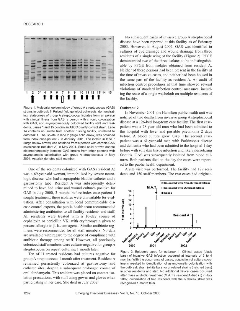
One of the residents colonized with GAS (resident A)was a 69-year-old woman, immobilized by severe neuro-logic disease, who had a suprapubic bladder catheter and agastrostomy tube. Resident A was subsequently deter-mined to have had urine and wound cultures positive forGAS in July 2000, 3 months before index case-patient 1sought treatment; these isolates were unavailable for eval-uation. After consultation with local communicable dis-ease control experts, the public health team recommendedadministering antibiotics to all facility residents and staff.All residents were treated with a 10-day course ofcephalexin or penicillin VK, with erythromycin given topersons allergic to β-lactam agents. Similar antibiotic reg-imens were recommended for all staff members. No dataare available with regard to the degree of compliance withantibiotic therapy among staff. However, all previouslycolonized staff members were culture-negative for group Astreptococcus on repeat culturing 1 month later.
Ten of 11 treated residents had cultures negative forgroup A streptococcus 1 month after treatment. Resident Aremained persistently colonized at gastrostomy andcatheter sites, despite a subsequent prolonged course oforal clindamycin. This resident was placed on contact iso-lation precautions, with staff using gowns and gloves whenparticipating in her care. She died in July 2002.
No subsequent cases of invasive group A streptococcaldisease have been reported at this facility as of February2003. However, in August 2002, GAS was identified incultures of eye drainage and wound drainage from threeresidents of a single wing of the facility (Figure 2). PFGEdemonstrated two of the three isolates to be indistinguish-able by PFGE from isolates obtained from resident A.Neither of these persons had been present in the facility atthe time of invasive cases, and neither had been housed inthe same part of the facility as resident A. An audit ofinfection control procedures at that time showed severalviolations of standard infection control measures, includ-ing the reuse of a single washcloth on multiple residents ofthe facility.
Outbreak 2In November 2001, the Hamilton public health unit was
notified of two deaths from invasive group A streptococcaldisease at a 126-bed long-term care facility. The first case-patient was a 78-year-old man who had been admitted tothe hospital with fever and possible pneumonia 2 daysbefore. A blood culture grew GAS. The second case-patient was a 61-year-old man with Parkinson's diseaseand dementia who had been admitted to the hospital 1 daybefore with soft skin tissue infection and likely necrotizingfasciitis. GAS was subsequently isolated from blood cul-tures. Both patients died on the day the cases were report-ed to the public health department.
A site visit was performed. The facility had 127 resi-dents and 150 staff members. The two cases had originat-
1262 Emerging Infectious Diseases • Vol. 9, No. 10, October 2003
RESEARCH
Figure 1. Molecular epidemiology of group A streptococcus (GAS)strains in outbreak 1. Pulsed-field gel electrophoresis, demonstrat-ing relatedness of group A streptococcal isolates from an personwith clinical illness from GAS, a person with chronic colonizationwith GAS, and asymptomatically colonized facility staff and resi-dents. Lanes 1 and 15 contain an ATCC quality control strain. Lane14 contains an isolate from another nursing facility, unrelated tooutbreak 1. The isolate in lane 2 (large solid arrow) was obtainedfrom index case-patient 2 in January 2001. The isolate in lane 7(large hollow arrow) was obtained from a person with chronic GAScolonization (resident A) in May 2001. Small solid arrows denoteelectrophoretically identical GAS strains from other persons withasymptomatic colonization with group A streptococcus in May2001. Asterisk denotes staff member.
Figure 2. Epidemic curve for outbreak 1. Clinical cases (blackbars) of invasive GAS infection occurred at intervals of 3 to 4months. With the occurrence of cases, acquisition of culture spec-imens resulted in identification of asymptomatic colonization withthe outbreak strain (white bars) or unrelated strains (hatched bars)in other residents and staff. No additional clinical cases occurredafter mass antibiotic treatment (M.A.T.); resident A died (†) in July2002; colonization of two residents with the outbreak strain wasrecognized 1 month later.
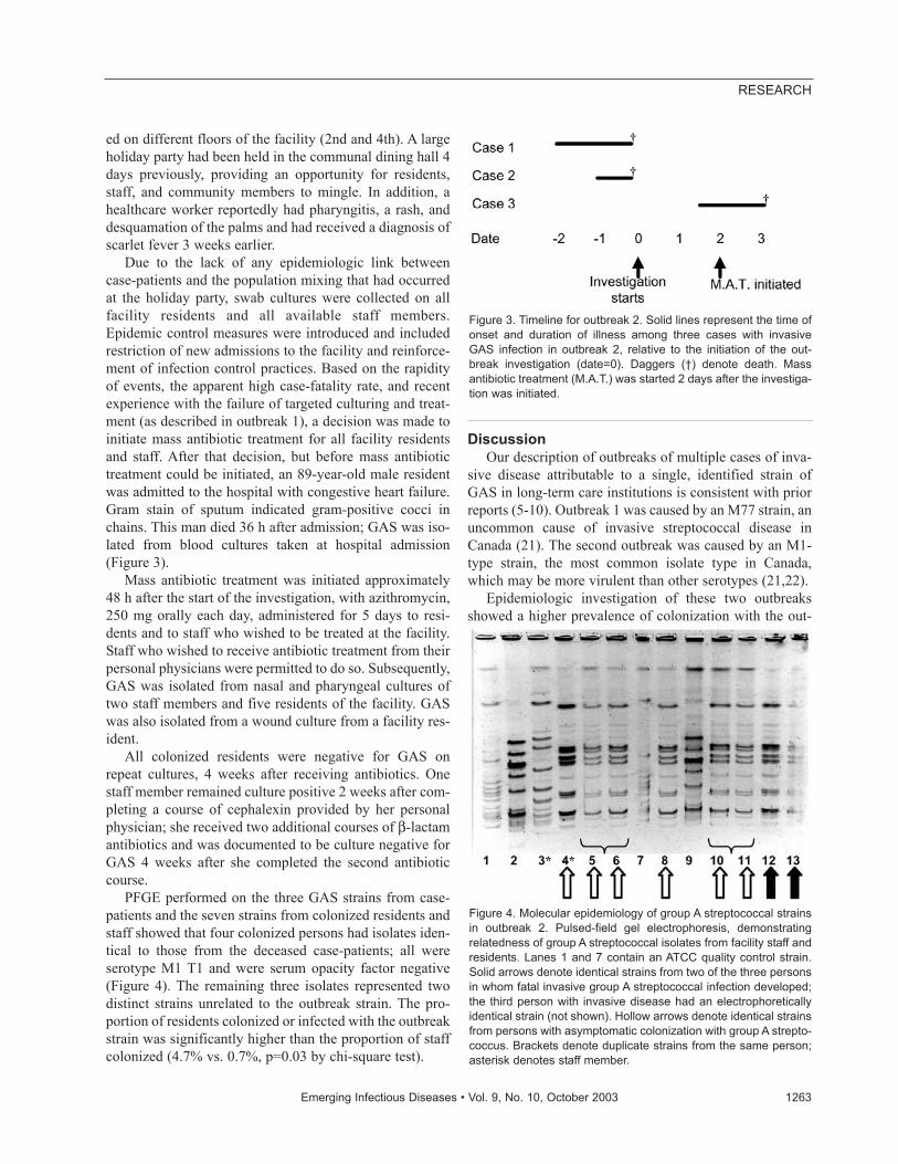
ed on different floors of the facility (2nd and 4th). A largeholiday party had been held in the communal dining hall 4days previously, providing an opportunity for residents,staff, and community members to mingle. In addition, ahealthcare worker reportedly had pharyngitis, a rash, anddesquamation of the palms and had received a diagnosis ofscarlet fever 3 weeks earlier.
Due to the lack of any epidemiologic link betweencase-patients and the population mixing that had occurredat the holiday party, swab cultures were collected on allfacility residents and all available staff members.Epidemic control measures were introduced and includedrestriction of new admissions to the facility and reinforce-ment of infection control practices. Based on the rapidityof events, the apparent high case-fatality rate, and recentexperience with the failure of targeted culturing and treat-ment (as described in outbreak 1), a decision was made toinitiate mass antibiotic treatment for all facility residentsand staff. After that decision, but before mass antibiotictreatment could be initiated, an 89-year-old male residentwas admitted to the hospital with congestive heart failure.Gram stain of sputum indicated gram-positive cocci inchains. This man died 36 h after admission; GAS was iso-lated from blood cultures taken at hospital admission(Figure 3).
Mass antibiotic treatment was initiated approximately48 h after the start of the investigation, with azithromycin,250 mg orally each day, administered for 5 days to resi-dents and to staff who wished to be treated at the facility.Staff who wished to receive antibiotic treatment from theirpersonal physicians were permitted to do so. Subsequently,GAS was isolated from nasal and pharyngeal cultures oftwo staff members and five residents of the facility. GASwas also isolated from a wound culture from a facility res-ident.
All colonized residents were negative for GAS onrepeat cultures, 4 weeks after receiving antibiotics. Onestaff member remained culture positive 2 weeks after com-pleting a course of cephalexin provided by her personalphysician; she received two additional courses of β-lactamantibiotics and was documented to be culture negative forGAS 4 weeks after she completed the second antibioticcourse.
PFGE performed on the three GAS strains from case-patients and the seven strains from colonized residents andstaff showed that four colonized persons had isolates iden-tical to those from the deceased case-patients; all wereserotype M1 T1 and were serum opacity factor negative(Figure 4). The remaining three isolates represented twodistinct strains unrelated to the outbreak strain. The pro-portion of residents colonized or infected with the outbreakstrain was significantly higher than the proportion of staffcolonized (4.7% vs. 0.7%, p=0.03 by chi-square test).
DiscussionOur description of outbreaks of multiple cases of inva-
sive disease attributable to a single, identified strain ofGAS in long-term care institutions is consistent with priorreports (5-10). Outbreak 1 was caused by an M77 strain, anuncommon cause of invasive streptococcal disease inCanada (21). The second outbreak was caused by an M1-type strain, the most common isolate type in Canada,which may be more virulent than other serotypes (21,22).
Epidemiologic investigation of these two outbreaksshowed a higher prevalence of colonization with the out-
Emerging Infectious Diseases • Vol. 9, No. 10, October 2003 1263
RESEARCH
Figure 3. Timeline for outbreak 2. Solid lines represent the time ofonset and duration of illness among three cases with invasiveGAS infection in outbreak 2, relative to the initiation of the out-break investigation (date=0). Daggers (†) denote death. Massantibiotic treatment (M.A.T.) was started 2 days after the investiga-tion was initiated.
Figure 4. Molecular epidemiology of group A streptococcal strainsin outbreak 2. Pulsed-field gel electrophoresis, demonstratingrelatedness of group A streptococcal isolates from facility staff andresidents. Lanes 1 and 7 contain an ATCC quality control strain.Solid arrows denote identical strains from two of the three personsin whom fatal invasive group A streptococcal infection developed;the third person with invasive disease had an electrophoreticallyidentical strain (not shown). Hollow arrows denote identical strainsfrom persons with asymptomatic colonization with group A strepto-coccus. Brackets denote duplicate strains from the same person;asterisk denotes staff member.
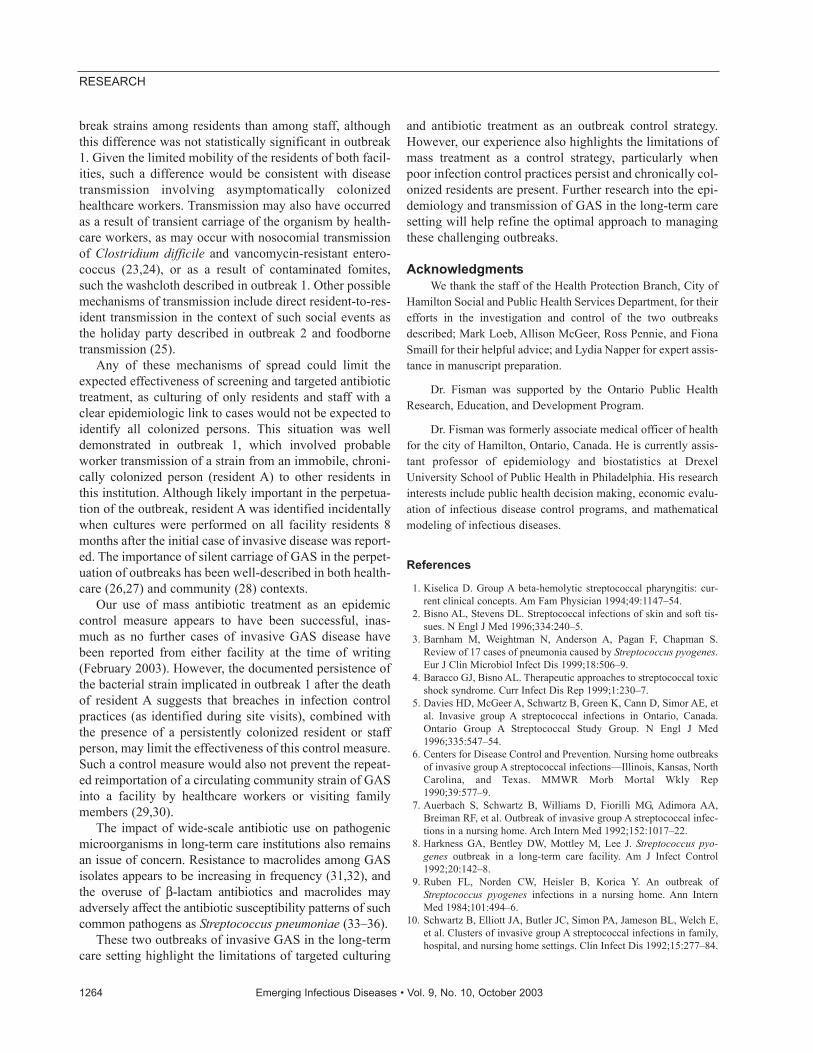
break strains among residents than among staff, althoughthis difference was not statistically significant in outbreak1. Given the limited mobility of the residents of both facil-ities, such a difference would be consistent with diseasetransmission involving asymptomatically colonizedhealthcare workers. Transmission may also have occurredas a result of transient carriage of the organism by health-care workers, as may occur with nosocomial transmissionof Clostridium difficile and vancomycin-resistant entero-coccus (23,24), or as a result of contaminated fomites,such the washcloth described in outbreak 1. Other possiblemechanisms of transmission include direct resident-to-res-ident transmission in the context of such social events asthe holiday party described in outbreak 2 and foodbornetransmission (25).
Any of these mechanisms of spread could limit theexpected effectiveness of screening and targeted antibiotictreatment, as culturing of only residents and staff with aclear epidemiologic link to cases would not be expected toidentify all colonized persons. This situation was welldemonstrated in outbreak 1, which involved probableworker transmission of a strain from an immobile, chroni-cally colonized person (resident A) to other residents inthis institution. Although likely important in the perpetua-tion of the outbreak, resident A was identified incidentallywhen cultures were performed on all facility residents 8months after the initial case of invasive disease was report-ed. The importance of silent carriage of GAS in the perpet-uation of outbreaks has been well-described in both health-care (26,27) and community (28) contexts.
Our use of mass antibiotic treatment as an epidemiccontrol measure appears to have been successful, inas-much as no further cases of invasive GAS disease havebeen reported from either facility at the time of writing(February 2003). However, the documented persistence ofthe bacterial strain implicated in outbreak 1 after the deathof resident A suggests that breaches in infection controlpractices (as identified during site visits), combined withthe presence of a persistently colonized resident or staffperson, may limit the effectiveness of this control measure.Such a control measure would also not prevent the repeat-ed reimportation of a circulating community strain of GASinto a facility by healthcare workers or visiting familymembers (29,30).
The impact of wide-scale antibiotic use on pathogenicmicroorganisms in long-term care institutions also remainsan issue of concern. Resistance to macrolides among GASisolates appears to be increasing in frequency (31,32), andthe overuse of β-lactam antibiotics and macrolides mayadversely affect the antibiotic susceptibility patterns of suchcommon pathogens as Streptococcus pneumoniae (33–36).
These two outbreaks of invasive GAS in the long-termcare setting highlight the limitations of targeted culturing
and antibiotic treatment as an outbreak control strategy.However, our experience also highlights the limitations ofmass treatment as a control strategy, particularly whenpoor infection control practices persist and chronically col-onized residents are present. Further research into the epi-demiology and transmission of GAS in the long-term caresetting will help refine the optimal approach to managingthese challenging outbreaks.
Acknowledgments We thank the staff of the Health Protection Branch, City of
Hamilton Social and Public Health Services Department, for theirefforts in the investigation and control of the two outbreaksdescribed; Mark Loeb, Allison McGeer, Ross Pennie, and FionaSmaill for their helpful advice; and Lydia Napper for expert assis-tance in manuscript preparation.
Dr. Fisman was supported by the Ontario Public HealthResearch, Education, and Development Program.
Dr. Fisman was formerly associate medical officer of healthfor the city of Hamilton, Ontario, Canada. He is currently assis-tant professor of epidemiology and biostatistics at DrexelUniversity School of Public Health in Philadelphia. His researchinterests include public health decision making, economic evalu-ation of infectious disease control programs, and mathematicalmodeling of infectious diseases.
References
1. Kiselica D. Group A beta-hemolytic streptococcal pharyngitis: cur-rent clinical concepts. Am Fam Physician 1994;49:1147–54.
2. Bisno AL, Stevens DL. Streptococcal infections of skin and soft tis-sues. N Engl J Med 1996;334:240–5.
3. Barnham M, Weightman N, Anderson A, Pagan F, Chapman S.Review of 17 cases of pneumonia caused by Streptococcus pyogenes.Eur J Clin Microbiol Infect Dis 1999;18:506–9.
4. Baracco GJ, Bisno AL. Therapeutic approaches to streptococcal toxicshock syndrome. Curr Infect Dis Rep 1999;1:230–7.
5. Davies HD, McGeer A, Schwartz B, Green K, Cann D, Simor AE, etal. Invasive group A streptococcal infections in Ontario, Canada.Ontario Group A Streptococcal Study Group. N Engl J Med1996;335:547–54.
6. Centers for Disease Control and Prevention. Nursing home outbreaksof invasive group A streptococcal infections—Illinois, Kansas, NorthCarolina, and Texas. MMWR Morb Mortal Wkly Rep1990;39:577–9.
7. Auerbach S, Schwartz B, Williams D, Fiorilli MG, Adimora AA,Breiman RF, et al. Outbreak of invasive group A streptococcal infec-tions in a nursing home. Arch Intern Med 1992;152:1017–22.
8. Harkness GA, Bentley DW, Mottley M, Lee J. Streptococcus pyo-genes outbreak in a long-term care facility. Am J Infect Control1992;20:142–8.
9. Ruben FL, Norden CW, Heisler B, Korica Y. An outbreak ofStreptococcus pyogenes infections in a nursing home. Ann InternMed 1984;101:494–6.
10. Schwartz B, Elliott JA, Butler JC, Simon PA, Jameson BL, Welch E,et al. Clusters of invasive group A streptococcal infections in family,hospital, and nursing home settings. Clin Infect Dis 1992;15:277–84.
1264 Emerging Infectious Diseases • Vol. 9, No. 10, October 2003
RESEARCH

11. Kaul R, McGeer A, Low DE, Green K, Schwartz B. Population-basedsurveillance for group A streptococcal necrotizing fasciitis: clinicalfeatures, prognostic indicators, and microbiologic analysis of seven-ty-seven cases. Am J Med 1997;103:18–24.
12. Bucher A, Martin PR, Hoiby EA, Halstensen A, Odegaard A, HelkumKB, et al. Spectrum of disease in bacteraemic patients during aStreptococcus pyogenes serotype M-1 epidemic in Norway in 1988.Eur J Clin Microbiol Infect Dis 1992;11:416–26.
13. Guidelines for management of contacts of cases of invasive group Astreptococcal disease (GAS) including streptococcal toxic shock syn-drome (STSS) and necrotizing fasciitis. 1995. Toronto, OntarioMinistry of Health. [Accessed January 28, 2003] Available from:URL: http://microbiology.mtsinai.on.ca/protocols/pdf/k5b.pdf
14. Pichichero ME. Culture and antigen detection tests for streptococcaltonsillopharyngitis. Am Fam Physician 1992;45:199–205.
15. Health Protection and Promotion Act. Ontario Regulation 559/91,R22.1. 4-23-1999.
16. Koneman E, Allen S, Janda W, Schreckenberger PC, Winn WC. Thegram-positive cocci part II: streptococci, enterococci, and “strepto-coccus-like bacteria.” Color atlas and textbook of diagnostic microbi-ology, 5th ed. Philadelphia: Lippincott; 1997. p. 577–649.
17. Johnson DR, Sramek J, Kaplan EI, Bicova R, Havlicek J, HavlickovaH, et al. Laboratory diagnosis of group A streptococcal infections.Geneva: World Health Organization; 1996.
18. Facklam R, Beall B, Efstratiou A, Fischetti V, Johnson D, Kaplan E,et al. emm typing and validation of provisional M types for group Astreptococci. Emerg Infect Dis 1999;5:247–53.
19. Maxted W, Widdowson J, Fraser C. The use of the serum opacityreaction in the typing of group A streptococci. J Med Microbiol1973;6:83–90.
20. Beall B, Gherardi G, Lovgren M, Facklam RR, Forwick BA, TyrrellGJ. emm and sof gene sequence variation in relation to serologicaltyping of opacity-factor-positive group A streptococci. Microbiology2000;146:1195–209.
21. Tyrrell GJ, Lovgren M, Forwick B, Hoe NP, Musser JM, Talbot JA.M types of group a streptococcal isolates submitted to the NationalCentre for Streptococcus (Canada) from 1993 to 1999. J ClinMicrobiol 2002;40:4466–71.
22. Talkington DF, Schwartz B, Black CM, Todd JK, Elliott J, BreimanRF, et al. Association of phenotypic and genotypic characteristics ofinvasive Streptococcus pyogenes isolates with clinical components ofstreptococcal toxic shock syndrome. Infect Immun 1993;61:3369–74.
23. Samore MH. Epidemiology of nosocomial Clostridium difficile diar-rhoea. J Hosp Infect. 1999;43 (Suppl):S183–90.
24. Austin DJ, Bonten MJ, Weinstein RA, Slaughter S, Anderson RM.Vancomycin-resistant enterococci in intensive-care hospital settings:transmission dynamics, persistence, and the impact of infection con-trol programs. Proc Natl Acad Sci U S A 1999;96:6908–13.
25. Farley TA, Wilson SA, Mahoney F, Kelso KY, Johnson DR, KaplanEL. Direct inoculation of food as the cause of an outbreak of group Astreptococcal pharyngitis. J Infect Dis 1993;167:1232–5.
26. Mastro T, Farley T, Elliott J, Facklam RR, Perks JR, Hadler JL, et al.An outbreak of surgical-wound infections due to group A streptococ-cus carried on the scalp. N Engl J Med 1990;323:968–72.
27. Paul S, Genese C, Spitalny K. Postoperative group a beta-hemolyticStreptococcus outbreak with the pathogen traced to a member of ahealthcare worker’s household. Infect Control Hosp Epidemiol1990;11:643–6.
28. Cockerill FR III, MacDonald KL, Thompson RL, Robertson F,Kohner PC, Besser-Wiek J, et al. An outbreak of invasive group Astreptococcal disease associated with high carriage rates of the inva-sive clone among school-aged children. JAMA 1997;277:38–43.
29. Weber DJ, Rutala WA, Denny FW, Jr. Management of healthcareworkers with pharyngitis or suspected streptococcal infections. InfectControl Hosp Epidemiol 1996;17:753–61.
30. Kaplan EL, Wotton JT, Johnson DR. Dynamic epidemiology of groupA streptococcal serotypes associated with pharyngitis. Lancet2001;358:1334–7.
31. Martin JM, Green M, Barbadora KA, Wald ER. Erythromycin-resist-ant group A streptococci in schoolchildren in Pittsburgh. N Engl JMed 2002;346:1200–6.
32. Weiss K, de Azavedo J, Restieri C, Galarneau LA, Gourdeau M,Harvey P, et al. Phenotypic and genotypic characterization ofmacrolide-resistant group A Streptococcus strains in the province ofQuebec, Canada. J Antimicrob Chemother 2001;47:345–8.
33. Fry AM, Jha HC, Lietman TM, Chaudhary JS, Bhatta RC, Elliott J,Hyde T, et al. Adverse and beneficial secondary effects of mass treat-ment with azithromycin to eliminate blindness due to trachoma inNepal. Clin Infect Dis 2002;35:395-402.
34. Granizo JJ, Aguilar L, Casal J, Dal-Re R, Baquero F. Streptococcuspneumoniae resistance to erythromycin and penicillin in relation tomacrolide and beta-lactam consumption in Spain (1979–1997). JAntimicrob Chemother 2000;46:767–73.
35. Kristinsson KG. Effect of antimicrobial use and other risk factors onantimicrobial resistance in pneumococci. Microb Drug Resist1997;3:117–23.
36. Morita JY, Kahn E, Thompson T, Laclaire L, Beall B, Gheradi G, etal. Impact of azithromycin on oropharyngeal carriage of group AStreptococcus and nasopharyngeal carriage of macrolide-resistantStreptococcus pneumoniae. Pediatr Infect Dis J 2000;19:41–6.
Address for correspondence: David N. Fisman, Drexel University Schoolof Public Health, Bellet Building, 1505 Race Street, Mail Stop 660,Philadelphia, PA 19104-1192 USA; fax: 215-762-4088; email:[email protected]
Emerging Infectious Diseases • Vol. 9, No. 10, October 2003 1265
RESEARCH
In recent years, increases in the urban fox populationhave been observed in many countries of the NorthernHemisphere. As a result, Echinococcus multilocularis hasentered the urban environment. Because of a possibleincreased risk for alveolar echinococcosis, interventionstrategies need to be evaluated. In Zürich, Switzerland, 50praziquantel-containing baits per km2 were distributedmonthly in six 1-km2 bait areas and one 6-km2 bait areafrom April 2000 through October 2001. The proportion of E.multilocularis coproantigen–positive fox fecal samples col-lected remained unchanged in six control areas butdecreased significantly in the 1-km2 bait areas (from 38.6%to 5.5%) and in the 6-km2 bait area (from 66.7% to 1.8%).E. multilocularis prevalence in the intermediate hostArvicola terrestris also decreased significantly in baitedareas. This controlled baiting study shows that a pro-nounced reduction of E. multilocularis egg contamination isfeasible in urban areas where the organism is highlyendemic.
The zoonotic tapeworm Echinococcus multilocularis istypically perpetuated in a wild life cycle, which
includes foxes (genera Vulpes and Alopex) as definitivehosts and various rodent species as intermediate hosts (1).In addition, eggs are accidentally ingested by humans; themetacestodes enter mainly the liver and cause alveolarechinococcosis, a severe, sometimes fatal disease if leftuntreated (2,3).
Few studies have been performed on the epidemiologyof alveolar echinococcosis. Risk factors for alveolarechinococcosis may include occupational and behavioralactivities. However, hunters, trappers, and persons work-ing with fur were not at increased risk for alveolarechinococcosis in South Dakota (4). Data from Europehave indicated that farming activities increase the risk forinfection (5,6). Contamination of the rural environmentwith E. multilocularis connected with farming activitieswas indirectly demonstrated by high prevalences of alveo-
lar echinococcosis in sows kept indoors but fed with grass(7). Areas with high water vole (Arvicola terrestris) densi-ties yielded a 10-fold higher risk for human alveolarechinococcosis compared with areas with low densities ofthis important intermediate host (8). In an area where theorganism is highly endemic, up to 39% of A. terrestris andup to 7% of dogs with free access to rodents were infectedwith E. multilocularis (9), and persons who have kept dogsaround dwellings were at higher risk for alveolarechinococcosis on St. Lawrence Island, Alaska (10).
Red foxes (Vulpes vulpes) are likely to be the mostimportant final host in many regions (11). In the past 2decades, foxes have started to colonize in cities around theworld (12–14), and evidence of the parasite cycle in urbanareas is increasing (13,15,16). In Zürich, Switzerland, onestudy found that 47% of the urban fox population wasinfected with E. multilocularis (17).
The high number of infected foxes in cities and vil-lages, in close contact with domestic pets and humans,could increase the risk of alveolar echinococcosis (16).The disease has an incubation period of 5 to 15 years;therefore, whether the actual incidence rate of alveolarechinococcosis reflects a continuing stable and low infec-tion risk or whether the increased infection pressure inhighly populated areas will lead to a delayed increase inthe incidence of alveolar echinococcosis cases in the futureis unclear (3). However, ecologic changes resulted in avery high alveolar echinococcosis prevalence of 4.0% inChina, which is highly endemic for the organism (18). Thehigh prevalence of E. multilocularis in densely populatedareas and the increase of foxes living in close vicinity tohumans strongly suggest that evaluating possible interven-tion strategies is prudent.
Few field studies focus on anthelmintic treatment ofdefinitive hosts. Rausch et al. (10) demonstrated in a vil-lage that was hyperendemic for the organism (St.Lawrence Island, Alaska) that continual treatment of dogswith praziquantel reduces infection pressure of E. multi-locularis, resulting in lower prevalence in locally trapped
1266 Emerging Infectious Diseases • Vol. 9, No. 10, October 2003
RESEARCH
Anthelmintic Baiting of Foxesagainst Urban Contamination with
Echinococcus multilocularisDaniel Hegglin,* Paul I. Ward,* and Peter Deplazes*
*University of Zürich, Zürich, Switzerland
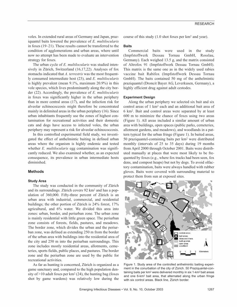
voles. In extended rural areas of Germany and Japan, praz-iquantel baits lowered the prevalence of E. multilocularisin foxes (19–21). These results cannot be transferred to thecondition of agglomerations and urban areas, where untilnow no attempt has been made to evaluate an interventionstrategy for foxes.
The urban cycle of E. multilocularis was studied inten-sively in Zürich, Switzerland (16,17,22). Analyses of foxstomachs indicated that A. terrestris was the most frequent-ly consumed intermediate host (23), and E. multilocularisis highly prevalent (mean 9.1%, maximum 20.9%) in thisvole species, which lives predominantly along the city bor-der (22). Accordingly, the prevalence of E. multilocularisin foxes was significantly higher in the urban peripherythan in more central areas (17), and the infection risk foralveolar echinococcosis might therefore be concentratedmainly in delimited areas in the urban periphery (16). Sinceurban inhabitants frequently use the zones of highest con-tamination for recreational activities and their domesticcats and dogs have access to infected voles, the urbanperiphery may represent a risk for alveolar echinococcosis.
In this controlled experimental field study, we investi-gated the effect of anthelmintic baiting in defined urbanareas where the organism is highly endemic and testedwhether E. multilocularis egg contamination was signifi-cantly reduced. We also examined whether, as an expectedconsequence, its prevalence in urban intermediate hostsdiminished.
Methods
Study AreaThe study was conducted in the community of Zürich
and its surroundings. Zürich covers 92 km2 and has a pop-ulation of 360,000. Fifty-three percent of Zürich is anurban area with industrial, commercial, and residentialbuildings; the other portion of Zürich is 24% forest, 17%agricultural, and 6% water. We divided this area intozones: urban, border, and periurban zone. The urban zoneis mainly residential with little green space. The periurbanzone consists of forests, fields, pastures, and meadows.The border zone, which divides the urban and the periur-ban zone, was defined as extending 250 m from the borderof the urban area with buildings into the residential area ofthe city and 250 m into the periurban surroundings. Thiszone includes mostly residential areas, allotments, ceme-teries, sports fields, public places, and pastures. The borderzone and the periurban zone are used by the public forrecreational activities.
As far as hunting is concerned, Zürich is organized as agame sanctuary and, compared to the high population den-sity of >10 adult foxes per km2 (24), the hunting bag (foxesshot by game wardens) was relatively low during the
course of this study (1.0 shot foxes per km2 and year).
BaitsCommercial baits were used in the study
(Impfstoffwerk Dessau Tornau GmbH, Rosslau,Germany). Each weighed 13.5 g, and the matrix consistedof Altrofox 91 (Impfstoffwerk Dessau Tornau GmbH).This matrix is the same one as in the widely used rabiesvaccine bait Rabifox (Impfstoffwerk Dessau TornauGmbH). The baits contained 50 mg of the anthelminticpraziquantel (Droncit Bayer AG, Leverkusen, Germany), ahighly efficient drug against adult cestodes.
Experiment DesignAlong the urban periphery we selected six bait and six
control areas of 1 km2 each and an additional bait area of6 km2. Bait and control areas were separated by at least600 m to minimize the chance of foxes using two areas(Figure 1). All areas included a similar amount of urbanarea with buildings, open spaces (public parks, cemeteries,allotment gardens, and meadows), and woodlands in a pat-tern typical for the urban fringe (Figure 1). In baited areas,50 praziquantel-containing baits per km2 were distributedmonthly (intervals of 25 to 35 days) during 19 monthsfrom April 2000 through October 2001. Baits were distrib-uted manually at places that were most likely to be fre-quented by foxes (e.g., where fox tracks had been seen, foxdens, and compost heaps) but not by dogs. To avoid olfac-tory contamination, baits were always handled with rubbergloves. Baits were covered with surrounding material toprotect them from sun at exposed sites.
Emerging Infectious Diseases • Vol. 9, No. 10, October 2003 1267
RESEARCH
Figure 1. Study area of the controlled anthelmintic baiting experi-ment in the conurbation of the city of Zürich. 50 Praziquantel-con-taining baits per km2 were delivered monthly in six 1-km2 bait areasand one 6-km2 bait area, that alternated along the urban fringewith six control areas. Black line, Zürich border.
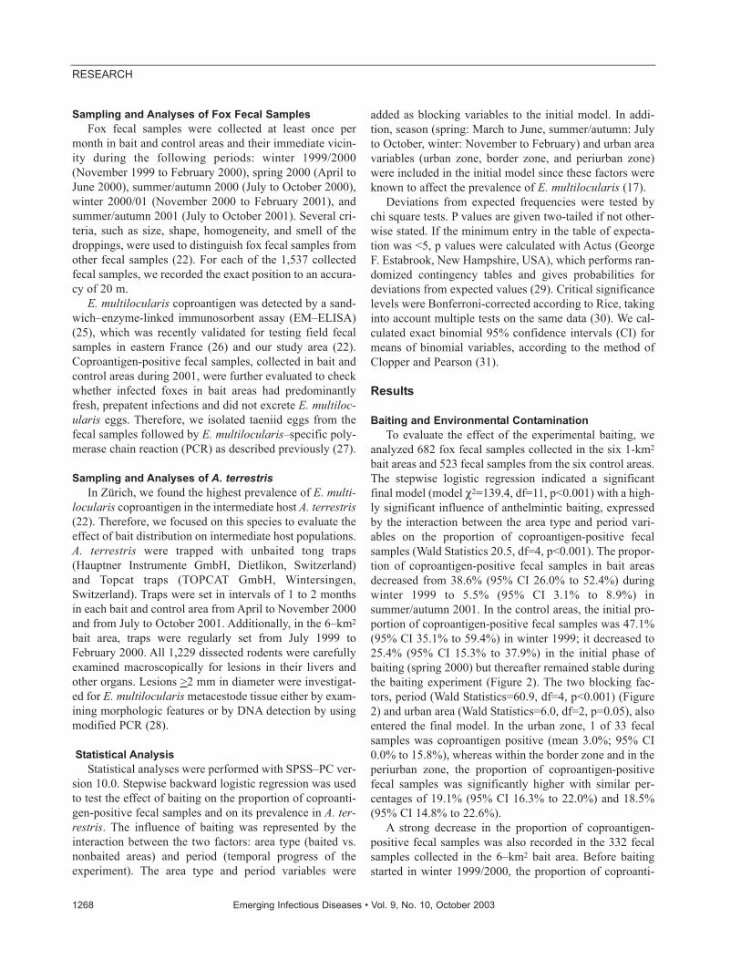
Sampling and Analyses of Fox Fecal SamplesFox fecal samples were collected at least once per
month in bait and control areas and their immediate vicin-ity during the following periods: winter 1999/2000(November 1999 to February 2000), spring 2000 (April toJune 2000), summer/autumn 2000 (July to October 2000),winter 2000/01 (November 2000 to February 2001), andsummer/autumn 2001 (July to October 2001). Several cri-teria, such as size, shape, homogeneity, and smell of thedroppings, were used to distinguish fox fecal samples fromother fecal samples (22). For each of the 1,537 collectedfecal samples, we recorded the exact position to an accura-cy of 20 m.
E. multilocularis coproantigen was detected by a sand-wich–enzyme-linked immunosorbent assay (EM–ELISA)(25), which was recently validated for testing field fecalsamples in eastern France (26) and our study area (22).Coproantigen-positive fecal samples, collected in bait andcontrol areas during 2001, were further evaluated to checkwhether infected foxes in bait areas had predominantlyfresh, prepatent infections and did not excrete E. multiloc-ularis eggs. Therefore, we isolated taeniid eggs from thefecal samples followed by E. multilocularis–specific poly-merase chain reaction (PCR) as described previously (27).
Sampling and Analyses of A. terrestrisIn Zürich, we found the highest prevalence of E. multi-
locularis coproantigen in the intermediate host A. terrestris(22). Therefore, we focused on this species to evaluate theeffect of bait distribution on intermediate host populations.A. terrestris were trapped with unbaited tong traps(Hauptner Instrumente GmbH, Dietlikon, Switzerland)and Topcat traps (TOPCAT GmbH, Wintersingen,Switzerland). Traps were set in intervals of 1 to 2 monthsin each bait and control area from April to November 2000and from July to October 2001. Additionally, in the 6–km2
bait area, traps were regularly set from July 1999 toFebruary 2000. All 1,229 dissected rodents were carefullyexamined macroscopically for lesions in their livers andother organs. Lesions >2 mm in diameter were investigat-ed for E. multilocularis metacestode tissue either by exam-ining morphologic features or by DNA detection by usingmodified PCR (28).
Statistical AnalysisStatistical analyses were performed with SPSS–PC ver-
sion 10.0. Stepwise backward logistic regression was usedto test the effect of baiting on the proportion of coproanti-gen-positive fecal samples and on its prevalence in A. ter-restris. The influence of baiting was represented by theinteraction between the two factors: area type (baited vs.nonbaited areas) and period (temporal progress of theexperiment). The area type and period variables were
added as blocking variables to the initial model. In addi-tion, season (spring: March to June, summer/autumn: Julyto October, winter: November to February) and urban areavariables (urban zone, border zone, and periurban zone)were included in the initial model since these factors wereknown to affect the prevalence of E. multilocularis (17).
Deviations from expected frequencies were tested bychi square tests. P values are given two-tailed if not other-wise stated. If the minimum entry in the table of expecta-tion was <5, p values were calculated with Actus (GeorgeF. Estabrook, New Hampshire, USA), which performs ran-domized contingency tables and gives probabilities fordeviations from expected values (29). Critical significancelevels were Bonferroni-corrected according to Rice, takinginto account multiple tests on the same data (30). We cal-culated exact binomial 95% confidence intervals (CI) formeans of binomial variables, according to the method ofClopper and Pearson (31).
Results
Baiting and Environmental ContaminationTo evaluate the effect of the experimental baiting, we
analyzed 682 fox fecal samples collected in the six 1-km2
bait areas and 523 fecal samples from the six control areas.The stepwise logistic regression indicated a significantfinal model (model χ2=139.4, df=11, p<0.001) with a high-ly significant influence of anthelmintic baiting, expressedby the interaction between the area type and period vari-ables on the proportion of coproantigen-positive fecalsamples (Wald Statistics 20.5, df=4, p<0.001). The propor-tion of coproantigen-positive fecal samples in bait areasdecreased from 38.6% (95% CI 26.0% to 52.4%) duringwinter 1999 to 5.5% (95% CI 3.1% to 8.9%) insummer/autumn 2001. In the control areas, the initial pro-portion of coproantigen-positive fecal samples was 47.1%(95% CI 35.1% to 59.4%) in winter 1999; it decreased to25.4% (95% CI 15.3% to 37.9%) in the initial phase ofbaiting (spring 2000) but thereafter remained stable duringthe baiting experiment (Figure 2). The two blocking fac-tors, period (Wald Statistics=60.9, df=4, p<0.001) (Figure2) and urban area (Wald Statistics=6.0, df=2, p=0.05), alsoentered the final model. In the urban zone, 1 of 33 fecalsamples was coproantigen positive (mean 3.0%; 95% CI0.0% to 15.8%), whereas within the border zone and in theperiurban zone, the proportion of coproantigen-positivefecal samples was significantly higher with similar per-centages of 19.1% (95% CI 16.3% to 22.0%) and 18.5%(95% CI 14.8% to 22.6%).
A strong decrease in the proportion of coproantigen-positive fecal samples was also recorded in the 332 fecalsamples collected in the 6–km2 bait area. Before baitingstarted in winter 1999/2000, the proportion of coproanti-
1268 Emerging Infectious Diseases • Vol. 9, No. 10, October 2003
RESEARCH
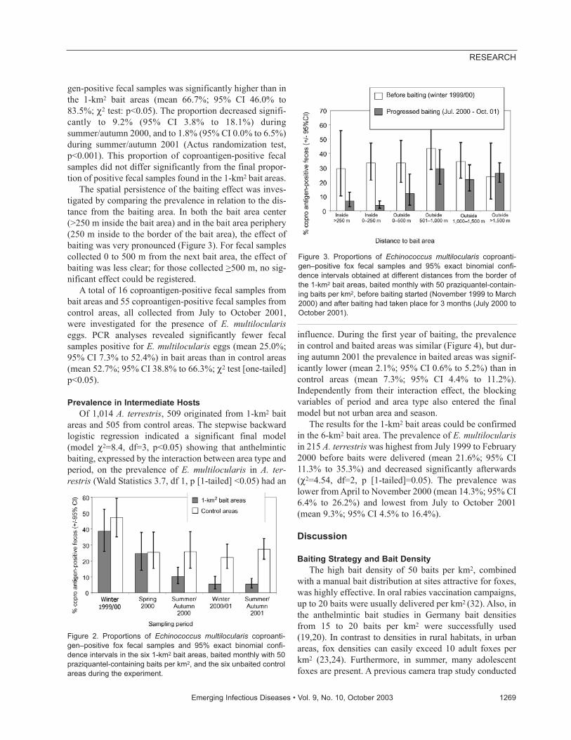
gen-positive fecal samples was significantly higher than inthe 1-km2 bait areas (mean 66.7%; 95% CI 46.0% to83.5%; χ2 test: p<0.05). The proportion decreased signifi-cantly to 9.2% (95% CI 3.8% to 18.1%) duringsummer/autumn 2000, and to 1.8% (95% CI 0.0% to 6.5%)during summer/autumn 2001 (Actus randomization test,p<0.001). This proportion of coproantigen-positive fecalsamples did not differ significantly from the final propor-tion of positive fecal samples found in the 1-km2 bait areas.
The spatial persistence of the baiting effect was inves-tigated by comparing the prevalence in relation to the dis-tance from the baiting area. In both the bait area center(>250 m inside the bait area) and in the bait area periphery(250 m inside to the border of the bait area), the effect ofbaiting was very pronounced (Figure 3). For fecal samplescollected 0 to 500 m from the next bait area, the effect ofbaiting was less clear; for those collected >500 m, no sig-nificant effect could be registered.
A total of 16 coproantigen-positive fecal samples frombait areas and 55 coproantigen-positive fecal samples fromcontrol areas, all collected from July to October 2001,were investigated for the presence of E. multiloculariseggs. PCR analyses revealed significantly fewer fecalsamples positive for E. multilocularis eggs (mean 25.0%;95% CI 7.3% to 52.4%) in bait areas than in control areas(mean 52.7%; 95% CI 38.8% to 66.3%; χ2 test [one-tailed]p<0.05).
Prevalence in Intermediate HostsOf 1,014 A. terrestris, 509 originated from 1-km2 bait
areas and 505 from control areas. The stepwise backwardlogistic regression indicated a significant final model(model χ2=8.4, df=3, p<0.05) showing that anthelminticbaiting, expressed by the interaction between area type andperiod, on the prevalence of E. multilocularis in A. ter-restris (Wald Statistics 3.7, df 1, p [1-tailed] <0.05) had an
influence. During the first year of baiting, the prevalencein control and baited areas was similar (Figure 4), but dur-ing autumn 2001 the prevalence in baited areas was signif-icantly lower (mean 2.1%; 95% CI 0.6% to 5.2%) than incontrol areas (mean 7.3%; 95% CI 4.4% to 11.2%).Independently from their interaction effect, the blockingvariables of period and area type also entered the finalmodel but not urban area and season.
The results for the 1-km2 bait areas could be confirmedin the 6-km2 bait area. The prevalence of E. multilocularisin 215 A. terrestris was highest from July 1999 to February2000 before baits were delivered (mean 21.6%; 95% CI11.3% to 35.3%) and decreased significantly afterwards(χ2=4.54, df=2, p [1-tailed]=0.05). The prevalence waslower from April to November 2000 (mean 14.3%; 95% CI6.4% to 26.2%) and lowest from July to October 2001(mean 9.3%; 95% CI 4.5% to 16.4%).
Discussion
Baiting Strategy and Bait DensityThe high bait density of 50 baits per km2, combined
with a manual bait distribution at sites attractive for foxes,was highly effective. In oral rabies vaccination campaigns,up to 20 baits were usually delivered per km2 (32). Also, inthe anthelmintic bait studies in Germany bait densitiesfrom 15 to 20 baits per km2 were successfully used(19,20). In contrast to densities in rural habitats, in urbanareas, fox densities can easily exceed 10 adult foxes perkm2 (23,24). Furthermore, in summer, many adolescentfoxes are present. A previous camera trap study conducted
Emerging Infectious Diseases • Vol. 9, No. 10, October 2003 1269
RESEARCH
Figure 2. Proportions of Echinococcus multilocularis coproanti-gen–positive fox fecal samples and 95% exact binomial confi-dence intervals in the six 1-km2 bait areas, baited monthly with 50praziquantel-containing baits per km2, and the six unbaited controlareas during the experiment.
Figure 3. Proportions of Echinococcus multilocularis coproanti-gen–positive fox fecal samples and 95% exact binomial confi-dence intervals obtained at different distances from the border ofthe 1-km2 bait areas, baited monthly with 50 praziquantel-contain-ing baits per km2, before baiting started (November 1999 to March2000) and after baiting had taken place for 3 months (July 2000 toOctober 2001).
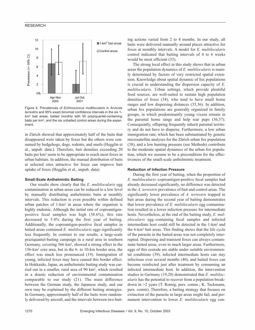
in Zürich showed that approximately half of the baits thatdisappeared were taken by foxes but the others were con-sumed by hedgehogs, dogs, rodents, and snails (Hegglin etal., unpub. data.). Therefore, bait densities exceeding 20baits per km2 seem to be appropriate to reach most foxes inurban habitats. In addition, the manual distribution of baitsat selected sites attractive for foxes can improve baituptake of foxes (Hegglin et al., unpub. data).
Small-Scale Anthelmintic Baiting Our results show clearly that the E. multilocularis egg
contamination in urban areas can be reduced to a low levelby manually distributing anthelmintic baits at monthlyintervals. This reduction is even possible within definedurban patches of 1-km2 in areas where the organism ishighly endemic. Although the initial rate of coproantigen-positive fecal samples was high (38.6%), this ratedecreased to 5.4% during the first year of baiting.Additionally, the coproantigen-positive fecal samples inbaited areas contained E. multilocularis eggs significantlyless frequently. In contrast to our results, a large-scalepraziquantel-baiting campaign in a rural area in southernGermany, covering 566 km2, showed a strong effect in the156-km2 core area, but in the 6- to 10-km border area, theeffect was much less pronounced (19). Immigration ofyoung, infected foxes may have caused this border effect.In Hokkaido, Japan, an anthelmintic baiting study was car-ried out in a smaller, rural area of 90 km2, which resultedin a drastic reduction of environmental contaminationcomparable to our study (21). The main differencebetween the German study, the Japanese study, and ourown may be explained by the different baiting strategies.In Germany, approximately half of the baits were random-ly delivered by aircraft, and the intervals between two bait-
ing actions varied from 2 to 4 months. In our study, allbaits were delivered manually around places attractive forfoxes at monthly intervals. A model for E. multiloculariscontrol indicated that baiting intervals of 4 to 6 weekswould be most efficient (33).
The strong local effect in this study shows that in urbanareas the population dynamics of E. multilocularis is main-ly determined by factors of very restricted spatial exten-sion. Knowledge about spatial dynamic of fox populationsis crucial in understanding the dispersion capacity of E.multilocularis. Urban settings, which provide plentifulfood sources, are well-suited to sustain high populationdensities of foxes (34), who tend to have small homeranges and low dispersing distances (35,36). In addition,urban fox populations are generally organized in familygroups, in which predominantly young vixens remain inthe parental home range and help rear pups (36,37).Consequently, offspring frequently inherit parental territo-ry and do not have to disperse. Furthermore, a low urbanimmigration rate, which has been substantiated by geneticmicrosatellite analyses for the Zürich urban fox population(38), and a low hunting pressure (see Methods) contributeto the moderate spatial dynamics of the urban fox popula-tion, which we assume to be a precondition for the effec-tiveness of the small-scale anthelmintic treatment.
Reduction of Infection PressureDuring the first year of baiting, when the proportion of
E. multilocularis–coproantigen-positive fecal samples hadalready decreased significantly, no difference was detectedin the A. terrestris prevalence of bait and control areas. Thesignificantly lower prevalence of A. terrestris trapped inbait areas during the second year of baiting demonstratesthat lower prevalence of E. multilocularis egg contamina-tion resulted in a lower infection pressure for intermediatehosts. Nevertheless, at the end of the baiting study, E. mul-tilocularis egg–containing fecal samples and infectedintermediate host could still be detected in the 1-km2 andthe 6-km2 bait areas. This finding shows that the life cycleof the parasite in the baited areas was not completely inter-rupted. Dispersing and transient foxes can always contam-inate baited areas, even in much larger areas. Furthermore,eggs of this cestode are stable under suitable environmen-tal conditions (39), infected intermediate hosts can stayinfectious over several months (40), and baited foxes canbecome reinfected just after treatment by consuming aninfected intermediate host. In addition, the interventionstudies in Germany (19,20) demonstrated that E. multiloc-ularis has the potential to recover from a population break-down in >2 years (T. Romig, pers. comm.; K. Tackmann,pers. comm). Therefore, a baiting strategy that focuses onextinction of the parasite in large areas might fail, and per-manent intervention to lower E. multilocularis egg con-
1270 Emerging Infectious Diseases • Vol. 9, No. 10, October 2003
RESEARCH
Figure 4. Prevalences of Echinococcus multilocularis in Arvicolaterrestris and 95% exact binomial confidence intervals in the six 1-km2 bait areas, baited monthly with 50 praziquantel-containingbaits per km2, and the six unbaited control areas during the exper-iment.
tamination in defined risk areas might be more realisticand cost efficient.
ConclusionWe demonstrated the feasibility of small-scale
anthelmintic baiting of foxes to reduce E. multilocularisegg contamination in urban areas intensively used by thepublic for recreational activities, such as gardening or out-door sports. In addition, the lower prevalence of infectedvoles also reduces the risk of domestic carnivores becom-ing infected by preying on voles and, consequently, therisk for egg transmission to pet animals. Therefore, we rec-ommend that public health policy should focus on suchdefined areas where the organism is highly endemic toreduce a potential risk for alveolar echinococcosis.
AcknowledgmentsThe authors thank Claudia Stieger, Isabelle Tanner, Rita
Bosshart, Gioia Schwarzenbach, Sasa Stefanic, and all other col-laborators for help in the extensive field and laboratory work;Christian Stauffer and the game wardens of the city of Zürich fortheir generous collaboration; and Fabio Bontadina and PaulTorgerson for valuable comments on an earlier draft of the man-uscript.
This investigation was carried out within the context of theIntegrated Fox Project, an interdisciplinary research project onthe dynamics of the fox population in Switzerland. The study wassupported by the Swiss Federal Office of Veterinary Medicine,Berne (Projekt Nr. 1.99.03) and the Swiss Federal Office forEducation and Science (EchinoRisk Projekt QLK2-CT-2001-001995 / BBW Nr. 00.0586-2).
The study was carried out at the Institute of Parasitology,University of Zürich, Switzerland.
Dr. Hegglin studied zoology at the University of Zürich,Switzerland, and is currently a Ph.D. research associate at theInstitute of Parasitology of the University of Zürich. His mainresearch interests are ecologic parasitology, urban ecology, andconservation biology.
References
1. Eckert J, Deplazes P. Alveolar echinococcosis in humans: the currentsituation in Central Europe and the need for countermeasures.Parasitol Today 1999;15:315–9.
2. Wilson JF, Rausch RL. Alveolar hydatid disease. A review of clinicalfeatures of 33 indigenous cases of Echinococcus multilocularis infec-tion in Alaskan Eskimos. Am J Trop Med Hyg 1980;29:1340–55.
3. Ammann R, Eckert J. Clinical diagnosis and treatment of echinococ-cosis in humans. In: Thompson RCA, Lymbery AJ, editors.Echinococcus and hydatid disease. Oxon (UK): CAB International;1995.
4. Hildreth MB, Sriram S, Gottstein B, Wilson M, Schantz PM. Failureto identify alveolar echinococcosis in trappers from South Dakota inspite of high prevalence of Echinococcus multilocularis in wildcanids. J Parasitol 2000;86:75–7.
5. Eckert J, Rausch RL, Gemmell MA, Giraudoux P, Kamiya M, LiuF–J, et al. Epidemiology of Echinococcus multilocularis,Echinococcus vogeli and Echinococcus oligarthus. In: Eckert J,Gemmel MA, Meslin F-X, Pawlowski ZS, editors. World HealthOrganization/OIE Manual on echinococcosis in humans and animals.Paris: World Health Organization; 2001. p. 164–94.
6. Kern P, Bardonnet K, Renner E, Auer H, Pawlowski Z, Ammann R,et al. European echinococcosis registry: human alveolar echinococco-sis, Europe, 1982–2000. Emerg Infect Dis 2003;9:343–9.
7. Deplazes P, Eckert J. Veterinary aspects of alveolar echinococcosis–azoonosis of public health significance. Vet Parasitol 2001;98:65–87.
8. Viel JF, Giraudoux P, Abrial V, Bresson-Hadni S. Water vole(Arvicola terrestris Scherman) density as risk factor for human alve-olar echinococcosis. Am J Trop Med Hyg 1999;61:559–65.
9. Gottstein B, Saucy F, Deplazes P, Reichen J, Demierre G, Busato A,et al. Is high prevalence of Echinococcus multilocularis in wild anddomestic animals associated with disease incidence in humans?Emerg Infect Dis 2001;7:408–12.
10. Rausch RL, Wilson JF, Schantz PM. A programme to reduce the riskof infection by Echinococcus multilocularis: the use of praziquantelto control the cestode in a village in the hyperendemic region ofAlaska. Ann Trop Med Parasitol 1990;84:239–50.
11. Rausch RL. Life cycle patterns and geographic distribution ofEchinococcus species. In: Thompson RCA, Lymbery AJ, editors.Echinococcus and hydatid disease. Oxon (UK): CAB International;1995.
12. Adkins CA, Stott P. Home ranges, movements and habitat associa-tions of red foxes Vulpes vulpes in suburban Toronto, Ontario,Canada. Can J Zool 1998;244:335–46.
13. Tsukada H, Morishima Y, Nonaka N, Oku Y, Kamiya M. Preliminarystudy of the role of red foxes in Echinococcus multilocularis trans-mission in the urban area of Sapporo, Japan. Parasitol2000;120:423–8.
14. Gloor S, Bontadina F, Hegglin D, Deplazes P, Breitenmoser U. Therise of urban fox populations in Switzerland. Mammalian Biology2001;66:155–64.
15. Romig T, Bilger B, Mackenstedt U. [Current spread and epidemiolo-gy of Echinococcus multilocularis]. Dtsch Tierarztl Wochenschr1999;106:352–7.
16. Deplazes P, Gloor S, Stieger C, Hegglin D. Urban transmission ofEchinococcus multilocularis. In: Craig P, Pawlowski Z, editors.Cestode zoonoses: echinococcosis and cysticercosis. Amsterdam:IOS Press; 2002. p. 287–97.
17. Hofer S, Gloor S, Muller U, Mathis A, Hegglin D, Deplazes P. Highprevalence of Echinococcus multilocularis in urban red foxes (Vulpesvulpes) and voles (Arvicola terrestris) in the city of Zürich,Switzerland. Parasitol 2000;120:135–42.
18. Craig PS, Giradoux P, Shi D, Bartholomot G, Barnish G, Delattre P, etal. An epidemiological and ecological study on human alveolarechinococcosis transmission in Gansu, China. Acta Trop2000;77:167–77.
19. Schelling U, Frank W, Will R, Romig T, Lucius R. Chemotherapywith praziquantel has the potential to reduce the prevalence ofEchinococcus multilocularis in wild foxes (Vulpes vulpes). Ann TropMed Parasitol 1997;91:179–86.
20. Tackmann K, Loschner U, Mix H, Staubach C, Thulke HH, Ziller M,et al. A field study to control Echinococcus multilocularis–infectionsof the red fox (Vulpes vulpes) in an endemic focus. Epidemiol Infect2001;127:577–87.
Emerging Infectious Diseases • Vol. 9, No. 10, October 2003 1271
RESEARCH
21. Tsukada H, Hamazaki K, Ganzorig S, Iwaki T, Konno K, Lagapa JT,et al. Potential remedy against Echinococcus multilocularis in wildred foxes using baits with anthelmintic distributed around fox breed-ing dens in Hokkaido, Japan. Parasitol 2002;125:119–29.
22. Stieger C, Hegglin D, Schwarzenbach G, Mathis A, Deplazes P.Spatial and temporal aspects of urban transmission of Echinococcusmultilocularis. Parasitol 2002;124:631–40.
23. Contesse P, Hegglin D, Gloor S, Bontadina F, Deplazes P. The diet ofurban foxes (Vulpes vulpes) and the availability of anthropogenicfood in the city of Zürich, Switzerland. Mammalian Biology; 2003 inpress.
24. Gloor S. The rise of urban foxes (Vulpes vulpes) in Switzerland andecological and parasitological aspects of a population in the recentlycolonised city of Zürich [Dissertation]. Zürich: University of Zürich;2002.
25. Deplazes P, Alther P, Tanner I, Thompson RCA, Eckert J.Echinococcus multilocularis coproantigen detection by enzyme-linked immunosorbent assay in fox, dog, and cat populations. JParasitol 1999;85:115–21.
26. Raoul F, Deplazes P, Nonaka N, Piarroux R, Vuitton DA, Giradoux P.Assessment of the epidemiological status of Echinococcus multilocu-laris in foxes in France using ELISA coprotests on fox faeces collect-ed in the field. Int J Parasitol 2001;31:1579–88.
27. Mathis A, Deplazes P, Eckert J. An improved test system for PCR-based specific detection of Echinococcus multilocularis eggs. JHelminthol 1996;70:219–22.
28. Dinkel A, von Nickisch-Rosenegk M, Bilger B, Merli M, Lucius R,Romig T. Detection of Echinococcus multilocularis in the definitivehost: coprodiagnosis by PCR as an alternative to necropsy. J ClinMicrobiol 1998;36:1871–6.
29. Estabrook CB, Estabrook GF. Actus: a solution to the problem ofsmall samples in the analysis of two-way contingency tables. HistMethods 1989;82:5–8.
30. Rice WR. Analyzing tables of statistical tests. Evolution1989;43:223–5.
31. Clopper CJ, Pearson ES. The use of confidence or fiducial limitsillustrated in the case of the binomial. Biometrika 1934;26:404–13.
32. Linhart SB, Kappeler K, Winderg LA. A review of baits and baitdelivery systems for free-ranging carnivores and ungulates. In:Contraception in wildlife: Animal and Plant Health InspectionService. United States Department of Agriculture; 1997. p. 69–132.
33. Hansen F, Tackmann K, Jeltsch F, Thulke H-H.Köderauslageintervalle und Dauer der Bekämpfung des kleinenFuchsbandwurmes: eine Modellierstudie. Berl. Munch. TierarztlWochenschr; 2003 in press.
34. Saunders G, White PCL, Harris S, Rayner JMV. Urban foxes (Vulpesvulpes): food acquisition, time and energy budgeting of a generalizedpredator. In: Dunstone N, Gorman ML, editors. Mammals as preda-tors. London: Oxford University Press; 1993. p. 215–34.
35. Trewhella WJ, Harris S, McAllister FE. Dispersal distance, home-range size and population density in the red fox (Vulpes vulpes): aquantitative analysis. Journal of Applied Ecology 1988;25:423–34.
36. Baker PJ, Funk SM, Harris S, White PCL. Flexible spatial organiza-tion of urban foxes, Vulpes vulpes, before and during an outbreak ofsarcoptic mange. Anim Behav 2000;59:127–46.
37. Macdonald DW. Resource dispersion and social organization of thered fox, Vulpes vulpes. In: Chapman A, Pursley D, editors.Proceedings of the Worldwide Furbearer Conference 1980 Aug 3–11;Forstburg, Maryland. Falls Church (VA): RR Donnelley and SonsCompany; 1981. p. 918–49.
38. Wandeler P, Funk SM, Largiadèr R, Gloor S, Breitenmoser U. Thecity-fox phenomenon: genetic consequences of a recent colonizationof urban habitat. Mol Ecol; 2003;12:647–56.
39. Veit P, Bilger B, Schad V, Schafer J, Frank W, Lucius R. Influence ofenvironmental factors on the infectivity of Echinococcus multilocu-laris eggs. Parasitol 1995;110:79–86.
40. Rausch RL, Wilson JF. Rearing of the adult Echinococcus multilocu-laris Leuckart, 1863, from sterile larvae from man. Am J Trop MedHyg 1973;22:357–60.
Address for correspondence: Peter Deplazes, Institute of Parasitology,University of Zürich, Winterthurerstr. 266a , CH–8057 Zürich; fax: +41 1635 85 07; email: [email protected]
1272 Emerging Infectious Diseases • Vol. 9, No. 10, October 2003
RESEARCH
The print journal is available at no charge to public health professionals
YES, I would like to receive Emerging Infectious Diseases.
Please print your name and businessaddress in the box and return by fax to404-371-5449 or mail to
EID EditorCDC/NCID/MS D611600 Clifton Road, NEAtlanta, GA 30333
Moving? Please give us your new address (in the box) and print the number of your oldmailing label here_________________________________________
�������������������� ���������������
Investigation of an acute gastroenteritis outbreakinvolving >100 persons at a summer camp in Girona,Spain, in June 2002 led to the detection of Salmonellaenterica and extended-spectrum cephalosporin-resistantEscherichia coli (ESCREC). Stool cultures were performedfor 22 symptomatic campers, three asymptomatic foodhandlers, and 10 healthy household members. Of the 22campers, 19 had Salmonella enterica, 9 had an ESCRECstrain carrying an extended-spectrum β-lactamase, and 2had a second ESCREC strain carrying a plasmidiccephamycinase. Related ESCREC were detected in two(salmonella-negative) asymptomatic food handlers and innone of the healthy household members. Fecal ESCRECand its β-lactamases and plasmids were extensively char-acterized. Three of the five ESCREC clones were recov-ered from multiple hosts. The apparent dissemination ofESCREC suggests a food or water vehicle. The observeddistribution of resistance plasmids and β-lactamase genesin several clones indicates a high degree of horizontaltransfer. Heightened vigilance and increased efforts mustbe made to discover the reservoirs and vehicles for com-munity dissemination of ESCREC.
Strains of Escherichia coli that produce enzymes capa-ble of degrading extended- spectrum cephalosporins
(ESCs), i.e., extended-spectrum β-lactamases (ESBLs), orthese drugs plus cephamycins, i.e., plasmidic or hyperpro-duction of chromosomal cephamycinases have recentlyemerged as important nosocomial pathogens (1,2). Someof these strains cannot be reliably detected by clinicalmicrobiology laboratories by using conventional suscepti-bility tests (3), and even when recognized, treating infec-tions caused by these strains can be challenging becausetherapeutic options are limited. Infections attributable tosuch strains are associated with prolonged hospital stays,increased healthcare costs, and an increased number ofdeaths if appropriate therapy is delayed (4,5).
To date, almost all reports of infection or colonizationwith ESBL- and plasmidic cephamycinase-producing E.coli (i.e., extended-spectrum cephalosporin-resistant E.coli [ESCREC]) have involved hospitalized patients ornursing home residents (3,6). The few reported patientswith community-acquired infection have been elderly anddebilitated and have had hospital contact, important coex-isting conditions, or both (3,6).
E. coli, including resistant strains, may be transmittedwithin the community through the food supply. Indeed,other gram-negative enteric pathogens, notably Salmonellaenterica, are a frequent cause of foodborne disease and,increasingly, are associated with antibiotic resistance,including antibiotic resistance to ESCs (7–11). Availabledata regarding other resistant E. coli suggest that ESCRECcould also be disseminated through the food supply(12–19).
The cefoperazone-containing medium routinely used inour laboratory for the isolation of Campylobacter occa-sionally yields other bacteria with hyperproduction ofchromosomal β-lactamases or their plasmidic derivatives,as well as strains carrying extended-spectrum β-lactamases(unpub. data). By using this media, we have isolated sever-al resistant enterobacteriaceae strains from patients withsporadic cases of gastroenteritis (unpub. data). During aninvestigation of a summer camp-associated salmonellosisoutbreak, we observed that stool cultures from ninecampers unexpectedly yielded, on cefoperazone-contain-ing medium, colonies resembling enterobacteriaceae, witha uniform mucoid appearance. This result suggested thepossibility that the same, probably ESC-resistant, enter-obacterial strain was present in all these persons, findingsconsistent with possible foodborne spread. Consequently,all samples were reevaluated on media containing cefo-taxime (see Methods) to increase sensitivity for detectionof ESC-resistant organisms. To gain more knowledge offoodborne spread as a potential mechanism of dissemina-tion of resistance genes, we undertook an extensive molec-ular epidemiologic analysis of these isolates.
Emerging Infectious Diseases • Vol. 9, No. 10, October 2003 1273
RESEARCH
Cephalosporin-resistant Escherichiacoli among Summer Camp
Attendees with Salmonellosis Guillem Prats,* Beatriz Mirelis,* Elisenda Miró,* Ferran Navarro,* Teresa Llovet,* James R. Johnson,†
Neus Camps,‡ Ángela Domínguez,‡ and Lluís Salleras‡
*Universitat Autònoma, Barcelona, Spain; †University ofMinnesota, Minneapolis, Minnesota, USA; and ‡Dept. de Sanitat iSeguretat Social, Barcelona, Spain
Methods
Description of the Outbreak and Stool SamplingTwo hundred twenty-five elementary and secondary
school students and 11 teachers were spending a week(June 11–15, 2001) at a summer camp in Palafrugell,Girona (Spain), when an outbreak of gastroenteritis beganon June 14. An epidemiologic investigation involving 200campers and staff failed to identify the source of the out-break. Clinical and epidemiologic studies were initiated atthe onset of the outbreak, but our participation as referencelaboratory began later. Consequently, a limited number ofstools from symptomatic patients and related persons wereavailable to us for analysis.
From June 16 to 19, stool samples from 22 ill camperswere collected for analysis. On July 6 (18–19 days later),additional stool samples were collected from four illcampers and from 10 asymptomatic household membersof the four ill campers. Stool samples were also collectedfrom three asymptomatic food handlers, on June 19, 22,and 28.
Microbiologic Studies
Human Samples Stool samples were processed according to convention-
al protocols for isolation of enteropathogenic bacteria, inaddition to special selection for resistant organisms (asdescribed below). Isolates were identified by conventionalmethods (20). Stools (fresh and after modified Ziehl-Neelsen staining) were microscopically examined for pro-tozoa (21). Latex agglutination was used to detectrotavirus immunochromatography to dectect adenovirus40/41, and reverse transcriptase–polymerase chain reac-tion (RT-PCR) (22) to detect calicivirus.
Selection of ESCRECStool suspended in saline was added to trypticase soy
broth containing 2 mg/L cefotaxime (TSB-CTX), whichafter 18 h of incubation at 35°C, was added to similarlysupplemented MacConkey broth (MacC-broth-CTX).After an 18-h incubation, a loopful of this broth was spreadon similarly supplemented MacConkey agar. A colony ofeach distinct morphotype was analyzed further.
Selection of Resistant S. enterica StrainsStool samples were processed as for isolation of
ESCREC, but after the initial growth the MacC-broth-CTX was added to selenite broth. After 8 h of incubationat 35°C, this broth was spread on cefotaxime-supplement-ed xylose-lysine-deoxycholate agar. Colonies suspected ofrepresenting Salmonella were analyzed further.
Food StudiesEight unprepared food items from the camp kitchen
were analyzed for Salmonella and ESCREC as describedfor stool samples, except that the initial TSB-CTX wasreplaced by peptone yeast extract broth.
Antibiotic Susceptibility TestingDisk diffusion susceptibility testing to 28 antibiotics
(Table) was performed according to National Committeefor Clinical Laboratory Standards (NCCLS) guidelines(23). The activity of cefotaxime and ceftazidime, com-bined with clavulanate, was determined by E test (ABBiodisk, Solna, Sweden). MICs to β-lactam antibioticswere determined by broth microdilution (Sensititre, TrekDiagnostic Systems LTD, West Sussex, England), accord-ing to NCCLS guidelines (24,25).
Transfer of Resistance DeterminantsFilter matings were performed with ESCREC donors,
using as recipients either E. coli HB101 (Nal Kan) forwhich transconjugants were selected, according todescribed methods (26), or the S. enterica isolate frompatient P12, for which transconjugants were selected byadding the filter containing mixed growth of E. coli andSalmonella to MacC-broth-CTX. This broth, which wassubsequently processed as described above for selection ofresistant S. enterica.
Extraction of ββ-Lactamases and Isoelectric Focusing (IEF)Crude extracts of β-lactamases were obtained by ultra-
sonication. Analytical IEF was performed as previouslydescribed (27).
Characterization of ββ-Lactamase GenesThe blaTEM, blaCTX-M-9 and blaCMY-2 genes were ampli-
fied as previously described (28–33). The DNA sequencewas directly determined from PCR products for both DNAstrands (34).
Biotyping and SerotypingThe biotype, as determined for 12 metabolic reactions,
was expressed as a 4-digit code (35). The serotype andphage type of Salmonella isolates were determined in theServicio de Enterobacterias del Centro Nacional deMicrobiología, Instituto Carlos III, Majadahonda, Spain(M.A. Usera and A. Echeita). The serogroup of theESCREC was determined at the Laboratorio de Referencia,Lugo, Universidad de Santiago de Compostela (J. Blanco).
Pulsed-Field Gel Electrophoresis (PFGE)Genomic profiles were analyzed by PFGE with XbaI
(Amersham Biosciences UK Limited, Buckinghamshire,
1274 Emerging Infectious Diseases • Vol. 9, No. 10, October 2003
RESEARCH
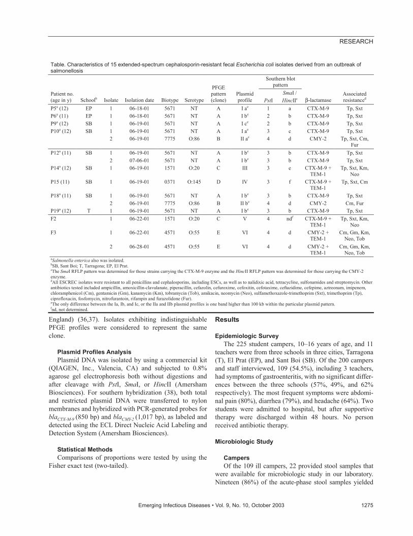
England) (36,37). Isolates exhibiting indistinguishablePFGE profiles were considered to represent the sameclone.
Plasmid Profiles Analysis Plasmid DNA was isolated by using a commercial kit
(QIAGEN, Inc., Valencia, CA) and subjected to 0.8%agarose gel electrophoresis both without digestions andafter cleavage with PstI, SmaI, or HincII (AmershamBiosciences). For southern hybridization (38), both totaland restricted plasmid DNA were transferred to nylonmembranes and hybridized with PCR-generated probes forblaCTX-M-9 (850 bp) and blaCMY-2 (1,017 bp), as labeled anddetected using the ECL Direct Nucleic Acid Labeling andDetection System (Amersham Biosciences).
Statistical MethodsComparisons of proportions were tested by using the
Fisher exact test (two-tailed).
Results
Epidemiologic SurveyThe 225 student campers, 10–16 years of age, and 11
teachers were from three schools in three cities, Tarragona(T), El Prat (EP), and Sant Boi (SB). Of the 200 campersand staff interviewed, 109 (54.5%), including 3 teachers,had symptoms of gastroenteritis, with no significant differ-ences between the three schools (57%, 49%, and 62%respectively). The most frequent symptoms were abdomi-nal pain (80%), diarrhea (79%), and headache (64%). Twostudents were admitted to hospital, but after supportivetherapy were discharged within 48 hours. No personreceived antibiotic therapy.
Microbiologic Study
CampersOf the 109 ill campers, 22 provided stool samples that
were available for microbiologic study in our laboratory.Nineteen (86%) of the acute-phase stool samples yielded
Emerging Infectious Diseases • Vol. 9, No. 10, October 2003 1275
RESEARCH
Table. Characteristics of 15 extended-spectrum cephalosporin-resistant fecal Escherichia coli isolates derived from an outbreak of salmonellosis
Southern blot pattern
Patient no. (age in y) Schoolb Isolate Isolation date Biotype Serotype
PFGE pattern (clone)
Plasmid profile PstI
SmaI / HincIIc β-lactamase
Associated resistanced
P5a (12) EP 1 06-18-01 5671 NT A I ae 1 a CTX-M-9 Tp, Sxt P6a (11) EP 1 06-18-01 5671 NT A I be 2 b CTX-M-9 Tp, Sxt P9a (12) SB 1 06-19-01 5671 NT A I ce 2 b CTX-M-9 Tp, Sxt
SB 1 06-19-01 5671 NT A I ae 3 c CTX-M-9 Tp, Sxt P10a (12) 2 06-19-01 7775 O:86 B II ae 4 d CMY-2 Tp, Sxt, Cm,
Fur SB 1 06-19-01 5671 NT A I be 3 b CTX-M-9 Tp, Sxt P12a (11)
2 07-06-01 5671 NT A I be 3 b CTX-M-9 Tp, Sxt P14a (12) SB 1 06-19-01 1571 O:20 C III 3 e CTX-M-9 +
TEM-1 Tp, Sxt, Km,
Neo P15 (11) SB 1 06-19-01 0371 O:145 D IV 3 f CTX-M-9 +
TEM-1 Tp, Sxt, Cm
SB 1 06-19-01 5671 NT A I be 3 b CTX-M-9 Tp, Sxt P18a (11) 2 06-19-01 7775 O:86 B II be 4 d CMY-2 Cm, Fur
P19a (12) T 1 06-19-01 5671 NT A I be 3 b CTX-M-9 Tp, Sxt F2 1 06-22-01 1571 O:20 C V 4 ndf CTX-M-9 +
TEM-1 Tp, Sxt, Km,
Neo 1 06-22-01 4571 O:55 E VI 4 d CMY-2 +
TEM-1 Cm, Gm, Km,
Neo, Tob F3
2 06-28-01 4571 O:55 E VI 4 d CMY-2 + TEM-1
Cm, Gm, Km, Neo, Tob
aSalmonella enterica also was isolated. bSB, Sant Boi; T, Tarragona; EP, El Prat. cThe SmaI RFLP pattern was determined for those strains carrying the CTX-M-9 enzyme and the HincII RFLP pattern was determined for those carrying the CMY-2 enzyme. dAll ESCREC isolates were resistant to all penicillins and cephalosporins, including ESCs, as well as to nalidixic acid, tetracycline, sulfonamides and streptomycin. Other antibiotics tested included ampicillin, amoxicillin-clavulanate, piperacillin, cefazolin, cefuroxime, cefoxitin, cefotaxime, ceftazidime, cefepime, aztreonam, imipenem, chloramphenicol (Cm), gentamicin (Gm), kanamycin (Km), tobramycin (Tob), amikacin, neomycin (Neo), sulfamethoxazole-trimethoprim (Sxt), trimethoprim (Tp), ciprofloxacin, fosfomycin, nitrofurantoin, rifampin and furazolidone (Fur). eThe only difference between the Ia, Ib, and Ic, or the IIa and IIb plasmid profiles is one band higher than 100 kb within the particular plasmid pattern. fnd, not determined.
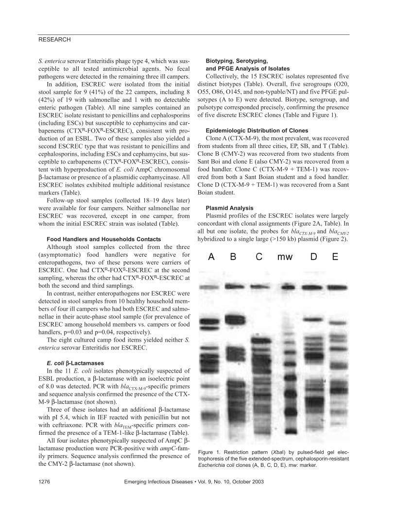
S. enterica serovar Enteritidis phage type 4, which was sus-ceptible to all tested antimicrobial agents. No fecalpathogens were detected in the remaining three ill campers.
In addition, ESCREC were isolated from the initialstool sample for 9 (41%) of the 22 campers, including 8(42%) of 19 with salmonellae and 1 with no detectableenteric pathogen (Table). All nine samples contained anESCREC isolate resistant to penicillins and cephalosporins(including ESCs) but susceptible to cephamycins and car-bapenems (CTXR-FOXR-ESCREC), consistent with pro-duction of an ESBL. Two of these samples also yielded asecond ESCREC type that was resistant to penicillins andcephalosporins, including ESCs and cephamycins, but sus-ceptible to carbapenems (CTXR-FOXR-ESCREC), consis-tent with hyperproduction of E. coli AmpC chromosomalβ-lactamase or presence of a plasmidic cephamycinase. AllESCREC isolates exhibited multiple additional resistancemarkers (Table).
Follow-up stool samples (collected 18–19 days later)were available for four campers. Neither salmonellae norESCREC was recovered, except in one camper, fromwhom the initial ESCREC strain was isolated (Table).
Food Handlers and Households ContactsAlthough stool samples collected from the three
(asymptomatic) food handlers were negative forenteropathogens, two of these persons were carriers ofESCREC. One had CTXR-FOXS-ESCREC at the secondsampling, whereas the other had CTXR-FOXR-ESCREC atboth the second and third samplings.
In contrast, neither enteropathogens nor ESCREC weredetected in stool samples from 10 healthy household mem-bers of four ill campers who had both ESCREC and salmo-nellae in their acute-phase stool sample (for prevalence ofESCREC among household members vs. campers or foodhandlers, p=0.03 and p=0.04, respectively).
The eight cultured camp food items yielded neither S.enterica serovar Enteritidis nor ESCREC.
E. coli ββ-LactamasesIn the 11 E. coli isolates phenotypically suspected of
ESBL production, a β-lactamase with an isoelectric pointof 8.0 was detected. PCR with blaCTX-M-9-specific primersand sequence analysis confirmed the presence of the CTX-M-9 β-lactamase (not shown).
Three of these isolates had an additional β-lactamasewith pI 5.4, which in IEF reacted with penicillin but notwith ceftriaxone. PCR with blaTEM-specific primers con-firmed the presence of a TEM-1-like β-lactamase (Table).
All four isolates phenotypically suspected of AmpC β-lactamase production were PCR-positive with ampC-fam-ily primers. Sequence analysis confirmed the presence ofthe CMY-2 β-lactamase (not shown).
Biotyping, Serotyping, and PFGE Analysis of IsolatesCollectively, the 15 ESCREC isolates represented five
distinct biotypes (Table). Overall, five serogroups (O20,O55, O86, O145, and non-typable/NT) and five PFGE pul-sotypes (A to E) were detected. Biotype, serogroup, andpulsotype corresponded precisely, confirming the presenceof five discrete ESCREC clones (Table and Figure 1).
Epidemiologic Distribution of ClonesClone A (CTX-M-9), the most prevalent, was recovered
from students from all three cities, EP, SB, and T (Table).Clone B (CMY-2) was recovered from two students fromSant Boi and clone E (also CMY-2) was recovered from afood handler. Clone C (CTX-M-9 + TEM-1) was recov-ered from both a Sant Boian student and a food handler.Clone D (CTX-M-9 + TEM-1) was recovered from a SantBoian student.
Plasmid Analysis Plasmid profiles of the ESCREC isolates were largely
concordant with clonal assignments (Figure 2A, Table). Inall but one isolate, the probes for blaCTX-M-9 and blaCMY-2hybridized to a single large (>150 kb) plasmid (Figure 2).
1276 Emerging Infectious Diseases • Vol. 9, No. 10, October 2003
RESEARCH
Figure 1. Restriction pattern (XbaI) by pulsed-field gel elec-trophoresis of the five extended-spectrum, cephalosporin-resistantEscherichia coli clones (A, B, C, D, E). mw: marker.
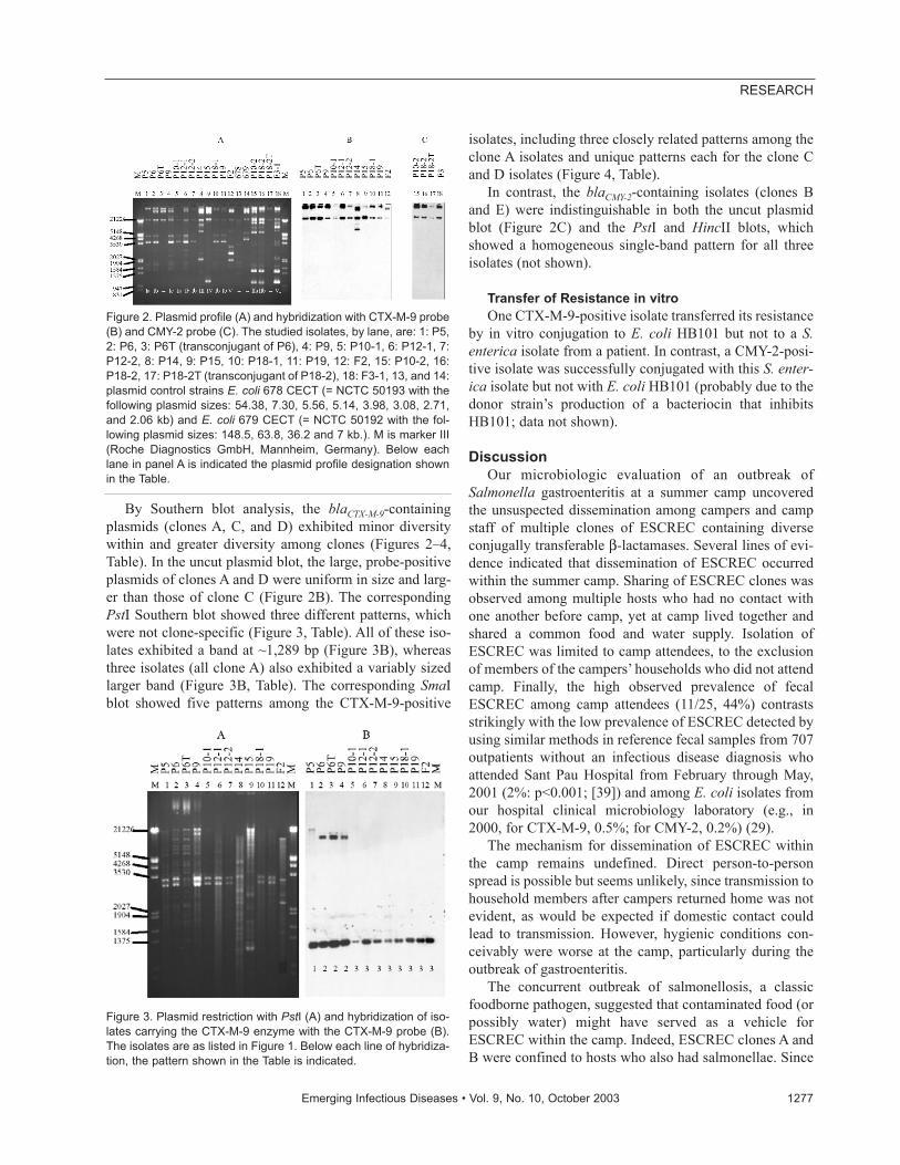
By Southern blot analysis, the blaCTX-M-9-containingplasmids (clones A, C, and D) exhibited minor diversitywithin and greater diversity among clones (Figures 2–4,Table). In the uncut plasmid blot, the large, probe-positiveplasmids of clones A and D were uniform in size and larg-er than those of clone C (Figure 2B). The correspondingPstI Southern blot showed three different patterns, whichwere not clone-specific (Figure 3, Table). All of these iso-lates exhibited a band at ~1,289 bp (Figure 3B), whereasthree isolates (all clone A) also exhibited a variably sizedlarger band (Figure 3B, Table). The corresponding SmaIblot showed five patterns among the CTX-M-9-positive
isolates, including three closely related patterns among theclone A isolates and unique patterns each for the clone Cand D isolates (Figure 4, Table).
In contrast, the blaCMY-2-containing isolates (clones Band E) were indistinguishable in both the uncut plasmidblot (Figure 2C) and the PstI and HincII blots, whichshowed a homogeneous single-band pattern for all threeisolates (not shown).
Transfer of Resistance in vitro One CTX-M-9-positive isolate transferred its resistance
by in vitro conjugation to E. coli HB101 but not to a S.enterica isolate from a patient. In contrast, a CMY-2-posi-tive isolate was successfully conjugated with this S. enter-ica isolate but not with E. coli HB101 (probably due to thedonor strain’s production of a bacteriocin that inhibitsHB101; data not shown).
DiscussionOur microbiologic evaluation of an outbreak of
Salmonella gastroenteritis at a summer camp uncoveredthe unsuspected dissemination among campers and campstaff of multiple clones of ESCREC containing diverseconjugally transferable β-lactamases. Several lines of evi-dence indicated that dissemination of ESCREC occurredwithin the summer camp. Sharing of ESCREC clones wasobserved among multiple hosts who had no contact withone another before camp, yet at camp lived together andshared a common food and water supply. Isolation ofESCREC was limited to camp attendees, to the exclusionof members of the campers’ households who did not attendcamp. Finally, the high observed prevalence of fecalESCREC among camp attendees (11/25, 44%) contrastsstrikingly with the low prevalence of ESCREC detected byusing similar methods in reference fecal samples from 707outpatients without an infectious disease diagnosis whoattended Sant Pau Hospital from February through May,2001 (2%: p<0.001; [39]) and among E. coli isolates fromour hospital clinical microbiology laboratory (e.g., in2000, for CTX-M-9, 0.5%; for CMY-2, 0.2%) (29).
The mechanism for dissemination of ESCREC withinthe camp remains undefined. Direct person-to-personspread is possible but seems unlikely, since transmission tohousehold members after campers returned home was notevident, as would be expected if domestic contact couldlead to transmission. However, hygienic conditions con-ceivably were worse at the camp, particularly during theoutbreak of gastroenteritis.
The concurrent outbreak of salmonellosis, a classicfoodborne pathogen, suggested that contaminated food (orpossibly water) might have served as a vehicle forESCREC within the camp. Indeed, ESCREC clones A andB were confined to hosts who also had salmonellae. Since
Emerging Infectious Diseases • Vol. 9, No. 10, October 2003 1277
RESEARCH
Figure 2. Plasmid profile (A) and hybridization with CTX-M-9 probe(B) and CMY-2 probe (C). The studied isolates, by lane, are: 1: P5,2: P6, 3: P6T (transconjugant of P6), 4: P9, 5: P10-1, 6: P12-1, 7:P12-2, 8: P14, 9: P15, 10: P18-1, 11: P19, 12: F2, 15: P10-2, 16:P18-2, 17: P18-2T (transconjugant of P18-2), 18: F3-1, 13, and 14:plasmid control strains E. coli 678 CECT (= NCTC 50193 with thefollowing plasmid sizes: 54.38, 7.30, 5.56, 5.14, 3.98, 3.08, 2.71,and 2.06 kb) and E. coli 679 CECT (= NCTC 50192 with the fol-lowing plasmid sizes: 148.5, 63.8, 36.2 and 7 kb.). M is marker III(Roche Diagnostics GmbH, Mannheim, Germany). Below eachlane in panel A is indicated the plasmid profile designation shownin the Table.
Figure 3. Plasmid restriction with PstI (A) and hybridization of iso-lates carrying the CTX-M-9 enzyme with the CTX-M-9 probe (B).The isolates are as listed in Figure 1. Below each line of hybridiza-tion, the pattern shown in the Table is indicated.
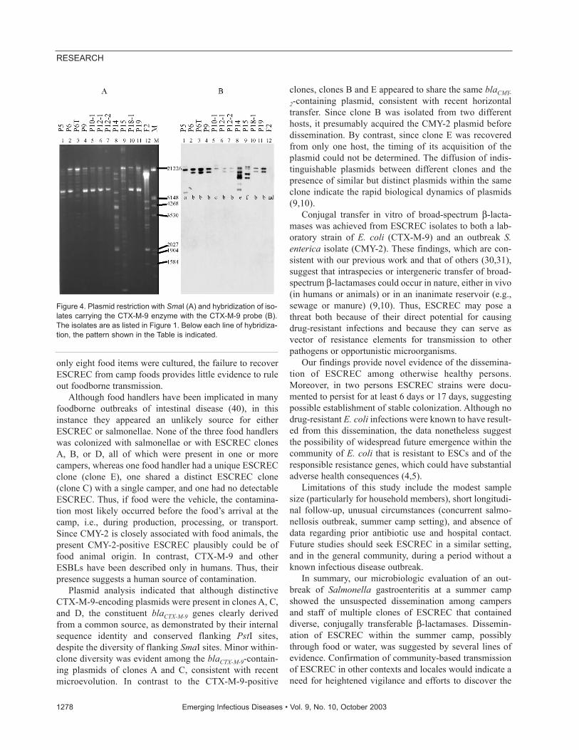
only eight food items were cultured, the failure to recoverESCREC from camp foods provides little evidence to ruleout foodborne transmission.
Although food handlers have been implicated in manyfoodborne outbreaks of intestinal disease (40), in thisinstance they appeared an unlikely source for eitherESCREC or salmonellae. None of the three food handlerswas colonized with salmonellae or with ESCREC clonesA, B, or D, all of which were present in one or morecampers, whereas one food handler had a unique ESCRECclone (clone E), one shared a distinct ESCREC clone(clone C) with a single camper, and one had no detectableESCREC. Thus, if food were the vehicle, the contamina-tion most likely occurred before the food’s arrival at thecamp, i.e., during production, processing, or transport.Since CMY-2 is closely associated with food animals, thepresent CMY-2-positive ESCREC plausibly could be offood animal origin. In contrast, CTX-M-9 and otherESBLs have been described only in humans. Thus, theirpresence suggests a human source of contamination.
Plasmid analysis indicated that although distinctiveCTX-M-9-encoding plasmids were present in clones A, C,and D, the constituent blaCTX-M-9 genes clearly derivedfrom a common source, as demonstrated by their internalsequence identity and conserved flanking PstI sites,despite the diversity of flanking SmaI sites. Minor within-clone diversity was evident among the blaCTX-M-9-contain-ing plasmids of clones A and C, consistent with recentmicroevolution. In contrast to the CTX-M-9-positive
clones, clones B and E appeared to share the same blaCMY-
2-containing plasmid, consistent with recent horizontaltransfer. Since clone B was isolated from two differenthosts, it presumably acquired the CMY-2 plasmid beforedissemination. By contrast, since clone E was recoveredfrom only one host, the timing of its acquisition of theplasmid could not be determined. The diffusion of indis-tinguishable plasmids between different clones and thepresence of similar but distinct plasmids within the sameclone indicate the rapid biological dynamics of plasmids(9,10).
Conjugal transfer in vitro of broad-spectrum β-lacta-mases was achieved from ESCREC isolates to both a lab-oratory strain of E. coli (CTX-M-9) and an outbreak S.enterica isolate (CMY-2). These findings, which are con-sistent with our previous work and that of others (30,31),suggest that intraspecies or intergeneric transfer of broad-spectrum β-lactamases could occur in nature, either in vivo(in humans or animals) or in an inanimate reservoir (e.g.,sewage or manure) (9,10). Thus, ESCREC may pose athreat both because of their direct potential for causingdrug-resistant infections and because they can serve asvector of resistance elements for transmission to otherpathogens or opportunistic microorganisms.
Our findings provide novel evidence of the dissemina-tion of ESCREC among otherwise healthy persons.Moreover, in two persons ESCREC strains were docu-mented to persist for at least 6 days or 17 days, suggestingpossible establishment of stable colonization. Although nodrug-resistant E. coli infections were known to have result-ed from this dissemination, the data nonetheless suggestthe possibility of widespread future emergence within thecommunity of E. coli that is resistant to ESCs and of theresponsible resistance genes, which could have substantialadverse health consequences (4,5).
Limitations of this study include the modest samplesize (particularly for household members), short longitudi-nal follow-up, unusual circumstances (concurrent salmo-nellosis outbreak, summer camp setting), and absence ofdata regarding prior antibiotic use and hospital contact.Future studies should seek ESCREC in a similar setting,and in the general community, during a period without aknown infectious disease outbreak.
In summary, our microbiologic evaluation of an out-break of Salmonella gastroenteritis at a summer campshowed the unsuspected dissemination among campersand staff of multiple clones of ESCREC that containeddiverse, conjugally transferable β-lactamases. Dissemin-ation of ESCREC within the summer camp, possiblythrough food or water, was suggested by several lines ofevidence. Confirmation of community-based transmissionof ESCREC in other contexts and locales would indicate aneed for heightened vigilance and efforts to discover the
1278 Emerging Infectious Diseases • Vol. 9, No. 10, October 2003
RESEARCH
Figure 4. Plasmid restriction with SmaI (A) and hybridization of iso-lates carrying the CTX-M-9 enzyme with the CTX-M-9 probe (B).The isolates are as listed in Figure 1. Below each line of hybridiza-tion, the pattern shown in the Table is indicated.
reservoirs and vehicles for dissemination of ESCRECwithin the community.
AcknowledgmentsWe thank Jorge Blanco for the serotyping of Escherichia
coli strains; Pilar Cortés, Montse Sabaté, and Laura Gómez fortechnical support; and Irene Barrabeig, Sofia Minguell, and RosaSala for collecting samples and clinical data from patients.
This study was partially supported by grants 97/062398/1293 from the “Fondo de Investigaciones Sanitarias de laSeguridad Social de España” (G.P.) and National ResearchInitiative Competitive Grants Program/ United StatesDepartment of Agriculture grant 00-35212-9408 (J.R.J.).
Prof. Prats is a microbiologist and director of theMicrobiology Department at the Hospital Universitari Valld’Hebron in Barcelona. His current research interests includeuropathogenic Escherichia coli and antibiotic resistance of enter-obacteria.
References
1. Nordmann P. Trends in beta-lactam resistance amongEnterobacteriaceae. Clin Infect Dis 1998;27( Suppl 1):S100–6.
2. Livermore DM. β-Lactamases in laboratory and clinical resistance.Clin Microbiol Rev 1995;8:557–84.
3. Bradford PA. Extended-spectrum β-lactamases in the 21st century:characterization, epidemiology, and detection of this important resist-ance threat. Clin Microbiol Rev 2001;14:933–51.
4. Lautenbach E, Patel JB, Bilker WB, Edelstein PH, Fishman NO.Extended-spectrum β-lactamase-producing Escherichia coli andKlebsiella pneumoniae: risk factors for infection and impact of resist-ance on outcomes. Clin Infect Dis 2001;32:1162–71.
5. Wong-Beringer A, Hindler J, Loeloff M, Queenan AM, Lee N,Pegues DA, et al. Molecular correlation for the treatment outcomes inbloodstream infections caused by Escherichia coli and Klebsiellapneumoniae with reduced susceptibility to ceftazidime. Clin InfectDis 2002;34:135–46.
6. Nathisuwan S, Burgess DS, Lewis JS II. Extended-spectrum β-lacta-mases: epidemiology, detection, and treatment. Pharmacotherapy2001;21:920–8.
7. Marsik FJ, Parisi JT, Blenden DC. Transmissible drug resistance ofEscherichia coli and Salmonella from humans, animals, and theirrural environments. J Infect Dis 1975;132:296–302.
8. White DG, Zhao S, Sudler R, Ayers S, Friedman S, Chen S, et al. Theisolation of antibiotic-resistant salmonella from retail ground meats.N Engl J Med 2001;345:1147–54.
9. Winokur PL, Brueggemann A, DeSalvo DL, Hoffmann L, Apley MD,Uhlenhopp EK, et al. Animal and human multidrug-resistant,cephalosporin-resistant salmonella isolates expressing a plasmid-mediated CMY-2 AmpC β-lactamase. Antimicrob Agents Chemother2000;44:2777–83.
10. Winokur PL, Vonstein DL, Hoffman LJ, Uhlenhopp EK, Doern GV.Evidence for transfer of CMY-2 AmpC β-lactamase plasmidsbetween Escherichia coli and Salmonella isolates from food animalsand humans. Antimicrob Agents Chemother 2001;45:2716–22.
11. Zhao S, White DG, McDermott PF, Friedman S, English L, Ayers S,et al. Identification and expression of cephamycinase bla(CMY) genes inEscherichia coli and Salmonella isolates from food animals andground meat. Antimicrob Agents Chemother 2001;45:3647–50.
12. Linton AH. Animal to man transmission of Enterobacteriaceae. R SocHealth J 1977;97:115–8.
13. Linton AH, Howe K, Bennett PM, Richmond MH, Whiteside EJ. Thecolonization of the human gut by antibiotic resistant Escherichia colifrom chickens. J Appl Bacteriol 1977;43:465–9.
14. Corpet DE. Antibiotic resistance from food. N Engl J Med1988;318:1206–7.
15. Garau J, Xercavins M, Rodríguez-Carballeira M, Gómez-Vera JR,Coll I, Vidal D, et al. Emergence and dissemination of quinolone-resistant Escherichia coli in the community. Antimicrob AgentsChemother 1999;43:2736–41.
16. Brinas L, Zarazaga M, Sáenz Y, Ruiz-Larrea F, Torres C. β-Lactamases in ampicillin-resistant Escherichia coli isolates fromfoods, humans, and healthy animals. Antimicrob Agents Chemother2002;46:3156–63.
17. Sáenz Y, Zarazaga M, Brinas L, Lantero M, Ruiz-Larrea F, Torres C.Antibiotic resistance in Escherichia coli isolates obtained from ani-mals, foods and humans in Spain. Int J Antimicrob Agents2001;18:353–8.
18. Shooter RA, Cooke EM, Faiers MC, Breaden AL, O’Farrell SM.Isolation of Escherichia coli, Pseudomonas aeruginosa, andKlebsiella from food in hospitals, canteens, and schools. Lancet1971;2:390–2.
19. van den Bogaard AE, London N, Driessen C, Stobberingh EE.Antibiotic resistance of faecal Escherichia coli in poultry, poultryfarmers and poultry slaughterers. J Antimicrob Chemother2001;47:763–71.
20. Murray P, Baron E, Pfaller M, Tenover F, Yolken R. Manual of clini-cal microbiology. Seventh ed. Washington: American Society ofMicrobiology; 1999.
21. Garcia LS. Practical guide to diagnostic parasitology. Washington:American Society for Microbiology; 1999.
22. Le Guyader F, Estes MK, Hardy ME, Neill FH, Green J, Brown DW,et al. Evaluation of a degenerate primer for the PCR detection ofhuman caliciviruses. Arch Virol 1996;141:2225–35.
23. National Committee for Clinical Laboratory Standards. Performancestandards for antimicrobial disk susceptibility test; NCCLS documentM2-A7. Wayne (PA): The Committee; 2000.
24. National Committee for Clinical Laboratory Standards. Methods fordilution antimicrobial susceptibility tests for bacteria that grow aero-bically; approved standard fifth edition. NCCLS document M7-A5.Wayne (PA): The Committee; 2000.
25. National Committee for Clinical Laboratory Standards Supplementaltables: disk diffusion; NCCLS document M100-S10. Wayne (PA):The Committee; 2000.
26. Miró E, del Cuerpo M, Navarro F, Sabaté M, Mirelis B, Prats G.Emergence of clinical Escherichia coli isolates with decreased sus-ceptibility to ceftazidime and synergic effect with co-amoxiclav dueto SHV-1 hyperproduction. J Antimicrob Chemother 1998;42:535–8.
27. Barthélémy M, Guionie M, Labia R. Beta-lactamases: determinationof their isoelectric points. Antimicrob Agents Chemother1978;13:695–8.
28. Sabaté M, Vergés C, Miró E, Mirelis B, Navarro F, del Rio E, et al.Incidencia de betalactamasas de espectro ampliado en Escherichiacoli en un hospital universitario durante 1994-1996. Enferm InfeccMicrobiol Clin 1999;17:401–4.
29. Sabaté M, Miró E, Navarro F, Vergés C, Aliaga R, Mirelis B, et al. β-Lactamases involved in resistance to broad-spectrum cephalosporinsin Escherichia coli and Klebsiella spp. clinical isolates collectedbetween 1994 and 1996, in Barcelona (Spain). J AntimicrobChemother 2002;49:989–97.
30. Sabaté M, Tarragó R, Navarro F, Miró E, Vergés C, Barbé J, et al.Cloning and sequence of the gene encoding a novel cefotaxime-hydrolyzing β-lactamase (CTX-M-9) from Escherichia coli in Spain.Antimicrob Agents Chemother 2000;44:1970–3.
Emerging Infectious Diseases • Vol. 9, No. 10, October 2003 1279
RESEARCH
31. Simarro E, Navarro F, Ruiz J, Miró E, Gómez J, Mirelis B.Salmonella enterica serovar Virchow with CTX-M-like beta-lacta-mase in Spain. J Clin Microbiol 2000;38:4676–8.
32. Marchese A, Arlet G, Schito GC, Lagrange PH, Philippon A.Characterization of FOX-3, an AmpC-type plasmid-mediated β-lac-tamase from an Italian isolate of Klebsiella oxytoca. AntimicrobAgents Chemother 1998;42:464–7.
33. Navarro F, Pérez-Trallero E, Marimón JM, Aliaga R, Gomariz M,Mirelis B. CMY-2-producing Salmonella enterica, Klebsiella pneu-moniae, Klebsiella oxytoca, Proteus mirabilis and Escherichia colistrains isolated in Spain (October 1999–December 2000). JAntimicrob Chemother 2001;48:383–9.
34. Fernández de Henestrosa AR, Rivera E, Tapias A, Barbé J.Identification of the Rhodobacter sphaeroides SOS box. MolMicrobiol 1998;28:991–1003.
35. LeMinor L, Richard C. Méthodes de laboratoire pour l’identificationdes enterobactéries. Paris: Institut Pasteur; 1993.
36. Smith CL, Klco S, Cantor CR. Pulsed-field gel electrophoresis andthe technology of large DNA molecules. In: Davis K, editor. Genomeanalysis: a practical approach. Oxford: IRL Press;, 1988. p. 41–72.
37. Bannerman TL, Hancock GA, Tenover FC, Miller JM. Pulsed-fieldgel electrophoresis as a replacement for bacteriophage typing ofStaphylococcus aureus. J Clin Microbiol 1995;33:551–5.
38. Sambrook J, Rusell DW. Molecular cloning. A laboratory manual.Third ed. Cold Spring Harbor (NY): Cold Spring Harbor LaboratoryPress; 2001.
39. Mirelis B. Navarro F, Miró E, Mesa RJ, Coll P, Prats G. Communitytransmission of extended- spectrum b-lactamase. Emerg Infect Dis.2003;9:1024-5.
40. Lee R, Peppe J, George H. Pulsed-field gel electrophoresis of genom-ic digests demonstrates linkages among food, food handlers, andpatrons in a foodborne Salmonella javiana outbreak inMassachusetts. J Clin Microbiol 1998;36:284–5.
Address for correspondence: Guillem Prats, Servei de Microbiologia,Hospital Vall d’Hebrón. P Vall d’Hebrón, 119-129, 08035, Barcelona,Spain; fax: 34 932746801; email: [email protected]
1280 Emerging Infectious Diseases • Vol. 9, No. 10, October 2003
RESEARCH
Search ppast iissues oof EEID aat wwww.cdc.gov/eid
An electronic reporting system for a network of 22 lab-oratories was implemented in Kansas City, Missouri, withan independent organization acting as a data clearing-house between the reporting laboratories and public healthdepartments. The system ran in tandem with conventionalreporting methods. Laboratory test orders and results wereaggregated and mapped to a common nomenclature.Reports were delivered through a secure Internet connec-tion to the Kansas City Health Department (KCHD); duringthe first 200 days of operation, 359 qualified results weredelivered electronically to KCHD. Data were received morequickly than they were with conventional reporting meth-ods: notification of chlamydia cases arrived 2 days earlier,invasive group A streptococcal disease cases arrived 2.3days sooner, and salmonellosis cases arrived 2.7 dayssooner. Data were more complete for all demographicfields, including address, age, sex, race, and date of birth.Two hundred fourteen cases reported electronically werenot received by conventional means.
Biosurveillance is the automated monitoring of infor-mation sources of potential value in detecting an
emerging epidemic, whether naturally occurring or theresult of bioterrorism. Information sources that can bemonitored for early warning include purchases of nonpre-scription medication (1) and symptoms reported duringambulatory care (2). Although these sources offer opportu-nities for early detection, they may also lead to high ratesof false-positive reactions. A more definitive tool for bio-surveillance is the electronic reporting of diagnostic resultsconfirming the presence of a pathogen.
Heightened concerns about bioterrorism have led pub-lic health organizations to reevaluate methods used toreport diseases. Currently, most healthcare providers noti-fy public health organizations of reportable diseases bytelephone, fax, or mail (3). These techniques generallydelay the communication of confirmatory test results andnotification of the appropriate public health organization
(4). Underreporting is a major concern with traditional dis-ease surveillance strategies (5); even cases of severe dis-eases sometimes go unreported (6). In addition, substantialvariability exists in the completeness of the informationsent to public health; initial reports often include only thetest result and the patient name. They lack demographicdetails that are useful to public health officials, requiringthem to perform followup calls to get the additional infor-mation (7). These delays and inconsistencies may impairthe ability of public health officials to detect or respond toa bioterrorist event. One solution to these deficiencies is touse an electronic system to report disease to public healthauthorities.
Three approaches to electronic disease reporting arefeasible. The first approach (Figure 1A) requires eachhealthcare provider to standardize clinical results (i.e., byusing the Systematized Nomenclature of Medicine[SNOMED]) before sending results electronically to theappropriate authority. Researchers in Pittsburgh, workingin an integrated delivery network that used a single datadictionary to minimize the difficulty of reconciling dis-parate coding systems, found this approach effective forelectronic disease reporting (8). However, developing thistype of system can be challenging because of the difficul-ty in updating multiple data dictionaries (translation tablesthat associate terms from multiple organizations). The sec-ond approach (Figure 1B) requires the use of result stan-dardization software (which collects data from multiplesites and attempts to automatically associate terms to stan-dard terms) at public health facilities. This approach placesthe responsibility for technology and personnel with thepublic health organizations. The third approach to elec-tronic reporting (Figure 1C) involves an intermediaryorganization that aggregates the data and distributes stan-dardized reports to public health organizations. Singlejurisdiction systems using this approach have been devel-oped in Hawaii and Indianapolis (9,10). We developed amultiple-jurisdiction data clearinghouse system (11) andassessed the benefits of the system in terms of timeliness,data completeness, and geographic depth of coverage.
Emerging Infectious Diseases • Vol. 9, No. 10, October 2003 1281
RESEARCH
Multijurisdictional Approach toBiosurveillance, Kansas City
Mark A. Hoffman,* Tiffany H. Wilkinson,† Aaron Bush,* Wayne Myers,* Ron G. Griffin,† Gerald L. Hoff,† and Rex Archer†
*Cerner Corporation, Kansas City, Missouri, USA; and †KansasCity Health Department, Kansas City, Missouri, USA

MethodsThe electronic reporting system consisted of three par-
ticipating groups: data contributors, the clearinghouse, andpublic health organizations (Figure 2). Four nonaffiliatedhealthcare organizations of varying size (consisting of 2, 2,5, and 13 facilities) participated in the system. The small-est facilities were regional care centers with 49 beds each;the largest was an urban hospital with 650 beds. All partic-ipating organizations used the same laboratory informationsystem (LIS) (PathNet, Cerner Corp., Kansas City, MO) todocument clinical microbiology results. Microbiologyreports were constructed from a combination of codifiedentries representing the pathogen and discrete observationsor free text entries added by the user. The data dictionariesand test catalogs at each organization were unique andwere not referenced to standardized vocabularies. Duringthe implementation of the electronic reporting system, thedata dictionaries and test catalogs from the participatingorganizations were uploaded to the data clearinghouse;entries representing reportable pathogens or tests with thepotential to yield reportable results were mapped to a stan-dardized vocabulary.
We established agreements to assure security within thesystem. Surveillance data reported to public health organi-zations are exempted from the Health Insurance Portabilityand Accountability Act. Legal agreements designating thesupplier of the clearinghouse system as a business associ-ate with full reporting capability were established with theparticipating organizations. These agreements, combinedwith stringent access controls (physical and proceduralsecurity measures), encryption of identifiable information,
and virtual private network (VPN) secured communica-tions and ensured protection of confidentiality. Interagencyagreements between public health organizations in theKansas City area provided a further framework for theappropriate exchange of data across jurisdictional bound-aries. For example, an agreement between KCHD and thestate of Kansas recognized Kansas City, Missouri, as acounty of Kansas for purposes of metropolitan surveil-lance.
Batch extraction scripts ran daily at each data contribu-tor, pulling new laboratory test orders, laboratory results,and patient demographic information. Demographic infor-mation was encrypted before being transferred to the dataclearinghouse through a VPN (Cisco Systems, San Jose,CA). The data were loaded into a data warehouse (OracleCorp., Redwood Shores, CA) and checked for errors andduplication; data mapping was then performed(Informatica Corp., Redwood City, CA). Microbiologicresults identifying reportable pathogens were mappedautomatically to a common nomenclature to standardizethe varying names between the participating organizations.Procedure orders (i.e., stool culture) deemed relevant tothe detection of an infectious disease outbreak were alsomapped to a common nomenclature. After these process-ing steps, the results were used to build two reports thatwere delivered through the Internet to KCHD using a VPNsecured account. One report provided trending informationon orderable procedures; the other provided results frommicrobiology tests (Figure 3). KCHD staff reviewed thereports using a report viewer (BusinessObjects Corp., SanJose, CA). Reports summarizing the results reported toKCHD were also delivered to the institution of origin.
1282 Emerging Infectious Diseases • Vol. 9, No. 10, October 2003
RESEARCH
Figure 1. Comparison of technical approaches to biosurveillance:A) standardization, filtering, and checking for duplication done atcontributor site; B) translation and checking for duplication at pub-lic health site; C) data repository.
Figure 2. Data clearinghouse system architecture. Data areextracted from the laboratory information network at contributingsites and encrypted into a flat file. These are then delivered by vir-tual private network (VPN)–secured file transfer protocol to theclearinghouse where they are subjected to data warehousingprocesses. Jurisdictional filters are applied to the data to constructreports with data appropriate for the recipient. KCHD, Kansas CityHealth Department; KDHE, Kansas Department of Health andEnvironment.
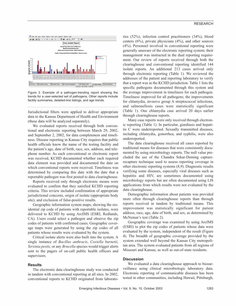
Jurisdictional filters were applied to deliver appropriatedata to the Kansas Department of Health and Environment(these data will be analyzed separately).
We evaluated reports received through both conven-tional and electronic reporting between March 29, 2002,and September 2, 2002, for data completeness and timeli-ness. Disease reporting in Kansas City requires that publichealth officials know the name of the testing facility andthe patient’s age, date of birth, race, sex, address, and tele-phone number. As each conventional or electronic reportwas received, KCHD documented whether each requireddata element was provided and documented the date onwhich conventional reports were received. Timeliness wasdetermined by comparing this date with the date that areportable pathogen was first posted to data clearinghouse.
Reports received only through electronic means wereevaluated to confirm that they satisfied KCHD reportingcriteria. This review included confirmation of appropriatejurisdictional concerns, origin of isolate (appropriate bodysite), and exclusion of false-positive results.
Geographic information system maps, showing the res-idential zip code of patients with reportable isolates, weredelivered to KCHD by using ArcIMS (ESRI, Redlands,CA). Users could select a pathogen and observe the zipcodes of patients with confirmed cases. Geographic cover-age maps were generated by using the zip codes of allpatients whose results were evaluated by the system.
Critical isolate alerts were also built into the system. Asingle instance of Bacillus anthracis, Coxiella burnetii,Yersinia pestis, or any Brucella species would trigger alertssent to the pagers of on-call public health officers andsupervisors.
ResultsThe electronic data clearinghouse study was conducted
in tandem with conventional reporting at all sites. In 2002,conventional reports to KCHD originated from laborato-
ries (52%), infection control practitioners (34%), bloodcenters (6%), private physicians (4%), and other sources(4%). Personnel involved in conventional reporting weregenerally unaware of the electronic reporting system; theirmanagement was instructed in the dual reporting require-ment. Our review of reports received through both theclearinghouse and conventional reporting identified 144isolate reports. An additional 213 cases arrived onlythrough electronic reporting (Table 1). We reviewed theaddresses of the patient and reporting laboratory to verifythat a report was in the KCHD jurisdiction. Table 1 lists thespecific pathogens documented through this system andthe average improvement in timeliness for each pathogen.Timeliness improved for all pathogens; the improvementsfor chlamydia, invasive group A streptococcal infections,and salmonellosis cases were statistically significant(Table 1). One chlamydia case arrived 20 days earlierthrough clearinghouse reports.
Many case reports were only received through electron-ic reporting (Table 1). In particular, giardiasis and hepati-tis C were underreported. Sexually transmitted diseases,including chlamydia, gonorrhea, and syphilis, were alsounderreported.
The data clearinghouse received all cases reported bytraditional means for diseases that were consistently docu-mented by using microbiology reports. The null value pre-cluded the use of the Chandra Sekar-Deming capture-recapture technique used to assess reporting coverage inother electronic reporting systems (8,9). Diagnostic resultsverifying some diseases, especially viral diseases such ashepatitis and HIV, are sometimes documented usingmicrobiology reports but are often documented using LISapplications from which results were not evaluated by thedata clearinghouse.
Demographic information about patients was providedmore often through clearinghouse reports than throughreports received in tandem by traditional means. Thisimprovement was statistically significant for patientaddress, race, age, date of birth, and sex, as determined byMcNemar’s test (Table 2).
Geographic coverage was examined by using ArcIMS(ESRI) to plot the zip codes of patients whose data wereevaluated by the system, independent of the result (Figure4). The breadth of geographic coverage provided by thesystem extended well beyond the Kansas City metropoli-tan area. The system evaluated patients from all regions ofMissouri and Kansas, as well as out-of-state residents.
DiscussionWe evaluated a data clearinghouse approach to biosur-
veillance using clinical microbiologic laboratory data.Electronic reporting of communicable diseases has beentested in other communities, including Hawaii, Pittsburgh,
Emerging Infectious Diseases • Vol. 9, No. 10, October 2003 1283
RESEARCH
Figure 3. Example of a pathogen-trending report showing thetrends for a user-selected set of pathogens. Other reports includefacility summaries, detailed-line listings, and age trends.
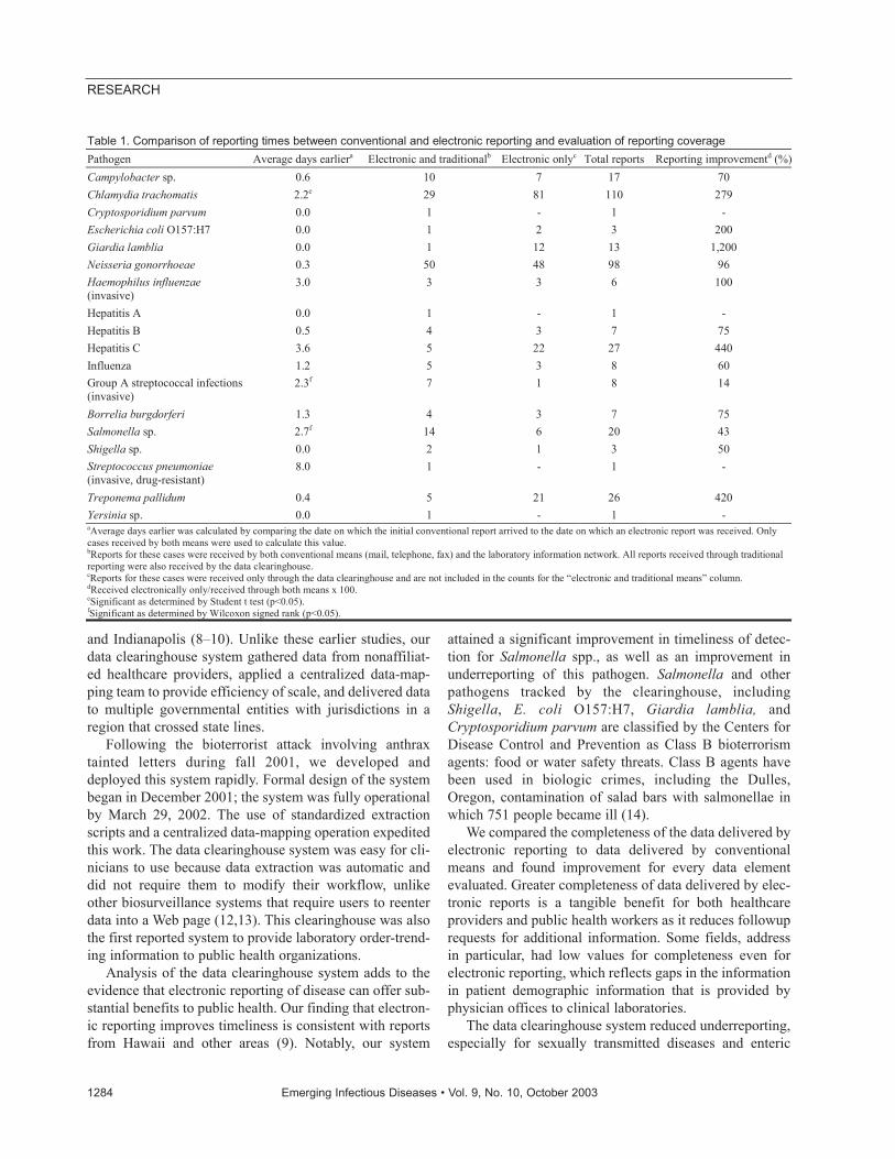
and Indianapolis (8–10). Unlike these earlier studies, ourdata clearinghouse system gathered data from nonaffiliat-ed healthcare providers, applied a centralized data-map-ping team to provide efficiency of scale, and delivered datato multiple governmental entities with jurisdictions in aregion that crossed state lines.
Following the bioterrorist attack involving anthraxtainted letters during fall 2001, we developed anddeployed this system rapidly. Formal design of the systembegan in December 2001; the system was fully operationalby March 29, 2002. The use of standardized extractionscripts and a centralized data-mapping operation expeditedthis work. The data clearinghouse system was easy for cli-nicians to use because data extraction was automatic anddid not require them to modify their workflow, unlikeother biosurveillance systems that require users to reenterdata into a Web page (12,13). This clearinghouse was alsothe first reported system to provide laboratory order-trend-ing information to public health organizations.
Analysis of the data clearinghouse system adds to theevidence that electronic reporting of disease can offer sub-stantial benefits to public health. Our finding that electron-ic reporting improves timeliness is consistent with reportsfrom Hawaii and other areas (9). Notably, our system
attained a significant improvement in timeliness of detec-tion for Salmonella spp., as well as an improvement inunderreporting of this pathogen. Salmonella and otherpathogens tracked by the clearinghouse, includingShigella, E. coli O157:H7, Giardia lamblia, andCryptosporidium parvum are classified by the Centers forDisease Control and Prevention as Class B bioterrorismagents: food or water safety threats. Class B agents havebeen used in biologic crimes, including the Dulles,Oregon, contamination of salad bars with salmonellae inwhich 751 people became ill (14).
We compared the completeness of the data delivered byelectronic reporting to data delivered by conventionalmeans and found improvement for every data elementevaluated. Greater completeness of data delivered by elec-tronic reports is a tangible benefit for both healthcareproviders and public health workers as it reduces followuprequests for additional information. Some fields, addressin particular, had low values for completeness even forelectronic reporting, which reflects gaps in the informationin patient demographic information that is provided byphysician offices to clinical laboratories.
The data clearinghouse system reduced underreporting,especially for sexually transmitted diseases and enteric
1284 Emerging Infectious Diseases • Vol. 9, No. 10, October 2003
RESEARCH
Table 1. Comparison of reporting times between conventional and electronic reporting and evaluation of reporting coverage Pathogen Average days earliera Electronic and traditionalb Electronic onlyc Total reports Reporting improvementd (%) Campylobacter sp. 0.6 10 7 17 70 Chlamydia trachomatis 2.2e 29 81 110 279 Cryptosporidium parvum 0.0 1 - 1 - Escherichia coli O157:H7 0.0 1 2 3 200 Giardia lamblia 0.0 1 12 13 1,200 Neisseria gonorrhoeae 0.3 50 48 98 96 Haemophilus influenzae (invasive)
3.0 3 3 6 100
Hepatitis A 0.0 1 - 1 - Hepatitis B 0.5 4 3 7 75 Hepatitis C 3.6 5 22 27 440 Influenza 1.2 5 3 8 60 Group A streptococcal infections (invasive)
2.3f 7 1 8 14
Borrelia burgdorferi 1.3 4 3 7 75 Salmonella sp. 2.7f 14 6 20 43 Shigella sp. 0.0 2 1 3 50 Streptococcus pneumoniae (invasive, drug-resistant)
8.0 1 - 1 -
Treponema pallidum 0.4 5 21 26 420 Yersinia sp. 0.0 1 - 1 - aAverage days earlier was calculated by comparing the date on which the initial conventional report arrived to the date on which an electronic report was received. Only cases received by both means were used to calculate this value. bReports for these cases were received by both conventional means (mail, telephone, fax) and the laboratory information network. All reports received through traditional reporting were also received by the data clearinghouse. cReports for these cases were received only through the data clearinghouse and are not included in the counts for the “electronic and traditional means” column. dReceived electronically only/received through both means x 100. eSignificant as determined by Student t test (p<0.05). fSignificant as determined by Wilcoxon signed rank (p<0.05).
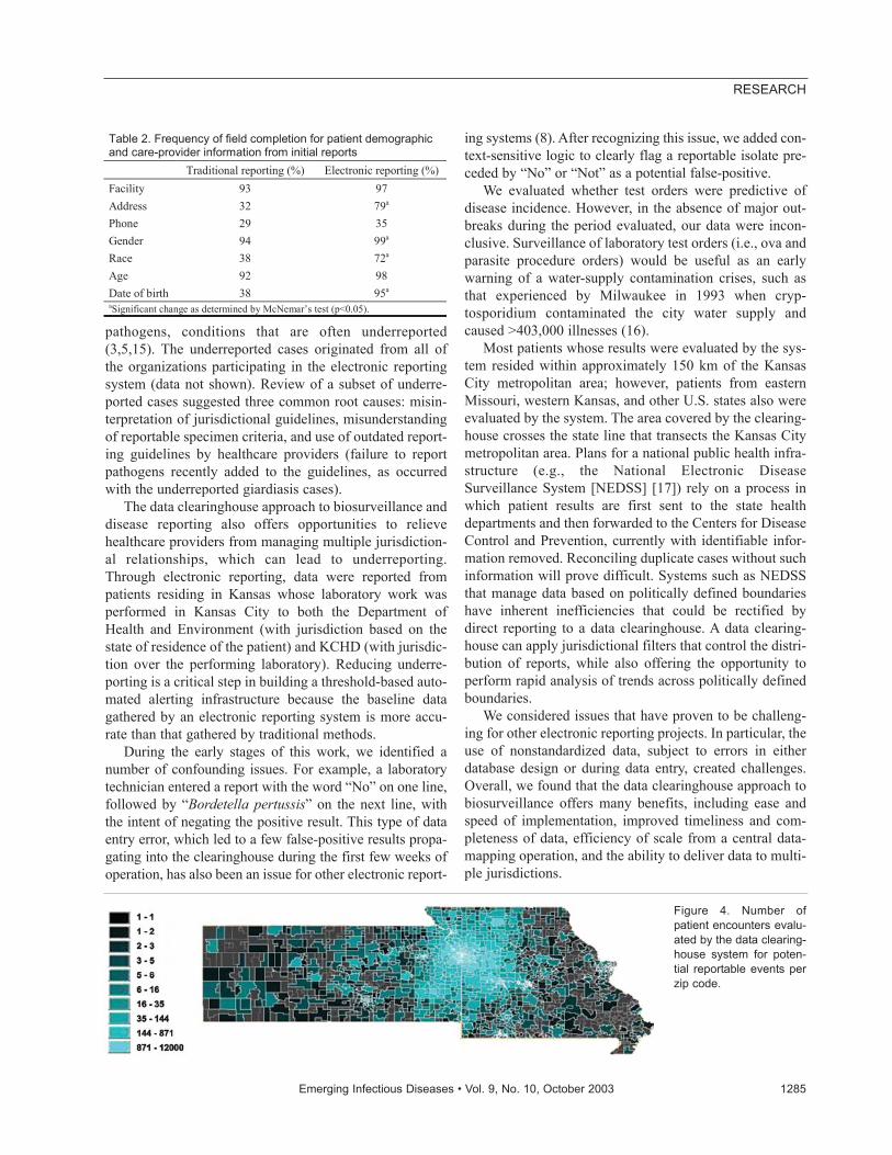
pathogens, conditions that are often underreported(3,5,15). The underreported cases originated from all ofthe organizations participating in the electronic reportingsystem (data not shown). Review of a subset of underre-ported cases suggested three common root causes: misin-terpretation of jurisdictional guidelines, misunderstandingof reportable specimen criteria, and use of outdated report-ing guidelines by healthcare providers (failure to reportpathogens recently added to the guidelines, as occurredwith the underreported giardiasis cases).
The data clearinghouse approach to biosurveillance anddisease reporting also offers opportunities to relievehealthcare providers from managing multiple jurisdiction-al relationships, which can lead to underreporting.Through electronic reporting, data were reported frompatients residing in Kansas whose laboratory work wasperformed in Kansas City to both the Department ofHealth and Environment (with jurisdiction based on thestate of residence of the patient) and KCHD (with jurisdic-tion over the performing laboratory). Reducing underre-porting is a critical step in building a threshold-based auto-mated alerting infrastructure because the baseline datagathered by an electronic reporting system is more accu-rate than that gathered by traditional methods.
During the early stages of this work, we identified anumber of confounding issues. For example, a laboratorytechnician entered a report with the word “No” on one line,followed by “Bordetella pertussis” on the next line, withthe intent of negating the positive result. This type of dataentry error, which led to a few false-positive results propa-gating into the clearinghouse during the first few weeks ofoperation, has also been an issue for other electronic report-
ing systems (8). After recognizing this issue, we added con-text-sensitive logic to clearly flag a reportable isolate pre-ceded by “No” or “Not” as a potential false-positive.
We evaluated whether test orders were predictive ofdisease incidence. However, in the absence of major out-breaks during the period evaluated, our data were incon-clusive. Surveillance of laboratory test orders (i.e., ova andparasite procedure orders) would be useful as an earlywarning of a water-supply contamination crises, such asthat experienced by Milwaukee in 1993 when cryp-tosporidium contaminated the city water supply andcaused >403,000 illnesses (16).
Most patients whose results were evaluated by the sys-tem resided within approximately 150 km of the KansasCity metropolitan area; however, patients from easternMissouri, western Kansas, and other U.S. states also wereevaluated by the system. The area covered by the clearing-house crosses the state line that transects the Kansas Citymetropolitan area. Plans for a national public health infra-structure (e.g., the National Electronic DiseaseSurveillance System [NEDSS] [17]) rely on a process inwhich patient results are first sent to the state healthdepartments and then forwarded to the Centers for DiseaseControl and Prevention, currently with identifiable infor-mation removed. Reconciling duplicate cases without suchinformation will prove difficult. Systems such as NEDSSthat manage data based on politically defined boundarieshave inherent inefficiencies that could be rectified bydirect reporting to a data clearinghouse. A data clearing-house can apply jurisdictional filters that control the distri-bution of reports, while also offering the opportunity toperform rapid analysis of trends across politically definedboundaries.
We considered issues that have proven to be challeng-ing for other electronic reporting projects. In particular, theuse of nonstandardized data, subject to errors in eitherdatabase design or during data entry, created challenges.Overall, we found that the data clearinghouse approach tobiosurveillance offers many benefits, including ease andspeed of implementation, improved timeliness and com-pleteness of data, efficiency of scale from a central data-mapping operation, and the ability to deliver data to multi-ple jurisdictions.
Emerging Infectious Diseases • Vol. 9, No. 10, October 2003 1285
RESEARCH
Figure 4. Number ofpatient encounters evalu-ated by the data clearing-house system for poten-tial reportable events perzip code.
Table 2. Frequency of field completion for patient demographic and care-provider information from initial reports Traditional reporting (%) Electronic reporting (%) Facility 93 97 Address 32 79a Phone 29 35 Gender 94 99a Race 38 72a Age 92 98 Date of birth 38 95a aSignificant change as determined by McNemar’s test (p<0.05).
AcknowledgmentsWe thank Lesha Dennis, Gary Dickerson, Suzanne Fiske,
Andrew Friede, Jody Gosch, Kim Hlobik, and Karen Miscavish;the Information Technology staff at Health Midwest; St. Luke’sShawnee Mission, Truman; Sisters of Mercy; and the KansasCity Health Department.
This project was funded independently by CernerCorporation and the Kansas City Health Department. CernerCorporation has a financial interest in the HealthSentry clearing-house system.
Dr. Hoffman is a molecular microbiologist at CernerCorporation. His research interests include biosurveillance,bioinformatics, medical informatics, and clinical decision sup-port software. In addition to his work on the HealthSentry biosur-veillance system, Dr. Hoffman is a co-investigator on an Agencyfor Healthcare Research and Quality HIV clinical decision sup-port grant and leads efforts at Cerner to capture and structuregenomic information in electronic medical records.
References1. Goldenberg A, Shmueli G, Caruana RA, Fienberg SE. Early statisti-
cal detection of anthrax outbreaks by tracking over-the-counter med-ication sales. Proc Natl Acad Sci U S A 2002;99:5237–40.
2. Lazarus R, Kleinman K, Dashevsky I, Adams C, Kludt P, DeMaria AJr, et al. Use of automated ambulatory-care encounter records fordetection of acute illness clusters, including potential bioterrorismevents. Emerg Infect Dis 2002;8:753–60.
3. Thacker SB, Choi K, Brachman PS. The surveillance of infectiousdiseases. JAMA 1983;249:1181–5.
4. Schwarcz SK, Hsu LC, Parisi MK, Katz MH. The impact of the 1993AIDS case definition on the completeness and timeliness of AIDSsurveillance. AIDS 1999;13:1109–14.
5. Kimball AM, Thacker SB, Levy ME. Shigella surveillance in a largemetropolitan area: assessment of a passive reporting system. Am JPublic Health 1980;70:164–6.
6. Ackman DM, Birkhead G, Flynn M. Assessment of surveillance formeningococcal disease in New York State, 1991. Am J Epidemiol1996;144:78–82.
7. Teutsch S, Churchill RE. Principles and practice of public health sur-veillance. New York: Oxford University Press; 1994.
8. Panackal AA, M’ikanatha NM, Tsui FC, McMahon J, Wagner MM,Dixon BW, et al. Automatic electronic laboratory-based reporting ofnotifiable infectious diseases at a large health system. Emerg InfectDis 2002;8:685–91.
9. Effler P, Ching-Lee M, Bogard A, Ieong MC, Nekomoto T, JerniganD. Statewide system of electronic notifiable disease reporting fromclinical laboratories: comparing automated reporting with conven-tional methods. JAMA 1999;282:1845–50.
10. Overhage JM, Suico J, McDonald CJ. Electronic laboratory report-ing: barriers, solutions and findings. J Public Health Manag Pract2001;7:60–6.
11. HealthSentry [computer program]. Kansas City (MO): Cerner Corp.;2002.
12. Syndromic surveillance for bioterrorism following the attacks on theWorld Trade Center—New York City, 2001. MMWR Morb MortalWkly Rep 2002;51:13–5.
13. Zelicoff A, Brillman J, Forslund DW, George JE, Zink S, Koenig S,et al. The rapid syndrome validation project (RSVP). Proc AMIASymp 2001;771–5.
14. Torok TJ, Tauxe RV, Wise RP, Livengood JR, Sokolow R, Mauvais S,et al. A large community outbreak of salmonellosis caused by inten-tional contamination of restaurant salad bars. JAMA1997;278:389–95.
15. Harkess JR, Gildon BA, Archer PW, Istre GR. Is passive surveillancealways insensitive? An evaluation of shigellosis surveillance inOklahoma. Am J Epidemiol 1988;128:878–81.
16. MacKenzie WR, Hoxie NJ, Proctor ME, Gradus MS, Blair KA,Peterson DE, et al. A massive outbreak in Milwaukee of cryptosporid-ium infection transmitted through the public water supply. N Engl JMed 1994;331:161–7.
17. National Electronic Disease Surveillance System (NEDSS): a stan-dards-based approach to connect public health and clinical medicine.J Public Health Manag Pract 2001;7:43–50.
Address for correspondence: Mark Hoffman, Cerner Corporation, 2800Rockcreek Parkway, Kansas City, MO 64117-2551, USA; fax: (816) 571-3291; email: [email protected]
1286 Emerging Infectious Diseases • Vol. 9, No. 10, October 2003
RESEARCH
Epidemics of meningococcal meningitis occur in areaswith particular environmental characteristics. We presentevidence that the relationship between the environmentand the location of these epidemics is quantifiable and pro-pose a model based on environmental variables to identifyregions at risk for meningitis epidemics. These findings,which have substantial implications for directing surveil-lance activities and health policy, provide a basis for moni-toring the impact of climate variability and environmentalchange on epidemic occurrence in Africa.
Epidemics of meningitis occur worldwide. However,the “meningitis belt” of Africa’s Sahel region has the
greatest incidence of the disease, with large epidemicsattributed to predominantly group A meningococci; theendemic levels found in this region would be consideredepidemic elsewhere. Although factors predisposing popu-lations to meningitis epidemics are poorly understood,population susceptibility, introduction of new strains, poorliving conditions, and concurrent infections have all beenimplicated. Epidemics occur throughout Africa in the dryseason, coincide with periods of very low humidity anddusty conditions, and disappear with the onset of the rains,suggesting that these environmental factors may also playan important role in the occurrence of the disease (1–3).
Lapeyssonnie (4) observed in 1963 that epidemicslargely occurred in a semi-arid zone south from the Sahara,with 300–1,100 mm mean annual rainfall, and Chees-brough et al. (5) suggested in 1995 that areas that arehumid throughout the year have low disease rates. In WestAfrica, Waddy (1958) described an area that suffered epi-demics as having “… only one definable frontier, the junc-tion of the forest…with the savanna…, when there is anabrupt change from a permanently humid climate to onewith a severe dry season” (6). Epidemics have been rarelyreported from the humid forested or coastal regions, evenwhen neighboring areas are severely affected.
The timing of future outbreaks is unpredictable; toolsthat identify the key environmental factors associated withareas prone to meningitis epidemics would help us tounderstand the basis for these outbreaks and eventuallyoptimize prevention and control activities. We describe amodel that predicts the probability, based on environmen-tal information, of an area experiencing an epidemic ofmeningitis.
Methods
Epidemiologic DataDetails of all known meningitis epidemics occurring
before 2000 in countries comprising continental Africawere compiled from information documented in the pub-lished literature and unpublished institutional reports at theend of June 2001. All epidemics reported in the medicalliterature were identified in PubMed’s online database ofmedical literature (United States National Library ofMedicine, available from: URL: http://www.ncbi.nlm.nih.gov/PubMed/) through manual searches and by cross-referencing publications. We obtained unpublished infor-mation from Web searches or directly from internationaland national organizations involved in disease control andhumanitarian aid (1).
Epidemics reported at the provincial (second adminis-trative level) and district level were located by usingadministrative boundaries available from the U.S.Geological Survey EROS Data Centre Africa DataDissemination Service, Sioux Falls, SD (available from:URL: http://edcintl.cr.usgs.gov/adds/). Locations of vil-lages or towns were verified by using the National Imageryand Mapping Agency’s GEONet gazetteer (available from:URL: http://www.nima.mil/), historical reference atlasesand maps contained in the original reports, and weremapped to the current administrative boundaries by usingArcView 3.1 geographic information system (GIS) (ESRI,Redlands, CA). We assumed that events reported at theprovincial level affected all constituent districts andexcluded epidemics reported only at the national level.
Emerging Infectious Diseases • Vol. 9, No. 10, October 2003 1287
RESEARCH
Environmental Risk and MeningitisEpidemics in Africa
Anna M. Molesworth,* Luis E. Cuevas,* Stephen J. Connor,* Andrew P. Morse,† and Madeleine C. Thomson*
*Liverpool School of Tropical Medicine, Liverpool, UnitedKingdom; and †University of Liverpool, Liverpool, United Kingdom
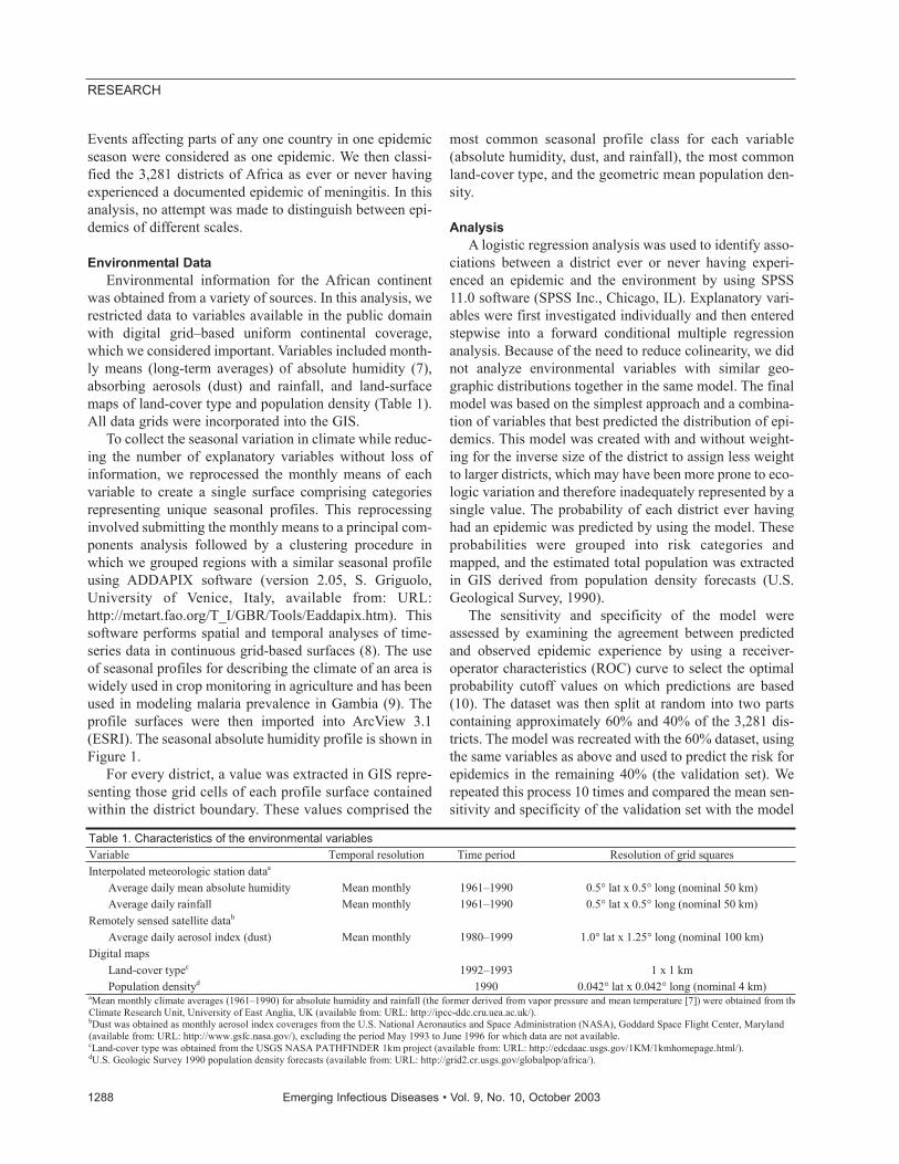
Events affecting parts of any one country in one epidemicseason were considered as one epidemic. We then classi-fied the 3,281 districts of Africa as ever or never havingexperienced a documented epidemic of meningitis. In thisanalysis, no attempt was made to distinguish between epi-demics of different scales.
Environmental DataEnvironmental information for the African continent
was obtained from a variety of sources. In this analysis, werestricted data to variables available in the public domainwith digital grid–based uniform continental coverage,which we considered important. Variables included month-ly means (long-term averages) of absolute humidity (7),absorbing aerosols (dust) and rainfall, and land-surfacemaps of land-cover type and population density (Table 1).All data grids were incorporated into the GIS.
To collect the seasonal variation in climate while reduc-ing the number of explanatory variables without loss ofinformation, we reprocessed the monthly means of eachvariable to create a single surface comprising categoriesrepresenting unique seasonal profiles. This reprocessinginvolved submitting the monthly means to a principal com-ponents analysis followed by a clustering procedure inwhich we grouped regions with a similar seasonal profileusing ADDAPIX software (version 2.05, S. Griguolo,University of Venice, Italy, available from: URL:http://metart.fao.org/T_I/GBR/Tools/Eaddapix.htm). Thissoftware performs spatial and temporal analyses of time-series data in continuous grid-based surfaces (8). The useof seasonal profiles for describing the climate of an area iswidely used in crop monitoring in agriculture and has beenused in modeling malaria prevalence in Gambia (9). Theprofile surfaces were then imported into ArcView 3.1(ESRI). The seasonal absolute humidity profile is shown inFigure 1.
For every district, a value was extracted in GIS repre-senting those grid cells of each profile surface containedwithin the district boundary. These values comprised the
most common seasonal profile class for each variable(absolute humidity, dust, and rainfall), the most commonland-cover type, and the geometric mean population den-sity.
AnalysisA logistic regression analysis was used to identify asso-
ciations between a district ever or never having experi-enced an epidemic and the environment by using SPSS11.0 software (SPSS Inc., Chicago, IL). Explanatory vari-ables were first investigated individually and then enteredstepwise into a forward conditional multiple regressionanalysis. Because of the need to reduce colinearity, we didnot analyze environmental variables with similar geo-graphic distributions together in the same model. The finalmodel was based on the simplest approach and a combina-tion of variables that best predicted the distribution of epi-demics. This model was created with and without weight-ing for the inverse size of the district to assign less weightto larger districts, which may have been more prone to eco-logic variation and therefore inadequately represented by asingle value. The probability of each district ever havinghad an epidemic was predicted by using the model. Theseprobabilities were grouped into risk categories andmapped, and the estimated total population was extractedin GIS derived from population density forecasts (U.S.Geological Survey, 1990).
The sensitivity and specificity of the model wereassessed by examining the agreement between predictedand observed epidemic experience by using a receiver-operator characteristics (ROC) curve to select the optimalprobability cutoff values on which predictions are based(10). The dataset was then split at random into two partscontaining approximately 60% and 40% of the 3,281 dis-tricts. The model was recreated with the 60% dataset, usingthe same variables as above and used to predict the risk forepidemics in the remaining 40% (the validation set). Werepeated this process 10 times and compared the mean sen-sitivity and specificity of the validation set with the model
1288 Emerging Infectious Diseases • Vol. 9, No. 10, October 2003
RESEARCH
Table 1. Characteristics of the environmental variables Variable Temporal resolution Time period Resolution of grid squares Interpolated meteorologic station dataa
Average daily mean absolute humidity Mean monthly 1961–1990 0.5° lat x 0.5° long (nominal 50 km) Average daily rainfall Mean monthly 1961–1990 0.5° lat x 0.5° long (nominal 50 km)
Remotely sensed satellite datab Average daily aerosol index (dust) Mean monthly 1980–1999 1.0° lat x 1.25° long (nominal 100 km)
Digital maps Land-cover typec 1992–1993 1 x 1 km Population densityd 1990 0.042° lat x 0.042° long (nominal 4 km)
aMean monthly climate averages (1961–1990) for absolute humidity and rainfall (the former derived from vapor pressure and mean temperature [7]) were obtained from theClimate Research Unit, University of East Anglia, UK (available from: URL: http://ipcc-ddc.cru.uea.ac.uk/). bDust was obtained as monthly aerosol index coverages from the U.S. National Aeronautics and Space Administration (NASA), Goddard Space Flight Center, Maryland (available from: URL: http://www.gsfc.nasa.gov/), excluding the period May 1993 to June 1996 for which data are not available. cLand-cover type was obtained from the USGS NASA PATHFINDER 1km project (available from: URL: http://edcdaac.usgs.gov/1KM/1kmhomepage.html/). dU.S. Geologic Survey 1990 population density forecasts (available from: URL: http://grid2.cr.usgs.gov/globalpop/africa/).
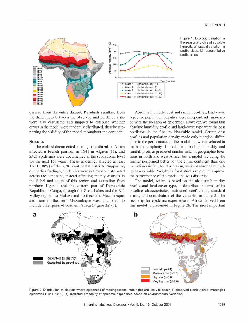
derived from the entire dataset. Residuals resulting fromthe differences between the observed and predicted riskswere also calculated and mapped to establish whethererrors in the model were randomly distributed, thereby sup-porting the validity of the model throughout the continent.
ResultsThe earliest documented meningitis outbreak in Africa
affected a French garrison in 1841 in Algiers (11), and≥425 epidemics were documented at the subnational levelfor the next 158 years. These epidemics affected at least1,231 (38%) of the 3,281 continental districts. Supportingour earlier findings, epidemics were not evenly distributedacross the continent, instead affecting mainly districts inthe Sahel and south of this region and extending fromnorthern Uganda and the eastern part of DemocraticRepublic of Congo, through the Great Lakes and the RiftValley regions to Malawi and northeastern Mozambique,and from northeastern Mozambique west and south toinclude other parts of southern Africa (Figure 2a) (1).
Absolute humidity, dust and rainfall profiles, land-covertype, and population densities were independently associat-ed with the location of epidemics. However, we found thatabsolute humidity profile and land-cover type were the bestpredictors in the final multivariable model. Certain dustprofiles and population density made only marginal differ-ence to the performance of the model and were excluded tomaintain simplicity. In addition, absolute humidity andrainfall profiles predicted similar risks in geographic loca-tions in north and west Africa, but a model including theformer performed better for the entire continent than oneincluding rainfall; for this reason, we kept absolute humid-ity as a variable. Weighting for district size did not improvethe performance of the model and was discarded.
The model, which is based on the absolute humidityprofile and land-cover type, is described in terms of itsbaseline characteristics, estimated coefficients, standarderrors, and contribution of the variables in Table 2. Therisk map for epidemic experience in Africa derived fromthis model is presented in Figure 2b. The most important
Emerging Infectious Diseases • Vol. 9, No. 10, October 2003 1289
RESEARCH
Figure 1. Ecologic variation inthe seasonal profile of absolutehumidity. a) spatial variation inprofile class; b) representativeprofile class.
Figure 2. Distribution of districts where epidemics of meningococcal meningitis are likely to occur. a) observed distribution of meningitisepidemics (1841–1999). b) predicted probabilty of epidemic experience based on environmental variables.
a b
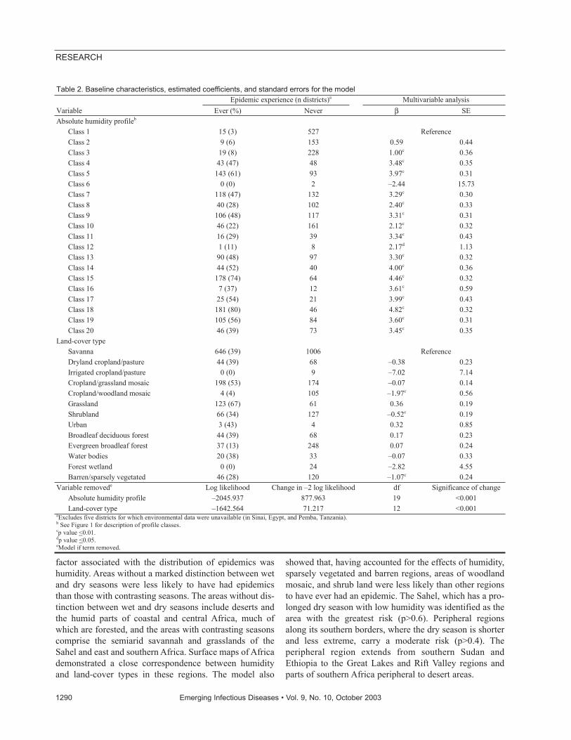
factor associated with the distribution of epidemics washumidity. Areas without a marked distinction between wetand dry seasons were less likely to have had epidemicsthan those with contrasting seasons. The areas without dis-tinction between wet and dry seasons include deserts andthe humid parts of coastal and central Africa, much ofwhich are forested, and the areas with contrasting seasonscomprise the semiarid savannah and grasslands of theSahel and east and southern Africa. Surface maps of Africademonstrated a close correspondence between humidityand land-cover types in these regions. The model also
showed that, having accounted for the effects of humidity,sparsely vegetated and barren regions, areas of woodlandmosaic, and shrub land were less likely than other regionsto have ever had an epidemic. The Sahel, which has a pro-longed dry season with low humidity was identified as thearea with the greatest risk (p>0.6). Peripheral regionsalong its southern borders, where the dry season is shorterand less extreme, carry a moderate risk (p>0.4). Theperipheral region extends from southern Sudan andEthiopia to the Great Lakes and Rift Valley regions andparts of southern Africa peripheral to desert areas.
1290 Emerging Infectious Diseases • Vol. 9, No. 10, October 2003
RESEARCH
Table 2. Baseline characteristics, estimated coefficients, and standard errors for the model Epidemic experience (n districts)a Multivariable analysis
Variable Ever (%) Never β SE Absolute humidity profileb
Class 1 15 (3) 527 Reference Class 2 9 (6) 153 0.59 0.44 Class 3 19 (8) 228 1.00c 0.36 Class 4 43 (47) 48 3.48c 0.35 Class 5 143 (61) 93 3.97c 0.31 Class 6 0 (0) 2 –2.44 15.73 Class 7 118 (47) 132 3.29c 0.30 Class 8 40 (28) 102 2.40c 0.33 Class 9 106 (48) 117 3.31c 0.31 Class 10 46 (22) 161 2.12c 0.32 Class 11 16 (29) 39 3.34c 0.43 Class 12 1 (11) 8 2.17d 1.13 Class 13 90 (48) 97 3.30c 0.32 Class 14 44 (52) 40 4.00c 0.36 Class 15 178 (74) 64 4.46c 0.32 Class 16 7 (37) 12 3.61c 0.59 Class 17 25 (54) 21 3.99c 0.43 Class 18 181 (80) 46 4.82c 0.32 Class 19 105 (56) 84 3.60c 0.31 Class 20 46 (39) 73 3.45c 0.35
Land-cover type Savanna 646 (39) 1006 Reference Dryland cropland/pasture 44 (39) 68 –0.38 0.23 Irrigated cropland/pasture 0 (0) 9 –7.02 7.14 Cropland/grassland mosaic 198 (53) 174 –0.07 0.14 Cropland/woodland mosaic 4 (4) 105 –1.97c 0.56 Grassland 123 (67) 61 0.36 0.19 Shrubland 66 (34) 127 –0.52c 0.19 Urban 3 (43) 4 0.32 0.85 Broadleaf deciduous forest 44 (39) 68 0.17 0.23 Evergreen broadleaf forest 37 (13) 248 0.07 0.24 Water bodies 20 (38) 33 –0.07 0.33 Forest wetland 0 (0) 24 –2.82 4.55 Barren/sparsely vegetated 46 (28) 120 –1.07c 0.24
Variable removede Log likelihood Change in –2 log likelihood df Significance of change Absolute humidity profile –2045.937 877.963 19 <0.001 Land-cover type –1642.564 71.217 12 <0.001
aExcludes five districts for which environmental data were unavailable (in Sinai, Egypt, and Pemba, Tanzania). b See Figure 1 for description of profile classes. cp value <0.01. dp value <0.05. eModel if term removed.
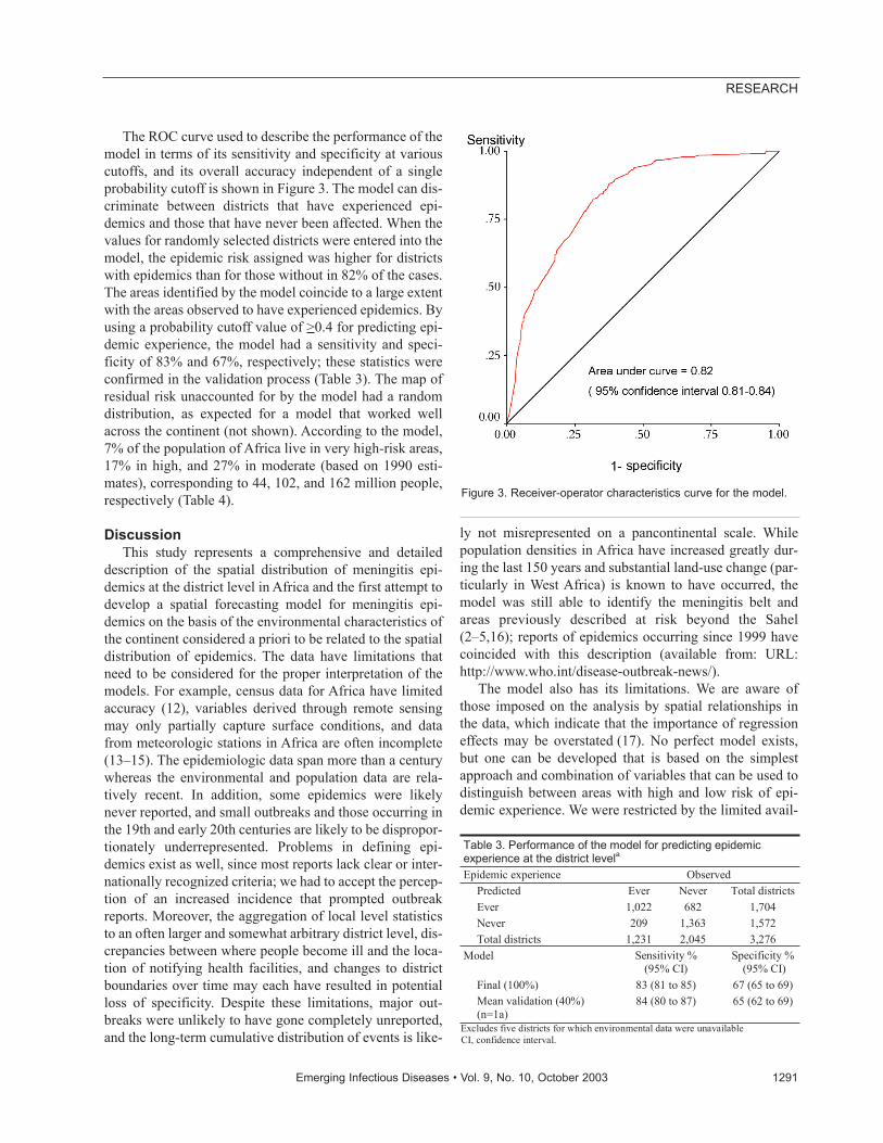
The ROC curve used to describe the performance of themodel in terms of its sensitivity and specificity at variouscutoffs, and its overall accuracy independent of a singleprobability cutoff is shown in Figure 3. The model can dis-criminate between districts that have experienced epi-demics and those that have never been affected. When thevalues for randomly selected districts were entered into themodel, the epidemic risk assigned was higher for districtswith epidemics than for those without in 82% of the cases.The areas identified by the model coincide to a large extentwith the areas observed to have experienced epidemics. Byusing a probability cutoff value of >0.4 for predicting epi-demic experience, the model had a sensitivity and speci-ficity of 83% and 67%, respectively; these statistics wereconfirmed in the validation process (Table 3). The map ofresidual risk unaccounted for by the model had a randomdistribution, as expected for a model that worked wellacross the continent (not shown). According to the model,7% of the population of Africa live in very high-risk areas,17% in high, and 27% in moderate (based on 1990 esti-mates), corresponding to 44, 102, and 162 million people,respectively (Table 4).
DiscussionThis study represents a comprehensive and detailed
description of the spatial distribution of meningitis epi-demics at the district level in Africa and the first attempt todevelop a spatial forecasting model for meningitis epi-demics on the basis of the environmental characteristics ofthe continent considered a priori to be related to the spatialdistribution of epidemics. The data have limitations thatneed to be considered for the proper interpretation of themodels. For example, census data for Africa have limitedaccuracy (12), variables derived through remote sensingmay only partially capture surface conditions, and datafrom meteorologic stations in Africa are often incomplete(13–15). The epidemiologic data span more than a centurywhereas the environmental and population data are rela-tively recent. In addition, some epidemics were likelynever reported, and small outbreaks and those occurring inthe 19th and early 20th centuries are likely to be dispropor-tionately underrepresented. Problems in defining epi-demics exist as well, since most reports lack clear or inter-nationally recognized criteria; we had to accept the percep-tion of an increased incidence that prompted outbreakreports. Moreover, the aggregation of local level statisticsto an often larger and somewhat arbitrary district level, dis-crepancies between where people become ill and the loca-tion of notifying health facilities, and changes to districtboundaries over time may each have resulted in potentialloss of specificity. Despite these limitations, major out-breaks were unlikely to have gone completely unreported,and the long-term cumulative distribution of events is like-
ly not misrepresented on a pancontinental scale. Whilepopulation densities in Africa have increased greatly dur-ing the last 150 years and substantial land-use change (par-ticularly in West Africa) is known to have occurred, themodel was still able to identify the meningitis belt andareas previously described at risk beyond the Sahel(2–5,16); reports of epidemics occurring since 1999 havecoincided with this description (available from: URL:http://www.who.int/disease-outbreak-news/).
The model also has its limitations. We are aware ofthose imposed on the analysis by spatial relationships inthe data, which indicate that the importance of regressioneffects may be overstated (17). No perfect model exists,but one can be developed that is based on the simplestapproach and combination of variables that can be used todistinguish between areas with high and low risk of epi-demic experience. We were restricted by the limited avail-
Emerging Infectious Diseases • Vol. 9, No. 10, October 2003 1291
RESEARCH
Figure 3. Receiver-operator characteristics curve for the model.
Table 3. Performance of the model for predicting epidemic experience at the district levela Epidemic experience Observed
Predicted Ever Never Total districts Ever 1,022 682 1,704 Never 209 1,363 1,572 Total districts 1,231 2,045 3,276
Model Sensitivity % (95% CI)
Specificity % (95% CI)
Final (100%) 83 (81 to 85) 67 (65 to 69) Mean validation (40%) (n=1a)
84 (80 to 87) 65 (62 to 69)
Excludes five districts for which environmental data were unavailable CI, confidence interval.
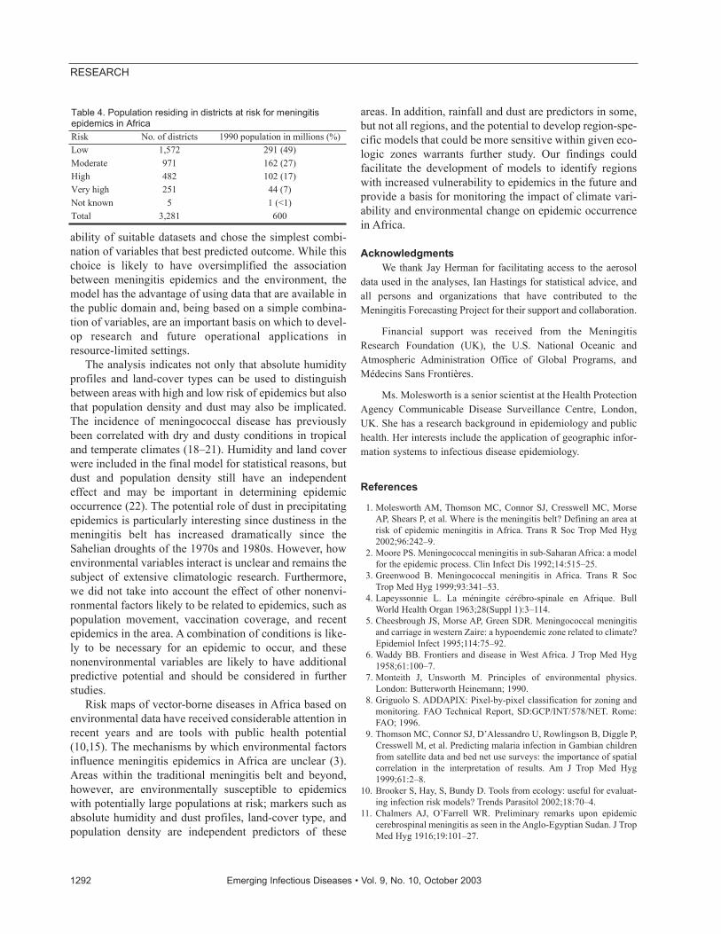
ability of suitable datasets and chose the simplest combi-nation of variables that best predicted outcome. While thischoice is likely to have oversimplified the associationbetween meningitis epidemics and the environment, themodel has the advantage of using data that are available inthe public domain and, being based on a simple combina-tion of variables, are an important basis on which to devel-op research and future operational applications inresource-limited settings.
The analysis indicates not only that absolute humidityprofiles and land-cover types can be used to distinguishbetween areas with high and low risk of epidemics but alsothat population density and dust may also be implicated.The incidence of meningococcal disease has previouslybeen correlated with dry and dusty conditions in tropicaland temperate climates (18–21). Humidity and land coverwere included in the final model for statistical reasons, butdust and population density still have an independenteffect and may be important in determining epidemicoccurrence (22). The potential role of dust in precipitatingepidemics is particularly interesting since dustiness in themeningitis belt has increased dramatically since theSahelian droughts of the 1970s and 1980s. However, howenvironmental variables interact is unclear and remains thesubject of extensive climatologic research. Furthermore,we did not take into account the effect of other nonenvi-ronmental factors likely to be related to epidemics, such aspopulation movement, vaccination coverage, and recentepidemics in the area. A combination of conditions is like-ly to be necessary for an epidemic to occur, and thesenonenvironmental variables are likely to have additionalpredictive potential and should be considered in furtherstudies.
Risk maps of vector-borne diseases in Africa based onenvironmental data have received considerable attention inrecent years and are tools with public health potential(10,15). The mechanisms by which environmental factorsinfluence meningitis epidemics in Africa are unclear (3).Areas within the traditional meningitis belt and beyond,however, are environmentally susceptible to epidemicswith potentially large populations at risk; markers such asabsolute humidity and dust profiles, land-cover type, andpopulation density are independent predictors of these
areas. In addition, rainfall and dust are predictors in some,but not all regions, and the potential to develop region-spe-cific models that could be more sensitive within given eco-logic zones warrants further study. Our findings couldfacilitate the development of models to identify regionswith increased vulnerability to epidemics in the future andprovide a basis for monitoring the impact of climate vari-ability and environmental change on epidemic occurrencein Africa.
AcknowledgmentsWe thank Jay Herman for facilitating access to the aerosol
data used in the analyses, Ian Hastings for statistical advice, andall persons and organizations that have contributed to theMeningitis Forecasting Project for their support and collaboration.
Financial support was received from the MeningitisResearch Foundation (UK), the U.S. National Oceanic andAtmospheric Administration Office of Global Programs, andMédecins Sans Frontières.
Ms. Molesworth is a senior scientist at the Health ProtectionAgency Communicable Disease Surveillance Centre, London,UK. She has a research background in epidemiology and publichealth. Her interests include the application of geographic infor-mation systems to infectious disease epidemiology.
References
1. Molesworth AM, Thomson MC, Connor SJ, Cresswell MC, MorseAP, Shears P, et al. Where is the meningitis belt? Defining an area atrisk of epidemic meningitis in Africa. Trans R Soc Trop Med Hyg2002;96:242–9.
2. Moore PS. Meningococcal meningitis in sub-Saharan Africa: a modelfor the epidemic process. Clin Infect Dis 1992;14:515–25.
3. Greenwood B. Meningococcal meningitis in Africa. Trans R SocTrop Med Hyg 1999;93:341–53.
4. Lapeyssonnie L. La méningite cérébro-spinale en Afrique. BullWorld Health Organ 1963;28(Suppl 1):3–114.
5. Cheesbrough JS, Morse AP, Green SDR. Meningococcal meningitisand carriage in western Zaire: a hypoendemic zone related to climate?Epidemiol Infect 1995;114:75–92.
6. Waddy BB. Frontiers and disease in West Africa. J Trop Med Hyg1958;61:100–7.
7. Monteith J, Unsworth M. Principles of environmental physics.London: Butterworth Heinemann; 1990.
8. Griguolo S. ADDAPIX: Pixel-by-pixel classification for zoning andmonitoring. FAO Technical Report, SD:GCP/INT/578/NET. Rome:FAO; 1996.
9. Thomson MC, Connor SJ, D’Alessandro U, Rowlingson B, Diggle P,Cresswell M, et al. Predicting malaria infection in Gambian childrenfrom satellite data and bed net use surveys: the importance of spatialcorrelation in the interpretation of results. Am J Trop Med Hyg1999;61:2–8.
10. Brooker S, Hay, S, Bundy D. Tools from ecology: useful for evaluat-ing infection risk models? Trends Parasitol 2002;18:70–4.
11. Chalmers AJ, O’Farrell WR. Preliminary remarks upon epidemiccerebrospinal meningitis as seen in the Anglo-Egyptian Sudan. J TropMed Hyg 1916;19:101–27.
1292 Emerging Infectious Diseases • Vol. 9, No. 10, October 2003
RESEARCH
Table 4. Population residing in districts at risk for meningitis epidemics in Africa Risk No. of districts 1990 population in millions (%) Low 1,572 291 (49) Moderate 971 162 (27) High 482 102 (17) Very high 251 44 (7) Not known 5 1 (<1) Total 3,281 600
12. Deichmann U. A medium resolution population database for Africa.Database documentation and digital database. Santa Barbara (CA):National Center for Geographic Information and Analysis, Universityof California; 1994.
13. Herman JR, Bhartia PK, Torres O, Hsu C, Seftor C, Celarier E.Global distribution of UV-absorbing aerosols from Nimbus 7/TOMSdata. J Geophys Res 1997;102:16911–22.
14. New M, Hulme M, Jones P. Representing twentieth-century space-time climate variability. Part 1: development of a 1961–90 meanmonthly terrestrial climatology. J Clim 1999;12:829–56.
15. Thomson MC, Connor SJ. Environmental information systems forthe control of arthropod vectors of disease. Med Vet Entomol2000;14:227–44.
16. Mpairwe Y, Matovu HL. Cerebrospinal meningitis in east Africa1911–1965. Trans R Soc Trop Med Hyg 1971;65:70–7.
17. Diggle P, Moyeed R, Rawlingson B, Thomson M. Childhood malar-ia in the Gambia: a case study in model-based geostatistics. Appl Stat2002;51:493–506.
18. Molineaux L. Climate and meningococcal disease. [PhD Thesis].Berkeley (CA): University of Berkeley; 1969.
19. Waddy BB. Climate and respiratory infections. Lancet1952;263:674–7.
20. Greenwood BM, Blakebrough IS, Bradley AK, Wali S, Whittle HC.Meningococcal disease and season in sub-Saharan Africa. Lancet1984;326:1339–42.
21. Besancenot JP, Boko M, Oke PC. Weather conditions and cere-brospinal meningitis in Benin (Gulf of Guinea, West Africa). Eur JEpidemiol 1997;13:807–15.
22. Molesworth AM, Cuevas LE, Morse AP, Herman JR, Thomson MC.Dust clouds and spread of infection. Lancet 2002;359:81–2.
Address for correspondence: Madeleine C. Thomson, InternationalResearch Institute for Climate Prediction (IRI), The Earth Institute ofColumbia University, Lamont Campus, POB 1000, Palisades, New York,10964, USA; fax: 1-845-680-4864; email: [email protected]
Emerging Infectious Diseases • Vol. 9, No. 10, October 2003 1293
RESEARCH
Search past issues of EID at www.cdc.gov/eid
Severe AcuteRespiratorySyndrome:
Lessons fromSingapore
Kamaljit Singh,* Li-Yang Hsu,† Jorge S. Villacian,† Abdulrazaq Habib,*
Dale Fisher,* and Paul A. Tambyah*An outbreak of severe acute respiratory syndrome
(SARS) occurred in Singapore in March 2003. To illustratethe problems in diagnosing and containing SARS in thehospital, we describe a case series and highlight changesin triage and infection control practices that have resulted.By implementing these changes, we have stopped thenosocomial transmission of the virus.
An outbreak of severe acute respiratory syndrome(SARS) was first recognized in Singapore on March
12, 2003. The index patient was hospitalized at Tan TockSeng Hospital, which has since become the country’s des-ignated SARS hospital. The patient infected 20 other peo-ple (including patients and healthcare workers), who sub-sequently became the sources for secondary spread of theinfection (1). As of June 12, 2003, a total of 206 cases and31 deaths attributed to SARS had been reported inSingapore.
We describe the important lessons learned during thetriage and containment of SARS at the National UniversityHospital, Singapore. Both involved expanding isolationcriteria to include all patients with undifferentiated fever(even in the absence of respiratory symptoms or chest x-ray changes), improving contact-tracing methods, enforc-ing the use of fit-tested personal protective equipment inall patient-care areas, avoiding aerosol-generating proce-dures, and carefully monitoring all healthcare workers forfever or respiratory symptoms. We also highlight theimpact of these measures on preventing the entry andnosocomial spread of infection.
The StudyFrom March 13 to May 5, 2003, we identified all epi-
demiologically linked patients whose disease met theCenters for Disease Control and Prevention’s case defini-tion of SARS issued on April 29, 2003 (2). Initial investi-
gations included a complete blood count (with a differen-tial count), serum biochemical measurements (includingelectrolytes, renal and liver function values, creatinekinase, and lactate dehydrogenase), and a chest x-ray.Since the cause of the virus was not known at the onset ofthe outbreak, routine microbiologic cultures of sputum,urine, and blood were done to rule out common bacterialcauses of pneumonia. In addition, mycoplasma serologyand urine Legionella antigen testing were carried out.When reverse transcriptase–polymerase chain reaction(RT-PCR) kits for coronavirus detection became available,later patients also provided samples for RT-PCR.
Probable SARS was diagnosed in 14 patients andhealthcare workers at National University Hospital. Themedian age of the patients (five men and nine women) was58 years (range 21–84). Detailed patient characteristics,including background, medical histories, symptoms, andsigns, are shown in Tables 1 and 2.
Case Histories
Case 2A woman 43 years of age was admitted to the hospital
on March 23; she had had fever, headache, vomiting, andcoughing for 7 days and diarrhea on the first day of her ill-ness, which spontaneously resolved. She reported noSARS contacts. The patient was admitted to an isolationroom with the diagnosis of community-acquired pneumo-nia, but her condition rapidly deteriorated. She was trans-ferred to the intensive care unit (ICU), where she died onMarch 31. On day 3 of her hospital stay, healthcare work-ers discovered that she had previously visited a friend withhepatitis at the Tan Tock Seng Hospital. Two unidentifiedSARS patients had been on that hospital ward.
Case 3Case-patient 3 was an ICU physician who performed a
bronchoscopy on case-patient 2 on March 26. This proce-dure was performed in a negative-pressure room withgloves, gown, and an N95 mask. He became ill with fever,headache, and myalgia on March 29. His initial chest x-raywas clear, and his fever resolved transiently for 30 hoursbefore recurring. Subsequent chest x-rays showed rightlower-lobe infiltrates that went on to involve both lungfields. He eventually required intubation and ventilatorysupport in the ICU. He was successfully extubated and hassince been discharged.
Case 6A man 63 years of age with coronary artery disease was
admitted to the hospital on April 8 after he reported dizzi-ness and shortness of breath. He had seen his general prac-titioner 2 days earlier with complaints of rhinorrhea, cough,
1294 Emerging Infectious Diseases • Vol. 9, No. 10, October 2003
DISPATCHES
*National University Hospital, Singapore; and †Tan Tock SengHospital, Singapore
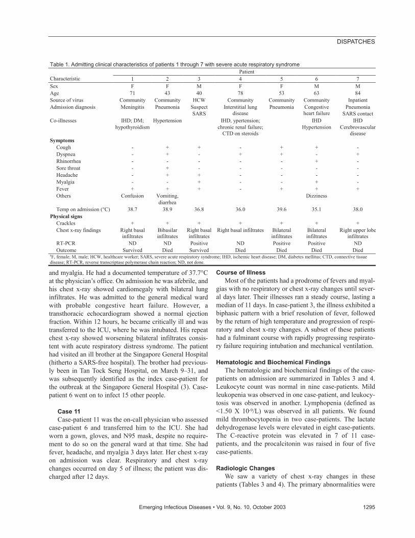
and myalgia. He had a documented temperature of 37.7°Cat the physician’s office. On admission he was afebrile, andhis chest x-ray showed cardiomegaly with bilateral lunginfiltrates. He was admitted to the general medical wardwith probable congestive heart failure. However, atransthoracic echocardiogram showed a normal ejectionfraction. Within 12 hours, he became critically ill and wastransferred to the ICU, where he was intubated. His repeatchest x-ray showed worsening bilateral infiltrates consis-tent with acute respiratory distress syndrome. The patienthad visited an ill brother at the Singapore General Hospital(hitherto a SARS-free hospital). The brother had previous-ly been in Tan Tock Seng Hospital, on March 9–31, andwas subsequently identified as the index case-patient forthe outbreak at the Singapore General Hospital (3). Case-patient 6 went on to infect 15 other people.
Case 11Case-patient 11 was the on-call physician who assessed
case-patient 6 and transferred him to the ICU. She hadworn a gown, gloves, and N95 mask, despite no require-ment to do so on the general ward at that time. She hadfever, headache, and myalgia 3 days later. Her chest x-rayon admission was clear. Respiratory and chest x-raychanges occurred on day 5 of illness; the patient was dis-charged after 12 days.
Course of IllnessMost of the patients had a prodrome of fevers and myal-
gias with no respiratory or chest x-ray changes until sever-al days later. Their illnesses ran a steady course, lasting amedian of 11 days. In case-patient 3, the illness exhibited abiphasic pattern with a brief resolution of fever, followedby the return of high temperature and progression of respi-ratory and chest x-ray changes. A subset of these patientshad a fulminant course with rapidly progressing respirato-ry failure requiring intubation and mechanical ventilation.
Hematologic and Biochemical FindingsThe hematologic and biochemical findings of the case-
patients on admission are summarized in Tables 3 and 4.Leukocyte count was normal in nine case-patients. Mildleukopenia was observed in one case-patient, and leukocy-tosis was observed in another. Lymphopenia (defined as<1.50 X 10⁻⁹/L) was observed in all patients. We foundmild thrombocytopenia in two case-patients. The lactatedehydrogenase levels were elevated in eight case-patients.The C-reactive protein was elevated in 7 of 11 case-patients, and the procalcitonin was raised in four of fivecase-patients.
Radiologic ChangesWe saw a variety of chest x-ray changes in these
patients (Tables 3 and 4). The primary abnormalities were
Emerging Infectious Diseases • Vol. 9, No. 10, October 2003 1295
DISPATCHES
Table 1. Admitting clinical characteristics of patients 1 through 7 with severe acute respiratory syndrome Patient
Characteristic 1 2 3 4 5 6 7 Sex F F M F F M M Age 71 43 40 78 53 63 84 Source of virus Community Community HCW Community Community Community Inpatient Admission diagnosis Meningitis Pneumonia Suspect
SARS Interstitial lung
disease Pneumonia Congestive
heart failure Pneumonia
SARS contact Co-illnesses IHD; DM;
hypothyroidism Hypertension IHD, ypertension;
chronic renal failure; CTD on steroids
IHD Hypertension
IHD Cerebrovascular
disease Symptoms
Cough - + + - + + - Dyspnea - + - + + - + Rhinorrhea - - - - - + - Sore throat - + - - - - - Headache - + + - - - - Myalgia - - + - - + - Fever + + + - + + + Others Confusion Vomiting,
diarrhea Dizziness
Temp on admission (°C) 38.7 38.9 36.8 36.0 39.6 35.1 38.0 Physical signs
Crackles + + + + + + + Chest x-ray findings Right basal
infiltrates Bibasilar infiltrates
Right basal infiltrates
Right basal infiltrates Bilateral infiltrates
Bilateral infiltrates
Right upper lobeinfiltrates
RT-PCR ND ND Positive ND Positive Positive ND Outcome Survived Died Survived Died Died Died Died
aF, female; M, male; HCW, healthcare worker; SARS, severe acute respiratory syndrome; IHD, ischemic heart disease; DM, diabetes mellitus; CTD, connective tissue disease; RT-PCR, reverse transcriptase polymerase chain reaction; ND, not done.
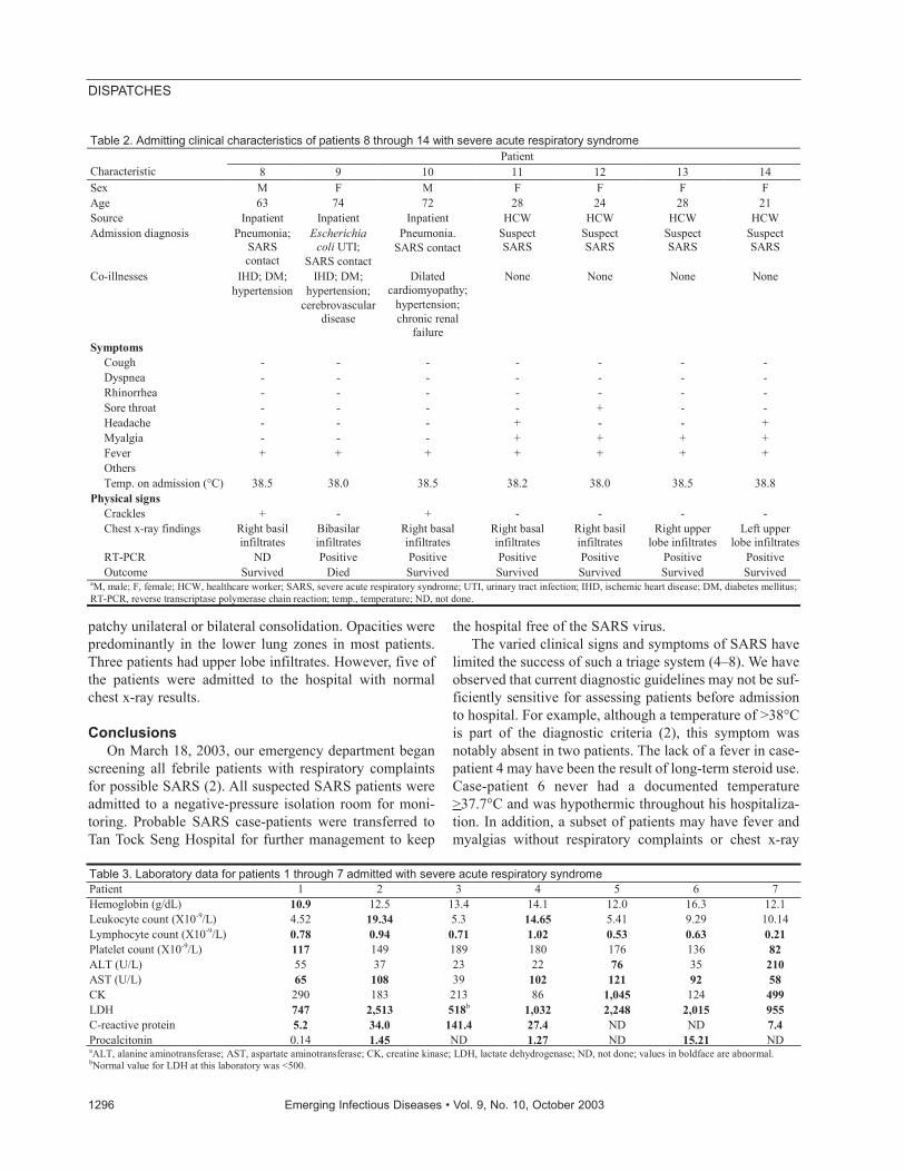
patchy unilateral or bilateral consolidation. Opacities werepredominantly in the lower lung zones in most patients.Three patients had upper lobe infiltrates. However, five ofthe patients were admitted to the hospital with normalchest x-ray results.
ConclusionsOn March 18, 2003, our emergency department began
screening all febrile patients with respiratory complaintsfor possible SARS (2). All suspected SARS patients wereadmitted to a negative-pressure isolation room for moni-toring. Probable SARS case-patients were transferred toTan Tock Seng Hospital for further management to keep
the hospital free of the SARS virus. The varied clinical signs and symptoms of SARS have
limited the success of such a triage system (4–8). We haveobserved that current diagnostic guidelines may not be suf-ficiently sensitive for assessing patients before admissionto hospital. For example, although a temperature of >38°Cis part of the diagnostic criteria (2), this symptom wasnotably absent in two patients. The lack of a fever in case-patient 4 may have been the result of long-term steroid use.Case-patient 6 never had a documented temperature>37.7°C and was hypothermic throughout his hospitaliza-tion. In addition, a subset of patients may have fever andmyalgias without respiratory complaints or chest x-ray
1296 Emerging Infectious Diseases • Vol. 9, No. 10, October 2003
DISPATCHES
Table 2. Admitting clinical characteristics of patients 8 through 14 with severe acute respiratory syndrome Patient
Characteristic 8 9 10 11 12 13 14 Sex M F M F F F F Age 63 74 72 28 24 28 21 Source Inpatient Inpatient Inpatient HCW HCW HCW HCW Admission diagnosis Pneumonia;
SARS contact
Escherichia coli UTI;
SARS contact
Pneumonia. SARS contact
Suspect SARS
Suspect SARS
Suspect SARS
Suspect SARS
Co-illnesses IHD; DM; hypertension
IHD; DM; hypertension;
cerebrovascular disease
Dilated cardiomyopathy;
hypertension; chronic renal
failure
None None None None
Symptoms Cough - - - - - - - Dyspnea - - - - - - - Rhinorrhea - - - - - - - Sore throat - - - - + - - Headache - - - + - - + Myalgia - - - + + + + Fever + + + + + + + Others Temp. on admission (°C) 38.5 38.0 38.5 38.2 38.0 38.5 38.8
Physical signs Crackles + - + - - - - Chest x-ray findings Right basil
infiltrates Bibasilar infiltrates
Right basal infiltrates
Right basal infiltrates
Right basil infiltrates
Right upper lobe infiltrates
Left upper lobe infiltrates
RT-PCR ND Positive Positive Positive Positive Positive Positive Outcome Survived Died Survived Survived Survived Survived Survived
aM, male; F, female; HCW, healthcare worker; SARS, severe acute respiratory syndrome; UTI, urinary tract infection; IHD, ischemic heart disease; DM, diabetes mellitus; RT-PCR, reverse transcriptase polymerase chain reaction; temp., temperature; ND, not done.
Table 3. Laboratory data for patients 1 through 7 admitted with severe acute respiratory syndrome Patient 1 2 3 4 5 6 7 Hemoglobin (g/dL) 10.9 12.5 13.4 14.1 12.0 16.3 12.1 Leukocyte count (X10-9/L) 4.52 19.34 5.3 14.65 5.41 9.29 10.14 Lymphocyte count (X10-9/L) 0.78 0.94 0.71 1.02 0.53 0.63 0.21 Platelet count (X10-9/L) 117 149 189 180 176 136 82 ALT (U/L) 55 37 23 22 76 35 210 AST (U/L) 65 108 39 102 121 92 58 CK 290 183 213 86 1,045 124 499 LDH 747 2,513 518b 1,032 2,248 2,015 955 C-reactive protein 5.2 34.0 141.4 27.4 ND ND 7.4 Procalcitonin 0.14 1.45 ND 1.27 ND 15.21 ND aALT, alanine aminotransferase; AST, aspartate aminotransferase; CK, creatine kinase; LDH, lactate dehydrogenase; ND, not done; values in boldface are abnormal. bNormal value for LDH at this laboratory was <500.
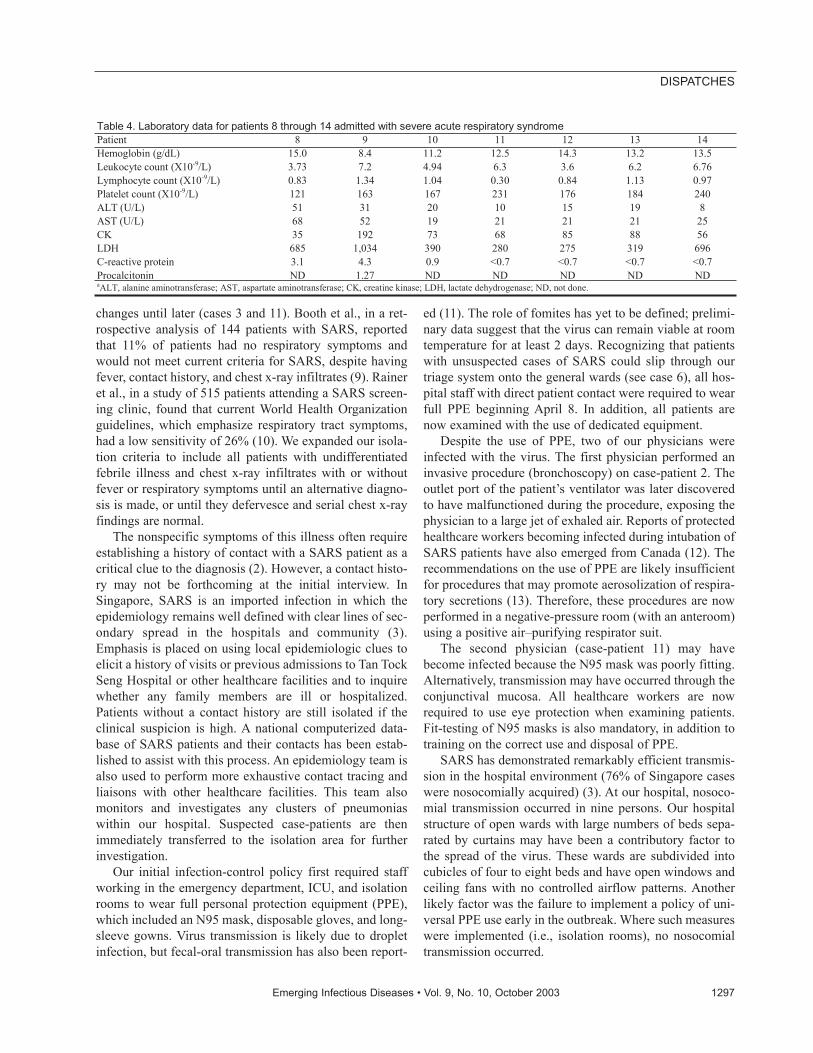
changes until later (cases 3 and 11). Booth et al., in a ret-rospective analysis of 144 patients with SARS, reportedthat 11% of patients had no respiratory symptoms andwould not meet current criteria for SARS, despite havingfever, contact history, and chest x-ray infiltrates (9). Raineret al., in a study of 515 patients attending a SARS screen-ing clinic, found that current World Health Organizationguidelines, which emphasize respiratory tract symptoms,had a low sensitivity of 26% (10). We expanded our isola-tion criteria to include all patients with undifferentiatedfebrile illness and chest x-ray infiltrates with or withoutfever or respiratory symptoms until an alternative diagno-sis is made, or until they defervesce and serial chest x-rayfindings are normal.
The nonspecific symptoms of this illness often requireestablishing a history of contact with a SARS patient as acritical clue to the diagnosis (2). However, a contact histo-ry may not be forthcoming at the initial interview. InSingapore, SARS is an imported infection in which theepidemiology remains well defined with clear lines of sec-ondary spread in the hospitals and community (3).Emphasis is placed on using local epidemiologic clues toelicit a history of visits or previous admissions to Tan TockSeng Hospital or other healthcare facilities and to inquirewhether any family members are ill or hospitalized.Patients without a contact history are still isolated if theclinical suspicion is high. A national computerized data-base of SARS patients and their contacts has been estab-lished to assist with this process. An epidemiology team isalso used to perform more exhaustive contact tracing andliaisons with other healthcare facilities. This team alsomonitors and investigates any clusters of pneumoniaswithin our hospital. Suspected case-patients are thenimmediately transferred to the isolation area for furtherinvestigation.
Our initial infection-control policy first required staffworking in the emergency department, ICU, and isolationrooms to wear full personal protection equipment (PPE),which included an N95 mask, disposable gloves, and long-sleeve gowns. Virus transmission is likely due to dropletinfection, but fecal-oral transmission has also been report-
ed (11). The role of fomites has yet to be defined; prelimi-nary data suggest that the virus can remain viable at roomtemperature for at least 2 days. Recognizing that patientswith unsuspected cases of SARS could slip through ourtriage system onto the general wards (see case 6), all hos-pital staff with direct patient contact were required to wearfull PPE beginning April 8. In addition, all patients arenow examined with the use of dedicated equipment.
Despite the use of PPE, two of our physicians wereinfected with the virus. The first physician performed aninvasive procedure (bronchoscopy) on case-patient 2. Theoutlet port of the patient’s ventilator was later discoveredto have malfunctioned during the procedure, exposing thephysician to a large jet of exhaled air. Reports of protectedhealthcare workers becoming infected during intubation ofSARS patients have also emerged from Canada (12). Therecommendations on the use of PPE are likely insufficientfor procedures that may promote aerosolization of respira-tory secretions (13). Therefore, these procedures are nowperformed in a negative-pressure room (with an anteroom)using a positive air–purifying respirator suit.
The second physician (case-patient 11) may havebecome infected because the N95 mask was poorly fitting.Alternatively, transmission may have occurred through theconjunctival mucosa. All healthcare workers are nowrequired to use eye protection when examining patients.Fit-testing of N95 masks is also mandatory, in addition totraining on the correct use and disposal of PPE.
SARS has demonstrated remarkably efficient transmis-sion in the hospital environment (76% of Singapore caseswere nosocomially acquired) (3). At our hospital, nosoco-mial transmission occurred in nine persons. Our hospitalstructure of open wards with large numbers of beds sepa-rated by curtains may have been a contributory factor tothe spread of the virus. These wards are subdivided intocubicles of four to eight beds and have open windows andceiling fans with no controlled airflow patterns. Anotherlikely factor was the failure to implement a policy of uni-versal PPE use early in the outbreak. Where such measureswere implemented (i.e., isolation rooms), no nosocomialtransmission occurred.
Emerging Infectious Diseases • Vol. 9, No. 10, October 2003 1297
DISPATCHES
Table 4. Laboratory data for patients 8 through 14 admitted with severe acute respiratory syndrome Patient 8 9 10 11 12 13 14 Hemoglobin (g/dL) 15.0 8.4 11.2 12.5 14.3 13.2 13.5 Leukocyte count (X10-9/L) 3.73 7.2 4.94 6.3 3.6 6.2 6.76 Lymphocyte count (X10-9/L) 0.83 1.34 1.04 0.30 0.84 1.13 0.97 Platelet count (X10-9/L) 121 163 167 231 176 184 240 ALT (U/L) 51 31 20 10 15 19 8 AST (U/L) 68 52 19 21 21 21 25 CK 35 192 73 68 85 88 56 LDH 685 1,034 390 280 275 319 696 C-reactive protein 3.1 4.3 0.9 <0.7 <0.7 <0.7 <0.7 Procalcitonin ND 1.27 ND ND ND ND ND aALT, alanine aminotransferase; AST, aspartate aminotransferase; CK, creatine kinase; LDH, lactate dehydrogenase; ND, not done.
In addition, because hospital staff are a recognizedsource of secondary transmission (3,4,6), all healthcareworkers are now required to monitor their temperaturesthree times a day. Anyone with a respiratory illness or atemperature >37.5ºC is removed from duty, pending fur-ther evaluation. This policy has successfully prevented thesecondary transmission of SARS from affected healthcareworkers.
The lessons gathered from our hospital outbreak haveresulted in dramatic changes to our triage and infection-control policies. All patients with undifferentiated febrileillness, respiratory complaints, or chest x-ray infiltrates areisolated, screened for SARS contacts, and nursed with fullPPE. Despite continued community transmission of SARSin Singapore (the last community case was identified onMay 10, 2003), the measures implemented since April 8,2003, enabled us to identify and contain eight additionalSARS cases and prevent the nosocomial transmission ofthe virus.
Dr. Singh is an associate consultant in infectious diseasesand microbiology at the National University Hospital. Hisresearch interests include serologic assays for the diagnosis ofsevere acute respiratory syndrome, and molecular epidemiology,and polymerase chain reaction for the rapid diagnosis of denguevirus.
References
1. Hsu Li-Yang, Lee CC, Green JA, Ang B, Paton N, Lee L, et al. Severeacute respiratory syndrome (SARS) in Singapore: features of indexpatient and initial contacts. Emerg Infect Dis 2003:9:713–7.
2. Centers for Disease Control and Prevention. Updated interim surveil-lance case definition for severe acute respiratory syndrome(SARS)—United States, April 29, 2003. MMWR Morb Mortal WklyRep 2003;52:391–3.
3. Centers for Disease Control and Prevention. Severe acute respiratorysyndrome—Singapore, 2003. MMWR Morb Mortal Wkly Rep2003;52:405–12.
4. Tsang KW, Ho PL, Ooi GC, Yee WK, Wang T, Chan-Yeung M, et al.A cluster of cases of severe acute respiratory syndrome in HongKong. N Engl J Med 2003;348:1977–85.
5. Poutanen SM, Low DE, Henry B, Finkelstein S, Rose D, Green K, etal. Identification of severe acute respiratory syndrome in Canada. NEngl J Med 2003;348:1995–2005.
6. Centers for Disease Control and Prevention. Preliminary clinicaldescription of severe acute respiratory syndrome. MMWR MorbMortal Wkly Rep 2003;52:255–6.
7. Peiris JSM, Chu CM, Cheng VCC, Chan KS, Hung IF, Poon LL, etal. Clinical progression and viral load in a community outbreak ofcoronavirus-associated SARS pneumonia: a prospective study.Lancet 2003;361:1767–72.
8. Fisher DA, Lim TK, Lim YT, Singh KS, Tambyah PA. Atypical pre-sentations of SARS. Lancet 2003;361:1740.
9. Booth CM, Matukas LM, Tomlinson GA, Rachlis AR, Rose DB,Dwash HA, et al. Clinical features and short-term outcomes of 144patients with SARS in the greater Toronto area. JAMA 2003;289:1–9.
10. Rainer TH, Cameron PA, Smit D, Ong KL, Hung AN, Nin DC, et al.Evaluation of WHO criteria for identifying patients with severe acuterespiratory syndrome out of hospital: prospective observational study.BMJ 2003;326:1354–8.
11. Hong Kong Department of Health. Atypical pneumonia. Availablefrom: URL: http://www.info.gov.hk/dh/ap.htm
12. Cluster of severe acute respiratory syndrome cases among protectedhealthcare workers—Toronto, April 2003. Canada CommunicableDiseases Report. Available from: URL: http://www.hc-sc.gc.ca
13. Centers for Disease Control and Prevention. Updated interim domes-tic guidelines for triage and disposition of patients who may havesevere acute respiratory syndrome (SARS). Available from: URL:http: //www.cdc.gov/ncidod/sars/triage_interim_guidance
Address for correspondence: Kamaljit Singh, Department of Medicine,National University Hospital, 5 Lower Kent Ridge Road, Singapore119074; fax: (65) 67750127; email: [email protected]
1298 Emerging Infectious Diseases • Vol. 9, No. 10, October 2003
DISPATCHES
Use of trade names is for identification only and does not implyendorsement by the Public Health Service or by the U.S.Department of Health and Human Services.
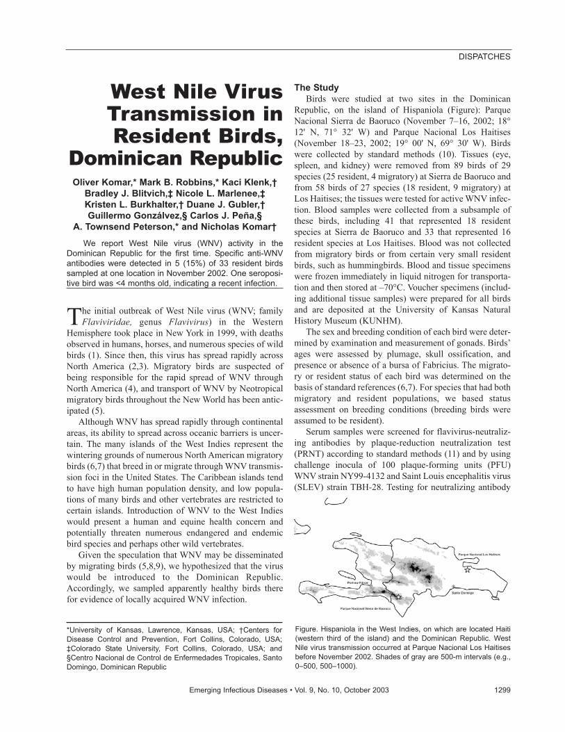
West Nile VirusTransmission inResident Birds,
Dominican Republic Oliver Komar,* Mark B. Robbins,* Kaci Klenk,†
Bradley J. Blitvich,‡ Nicole L. Marlenee,‡ Kristen L. Burkhalter,† Duane J. Gubler,†Guillermo Gonzálvez,§ Carlos J. Peña,§
A. Townsend Peterson,* and Nicholas Komar†We report West Nile virus (WNV) activity in the
Dominican Republic for the first time. Specific anti-WNVantibodies were detected in 5 (15%) of 33 resident birdssampled at one location in November 2002. One seroposi-tive bird was <4 months old, indicating a recent infection.
The initial outbreak of West Nile virus (WNV; familyFlaviviridae, genus Flavivirus) in the Western
Hemisphere took place in New York in 1999, with deathsobserved in humans, horses, and numerous species of wildbirds (1). Since then, this virus has spread rapidly acrossNorth America (2,3). Migratory birds are suspected ofbeing responsible for the rapid spread of WNV throughNorth America (4), and transport of WNV by Neotropicalmigratory birds throughout the New World has been antic-ipated (5).
Although WNV has spread rapidly through continentalareas, its ability to spread across oceanic barriers is uncer-tain. The many islands of the West Indies represent thewintering grounds of numerous North American migratorybirds (6,7) that breed in or migrate through WNV transmis-sion foci in the United States. The Caribbean islands tendto have high human population density, and low popula-tions of many birds and other vertebrates are restricted tocertain islands. Introduction of WNV to the West Indieswould present a human and equine health concern andpotentially threaten numerous endangered and endemicbird species and perhaps other wild vertebrates.
Given the speculation that WNV may be disseminatedby migrating birds (5,8,9), we hypothesized that the viruswould be introduced to the Dominican Republic.Accordingly, we sampled apparently healthy birds therefor evidence of locally acquired WNV infection.
The StudyBirds were studied at two sites in the Dominican
Republic, on the island of Hispaniola (Figure): ParqueNacional Sierra de Baoruco (November 7–16, 2002; 18°12' N, 71° 32' W) and Parque Nacional Los Haitises(November 18–23, 2002; 19° 00' N, 69° 30' W). Birdswere collected by standard methods (10). Tissues (eye,spleen, and kidney) were removed from 89 birds of 29species (25 resident, 4 migratory) at Sierra de Baoruco andfrom 58 birds of 27 species (18 resident, 9 migratory) atLos Haitises; the tissues were tested for active WNV infec-tion. Blood samples were collected from a subsample ofthese birds, including 41 that represented 18 residentspecies at Sierra de Baoruco and 33 that represented 16resident species at Los Haitises. Blood was not collectedfrom migratory birds or from certain very small residentbirds, such as hummingbirds. Blood and tissue specimenswere frozen immediately in liquid nitrogen for transporta-tion and then stored at –70°C. Voucher specimens (includ-ing additional tissue samples) were prepared for all birdsand are deposited at the University of Kansas NaturalHistory Museum (KUNHM).
The sex and breeding condition of each bird were deter-mined by examination and measurement of gonads. Birds’ages were assessed by plumage, skull ossification, andpresence or absence of a bursa of Fabricius. The migrato-ry or resident status of each bird was determined on thebasis of standard references (6,7). For species that had bothmigratory and resident populations, we based statusassessment on breeding conditions (breeding birds wereassumed to be resident).
Serum samples were screened for flavivirus-neutraliz-ing antibodies by plaque-reduction neutralization test(PRNT) according to standard methods (11) and by usingchallenge inocula of 100 plaque-forming units (PFU)WNV strain NY99-4132 and Saint Louis encephalitis virus(SLEV) strain TBH-28. Testing for neutralizing antibody
Emerging Infectious Diseases • Vol. 9, No. 10, October 2003 1299
DISPATCHES
*University of Kansas, Lawrence, Kansas, USA; †Centers forDisease Control and Prevention, Fort Collins, Colorado, USA;‡Colorado State University, Fort Collins, Colorado, USA; and§Centro Nacional de Control de Enfermedades Tropicales, SantoDomingo, Dominican Republic
Figure. Hispaniola in the West Indies, on which are located Haiti(western third of the island) and the Dominican Republic. WestNile virus transmission occurred at Parque Nacional Los Haitisesbefore November 2002. Shades of gray are 500-m intervals (e.g.,0–500, 500–1000).
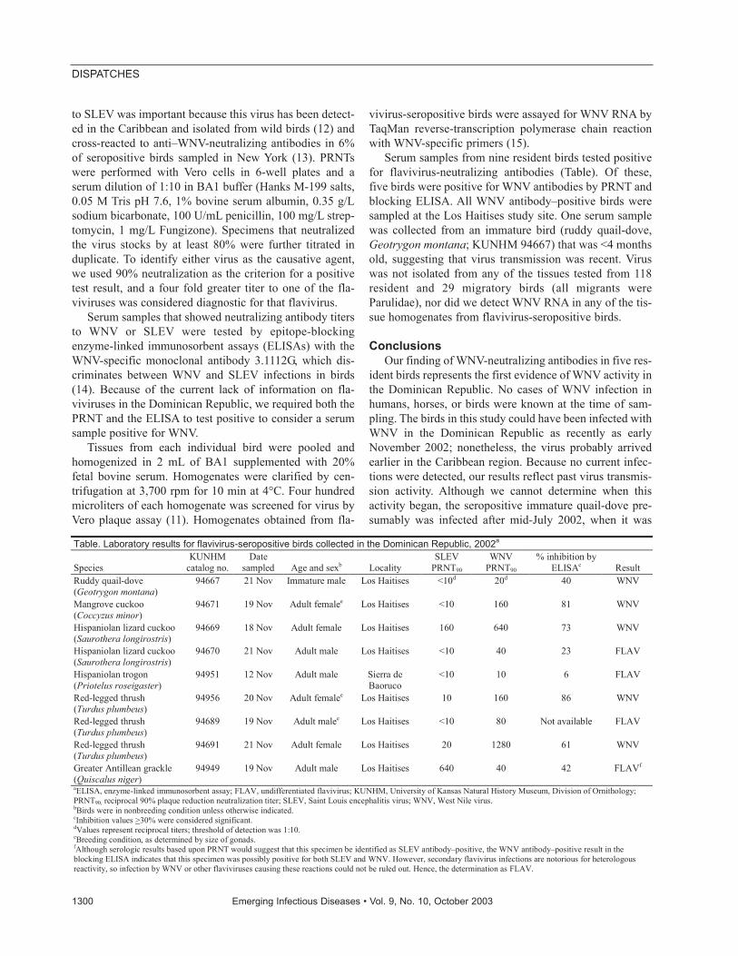
to SLEV was important because this virus has been detect-ed in the Caribbean and isolated from wild birds (12) andcross-reacted to anti–WNV-neutralizing antibodies in 6%of seropositive birds sampled in New York (13). PRNTswere performed with Vero cells in 6-well plates and aserum dilution of 1:10 in BA1 buffer (Hanks M-199 salts,0.05 M Tris pH 7.6, 1% bovine serum albumin, 0.35 g/Lsodium bicarbonate, 100 U/mL penicillin, 100 mg/L strep-tomycin, 1 mg/L Fungizone). Specimens that neutralizedthe virus stocks by at least 80% were further titrated induplicate. To identify either virus as the causative agent,we used 90% neutralization as the criterion for a positivetest result, and a four fold greater titer to one of the fla-viviruses was considered diagnostic for that flavivirus.
Serum samples that showed neutralizing antibody titersto WNV or SLEV were tested by epitope-blockingenzyme-linked immunosorbent assays (ELISAs) with theWNV-specific monoclonal antibody 3.1112G, which dis-criminates between WNV and SLEV infections in birds(14). Because of the current lack of information on fla-viviruses in the Dominican Republic, we required both thePRNT and the ELISA to test positive to consider a serumsample positive for WNV.
Tissues from each individual bird were pooled andhomogenized in 2 mL of BA1 supplemented with 20%fetal bovine serum. Homogenates were clarified by cen-trifugation at 3,700 rpm for 10 min at 4°C. Four hundredmicroliters of each homogenate was screened for virus byVero plaque assay (11). Homogenates obtained from fla-
vivirus-seropositive birds were assayed for WNV RNA byTaqMan reverse-transcription polymerase chain reactionwith WNV-specific primers (15).
Serum samples from nine resident birds tested positivefor flavivirus-neutralizing antibodies (Table). Of these,five birds were positive for WNV antibodies by PRNT andblocking ELISA. All WNV antibody–positive birds weresampled at the Los Haitises study site. One serum samplewas collected from an immature bird (ruddy quail-dove,Geotrygon montana; KUNHM 94667) that was <4 monthsold, suggesting that virus transmission was recent. Viruswas not isolated from any of the tissues tested from 118resident and 29 migratory birds (all migrants wereParulidae), nor did we detect WNV RNA in any of the tis-sue homogenates from flavivirus-seropositive birds.
ConclusionsOur finding of WNV-neutralizing antibodies in five res-
ident birds represents the first evidence of WNV activity inthe Dominican Republic. No cases of WNV infection inhumans, horses, or birds were known at the time of sam-pling. The birds in this study could have been infected withWNV in the Dominican Republic as recently as earlyNovember 2002; nonetheless, the virus probably arrivedearlier in the Caribbean region. Because no current infec-tions were detected, our results reflect past virus transmis-sion activity. Although we cannot determine when thisactivity began, the seropositive immature quail-dove pre-sumably was infected after mid-July 2002, when it was
1300 Emerging Infectious Diseases • Vol. 9, No. 10, October 2003
DISPATCHES
Table. Laboratory results for flavivirus-seropositive birds collected in the Dominican Republic, 2002a
Species KUNHM
catalog no. Date
sampled Age and sexb Locality SLEV
PRNT90 WNV
PRNT90 % inhibition by
ELISAc Result Ruddy quail-dove (Geotrygon montana)
94667 21 Nov Immature male Los Haitises <10d 20d 40 WNV
Mangrove cuckoo (Coccyzus minor)
94671 19 Nov Adult femalee Los Haitises <10 160 81 WNV
Hispaniolan lizard cuckoo (Saurothera longirostris)
94669 18 Nov Adult female Los Haitises 160 640 73 WNV
Hispaniolan lizard cuckoo (Saurothera longirostris)
94670 21 Nov Adult male Los Haitises <10 40 23 FLAV
Hispaniolan trogon (Priotelus roseigaster)
94951 12 Nov Adult male Sierra de Baoruco
<10 10 6 FLAV
Red-legged thrush (Turdus plumbeus)
94956 20 Nov Adult femalee Los Haitises 10 160 86 WNV
Red-legged thrush (Turdus plumbeus)
94689 19 Nov Adult malee Los Haitises <10 80 Not available FLAV
Red-legged thrush (Turdus plumbeus)
94691 21 Nov Adult female Los Haitises 20 1280 61 WNV
Greater Antillean grackle (Quiscalus niger)
94949 19 Nov Adult male Los Haitises 640 40 42 FLAVf
aELISA, enzyme-linked immunosorbent assay; FLAV, undifferentiated flavivirus; KUNHM, University of Kansas Natural History Museum, Division of Ornithology; PRNT90, reciprocal 90% plaque reduction neutralization titer; SLEV, Saint Louis encephalitis virus; WNV, West Nile virus. bBirds were in nonbreeding condition unless otherwise indicated. cInhibition values >30% were considered significant. dValues represent reciprocal titers; threshold of detection was 1:10. eBreeding condition, as determined by size of gonads. fAlthough serologic results based upon PRNT would suggest that this specimen be identified as SLEV antibody–positive, the WNV antibody–positive result in the blocking ELISA indicates that this specimen was possibly positive for both SLEV and WNV. However, secondary flavivirus infections are notorious for heterologous reactivity, so infection by WNV or other flaviviruses causing these reactions could not be ruled out. Hence, the determination as FLAV.
born, and before early November 2002, in order to havestimulated detectable antibody production by mid-November.
The earliest evidence of WNV transmission in the WestIndies is a human case from the Cayman Islands in 2001(16). WNV-seropositive birds captured in Jamaica early in2002 may have been infected in 2001 or earlier (17).Additional evidence of WNV transmission in theCaribbean region includes the report of two seropositivehorses in Yucatán state, Mexico, sampled in July 2002(18). Although the seropositivity of vertebrates in CaymanIslands, Jamaica, Mexico, and now the DominicanRepublic is strong evidence for WNV activity in theregion, it is indirect evidence and does not entirely rule outthe possibility of cross-reactions with another flavivirus inlaboratory assays. WNV remains to be isolated from theregion.
The presence of WNV at Los Haitises may have result-ed from transportation by viremic migratory birds fromNorth America, where WNV transmission foci are wide-spread (2). Several migratory bird species, in particular,parulid warblers (order Passeriformes), were observed atthis site. At least some passerine birds are capable of trans-mitting virus during their few days of viremia (19).Therefore, transmission to mosquitoes or predators fromviremic migrants would be possible for a brief period (afew days, at most) after arrival at a site. Virus introductioninto Caribbean ecosystems is therefore likely to occur atcoastal sites where transoceanic migrants make first land-fall.
We found no evidence of active virus in bird tissues ofboth resident and migratory species. We did not test serumsamples from migrants because the presence of antibodieswould not be informative, given the history of these birdstraveling through areas of WNV transmission in or nearNorth American breeding grounds. We presume that thefive seropositive resident birds were infected locallybecause the four species involved are not migratory (7).Although young birds may disperse several kilometersfrom natal sites (20), adults probably live entirely withinbreeding territories.
Although only five birds (15%, 95% confidence inter-val [CI] 5% to 32%) at Los Haitises were found to beseropositive, the results suggest that transmission of WNVamong bird populations at that site was widespread. Forcomparison, seroprevalence of WNV-neutralizing antibod-ies in resident birds was 50% (CI 44% to 57%) in Queens,New York City, after the 1999 outbreak (13), and 23% (CI18% to 29%) in Staten Island after the 2000 outbreak (21).We used conservative criteria for determining a positiveresult because the background diversity of flaviviruses inthe Dominican Republic has not been studied recently. Ifonly the PRNT had been used (as was the case in the New
York studies), then seven (21%, CI 7% to 35%) of the birdsfrom Los Haitises would have been reported as positive forantibodies to WNV.
The evidence for local WNV transmission in theDominican Republic indicates risk for West Nile fever andmeningoencephalitis in the human, equine, and avian pop-ulations of Hispaniola. We suggest that WNV be consid-ered in the differential diagnosis of humans and other ver-tebrates with central nervous system disease in Hispaniola.
AcknowledgmentsWe thank Bolivar Cabrera, Tristan Davis, Juan Cepedes
Hidalgo, José Dolores Jiménez, Esteban López-Medrano, andDomingo Sirí for assistance in the field; José Ramón Albaine andSimón Guerrero for logistical assistance and assistance with per-mits; John Roehrig for assisting with the interpretation of sero-logic test results; and the two anonymous reviewers who provid-ed comments on the manuscript.
This study was supported in part by National ScienceFoundation Grant #0211388, and in part by the Centers forDisease Control and Prevention contract U50/CCU 820510-02.
Mr. Komar is an ornithologist working on a Ph.D. in biolo-gy at the University of Kansas Natural History Museum andDepartment of Ecology and Evolutionary Biology in Lawrence,Kansas. His primary research interests are avian ecology andconservation in the Neotropical region.
References
1. Lanciotti RS, Roehrig JT, Deubel V, Smith J, Parker M, Steele K, etal. Origin of the West Nile virus responsible for an outbreak ofencephalitis in the northeastern United States. Science1999;286:2333–7.
2. Chow CC, Montgomery SP, O’Leary DR, Nasci RS, Campbell GL,Kipp AM, et al. Provisional surveillance summary of the West Nilevirus epidemic—United States, January–November 2002. MMWRMorb Mortal Wkly Rep 2002;51:1129–33.
3. Health Canada. Population and Public Health Branch WNV surveil-lance updates, December 4, 2002. Available from: URL:http://www.hc-sc.gc.ca/pphb-dgspsp/wnv-vwn/mon_e.html#sitrep
4. McLean RG, Ubico SR, Bourne D, Komar N. West Nile virus in live-stock and wildlife. Curr Top Microbiol Immunol 2002;267:271–308.
5. Rappole JH, Derrickson SR, Hubalek Z. Migratory birds and spreadof West Nile virus in the Western Hemisphere. Emerg Infect Dis2000;6:319–28.
6. American Ornithologists’ Union. Check-list of North American birds.7th ed. Washington: American Ornithologists’ Union; 1998.
7. Raffaele H, Wiley J, Garrido O, Keith A, Raffaele J. A guide to thebirds of the West Indies. Princeton (NJ): Princeton Univ. Press; 1998.
8. Malkinson M, Banet C, Weisman Y, Pokamunski S, King R, DrouetMT, et al. Introduction of West Nile virus in the Middle East bymigrating white storks. Emerg Infect Dis 2002;8:392–7.
9. Peterson AT, Vieglais DA, Andreasen JK. Migratory birds modeled ascritical transport agents for West Nile virus in North America. VectorBorne Zoonotic Dis 2003;3:27–37.
10. Gaunt AS, Oring LW, editors. Guidelines to the use of wild birds inresearch. The Ornithological Council; 1997. Available from: URL:www.nmnh.si.edu/BIRDNET/GuideToUse
Emerging Infectious Diseases • Vol. 9, No. 10, October 2003 1301
DISPATCHES
11. Beaty BJ, Calisher CH, Shope RE. Arboviruses. In: Lennette EH,Lennette DA, Lennette ET, editors. Diagnostic procedures for viral,rickettsial, and chlamydial infections, 7th ed. Washington: AmericanPublic Health Association; 1995. p. 189–212.
12. Tsai TF, Mitchell CJ. St. Louis encephalitis. In: Monath TP, editor.Vol. IV, The arboviruses: epidemiology and ecology. Boca Raton(FL): CRC Press; 1988. p. 113–43.
13. Komar N, Panella NA, Burns JE, Dusza SW, Mascarenhas TM,Talbot TO. Serologic evidence for West Nile virus infection in birdsin the New York City vicinity during an outbreak in 1999. EmergInfect Dis 2001;7:621–5.
14. Blitvich BJ, Marlenee NL, Hall RA, Calisher CH, Bowen RA,Roehrig JT, et al. Epitope-blocking enzyme-linked immunosorbentassays for the detection of serum antibodies to West Nile virus in mul-tiple avian species. J Clin Microbiol 2003;41:1041–7.
15. Lanciotti RS, Kerst AJ, Nasci RS, Godsey MS, Mitchell CJ, SavageHM, et al. Rapid detection of West Nile virus from human clinicalspecimens, field-collected mosquitoes, and avian samples by aTaqMan reverse transcriptase-PCR assay. J Clin Microbiol2000;38:4066–71.
16. O’Leary DR, Nasci RS, Campbell GL, Marfin AA. West Nile virusactivity—United States, 2001. MMWR Morb Mortal Wkly Rep2002;51:497–501.
17. Dupuis II AP, Marra PP, Kramer LD. Serologic evidence for WestNile virus transmission in Jamaica, West Indies. Emerg Infect Dis2003;9:860–3.
18. Loroño-Pino MA, Blitvich BJ, Farfán-Ale JA, Puerto FI, Blanco JM,Marlenee NL, et al. Serologic evidence of West Nile virus infectionin horses, Yucatan State, Mexico. Emerg Infect Dis 2003;9:857–9.
19. Komar N, Langevin S, Hinten S, Nemeth N, Edwards E, Hettler D, etal. Experimental infection of North American birds with the NewYork 1999 strain of West Nile virus. Emerg Infect Dis2003;9:311–22.
20. Greenwood PJ. Mating systems, philopatry and dispersal in birds andmammals. Animal Behavior 1980;28:1140–62.
21. Komar N, Burns J, Dean C, Panella NA, Dusza S, Cherry B.Serologic evidence for West Nile virus infection in birds in StatenIsland, New York, after an outbreak in 2000. Vector Borne ZoonoticDis 2001;1:191–6.
Address for correspondence: Oliver Komar, Natural History Museum andBiodiversity Research Center, University of Kansas, 1345 JayhawkBoulevard, Lawrence, KS 66045-7561, USA; fax: 785-864-5335; email:[email protected]
1302 Emerging Infectious Diseases • Vol. 9, No. 10, October 2003
DISPATCHES
SS ee aa rr cc hh pp aa ss tt ii ss ss uu ee ss oo ff EE II DD aa tt ww ww ww.. cc dd cc .. gg oo vv // ee ii dd
West Nile VirusEncephalitis and
Myocarditis in Wolf and Dog
Carol A. Lichtensteiger,* Kathleen Heinz-Taheny,* Tanasa S. Osborne,*
Robert J. Novak,* Beth A. Lewis,* and Margaret L. Firth†
In the third season (2002) of the West Nile virus epi-demic in the United States, two canids (wolf and dog) werediagnosed with West Nile virus encephalitis and myocardi-tis with similarities to known affected species (humans,horses, and birds). The West Nile virus infections were con-firmed by immunohistochemistry and polymerase chainreaction.
Since its 1999 introduction in New York, West Nilevirus (WNV) has spread to >40 states, causing season-
al mosquito-borne disease in humans, horses, and birds(1–7). We recently identified two Illinois canids (a captivewolf and a domestic dog) with severe disease associatedwith WNV infection.
Outside the Western Hemisphere, WNV has beenendemic for decades (4,8–11). Canids have not beenthought to be important in the epidemiology of this virus.However, a dog with neurologic disease that died in Africain 1977 is now thought to have been infected with WNV(9,12). In a recent study in which four dogs were experi-mentally infected, no clinical disease was observed, and alow viremia developed in one dog (11). Natural infectionsoccur in dogs, as indicated by serum antibodies; seroposi-tivity in surveys was 37% in the 1980s in South Africa,24% in the 1990s in India, and 5% in 1999 in New YorkCity (10,11,13). In addition, a few individuals of somemammalian species have been listed as infected (not dis-eased): 14 bats, four rodents, three rabbits, two cats, tworaccoons, and a skunk (6).
The StudyTwo Illinois canids were brought in for necropsy in
August 2002: 1) a 3-month-old female wolf pup, whichdied after 2 days of lethargy, depression, and irritabilitythat progressed to anorexia, weakness, ataxia, and blind-ness and 2) an overweight 8-year-old castrated male IrishSetter-Golden Retriever mixed breed dog that was eutha-
nized in moribund condition after a 7-day illness. The dogwas hospitalized after 3 days of lethargy, anorexia, poly-dipsia, ocular discharge, and difficulty in rising that hadprogressed to fever, listlessness, weakness, ptyalism, nasaland ocular discharge, watery diarrhea, and abdominal pain.Although the diarrhea resolved and the dog was strongerand more alert the morning after hospitalization, over thenext 3 days, multisystemic clinical signs developed,including dyspnea, diarrhea with melena, ataxia, a head tiltwith head bobbing, pulmonary edema, and cardiacarrhythmias. While hospitalized, the dog had mild anemiawithout spherocytes, moderate to severe thrombocytopeniawith large platelets, moderate leukocytosis with a left shift,a moderate hypokalemia, a mild hypoglycemia, andhypoproteinemia. Symptomatic and supportive care treat-ment included broad-spectrum antibiotics, a gastrointesti-nal protectant, a diuretic, fluid therapy, and systemic corti-costeroids (for immune-mediated thrombocytopenia andanemia).
Both the wolf and dog were necropsied. Samples ofmultiple tissues were fixed and processed for routine diag-nostic histopathology. In addition, tissues were screenedwith peroxidase immunohistochemistry for distemper,rabies, WNV, Toxoplasma, and Neospora. Sections forWNV immunohistochemistry were pretreated with 0.1%protease (20 min at 37°C) and nonspecific antibody bind-ing blocked with Power Block (BioGenx, San Ramon,CA). The primary antibody was mouse anti-WNV mono-clonal ascitic fluid diluted 1:1,000 (ATTC, Manassas, VA).The wolf brain was assayed for rabies antigen by fluores-cent antibody (Public Health Laboratory, Springfield, IL).
West Nile viral RNA in the brain of the wolf and thekidney and liver of the dog (available cyropreserved tis-sue) was assayed by using a modified 5′ nuclease fluoro-genic real-time, reverse transcriptase polymerase chainreaction assay (RT-PCR) (TaqMan, Applied Biosystems,Foster City, CA) (14). Each amplification reaction con-tained primers and probes (Table) (14,15) for both WNVand St. Louis encephalitis virus (SLEV) with FAM (6-car-boxyfluorescein) and VIC (Applied Biosystems) fluores-cent-labeled probes, respectively. A tissue sample (about200 µg) was homogenized in 1.5 mL of medium 199 withL-glutamine, Hanks balanced salt solution, and 25 mMHEPES in a microfuge tube with three 3.2-mm stainlesssteel beads (Biospec Products, Bartlesville, OK). The sam-ple was ground in a mixer mill for four min at a rate of 1/30sec-1, and then centrifuged (4,000 X g, 4 min). RNA waspurified from 220 µL of the supernatant by using a viralRNA purification kit and the Biorobot 9604 (QIAGEN,Inc., Alameda, CA), eluting in a final volume of 85 µL.Real-time, RT-PCR was done in a 25-µL reaction withOne-step RT-PCR Master Mix kit (Applied Biosystems)containing 25 pmol of each primer set, 3.25 pmol of each
Emerging Infectious Diseases • Vol. 9, No. 10, October 2003 1303
DISPATCHES
*University of Illinois, Champaign-Urbana, Illinois, USA; and†Town and Country Animal Hospital, Normal, Illinois, USA
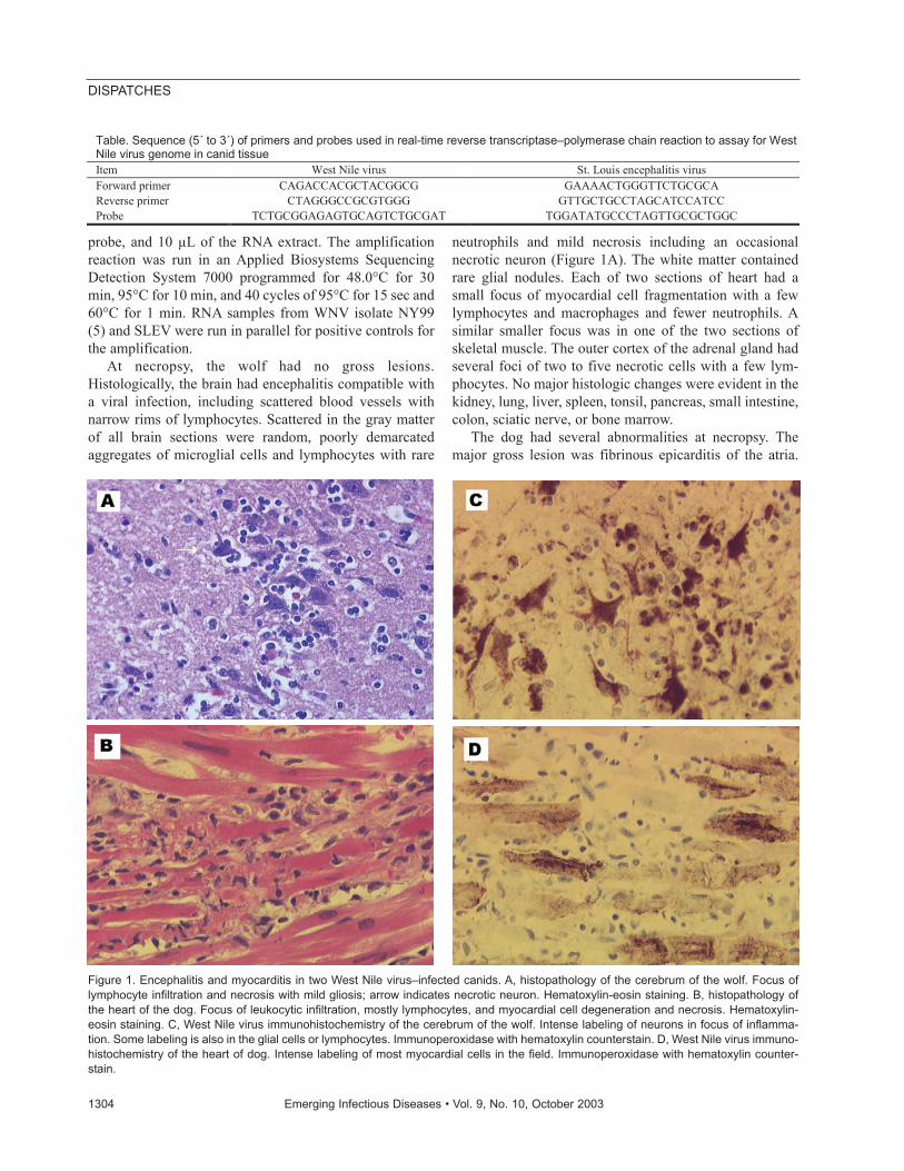
probe, and 10 µL of the RNA extract. The amplificationreaction was run in an Applied Biosystems SequencingDetection System 7000 programmed for 48.0°C for 30min, 95°C for 10 min, and 40 cycles of 95°C for 15 sec and60°C for 1 min. RNA samples from WNV isolate NY99(5) and SLEV were run in parallel for positive controls forthe amplification.
At necropsy, the wolf had no gross lesions.Histologically, the brain had encephalitis compatible witha viral infection, including scattered blood vessels withnarrow rims of lymphocytes. Scattered in the gray matterof all brain sections were random, poorly demarcatedaggregates of microglial cells and lymphocytes with rare
neutrophils and mild necrosis including an occasionalnecrotic neuron (Figure 1A). The white matter containedrare glial nodules. Each of two sections of heart had asmall focus of myocardial cell fragmentation with a fewlymphocytes and macrophages and fewer neutrophils. Asimilar smaller focus was in one of the two sections ofskeletal muscle. The outer cortex of the adrenal gland hadseveral foci of two to five necrotic cells with a few lym-phocytes. No major histologic changes were evident in thekidney, lung, liver, spleen, tonsil, pancreas, small intestine,colon, sciatic nerve, or bone marrow.
The dog had several abnormalities at necropsy. Themajor gross lesion was fibrinous epicarditis of the atria.
1304 Emerging Infectious Diseases • Vol. 9, No. 10, October 2003
DISPATCHES
Table. Sequence (5´ to 3´) of primers and probes used in real-time reverse transcriptase–polymerase chain reaction to assay for West Nile virus genome in canid tissue Item West Nile virus St. Louis encephalitis virus Forward primer CAGACCACGCTACGGCG GAAAACTGGGTTCTGCGCA Reverse primer CTAGGGCCGCGTGGG GTTGCTGCCTAGCATCCATCC Probe TCTGCGGAGAGTGCAGTCTGCGAT TGGATATGCCCTAGTTGCGCTGGC
Figure 1. Encephalitis and myocarditis in two West Nile virus–infected canids. A, histopathology of the cerebrum of the wolf. Focus oflymphocyte infiltration and necrosis with mild gliosis; arrow indicates necrotic neuron. Hematoxylin-eosin staining. B, histopathology ofthe heart of the dog. Focus of leukocytic infiltration, mostly lymphocytes, and myocardial cell degeneration and necrosis. Hematoxylin-eosin staining. C, West Nile virus immunohistochemistry of the cerebrum of the wolf. Intense labeling of neurons in focus of inflamma-tion. Some labeling is also in the glial cells or lymphocytes. Immunoperoxidase with hematoxylin counterstain. D, West Nile virus immuno-histochemistry of the heart of dog. Intense labeling of most myocardial cells in the field. Immunoperoxidase with hematoxylin counter-stain.
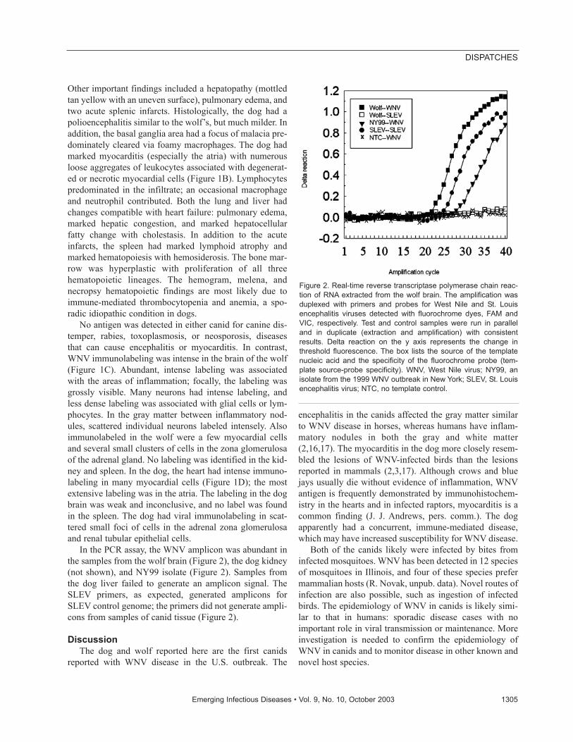
Other important findings included a hepatopathy (mottledtan yellow with an uneven surface), pulmonary edema, andtwo acute splenic infarcts. Histologically, the dog had apolioencephalitis similar to the wolf’s, but much milder. Inaddition, the basal ganglia area had a focus of malacia pre-dominately cleared via foamy macrophages. The dog hadmarked myocarditis (especially the atria) with numerousloose aggregates of leukocytes associated with degenerat-ed or necrotic myocardial cells (Figure 1B). Lymphocytespredominated in the infiltrate; an occasional macrophageand neutrophil contributed. Both the lung and liver hadchanges compatible with heart failure: pulmonary edema,marked hepatic congestion, and marked hepatocellularfatty change with cholestasis. In addition to the acuteinfarcts, the spleen had marked lymphoid atrophy andmarked hematopoiesis with hemosiderosis. The bone mar-row was hyperplastic with proliferation of all threehematopoietic lineages. The hemogram, melena, andnecropsy hematopoietic findings are most likely due toimmune-mediated thrombocytopenia and anemia, a spo-radic idiopathic condition in dogs.
No antigen was detected in either canid for canine dis-temper, rabies, toxoplasmosis, or neosporosis, diseasesthat can cause encephalitis or myocarditis. In contrast,WNV immunolabeling was intense in the brain of the wolf(Figure 1C). Abundant, intense labeling was associatedwith the areas of inflammation; focally, the labeling wasgrossly visible. Many neurons had intense labeling, andless dense labeling was associated with glial cells or lym-phocytes. In the gray matter between inflammatory nod-ules, scattered individual neurons labeled intensely. Alsoimmunolabeled in the wolf were a few myocardial cellsand several small clusters of cells in the zona glomerulosaof the adrenal gland. No labeling was identified in the kid-ney and spleen. In the dog, the heart had intense immuno-labeling in many myocardial cells (Figure 1D); the mostextensive labeling was in the atria. The labeling in the dogbrain was weak and inconclusive, and no label was foundin the spleen. The dog had viral immunolabeling in scat-tered small foci of cells in the adrenal zona glomerulosaand renal tubular epithelial cells.
In the PCR assay, the WNV amplicon was abundant inthe samples from the wolf brain (Figure 2), the dog kidney(not shown), and NY99 isolate (Figure 2). Samples fromthe dog liver failed to generate an amplicon signal. TheSLEV primers, as expected, generated amplicons forSLEV control genome; the primers did not generate ampli-cons from samples of canid tissue (Figure 2).
DiscussionThe dog and wolf reported here are the first canids
reported with WNV disease in the U.S. outbreak. The
encephalitis in the canids affected the gray matter similarto WNV disease in horses, whereas humans have inflam-matory nodules in both the gray and white matter(2,16,17). The myocarditis in the dog more closely resem-bled the lesions of WNV-infected birds than the lesionsreported in mammals (2,3,17). Although crows and bluejays usually die without evidence of inflammation, WNVantigen is frequently demonstrated by immunohistochem-istry in the hearts and in infected raptors, myocarditis is acommon finding (J. J. Andrews, pers. comm.). The dogapparently had a concurrent, immune-mediated disease,which may have increased susceptibility for WNV disease.
Both of the canids likely were infected by bites frominfected mosquitoes. WNV has been detected in 12 speciesof mosquitoes in Illinois, and four of these species prefermammalian hosts (R. Novak, unpub. data). Novel routes ofinfection are also possible, such as ingestion of infectedbirds. The epidemiology of WNV in canids is likely simi-lar to that in humans: sporadic disease cases with noimportant role in viral transmission or maintenance. Moreinvestigation is needed to confirm the epidemiology ofWNV in canids and to monitor disease in other known andnovel host species.
Emerging Infectious Diseases • Vol. 9, No. 10, October 2003 1305
DISPATCHES
Figure 2. Real-time reverse transcriptase polymerase chain reac-tion of RNA extracted from the wolf brain. The amplification wasduplexed with primers and probes for West Nile and St. Louisencephalitis viruses detected with fluorochrome dyes, FAM andVIC, respectively. Test and control samples were run in paralleland in duplicate (extraction and amplification) with consistentresults. Delta reaction on the y axis represents the change inthreshold fluorescence. The box lists the source of the templatenucleic acid and the specificity of the fluorochrome probe (tem-plate source-probe specificity). WNV, West Nile virus; NY99, anisolate from the 1999 WNV outbreak in New York; SLEV, St. Louisencephalitis virus; NTC, no template control.
AcknowledgmentsWe thank Marshall Van de Wyngaerde and Nina Krasavin
for the polymerase chain reaction assays, Jane Chladny and herco-workers for the histology preparation and immunohistochem-istry assays, Chris Johnson and Hope Lopez for clinical care dataon the two canine patients, and Laura Kramer for providing St.Louis encephalitis virus.
The work was supported in part by Centers for DiseaseControl and Prevention grant U50/CCU520518-01 (RJN) andDepartment of Natural Resources Illinois Waste Tire Fund (RJN).
Dr. Lichtensteiger is a veterinary pathologist on the facultyat the University of Illinois (Veterinary Diagnostic Laboratoryand Department of Veterinary Pathobiology) with more than 10years of experience in diagnostic pathology and research.Research experience includes antigenic variation of the goatlentivirus and molecular pathogenicity of Haemophilus andSalmonella.
References1. Trock SC, Meade BJ, Glaser AL, Ostlund EN, Lanciotti RS, Cropp
BC, et al. West Nile virus outbreak among horses in New York State,1999 and 2000. Emerg Infect Dis 2001;7:745–7.
2. Cantile C, Del Piero F, Di Guardo G, Arispici M. Pathologic andimmunohistochemical findings in naturally occurring West Nile virusinfection in horses. Vet Pathol 2001;38:414–21.
3. Steele KE, Linn MJ, Schoepp RJ, Komar N, Geisbert TW, ManducaRM, et al. Pathology of fatal West Nile virus infections in native andexotic birds during the 1999 outbreak in New York City, New York.Vet Pathol 2000;37:208–24.
4. Komar N. West Nile viral encephalitis. Rev Sci Tech2000;19:166–76.
5. Lanciotti RS, Roehrig JT, Deubel V, Smith J, Parker M, Steele K, etal. Origin of the West Nile virus responsible for an outbreak ofencephalitis in the Northeastern United States. Science1999;286:2333–7.
6. Centers for Disease Control and Prevention. Update: West Nile Virusactivity—eastern United States, 2000. MMWR Morb Mortal WeeklyRep 2000;49:1044–7.
7. Centers for Disease Control and Prevention. West Nile virus activity-United States, August 21–28, 2002, and Illinois, January 1–August27, 2002. MMWR Morb Mortal Weekly Rep 2002;51:764–6.
8. Hubálek Z, Halouzka J. West Nile fever—a reemerging mosquito-borne viral disease in Europe. Emerg Infect Dis 1999;5:643–50.
9. Burt FJ, Grobbelaar AA, Leman PA, Anthony FS, Gibson GVF,Swanepoel R. Phylogenetic relationships of Southern African WestNile virus isolates. Emerg Infect Dis 2002;8:820–6.
10. Mall MP, Kumar A, Malik SVS. Sero-positivity of domestic animalsagainst Japanese encephalitis in Bareilly area, U.P. J Commun Dis1995;27:242–6.
11. Blackburn NK, Reyers F, Berry WL, Shepherd AJ. Susceptibility ofdogs to West Nile virus: a survey and pathogenicity trial. J CompPathol 1989;100:59–66.
12. Simpson VR, Kuebart G. A fatal case of Wesselsbron disease in a dog.Vet Rec 1979;105:329.
13. Komar N, Panella NA, Boyce E. Exposure of domestic mammals toWest Nile virus during an outbreak of human encephalitis, New YorkCity, 1999. Emerg Infect Dis 2001;7:736–8.
14. Lanciotti RS, Kerst AJ, Nasci RS, Godsey MS, Mitchell CJ, SavageHM, et al. Rapid detection of West Nile virus from human clinicalspecimens, field-collected mosquitoes, and avian samples by aTaqMan reverse transcriptase-PCR assay. J Clin Microbiol2000;38:4066–71.
15. Lanciotti RS, Kerst AJ. Nucleic acid sequence-based amplificationassays for rapid detection of West Nile and St. Louis encephalitisviruses. J Clin Microbiol 2001;39:4506–13.
16. Bernard KA, Kramer LD. West Nile virus activity in the UnitedStates, 2001. Viral Immunol 2001;14:319–38.
17. Sampson BA, Ambrosi C, Charlot A, Reiber K, Veress JF,Armbustmacher V. The pathology of human West Nile virus infec-tion. Hum Pathol 2000;31:527–31.
Address for correspondence: Carol Lichtensteiger, 2001 South LincolnAvenue, Urbana, IL 61802, USA; fax: (217) 244-2439; email:[email protected]
1306 Emerging Infectious Diseases • Vol. 9, No. 10, October 2003
DISPATCHES
Weissella confusaInfection in Primate
(Cercopithecusmona)
Ana I. Vela,* Concepción Porrero,* Joaquín Goyache,* Ana Nieto,* Belen Sánchez,*
Víctor Briones,* Miguel Angel Moreno,* Lucas Domínguez,*
and José F. Fernández-Garayzábal*We describe the first systemic infection by Weissella
confusa in a mona monkey (Cercopithecus mona) on thebasis of microbiologic, molecular genetic, and histologicdata. The same strain of W. confusa, as determined bypulsed-field gel electrophoresis, was isolated in pure cul-ture from the brain, liver, spleen, and intestine of this pri-mate, illustrating the clinical importance of the isolations.
Weissella microorganisms are gram-positive, catalase-negative coccobacilli, which have been isolated from
a wide variety of habitats such as soil, fresh vegetables,fermented foods, or meat and meat products (1,2). Thegenus Weissella is peculiar since it currently includes 11validated species, but only Weissella confusa (basonymLactobacillus confusus) and W. cibaria have been isolatedfrom human or animal clinical sources. W. cibaria hasbeen isolated from human bile and feces, the liver of acanary, and material from an infected ear in a dog (1). W.confusa has been isolated from feces of children with bac-teremic infections (3) and liver transplants (4), and fromthe peritoneal fluids and abdominal walls of two patients(5). In animals, W. confusa has been isolated from necrop-sy specimens from a dog and from the ear of a dog withotitis (1). However, with the exception of a thumb abscesscaused by W. confusa in a healthy 49-year-old man (6), theclinical significance of all other clinical isolates was notclearly established. This article describes the first well-documented systemic infection caused by W. confusa in aprimate.
Case ReportA juvenile female mona monkey (Cercopithecus mona)
was found dead without clinical signs of disease in the pre-vious 24 h. The animal had no previous relevant medicalhistory. The monkey was housed in a cage with anothermonkey, which formed part of a primate bioacousticresearch unit. None of the other monkeys housed in the
same research unit died or exhibited any clinical sign. Thedead monkey was sent to the hospital of the veterinaryschool at the Complutense University in Madrid fornecropsy. Postmortem examination showed the existenceof congestion, edema, and petechial hemorrhages in mostinternal organs, especially marked in the brain, liver, andspleen, which are typical lesions associated with systemicinfections. Samples from intestine, lung, liver, and brainwere collected under aseptic conditions for microbiologicstudies. Tissue samples were surface-plated on Columbiablood agar (bio-Mérieux España, s.a. Madrid) and incubat-ed aerobically and under anaerobic conditions for 48 h at37°C. Gram-positive, catalase-negative, facultative anaer-obic coccobacilli were isolated in pure culture from lung,liver, brain, and intestine. Biochemical characterizationwas achieved by using the commercial Rapid ID32 Strepversion 2.0 system (bioMérieux España, s.a. Madrid)according to the manufacturer’s instructions. The four iso-lates had identical biochemical profile (numerical code72007000000), being identified as Leuconostoc spp. Acidproduction from ribose, L-arabinose, and galactose wasalso tested by using phenol red base medium (DifcoLaboratories, Detroit, MI), supplemented with 1% (w/v)sugar, after 48 h of incubation at 37°C. Antimicrobial sus-ceptibility was tested by the microdilution method andhaemophilus test medium with lysed horse blood (7) witha commercially prepared dehydrated panel (Sensititre,Trek Diagnostic System, East Grinstead, UK) as previous-ly described (8). MICs (in µg/mL) were as follows: tetra-cycline, <1; amoxicillin, <0.25; trimethoprim, 32; erythro-mycin, <0.5; penicillin, <0.5; chloramphenicol, 8;ciprofloxacin, <0.25; and vancomycin >128. Resistance ofW. confusa to vancomycin has been reported previously(4,6,7).
For histopathologic studies, tissues were fixed in 10%neutral-buffered formalin, embedded in paraffin, cut in 4-µm sections, and stained with hematoxylin and eosin.Histologic examination of the lungs, spleen, and livershowed the existence of inflammatory infiltrates com-posed mainly of neutrophils, and in lower proportion, oflymphocytes and macrophages (Figure 1), suggesting theexistence of an acute septicemic process. Gram-positivecoccobacilli emboli were observed in some hepatic ves-sels, suggesting a hematogenous dissemination.
Identifying Weissella species by classic phenotypicmethods can be difficult (1,9). Comparing the 16S rRNAgene sequences of bacterial species is a useful approach forthe identifying unusual clinical isolates or those whichcannot be properly identified by conventional phenotypicmethods (10,11). The 16S rRNA gene of each isolate wasamplified by polymerase chain reaction (PCR) and furthersequenced to determine genotypic identity (12). The deter-mined sequences consisted of approximately 1,400
Emerging Infectious Diseases • Vol. 9, No. 10, October 2003 1307
DISPATCHES
*Universidad Complutense, Madrid, Spain
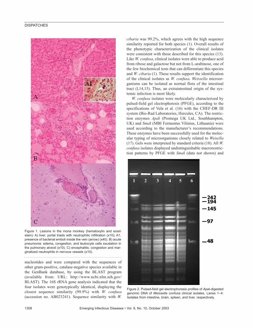
nucleotides and were compared with the sequences ofother gram-positive, catalase-negative species available inthe GenBank database, by using the BLAST program(available from: URL: http://www.ncbi.nlm.nih.gov/BLAST). The 16S rRNA gene analysis indicated that thefour isolates were genotypically identical, displaying theclosest sequence similarity (99.9%) with W. confusa(accession no. AB023241). Sequence similarity with W.
cibaria was 99.2%, which agrees with the high sequencesimilarity reported for both species (1). Overall results ofthe phenotypic characterization of the clinical isolateswere consistent with those described for this species (13).Like W. confusa, clinical isolates were able to produce acidfrom ribose and galactose but not from L-arabinose, one ofthe few biochemical tests that can differentiate this speciesand W. cibaria (1). These results support the identificationof the clinical isolates as W. confusa. Weissella microor-ganisms can be isolated as normal flora of the intestinaltract (l,14,15). Thus, an extraintestinal origin of the sys-temic infection is most likely.
W. confusa isolates were molecularly characterized bypulsed-field gel electrophoresis (PFGE), according to thespecifications of Vela et al. (16) with the CHEF-DR IIIsystem (Bio-Rad Laboratories, Hercules, CA). The restric-tion enzymes ApaI (Promega UK Ltd., Southhampton,UK) and SmaI (MBI Fermentas Vilinius, Lithuania) wereused according to the manufacturer’s recommendations.These enzymes have been successfully used for the molec-ular typing of microorganisms closely related to Weisella(17). Gels were interpreted by standard criteria (18). All W.confusa isolates displayed undistinguishable macrorestric-tion patterns by PFGE with SmaI (data not shown) and
1308 Emerging Infectious Diseases • Vol. 9, No. 10, October 2003
DISPATCHES
Figure 1. Lesions in the mona monkey (hematoxylin and eosinstain): A) liver: portal triads with neutrophilic infiltration (x10); A1,presence of bacterial emboli inside the vein (arrow) (x40). B) acutepneumonia: edema, congestion, and leukocyte cells exudation inthe pulmonary alveoli (x10). C) encephalitis: congestion and mar-ginalized neutrophils in nervous vessels (x10).
Figure 2. Pulsed-field gel electrophoresis profiles of ApaI-digestedgenomic DNA of Weissella confusa clinical isolates. Lanes 1–4:Isolates from intestine, brain, spleen, and liver, respectively.
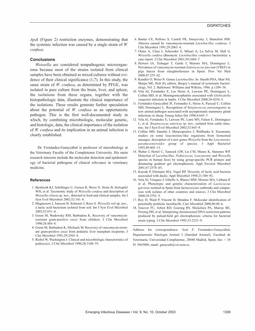
ApaI (Figure 2) restriction enzymes, demonstrating thatthe systemic infection was caused by a single strain of W.confusa.
ConclusionsWeissella are considered nonpathogenic microorgan-
isms because most of the strains isolated from clinicalsamples have been obtained as mixed cultures without evi-dence of their clinical significance (1,7). In this study, thesame strain of W. confusa, as determined by PFGE, wasisolated in pure culture from the brain, liver, and spleen;the isolations from these organs, together with thehistopathologic data, illustrate the clinical importance ofthe isolations. These results generate further speculationabout the potential of W. confusa as an opportunisticpathogen. This is the first well-documented study inwhich, by combining microbiologic, molecular genetic,and histologic, data, the clinical importance of the isolationof W. confusa and its implication in an animal infection isclearly established.
Dr. Fernández-Garayzábal is professor of microbiology atthe Veterinary Faculty of the Complutense University. His mainresearch interests include the molecular detection and epidemiol-ogy of bacterial pathogens of clinical relevance in veterinarymedicine.
References
1. Bjorkroth KJ, Schillinger U, Geisen R, Weiss N, Hoste B, HolzapfelWH, et al. Taxonomic study of Weissella confusa and description ofWeissella cibaria sp. nov., detected in food and clinical samples. Int JSyst Evol Microbiol 2002;52:141–8.
2. Magnusson J, Jonsson H, Schnurer J, Roos S. Weissella soli sp. nov.,a lactic acid bacterium isolated from soil. Int J Syst Evol Microbiol2002;52:831–4.
3. Green M, Wadowsky RM, Barbadora K. Recovery of vancomycin-resistant gram-positive cocci from children. J Clin Microbiol1990;28:484–8.
4. Green M, Barbadora K, Michaels M. Recovery of vancomycin-resist-ant gram-positive cocci from pediatric liver transplant recipients. JClin Microbiol 1991;29:2503–6.
5. Riebel W, Washington J. Clinical and microbiologic characteristics ofpediococci. J Clin Microbiol 1990;28:1348–55.
6. Bantar CE, Relloso S, Castell FR, Smayevsky J, Bianchini HM.Abscess caused by vancomycin-resistant Lactobacillus confusus. JClin Microbiol 1991;29:2063–4.
7. Olano A, Chua J, Schroeder S, Minari A, La Salvia M, Hall G.Weissella confusa (Basonym: Lactobacillus confusus) bacteremia: acase report. J Clin Microbiol 2001;39:1604–7.
8. Herrero IA, Teshager T, Garde J, Moreno MA, Domínguez L.Prevalence of vancomycin-resistant Enterococcus faecium (VREF) inpigs faeces from slaughterhouses in Spain. Prev Vet Med2000;47:255–62.
9. Kandler O, Weiss N. Genus Lactobacillus. In: Sneath PHA, Mair NS,Sharpe ME, Holt JG, editors. Bergey’s manual of systematic bacteri-ology, Vol. 2. Baltimore: Williams and Wilkins; 1986. p.1209–34.
10. Vela AI, Fernández E, Las Heras A, Lawson PE, Domínguez L,Collins MD, et al. Meningoencephalitis associated with Globicatellasanguinis infection in lambs. J Clin Microbiol 2000;38:4254–5.
11. Fernández-Garayzábal JF, Fernandez E, Heras A, Pascual C, CollinsMD, Dominguez L. Recognition of Streptococcus parasanguinis asnew animal pathogen associated with asymptomatic mammary glandinfections in sheep. Emerg Infect Dis 1998;4:645–7.
12. Vela AI, Fernández E, Lawson PE, Latre MV, Falsen E, DomínguezL, et al. Streptococcus entericus sp. nov., isolated from cattle intes-tine. Int J Syst Evol Microbiol 2002;52:665–9.
13. Collins MD, Samelis J, Metaxopoulos J, Wallbanks S. Taxonomicstudies on some leuconostoc-like organisms from fermentedsausages: description of a new genus Weissella from the Leuconostocparamesenteroides group of species. J Appl Bacteriol1993;49:405–13.
14. Walter J, Hertel C, Tannock GW, Lis CM, Munro K, Hammes WP.Detection of Lactobacillus, Pediococcus, Leuconostoc and Weissellaspecies in human feces by using group-specific PCR primers anddenaturing gradient gel electrophoresis. Appl Environ Microbiol2001;67:2578–85.
15. Kurzak P, Ehrmann MA, Vogel RP. Diversity of lactic acid bacteriaassociated with ducks. Appl Microbiol 1998;21:588–92.
16. Vela AI, Vázquez J, Gibello A, Blanco MM, Moreno MA, Liébana P,et al. Phenotypic and genetic characterization of Lactococcusgarvieae isolated in Spain from lactococcosis outbreaks and compar-ison with isolates of other countries and sources. J Clin Microbiol2000;38:3791–5.
17. Roy D, Ward P, Vincent D, Mondou F. Molecular identification ofpotentially probiotic lactobacilli. Curr Microbiol 2000;40:40–6.
18. Tenover FC, Arbeit RD, Goering RV, Mickelsen PA, Murray BE,Persing DH, et al. Interpreting chromosomal DNA restriction patternsproduced by pulsed-field gel electrophoresis: criteria for bacterialstrain typing. J Clin Microbiol 1995;33:2233–9.
Address for correspondence: José F. Fernández-Garayzábal,Departamento Patología Animal I (Sanidad Animal), Facultad deVeterinaria, Universidad Complutense, 28040 Madrid, Spain; fax: + 3491 3943908; email: [email protected]
Emerging Infectious Diseases • Vol. 9, No. 10, October 2003 1309
DISPATCHES
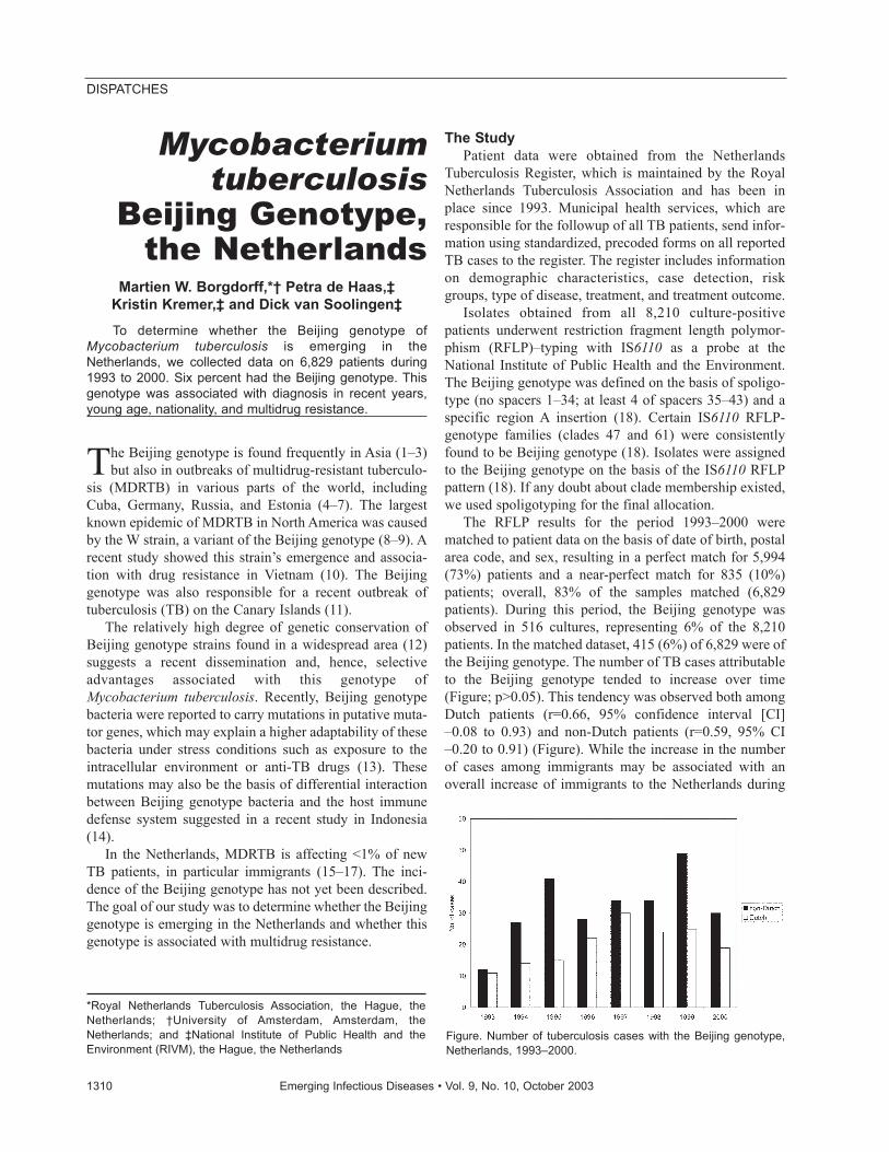
Mycobacteriumtuberculosis
Beijing Genotype,the Netherlands
Martien W. Borgdorff,*† Petra de Haas,‡ Kristin Kremer,‡ and Dick van Soolingen‡To determine whether the Beijing genotype of
Mycobacterium tuberculosis is emerging in theNetherlands, we collected data on 6,829 patients during1993 to 2000. Six percent had the Beijing genotype. Thisgenotype was associated with diagnosis in recent years,young age, nationality, and multidrug resistance.
The Beijing genotype is found frequently in Asia (1–3)but also in outbreaks of multidrug-resistant tuberculo-
sis (MDRTB) in various parts of the world, includingCuba, Germany, Russia, and Estonia (4–7). The largestknown epidemic of MDRTB in North America was causedby the W strain, a variant of the Beijing genotype (8–9). Arecent study showed this strain’s emergence and associa-tion with drug resistance in Vietnam (10). The Beijinggenotype was also responsible for a recent outbreak oftuberculosis (TB) on the Canary Islands (11).
The relatively high degree of genetic conservation ofBeijing genotype strains found in a widespread area (12)suggests a recent dissemination and, hence, selectiveadvantages associated with this genotype ofMycobacterium tuberculosis. Recently, Beijing genotypebacteria were reported to carry mutations in putative muta-tor genes, which may explain a higher adaptability of thesebacteria under stress conditions such as exposure to theintracellular environment or anti-TB drugs (13). Thesemutations may also be the basis of differential interactionbetween Beijing genotype bacteria and the host immunedefense system suggested in a recent study in Indonesia(14).
In the Netherlands, MDRTB is affecting <1% of newTB patients, in particular immigrants (15–17). The inci-dence of the Beijing genotype has not yet been described.The goal of our study was to determine whether the Beijinggenotype is emerging in the Netherlands and whether thisgenotype is associated with multidrug resistance.
The StudyPatient data were obtained from the Netherlands
Tuberculosis Register, which is maintained by the RoyalNetherlands Tuberculosis Association and has been inplace since 1993. Municipal health services, which areresponsible for the followup of all TB patients, send infor-mation using standardized, precoded forms on all reportedTB cases to the register. The register includes informationon demographic characteristics, case detection, riskgroups, type of disease, treatment, and treatment outcome.
Isolates obtained from all 8,210 culture-positivepatients underwent restriction fragment length polymor-phism (RFLP)–typing with IS6110 as a probe at theNational Institute of Public Health and the Environment.The Beijing genotype was defined on the basis of spoligo-type (no spacers 1–34; at least 4 of spacers 35–43) and aspecific region A insertion (18). Certain IS6110 RFLP-genotype families (clades 47 and 61) were consistentlyfound to be Beijing genotype (18). Isolates were assignedto the Beijing genotype on the basis of the IS6110 RFLPpattern (18). If any doubt about clade membership existed,we used spoligotyping for the final allocation.
The RFLP results for the period 1993–2000 werematched to patient data on the basis of date of birth, postalarea code, and sex, resulting in a perfect match for 5,994(73%) patients and a near-perfect match for 835 (10%)patients; overall, 83% of the samples matched (6,829patients). During this period, the Beijing genotype wasobserved in 516 cultures, representing 6% of the 8,210patients. In the matched dataset, 415 (6%) of 6,829 were ofthe Beijing genotype. The number of TB cases attributableto the Beijing genotype tended to increase over time(Figure; p>0.05). This tendency was observed both amongDutch patients (r=0.66, 95% confidence interval [CI]–0.08 to 0.93) and non-Dutch patients (r=0.59, 95% CI–0.20 to 0.91) (Figure). While the increase in the numberof cases among immigrants may be associated with anoverall increase of immigrants to the Netherlands during
1310 Emerging Infectious Diseases • Vol. 9, No. 10, October 2003
DISPATCHES
*Royal Netherlands Tuberculosis Association, the Hague, theNetherlands; †University of Amsterdam, Amsterdam, theNetherlands; and ‡National Institute of Public Health and theEnvironment (RIVM), the Hague, the Netherlands
Figure. Number of tuberculosis cases with the Beijing genotype,Netherlands, 1993–2000.
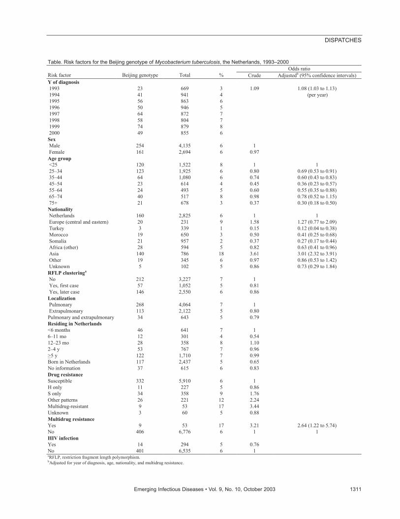
Emerging Infectious Diseases • Vol. 9, No. 10, October 2003 1311
DISPATCHES
Table. Risk factors for the Beijing genotype of Mycobacterium tuberculosis, the Netherlands, 1993–2000 Odds ratio
Risk factor Beijing genotype Total % Crude Adjustedb (95% confidence intervals) Y of diagnosis 1993 23 669 3 1.09 1.08 (1.03 to 1.13) 1994 41 941 4 (per year) 1995 56 863 6 1996 50 946 5 1997 64 872 7 1998 58 804 7 1999 74 879 8 2000 49 855 6 Sex Male 254 4,135 6 1 Female 161 2,694 6 0.97 Age group <25 120 1,522 8 1 1 25–34 123 1,925 6 0.80 0.69 (0.53 to 0.91) 35–44 64 1,080 6 0.74 0.60 (0.43 to 0.83) 45–54 23 614 4 0.45 0.36 (0.23 to 0.57) 55–64 24 493 5 0.60 0.55 (0.35 to 0.88) 65–74 40 517 8 0.98 0.78 (0.52 to 1.15) 75+ 21 678 3 0.37 0.30 (0.18 to 0.50) Nationality Netherlands 160 2,825 6 1 1 Europe (central and eastern) 20 231 9 1.58 1.27 (0.77 to 2.09) Turkey 3 339 1 0.15 0.12 (0.04 to 0.38) Morocco 19 650 3 0.50 0.41 (0.25 to 0.68) Somalia 21 957 2 0.37 0.27 (0.17 to 0.44) Africa (other) 28 594 5 0.82 0.63 (0.41 to 0.96) Asia 140 786 18 3.61 3.01 (2.32 to 3.91) Other 19 345 6 0.97 0.86 (0.53 to 1.42) Unknown 5 102 5 0.86 0.73 (0.29 to 1.84) RFLP clusteringa No 212 3,227 7 1 Yes, first case 57 1,052 5 0.81 Yes, later case 146 2,550 6 0.86 Localization Pulmonary 268 4,064 7 1 Extrapulmonary 113 2,122 5 0.80 Pulmonary and extrapulmonary 34 643 5 0.79 Residing in Netherlands <6 months 46 641 7 1 6–11 mo 12 301 4 0.54 12–23 mo 28 358 8 1.10 2–4 y 53 767 7 0.96 >5 y 122 1,710 7 0.99 Born in Netherlands 117 2,437 5 0.65 No information 37 615 6 0.83 Drug resistance Susceptible 332 5,910 6 1 H only 11 227 5 0.86 S only 34 358 9 1.76 Other patterns 26 221 12 2.24 Multidrug-resistant 9 53 17 3.44 Unknown 3 60 5 0.88 Multidrug resistance Yes 9 53 17 3.21 2.64 (1.22 to 5.74) No 406 6,776 6 1 1 HIV infection Yes 14 294 5 0.76 No 401 6,535 6 1 aRFLP, restriction fragment length polymorphism. bAdjusted for year of diagnosis, age, nationality, and multidrug resistance.
the study period, the increasing numbers among Dutchpatients likely reflect an increasing incidence rate.
The proportion of TB patients who had the Beijinggenotype was significantly associated in univariate analy-sis with a later year of diagnosis, young age, nationality(increased among immigrants from Asia and decreasedamong those from Morocco, Turkey, Somalia, and otherAfrican countries, compared with Dutch citizens), andmultidrug resistance (Table). These associations persistedin multivariate analysis (Table).
The association between the proportion of TB caseswith the Beijing genotype and age was observed in partic-ular in Dutch patients (chi squaretrend 15.5; p<0.0001), andwas not clear among non-Dutch patients (chi squaretrend0.8; p>0.2). Among Dutch patients, the Beijing genotypewas more commonly found in new cases (6%, 133/2,130)than among retreated cases (4%, 18/498; p<0.05). Amongnon-Dutch patients, the Beijing genotype was slightlymore common in retreated cases (8%, 19/229) than in newcases (6%, 204/3,218); however, this occurrence was notsignificant (p>0.2). The incidence pattern by person andtime appears consistent with transmission of the Beijinggenotype from immigrants to the Dutch population (19).
The proportion of TB cases with the Beijing genotypewas not substantially associated with HIV infection, pul-monary localization, RFLP clustering, or duration of stayin the Netherlands (Table). The lack of association withRFLP-clustering suggests that the Beijing genotype is notspreading more quickly than other M. tuberculosis strains.
Of the nine patients with the Beijing genotype and mul-tidrug resistance, four were from central and easternEurope, three from Asia, one from Africa, and one fromthe Netherlands. One of these nine patients was RFLP-clustered; the other patient in that RFLP cluster had beendiagnosed previously and had isolated isoniazid resistance.
During 1993 to 2000 in the Netherlands, the Beijinggenotype represented 6% of TB cases. The Beijing geno-type was associated with recent diagnoses, young age (inparticular among Dutch citizens), nationality (EasternEurope and Asia), and multidrug resistance. Although theBeijing genotype was associated with multidrug resist-ance, the number of MDRTB cases was small. No second-ary cases were observed with the Beijing genotype andMDRTB. Of the nine cases of MDRTB, only one case mayhave been caused by transmission within the Netherlandsfrom a case-patient who had isoniazid resistance at thestart of treatment and may have acquired rifampicin resist-ance during treatment. However, the spread of the Beijinggenotype among young people suggests the possible emer-gence of multidrug resistance and emphasizes the need forcontinued surveillance.
AcknowledgmentsWe thank all municipal health services in the Netherlands
for their contribution to the Netherlands Tuberculosis Register.
This study was performed within the framework ofConcerted Action project QLK2-2000-00630 on MolecularEpidemiology of Tuberculosis.
Dr. Borgdorff is the chief epidemiologist of the RoyalNetherlands Tuberculosis Association (KNCV) and professor ofinternational health with special reference to tuberculosis at theUniversity of Amsterdam. His areas of interest include the molecu-lar epidemiology of tuberculosis and other communicable diseases.
References
1. van Soolingen D, Qian L, de Haas PE, Douglas JT, Traore H, PortaelsF, et al. Predominance of a single genotype of Mycobacterium tuber-culosis in countries of East Asia. J Clin Microbiol 1995;33:3234–8.
2. Chan MY, Borgdorff M, Yip CW, de Haas PE, Wong WS, Kam KM,et al. Seventy percent of the Mycobacterium tuberculosis isolates inHong Kong represent the Beijing genotype. Epidemiol Infect2001;127:169–71.
3. Bifani PJ, Mathema B, Kurepina NE, Kreiswirth BN. Global dissem-ination of the Mycobacterium tuberculosis W-Beijing family strains.Trends Microbiol 2002;10:45–52.
4. Diaz R, Kremer K, de Haas PE, Gomez RI, Marrero A, Valdivia JA,et al. Molecular epidemiology of tuberculosis in Cuba outside ofHavana, July 1994–June 1995: utility of spoligotyping versus IS6110restriction fragment length polymorphism. Int J Tuberc Lung Dis1998;2:743–50.
5. Niemann S, Rusch-Gerdes S, Richter E. IS6110 fingerprinting ofdrug-resistant Mycobacterium tuberculosis strains isolated inGermany during 1995. J Clin Microbiol 1997;35:3015–20.
6. Marttila HJ, Soini H, Eerola E, Vyshevskaya E, Vyshnevskyi BI,Otten TF. A Ser315Thr substitution in katG is predominant in genet-ically heterogeneous multi-drug resistant Mycobacterium tuberculo-sis isolates originating from the St. Petersburg area in Russia.Antimicrobiol Agents Chemother 1998;42:2443–5.
7. Kruuner A, Hoffner SE, Sillastu H, Danilovits M, Levina K, SvensonSB, et al. Spread of drug-resistant pulmonary tuberculosis in Estonia.J Clin Microbiol 2001;39:3339–45.
8. Bifani PJ, Plikaytis BB, Kapur V, Stockbauer K, Pan X, Lutfey ML.Origin and interstate spread of a New York City multidrug-resistantMycobacterium tuberculosis clone family. JAMA 1996;275:452–7.
9. Frieden TR, Sherman LF, Maw KL, Fujiwara PI, Crawford JT, NivinB, et al. A multi-institutional outbreak of highly drug-resistant tuber-culosis: epidemiology and clinical outcomes. JAMA1996;276:1229–35.
10. Anh DD, Borgdorff MW, Van LN, Lan NT, van Gorkom T, KremerK, et al. Mycobacterium tuberculosis Beijing genotype emerging inVietnam. Emerg Infect Dis 2000;6:302–5.
11. Caminero JA, Pena MJ, Campos-Herrero MI, Rodriguez JC, GarciaI, Cabrera P, et al. Epidemiological evidence of the spread of aMycobacterium tuberculosis strain of the Beijing genotype on GranCanaria Island. Am J Respir Crit Care Med 2001;164:1165–70.
12. Glynn JR, Whiteley J, Bifani PJ, Kremer K, van Soolingen D.Worldwide occurrence of Beijing/W strains of Mycobacterium tuber-culosis: a systematic review. Emerg Infect Dis 2002;8:843–9.
13. Rad ME, Bifani P, Martin C, Kremer K, Samper S, Rauzier J, et al.Mutations in putative mutator genes of Mycobacterium tuberculosisstrains of the W-Beijing family. Emerg Infect Dis 2003;7:838–45.
1312 Emerging Infectious Diseases • Vol. 9, No. 10, October 2003
DISPATCHES
14. Van Crevel R, Nelwan RH, de Lenne W, Veeraga Y, van der ZandenAG, Amin Z, et al. Mycobacterium tuberculosis Beijing genotypestrains associated with febrile response to treatment. Emerg InfectDis 2001;7:880–3.
15. Espinal MA, Laszlo A, Simonsen L, Boulahbal F, Kim SJ, Reniero A,et al. Global trends in resistance to antituberculosis drugs. WorldHealth Organization—International Union against Tuberculosis andLung Disease Working Group on Anti-Tuberculosis Drug ResistanceSurveillance. N Engl J Med 2001;344:1294–303.
16. Lambregts-van Weezenbeek CS, Jansen HM, Nagelkerke NJ, vanKlingeren B, Veen J. Nationwide surveillance of drug-resistant tuber-culosis in the Netherlands: rates, risk factors and treatment outcome.Int J Tuberc Lung Dis 1998;2:288–95.
17. Lambregts-van Weezenbeek CS, Jansen HM, Veen J, Nagelkerke NJ,Sebek MM, van Soolingen D. Origin and management of primary andacquired drug-resistant tuberculosis in the Netherlands: the truthbehind the rates. Int J Tuberc Lung Dis 1998;2:296–302.
18. Kremer K, van den Brandt A, Kurepina NE, Glynn J, Bifani PJ, vanSoolingen D. Definition of the Mycobacterium tuberculosis Beijinggenotype. European Union Concerted Action project meeting,Cascais, May 27–30, 2002. Abstract 019.
19. Borgdorff MW, Nagelkerke NJ, De Haas PE, Van Soolingen D.Transmission of tuberculosis depending on the age and sex of sourcecases. Am J Epidemiol 2001;154:934–43.
Address for correspondence: M.W. Borgdorff, Royal NetherlandsTuberculosis Association (KNCV), P.O. Box 146, 2501 CC the Hague,the Netherlands; fax: +31 70 3584004; email: [email protected]
Emerging Infectious Diseases • Vol. 9, No. 10, October 2003 1313
DISPATCHES
All material published in Emerging Infectious Diseases is in thepublic domain and may be used and reprinted without special per-mission; proper citation, however, is appreciated.
Search past issues of EID at www.cdc.gov/eid
Saliva andMeningococcal
Transmission Hilary J. Orr,* Steve J. Gray,† Mary Macdonald,‡
and James M. Stuart*1
Neisseria meningitidis carriage was compared in swabspecimens of nasopharynx, tonsils, and saliva taken from258 students. We found a higher yield in nasopharyngealthan in tonsillar swabs (32% vs. 19%, p<0.001). Low preva-lence of carriage in saliva swabs (one swab [0.4%]) sug-gests that low levels of salivary contact are unlikely totransmit meningococci.
Invasive meningococcal disease has a high case-fatalityrate and an immediate risk of further cases among
household contacts. Public health measures thereforeinclude prompt identification of contacts for chemopro-phylaxis (1). One question that commonly arises iswhether salivary contact through sharing cups or glasses isan indication for prophylaxis, but the evidence base toinform an answer is weak, and national guidelines areinconsistent (1,2). Although saliva is thought to inhibitmeningococcal growth (3), carriage rates in saliva are notknown, and swabs to detect carriage are usually taken fromtonsils or nasopharynx (4–6). We compared meningococ-cal isolation rates in swabs of saliva (front of mouth), ton-sils, and nasopharynx.
We recruited volunteers among students from two col-leges in Hereford, England. After giving written consent,students completed a short questionnaire on age, sex,smoking, recent antimicrobial drug use, and meningococ-cal vaccine status. Three sterile, dry, cotton-tipped swabswere used to take samples from each volunteer: one fromthe nasopharynx (through the mouth and swept up behindthe uvula), one from both tonsils, and one swab of salivabetween the lower gum and lips. Swabs were plated direct-ly onto a selective culture medium primarily designed forthe isolation of pathogenic Neisseria species (modifiedNew York City base containing vancomycin, colistin, andtrimethoprim), prepared by Taunton Media Services, UK(7). The plates were transported to Hereford Public HealthLaboratory, where they were spread once from the primaryinoculum and incubated in 7% CO2 at 37°C for 48 h.Putative Neisseria species isolated were sent to the
Meningococcal Reference Unit, Manchester Public HealthLaboratory, for Neisseria meningitidis confirmation andserologic phenotypic characterization. Data were enteredinto the computer using Excel (Microsoft Corp., Redmond,WA). Carriage rates by site were compared withMcNemar’s test and by risk factor using chi-square tests.Ethical approval was obtained from the Public HealthLaboratory Service Ethics Committee and HerefordshireDistrict Ethics Committee.
Of the 258 participants, 90 (35%) were identified ascarrying Neisseria meningitidis from one or more sites.The site with the highest yield was the nasopharynx(32.2%), whereas tonsillar carriage was 19.4% (Table).One (0.4%) of the 258 saliva swab specimens was positive.No one had positive specimens from all three sites, and theperson with the positive saliva swab had negative swabsfrom the other two sites. Differences in carriage ratesbetween the nasopharynx and tonsils and between thenasopharynx and saliva were statistically significant(p<0.001 in both cases).
The predominant serogroup among carried strains wasB. No serogroup C strains were identified. Of the 44 carri-ers with positive swabs from both nasopharynx and tonsils,each pair of isolates was considered to be phenotypicallyindistinguishable by serogroup, serotype, and sero-sub-type. In three of these pairs, one isolate expressedserogroup B, and the paired isolate could not beserogrouped but had identical serotype and sero-subtype.
Of the 258 participants, 116 (45%) were men, and 142were women. Most (86%) were 18 to 21 years of age.Carriage rates were higher among men than women(54/116 vs. 36/142, p<0.001), and among smokers thannonsmokers (49/90 vs. 51/168, p<0.001). Carriage rateswere similar when persons were stratified by age,meningococcal vaccination status, and recent antimicro-bial drug use. Although duplicate swabs from thenasopharynx sometimes yield different meningococcalstrains (3), none of the paired isolates in this study weredistinguishable by phenotype.
The yield of meningococci from nasopharyngeal swabswas nearly twice as high as that from tonsillar swabs.Previous researchers have found a lower sensitivity ofnasopharyngeal swabs taken through the nose using smallcotton-tipped wire swabs compared to tonsillar swabstaken using larger cotton-tipped swabs (5,6). Our use ofthe same type of swab to sample both sites provided amore valid comparison. The carriage rate was higher thanexpected for this age group (4), suggesting that we had
1314 Emerging Infectious Diseases • Vol. 9, No. 10, October 2003
DISPATCHES
*Health Protection Agency (South West), Gloucester, England,United Kingdom; †Meningococcal Reference Unit, Manchester,England, United Kingdom; and ‡County Hospital, Hereford,England, United Kingdom
1H.O. was responsible for recruiting students, obtaining speci-mens, swabs, and drafting the paper with J.S.; S.G. and M.M. wereresponsible for microbiologic processing and analysis; and J.S.designed the study and drafted the paper with H.O. All authorscontributed to the final draft.
efficient swabbing and microbiologic techniques. We sug-gest that throat swabs to detect meningococcal carriageshould always be taken from the nasopharynx (through themouth whenever practical) and not from the tonsils.
The very low isolation rate from saliva swabs suggeststhat low levels of salivary contact are unlikely to transmitmeningococci (1). This observation is supported by resultsof a case-control study among university students thatfound no association between meningococcal acquisitionand sharing of glasses or cigarettes (8). On the basis of thisevidence, we propose that guidelines for public healthmanagement of meningococcal disease should not includelow-level salivary contact (e.g., sharing drinks) with acase-patient as an indication for chemoprophylaxis.
AcknowledgmentsWe thank all staff and students at Hereford College of
Technology and Hereford College of Art and Design who wereinvolved in this study and Erika Duffell and Nicky Maxwell fortheir help with sampling.
This study was supported by Gloucester Public HealthLaboratory Trust Fund.
Ms. Orr is an epidemiology research nurse in southwestEngland, working for the Health Protection Agency. She has aresearch interest in the epidemiology of infectious diseases.
References
1. Public Health Laboratory Service. Guidelines for public health man-agement of meningococcal disease in the UK. Commun Dis PublicHealth 2002;5:187–204.
2. Communicable Diseases Network Australia. Guidelines on the earlyclinical and public health management of meningococcal disease inAustralia. [Accessed 3 June 2003]. Available from: URL:http://www.health.gov.au/pubhlth/cdi/pubs/mening.htm
3. Gordon MH. The inhibitory action of saliva on growth of themeningococcus. Great Britain Medical Research Committee, SpecialReport Series 3; 1917. p. 106–11.
4. Cartwright KAV, Stuart JM, Jones DM, Noah ND. The Stonehousesurvey: nasopharyngeal carriage of meningococci and Neisserialactamica. Epidemiol Infect 1987;99:591–601.
5. Olcén P, Kjellander J, Danielsson D, Lindquist BL. Culture diagnosisof meningococcal carriers: yield from different sites and influence ofstorage in transport medium. J Clin Pathol 1979;32:1222–5.
6. Hoeffler DF. Recovery of Neisseria meningitidis from the nasophar-ynx. Comparison of two techniques. Am J Dis Child 1974;128:54–6.
7. Cunningham R, Matthews R, Lewendon G, Harrison S, Stuart JM.Improved rate of isolation of Neisseria meningitidis by direct platingof pharyngeal swabs. J Clin Microbiol 2001;39:4575–6.
8. Neal KR, Nguyen-Van-Tam J, Jeffrey N, Slack RC, Madeley RJ, Ait-Tahar K, et al. Changing carriage rate of Neisseria meningitidisamong university students during the first week of term: cross sec-tional study. BMJ 2000;320:846–9.
Address for correspondence: James Stuart, Consultant Epidemiologist,Health Protection Agency (South West), Microbiology Laboratory,Gloucestershire Royal Hospital, Gloucester GL1 3NN, England, UK; fax:++44(0) 1452 412946; email: [email protected]
Emerging Infectious Diseases • Vol. 9, No. 10, October 2003 1315
DISPATCHES
Table. Carriage of Neisseria meningitidis by site Site of swab One site positive Two sites positive Three sites positive Total positive Overall carriage % Nasopharynx 39 44 0 83 32.2 Tonsils 6 44 0 50 19.4 Saliva 1 0 0 1 0.4 a(n=258).
Small ColonyVariants of
Staphylococcusaureus and
Pacemaker-relatedInfection
Harald Seifert,* Hilmar Wisplinghoff,* Petra Schnabel,* and Christof von Eiff†
We describe the first known case of a device-relatedbloodstream infection caused by Staphylococcus aureussmall colony variants. Recurrent pacemaker-related blood-stream infection within a 7-month period illustrates the poorclinical and microbiologic response to prolonged antimicro-bial therapy in a patient infected with this S. aureus sub-population.
Infections caused by Staphylococcus aureus range frommild skin infections to acute life-threatening diseases
such as pneumonia, osteomyelitis, and endocarditis.However, S. aureus may also cause a chronic disease withpersistent and recurrent infections. Skin and soft tissueinfections, chronic osteomyelitis, and persistent infectionsin patients with cystic fibrosis have been associated withsmall colony variants, a naturally occurring subpopulationof the species S. aureus (1–6). S. aureus small colony vari-ants are characterized as electron transport deficient bacte-ria because of their auxotrophism to hemin or menadioneor are recognized as thymidine-dependent. These variantsproduce very small, mostly nonpigmented and non-hemolytic colonies. In addition, they also demonstrate var-ious other features that are atypical for S. aureus, includingreduced coagulase production, failure to use mannitol, andincreased resistance to aminoglycosides and cell-wallactive antibiotics (3–10). Furthermore, the ability of thesevariants to persist intracellularly within nonprofessionalphagocytes has been described (3,5,11). Because of theirfastidious growth characteristics and unusual morphologicappearance, small colony variants present a challenge bothto the microbiologist and the clinician, often resulting inmisidentification and misinterpretation (1,2,7,8).Prerequisite for recovering and identifying these variantsis the application of extended conventional culture andidentification techniques (3,5,8). We report the first case of
a pacemaker-related bloodstream infection caused by S.aureus small colony variants. This case illustrates the poorclinical and microbiologic response to prolonged antimi-crobial therapy in patients infected with these variants.
Case ReportA 63-year-old man was transferred to our facility with
the presumptive diagnosis of endocarditis related to apacemaker-lead infection. Past medical history includedhypertension, coronary artery disease, and noninsulin-dependent diabetes mellitus. A VVI (ventricular ventricu-lar inhibited) pacemaker had been implanted for treatmentof sick sinus syndrome 9 years earlier. Six weeks beforeadmission, this device had been removed because of apocket infection after blunt trauma with dislocation of thedevice and perforation of the skin. Specimens for microbi-ologic culture were not obtained at this time. The pace-maker leads were left in place, a gentamicin-containingsponge was applied to the infection site, and a new pace-maker was implanted on the other side of the chest. Fourweeks later, the patient sought treatment at the local hospi-tal for a high fever (39.7°C) and chills and a subcutaneousabscess with oxacillin-susceptible S. aureus at the primaryinsertion site. After surgical drainage, antimicrobial thera-py was initiated with intravenous cefuroxime. The remain-ing pacemaker leads were partially cut but not completelyremoved. Ten days later, spiking fever and chills unrespon-sive to the administration of meropenem and vancomycindeveloped, and the patient was transferred to our medicalcenter for pacemaker ablation. The physical examinationdid not indicate auscultation abnormalities or stigmata ofendocarditis. Laboratory studies were unremarkableexcept a C-reactive protein (CRP) level of 170 mg/L (nor-mal value <8 mg/L) and a blood sedimentation rate of 79mm/h. Multiple blood cultures taken on admissionremained negative. Transesophageal echocardiography didnot show vegetations or other evidence of endocarditis. Onhospital day 6, the new pacemaker was completelyremoved by percutaneous ablation as were the remainingleads of the old device. Only the tip of the pacemaker leadremained fixed in the myocardium, and surgical removalinvolving extracorporal circulation was not attempted. Thepatient’s condition improved rapidly, CRP level returned tonormal, and on hospital day 32, the patient was transferredto the local hospital to complete a 6-week course of intra-venous vancomycin and rifampin as empirical antistaphy-lococcal therapy. Before transfer, the daily vancomycindose had been reduced to 250 mg twice a day after an ele-vated vancomycin serum level. Eight days later, the patientwas readmitted with recurrent high fever. Blood culturestaken on readmission were again negative. After the van-comycin dose was increased to 500 mg every 12 hours, thepatient promptly became afebrile. Antimicrobial therapy
1316 Emerging Infectious Diseases • Vol. 9, No. 10, October 2003
DISPATCHES
*University of Köln, Germany; and †University of Münster Hospitaland Clinics, Münster, Germany
was discontinued after the patient had completed a 10-week course of vancomycin and rifampin. Three days later,the patient again had spiking fever. After 6 to 48 hours ofincubation, four sets of blood cultures obtained on fourconsecutive days yielded nonpigmented and nonhemolyticstaphylococci, initially identified on the basis of a negativetube coagulation test and the API ID 32 Staph system(bioMérieux, Marcy-L’Etoile, France) as coagulase-nega-tive staphylococci, susceptible to oxacillin (MIC 0.5mg/mL) and vancomycin (MIC 1.0 mg/mL) but resistantto rifampin (MIC >32 mg/mL). However, the colony mor-phologic findings were suggestive of small colony variantsof S. aureus, confirmed by polymerase chain reactionamplification of the nuc and coa genes as well as by deter-mination of the strain’s auxotrophy for hemin. The patientresponded promptly to flucloxacillin, 4 g intravenouslythree times a day. After another 6-week course of parenter-al therapy, antimicrobial therapy was discontinued, and thepatient was discharged (CRP 7 mg/L, ESR 35 mm/h); hewas readmitted after 6 days with chills and high fever.Antimicrobial therapy with intravenous flucloxacillin wasresumed and followed by immediate defervescence. Threeblood cultures taken on readmission were again positivewith S. aureus small colony variants. Clonal identity of allisolates was demonstrated by pulsed-field gel elec-trophoresis of bacterial DNA (data not shown). A trans-esophageal echocardiogram showed the residual tip of thepacemaker lead fixed in the myocardial septum withoutvegetations. The remaining device was finally removed byopen-heart surgery with use of cardiopulmonary bypass.Microbiologic culture of the pacemaker electrode per-formed at a different institution yielded abundant growthof staphylococci that were misidentified as S. warneri,showing the same biochemical profile as the previouslyisolated bacteria as determined by the ID 32 Staph system.The patient recovered completely and was discharged onthe 10th postoperative day after a total hospital course of 7months.
ConclusionsS. aureus small colony variants have been implicated in
persistent and recurrent infections that give a poor clinicaland bacteriologic response to standard antimicrobial thera-py in patients with abscess, chronic osteomyelitis, andbronchopulmonary infections, particularly after prolongedexposure to antibiotics (1–6). Bloodstream infection relat-ed to an implantable intravascular device with this S.aureus variant has not been reported before. These pheno-typic variants are characterized by their fastidious growthand atypical colony morphologic findings on routinemedia, making recovery as well as correct identificationdifficult for microbiologic laboratories (3,5,8). The abilityto interrupt electron transport and to form a variant sub-
population affords S. aureus a number of survival advan-tages, including the ability of this subpopulation to persistintracellularly within nonprofessional phagocytes (11,12).The intracellular position may shield small colony variantsfrom host defenses and decrease exposure to antibiotics(3,5,11). S. aureus small colony variants can be selected bygentamicin in vitro and in vivo as shown in patients withosteomyelitis after gentamicin bead placement (4,12).Chuard et al. demonstrated that, in contrast to their normalphenotype parental strain, S. aureus small colony variantsthat were attached to fibronectin-coated coverslips werehighly resistant to cell-wall–active antimicrobial agentssuch as oxacillin and vancomycin (13).
In our case, findings suggest that S. aureus smallcolony variants might have been selected from the parentstrain population with a normal phenotype after exposureto the locally applied aminoglycoside or to the prolongedadministration of vancomycin. Continually positive bloodcultures with the same strain as demonstrated by molecu-lar typing and the presumable persistence of these organ-isms on the pacemaker lead tip may partly be explained bythe poor effectiveness of vancomycin and flucloxacillinagainst these slow-growing organisms that were adheringto the remaining foreign body and the ability of these vari-ants to persist intracellularly (7,8,13).
This case adds to the spectrum of persistent and relaps-ing infections attributed to S. aureus small colony variantsand emphasizes that these variants may also play a role inintravascular device–related infections. It also illustratesthat complete removal of any foreign body material isessential for the complete cure of prosthetic intravasculardevice–related S. aureus infection. Laboratories should beparticularly alert for S. aureus small colony variants whensamples are submitted from patients who have receivedlong-term antimicrobial therapy, especially if the infectionis unusually persistent or recurrent.
Dr. Seifert is professor of clinical microbiology at theInstitute for Medical Microbiology, Immunology and Hygiene atthe University of Cologne, Köln, Germany. His research interestsinclude the molecular epidemiology of nosocomial pathogens, inparticular Acinetobacter species and methicillin-resistantStaphylococcus aureus, catheter-related infections, and antimi-crobial resistance and its mechanisms.
References
1. Proctor RA, Bates DM, McNamara PJ. Electron transport deficientStaphylococcus aureus small-colony variants as emerging pathogens.In: Scheld WM, Craig WA, Hughes JM, editors. Emerging infections5. Washington: ASM Press; 2001.
2. Proctor RA, van Langevelde P, Kristjansson M, Maslow JN, ArbeitRD. Persistent and relapsing infections associated with small-colonyvariants of Staphylococcus aureus. Clin Infect Dis 1995;20:95–102.
Emerging Infectious Diseases • Vol. 9, No. 10, October 2003 1317
DISPATCHES
3. von Eiff C, Becker K, Metze D, Lubritz G, Hockmann J, Schwarz T,et al. Intracellular persistence of Staphylococcus aureus small-colonyvariants within keratinocytes: a cause for antibiotic treatment failurein a patient with Darier’s disease. Clin Infect Dis 2001;32:1643–7.
4. von Eiff C, Bettin D, Proctor RA, Rolauffs B, Lindner N,Winkelmann W, et al. Recovery of small colony variants ofStaphylococcus aureus following gentamicin bead placement forosteomyelitis. Clin Infect Dis 1997;25:1250–1.
5. Kahl B, Herrmann M, Schulze-Everding A, Koch HG, Becker K,Harms E, et al. Persistent infection with small colony variant strainsof Staphylococcus aureus in patients with cystic fibrosis. J Infect Dis1998;177:1023–9.
6. Seifert H, von Eiff C, Fätkenheuer G. Fatal case due to methicillin-resistant Staphylococcus aureus small colony variants in an AIDSpatient. Emerg Infect Dis 1999:5:450–3.
7. Proctor RA, Kahl B, von Eiff C, Vaudaux PE, Lew DP, Peters G.Staphylococcal small colony variants have novel mechanisms forantibiotic resistance. Clin Infect Dis 1997;27(Suppl 1):S68–S74.
8. Proctor RA, Peters G. Small colony variants in staphylococcal infec-tions: diagnostic and therapeutic implications. Clin Infect Dis1998;27:419–23.
9. Kahl BC, Belling G, Reichelt R, Herrmann M, Proctor RA, Peters G.Thymidine-dependent small-colony variants of Staphylococcusaureus exhibit gross morphological and ultrastructural changesconsistent with impaired cell separation. J Clin Microbiol2003:41:410–3.
10. Baumert N, von Eiff C, Schaaff F, Peters G, Proctor RA, Sahl HG.Physiology and antibiotic susceptibility of Staphylococcus aureussmall colony variants. Microb Drug Resist 2002;8:253–60.
11. Vaudaux P, Francois P, Bisognano C, Kelley WL, Lew DP, SchrenzelJ, et al. Increased expression of clumping factor and fibronectin-binding proteins by hemB mutants of Staphylococcus aureusexpressing small colony variant phenotypes. Infect Immun2002;70:5428–37.
12. Balwit JM, van Langevelde P, Vann JM, Proctor RA. Gentamicin-resistant menadione and hemin auxotrophic Staphylococcus aureuspersist within cultured endothelial cells. J Infect Dis1994;170:1033–7.
13. Chuard C, Vaudaux PE, Proctor RA, Lew DP. Decreased susceptibil-ity to antibiotic killing of a stable small colony variant ofStaphylococcus aureus in fluid phase and on fibronectin-coated sur-faces. J Antimicrob Chemother 1997;39:603–8.
Address for correspondence: Harald Seifert, Institut für MedizinischeMikrobiologie, Immunologie und Hygiene, Goldenfelsstr.19-21, 50935Köln, Germany; fax: 0049 221-4783979; email: [email protected]
1318 Emerging Infectious Diseases • Vol. 9, No. 10, October 2003
DISPATCHES
Research Studies: Articles should be 2,000 to 3,500 words andshould include references, not to exceed 40. Use of subheadings inthe main body of the text is recommended. Photographs and illus-trations are encouraged. Provide a short abstract (150 words) and abrief biographical sketch of first author—both authors if only two.
These articles report laboratory and epidemiologic results with-in a public health perspective. Although these reports may be writ-ten in the style of traditional research articles, they should explainthe value of the research in public health terms and place the find-ings in a larger perspective (e.g., “Here is what we found, and hereis what the findings mean”).
Manuscript Preparation. For word processing, use MS Word.Begin each of the following sections on a new page and in thisorder: title page, keywords, abstract, text, acknowledgments, biog-raphical sketch, references, tables, figure legends, appendixes, andfigures. Each figure should be in a separate file.
Title Page. Give complete information about each author (i.e.,full name, graduate degree(s), affiliation, and the name of the insti-tution in which the work was done). Clearly identify the correspon-ding author and provide that author's mailing address (includephone number, fax number, and e-mail address). Include separateword counts for abstract and text.
Keywords. Include up to 10 keywords; use terms listed inMedical Subject Headings Index Medicus.
Text. Double-space everything, including the title page,abstract, references, tables, and figure legends. Printed manuscriptshould be single-sided, beginning with the title page. Indent para-graphs; leave no extra space between paragraphs. After a period,leave only one space before beginning the next sentence. Use 12-point Times New Roman font and format with ragged right margins
(left align). Italicize (rather than underline) scientific names whenneeded.
Biographical Sketch. Include a short biographical sketch of thefirst author—both authors if only two. Include affiliations and theauthor’s primary research interests.
References. Follow Uniform Requirements (www.icmje.org/index.html). Do not use endnotes for references. Place refer-ence numbers in parentheses, not superscripts. Number citations inorder of appearance (including in text, figures, and tables). Cite per-sonal communications, unpublished data, and manuscripts in prepa-ration or submitted for publication in parentheses in text. ConsultList of Journals Indexed in Index Medicus for accepted journalabbreviations; if a journal is not listed, spell out the journal title.List the first six authors followed by “et al.” Do not cite referencesin the abstract.
Tables and Figures. Create tables within MS Word’s table tool.Do not format tables as columns or tabs. Send graphics in native,high-resolution (200-dpi minimum) .TIF (Tagged Image File), or.EPS (Encapsulated Postscript) format. Graphics should be in a sep-arate electronic file from the text file. For graphic files, use Arialfont. Convert Macintosh files into the suggested PC format.Figures, symbols, letters, and numbers should be large enough toremain legible when reduced. Place figure keys within the figure.For more information see EID Style Guide (http://www.cdc.gov/ncidod/EID/style_guide .htm).
Manuscript Submission. Include a cover letter indicating theproposed category of the article (e.g., Research, Dispatch) andverifying that the final manuscript has been seen and approved byall authors. Submit an electronic copy (by e-mail) to the Editor,[email protected].
Instructions for Emerging Infectious Diseases Authors
West Nile VirusDetection in
American Crows Sarah A. Yaremych,* Richard E. Warner,*
Marshall T. Van de Wyngaerde,† Adam M. Ringia,† Richard Lampman,†
and Robert J. Novak†
A dipstick immunochromatographic assay used forWest Nile virus (WNV) detection in mosquitoes was inves-tigated for application to testing of fecal, saliva, and tissuesamples from dead American Crows (Corvus brachyrhyn-chos). Results suggest that VecTest may be an efficientmethod for WNV detection in field-collected, deadAmerican Crows, although confirmation of results and fur-ther investigation are warranted.
The American Crow (Corvus brachyrhynchos) has beendesignated as a West Nile virus (WNV) surveillance
species (1), and dead American Crows have been used tomonitor WNV activity across the nation. The AmericanCrow is a useful species for monitoring disease activitybecause it is highly visible and recognizable; furthermore,all American Crows experimentally inoculated with WNVhave died within 7 days of inoculation after attainingviremias of sufficient titer to infect mosquitoes (2). Aviandeaths early in the transmission season are a warning forincreased risk for human WNV cases (3); by monitoringWNV infection in dead American Crows, we can detectareas of epidemiologic public health concern.
Standard methods of identifying WNV in dead crowsinclude two direct tests of tissues by immunohistochem-istry (IHC) and reverse transcriptase-polymerase chainreaction (RT-PCR TaqMan (Applied Biosystems, FosterCity, CA). Komar et al. (4) determined that postmortemcloacal and oral swabs could replace brain tissue as a spec-imen for WNV detection in crows and jays. Addressingthis, and the need for a simple, quick, and cost-effectivemethod for viral surveillance in dead crows, we conducteda study to determine whether the VecTest WNV/SaintLouis encephalitis virus (SLEV) Antigen Panel Assay(Medical Analysis Systems Inc., Camarillo, CA) could beused for WNV testing of fecal, saliva, and tissue samplesfrom American Crows.
The VecTest was designed as a rapid wicking assay toidentify the presence or absence of viral antigen specific to
WNV or SLEV in infected mosquitoes. This test employsmonoclonal antibodies against WNV and SLEV in a one-step procedure with a wicking test strip. All componentsnecessary to carry out the test are provided in a kit, includ-ing vials, buffer solution, and dipsticks. The tests can beperformed in the field, highly trained personnel and spe-cialized equipment are not necessary, and results can beobtained quickly (<20 minutes). Each dipstick contains aninternal positive control to indicate that the test has per-formed properly. After mosquitoes are ground in a buffersolution, a dipstick is inserted; WNV and SLEV antigenpresent in the mosquito slurry will bind to the specific anti-body-colloidal gold conjugate. Antigen presence is indicat-ed by a red line that develops in the test zone of the dip-stick, specific to WNV or SLEV; pictures of dipsticks canbe obtained from Ryan et al. (5). Ryan evaluated the prod-uct for detection of WNV in mosquitoes in a laboratoryand suggested that sensitivity of VecTest is comparable tothat of an antigen-capture enzyme-linked immunosorbentassay but less sensitive than Vero cell plaque assays or RT-PCR. Our study is a preliminary evaluation of the VecTestassay to determine whether it can be used for accurateWNV testing of field-collected dead American Crows.
The StudyIn February–October 2002, as part of a broader study,
we captured, banded, and marked 156 American Crows (C.brachyrhynchos) in Champaign and Urbana, Illinois(Yaremych SA. West Nile virus and American Crows ineast-central Illinois. University of Illinois; 2003). Radio-transmitters were attached to individuals of a subsample ofcaptured crows. Many of the study crows died in the sum-mer; all recovered crows were tested for WNV. Deadcrows were retrieved by tracking the radio signal to thecarcasses, by chance encounter, or by notification from thepublic. Dead study crows with transmitters were typicallyrecovered <36 hours after death, as this period was themaximum amount of time elapsed between the last liveobservation of a crow during radio tracking and recoveryof the carcass upon death. We estimated that other markedstudy crows without transmitters were retrieved up to 72hours after death. The abundance of dead crows allowedfor a simple comparative study involving the use ofVecTest, RT-PCR, and IHC tests to detect WNV in crowfecal, saliva, and tissue samples.
For each dead crow, we diluted fecal scrapings from thecloaca and salivary scrapings from the mouth in VecTestbuffer solution in the vials provided in the kit. A metalspatula was used to obtain samples, and instruments weresanitized with a wash of soap and water and 70% ethanolbefore and after use. In some cases, maggots were in suchabundance in the mouth and cloaca that they could not beremoved from the sample; they were added to the vials
Emerging Infectious Diseases • Vol. 9, No. 10, October 2003 1319
DISPATCHES
*University of Illinois, Urbana, Illinois, USA; and †Illinois NaturalHistory Survey, Champaign, Illinois, USA
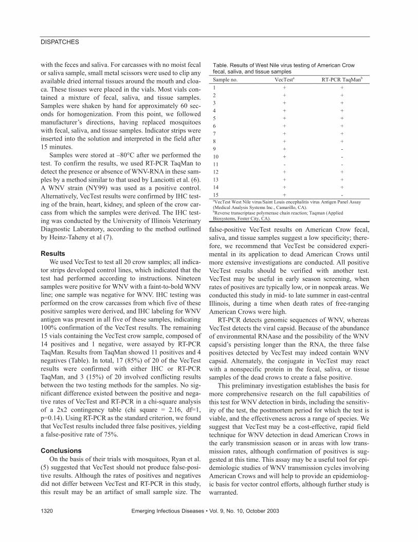
with the feces and saliva. For carcasses with no moist fecalor saliva sample, small metal scissors were used to clip anyavailable dried internal tissues around the mouth and cloa-ca. These tissues were placed in the vials. Most vials con-tained a mixture of fecal, saliva, and tissue samples.Samples were shaken by hand for approximately 60 sec-onds for homogenization. From this point, we followedmanufacturer’s directions, having replaced mosquitoeswith fecal, saliva, and tissue samples. Indicator strips wereinserted into the solution and interpreted in the field after15 minutes.
Samples were stored at –80°C after we performed thetest. To confirm the results, we used RT-PCR TaqMan todetect the presence or absence of WNV-RNA in these sam-ples by a method similar to that used by Lanciotti et al. (6).A WNV strain (NY99) was used as a positive control.Alternatively, VecTest results were confirmed by IHC test-ing of the brain, heart, kidney, and spleen of the crow car-cass from which the samples were derived. The IHC test-ing was conducted by the University of Illinois VeterinaryDiagnostic Laboratory, according to the method outlinedby Heinz-Taheny et al (7).
ResultsWe used VecTest to test all 20 crow samples; all indica-
tor strips developed control lines, which indicated that thetest had performed according to instructions. Nineteensamples were positive for WNV with a faint-to-bold WNVline; one sample was negative for WNV. IHC testing wasperformed on the crow carcasses from which five of thesepositive samples were derived, and IHC labeling for WNVantigen was present in all five of these samples, indicating100% confirmation of the VecTest results. The remaining15 vials containing the VecTest crow sample, composed of14 positives and 1 negative, were assayed by RT-PCRTaqMan. Results from TaqMan showed 11 positives and 4negatives (Table). In total, 17 (85%) of 20 of the VecTestresults were confirmed with either IHC or RT-PCRTaqMan, and 3 (15%) of 20 involved conflicting resultsbetween the two testing methods for the samples. No sig-nificant difference existed between the positive and nega-tive rates of VecTest and RT-PCR in a chi-square analysisof a 2x2 contingency table (chi square = 2.16, df=1,p=0.14). Using RT-PCR as the standard criterion, we foundthat VecTest results included three false positives, yieldinga false-positive rate of 75%.
ConclusionsOn the basis of their trials with mosquitoes, Ryan et al.
(5) suggested that VecTest should not produce false-posi-tive results. Although the rates of positives and negativesdid not differ between VecTest and RT-PCR in this study,this result may be an artifact of small sample size. The
false-positive VecTest results on American Crow fecal,saliva, and tissue samples suggest a low specificity; there-fore, we recommend that VecTest be considered experi-mental in its application to dead American Crows untilmore extensive investigations are conducted. All positiveVecTest results should be verified with another test.VecTest may be useful in early season screening, whenrates of positives are typically low, or in nonpeak areas. Weconducted this study in mid- to late summer in east-centralIllinois, during a time when death rates of free-rangingAmerican Crows were high.
RT-PCR detects genomic sequences of WNV, whereasVecTest detects the viral capsid. Because of the abundanceof environmental RNAase and the possibility of the WNVcapsid’s persisting longer than the RNA, the three falsepositives detected by VecTest may indeed contain WNVcapsid. Alternately, the conjugate in VecTest may reactwith a nonspecific protein in the fecal, saliva, or tissuesamples of the dead crows to create a false positive.
This preliminary investigation establishes the basis formore comprehensive research on the full capabilities ofthis test for WNV detection in birds, including the sensitiv-ity of the test, the postmortem period for which the test isviable, and the effectiveness across a range of species. Wesuggest that VecTest may be a cost-effective, rapid fieldtechnique for WNV detection in dead American Crows inthe early transmission season or in areas with low trans-mission rates, although confirmation of positives is sug-gested at this time. This assay may be a useful tool for epi-demiologic studies of WNV transmission cycles involvingAmerican Crows and will help to provide an epidemiolog-ic basis for vector control efforts, although further study iswarranted.
1320 Emerging Infectious Diseases • Vol. 9, No. 10, October 2003
DISPATCHES
Table. Results of West Nile virus testing of American Crow fecal, saliva, and tissue samples Sample no. VecTesta RT-PCR TaqManb 1 + + 2 + + 3 + + 4 + + 5 + + 6 + + 7 + + 8 + + 9 + - 10 + - 11 - - 12 + + 13 + + 14 + + 15 + - aVecTest West Nile virus/Saint Louis encephalitis virus Antigen Panel Assay (Medical Analysis Systems Inc., Camarillo, CA). bReverse transcriptase polymerase chain reaction; Taqman (Applied Biosystems, Foster City, CA).
AcknowledgmentsWe thank Arlo Raim, Adam Arnold, Gabe Hamer, and Chris
Warner for aid in capturing and tracking crows; Nina Krasavinand Hyun-Young Koo for laboratory assistance; John Andrewsfor interpreting immunohistochemistry results; and Jane Chladnyand the necropsy and histology technicians at the University ofIllinois Veterinary Medicine Diagnostic Laboratory for immuno-histochemistry testing.
This research was supported by McIntire-Stennis ForestryResearch Act Project, Centers for Disease Control and PreventionGrant U50/CCU520518-02 (RJN) and Department of NaturalResources Illinois Waste Tire Fund (RJN).
The authors declare no financial interests in the producers ofVecTest.
Ms. Yaremych conducted this research while workingtowards her master’s degree in the Department of NaturalResources and Environmental Sciences at the University ofIllinois. She is a doctoral candidate studying wildlife diseases inthe Department of Fisheries and Wildlife at Michigan StateUniversity. Her primary research interests are in wildlife ecologyand epidemiology.
References
1. Eidson M, Komar N, Sorhage F, Melson R, Talbot T, Mostashari F, etal. Crow deaths as a sentinel surveillance system for West Nile virusin the northeastern United States. Emerg Infect Dis 2001;7:615–20.
2. McLean RG, Ubico SR, Docherty DE, Hansen WR, Sielo L,McNamera TS. West Nile virus transmission and ecology in birds.Ann N Y Acad Sci 2001;951:54–7.
3. Guptill SC, Julian KG, Campbell GL, Price SD, Marfin AA. Early-season avian deaths from West Nile virus as warnings of humaninfection. Emerg Infect Dis 2003;9:483–4.
4. Komar N, Lanciotti R, Bowen R, Langevin S, Bunning M. Detectionof West Nile virus in oral and cloacal swabs collected from bird car-casses. Emerg Infect Dis 2002;8:741–2.
5. Ryan J, Dave K, Emmerich E, Fernandez B, Turell M, Johnson J, etal. Wicking assays for the rapid detection of West Nile and St. Louisencephalitis viral antigens in mosquitoes (Diptera: Culicidae). J MedEntomol 2003;40:95–9.
6. Lanciotti RS, Kerst AJ, Nasci RS, Godsey MS, Mitchell CJ, SavageHM, et al. Rapid detection of West Nile virus from human clinicalspecimens, field-collected mosquitoes, and avian samples by aTaqMan reverse transcriptase-PCR assay. J Clin Microbiol2000;38:4066–71.
7. Heinz-Taheny KM, Andrews JJ, Kinsel MJ, Pessier AP, PinkersonME, Lemberger KY, et al. West Nile virus infection in free-rangingsquirrels in Illinois. J Vet Diagn Invest. In press 2003.
Address for correspondence: Sarah A. Yaremych, Department ofFisheries and Wildlife, Michigan State University, 13 Natural ResourcesBldg., East Lansing, MI 48824, USA; fax: 517-432-1699; email:[email protected]
Emerging Infectious Diseases • Vol. 9, No. 10, October 2003 1321
DISPATCHES
The print journal is available at no charge to public health professionals
YES, I would like to receive Emerging Infectious Diseases.
Please print your name and businessaddress in the box and return by fax to404-371-5449 or mail to
EID EditorCDC/NCID/MS D611600 Clifton Road, NEAtlanta, GA 30333
Moving? Please give us your new address (in the box) and print the number of your oldmailing label here_________________________________________
�������������������� ���������������
SevereHistoplasmosis in
Travelers toNicaragua
Michelle Weinberg,* Julia Weeks,† Susan Lance-Parker,‡ Marc Traeger,*§
Steven Wiersma,§ Quyen Phan,¶ David Dennison,# Pia MacDonald,* ** Mark Lindsley,* Jeannette Guarner,* Patricia Connolly,†† Martin Cetron,*
and Rana Hajjeh*We investigated an outbreak of unexpectedly severe
histoplasmosis among 14 healthy adventure travelers fromthe United States who visited a bat-infested cave inNicaragua. Although histoplasmosis has rarely been report-ed to cause serious illness among travelers, this outbreakdemonstrates that cases may be severe among travelers,even young, healthy persons.
Histoplasmosis is a systemic infection caused by thedimorphic fungus, Histoplasma capsulatum. Infection
results from inhaling spores, usually through exposure to batand bird droppings in barnyards and caves. Although out-breaks have occurred after visits to bat-infested caves, histo-plasmosis has not been frequently recognized as travel-relat-ed and has rarely led to serious illness among young, healthytravelers. This fungus is endemic in the United States alongthe Ohio and Mississippi River valleys and many other partsof the world, particularly Latin America. Histoplasmosis isoften asymptomatic in endemic settings, but infection canresult in a spectrum of illness, ranging from mild influenza-like illness to acute pulmonary infection and disseminatedextrapulmonary disease. Immunocompromised persons andthe elderly are at greater risk for disseminated disease (1).This report describes a recent outbreak of histoplasmosisamong U.S. adventure travelers to Nicaragua that was asso-ciated with a high attack rate and hospitalizations amongpreviously healthy travelers.
The StudyIn June 2001, five persons with a febrile respiratory ill-
ness visited a community hospital. The patients wereamong 15 persons (age range 19–61 years; median 38years), residents of four states, who had participated in ageology-biology community college class trip toNicaragua. To determine the cause and describe character-istics of the outbreak, we interviewed trip participants andreviewed medical records to collect demographic informa-tion, clinical history, and activities during the trip. Basedon the partcipant’s exposure to a cave with bats and clini-cal characteristics suggestive of acute histoplasmosis,diagnostic testing for histoplasmosis was conducted.
A case of acute histoplasmosis was defined as illness ina person who tested positive by one of the following labo-ratory tests: serology, urine antigen, histopathology, or cul-ture. Immunodiffusion and complement- fixation serolog-ic tests were performed at the Centers for Disease Controland Prevention (CDC). The immunodiffusion test was con-sidered positive if an H or M band or both were detected.A complement-fixation titer to the yeast or mycelial anti-gens of >1:32 or a fourfold increase in the titer betweenacute- and convalescent-phase serum specimens was con-sidered evidence of acute infection. Acute-phase serumspecimens were obtained 2–3 weeks after exposure.Convalescent-phase serum specimens were collectedapproximately 3 months after exposure. One patient under-went bronchoalveolar lavage and bronchoscopy, and abiopsy specimen was obtained. Lung tissue was stainedwith hematoxylin and eosin and Gomori methenamine sil-ver stains. Bronchoalveolar lavage washings were culturedby conventional methods (2). Identification of the organ-ism as H. capsulatum was confirmed by GenProbe assay(GenProbe, San Diego, CA). Urine antigen tests, whichwere performed 2 weeks after exposure, are consideredpositive if >1 unit is detected (3,4).
The travelers began their trip on May 19 and returnedto the United States on May 30. During their trip, theyexplored rock quarries, visited a biological research sta-tion, swam in freshwater lakes, and were exposed to par-rots and insects. On May 21, a total of 14 of the 15 travel-ers entered a small cave that had previously been used as asilver mine. They remained in the cave for 10 minutes,during which time they saw several flying bats and batguano on the ground.
The infection rate was 100% among the 14 travelerswho entered the cave, and 12 (86%) were symptomatic. All14 patients had serologically confirmed infection. Of the14 patients, 12 (86%) had urine antigen tests (Table). Theonly traveler who did not enter the cave tested negative byserology and urine antigen.
Of the infected travelers, 8 (57%) were female. Theaverage incubation period was 11 days (range 10–12 days),
1322 Emerging Infectious Diseases • Vol. 9, No. 10, October 2003
DISPATCHES
*Centers for Disease Control and Prevention, Atlanta, Georgia,USA; †Archbold Urgent Care Center, Thomasville, Georgia, USA;‡Georgia Department of Human Resources, Division of PublicHealth, Atlanta, Georgia, USA; §Florida Department of Health,Tallahassee, Florida, USA; ¶Connecticut Department of PublicHealth, Hartford, Connecticut, USA; #Highlands-CashiersHospital, Highlands, North Carolina, USA; **North CarolinaDivision of Public Health, Raleigh, North Carolina, USA; and††Indiana University School of Medicine and HistoplasmosisReference Laboratory, Indianapolis, Indiana, USA
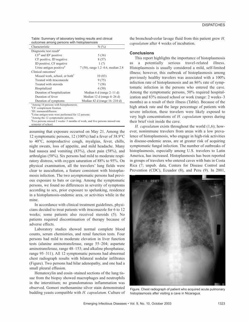
assuming that exposure occurred on May 21. Among the12 symptomatic persons, 12 (100%) had a fever of 38.9°Cto 40°C, nonproductive cough, myalgias, fever, chills,night sweats, loss of appetite, and mild headache. Manyhad nausea and vomiting (83%), chest pain (58%), andarthralgias (58%). Six persons had mild to moderate respi-ratory distress, with oxygen saturation of 88% to 95%. Onphysical examination, all the travelers’ lung fields wereclear to auscultation, a feature consistent with histoplas-mosis infection. The two asymptomatic persons had previ-ous exposure to bats or caving. Among the symptomaticpersons, we found no differences in severity of symptomsaccording to sex, prior exposure to spelunking, residencein a histoplamosis-endemic area, or activities while in themine.
In accordance with clinical treatment guidelines, physi-cians decided to treat patients with itraconazole for 6 to 12weeks; some patients also received steroids (5). Nopatients required discontinuation of therapy because ofadverse effects.
Laboratory studies showed normal complete bloodcounts, serum chemistries, and renal function tests. Fourpersons had mild to moderate elevation in liver functiontests (alanine aminotransferase, range 55–204; aspartateaminotransferase, range 48–153; and alkaline phosphatase,range 95–311). All 12 symptomatic persons had abnormalchest radiograph results with bilateral nodular infiltrates(Figure). Two persons had hilar adenopathy, and one had asmall pleural effusion.
Hematoxylin and eosin–stained sections of the lung tis-sue from the biopsy showed macrophages and neutrophilsin the interstitium; no granulomatous inflammation wasobserved. Gomori methenamine silver stain demonstratedbudding yeasts compatible with H. capsulatum. Culture of
the bronchoalveolar lavage fluid from this patient grew H.capsulatum after 4 weeks of incubation.
ConclusionsThis report highlights the importance of histoplasmosis
as a potentially serious travel-related illness.Histoplasmosis is usually considered a mild, self-limitedillness; however, this outbreak of histoplasmosis amongpreviously healthy travelers was associated with a 100%infection rate of histoplasmosis and an 86% rate of symp-tomatic infection in the persons who entered the cave.Among the symptomatic persons, 50% required hospital-ization and 83% missed school or work (range: 2 weeks–3months) as a result of their illness (Table). Because of thehigh attack rate and the large percentage of patients withsevere infection, these travelers were likely exposed tovery high concentrations of H. capsulatum spores duringtheir brief visit inside the cave.
H. capsulatum exists throughout the world (1,6); how-ever, nonimmune travelers from areas with a low preva-lence of histoplasmosis, who engage in high-risk activitiesin disease-endemic areas, are at greater risk of acquiringsymptomatic fungal infection. The number of outbreaks ofhistoplasmosis, especially among U.S. travelers to LatinAmerica, has increased. Histoplasmosis has been reportedin groups of travelers who entered caves with bats in CostaRica (7; unpub. data, Centers for Disease Control andPrevention (CDC), Ecuador (8), and Peru (9). In 2001,
Emerging Infectious Diseases • Vol. 9, No. 10, October 2003 1323
DISPATCHES
Table: Summary of laboratory testing results and clinical outcomes among persons with histoplasmosis Characteristic N (%) Diagnostic test resulta
CFb and IDc positive 5 (36) CF positive, ID negative 8 (57) ID positive, CF negative 1 (7) Urine antigen positived 7 (58), range 1.2–4.6, median 2.8
Clinical outcomese Missed work, school, or bothf 10 (83) Treated with itraconazole 9 (75) Treated with steroids 7 (58) Hospitalized 6 (50) Duration of hospitalization Median 6 d (range 2–11 d) Duration of fever Median 12 d (range 4–26 d) Duration of symptoms Median 42 d (range 16–210 d)
aAmong 14 persons with histoplasmosis. bCF: complement fixation. cID: immunodiffusion. dUrine antigen tests were performed for 12 persons. eAmong the 12 symptomatic persons. fFive persons missed 2 weeks–3 months of work, and five persons missed one semester of school.
Figure. Chest radiograph of patient who acquired acute pulmonaryhistoplasmosis after visiting a cave in Nicaragua.
more than 200 college students became infected withhistoplasmosis during a spring break trip to Acapulco,Mexico (10,11). Outbreaks and isolated cases of histoplas-mosis have also been described among non–-U.S. travelersto Latin America and other areas of the world (12–19).
Clinicians should consider fungal pathogens whenevaluating returning travelers who have a febrile respirato-ry syndrome. The differential diagnosis of acute febrilerespiratory illness in international adventure travelers isextensive and includes legionellosis, psittacosis, lep-tospirosis, schistosomiasis, histoplasmosis, coccid-ioidomycosis, influenza, parainfluenza, mycoplasma,dengue, and malaria.
The timely diagnosis of histoplasmosis in travelers maybe particularly challenging for clinicians evaluating onesporadic case or a patient involved in an undetected multi-state outbreak. Antibody response can take 4–6 weeks todevelop, reducing its usefulness in the acute setting andrequiring the collection of a convalescent-phase specimenfor confirmation. Antibodies may also persist for 5 years,making it difficult to distinguish between prior and recentinfection without obtaining a convalescent-phase speci-men. The sensitivity of immunodiffusion is 72% to 85%,while that of complement fixation is 80% to 90% (20,21).The antigen test can be used for urine, cerebrospinal fluid,serum, and bronchoalveolar lavage fluid, and results areavailable in 24 hours. This test had been used most exten-sively in AIDS patients; however, it is less sensitive innonimmunocompromised persons with acute pulmonaryinfection. Sensitivity of the urine antigen test for detectionof acute pulmonary infection ranges from 44% to 75% andincreases when performed early after the onset of symp-toms (3,4).
In this outbreak, 80% of the travelers were prescribedappropriate malaria chemoprophylaxis during a pretravelassessment. However, most clinicians may not havethought to counsel them about histoplasmosis risk and pre-vention. During pretravel visits, clinicians should reviewpatients’ itineraries for possible fungal exposures andshould counsel them to avoid entering caves, especiallythose known to be bat infested. Since travelers may notknow whether their itinerary includes visits to bat-infestedcaves, adventure travelers and persons who are at high riskfor severe histoplasmosis infection, such as immunocom-promised travelers (especially persons with AIDS), shouldbe counseled to avoid possible exposures, either by avoid-ing caves or by using special protective masks. TheNational Institute for Occupational Safety and Health haspublished guidelines for special masks that can be used toreduce occupational and other exposures (e.g., among spe-lunkers). Unfortunately, these special masks are bulky andmay be impractical for many travelers. (22). While mostnonimmunocompromised patients with acute pulmonary
histoplasmosis improve without treatment, persons withhypoxemia, diffuse pulmonary histoplasmosis, or severeillness requiring hospitalization may benefit from antifun-gal therapy and, in some cases, corticosteroids (5).
The contribution of histoplasmosis to infectious diseaseillness in travelers is likely underestimated. As adventuretravel becomes increasingly accessible, especially amongpersons at high risk, immunocompromised persons, andthe elderly, histoplasmosis and other fungal infections intravelers may become more common. Enhanced surveil-lance for histoplasmosis and other fungal infections bypublic health officials, combined with heightened aware-ness by clinicians who are evaluating symptomatic, post-travel patients, can lead to greater understanding of theepidemiology of fungal infections among travelers and,ultimately, to improved prevention measures.
Acknowledgments We thank Juan Jose Amador of Nicaraguan Ministry of
Health, Phyllis Kozarsky, the members of the group who partici-pated in this trip, and the Histoplasmosis Reference Laboratory,which performed the urine antigen testing, for their help with thisinvestigation, and Ava Navin for editorial assistance.
When the work was done for this outbreak investigation,Ms. Connolly was associated with Indiana University. She is cur-rently an employee of MiraVista Diagnostics, Indianapolis,Indiana.
Dr. Weinberg is the team leader for Migrant and BorderHealth in the Surveillance and Epidemiology Branch, Division ofGlobal Migration and Quarantine, Centers for Disease Controland Prevention. Her research interests include internationalhealth, particularly issues in Hispanic populations. She has coor-dinated the binational activities of the U.S–Mexico BorderInfectious Diseases Surveillance network since 1999.
References
1. Deepe G. Histoplasma capsulatum. In: Mandel GL, Bennett JE, DolinR, editors. Principles and practice of infectious diseases. 5th ed. NewYork: Churchill Livingston; 2000. p. 2718–33.
2. Larone DH, Mitchell TG, Walsh TJ. Histoplasma, blastomyces, coc-cidioides, and other dimorphic fungi causing systemic mycoses. In:Murray PR, editor. Manual of clinical microbiology. 7th ed.Washington: ASM Press; 1999. p. 1259–68.
3. Williams B, Fojtasek M, Connolly-Stringfield P, Wheat J. Diagnosisof histoplasmosis by antigen detection during an outbreak inIndianapolis, Ind. Arch Pathol Lab Med 1994;118:1205–8.
4. Wheat LJ, Kohler RB, Tewari RP. Diagnosis of disseminated histo-plasmosis by detection of Histoplasma capsulatum antigen in serumand urine specimens. N Engl J Med 1986;314:83–8.
5. Wheat J, Sarosi G, McKinsey D, Hamill R, Bradsher R, Johnson P, etal. Practice guidelines for the management of patients with histoplas-mosis. Clin Infect Dis 2000;30:688–95.
6. Wilson ME. A world guide to infections: diseases, distribution, diag-nosis. Oxford: Oxford University Press; 1991. p.190.
1324 Emerging Infectious Diseases • Vol. 9, No. 10, October 2003
DISPATCHES
7. Centers for Disease Control and Prevention. Cave-associated histo-plasmosis—Costa Rica. MMWR Morb Mortal Wkly Rep1988;37:312–3.
8. Valdez H, Salata RA. Bat-associated histoplasmosis in returning trav-elers: case presentation and description of a cluster. J Travel Med1999;6:258–60.
9. Nasta P, Donisi A, Cattane A, Chiodera A, Cassari S. Acute histoplas-mosis in spelunkers returning from Mato Grosso, Peru. J Travel Med1997;4:176–8.
10. Centers for Disease Control and Prevention. Outbreak of acute respi-ratory febrile illness among college students—Acapulco, Mexico,March 2001. MMWR Morb Mortal Wkly Rep 2001;50:261–2.
11. Centers for Disease Control and Prevention. Update: outbreak ofacute febrile respiratory illness among college students—Acapulco,Mexico, March 2001. MMWR Morb Mortal Wkly Rep2001;50:359–60.
12. Bonnet D, Balandraud P, Lonjon T, Rey P, Van de Walle JP, Cador L,et al. Round pulmonary lesions after returning from French Guyana.Six cases of American pulmonary histoplasmosis. Med Trop (Mars)1995;55:55–60.
13. Buxton JA, Dawar M, Wheat LJ, Black WA, Ames NG, Mugford M,et al. Outbreak of histoplasmosis in a school party that visited a cavein Belize: role of antigen testing in diagnosis. J Travel Med2002;9:48–50.
14. Erkens K, Lademann M, Tintelnot K, Lafrenz M, Kaben U, ReisingerEC. Histoplasmosis group disease in bat researchers returning fromCuba. Dtsch Med Wochenschr 2002;127:21–25.
15. Fujio J, Nishimura K, Miyaji M. Epidemiological survey of theimported mycoses in Japan. Nippon Ishinkin Gakkai Zasshi1999;40:103–9.
16. Gascon J, Torres JM, Luburich P, Ayuso JR, Xaubet A, Corachan M.Imported histoplasmosis in Spain. J Travel Med 2000;7:89–91.
17. Hatakeyama S, Kashiyama T, Takechi A, Sasaki S, Akamatsu E.Cave-associated acute pulmonary histoplasmosis in two Japanesereturning from Mexico. Nihon Kokyuki Gakkai Zasshi2001;39:293–7.
18. Hirsch D, Leupold W, Rupprecht E. Pulmonary histoplasmoma aftertravel abroad. Pneumologie 1996;50:242–4.
19. Suzaki A, Kimura M, Kimura S, Shimada K, Miyaji M, Kaufman L.An outbreak of acute pulmonary histoplasmosis among travelers to abat-inhabited cave in Brazil. Kansenshogaku Zasshi 1995;69:444–9.
20. Morrison CJ, Lindsley MD. Serological approaches to the diagnosisof invasive fungal infections. In: Calderone R, Cihlar R, editors.Fungal pathogenesis: principles and practice. New York: MarcelDekker; 2001. p. 667–716.
21. Reiss E, Kaufman L, Kovacs JA, Lindsley MD. Clinical immunomy-cology. In: Rose NR, Hamilton RG, Detrick B, editors. Manual ofclinical laboratory immunology. Sixth edition. Washington: AmericanSociety for Microbiology; 2002. p. 559–83.
22. Centers for Disease Control and Prevention. National Institute forOccupational Safety and Health. Histoplasmosis: protecting workersat risk, revised guidelines for preventing histoplasmosis; 1997.Available from: URL: http://www.cdc.gov/niosh/97-146.html
Address for correspondence: Michelle Weinberg, Division of GlobalMigration and Quarantine, Centers for Disease Control and Prevention,1600 Clifton Rd, Mailstop E03, Atlanta, Georgia, USA, 30333; fax: 404-498-1633; email: [email protected]
Emerging Infectious Diseases • Vol. 9, No. 10, October 2003 1325
DISPATCHES
Mayaro Virus inWild Mammals,French Guiana
Benoît de Thoisy,* Jacques Gardon,* Rosa Alba Salas,† Jacques Morvan,*
and Mirdad Kazanji*A serologic survey for Mayaro virus (Alphavirus,
Togaviridae) in 28 wild nonflying forest mammal species inFrench Guiana showed a prevalence ranging from 0% to52% and increasing with age. Species active during the dayand those who spent time in trees were significantly moreinfected, results consistent with transmission implicatingdiurnal mosquitoes and continuous infectious pressure.
The Mayaro virus is a zoonotic Alphavirus (familyTogaviridae) responsible for epidemics of febrile
exanthematous illness in Latin America and Amazonia (1).Although the death rate is low, Mayaro fever is a majorarboviral infection relevant to public health in rural popu-lations, with an increasing incidence of human cases in theAmazonian basin following ecosystem disturbances (2).The activity of the Mayaro virus can be described as a con-stantly moving wave, transmitted among susceptible verte-brates by sylvatic Culicidae mosquitoes (3). Haemagogusmosquitoes are the main vectors; they are diurnal canopydwellers common in high pristine rainforests (4). Severalvertebrate hosts, mainly primates, rodents, and birds, areconsidered to be reservoirs, although their exact role inmaintaining the virus is insufficiently understood (2).Previous serologic surveys have shown high prevalencerates in primates, but the virus has been isolated only fromlizards and one marmoset (5,6); experimental inoculationof marmosets resulted in a short period of viremia,although the titer was nevertheless probably high enoughto infect vectors (4). Human disease outbreaks could occurwhen birds or vectors introduce the virus into rural areaswith high densities of both Haemagogus and potentialreservoirs.
This emerging disease has recently been reported inFrench Guiana, a French administrative unit on the north-ern coast of South America (6). To investigate the diversi-ty of the reservoir species, a serologic survey for Mayarovirus was conducted on 28 nonflying mammalian rainfor-est species, in a total of 579 animals; no previous surveysin wild vertebrate species has included so many samples.
Investigations on antibody responses in potential hosts areimportant first steps for understanding viral dynamics (7).Since the infectious process plays a major role in wildlifeecology (8), we used Mayaro infection as a case study toinvestigate the correlation between ecologic and biologicpatterns of potential hosts and their susceptibility to infec-tion.
The StudyBlood samples were collected in 1994–95 during a
wildlife rescue operation at the Petit Saut hydroelectricdam site (4°55′ N, 53°05′ W), French Guiana (9). Theoverall habitat of all species was pristine high rainforest.Each serum sample was tested by hemagglutination inhibi-tion (HI) for antibodies to Mayaro and Tonate (Venezualanequine encephalitis complex) viruses. Serum samples withtiters >1:20 were confirmed by seroneutralization at a 1:20dilution (10). Briefly, equal volumes of diluted serum weremixed with a Mayaro virus suspension containing 100 tis-sue culture infectious dose 50 per 0.1 mL. The reactionwas incubated at 37°C for 1 h. As control of the test doseof virus, the working dilution was successively diluted 10-fold. One hundred microliters of the serum-virus mixture,control virus, and diluted serum was inoculated in dupli-cate in monolayers of Vero-E6 cell line and incubated at37°C for 5 to 7 days. A positive reaction by neutralizationwas considered with the total inhibition of the cytopathiceffect in the cell monolayer induced by Mayaro virus.Analysis of variance (p<0.05 considered significant) wasused to study correlations between ecologic patterns andarcsine-transformed seropositivity of species (XlStat-Pro,Addinsoft, Paris, France). The following ecologic parame-ters were considered: vertical use of space (arboreal, ter-restrial, and both); density (low <10 individualanimals/km2; medium 10–20 individual animals, and high>20 individual animals/km2); rhythm of activity (strictlydiurnal, strictly nocturnal, or both); and lifespan. Becauselifespan data in the wild are not available for most species,we used last reproduction age as an indicator, and we clas-sified lifespan as short (last reproduction age <5 y), inter-mediate (last reproduction age between 5 and 15 y) , andlong (last reproduction age >15 y). The data used are sum-marized in Table 1.
All the serum samples were negative for Tonate virus,which suggests that no cross-reaction occurred withMayaro virus. The possibility of cross-reaction with Unavirus, a closely genetically related Togavirus, was not con-sidered since Una is known only in subtropical areas (11)and open habitats (12,13). The seroprevalence rates ofMayaro virus, on the basis of seroneutralization confirma-tion, ranged from 0% to 52% and reached 80% in twospecies (Table 2); however, only a limited sample wasavailable. Gender had no apparent effect on the frequency
1326 Emerging Infectious Diseases • Vol. 9, No. 10, October 2003
DISPATCHES
*Institut Pasteur de la Guyane, French Guiana; and †CaribbeanEpidemiology Center (CAREC), Port of Spain, Republic of Trinidadand Tobago
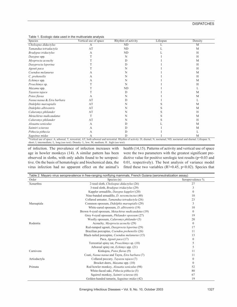
of infection. The prevalence of infection increases withage in howler monkeys (14). A similar pattern has beenobserved in sloths, with only adults found to be seroposi-tive. On the basis of hematologic and biochemical data, thevirus infection had no apparent effect on the animal’s
health (14,15). Patterns of activity and vertical use of spacewere the two parameters with the greatest significant pre-dictive value for positive serologic test results (p=0.03 and0.01, respectively). The best analysis of variance modelfitted these two variables (R2=0.45, p<0.02). Species that
Emerging Infectious Diseases • Vol. 9, No. 10, October 2003 1327
DISPATCHES
Table 1. Ecologic data used in the multivariate analysis Species Vertical use of space Rhythm of activity Lifespan Density Choloepus didactylus A ND L M Tamandua tetradactyla AT ND L M Bradypus tridactylus A ND L H Dasypus spp. T N I H Myoprocta acouchy T D I M Dasyprocta leporina T D I H Agouti paca T N I H Coendou melanurus A N I M C. prehensilis A N I H Echimys spp. A N I M Proechimys sp. T N I H Mazama spp. T ND I L Tayassu tajacu T D I M Potos flavus A N I M Nasua nasua & Eira barbara AT D I L Didelphis marsupialis AT N S M Didelphis albiventris AT N S M Caluromys philander AT N S H Metachirus nudicaudatus T N S M Caluromys philander AT N S H Alouatta seniculus A D L M Saimiri sciureus A D I M Pithecia pithecia A D I L Saguinus midas A D I M aVertical use of space: A, arboreal, T, terrestrial, AT, both arboreal and terrestrial. Rhythm of activity: D, diurnal; N, nocturnal; ND, nocturnal and diurnal. Lifespan: S, short; I, intermediate; L, long (see text). Density: L, low; M, medium; H , high (see text).
Table 2. Mayaro virus seroprevalence in free-ranging nonflying mammals, French Guiana (seroneutralization assay) Order Species (n) Seroprevalence % Xenarthra 2-toed sloth, Choloepus didactylus (26) 27 3-toed sloth, Bradypus tridactylus (29) 3 Kappler armadillo, Dasypus kappleri (20) 0 Nine-banded armadillo, D. novemcinctus (40) 10 Collared anteater, Tamandua tetradactyla (26) 23 Marsupiala Common opossum, Didelphis marsupialis (29) 3 White-eared opossum, D. albiventris (19) 10 Brown 4-eyed opossum, Metachirus nudicaudatus (19) 0 Grey 4-eyed opossum, Philander opossum (27) 19 Woolly opossum, Caluromys philander (5) 20 Rodentia Acouchy, Myoprocta acouchy (29) 0 Red-rumped agouti, Dasyprocta leporina (29) 17 Brazilian porcupine, Coendou prehensilis (26) 11 Black-tailed porcupine, Coendou melanurus (15) 13 Paca, Agouti paca (17) 0 Terrestrial spiny rat, Proechimys sp. (18) 5 Arboreal spiny rat, Echimys spp. (21) 5 Carnivora Kinkajou, Potos flavus (9) 11 Coati, Nasua nasua and Tayra, Eira barbara (7) 11 Artiodactyla Collared peccary, Tayassu tajacu (7) 0 Brocket deers, Mazama spp. (10) 0 Primata Red howler monkey, Alouatta seniculus (98) 52 White-faced saki, Pithecia pithecia (5) 80 Squirrel monkey, Saimiri sciureus (6) 67 Golden-handed tamarin, Saguinus midas (42) 19
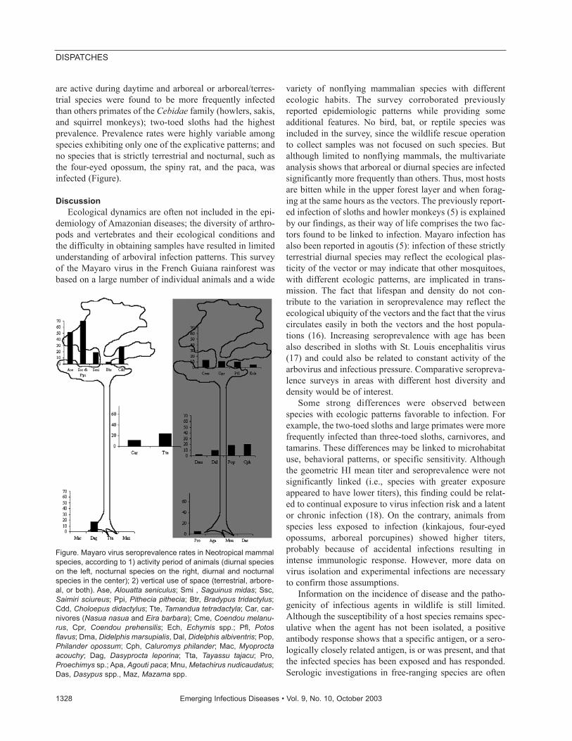
are active during daytime and arboreal or arboreal/terres-trial species were found to be more frequently infectedthan others primates of the Cebidae family (howlers, sakis,and squirrel monkeys); two-toed sloths had the highestprevalence. Prevalence rates were highly variable amongspecies exhibiting only one of the explicative patterns; andno species that is strictly terrestrial and nocturnal, such asthe four-eyed opossum, the spiny rat, and the paca, wasinfected (Figure).
DiscussionEcological dynamics are often not included in the epi-
demiology of Amazonian diseases; the diversity of arthro-pods and vertebrates and their ecological conditions andthe difficulty in obtaining samples have resulted in limitedunderstanding of arboviral infection patterns. This surveyof the Mayaro virus in the French Guiana rainforest wasbased on a large number of individual animals and a wide
variety of nonflying mammalian species with differentecologic habits. The survey corroborated previouslyreported epidemiologic patterns while providing someadditional features. No bird, bat, or reptile species wasincluded in the survey, since the wildlife rescue operationto collect samples was not focused on such species. Butalthough limited to nonflying mammals, the multivariateanalysis shows that arboreal or diurnal species are infectedsignificantly more frequently than others. Thus, most hostsare bitten while in the upper forest layer and when forag-ing at the same hours as the vectors. The previously report-ed infection of sloths and howler monkeys (5) is explainedby our findings, as their way of life comprises the two fac-tors found to be linked to infection. Mayaro infection hasalso been reported in agoutis (5): infection of these strictlyterrestrial diurnal species may reflect the ecological plas-ticity of the vector or may indicate that other mosquitoes,with different ecologic patterns, are implicated in trans-mission. The fact that lifespan and density do not con-tribute to the variation in seroprevalence may reflect theecological ubiquity of the vectors and the fact that the viruscirculates easily in both the vectors and the host popula-tions (16). Increasing seroprevalence with age has beenalso described in sloths with St. Louis encephalitis virus(17) and could also be related to constant activity of thearbovirus and infectious pressure. Comparative seropreva-lence surveys in areas with different host diversity anddensity would be of interest.
Some strong differences were observed betweenspecies with ecologic patterns favorable to infection. Forexample, the two-toed sloths and large primates were morefrequently infected than three-toed sloths, carnivores, andtamarins. These differences may be linked to microhabitatuse, behavioral patterns, or specific sensitivity. Althoughthe geometric HI mean titer and seroprevalence were notsignificantly linked (i.e., species with greater exposureappeared to have lower titers), this finding could be relat-ed to continual exposure to virus infection risk and a latentor chronic infection (18). On the contrary, animals fromspecies less exposed to infection (kinkajous, four-eyedopossums, arboreal porcupines) showed higher titers,probably because of accidental infections resulting inintense immunologic response. However, more data onvirus isolation and experimental infections are necessaryto confirm those assumptions.
Information on the incidence of disease and the patho-genicity of infectious agents in wildlife is still limited.Although the susceptibility of a host species remains spec-ulative when the agent has not been isolated, a positiveantibody response shows that a specific antigen, or a sero-logically closely related antigen, is or was present, and thatthe infected species has been exposed and has responded.Serologic investigations in free-ranging species are often
1328 Emerging Infectious Diseases • Vol. 9, No. 10, October 2003
DISPATCHES
Figure. Mayaro virus seroprevalence rates in Neotropical mammalspecies, according to 1) activity period of animals (diurnal specieson the left, nocturnal species on the right, diurnal and nocturnalspecies in the center); 2) vertical use of space (terrestrial, arbore-al, or both). Ase, Alouatta seniculus; Smi , Saguinus midas; Ssc,Saimiri sciureus; Ppi, Pithecia pithecia; Btr, Bradypus tridactylus;Cdd, Choloepus didactylus; Tte, Tamandua tetradactyla; Car, car-nivores (Nasua nasua and Eira barbara); Cme, Coendou melanu-rus, Cpr, Coendou prehensilis; Ech, Echymis spp.; Pfl, Potosflavus; Dma, Didelphis marsupialis, Dal, Didelphis albiventris; Pop,Philander opossum; Cph, Caluromys philander; Mac, Myoproctaacouchy; Dag, Dasyprocta leporina; Tta, Tayassu tajacu; Pro,Proechimys sp.; Apa, Agouti paca; Mnu, Metachirus nudicaudatus;Das, Dasypus spp., Maz, Mazama spp.
limited to transversal surveys in several animals at a singletime. The levels and distribution of seropositivity and titerscan, however, be used for a better understanding of bothvirus dynamics and host susceptibility. In infections withalphaviruses, the viremia is intense but short (7), but thediversity of the potential reservoirs may compensate forthis short period. When the virus is introduced into a newarea, many individual animals can be infected rapidly. Thelow individual excretion rate is compensated for by thenumber of animals possibly affected by the virus. This viralstrategy is completely opposite to that of the Arenaviridae,where the reservoirs are much less diverse (usually a singlespecies), but the hosts occur at high density and viral excre-tion is long-lasting (19). Other arboviruses with an ecolog-ic niche similar to that of the Mayaro virus may also havea wide diversity of potential reservoirs. The Mayaro virusis active mainly in forests, but Haemagogus spp. can flyover large areas. Howler monkeys, tamarins, squirrel mon-keys, and agoutis are also still common in the vicinity ofthe main cities of French Guiana, and periurban speciesmay also be infected and act as reservoirs. Moreover,alphaviruses are prone to be hosted by a large range of vec-tors (3) and the risk for new epidemic patterns, comparableto the emergence of periurban Chikungunya cycle inAfrica, should be considered (1). Demographic pressure isresulting in increasing human contact with forest habitatsand fauna; the predictable health consequences of this evo-lution have been emphasized in parasites (20) and may alsobe applicable in arboviruses.
AcknowledgmentsWe are grateful to J. Lelarge and E. Bourreau for technical
help.
The serum samples were collected during the ProgrammeFaune Sauvage, funded by Electricité de France–Centre Nationald’Equipement Hydroélectrique.
Dr. de Thoisy is a veterinarian as well as a doctoral student inthe retrovirology laboratory of the Pasteur Institute of FrenchGuiana. His areas of expertise are Neotropical wildlife diseases,virology, and ecology.
References 1. Tesh RB, Watts DM, Russel KL, Damodaran C, Calampa C, Cabezas
C, et al. Mayaro virus disease: an emerging mosquito-borne zoonosisin tropical South America. Clin Infect Dis 1999;28:67–73.
2. Vasconcelos PFC, Travassos APA, Rodrigues SG, Travassos da RosaES, Dégallier N, Travassos da Rosa JFS. Inadequate management ofnatural ecosystem in the Brazilian Amazon region results in the emer-gence and reemergence of arbovirus. Cad Saude Publica2001;17(suppl):155–64.
3. Pinheiro FP, LeDuc JW. Mayaro virus disease. In: Monath TP, editor.The arboviroses: epidemiology and ecology. Vol. 3. Boca Raton (FL):CRC Press; 1998. p.137–50.
4. Hoch AL, Peterson NE, Le Duc JN, Pinheiro FP. An outbreak ofMayaro virus disease in Belterra, Brazil. III. Entomological and eco-logical studies. Am J Trop Med Hyg 1981;30:689–98.
5. Woodall JP. Virus research in Amazonia. Atas do Simposia sobre aBiota Amazónia 1967; 6(Patologia):31–63.
6. Talarmin A, Chandler LJ, Kazanji M, de Thoisy B, Debon P, LelargeJ, et al. Mayaro virus fever in French Guiana: isolation, identificationand seroprevalence. Am J Trop Med Hyg 1998;59:452–6.
7. Peters CJ, Dalrymple JM. Alphaviruses. In: Fields BN, Knipe DM,editors. Virology. 2nd edition. New York: Raven Press; 1990.
8. Spalding MG, Forester DJ. Disease monitoring of free-ranging andreleased wildlife. Journal of Zoo and Wildlife Medicine1993;24:271–81.
9. Vié JC. Wildlife rescues: the case of Petit Saut hydroelectric dam inFrench Guiana. Oryx 1999;33:115–26.
10. Lindsley HS, Calisher CH, Mathews JH. Serum dilution neutraliza-tion test for California group virus identification and serology. J ClinMicrobiol 1976;4:503–10.
11. Monath TP, Sabattini MS, Pauli R, Daffner JF, Mitchell CJ, BowerGS, et al. Arbovirus investigations in Argentina, 1977–1980. IV.Serologic surveys and sentinel equine program. Am J Trop Med Hyg1985;34:966–75.
12. Walder R, Suarez OM, Calisher CH. Arbovirus studies insouthwestern Venezuela during 1973–1981. II. Isolations and furtherstudies of Venezuelan and eastern equine encephalitis, Una, Itaqui,and Moju viruses. Am J Trop Med Hyg 1984;33:483–91.
13. Haas RA, Arron-Leeuwin AE. Arboviruses isolated from mosquitoesand man in Surinam. Trop Geogr Med 1975;27:409–12.
14. de Thoisy B, Vogel I, Reynes JM, Pouliquen JF, Carme B, Kazanji M,et al. Health evaluation of translocated free-ranging primates inFrench Guiana. Am J Primatol 2001;54:1–16.
15. Vogel I, Vie JC, de Thoisy B, Moreau B. Hematological and serumchemistry profiles of free-ranging southern two-toed sloths in FrenchGuiana. J Wildlife Dis 1999;35:531–5.
16. Scott ME. The impact of infection and disease on animal populations:implications for conservation biology. Conserv Biol 1988;2:40–56.
17. Seymour C, Peralta PH, Montgomery GG. Serological evidence ofnatural Togavirus infections in Panamanian sloths and other verte-brates. Am J Trop Med Hyg 1983;32:854–61.
18. Fischer-Tenhagen C, Hamblin C, Quandt S, Frölich K. Serosurvey forselected infectious disease agents in free-ranging black and white rhi-noceros in Africa. J Wildl Dis 2000;36:316–23.
19. Bowen MD, Peters CJ, Nichol ST. Phylogenetic analysis of theArenaviridae: patterns of virus evolution and evidence for cospecia-tion between Arenaviruses and their rodent hosts. Mol PhylogenetEvol 1997;8:301–16.
20. Fandeur T, Volney B, Peneau C, de Thoisy B. Monkeys of the rain-forest in French Guiana are natural reservoirs for P. brasilianum/P.malariae malaria. Parasitology 2000;120:11–21.
Address for correspondence: M. Kazanji, Laboratoire de rétrovirologie,Institut Pasteur de la Guyane, BP 6010, 23 avenue Pasteur, F-97306Cayenne cedex, French Guiana; fax: +(594) 594 309 416; email:[email protected]
Emerging Infectious Diseases • Vol. 9, No. 10, October 2003 1329
DISPATCHES
The EuropeanCommission’s Task
Force onBioterrorism
Anders Tegnell,* Philippe Bossi,* Agoritsa Baka,*Frank Van Loock,* Jan Hendriks,*
Solvejg Wallyn,* and Georgios Gouvras*
In response to the increased threat of bioterrorism, atask force on health security was established in theEuropean Commission. Task force members address abroad range of issues related to preparedness for andresponse to bioterrorist events and seek to bring about agreater collaboration between the European Union mem-ber states.
The deliberate release of anthrax in the United States ofAmerica in September and October 2001 completely
changed the international perception of the risk of bioter-rorism. Before that time, public health preparedness forbioterrorism was not a political priority in the EuropeanUnion (EU) member states. Preparedness and responseplans and actions have since been given a higher priorityon EU member agendas. Bioterrorism also initiated a dis-cussion on the need to improve preparedness through rein-forcing existing public health structures that are in chargeof monitoring and controlling diseases.
On October 19, 2001, at the European Council inGhent, Belgium, the Council of Ministers of the EU andthe European Commission (EC) were asked by the headsof state and government to prepare a program to improvethe cooperation between member states for the evaluationof risks, alerts, and intervention, the storage of the neces-sary means, and the collaboration in the field of research.
In parallel with these developments at the Europeanlevel, many well-focused national activities were also car-ried out. Countries reexamined their preparedness plansand adapted them to the new threats, and legislation need-ed to implement countermeasures was reviewed. Thenational authorities also discussed the necessity of stock-piling medicines and other medical supplies and mostupgraded their stocks of vaccines and antibiotics, especial-ly smallpox vaccine. To improve the ability to recognize adeliberate release, countries developed training programsfor different kinds of first responders.
On the initiative of the European Commissioner forHealth and Consumer Protection, the Health SecurityCommittee (HSC) was set up with representatives of theministries of health from each member state. Following aproposal by the commission, the HSC agreed on December17, 2001, on a program for cooperation on preparednessand response to biologic and chemical agent attacks (orHealth Security Programme). The program comprises 25actions grouped under four objectives (available from:URL: http://europa.eu.int/comm/health/ph_threats/Bio-terrorisme/bioterrorisme_en.htm). The overall goal is toimprove cooperation between the member states with theaid of the commission and to facilitate collaborationbetween the different national authorities involved in pub-lic health preparedness for bioterrorism. During thisprocess, gaps in resources and methods are identified andprojects initiated to fill these gaps. As is standard practicein many sectors of activity at EU level, existing examplesof preparedness in the member states are shared and can beextended or adapted for use throughout the EU. Projectsand activities under the health security program are fund-ed through existing funds, especially from the budget fore-seen for the EU’s public health programs. Other funds suchas from the sixth framework program for research couldalso be used for activities of relevance to the health secu-rity program.
Since the beginning of May 2002, a 15-member strongtask force has been set up by the Directorate-General ofHealth and Consumer Protection, comprising nine nation-al experts seconded from different institutions in the EUcountries and six commission officials. The task forceincludes representatives from the Robert Koch Instituteand the Paul Erhlich Institute (Germany), the ScientificInstitute of Public Health (Belgium), the Swedish Institutefor Infectious Diseases Control (SMI, Sweden), theNational Institute of Public Health and the Environment(RIVM, Netherlands), the Hellenic Institute for InfectiousDisease Control (Greece), the Italian National Institute forHealth (Italy), as well as the Pitié-Salpêtrière Hospital inParis (France). The task force’s main objective is to imple-ment the health security program. To this end, membershave initiated further activities with key players in themember states, some of which are described below.
One of the initial priority activities was the setting up ofa mechanism for rapid information exchange, consultation,and coordination on all aspects dealing with bioterrorism.All designated authorities of the EU member states havenow been connected through this network by means ofemail, fax, and telephone. This tool makes it possible forthe task force and the nine member states to disseminateinformation rapidly about suspected or confirmed inci-dents and for national authorities to evaluate measuresplanned with their implications for EU and for other mem-
1330 Emerging Infectious Diseases • Vol. 9, No. 10, October 2003
DISPATCHES
*Task Force on Biological and Chemical Agent Threats, EuropeanCommission, Luxembourg, Belgium
ber states. The task force is available 24 hours a day, 7 daysa week to facilitate the process. By May 2003, the systemhad been used in two major EU-wide exercises and fornine alerts about suspicious events communicated by themember states.
The early detection of a deliberate release or bioterror-ist attack is an essential part of a program of preparedness.One essential tool to make this possible will be the regularsurveillance activities already in place at national andEuropean level. Certain modifications to these systems,such as the addition of certain diseases considered as hav-ing a high potential for deliberate release, would have to bedone to make the systems better adapted to the new chal-lenges of bioterrorism. To achieve this, the CommunityNetwork on Communicable Diseases (the already existingmechanism for EU collaboration in the areas of infectiousdisease surveillance and control) extended its list of dis-eases to be reported, to include additional threats such astularemia, anthrax, Q fever, and smallpox.
When the task force was established, several lists ofagents that could be involved in a biologic attack and con-sidered of major bioterrorist threat had been establishedand published. Against this background, an approach moresuitable to the demands for focused action in Europe wasdeveloped though a matrix model designed to provide anevaluation of the public health impact of any given agentthat could be used in bioterrorism. The matrix model mayalso be used to identify areas where the public healthresponse needed strengthening for any given agent. Thismeans that for a given type of action a different set ofagents would emerge as deserving the highest attention.The matrix model is currently being validated and has pro-duced lists targeting specific areas of public health pre-paredness, which are currently being reviewed by mem-bers of the HSC. Using this model, identifying the publichealth impact of new threats should be possible, such assevere acute respiratory syndrome (SARS) (once it hasbeen better characterized), multidrug-resistant tuberculo-sis, or any other new or drastically changed disease-caus-ing agent.
The clinical management, including recognition, ofmany of the diseases caused by the bioterrorist agents islikely to be difficult, since they are, to a large extent,unknown to the average clinician in Europe today. To pro-vide clinicians with guidance and a source of generallyagreed-upon information, 10 different articles on 48 majoragents have been compiled (Bacillus anthracis (anthrax),Yersinia pestis (plague), Francisella tularensis (tularemia),smallpox and monkeypox viruses, Clostridium botulinum(botulism), hemorrhagic fever viruses, encephalitis virus-es, Coxiella burnetii (Q fever), Brucella species (brucel-losis), Pseudomonas mallei (glanders), and Pseudomonaspseudomallei). They have been sent for comments to
experts in all member states, have been adopted by HSC,and will be made available to the European medical com-munity. Their publication in a peer-reviewed medical jour-nal is currently being considered.
The laboratory capacity required to diagnose most ofthe potentially toxic and infectious agents that might beused in case of deliberate release is believed to exist with-in the EU as a whole; however, no member state has thecomplete spectrum of the diagnostic capabilities needed.To improve the collective preparedness and to make moreefficient use of the available resources, the laboratorycapacities already in place are currently under review. As along-term goal these capabilities would be made availableto all member states of the EU. Additionally, more con-crete collaboration between laboratories was initiated toshare techniques and participate in common quality assur-ance schemes. Assisted by the task force, the P4 (U.S.BSL-4) laboratories in the EU are currently forming a net-work that will enhance their research activities throughcommon projects. They have been asked to develop proce-dures to make P4 diagnostics available to all memberstates and to handle, under high safety level requirements,a substantial workload of analysis of environmental sam-ples that might arise following a series of bioterrorist inci-dents or threats.
The European Agency for the Evaluation of MedicinalProducts (EMEA) has already developed guidelines on theuse of authorized pharmaceuticals in case of deliberaterelease of biologic and chemical agents (available from:URL: http://www.emea.eu.int/index/indexh1.htm). Thisincludes guidelines for treatment and prophylaxis of theagents on the so-called CDC A-list of the most importantthreats as bioterrorist agents (1).
Stockpiling of medicines and vaccines (in particular,smallpox vaccines) has been extensively discussedbetween the different member states and the EC. Althoughthe member states initially looked into the possibility of anEU-level vaccine stockpile, the member states opted toestablish national stockpiles sufficient for their projectedneeds.
Smallpox has become the model for much of the workperformed in the area of preparedness for bioterrorist inci-dents. Vaccination is certainly an essential countermeasureagainst possible smallpox threats, yet finding the right bal-ance under different vaccine administration scenariosbetween personal and societal cost-benefit ratios hasproved extremely difficult. To find optimal solutions, var-ious attempts have been made to construct appropriatemathematical models. Currently a group of European mod-elers has been called by the task force to work together ina network to develop models for EU purposes. In addition,this group has been asked to give expert opinions on newmodels published and to establish an infrastructure for
Emerging Infectious Diseases • Vol. 9, No. 10, October 2003 1331
DISPATCHES
data-exchange that could be used for real-time modeling,should the need arise.
Many member states have developed (or are in theprocess of developing) specific preparedness plans forsmallpox. The task force has put in place a compilation andreview process, which includes the tabulation and compar-ison of these plans to improve the understanding of howand with what measures individual countries respond tospecific triggers and events and how components of theirplans, which are important for the whole EU, will interact.
National authorities had already developed substantialcapacities to deal with chemical incidents. Much of the EUwork on chemical incidents has been done in close collab-oration with other organizations like World HealthOrganization (WHO) and Organization for the Prohibitionof Chemical Weapons. A special working-group has beenformed under the auspices of HSC, and a project will beinitiated to take work on certain key aspects forward.
In most areas investigated, existing European expertisewas identified that could ensure a high level of prepared-ness against biologic or chemical agents. Considerableeffort is needed, however, to determine the exact kind ofexpertise needed for specific cases and to identify special-ists and develop the modalities required for effective shar-ing of this expertise between countries. As a first step, aplan for the development of a common directory of expertshas been developed, and countries have been asked tomake their national expertise available on behalf of theEU. Further modalities will be developed when this firststep is implemented.
Better preparedness should also include improved coor-dination between the commission and international organ-izations working in this area such as the WHO and the par-ties to the Global Health Security Action Initiative(Canada, France, Germany, Italy, Japan, Mexico, theUnited States, the United Kingdom, and the EuropeanCommission with WHO participating as a resource).Multiple initiatives regarding coordination of actions inthis area are being pursued.
The EU program on health security is expected to beconcluded in November 2003. However, this deadline like-ly does not provide enough time to complete the activities
anticipated in many of the areas included in the 25-pointaction program. An extension of the task force’s durationis therefore being sought. In the areas where activities havetaken place, a centrally launched initiative at EU level hasbeen found to be beneficial both for the member states andfor the EU as a whole. The work has opened new commu-nication channels between member states, improving boththe understanding of the preparedness in other countries aswell as giving the possibility to learn from good practiceand tried solutions. The EC has also gained experience inworking with a group of experts who initiate activities withmore operational effectiveness than has been the case inthe past. This experience will be very useful in the contin-ued discussion on the development of a European Centrefor Disease Prevention and Control since the task force canbe seen as a precursor to this centre, albeit in a narrow areaof activity. During the remaining 6 months and the periodof extension, if it is approved, the work in the area ofbioterrorism will continue to strengthen the capabilities atEU level to respond to biologic and chemical threats. Thecapabilities to manage diseases in emergency situations ingeneral will be improved, and knowledge gained will bemost useful for other areas of emergency preparedness.
Dr. Tegnell is an epidemiologist and infectious disease clini-cian currently seconded from the Swedish Institute for InfectiousDisease Control to the Task Force on Health Security at theEuropean Commission. His work relates to surveillance andinfectious disease control with emphasis on emerging and unusu-al diseases as well as bioterrorism preparedness.
Reference
1. Biological and chemical terrorism: strategic plan for preparednessand response. Recommendations of the CDC Strategic PlanningWorkgroup. MMWR Recomm Rep 2000; 49(RR-4):1–14.
Address for correspondence: Anders Tegnell, Task Force on Biologicaland Chemical Agent Threats, Public Health Directorate, EuropeanCommission, L-2920 Luxembourg, Belgium; fax: +352 4301 33449;email: [email protected]
1332 Emerging Infectious Diseases • Vol. 9, No. 10, October 2003
DISPATCHES
Wild-type MeaslesVirus in Brain
Tissue of Childrenwith Subacute
SclerosingPanencephalitis,
ArgentinaPaola Roxana Barrero,* Jorge Grippo,*
Mariana Viegas,* and Alicia Susana Mistchenko*We studied eight children who had measles at 6 to 10
months of age during the 1998 Argentine measles outbreakand in whom subacute sclerosing panencephalitis devel-oped 4 years later. We report the genetic characterizationof brain tissue–associated measles virus samples fromthree patients. Phylogenetic relationships clustered theseviruses with the wild-type D6 genotype isolated during the1998 outbreak. The children received measles vaccine;however, vaccinal strains were not found.
Measles is often incorrectly regarded as a mild disease,and priority is not given to measles elimination pro-
grams in some countries (1). Nevertheless, substantialprogress has been made in eliminating measles virus fromthe Americas through massive vaccination campaigns andmaintaining high measles population immunity over time.From 1990 to 1996, measles cases declined from 250,000to 2,109; however, in 1997, measles reemerged in theAmericas, with 70,983 confirmed cases. From the lastArgentine outbreak (July 1997–May 1999), 10,354 con-firmed measles cases were reported, most of them inunvaccinated preschool-aged children in the greaterBuenos Aires metropolitan area (2).
Although measles virus (Morbillivirus genus,Paramyxovirus family) is not highly neurotropic, it canestablish long-term persistent infection in brain cells.Three different neurologic complications result from inter-actions of measles virus with neural tissue. Acute postin-fectious encephalitis, usually appearing 5–14 days after therash, is thought to be a virus-induced autoimmune disease.Measles inclusion–body encephalitis, which occurs inimmunocompromised patients after a latent period of 3–6months, is believed to be a direct measles virus infection ofneural tissue. The third form, subacute sclerosing panen-
cephalitis (SSPE), manifests 2–10 years after primarymeasles infection as a progressive and fatal chronic neu-rodegenerative disease caused by persistent defectivemeasles virus in neurons and oligodendrocytes. SSPEdevelops with high titers of anti-measles antibodies both incerebrospinal fluid (CSF) and serum, which seem unableto eliminate measles virus from the brain (3).
Although measles virus is serologically monotypic,genetic variability has defined eight clades, including 20genotypes and a putative new genotype that are geograph-ically and temporally restricted (4). Evidence does notindicate that wild-type measles virus strains differ in termsof either pathogenesis or neurovirulence. Measles virusrecovered from patients with SSPE differs from wild-typeviruses in a number of mutations that mainly affect thematrix, hemagglutinin (H), nucleocapsid (N), and fusiongenes. The matrix accumulates the highest level of muta-tions in the entire open reading frame; by contrast, the N ismodified in the carboxyl terminus, and the H has biasedhypermutation in a limited region (5,6). Despite the pauci-ty of studies in molecular epidemiology of SSPE, measlesvirus sequences obtained from brain tissues are homolo-gous to the genotype circulating at the time of primaryexposure to measles virus (7).
We studied eight children who had measles as infantsduring the 1998 measles outbreak in Argentina and inwhom SSPE developed 4 years later. We report the genet-ic characterization of brain tissue–associated measles virusfrom three of these patients.
The StudyDiagnosis of SSPE was based on Dyken’s criteria,
which include progressive cognitive decline and stereotyp-ical myoclonus, characteristic electroencephalogram(EEG) changes, raised CSF globulin levels without pleo-cytosis, raised CSF measles antibody titers, and typicalhistopathologic findings in brain biopsy materials (8).
Antimeasles immunoglobulin (Ig) G antibodies in CSFand serum samples were assessed by using an automatedqualitative enzyme-linked fluorescent immunoassay(bioMérieux, Marcy l’Etoile, France) and a quantitativeindirect immunofluorescence test (Bion, Des Plaines, IL).In three patients, measles virus RNA was isolated from apunch of white matter obtained in the course of Ommayareservoir implantation at SSPE onset. RNA was purifiedby the guanidinium-thiocyanate-phenol-chloroformmethod, and genetic characterization of measles virus wasperformed by sequencing the 450 nt from the carboxyl ter-minus of nucleoprotein (N) gene and a 377-bp fragment ofthe H gene (9). Viral fragments were reverse-transcribedwith omniscript and sensiscript enzymes, and amplifiedwith Hot Start Taq DNA Polymerase (QIAGEN GmbH,Hilden, Germany). Purified polymerase chain reaction
Emerging Infectious Diseases • Vol. 9, No. 10, October 2003 1333
DISPATCHES
*Hospital de Niños Dr. Ricardo Gutiérrez, Buenos Aires, Argentina
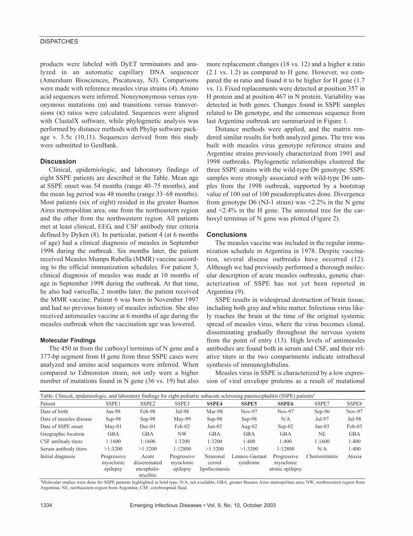
products were labeled with DyET terminators and ana-lyzed in an automatic capillary DNA sequencer(Amersham Biosciences, Piscataway, NJ). Comparisonswere made with reference measles virus strains (4). Aminoacid sequences were inferred. Nonsynonymous versus syn-onymous mutations (ω) and transitions versus transver-sions (κ) ratios were calculated. Sequences were alignedwith ClustalX software, while phylogenetic analysis wasperformed by distance methods with Phylip software pack-age v. 3.5c (10,11). Sequences derived from this studywere submitted to GenBank.
DiscussionClinical, epidemiologic, and laboratory findings of
eight SSPE patients are described in the Table. Mean ageat SSPE onset was 54 months (range 40–75 months), andthe mean lag period was 48 months (range 33–68 months).Most patients (six of eight) resided in the greater BuenosAires metropolitan area, one from the northeastern regionand the other from the northwestern region. All patientsmet at least clinical, EEG, and CSF antibody titer criteriadefined by Dyken (8). In particular, patient 4 (at 6 monthsof age) had a clinical diagnosis of measles in September1998 during the outbreak. Six months later, the patientreceived Measles Mumps Rubella (MMR) vaccine accord-ing to the official immunization schedules. For patient 5,clinical diagnosis of measles was made at 10 months ofage in September 1998 during the outbreak. At that time,he also had varicella; 2 months later, the patient receivedthe MMR vaccine. Patient 6 was born in November 1997and had no previous history of measles infection. She alsoreceived antimeasles vaccine at 6 months of age during themeasles outbreak when the vaccination age was lowered.
Molecular FindingsThe 450 nt from the carboxyl terminus of N gene and a
377-bp segment from H gene from three SSPE cases wereanalyzed and amino acid sequences were inferred. Whencompared to Edmonston strain, not only were a highernumber of mutations found in N gene (36 vs. 19) but also
more replacement changes (18 vs. 12) and a higher κ ratio(2.1 vs. 1.2) as compared to H gene. However, we com-pared the ω ratio and found it to be higher for H gene (1.7vs. 1). Fixed replacements were detected at position 357 inH protein and at position 467 in N protein. Variability wasdetected in both genes. Changes found in SSPE samplesrelated to D6 genotype, and the consensus sequence fromlast Argentine outbreak are summarized in Figure 1.
Distance methods were applied, and the matrix ren-dered similar results for both analyzed genes. The tree wasbuilt with measles virus genotype reference strains andArgentine strains previously characterized from 1991 and1998 outbreaks. Phylogenetic relationships clustered thethree SSPE strains with the wild-type D6 genotype. SSPEsamples were strongly associated with wild-type D6 sam-ples from the 1998 outbreak, supported by a bootstrapvalue of 100 out of 100 pseudoreplicates done. Divergencefrom genotype D6 (NJ-1 strain) was <2.2% in the N geneand <2.4% in the H gene. The unrooted tree for the car-boxyl terminus of N gene was plotted (Figure 2).
ConclusionsThe measles vaccine was included in the regular immu-
nization schedule in Argentina in 1978. Despite vaccina-tion, several disease outbreaks have occurred (12).Although we had previously performed a thorough molec-ular description of acute measles outbreaks, genetic char-acterization of SSPE has not yet been reported inArgentina (9).
SSPE results in widespread destruction of brain tissue,including both gray and white matter. Infectious virus like-ly reaches the brain at the time of the original systemicspread of measles virus, where the virus becomes clonal,disseminating gradually throughout the nervous systemfrom the point of entry (13). High levels of antimeaslesantibodies are found both in serum and CSF, and their rel-ative titers in the two compartments indicate intrathecalsynthesis of immunoglobulins.
Measles virus in SSPE is characterized by a low expres-sion of viral envelope proteins as a result of mutational
1334 Emerging Infectious Diseases • Vol. 9, No. 10, October 2003
DISPATCHES
Table. Clinical, epidemiologic, and laboratory findings for eight pediatric subacute sclerosing panencephalitis (SSPE) patientsa Patient SSPE1 SSPE2 SSPE3 SSPE4 SSPE5 SSPE6 SSPE7 SSPE8 Date of birth Jan-98 Feb-98 Jul-98 Mar-98 Nov-97 Nov-97 Sep-96 Nov-97 Date of measles disease Sep-98 Sep-98 May-99 Sep-98 Sep-98 N/A Jul-97 Jul-98 Date of SSPE onset May-01 Dec-01 Feb-02 Jun-02 Aug-02 Sep-02 Jan-03 Feb-03 Geographic location GBA GBA NW GBA GBA GBA NE GBA CSF antibody titers 1:1600 1:1600 1:3200 1:3200 1:400 1:400 1:1600 1:400 Serum antibody titers >1:3200 >1:3200 1:12800 >1:3200 >1:3200 1:12800 N/A 1:400 Initial diagnosis Progressive
myoclonic epilepsy
Acute disseminated encephalo-
myelitis
Progressive myoclonic epilepsy
Neuronal ceroid
lipofucsinosis
Lennox-Gastaut syndrome
Progressive myoclonic
atonic epilepsy
Chorioretinitis Ataxia
aMolecular studies were done for SSPE patients highlighted in bold type. N/A, not available; GBA, greater Buenos Aires metropolitan area; NW, northwestern region from Argentina; NE, northeastern region from Argentina; CSF, cerebrospinal fluid.
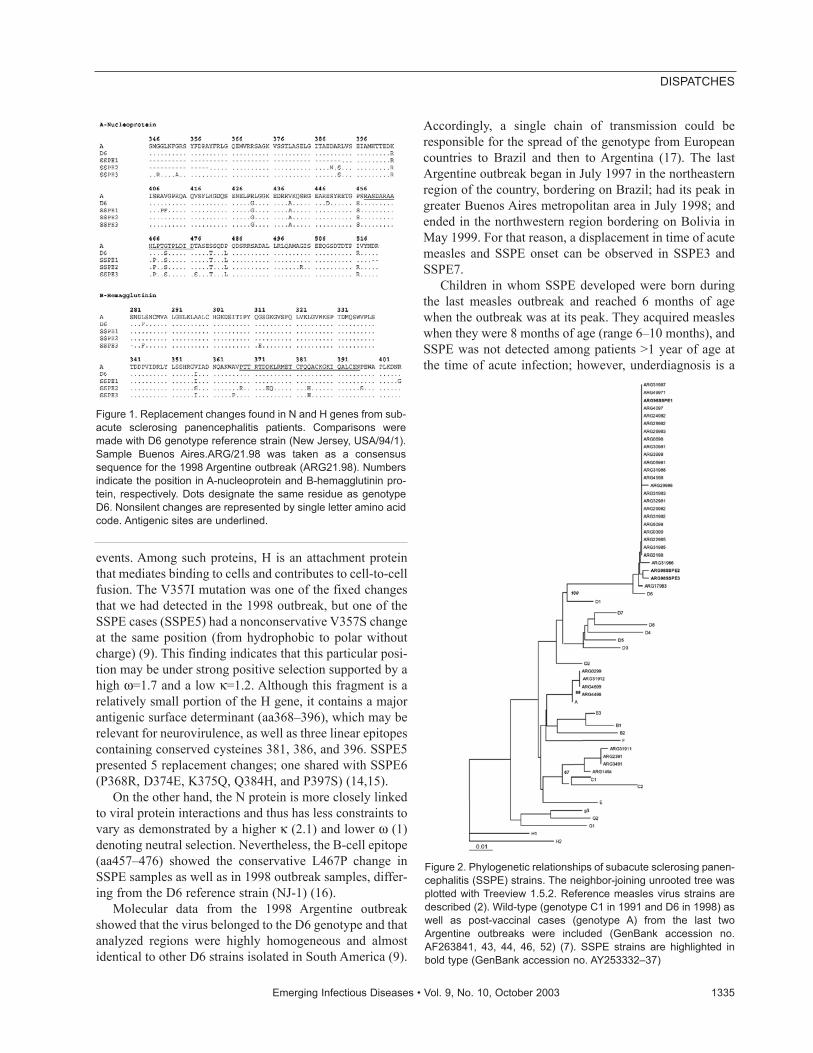
events. Among such proteins, H is an attachment proteinthat mediates binding to cells and contributes to cell-to-cellfusion. The V357I mutation was one of the fixed changesthat we had detected in the 1998 outbreak, but one of theSSPE cases (SSPE5) had a nonconservative V357S changeat the same position (from hydrophobic to polar withoutcharge) (9). This finding indicates that this particular posi-tion may be under strong positive selection supported by ahigh ω=1.7 and a low κ=1.2. Although this fragment is arelatively small portion of the H gene, it contains a majorantigenic surface determinant (aa368–396), which may berelevant for neurovirulence, as well as three linear epitopescontaining conserved cysteines 381, 386, and 396. SSPE5presented 5 replacement changes; one shared with SSPE6(P368R, D374E, K375Q, Q384H, and P397S) (14,15).
On the other hand, the N protein is more closely linkedto viral protein interactions and thus has less constraints tovary as demonstrated by a higher κ (2.1) and lower ω (1)denoting neutral selection. Nevertheless, the B-cell epitope(aa457–476) showed the conservative L467P change inSSPE samples as well as in 1998 outbreak samples, differ-ing from the D6 reference strain (NJ-1) (16).
Molecular data from the 1998 Argentine outbreakshowed that the virus belonged to the D6 genotype and thatanalyzed regions were highly homogeneous and almostidentical to other D6 strains isolated in South America (9).
Accordingly, a single chain of transmission could beresponsible for the spread of the genotype from Europeancountries to Brazil and then to Argentina (17). The lastArgentine outbreak began in July 1997 in the northeasternregion of the country, bordering on Brazil; had its peak ingreater Buenos Aires metropolitan area in July 1998; andended in the northwestern region bordering on Bolivia inMay 1999. For that reason, a displacement in time of acutemeasles and SSPE onset can be observed in SSPE3 andSSPE7.
Children in whom SSPE developed were born duringthe last measles outbreak and reached 6 months of agewhen the outbreak was at its peak. They acquired measleswhen they were 8 months of age (range 6–10 months), andSSPE was not detected among patients >1 year of age atthe time of acute infection; however, underdiagnosis is a
Emerging Infectious Diseases • Vol. 9, No. 10, October 2003 1335
DISPATCHES
Figure 1. Replacement changes found in N and H genes from sub-acute sclerosing panencephalitis patients. Comparisons weremade with D6 genotype reference strain (New Jersey, USA/94/1).Sample Buenos Aires.ARG/21.98 was taken as a consensussequence for the 1998 Argentine outbreak (ARG21.98). Numbersindicate the position in A-nucleoprotein and B-hemagglutinin pro-tein, respectively. Dots designate the same residue as genotypeD6. Nonsilent changes are represented by single letter amino acidcode. Antigenic sites are underlined.
Figure 2. Phylogenetic relationships of subacute sclerosing panen-cephalitis (SSPE) strains. The neighbor-joining unrooted tree wasplotted with Treeview 1.5.2. Reference measles virus strains aredescribed (2). Wild-type (genotype C1 in 1991 and D6 in 1998) aswell as post-vaccinal cases (genotype A) from the last twoArgentine outbreaks were included (GenBank accession no.AF263841, 43, 44, 46, 52) (7). SSPE strains are highlighted inbold type (GenBank accession no. AY253332–37)
permanent challenge. Time lag between acute and SSPEonset was approximately 4 years, according to the report-ed data, but the rate for developing SSPE was higher thanpreviously described (8).
Phylogenetic analysis of three SSPE cases from the lastoutbreak clustered with D6 genotype that circulated inArgentina in 1998. Although the original sequence of thewild-type virus that caused acute infection is unknown, wehave a consensus sequence that summarizes the outbreak;therefore, we infer that changes may have occurred sincethen and contributed to the development of SSPE.Although all three patients had been immunized accordingto the schedule, vaccinal strains were not detected in braintissue. These results agree with those recently reported forSSPE patients in the United Kingdom and Papua, NewGuinea (18,19). Our data show that these three patients hadbeen infected with wild-type circulating D6 virus beforeimmunization. This primary measles virus infection innonimmunized infants may be the leading cause of thehigh rate of SSPE inferred from our data. After brain tis-sues of deceased adults were randomly autopsied,Lawrence et al. raised the possibility that brain measlesinfection does not invariably lead to neurologic diseasecaused by measles virus. Therefore, neurologic diseasemediated by measles virus may depend on the primaryinfection age (20). Although the basis for measles-associ-ated neurologic disease is unclear and more thoroughmolecular studies need to be performed, our findings con-tribute to worldwide efforts in molecular characterizationof SSPE strains and aim to increase awareness amongphysicians to improve diagnosis at early stages.
A.S.M is a member of Comisión de InvestigacionesCientíficas de la Provincia de Buenos Aires (CIC). P.R.B. andM.V. are fellows from the National Council of Investigation andTechnology (CONICET). The Virology Laboratory at Dr. R.Gutiérrez Children’s Hospital is the Reference Laboratory for theNational Measles Elimination Program of Buenos Aires City Hall(GCBA).
Ms. Barrero has a fellowship from the National Council ofInvestigation and Technology (CONICET) in the virology labo-ratory at the Dr Ricardo Gutiérrez Children’s Hospital, BuenosAires, Argentina. Her work is focused on molecular epidemiolo-gy of emerging and respiratory viruses.
References
1. Cutts FT, Henao-Restrepo A, Olive JM, Cutts FT, Henao-Restrepo A,Olive JM. Measles elimination: progress and challenges. Vaccine1999;17(Suppl 3):S47–52.
2. Hersh BS, Tambini G, Nogueira AC, Carrasco P, de Quadros C.Review of regional measles surveillance data in the Americas,1996–99. Lancet 2000;355:1943–8.
3. Norrby E, Kristensson K. Measles virus in the brain. Brain Res Bull1997;44:213–20.
4. World Health Organization. Nomenclature for describing the geneticcharacteristics of wild-type measles viruses (update). Part I. WklyEpidemiol Rec 2001;76:242–7.
5. Cattaneo R, Schmid A, Spielhofer P, Kaelin K, Baczko K, ter MeulenV, et al. Mutated and hypermutated genes of persistent measles virus-es which caused lethal human brain diseases. Virology1989;173:415–25.
6. Baczko K, Liebert UG, Billeter M, Cattaneo R, Budka H, ter MeulenV. Expression of defective measles virus genes in brain tissues ofpatients with subacute sclerosing panencephalitis. J Virol1986;59:472–8.
7. Rima BK, Earle AP, Baczko K, ter Meulen V, Liebert UG, Carstens,et al. Sequence divergence of measles virus haemagglutinin duringnatural evolution and adaptation to cell culture. J Gen Virol1997;78:97–106.
8. Gark RK. Subacute sclerosing panencephalitis. Postgrad Med J2002;78:63–70.
9. Barrero PR, Zandomeni RO, Mistchenko AS. Measles virus circula-tion in Argentina: 1991–1999. Arch Virol 2001;146:815–23.
10. Thompson JD, Gibson TJ, Plewniak F, Jeanmougin F, Higgins DG.The ClustalX windows interface: flexible strategies for multiplesequence alignment aided by quality analysis tools. Nucleic AcidsRes 1997;24:4876–82.
11. Felsestein J. PHYLIP- Phylogeny inference package. Cladistics1989;5:164–6.
12. Bilkis MD, Barrero PR, Mistchenko AS. Measles resurgence inArgentina: 1997–98 outbreak. Epidemiol Infect 2000;124:289–93.
13. Backzo K, Lampe J, Liebert UG, Brinckmann U, ter Meulen V,Pardowitz I, et al. Clonal expansion of hypermutated measles virus ina SSPE brain. Virology 1993;197:188–95.
14. Liebert UG, Flanagan SG, Löffler S. Antigenic determinants ofmeasles virus hemagglutinin associated with neurovirulence. J Virol1994;68:1486–93.
15. Ziegler D, Fournier P, Berbers GAH, Steuer H, Wiesmüller KH,Fleckenstein B, et al. Protection against measles virus encephalitis bymonoclonal antibodies binding to a cysteine loop domain of the Hprotein mimicked by peptides which are not recognised by maternalantibodies. J Gen Virol 1996;77:2479–89.
16. Buckland R, Giraldon P, Wild TF. Expression of measles virus nucle-oprotein in Escherichia coli: use of deletion mutants to locate theantigenic sites. J Gen Virol 1989;70:435–41.
17. Oliveira MI, Rota PA, Suely PC, Figueiredo CA, Afonso AMS,Theobaldo M, et al. Genetic homogeneity of measles viruses associ-ated with a measles outbreak, São Paulo, Brazil, 1997. Emerg InfectDis 2002;8:808–13.
18. Jin L, Beard S, Hunjan R, Brown DW, Miller E. Characterization ofmeasles virus strains causing SSPE: a study of 11 cases. J Neurovirol2002;8:335–44.
19. Miki K, Komase K, Mgone CS, Kawanishi R, Iijima M, Mgone JM,et al. Molecular analysis of measles virus genome derived from SSPEand acute measles patients in Papua, New Guinea. J Med Virol2002;68:105–12 .
20. Lawrence DMP, Vaughn MM, Belman AR, Cole JS, Rall GF. Immuneresponse-mediated protection of adult but not neonatal mice fromneuron-restricted measles virus infection and central nervous systemdisease. J Virol 1999;73:1795–801.
Address for correspondence: Alicia S. Mistchenko, Laboratorio deVirología, Hospital de Niños Dr. Ricardo Gutiérrez, Gallo 1330 (1425)Buenos Aires, Argentina; fax: 54-11-49626770; email: [email protected]
1336 Emerging Infectious Diseases • Vol. 9, No. 10, October 2003
DISPATCHES
Cat or DogOwnership and
Seroprevalence of Ehrlichiosis,
Q Fever, and Cat-Scratch Disease
Martina Skerget,* Christoph Wenisch,* Florian Daxboeck,† Robert Krause,*
Renate Haberl,* and Doris Stuenzner* Concerns have been raised about the role of domestic
cats or dogs in the acquisition of zoonoses, in particular inpregnant women or immune-suppressed persons. Wereport that cat or dog ownership is not associated with anincreased seroprevalence of antibodies to Anaplasmaphagozytophilum, Coxiella burnetii, and Bartonella hense-lae in symptom-free persons in Styria, Austria.
Keeping pet cats and dogs is very popular in Austria.However, these animals can serve as reservoirs for the
agents of important bacterial infectious diseases and as apotential source of infection for humans, even though theinfectious animals may be asymptomatic. Infections arepotentially transmitted from domestic animals to humansby scratches, bites, or close contact. Examples for suchinfections include human granulocytic ehrlichiosis(Anaplasma phagocytophilum), cat-scratch disease (CSD,Bartonella henselae), and Q fever (Coxiella burnetii).
Cats are known to be the most important source ofinfections for B. henselae (aeroprevalence in Austrian catsis 33%) (1). However, dogs may also transmit B. henselae(2). Animals are contagious through their blood, whichmay contaminate saliva in cases of gum bleeding. Fleasfrom infected animals may contain the infectious agent,and bites from these fleas can transmit CSD. Typically,CSD is a benign and self-limiting disease in humans,occurring with lymphadenopathy, low-grade fever, pri-mary cutaneous inoculation lesion, and weight loss, lasting6–12 weeks. Rarely observed atypical signs and symptomsinclude erythema nodosum, figurate erythemas, thrombo-cytopenic purpura, Perinaud’s oculoglandular syndrome,encephalopathy, hepatic granulomas, osteomyelitis, pul-monary disease, and optic neuritis (3). These severe mani-festations occur in immunocompetent patients, whereas
bacillary angiomatosis or peliosis hepatitis are more likelyto develop in immunosuppressed patients.
C. burnetii infection has been associated with a chron-ic fatigue–like syndrome (4). Both cats and dogs are well-described reservoirs for C. burnetii (5). In humans, C. bur-netii infection usually is asymptomatic (60%) or manifestsas a mild disease with fever, headache, myalgias, andspontaneous recovery (5). However, this infection maylead to serious complications and even death in patientswith acute disease, especially those with meningoen-cephalitis and myocarditis and, more frequently, in chron-ically infected patients with endocarditis. Q fever in preg-nancy has been associated with abortion, premature birth,and low weight in newborns (6,7).
Within the past several decades, the number ofEhrlichia and Anaplasma spp. recognized to infect cats,dogs, and humans has expanded substantially (8). Theagent of human granulocytic ehrlichiosis (HGE) hasrecently been classified as A. phagozytophilum (9). Thedisease has influenzalike symptoms with variable degreesof anemia, thrombocytopenia, leukopenia, and elevatedliver enzymes. Dogs are thought to be sentinels for assess-ing risk for HGE in humans (10). Cases of HGE in theUnited States are increasing in incidence (11). Reports ofacute cases in Europe have been rare, although serosurveysof the prevalence of antibodies to A. phagocytophilumhave been conducted. A survey in Slovenia showed that15.4% of the examined population had detectable antibod-ies to the pathogen and several cases of HGE had beenconfirmed (12). No similar serosurvey has been conductedin Austria, although Ixodes ricinus, thought to be the prin-cipal vector in Europe, is present in Austria (13). We reportthat the rate of seropositivity of A. phagocytophilum(immunoglobulin [Ig] G antibodies 25/376 [7%], IgM anti-bodies 6/376 [2%]), B. henselae (88/376 [23%]), and C.burnetii (23/376 [6%]) in Styria, Austria, is not affected bycat or dog ownership.
The StudyWe examined serum specimens from 376 persons that
were collected at the University Hospital Graz, Austria,from December 2001 to April 2002. These persons had nohistory of a tick bite for at least 1 year. A total of 202 per-sons with domestic dogs, cats, or both (dogs n=77, catsn=106, dogs and cats n=19) were compared with 174 per-sons who had no domestic pet contact for at least 1 year.Study participants in the no-pet group had never lived witha cat or a dog. The domestic pets had no symptoms or signsof infection, as determined by veterinarians. All partici-pants were outpatients, and blood samples were drawn forroutine blood tests. Each participant completed a question-naire about medical history. These persons had no knownhistory of rickettsiosis, borreliosis, or tick-borne
Emerging Infectious Diseases • Vol. 9, No. 10, October 2003 1337
DISPATCHES
*University Hospital Graz, Graz, Austria; and †University Hospital,Vienna, Austria
encephalitis and reported no febrile or influenzalike illnessduring the previous 6 months. Each participant providedverbal consent for the serum to be used for detecting anti-bodies against several infectious agents related tozoonoses. The following information was collected foreach participant: age, sex, area of residence, and medicalhistory. Demographic data for the participants are shownin Table 1.
Serum samples were tested for the infectious agentswith the following methods: B. henselae by indirectimmunofluorescence (Biognost, Gräfelfing, Germany), C.burnetii by microagglutination reaction (Bodybion Mar;Bioveta National Enterprise Nitra, Slovakia), and HGEIgM indirect immunofluorescent antibody test (IFA, MRLDiagnostics, Cypress, CA), HGE by IFA IgG (FocusTechnologies, Cypress, CA) with titers >64 were consid-ered positive. All assays were performed in duplicateaccording to manufacturers’ instructions. Biostatisticalanalysis was performed with the statistical package JandelSigmaStat Statistical Software, version 2.0, NL (JandelSigmaStat, Jandel, San Rafael, CA). Chi-square tests or ttests were used to determine differences between the pres-ence of antibodies to the test organisms and demographicdata, as appropriate. A two-tailed p value <0.05 was con-sidered significant.
A total of 88 (23%) of 376 persons had antibodiesagainst B. henselae. No differences in terms of age, sex,urban or rural residence, or concomitant diseases werenoted. No difference in persons with and without domesticpets (46/174 [26%] and 42/202 [20%], respectively) wasseen (Table 2).
The overall prevalence of antibodies to C. burnetii was6% (23/376 persons). Again, no differences in age, sex,urban or rural residence, or concomitant diseases (13/202[6%]) and persons with and without domestic pets (10/174[6%]), were seen (Table 2).
The overall prevalence of IgG/IgM antibodies to A.phagocytophilum was 7% and 2% (25/376, and 6/376,respectively). No differences in terms of age, sex, urban orrural residence, or concomitant diseases were seen. IgGantibody titers were low (of 25 positive serum specimens,17 had titers >64, and 8 had titers >128). In the six patientswith positive IgM antibodies, no clinical evidence of ehrli-chiosis was present, and additional blood samples showedno cytopenia. No difference in persons with (8% and 2%IgG/IgM positive, respectively) and without domestic pets(6% and 1% IgG/IgM positive, respectively) was seen(Table 2).
The seroreactivity to C. burnetii and B. henselae did notdiffer between A. phagocytophilum–positive patients andA. phagocytophilum–negative patients (p>0.05). Likewise,the seroreactivity to A. phagocytophilum and B. henselaedid not differ between C. burnetii–positive patients and C.burnetii–negative patients (p>0.05). In addition, theseroreactivity to A. phagocytophilum and C. burnetii didnot differ between B. henselae–positive patients and B.henselae–negative patients (p>0.05).
ConclusionsVeterinarians have the responsibility of providing accu-
rate information to their clients about the zoonotic trans-mission of infections from pets, especially to those mostvulnerable, such as children, pregnant women, the elderly,and the immunocompromised. Effective education is vitalto allay public concerns and promote responsible pet own-ership (14). With respect to A. phagocytophilum, B. hense-lae, and C. burnetii, cat or dog ownership was not relatedto an increased incidence of antibodies in our study.
CSD has been reported worldwide and seems to be themost common B. henselae infection in humans. In theUnited States, epidemiologic databases estimate thatapproximately 24,000 cases of CSD occur each year, witha calculated incidence of 9.3/10,000 ambulatory patientsper year. In various studies, the seroprevalence of antibod-ies to B. henselae in humans ranges from 3.6% to 6%(15–17). Although CSD may occur in persons of any age,most patients are <18 years of age, perhaps because chil-dren are more likely to have close and rough contact withcats. The observed high incidence of antibody positivity inadults could be related to the persistence of antibodies afterasymptomatic infection. Alternatively, it may be duechronic low-grade infection, which has been demonstratedfor B. quintana and B. bacilliformis (18,19). The incidenceof CSD is seasonal; most cases occur in August to Octoberin northern temperate areas. The prevalence of the diseasealso varies by geographic location. The prevalence of anti-bodies to B. henselae is reportedly higher in areas withwarm humid climates, where the prevalence and intensityof cat flea infestations are higher (20). Cats may infecthumans either directly through scratches and bites or indi-rectly by means of the cat flea (Ctenocephalides felis),which is the arthropod vector (20). Recent research hasdemonstrated that B. henselae seroprevalence is elevatedin patients with coronary vascular disease (21).Considering the comparatively high mean age of thepatients tested in this study and the high percentage of
1338 Emerging Infectious Diseases • Vol. 9, No. 10, October 2003
DISPATCHES
Table 1. Demographic data of patients with and without domestic pets No pet (n=174) Cat (n=106) Dog (n=77) Cat and dog (n=19) Age (y) 55±19 51±14 53±20 55±15 Sex (male/female) 55/119 32/74 37/38 6/13 Urban/rural 133/41 63/43 39/36 7/12
patients admitted for cardiovascular disease (Table 1), ourresults provide indirect support for this finding.
Because Q fever is rarely a notifiable disease, its inci-dence in humans cannot be assessed in most countries.Current epidemiologic studies indicate, however, that Qfever should be considered a public health problem inmany countries, including France, United Kingdom, Italy,Spain, Germany, Israel, Greece, and Canada (NovaScotia), as well as in many countries where Q fever isprevalent but unrecognized because of poor surveillance.Q fever remains primarily an occupational hazard in per-sons in contact with domestic animals such as cattle,sheep, and, less frequently, goats. Persons at risk from Qfever include farmers, veterinarians, abattoir workers,those in contact with dairy products, and laboratory per-sonnel performing C. burnetii culture and working with C.burnetii–infected animals. However, reports of sporadiccases in persons living in urban areas after occasional con-tact with farm animals or after contact with infected petssuch as dogs and cats have increased. Our data suggest thatQ fever also occurs in Austria but that pet ownership orrural residence has no effect on seroprevalence. C. burnetiihas also been isolated from the locally widespread I. rici-nus ticks (22), although no tick-borne Q fever has beendescribed in Austria.
For A. phagocytophilum, our seroprevalence is similarto those observed in Greece, much lower than that inSlovenia (15.4%), and higher than those seen in Bulgaria(2.9%) and Germany (1.9%) (23). In Austria (Styria), theprevalence in blood donors is 4% (24). We agree withDaniel et al. (23) that this Austrian prevalence could bedue to the fact that the prevalence in blood donors is lower,unlike the survey in Greece, Slovenia, or this assessment.Even though the titers to A. phagocytophilum were low inour study, they suggest infection at an undetermined time.
Our data also suggest a discrepancy between the com-paratively high seroprevalence of specific antibodies to A.phagozytophilum, C. burnetii, and B. henselae, respective-ly, and the few diagnosed cases of human disease. Clearly,cat or dog ownership is not related to an increased inci-dence of antibodies against these pathogens. However,because of the widespread distribution of I. ricinus, anorganism also capable of transmission, this finding must
be interpreted cautiously. In addition, the small sample sizemay have been insufficient to detect differences betweenthe groups. Hence, recommendations for the population atrisk for a severe course of these infections (pregnantwomen, the elderly, immunosuppressed patients, HIV-pos-itive persons, and infants) are not possible. Nonetheless,such groups of patients may benefit from efficient tick andflea control on dogs and cats, as an adjunct to decreasemechanical transport of the parasites into their homes.Further research is needed to clarify the importance of petsor ticks for these diseases in Austria.
Dr. Skerget is a research fellow at the Department ofMedicine, University of Graz. She is interested in zoonoses.
References
1. Allerberger F, Schönbauer M, Zangerle R, Dierich M. Prevalence ofantibody to Rochalimaea henselae among Austrian cats. Eur J Pediatr1995;154:165.
2. Murano I, Tsuneoka H, Iino H, Kamei T, Nakamura I, Tsukahara M.Two patients with Bartonella henselae infection from a dog.Kansenshogaku Zasshi 2001;75:808–11.
3. Windsor JJ. Cat-scratch disease: epidemiology, aetiology and treat-ment. Br J Biomed Sci 2001;58:101–10.
4. Wildman MJ, Smith EG, Groves J, Beattie JM, Caul EO, Ayres JG.Chronic fatigue following infection by Coxiella burnetii (Q fever):ten-year follow-up of the 1989 UK outbreak cohort. QJM2002;95:527–38.
5. Caron F, Meurice JC, Ingrand P, Bourgoin A, Masson P, Roblot P, etal. Acute Q fever pneumonia: a review of 80 hospitalized patients.Chest 1998;114:808–13.
6. Pinsky RL, Fishbein DB, Greene CR, Gensheimer KF. An outbreakof cat-associated Q-fever in the United States. J Infect Dis1991;164:202–4.
7. Maurin M, Raoult D. Q fever. Clin Microbiol Rev 1999;12:518–53.8. Neer TM, Breitschwerdt EB, Greene RT, Lappin MR. Consensus
statement on ehrlichial disease of small animals from the infectiousdisease study group of the ACVIM. American College of VeterinaryInternal Medicine. J Vet Intern Med 2002;16:309–15.
9. Dumler JS, Barbet AF, Bekker CP, Dasch GA, Palmer GH, Ray SC,et al. Reorganization of genera in the families Rickettsiaceae andAnaplasmataceae in the order Rickettsiales: unification of somespecies of Ehrlichia with Anaplasma, Cowdria with Ehrlichia andEhrlichia with Neorickettsia, descriptions of six new species combi-nations and designation of Ehrlichia equi and ‘HGE agent’ as subjec-tive synonyms of Ehrlichia phagocytophila. Int J Syst Evol Microbiol2001;51:2145–65.
Emerging Infectious Diseases • Vol. 9, No. 10, October 2003 1339
DISPATCHES
Table 2. Seroprevalence of antibodies against Anaplasma phagocytophilum, Coxiella burnetti, and Bartonella henselae in persons with and without domestic petsa
Organism No pet
n=174 (%) Cat
n=106 (%) Dog
n=77 (%) Cat and dog
n=19 (%) No pet/cat
p value No pet/dog
p value No pet/cat and
dog p value A. phagocytophilum IgGb 12 (6) 7 (7) 2 (5) 4 (13) 0.239 0.332 0.162 IgM 2 (1) 1 (1) 1 (2) 2 (6) 0.856 0.849 0.611 C. burnetii 13 (6) 6 (6) 2 (5) 2 (6) 0.235 0.293 0.257 B. henselae 46 (26) 29 (28) 9 (23) 4 (13) 0.272 0.143 0.113 aNo differences are evident by chi-square test for comparisons between the groups for all pathogens. bIg, immunoglobulin.
10. Foley JE, Foley P, Madigan JE. Spatial distribution of seropositivityto the causative agent of granulocytic ehrlichiosis in dogs inCalifornia. Am J Vet Res 2001;62:1599–605.
11. Ijdo JW, Meek JI, Cartter ML, Magnarelli LA, Wu C, Tenuta SW, etal. The emergence of another tickborne infection in the 12-town areaaround Lyme, Connecticut: human granulocytic ehrlichiosis. J InfectDis 2000;181:1388–93.
12. Cizman M, Avsic-Zupanc T, Petrovec M, Ruzic-Sabljic E, Pokorn M.Seroprevalence of ehrlichiosis, Lyme borreliosis and tick borneencephalitis infections in children and young adults in Slovenia. WienKlin Wochenschr 2000;112:842–5.
13. Sixl W, Hinaidy HK, Kutzer E. Tick fauna of Austrian carnivores.Wiener Tierärztliche Monatsschrift 1971;58:427.
14. Rehacek J, Kaaserer B, Urvolgyi J, Lukacova M, Kovacova E,Kocianova E. Isolation of Coxiella burnetii and of an unknownrickettsial organism from Ixodes ricinus ticks collected in Austria.Eur J Epidemiol 1994;10:719–23.
15. Zangwill KM, Hamilton DH, Perkins BA, Regnery RL, Plikaytis BD,Hadler JL, et al. Cat scratch disease in Connecticut: epidemiology,risk factors, and evaluation of a new diagnostic test. N Engl J Med1993;329:8–13.
16. Jackson LA, Perkins BA, Wenger JD. Cat-scratch disease in theUnited States: an analysis of three national databases. Am J PublicHealth 1993;83:1707–11.
17. Hamilton DH, Zangwill KM, Hadler JL, Carter ML. Cat-scratchdisease—Connecticut, 1992–1993. J Infect Dis 1995;172:570–3.
18. Kosek M, Lavarello R, Gilman RH, Delgado J, Maguina C,Verastegui M, et al. Natural history of infection with Bartonellabacilliformis in a nonendemic population. J Infect Dis2000;182:865–72.
19. Foucault C, Barrau K, Brouqui P, Raoult D. Bartonella quintanabacteremia among homeless people. Clin Infect Dis 2002;35:684–9.
20. Irwin PJ. Companion animal parasitology: a clinical perspective. IntJ Parasitol 2002;32:581–93.
21. Ender PT, Phares J, Gerson G, Taylor SE, Regnery R, Challener RC,et al. Association of Bartonella species and Coxiella burnetii infec-tion with coronary artery disease: J Infect Dis 2001;183:831–4.
22. Jacomo V, Kelly PJ, Raoult D. Natural history of Bartonellainfections (an exception to Koch’s postulate). Clin Diagn LabImmunol 2002;9:8–18.
23. Daniel SA, Manika K, Arvanitidou M, Diza E, Symeonidis N,Antoniadis A. Serological evidence of human granulocytic ehrlichio-sis, Greece. Emerg Infect Dis 2002;8:643–4.
24. Sixl W, Ladurner G, Stuenzner D, Marth E. Epidemiological exami-nations of human sera for Ehrlichia in Austria. InternationalConference on Lyme Borreliosis and other Emerging Tick-BorneDiseases. Jun 20–24, 1999. Munich, Germany. Abstract book. Basel,Switzerland: AKM Congress Service; 1999. p. 55.
Address for correspondence: Christoph Wenisch, Division of InfectiousDiseases, Department of Medicine, University Hospital Graz,Auenbruggerplatz 15, A-8036 Graz, Austria; fax: +43-316-385-4622;email: [email protected]
1340 Emerging Infectious Diseases • Vol. 9, No. 10, October 2003
DISPATCHES
SS ee aa rr cc hh pp aa ss tt ii ss ss uu ee ss oo ff EE II DD aa tt ww ww ww.. cc dd cc .. gg oo vv // ee ii dd
Flying Squirrel–associated Typhus,
United StatesMary G. Reynolds,* John W. Krebs,* James A. Comer,* John W. Sumner,*
Thomas C. Rushton,† Carlos E. Lopez,‡ William L. Nicholson,* Jane A. Rooney,§
Susan E. Lance-Parker,¶ Jennifer H. McQuiston,*Christopher D. Paddock,* and James E. Childs*
In March 2002, typhus fever was diagnosed in twopatients residing in West Virginia and Georgia. Bothpatients were hospitalized with severe febrile illnesses, andboth had been recently exposed to or had physical contactwith flying squirrels or flying squirrel nests. Laboratoryresults indicated Rickettsia prowazekii infection.
Typhus fever from Rickettsia prowazekii infection is asevere and occasionally fatal disease in humans.
Frequently referred to as epidemic typhus or louse-bornetyphus, this disease can cause large epidemics when con-ditions are favorable for person-to-person spread of bodylice (Pediculus humanus humanus). For the last fewdecades, reported outbreaks have been confined mainly tothe cold mountainous regions of Africa and South Americaand have disproportionately affected impoverished anddisplaced communities (1).
Infections with R. prowazekii are rarely described in theUnited States. From 1976 to 2001, a total of 39 human R.prowazekii infections were documented in persons with noreported contact with body lice or persons with lice (2–5).Nearly all of these cases were in the eastern United States,and in approximately one third of cases, contact with fly-ing squirrels (Glaucomys spp.) or with flying squirrel nestsoccurred before disease onset.
Flying squirrels are the only known vertebrate reservoirof R. prowazekii, other than humans, and contact withthese animals has been linked to most sporadic typhuscases in the United States. Interest in this disease was highin the 10 years after the first isolation of R. prowazekiifrom flying squirrels (6,7), but few cases have been report-ed since 1985. We describe two cases of flyingsquirrel–associated typhus that occurred in West Virginiaand Georgia in 2002 and provide a contemporary summa-ry of this disease in the United States.
Case Reports
West VirginiaDuring February 2002, a 44-year-old man in West
Virginia arrived in the emergency department, withheadache, fever, and chills. The patient also had hematuria,joint pain, discomfort on the left side of his abdomen, andvomiting. Laboratory findings included elevated levels ofalanine transaminase (ALT) and aspartate transaminase(AST) (100 and 91 U/L, respectively), leukocyte count of4.1 x 109/L, platelet count of 249 x 109/L, and erythrocytesedimentation rate of 42 mm/h. The patient also had diver-ticulosis. A treatment regimen of levofloxacin and metron-idazole was begun, and the patient was admitted to thehospital. The condition worsened, and an infectious dis-ease specialist was consulted on day 4 of hospitalization.At this time, the patient was febrile (maximum tempera-ture, 38°C), reported myalgia and malaise, and had mildlyinjected sclerae (without photophobia). AST and ALT lev-els remained slightly elevated. Levofloxacin and metron-idazole were discontinued. Because the patient was arecreational hunter, serologic tests for Rocky Mountainspotted fever, ehrlichiosis, adenovirus, Lyme disease, andcytomegalovirus (CMV) were ordered, and doxycyclinewas given as empiric therapy. Serologic tests were nega-tive for all agents. The patient was discharged on day 7 ofhospitalization with a diagnosis of immunoglobulin (Ig) Anephropathy and hepatitis. At a follow-up visit (day 27after illness onset), the patient still had myalgias, fatigue,and conjunctivitis, although his fever and abdominal painhad resolved. At this time, serologic testing for typhusgroup rickettsiae showed reactive IgM antibodies at a titerof 512 and IgG at a titer of <64. An additional serum sam-ple obtained on day 53 after illness onset was tested at theCenters for Disease Control and Prevention (CDC) byindirect-immunofluorescence assay (IFA) and showedtiters of IgM and IgG antibodies reactive with R.prowazekii of 128 and 1,024, respectively, indicating arecent acute infection.
During January, the patient had spent several nights ina hunting cabin in a rural area of Hardy County, WestVirginia. Flying squirrels had infested the cabin every win-ter for several years; evidence of nesting materials androdent feces in the attic and wall spaces was visible. Thepatient did not report seeing flying squirrels during visitsto the cabin in 2002 but reported having removed rodentnesting materials and debris from a wall space 10–15 daysbefore becoming ill.
GeorgiaDuring March 2002, a 57-year-old man from Fulton
County, Georgia, received medical treatment for confusionassociated with a febrile illness of approximately 1-week
Emerging Infectious Diseases • Vol. 9, No. 10, October 2003 1341
DISPATCHES
*Centers for Disease Control and Prevention, Atlanta, Georgia,USA; †Marshall University, Huntington, West Virginia, USA;‡Atlanta I.D. Group, Atlanta, Georgia, USA; §West VirginiaDivision of Public Health, Charleston, West Virginia, USA; and¶Georgia Division of Public Health, Atlanta, Georgia, USA
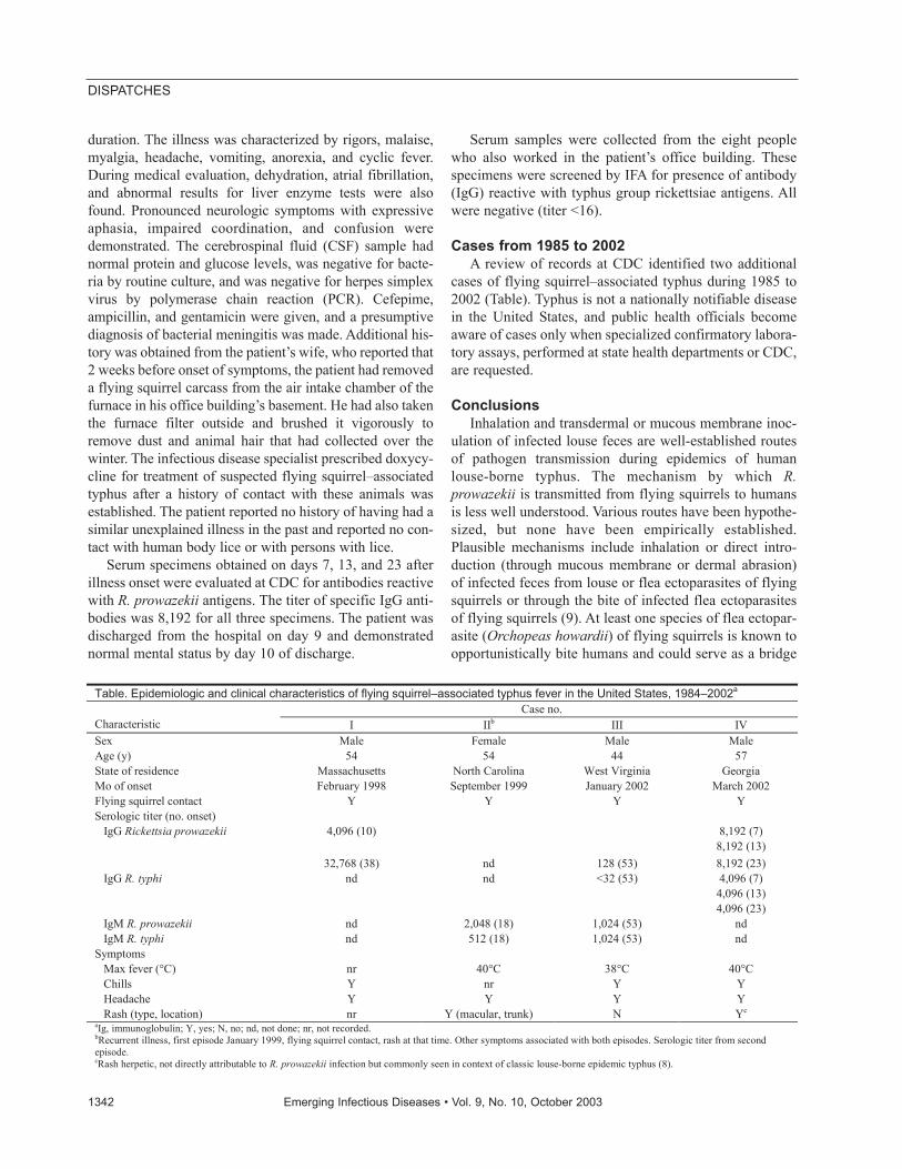
duration. The illness was characterized by rigors, malaise,myalgia, headache, vomiting, anorexia, and cyclic fever.During medical evaluation, dehydration, atrial fibrillation,and abnormal results for liver enzyme tests were alsofound. Pronounced neurologic symptoms with expressiveaphasia, impaired coordination, and confusion weredemonstrated. The cerebrospinal fluid (CSF) sample hadnormal protein and glucose levels, was negative for bacte-ria by routine culture, and was negative for herpes simplexvirus by polymerase chain reaction (PCR). Cefepime,ampicillin, and gentamicin were given, and a presumptivediagnosis of bacterial meningitis was made. Additional his-tory was obtained from the patient’s wife, who reported that2 weeks before onset of symptoms, the patient had removeda flying squirrel carcass from the air intake chamber of thefurnace in his office building’s basement. He had also takenthe furnace filter outside and brushed it vigorously toremove dust and animal hair that had collected over thewinter. The infectious disease specialist prescribed doxycy-cline for treatment of suspected flying squirrel–associatedtyphus after a history of contact with these animals wasestablished. The patient reported no history of having had asimilar unexplained illness in the past and reported no con-tact with human body lice or with persons with lice.
Serum specimens obtained on days 7, 13, and 23 afterillness onset were evaluated at CDC for antibodies reactivewith R. prowazekii antigens. The titer of specific IgG anti-bodies was 8,192 for all three specimens. The patient wasdischarged from the hospital on day 9 and demonstratednormal mental status by day 10 of discharge.
Serum samples were collected from the eight peoplewho also worked in the patient’s office building. Thesespecimens were screened by IFA for presence of antibody(IgG) reactive with typhus group rickettsiae antigens. Allwere negative (titer <16).
Cases from 1985 to 2002A review of records at CDC identified two additional
cases of flying squirrel–associated typhus during 1985 to2002 (Table). Typhus is not a nationally notifiable diseasein the United States, and public health officials becomeaware of cases only when specialized confirmatory labora-tory assays, performed at state health departments or CDC,are requested.
ConclusionsInhalation and transdermal or mucous membrane inoc-
ulation of infected louse feces are well-established routesof pathogen transmission during epidemics of humanlouse-borne typhus. The mechanism by which R.prowazekii is transmitted from flying squirrels to humansis less well understood. Various routes have been hypothe-sized, but none have been empirically established.Plausible mechanisms include inhalation or direct intro-duction (through mucous membrane or dermal abrasion)of infected feces from louse or flea ectoparasites of flyingsquirrels or through the bite of infected flea ectoparasitesof flying squirrels (9). At least one species of flea ectopar-asite (Orchopeas howardii) of flying squirrels is known toopportunistically bite humans and could serve as a bridge
1342 Emerging Infectious Diseases • Vol. 9, No. 10, October 2003
DISPATCHES
Table. Epidemiologic and clinical characteristics of flying squirrel–associated typhus fever in the United States, 1984–2002a Case no.
Characteristic I IIb III IV Sex Male Female Male Male Age (y) 54 54 44 57 State of residence Massachusetts North Carolina West Virginia Georgia Mo of onset February 1998 September 1999 January 2002 March 2002 Flying squirrel contact Y Y Y Y Serologic titer (no. onset)
8,192 (7) 4,096 (10) 8,192 (13)
IgG Rickettsia prowazekii
32,768 (38) nd 128 (53) 8,192 (23) 4,096 (7)
4,096 (13) IgG R. typhi nd nd <32 (53)
4,096 (23) IgM R. prowazekii nd 2,048 (18) 1,024 (53) nd IgM R. typhi nd 512 (18) 1,024 (53) nd
Symptoms Max fever (°C) nr 40°C 38°C 40°C Chills Y nr Y Y Headache Y Y Y Y Rash (type, location) nr Y (macular, trunk) N Yc
aIg, immunoglobulin; Y, yes; N, no; nd, not done; nr, not recorded. bRecurrent illness, first episode January 1999, flying squirrel contact, rash at that time. Other symptoms associated with both episodes. Serologic titer from second episode. cRash herpetic, not directly attributable to R. prowazekii infection but commonly seen in context of classic louse-borne epidemic typhus (8).
vector for transmission from flying squirrel to human.Rickettsiae transmission among captive flying squirrels,however, has only been demonstrated with a louse vector(9,10). While the exact mechanism of pathogen transmis-sion has not yet been determined, the lack of detectableexposure to R. prowazekii in the household members or co-workers of documented cases (3,5) supports the idea thatthe risk for R. prowazekii infection after casual or indirectexposure to flying squirrels is low. Rather, existing evi-dence suggests that infection follows from close physicalcontact with flying squirrels or from exposure to a concen-trated source of infectious materials (e.g., nests, dander, orinfected ectoparasites).
Currently, no formal system for epidemic typhus sur-veillance exists in the United States, and diagnosis is hin-dered by the lack of rapid and reliable commercial tests. R.prowazekii infections can be confirmed by serologic test-ing, PCR, or organism culture. Commercial testing is,however, not widely available, and commercial serologictests lack specificity because most detect antibodies reac-tive with a surrogate typhus-group rickettsial antigen (typ-ically R. typhi). PCR (in conjunction with DNA sequenc-ing) is a highly specific diagnostic tool (11) but has lowsensitivity for commonly available clinical specimens,such as whole blood or serum. PCR may be used to greatereffect on other clinical specimens (e.g., cerebrospinalfluid, lymphocytes, and skin biopsy) if they are collectedand submitted for testing. Specific serologic and moleculartesting is available at CDC for specimens submittedthrough state public health laboratories.
Sporadic epidemic typhus occurs in the United States,primarily during winter and spring, and in regions withinthe normal range of the southern flying squirrel(Glaucomys volans) (12). This illness can be severe, result-ing in protracted hospital stays, particularly when diagno-sis and appropriate treatment are delayed. In these twocases, treatment with tetracycline antibiotics was initiatedafter other broad-spectrum antibiotics were used. Whentherapy was changed to the appropriate antibiotic, theresponse was rapid. Tetracycline antibiotics are highlyeffective therapies for typhus (13). These cases underscorethe importance of obtaining a thorough history of animaland arthropod contact in patients with acute febrile illness.Physicians and healthcare providers should remain alert tothe signs and symptoms of epidemic typhus and be awareof appropriate diagnostic methods and antibiotic treat-ments (13,14).
Dr. Reynolds has a Ph.D. in biomedical science and has pur-sued postdoctoral research in evolutionary biology. She is a fel-low in the Epidemic Intelligence Service at the Centers forDisease Control and Prevention in the Viral and RickettsialZoonoses Branch, Division of Viral and Rickettsial Zoonoses,National Center for Infectious Diseases.
References
1. Perine PL, Chandler BP, Krause DK, McCardle P, Awoke S, Habte-Gabr E, et al. A clinico-epidemiological study of epidemic typhus inAfrica. Clin Infect Dis 1992;14:1149–58.
2. Epidemic typhus associated with flying squirrels—United States.MMWR Morb Mortal Wkly Rep 1982;31:555–6.
3. Epidemic typhus—Georgia. MMWR Morb Mortal Wkly Rep1984;33:618–9.
4. Agger WA, Songsiridej V. Epidemic typhus acquired in Wisconsin.Wis Med J 1985;84:27–30.
5. Duma RJ, Sonenshine DE, Bozeman FM, Veazey JM Jr, Elisberg BL,Chadwick DP, et al. Epidemic typhus in the United States associatedwith flying squirrels. JAMA 1981;245:2318–23.
6. Bozeman FM, Masiello SA, Williams MS, Elisberg BL. Epidemictyphus rickettsiae isolated from flying squirrels. Nature1975;255:545–7.
7. McDade JE, Shepard CC, Redus MA, Newhouse VF, Smith JD.Evidence of Rickettsia prowazekii infections in the United States. AmJ Trop Med Hyg 1980;29:277–84.
8. Strong RP, Shattuck GC, Sellards AW, Zinnser H, Hopkins JG.Typhus fever with particular reference to the Serbian epidemic.Cambridge (MA): American Red Cross at the Harvard UniversityPress; 1920.
9. Bozeman FM, Sonenshine DE, Williams MS, Chadwick DP, LauerDM, Elisberg BL. Experimental infection of ectoparasitic arthropodswith Rickettsia prowazekii (GvF-16 strain) and transmission to flyingsquirrels. Am J Trop Med Hyg 1981;30:253–63.
10. Lauer DM, Sonenshine DE. Bionomics of the squirrel flea,Orchopeas howarde (Siphonaptera: Ceratophyllidae), in laboratoryand field colonies of the southern flying squirrel, Glaucomys volans,using radiolabeling techniques. J Med Entomol 1978;15:1–10.
11. Massung RF, Davis LE, Slater K, McKechnie DB, Puerzer M.Epidemic typhus meningitis in the southwestern United States. ClinInfect Dis 2001;32:979–82.
12. Kaplan JE, McDade JE, Newhouse VF. Suspected Rocky Mountainspotted fever in the winter—epidemic typhus? N Engl J Med1981;305:1648.
13. Huys J, Kayhigi J, Freyens P, Berghe GV. Single-dose treatment ofepidemic typhus with doxycyline. Chemotherapy 1973;18:314–7.
14. Ormsbee R, Peacock M, Philip R, Casper E, Plorde J, Gabre-Kidan T,et al. Serologic diagnosis of epidemic typhus fever. Am J Epidemiol1977;105:261–71.
Address for correspondence: Mary G. Reynolds; Centers for DiseaseControl and Prevention, 1600 Clifton Road, Mailstop G13, Atlanta, GA30338; fax: 404-639-2118; email: [email protected]
Emerging Infectious Diseases • Vol. 9, No. 10, October 2003 1343
DISPATCHES

Chlamydia trachomatisInfections in
Female Soldiers,Israel
Ellen S. Bamberger,*1 Efraim Siegler,†1
Einat Makler-Shiran,† Mihir V. Patel,† Jordan M. Steinberg,‡ Rosa Gershtein,*
and Isaac Srugo*We examined the prevalence of Chlamydia trachoma-
tis infection in Israeli female soldiers. The prevalence was3.2% among soldiers seeking medical care; rural residencewas identified as a significant risk factor. Nevertheless,given the study design, recommending broad-scale screen-ing of Israeli female soldiers may be premature.
Recent studies from the United States and Europe reportthat the prevalence of Chlamydia trachomatis ranges
from 5% to 20% in sexually active persons (1,2). Amongwomen, the consequences of the disease include pelvicinflammatory disease, ectopic pregnancy, and infertility,sequelae often accompanied by a substantial economicimpact (3). Most importantly, for up to 80% of infectedwomen, infection is asymptomatic, resulting in failure toseek timely medical care and the exacerbation of suchsequelae (4). Consequently, screening programs have beenrecommended to reduce infection, transmission, and dis-ease consequences (4,5). However, before such programsare universally advocated, since the cost-effectiveness ofsuch control programs is at least partially contingent upondisease prevalence (5), more data are required to assess theextent to which such prevalence rates may indeed be gen-eralizable to other countries and populations.
Epidemiologic research indicates that prevalence ratesof C. trachomatis vary, with a number of risk factorsaccounting for a substantial portion of this variance (4).For example, in the landmark study by Gaydos et al., sig-nificant risk factors for the prevalence of this infectionamong American female military recruits included age(women <25 years of age were at higher risk), vaginalintercourse, lack of condom use, and multiple sexual part-ners (4). Given that sexual practices such as condom useand multiple sex partners are likely to vary from country to
country, we designed a study to assess the prevalence of C.trachomatis in a population of Israeli women, namelyfemale soldiers, who, according to previous research, hada heightened risk of having the disease because of their ageand sexual activity (4–6).
The StudyWhereas Gaydos et al. invited all female army recruits
undergoing a physical examination at a reception base overthe course of 2-year period to participate in their study, oursample consisted of Israeli women actively seeking med-ical care. Specifically, participants in the current studywere women serving in the Israel Defense Forces, who,over a 12-month period attended a women’s health clinicfor a routine gynecologic examination, treatment of geni-tourinary symptoms, or contraceptive counseling. Theclinic was associated with a major tertiary hospital innorthern Israel. Of the 800 women we asked, 708 volun-teered to participate, for a response rate of 88.5%.Participants were between the ages of 18 and 45 (medianage 19, mean age 20). Most participants (77%) were bornin Israel.
After signing an informed consent form, participantswere asked to complete a short questionnaire (includingdemographic and sexual history items) and provide a urinespecimen. We performed the polymerase chain reaction(PCR) of urine specimens to test for C. trachomatis andNeisseria gonorrhoeae (Roche Amplicor, Branchburg,NJ). The PCR results and questionnaire data were coded asdichotomous variables and examined on the basis of bothunivariate (chi-square and Fisher exact test) and multivari-ate (logistic regression) analysis.
Of the 708 participants, 23 (3.25%) tested positive forC. trachomatis, and 8 (1%) tested positive for N. gonor-rhoeae. These rates are substantially lower than the 9.2%prevalence rate for C. trachomatis reported by Gaydos inthe U. S. military study (4). The median and average ageof those affected was 20. Most of those testing positivereported 1) having had sex in the past 3 months (86%), 2)having had more than one partner (45%) or having had apartner who had had more than one partner (57%), or 3)never having used a condom (70%) (Table). Nevertheless,none of these factors, identified by Gaydos as significantrisk factors in the American sample, was found to be a sig-nificant predictor of C. trachomatis infection in the Israelisoldiers. Similarly, while most of those testing positivewere born in Israel (81%), country of origin was not a sig-nificant predictor of infection. Neither were gynecologicsymptoms found to significantly distinguish between C.trachomatis–positive and –negative soldiers. Indeed, theonly factor that we found to significantly distinguish
1344 Emerging Infectious Diseases • Vol. 9, No. 10, October 2003
DISPATCHES
1Dr. Bamberger and Dr. Siegler contributed equally.
*B’nai Zion Medical Center, Haifa, Israel; †Rambam MedicalCenter, Haifa, Israel; and ‡Albert Einstein College of Medicine,Bronx Burough of New York, New York, USA
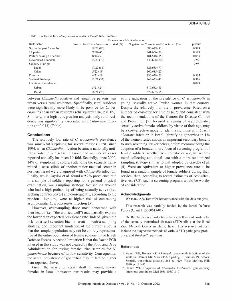
between Chlamydia-positive and -negative persons wasurban versus rural residence. Specifically, rural residentswere significantly more likely to be positive for C. tra-chomatis than urban residents (chi square=3.86, p<0.05).Similarly, in a logistic regression analysis, only rural resi-dence was significantly associated with Chlamydia infec-tion (p=0.043) (Table).
ConclusionsThe relatively low rate of C. trachomatis prevalence
was somewhat surprising for several reasons. First, since1994, when Chlamydia infection became a nationally noti-fiable infectious disease in Israel, the number of casesreported annually has risen 10-fold. Secondly, since 2000,14% of symptomatic soldiers attending the sexually trans-mitted disease clinic of another major medical center innorthern Israel were diagnosed with Chlamydia infection.Finally, while Gaydos et al. found a 9.2% prevalence ratein a sample of soldiers reporting for a general physicalexamination, our sampling strategy focused on womenwho had a high probability of being sexually active (i.e.,seeking contraceptives) and consequently, according to theprevious literature, were at higher risk of contractingasymptomatic C. trachomatis infection (5).
However, oversampling those most concerned withtheir health (i.e., “the worried well”) may partially explainthe lower than expected prevalence rate. Indeed, given therisk for a self-selection bias inherent in such a samplingstrategy, one important limitation of the current study isthat the sample population may not be entirely representa-tive of the entire population of female soldiers in the IsraeliDefense Forces. A second limitation is that the Roche PCRkit used in this study was not cleared by the Food and DrugAdministration for testing female urine samples for N.gonorrhoeae because of its low sensitivity. Consequently,the actual prevalence of gonorrhea may in fact be higherthan reported above.
Given the nearly universal draft of young Jewishfemales in Israel, however, our results may provide a
strong indication of the prevalence of C. trachomatis inyoung, sexually active Jewish women in that country.Despite the relatively low rate of prevalence, based on anumber of cost-efficacy studies (6,7) and consistent withthe recommendations of the Centers for Disease Controland Prevention (5), focused screening of asymptomatic,sexually active female soldiers, by virtue of their age, maybe a cost-effective mode for identifying those with C. tra-chomatis infection in Israel. Identifying gonorrhea in 1%of the women tested shows an important secondary benefitto such screening. Nevertheless, before recommending theadoption of a broader, more focused screening program offemale soldiers, whether symptomatic or not, we recom-mend collecting additional data with a more randomizedsampling strategy similar to that adopted by Gaydos et al.(4). Were an equivalent or higher prevalence rate to befound in a random sample of female soldiers during theirservice, then, according to recent estimates of cost-effec-tiveness (7,8), such a screening program would be worthyof consideration.
AcknowledgmentsWe thank Ada Tamir for her assistance with the data analysis.
This research was partially funded by the Israel DefenseForces (Grant # 15000615-01).
Dr. Bamberger is an infectious disease fellow and co-directorof the sexually transmitted diseases (STD) clinic at the B’naiZion Medical Center in Haifa, Israel. Her research interestsinclude the diagnostic methods of various STD pathogens, probi-otics, and Bordetella pertussis.
References
1. Stamm WE, Holmes KK. Chlamydia trachomatis infections of theadult. In: Holmes KK, Mardh P-A, Sparling PF, Wiesner PJ, editors.Sexually transmitted diseases. 2nd ed. New York: McGraw-Hill;1990. p. 181–93.
2. Stamm WE. Diagnosis of Chlamydia trachomatis genitourinaryinfections. Ann Intern Med 1988;108:710–7.
Emerging Infectious Diseases • Vol. 9, No. 10, October 2003 1345
DISPATCHES
Table. Risk factors for Chlamydia trachomatis in female Israeli soldiers Presence in soldiers who were
Risk factor Positive for C. trachomatis/no. tested (%) Negative for C. trachomatis/no. tested (%) p value Sex in the past 3 months 19/22 (86) 585/629 (93) 0.999 >1 partner 9/20 (45) 241/636 (38) 0.519 Partner having >1 partner 8/14 (57) 181/518 (35) 0.093 Never used a condom 14/20 (70) 442/628 (70) 0.99 Country of origin
Israel 17/22 (81) 525/685 (77) Other 5/22 (19) 160/685 (23)
0.95
Dysuria 4/21 (19) 136/639 (21) 0.805 Vaginal discharge 11/21 (52) 263/635 (41) 0.316 Location of residence
Urban 5/21 (24) 310/682 (45) Rural 16/21 (76) 372/682 (55)
0.049
3. Stamm WE. Expanding efforts to prevent chlamydial infection. NEngl J Med 1998;339:768–70.
4. Gaydos CA, Howell MR, Pare B, Clark KL, Ellis DA, Hendrix RM,et al. Chlamydia trachomatis infections in female military recruits. NEngl J Med 1998;339:739–44.
5. Centers for Disease Control and Prevention. Chlamydia trachomatisscreening recommendation (Appendix E). MMWR Recomm Rep2002;51(RR15):37. Available from: URL: www/cdc.gov/mmwr/pre-view/mmwrhtml/rr5115a6.htm
6. Lechner BL, Baker JA, Chastain DO, Cuda SE, Lynch J. The preva-lence of asymptomatic Chlamydia trachomatis in military dependentadolescents. Mil Med 2002;167:600–1.
7. Howell MR, Quinn TC, Gaydon, CA. Screening for Chlamydia tra-chomatis in asymptomatic women attending family planning clinics.Ann Intern Med 1998;128:277–84.
8. Honey E, Augood C, Templeton A, Russell I, Paavonen J, Mardh PA,et al. Cost effectiveness of screening for Chlamydia trachomatis: areview of published studies. Sex Transm Infect 2002;78:406–12.
Address for correspondence: Ellen S. Bamberger, Pediatric InfectiousDisease Unit, B’nai Zion Medical Center, 47 Golomb Street P.O.B.4940, Haifa, Israel 31048; fax: 972-4-826-3936; email: esbamberger@ yahoo.com
1346 Emerging Infectious Diseases • Vol. 9, No. 10, October 2003
DISPATCHES
Search past issues of EID at www.cdc.gov/eid
Emerging Infectious Diseases • Vol. 9, No. 10, October 2003 1347
LETTERS
Clostridium tertiumin NecrotizingFasciitis and
Gangrene To the Editor: Bacterial species of
the genus Clostridium are anaerobicor aerotolerant, gram-positive,endospore-forming bacilli found inthe soil and gut of humans and otheranimals. These species cause botu-lism, tetanus, gas gangrene, antibiot-ic-associated diarrhea, pseudomem-branous colitis, foodborne diarrhea,and necrotic enteritis in humans andinfections in other animals.Clostridium tertium, a non–exotoxin-producing, aerotolerant species, is anuncommon human pathogen. Firstisolated by Henry from war wounds in1917 (1), C. tertium was recognizedas a human pathogen when cases ofbacteremia were reported in 1963 (2).The organism has been implicated inbacteremia (3,4), meningitis (5), sep-tic arthritis (6), enterocolitis (7), spon-taneous bacterial peritonitis (8), post-traumatic brain abscess (9), and pneu-monia (4). Miller and colleagues, in arecent review of 32 cases, highlightedthe role of neutropenia, intestinalmucosal injury, and exposure to β-lac-tam antibiotics predisposing to C. ter-tium bacteremia (3). C. tertium as thesole pathogen causing necrotizingfasciitis and gangrene has not beenreported. We report the first two casesof necrotizing fasciitis and gangrenecaused by C. tertium.
A 58-year-old man was seen at thePostgraduate Institute of MedicalEducation and Research, Chandigarh,India, on August 17, 2001, with twononhealing, punched-out ulcers nearthe right lateral malleolus. The ulcerswere approximately 2 cm x 2.5 cm insize, with necrotic margins, purulentexudate, and a foul odor. On the sec-ond day, temperature of 39.4°C andgangrene of the right leg developed.The patient also had alcoholic liverdisease and non-Hodgkin lymphoma,
for which he had been receivingchemotherapy for last 6 months, andhe had had a 6 months’ course of anti-tubercular combination therapy forpulmonary tuberculosis 2 years earlier.
Peripheral blood showed a leuko-cyte count of 10,000/mm3 with 80%neutrophils, 11% lymphocytes, and9% monocytes. The fasting blood glu-cose level was 400 mg/dL with nor-mal electrolytes and renal functiontest results. Liver function testsshowed a serum glutamic oxalacetictransaminase level of 80 IU/L and aserum glutamic pyruvic transaminaselevel of 75 IU/L. Sputum microscopyshowed no acid-fast bacilli.Microscopy of necrotic tissue showedgram-positive bacilli (1–1.5 µm x 5–6µm) with frequent oval terminalspores. Aerobic blood culture wassterile. Insulin was given to controlhis blood glucose level. Antimicrobialtherapy included intravenous metron-idazole, vancomycin, and imipenem.Skin and subcutaneous tissue debride-ment and fasciotomy were also under-taken.
A 40-year-old man was seen at thePostgraduate Institute on October 17,2001, with multiple injuries of lowerextremities and abdomen following amotor vehicle accident. After 24 h, thepatient’s left leg and thigh turned gan-grenous, and a high-grade fever(38.6°C) and an elevated leukocytecount of 14,000/mm3 (70% neu-trophils, 20% lymphocytes, 10%monocytes) developed. Microscopyof necrotic tissue showed gram-posi-tive rods (1 µm x 5 µm) with oval, ter-minal spores. Aerobic blood culturewas sterile. Skin and subcutaneoustissue were extensively debrided.Antibiotic therapy with intravenouspenicillin, metronidazole, andamikacin was instituted.
Necrotic tissues from both caseswere cultured on Columbia sheepblood agar plates incubated aerobical-ly and anaerobically (ANOXOMATsystem, MART Microbiology BV,Lictenvoorde, the Netherlands) at
37°C for 24 h and 48 h, respectively.Overnight aerobic culture grew smallgray colonies (<1 mm in diameter) ofnon–spore-forming gram-variablebacilli (1 µm x 5 µm), which, on sub-culture anaerobically grew gram-pos-itive rods with oval, terminal spores.Anaerobic culture directly from spec-imen yielded similar spore-forming,gram-positive bacilli. The isolateswere presumptively identified asClostridium species by colony charac-teristics, Gram-stain morphology, andnegative catalase test results; theywere confirmed as C. tertium basedon aerotolerance; shape and locationof endospores; fermentation of glu-cose, lactose, maltose, and sucrose;nitrate reduction; and absence of pro-teolysis. Both isolates of C. tertiumwere susceptible in vitro to penicillin,ampicillin, vancomycin, and metron-idazole.
C. tertium has been traditionallyconsidered nonpathogenic. Theorganism was earlier isolated alongwith pathogens such as C. perfrin-gens, C. septicum, and C. sordelliifrom war wounds and cases of gan-grene (1). C. tertium is being increas-ingly reported as a human pathogen(3–9), and the strongest associationhas been with septicemia in patientswith neutropenia and hematologicmalignancies (3,4). Predisposing fac-tors for C. tertium bacteremia includeintestinal mucosal injury, neutropenia,β-lactam antibiotics (third-generationcephalosporins), cytotoxic chemo-therapy, and severe liver disease, asreviewed by Miller and co-workers(3). Necrotizing fasciitis and gas gan-grene caused by C. tertium as the solepathogen have not been reported,although Miller reported necrotizingfasciitis in a patient with acute lym-phocytic leukemia with C. tertium andC. septicum isolated from blood (3).The importance of isolation of C. ter-tium, particularly in polymicrobialcultures, is not well-established. Inour report, the first patient sufferedfrom alcoholic liver disease, had very
1348 Emerging Infectious Diseases • Vol. 9, No. 10, October 2003
LETTERS
high blood glucose levels, and was oncytotoxic chemotherapy for previous6 months. The risk for intestinalinjury is high in severe liver diseaseand cytotoxic chemotherapy.Intestinal mucosal compromise maypotentiate translocation of C. tertiumto systemic circulation and metastaticfoci. The second patient had no pre-disposing medical history before thepresent episode that might haveresulted in acquisition of C. tertiumfrom the soil. Both patients hadpyrexia, necrotizing fasciitis, and gan-grene of a lower limb with C. tertiumas the sole bacterial isolate. Neitherpatient had neutropenia when theywere first seen. This contrasts withearlier reports of C. tertium infections(predominantly bacteremia), whichusually occurred in patients with pre-existing neutropenia (3). Both patientsimproved with penicillin or van-comycin and metronidazole, and bothisolates were susceptible to thesethree antibiotics in vitro. Therefore,we consider both isolates to be clini-cally important. The pathogenesis ofinfection caused by C. tertium is notwell understood, since the organismdoes not produce exotoxins. No evi-dence exists to correlate oxygen sen-sitivity with bacterial enzyme produc-tion and pathogenicity in aerotolerantclostridia. Our report adds to the listof recently emerging diseases causedby C. tertium. The growing accept-ance of this organism as a humanpathogen will lead to better delin-eation and understanding of its patho-genic potential.
Pallab Ray,* Anindita Das,*Kundan Singh,*Anil Bhansali,*
and T.D. Yadav* *Postgraduate Institute of MedicalEducation and Research, Chandigarh,India
References
1. Henry H. An investigation of the culturalreactions of certain anaerobes found inwounds. J Pathol Bacteriol 1917;21:344–85.
2. King BM, Ranck BA, Daugherty MD, RauCA. Clostridium tertium septicemia. NEngl J Med 1963;269:467–9.
3. Miller DL, Brazer S, Murdoch D, RellerLB, Corey GR. Significance of Clostridiumtertium bacteremia in neutropenic and non-neutropenic patients: review of 32 cases.Clin Infect Dis 2001;32:975–8.
4. Valtonen M, Sivonen A, Elonen E. A clusterof seven cases of Clostridium tertium sep-ticemia in neutropenic patients. Eur J ClinMicrobiol Infect Dis 1990;9:40–2.
5. Kourtis AP, Weiner R, Belson K, RichardsFO Jr. Clostridium tertium meningitis as thepresenting sign of a meningocele in atwelve-year-old child. Pediatr Infect Dis J1997;16:527–9.
6. Gredlein CM, Silverman ML, Downey MS.Polymicrobial septic arthritis due toClostridium species: case report andreview. Clin Infect Dis 2000;30:590–4.
7. Coleman N, Speirs G, Khan J, Broadbent V,Wight DG, Warren RE. Neutropenic entero-colitis associated with Clostridium tertium.J Clin Pathol 1993;46:180–3.
8. Butler T, Pitt S. Spontaneous bacterial peri-tonitis due to Clostridium tertium.Gastroenterology 1982;82:133–4.
9. Lew JF, Wiedermann BL, Sneed J, CamposJ, McCullough D. Aerotolerant Clostridiumtertium brain abscess following a lawn dartinjury. J Clin Microbiol 1990;28:2127–9.
Address for correspondence: Pallab Ray,Department of Medical Microbiology,PGIMER, Chandigarh-160012, India; fax:+91(172)744401; email: [email protected]
DengueHemorrhagic Fever,Uttaradit, Thailand
To the Editor: Dengue hemor-rhagic fever (DHF) has been recog-nized as a disease of young children inthe past. Three decades ago mostreported case-patients in Thailandwere 3–6 years of age (1). Increasingevidence shows that the age groupmost affected is changing (2). Wereport evidence that in Uttaradit,Thailand, the predominant age ofthose who acquire DHF has increasedby at least 2 years during the 1990s.
Uttaradit is a province in the north-ern part of Thailand. DHF is endemicin Uttaradit, as it is in most parts ofthe country. Between 1992 and 2001,three major outbreaks of DHFoccurred, in 1993, 1998, and 2001.
The number of DHF cases report-ed to the Provincial Health Officefrom January 1992 to December 2001(classified by age groups) was used asthe estimated annual DHF incidence.Case definition and categorizationfollowed the International StatisticalClassification of Diseases and RelatedHealth Problems (ICD-10). DHF cat-egories reported in this study includedboth DHF without shock and thedengue shock syndrome (the numberof cases and deaths combined).Dengue fever, a milder disease mani-festation, was not included.
The age distribution of DHF casesshowed that, in the 1993 epidemic,children 5–9 years of age had thelargest proportion of cases, whereas in2001, the peak age of those infectedwas 10–14 years. The transitionalstage (mean age 11.3 years) wasobserved in 1998.
During the observed period, theannual mean age of DHF case-patients ranged from 8.4 to 15.1 years.Despite some fluctuation, the meanage of DHF case-patients was <10years of age before 1996. From 1997onward, the mean age was consistent-ly >10 years.
The incidence of DHF in children<4 years of age decreased from586.0/100,000 in the 1993 epidemicto 197.5/100,000 in 2001. The inci-dence in children 5–9 years of agealso decreased from 1,330.3/100,000to 676.6/100,000 in the correspondingyears. While the incidence in children10–14 years of age remainedunchanged, the incidence in those15–24 years of age increased from122.8/100,000 to 323.5/100,000, andfrom 20.0 to 52.6 per 100,000, a morethan twofold increase.
Our results clearly showed that themean age of DHF case-patients in-
Emerging Infectious Diseases • Vol. 9, No. 10, October 2003 1349
LETTERS
creased from 10.0 years in the 1993epidemic to 11.3 years in 1998 and to13.2 years in 2001, as a consequenceof a decrease in the incidence amongchildren <9 years, and an increase inthe incidence among the older agegroups. This finding was similar towhat had been observed earlier inSingapore and Indonesia (2,3).
Some researchers have found thatwhen the average number of annualdengue infections declines, the chanceof persons acquiring dengue infectionsdeclines, resulting in delays in the agewhen a person has experienced thefirst, then second, dengue infection(4). However, in Uttaradit, as well asin other parts of Thailand, dengueinfection is endemic, with large out-breaks occurring at 2- to 3-year inter-vals: later epidemics have also shownan increase in the overall incidencerates. Thus, this explanation is unlike-ly to be the reason for a shift in the agedistribution of DHF in Uttaradit.
We reviewed information that indi-cated that the shift in age predomi-nance could be caused by the changesin places of transmission. Amongthese was the study in Singapore,which proposed that an effective mos-quito-control program in householdshad resulted in changes in which agegroup had the largest number of DHFcases (5). A significant (p<0.001) risein seroconversion in children >6 yearsof age coincided with the start of for-mal schooling. The likelihood ofdengue infection increased with timespent away from home, suggestingthat the location where dengue wasacquired may have changed (5). Therecent study in Thailand also suggest-ed that, although dengue infectionmay be transmitted in the home envi-ronment, transmission within schoolsmay also be important (6).
The changing of the populationage structure also explained the ageshifting phenomenon in some studies(7). In Uttaradit, however, changes inthe age structure of the populationwere small from 1992 to 2001.
The intervening effect of vaccina-tion against Japanese encephalitisvirus, a different but related fla-vivirus, could also explain why themean age for most cases of DHFincreased. Cross-reaction betweendengue virus and Japanese encephali-tis virus is well established (8).Vaccination against Japaneseencephalitis virus may temporarilyprotect persons, primarily young chil-dren, against dengue infection or atleast reduce its severity, resulting in adecline in the observed incidence. Thecohort of these vaccine recipientswere then exposed to dengue infec-tion later in life and exhibited diseaseswhen they shifted into an older agegroup. An increase of Japaneseencephalitis vaccine coverage from96% in 1995 to 100% in 2001 (9)appeared to confirm the above expla-nation. Nevertheless, areas whereJapanese encephalitis vaccination hadnot been implemented also experi-enced a change in the age group withthe most DHF. A final alternativeexplanation is the effect of herdimmunity. Some researchers haveobserved that in places where denguedoes not occur yearly, older agegroups have higher rates of infection(10). However, dengue cases had beenreported every year in Uttaradit, andthe intervals between each epidemicwere not long. We therefore, believedthat the herd immunity hypothesis didnot explain the observed changing agepredominance in our study.
The mean age of DHF case-patients in Uttaradit, Thailand,increased by >3 years between 1992and 2001. This phenomenon may beimportant from a public health stand-point, as community and health-relat-ed personnel may still perceive DHFas a disease of only small children andunintentionally leave older childrenless protected or ignored. Furtherstudy is needed to confirm that the agegroup shifting of DHF predominancecan be explained by the changes inlocations where disease transmission
takes place and possibly by effectivehousehold mosquito- elimination pro-grams.
AcknowledgmentsWe thank the staff of Uttaradit
Provincial Health Office for their contri-bution on dengue reports.
Jayanton Patumanond,*Chamaiporn Tawichasri,* and
Seree Nopparat†*Chiang Mai University, Chiang Mai,Thailand; and †Tha Pla Hospital, Uttaradit,Thailand
References
1. Nimmannitaya S, Halstead SB, Cohen SN,Margiotta MR. Dengue and chikungunyavirus infection in man in Thailand,1962–1964. I. Observations on hospitalizedpatients with hemorrhagic fever. Am J TropMed Hyg 1969;18:954–71.
2. Goh KT. Dengue—a re-emerging infec-tious disease in Singapore. Ann Acad MedSingapore 1997;26:664–70.
3. Corwin AL, Larasati RP, Bangs MJ,Wuryadi S, Arjoso S, Sukri N, et al.Epidemic dengue transmission in southernSumatra, Indonesia. Trans R Soc Trop MedHyg 2001;95:257–65.
4. Fischer DB, Halstead SB. Observationsrelated to pathogenesis of dengue hemor-rhagic fever. V. Examination of age specif-ic sequential infection rates using a mathe-matical model. Yale J Biol Med1970;42:329–49.
5. Ooi EE, Hart TJ, Tan HC, Chan SH.Dengue seroepidemiology in Singapore.Lancet 2001;357:685–6.
6. Strickman D, Sithiprasasna R, KittayapongP, Innis B. Distribution of dengue andJapanese encephalitis among children inrural and suburban Thai villages. Am J TropMed Hyg 2000;63:27–35.
7. Pongsumpun P, Tang IM. A realistic agestructured transmission model for denguehemorrhagic fever in Thailand. SoutheastAsian J Trop Med Public Health2001;32:336–40.
8. Chang KJ. Studies on the serological cross-reaction between dengue and Japaneseencephalitis. J Microbiol Immunol Infect1997;30:207–18.
9. Uttaradit Provincial Health Office. Annualreport 2001. Thailand: CommunicableDisease Control Unit, Uttaradit ProvincialHealth Office;, Thailand; 2002.
10. Kaplan JE, Eliason DA, Moore M, SatherGE, Schonberger LB, Cabrera-Coello L, etal. Epidemiologic investigations of dengue
1350 Emerging Infectious Diseases • Vol. 9, No. 10, October 2003
LETTERS
infection in Mexico, 1980. Am J Epidemiol1983;117:335–43.
Address for correspondence: JayantonPatumanond, Department of CommunityMedicine, Faculty of Medicine, Chiang MaiUniversity, Chiang Mai 50200, Thailand; fax:66+53+945476; email: [email protected]
Antimicrobial Drug–resistant
SalmonellaTyphimurium
(Reply to Helms)In Reply to Helms: In the article
by Helms et al., Helms concludes thatinfections with Salmonella Typhim-urium strains resistant to ampicillin,chloramphenicol, streptomycin, sul-fonamide, and tetracycline (hereafterreferred to as penta-resistant) wereassociated with higher death rates thaninfections with non–penta-resistant S.Typhimurium. Helms also concludedthat infections with quinolone-resist-ant (nalidixine-resistant) S. Typhimu-rium were associated with higherdeath rates than quinolone-susceptibleS. Typhimurium (1).
Table 2 in Helms’ article providesinformation that enables close scruti-ny of this conclusion and comparisonof the excess mortality associatedwith penta-resistant, quinolone-sus-ceptible S. Typhimurium with theexcess mortality of non–penta-resist-
ant S. Typhimurium (1). In this letter,the Table is based on the originaltable. However, two additional com-parisons have been added: the p val-ues, which are not based on the databut are approximations based on theparameters in the table.
The conclusion is that onlyquinolone resistance is associatedwith excess mortality compared withnonresistant isolates. Penta-resistant,quinolone-susceptible S. Typhim-urium has a risk ratio of 2.9 (1.1 to7.9) compared to the ratio ofnon–penta-resistant isolates 2.1 (1.5to 2.9). When these figures are com-pared, the approximate p value is0.55, which, of course, is far frombeing significant. Thus, on the basisof the article by Helms, penta resist-ance may not pose a greater threat tohuman health than non–penta resist-ance. However, the measured effect ofpenta resistance is achieved by theinclusion of quinolone-resistant S.Typhimurium in the group.
Jan Dahl**Danish Bacon and Meat Council,Copenhagen, Denmark
Reference
1. Helms M, Vastrup P, Gerner-Smidt P,Mølbak K. Excess mortality associatedwith antimicrobial drug-resistantSalmonella Typhimurium. Emerg InfectDis 2002;8:490–5.
Address for correspondence: Jan Dahl, DanishBacon and Meat Council, Axeltory 3, 1609Copenhagen V, Denmark; fax: 4533145756;email: [email protected]
Antimicrobial Drug–resistant
SalmonellaTyphimurium
(Reply to Dahl)In Reply to Dahl: The emergence
and spread of multidrug-resistantSalmonella enterica serovarTyphimurium DT104 (MDR DT104)contributed to an internationalincrease in antimicrobial drug resist-ance in S. Typhimurium in the late1990s (1,2). This type of Salmonellais usually resistant to five drugs:ampicillin, chloramphenicol, strepto-mycin, sulfonamides, and tetracycline(R-type ACSSuT) and easily acquiresresistance to other drugs, includingquinolones, trimethoprim, and amino-glycosides (1,3–5). To determinedeath rates after infection with MDRDT104 or closely related strains, weidentified patients who were infectedwith strains at least resistant toACSSuT (6). Analysis limited tostrains that were only R-type ACSSuTwould have given a misleading resultsince MDR DT104 often, as men-tioned, develops additional resistanceto other classes of antimicrobial drugsin addition to the ACSSuT-complex.This fact needs to be taken intoaccount in any attempt to quantify theoverall public health impact of MDRDT104 and related strains.
We found, in our matched cohortstudy (6), that 283 patients infectedwith strains resistant to at leastACSSuT were 4.8 times more likelyto die than the general Danish popula-tion, compared with 2.3 for 953patients infected with pansusceptiblestrains. This difference in death ratesocurred mainly because 40 of the 283strains had R-type ACSSuTNx (i.e.,additional resistance to nalidixicacid), and infection with this strain inparticular is associated with a highdeath rate (relative mortality 13.1). AsDahl suggests, infection with R-typeACSSuT (Nx susceptible) was not
Table. Table showing additional comparisons (1)a Resistant Susceptible
Deaths/ cases RRb (95% CI)
Deaths/ cases RRb (95 % CI) p value
Penta with and without quinolone
12/283 4.8 (2.2 to 10.5) 47/1,764 2.1 (1.5 to 2.9) 0.06
Penta with quinolone
5/40 13.1 (3.3 to 51.9) 47/1,764 2.1 (1.5 to 2.9)c 0.01d
Penta without quinolone
7/243 2.9 (1.1 to 7.9) 47/1764 2.1 (1.5 to 2.9)c 0.55cd
aRR, relative risk; CI, confidence interval. bAdjusted for coexisting conditions. cCompared to the non-penta group. dApproximations based on the parameters from the table.
Emerging Infectious Diseases • Vol. 9, No. 10, October 2003 1351
LETTERS
associated with excess mortality in the243 patients included in the analysis,and the measured effect of ACSSuTwas achieved by the inclusion of thenalidixic acid–resistant strains in thisgroup. However, all deaths associatedwith nalidixic acid–resistant strainsoccurred in the 40 patients with R-type ACSSuTNx (being DT104s),whereas none of the 43 patients infect-ed with non-ACSSuT strains resistantto nalidixic acid died. This findingmay be related to small numbers inthese subanalyses. However, because25 of the patients with R-typeACSSuTNx were part of an outbreak,they may have had an average higherexposure dose, which may have con-tributed to some deaths (3). In addi-tion, an interaction between differentresistance traits in Salmonella mayexist, which may lead to more deathsand disease, or DT104 may be some-what more virulent than most other S.Typhimurium subtypes.
The database that we used for ouranalysis was updated in May 2002.We have now identified 13 deaths in342 patients infected with strainsresistant to ACSSuT (but Nx suscepti-ble), which corresponds to a relativemortality rate of 4.18 (95% confi-dence interval [CI]) 2.18 to 8.02)compared with a matched sample ofthe general population. Of 1,432patients infected with pansusceptiblestrains, 43 patients died (relative mor-tality rate 2.64; 95% CI 1.88 to 3.70).In other words, the mortality rate inpatients infected with strains resistantto ACSSuT (Nx susceptible) was 1.6times higher than in patients with pan-susceptible strains (p value for homo-geneity 0.22). These estimates werenot adjusted for coexisting conditionsas were the estimates in the paper (6).
We agree with Dahl that particularproblems are associated withquinolone resistance in zoonotic sal-monellae and that fluoroquinolonesmay have reduced efficacy to treatpatients infected with Salmonellastrains that are nalidixic acid
(quinolone) resistant (7). We thereforeencourage initiatives to preserve theefficacy of fluoroquinolones, includ-ing a limitation of their use in agricul-ture. Whether infection with S.Typhimurium R-type ACSSuT, withno additional resistance, is associatedwith higher disease or death rates thanpansusceptible S. Typhimuriumremains unclear. Although the differ-ence was not significant (p=0.22), ourrecent estimates suggest that the deathrate is approximately 60% higher inpatients infected with such strains.This view is corroborated by recentstudies from the United States, whichsuggest that S. Typhimurium R-typeACSSuT is associated with anincreased risk for blood stream infec-tion (8) and that resistance in nonty-phoid Salmonella is associated withan increased risk for admission to hos-pital (9).
Morten Helms,* Pernille Vastrup,*and Kåre Mølbak*
*Statens Serum Institut, Copenhagen,Denmark
References
1. Threlfall EJ, Frost JA, Ward LR, Rowe B.Increasing spectrum of resistance in mul-tiresistant Salmonella Typhimurium.Lancet 1996;347:1053–4.
2. Glynn MK, Bopp C, Dewitt W, Dabney P,Mokhtar M, Angulo FJ. Emergence of mul-tidrug-resistant Salmonella entericaserotype Typhimurium DT104 infections inthe United States. N Engl J Med1998;338:1333–8.
3. Mølbak K, Baggesen DL, Aarestrup FM,Ebbesen JM, Engberg J, Frydendahl K, etal. An outbreak of multidrug-resistant,quinolone-resistant Salmonella entericaserotype Typhimurium DT 104. N Engl JMed 1999;341:1420–5.
4. Baggesen DL, Sandvang D, Aarestrup FM.Characterization of Salmonella entericaserovar Typhimurium DT104 isolated fromDenmark and comparison with isolatesfrom Europe and the United States. J ClinMicrobiol 2000;38:1581–6.
5. Walker RA, Lawson AJ, Lindsay EA, WardLR, Wright PA, Bolton FJ, et al. Decreasedsusceptibility to ciprofloxacin in outbreak-associated multiresistant SalmonellaTyphimurium DT104. Vet Rec2000;147:395–6.
6. Helms M, Vastrup P, Gerner-Smidt P,Mølbak K. Excess mortality associatedwith antimicrobial drug-resistantSalmonella typhimurium. Emerg Infect Dis2002:8:490–5.
7. Aarestrup FM, Wiuff C, Mølbak K,Threlfall EJ. Is it time to change the breakpoints for fluoroquinolones for Salmonella?Antimicrob Agents Chemother 2003;47:827–9.
8. Mølbak K, Varma J, Rossiter S, Lay J,Joyce K, Stamey K, et al. Antimicrobialresistance in Salmonella serotypeTyphimurium, R-type ACSSuT, is associat-ed with bacteremia; NARMS, 1996-2000.Proceedings of the InternationalConference of Emerging InfectiousDiseases, 2002 Mar 24–27, Atlanta,Georgia, USA. Available from: URL:http://www.cdc.gov/iceid/
9. Varma JK, Mølbak K, Rossiter S, HawkinsMA, Jones TF, Mauvais SH, et al.Antimicrobial resistance in Salmonella isassociated with increased hospitalization;NARMS 1996-2000. Proceedings of theInternational Conference of EmergingInfectious Diseases, 2002 Mar 24–27,Atlanta, Georgia, USA. Available from:URL: http://www.cdc.gov/iceid/
Address for correspondence. Kåre Mølbak,Department of Epidemiology, Statens SerumInstitut, Artillerivej 5, DK-2300 Copenhagen S,Denmark; fax: 45 3268 3874; email:[email protected]
Serogroup ANeisseria
meningitidisOutside MeningitisBelt in Southwest
Cameroon To the Editor: Epidemic meningi-
tis associated with serogroup ANeisseria meningitidis is a devastat-ing disease in the absence of vaccina-tion (1). Without treatment, the case-fatality rate is high, approaching100%. In Africa, such epidemicsoccur regularly (1) within a well-lim-ited geographic zone, the so-called
1352 Emerging Infectious Diseases • Vol. 9, No. 10, October 2003
LETTERS
African meningitis belt (2). In thecountries within the meningitis belt,the illness is endemic and sporadic:numerous cases of meningococcalmeningitis are reported each year dur-ing the dry season, and every 6–12years a large outbreak occurs.Serogroup A N. meningitidis alsocauses sporadic cases of meningitisoutside the meningitis belt, account-ing for 10% to 30% of cases withidentified causes (1,3). Outbreaksmay also occur outside of the belt, butthey do not exhibit the same epidemi-ologic aspects. We report an epidemicof meningococcal meningitis in theSouth-West Province of Cameroon(~500 km south of the African menin-gitis belt and 350 km east of Yaoundé,the country’s capital), involving 61cases and 13 (21%) deaths.
Clinical and epidemiologicinformation was collected frommedical records at Bechati HealthCentre and Fontem MissionaryHospital, the only two care centers inthe epidemic area. A case was definedas sudden fever >38°C and neckstiffness if the patient was >12 monthsof age, or bulging fontanelle if thepatient was <12 months of age. Othersymptoms of meningitis, such asnausea, vomiting, irritability,confusion, and lethargy to the point ofcoma, were observed in severalpatients. An epidemic threshold of 15cases reported in a 2-week period in apopulation of >100,000 has thespecificity and probability to predict ameningococcal epidemic within themeningitis belt (1). In the Bechati area(10,326 inhabitants), nine fatal casesoccurred during the first week (March5–12, 2000), for an attack rate of 87per 100,000, a figure that local healthauthorities considered as epidemic.
The outbreak extended fromMarch 6 to April 6, 2000, and peakedMarch 21–22 (10 cases in 48 hours),in the area of Bechati (5°40′ north ofthe equator, 300 km2). The first case-patient was retrospectively identifiedas a 9-month-old child from Bechati,
treated for meningitis at FontemMissionary Hospital on February 25.This child was shown to have a gram-negative Diplococcus infection bymicroscopic analysis of a cere-brospinal fluid (CSF) sample.(Fontem is a rural city, 15 km fromBechati, by a poor-quality railroadtrack.) The epidemic began on March5 in Bechati and spread to seven othervillages. The last case was recordedon April 6. After the index case, 6cases occurred in week 1; 16 cases inweeks 2, 3, and 4; and 6 cases in week5. A total of 61 cases were registeredin the Bechati Health District (33male and 28 female patients), with amean age of 22 years (range 9 monthsto 70 years) and an attack rate of 591per 100,000 (61/10,326).
The first patient was cured byappropriate treatment with thi-amphenicol at Fontem MissionaryHospital. The next nine case-patientsin these remote villages all died,either without treatment or despitetraditional treatment. As thismeningococcal epidemic was the firstof its kind in this area, introducing anefficient response took some time.The public health authorities intro-duced chloramphenicol treatment onMarch 13; subsequently, four moredeaths occurred, including twountreated patients, among the next 51case-patients. The death rate was100% in week 1 (6/6), 31.3% in week2 (5/16), 0% in week 3, 12.5% inweek 4 (2/16), and 0% in week 5.Deaths were more frequent in patients>20 years of age (12/34; 35%) than inyounger patients (1/27; 4%, p<0.01),and deaths were highest in 20- to 29-year-old patients (8/17; 47%). Thesefindings suggest that the adults wereaffected earlier than children andteenagers. Two (4%) of the 50 patientstreated with chloramphenicol died,whereas all 11 (100%) untreatedpatients died.
Nine of the 61 patients underwentsome sort of CSF analysis. One had apositive direct microscopic examina-
tion. Four of the eight CSF samplestaken in the field tested positive formeningococcus A in the rapid aggluti-nation test; from one of these samples,serogroup A N. meningitidis was iso-lated. Thus, we considered that theepidemic was due to meningococcusA. The strain isolated was susceptibleto major antibiotics, resistant totrimethoprim/sulfamethoxazole, andbelonged to the epidemic cloneA:4:P1.9 (sequence type ST-7), whichwas circulating simultaneously innorth Cameroon and south Chad. Thethird pandemic caused by this strainbegan in China in 1993, causing largeepidemics in Mongolia in 1994 andMoscow in 1996 (4). This sequencetype seemed to emerge in Africa since1995 (5), and researchers hypothe-sized that severe epidemics attributa-ble to this ST-7 clone occurred inCameroon and Niger, since suchstrains were circulating in the popula-tion and this ST-7 clone was responsi-ble for severe outbreaks in Chad(1998) and Sudan (1999) (5). Aftermeningococcus A was identified onMarch 22, the decision was made tovaccinate the local population.Polysaccharide A-C meningococcalvaccines were administered soon afterweek 6 in the outbreak area (i.e., afterthe epidemic ended).
Bechati is located in an area oftropical rainforest, with mountains atan altitude of ~1,000 m and deephumid valleys in which the villagesare situated. This ecosystem is verydifferent from the dry Sahelianecosystem of the African meningitisbelt. Nonetheless, an outbreak ofserogroup A meningococcal infec-tions occurred in this zone, so weinvestigated the possible causes ofthis epidemic.
The introduction of the strain intothis remote population, probably inFebruary 2000, was almost certainlyfavored by intense commercialexchanges with surrounding popula-tions during the coffee harvest periodat the end of the dry season, when
Emerging Infectious Diseases • Vol. 9, No. 10, October 2003 1353
LETTERS
roads are more navigable than duringthe rainy season. The epidemic strainthen spread in the nonimmune popu-lation, which had no cohort immunebarrier. All age groups had similarattack rates, in contrast to epidemicswithin the meningitis belt, whichessentially affect children; the deathrate in the absence of appropriatetreatment was 100%. We showed thatin 1999 to 2000 in Yaoundé, a largecity situated in the tropical rainforestat about 600 km south of the meningi-tis belt, N. meningitidis was isolatedin 13.4% of cases of bacterial menin-gitis, and most of the strains isolatedbelonged to serogroup A (3).Serogroup A and W135 meningococ-cal meningitis increased in Yaoundébetween 1995 and 2000, possiblyattributable to increases in humanexchanges between the northernprovinces (situated within the menin-gitis belt) and the central and southernprovinces (6).
Other trigger factors frequentlyconsidered responsible for epidemicswithin the African meningitis belt aredrought and the “Harmattan” windbecause all major epidemics start atthe driest period of the dry season andstop with the first rains. TheHarmattan wind rarely reaches SouthCameroon. Precipitation has beenrecorded over a number of years atFontem Missionary Hospital. From1995 to 1999, yearly rainfall averaged2,300–2,500 mm, with only 0–50 mmfrom November to March. In the past5 years, an average of no more thantwo consecutive months have beenwithout rain, whereas almost fourconsecutive months without rain(December to the end of March)occurred just before the epidemic.
Thus, this outbreak appeared toresult from several factors: 1) avirulent serogroup A strain belongingto ST-7 that had been responsible forrecent epidemics in surroundingcountries and was circulating inCameroon; 2) the expansion of thisstrain, favored by the absence of an
immune barrier in the population andby commercial exchanges; and 3) anexceptionally dry season. Outbreaksof meningococcal disease are notstrictly bound to certain ecologicconditions occurring within themeningitis belt but may break outelsewhere. Since the epidemicreported here, another meningococcusA epidemic (~200 cases) has occurredat a similar equatorial latitude, nearBamenda (approximately 100 kmnorth of Fontem), in 2001 (J.Kamgno, pers. comm.).
Health authorities should be awareof the possibility of such epidemics,be ready to alert medical practitionersand the public about them as theyoccur, and ensure that patients receiveproper treatment and vaccines in thesezones.
Patrick Cunin,* Marie-ChristineFonkoua,* Basile Kollo,†
B. Atembeh Bedifeh,† Paul Bayanak,*
and Paul M.V. Martin* *Centre Pasteur du Cameroun, Yaoundé,Cameroun; and †Direction Générale de laSanté, Yaoundé, Cameroun
References
1. Greenwood B. Meningococcal meningitisin Africa. Trans R Soc Trop Med Hyg1999:93:341–53.
2. Lapeyssonie L. La méningitecérébrospinale en Afrique. Bull WorldHealth Organ 1963:28(Suppl):3–114.
3. Fonkoua MC, Cunin P, Sorlin P, Musi J,Martin PMV. Les méningites d’étiologiebactérienne à Yaoundé (Cameroun) en1999–2000. Bull Soc Pathol Exot2001:94:300–3.
4. Zhu P, Van der Ende A, Falush D, BrieskeD, Morelli G, Linz B, et al. Fit genotypesand escape variants of subgroup IIINeisseria meningitidis during three pan-demics of epidemic meningitis. PNAS2001:98:5234–9.
5. Nicolas P, Décousset L, Riglet V, Castelli P,Stor R, Blanchet G. Clonal expansion of ST-5 and emergence of ST-7 serogroup Ameningococci in Africa. Emerg Infect Dis2001:7:849–54.
6. Fonkoua MC, Taha M-K, Nicolas P, CuninP, Alonso JM, Bercion R, et al. Recentincrease in meningitis caused by Neisseriameningitidis serogroups A and W135,
Yaoundé, Cameroon. Emerg Infect Dis2002:8:327–9.
Address for correspondence: Paul M.V. Martin,Laboratoire des Listeria, Institut Pasteur, 25-28rue du Dr Roux, 75724 Paris Cedex-15, France;fax: 01 40 61 35 67; email: [email protected]
West Nile VirusMeningitis inPatient with
Common VariableImmunodeficiency
To the Editor: Infection by WestNile virus (WNV) was first recog-nized in the Western Hemisphere in1999 in New York (1). Subsequently,this mosquito-borne flavivirus hasspread westward and has emerged asan important cause of infectiousmeningoencephalitis in the UnitedStates (2). In September 2002, duringa WNV epidemic in Michigan (2), a38-year-old woman with commonvariable immunodeficiency (CVID)sought treatment at the University ofMichigan Hospital with acute WNV-associated meningitis. Although per-sons with CVID are at increased riskfor enteroviral meningoencephalitis, agreater susceptibility to arthropod-borne flavivirus infections has notbeen reported.
The patient had a history of recur-rent sino-pulmonary infections andgastrointestinal giardiasis and salmo-nellosis; at 33 years of age, she wasdiagnosed with CVID that has beensubsequently treated with intravenousimmunoglobulin (IVIG) every 3weeks. She was in her usual state ofhealth until 5 days before admission,when she noted the abrupt onset ofsevere headache, followed by temper-atures up to 39.4°C, progressive pho-tophobia, nausea, vomiting, and a
1354 Emerging Infectious Diseases • Vol. 9, No. 10, October 2003
LETTERS
transient papular rash on her trunk andextremities.
On arrival, the patient reportedmarked photophobia. Physical exami-nation showed a temperature of40.6°C, heart rate 80 beats per minute,and blood pressure 122/70 mmHg.She had cervical tenderness to palpa-tion and active range of motion withminimal rigidity. Small, diffuse, non-tender lymphadenopathy was noted inthe cervical region. No focal or globaldeficits were found on neurologicexam. Results of the remainder of thephysical examination was unremark-able. Initial laboratory values includ-ed a peripheral leukocyte count of 3.5K/mm3 (normal: 4.0–10.0 K/ mm3;42% neutrophils, 52% lymphocytes,and 7% monocytes), which wasunchanged from the patient’s baselineleukopenia. Her serum IgG level was1,081 mg/dL (620–1,520 mg/dL).
Cerebrospinal fluid (CSF) sam-pling indicated the following: erythro-cytes 3/mm3, leukocytes 77/mm3
(41% neutrophils, 51% lymphocytes,7% histiocytes), glucose 50 mg/dL(50–70 mg/dL), and protein 75 mg/dL(15–45 mg/dL). Results of routineGram stain, bacterial and fungal cul-tures, polymerase chain reaction test-ing for herpes simplex virus andCryptococcus neoformans antigenwere negative. Assay results of thepatient’s CSF for WNV by IgM-cap-ture enzyme-linked immunosorbentassay, performed by the MichiganDepartment of Community Health,were positive.
The patient was initially treatedwith parenteral ampicillin, cef-tazidime, and acyclovir, which werediscontinued within 48 hours. Hersymptoms improved with routinemedical support, and she was dis-charged on hospital day 5. We werenotified of the positive CSF IgM forWNV approximately 2 weeks afterthe patient was discharged, at whichtime her symptoms had completelyresolved. At a follow-up visit 3 weeksafter her hospitalization, the patient
had no residual symptoms of menin-gitis.
Patients with agammaglobuline-mia, either common variable or X-linked, are known to be susceptible torecurrent infections (3). Bacterialinfections are the best described;however, chronic enteroviral menin-goencephalitis is also associated withdeficiencies in B-cell immunity (4–6).Although the role of immunoglobu-lins in host defense against WNVinfection is not completely under-stood, evidence suggests that humoralimmunity protects against WNVinfection and severe disease (7–9).
WNV is a single-stranded RNAvirus of the family Flaviviridae. Itsgenome is processed to eight proteins,including the envelope (E) glycopro-tein, the matrix protein, the nucleocap-sid protein, and five nonstructural pro-teins (7). E-glycoprotein antibodiesdevelop during human WNV infection(8), and passive immunization of micewith E-glycoprotein antiserum pro-tects against WNV infection and death(7,8). In addition, IVIG therapy wasassociated with recovery from WNVmeningoencephalitis of an immuno-suppressed 70-year-old woman (9).Although our patient had normal lev-els of serum IgG at the time of illness,viral meningitis may occur in agam-maglobulinemic patients despite regu-lar IVIG therapy (10).
This case demonstrates the need toconsider WNV in patients with CVID.Our patient recovered promptly, with-out evidence of neurologic sequelae,despite her underlying immunodefi-ciency. More experience is needed toprovide a better understanding of therelationship between CVID andWNV.
Augusto M. Alonto,* David M. Aronoff,*
and Preeti N. Malani**University of Michigan Health System,Ann Arbor, Michigan, USA
References
1. Nash D, Mostashari F, Fine A, Miller J,O’Leary D, Murray K, et al. The outbreakof West Nile virus infection in the NewYork City area in 1999. N Engl J Med2001;344:1807–14.
2. Centers for Disease Control andPrevention. Provisional surveillance sum-mary of the West Nile virus epidemic—United States, January–November 2002.MMWR Morb Mortal Wkly Rep2002;51:1129–33.
3. Sneller MC, Strober W, Eisenstein E, JaffeJS, Cunningam-Rundles C. New insightsinto common variable immunodeficiency.Ann Intern Med 1993;118:720–30.
4. Wilfert CM, Buckley RH, Mohanakumar T,Griffith JF, Katz SL, Whisnant JK, et al.Persistent and fatal central nervous systemechovirus infections in patients with agam-maglobulinemia. N Engl J Med1977;296:1485–9.
5. McKinney RE, Katz SL, Wilfert CM.Chronic enteroviral meningoencephalitis inagammaglobulinemic patients. Rev InfectDis 1987;9:334–56.
6. Oneil KM, Pallansch MA, Winkelstein JA,Lock TM, Modlin JF. Chronic group Acoxsackievirus infection inagammaglobulinemia: demonstration ofgenomic variation of serotypically identicalisolates persistently excreted by the samepatient. J Infect Dis 1988;157:183–6.
7. Wang T, Anderson JF, Magnarelli LA,Bushmich S, Wong S, Koski RA, Fikrig E.West Nile virus envelope protein: role indiagnosis and immunity. Ann NY Acad Sci2001;951:325–7.
8. Wang T, Anderson JF, Magnarelli LA,Wong SJ, Koski RA, Fikrig E.Immunization of mice against West Nilevirus with recombinant envelope protein. JImmunol 2001;167:5273–7.
9. Shimoni Z, Niven MJ, Pitlick S, Bulvik S.Treatment of West Nile virus encephalitiswith intravenous immunoglobulin. EmergInfect Dis 2001;7:759.
10. Misbah SA, Spickett GP, Ryba PC,Hockaday JM, Kroll JS, Sherwood C, et al.Chronic enteroviral meningoencephalitis inagammaglobulinemia: a case report and lit-erature review. J Clin Immunol1992;12:266–70.
Address for correspondence: Preeti N. Malani,Division of Infectious Diseases, University ofMichigan Health System, 1500 E. MedicalCenter Drive, Room 3116, Ann Arbor, MI48109-0378, USA; fax: (734) 769-7039; email:[email protected]
Emerging Infectious Diseases • Vol. 9, No. 10, October 2003 1355
LETTERS
Isolation ofEnterobacter
sakazakiifrom Midgut of
Stomoxys calcitrans
To the Editor: Enterobacter saka-zakii, a gram-negative rod-shapedbacterium, is an emerging foodbornepathogen that can cause meningitis,sepsis, or necrotizing enterocolitis innewborns, particularly affecting pre-mature or other immunocompromisedinfants. Although an environmentalreservoir and mode of transmissionfor E. sakazakii has not been clearlyidentified, a growing number ofreports suggest that powderedmilk–based infant formulas can be avehicle for infection. We report theisolation of E. sakazakii from the gutsof larvae of the stable fly, Stomoxyscalcitrans, demonstrating an environ-mental reservoir for E. sakazakii andraising the possibility that environ-mental contamination by insects maybe important in the spread of thisopportunistic organism.
The first two cases of neonatalmeningitis caused by E. sakazakiiwere reported in the United Kingdomin 1961; both infants died (1).Subsequently, cases of meningitis,septicemia, necrotizing enterocolitis,brain abscesses, cerebral infarctionsand dermoid cysts of the posteriorfossa caused by E. sakazakii havebeen reported worldwide.
Although most documented casesinvolve infants, reports also describeadult infections. Adults with E.sakazakii infections usually have seri-ous underlying diseases or malignan-cies, but adult meningitis caused by E.sakazakii infection has never beenreported. Concern is growing regard-ing antibiotic resistance in E. sakaza-kii. Recently, an E. sakazakii woundinfection in an adult patient has beenreported, which was resistant to mul-
tiple antibiotics and required pro-longed treatment with broad-spectrumantibiotics (2).
Illness and death associated withE. sakazakii infection in neonates varyconsiderably, and mortality rates ashigh as 80% have been recorded (3).Implicated infection sources includeincubators, the birth canal, a blenderin a milk kitchen where powderedinfant formula was reconstituted andhospital environments (E. sakazakiigrowth on an agar plate in a wardspecifically under infection controlagainst multidrug-resistant Staphylo-coccus aureus [MRSA]). The pres-ence of E. sakazakii in powderedinfant formula has been reportedoften. An investigation of Entero-bacteriaceae found in powdered sub-stitutes for breast milk from 35 coun-tries indicated that 52.2% of the 141samples tested contained members ofthis family. E. agglomerans, E. cloa-cae, E. sakazakii, and Klebsiella pneu-moniae were the most frequently iso-lated organisms (4). More recently, E.sakazakii has been detected inultra–heat-treated milk, spoiled tofu,lettuce, and traditional fermentedbread (khamir), and in beer mugsrinsed mechanically or in open vats.Although more vehicles for transmis-sion are being identified, the environ-mental reservoir has remained elusive.A study in 1990 did not isolate thisorganism from samples of surfacewater, mud, soil, rotting wood, birddung, grain, rodents, domestic ani-mals, cattle, or raw cow’s milk (5). Wereport the isolation of E. sakazakiifrom the gut of larvae of the blood-sucking insect Stomoxys calcitrans.
S. calcitrans were reared in a labo-ratory culture (originally isolatedfrom farms in North Wales, U.K.) aspreviously described (6). Larvae wereremoved from culture medium,washed in sterile isotonic saline solu-tion, and surface sterilized by immer-sion in 100% ethanol. Larval gutswere dissected aseptically and placedindividually onto standard LB agar
plates. Plates were incubated at 24°Covernight and were examined for thepresence of bacteria the next day.Subcultures of each different colonytype were streaked onto fresh LBplates until pure colonies wereobtained.
A sample was taken from eachpure colony and an attempt was madeto identify the bacteria by using stan-dard polymerase chain reaction (PCR)amplification of the genes encodingbacterial 16S rRNA (7). We identifiedE. sakazakii, Providencia stuartii,Erwinia carotovora, Micrococcusluteus, and Serratia marcescens. TheE. sakazakii colony had the typicalyellow pigmentation and both colonymorphologic features reported for thisbacterium (3).
The growing reports of E. sakaza-kii infections and its implied role as anemerging foodborne pathogen are ofconcern to clinicians, the food indus-try, and consumers. The high mortali-ty rate and severity of this infection ininfants, coupled with the lack of eco-logic information about this organism,have fueled much debate. The volun-tary product recall of a powderedinfant formula by its manufacturer in2001, after E. sakazakii was found inthe finished product (8), exemplifiesthe need for stringent process controland the use of aseptic techniques dur-ing preparation, handling, storage,and use of reconstituted infant milk.The problem of foodborne transmis-sion is further exacerbated by the hightolerance to heat of E. sakazakii (3).The overall risk for infection maydepend on several factors, includingnumbers of bacteria present in theproduct, handling after preparation,and underlying patient characteristics.
We have identified the larval gutof the stable fly, Stomoxys calcitrans,as an environmental reservoir for E.sakazakii. Stable flies have a world-wide distribution; they are bloodfeeders that primarily feed on cattle,horses, dogs, pigs, and humans, butthey will also take a blood meal from
1356 Emerging Infectious Diseases • Vol. 9, No. 10, October 2003
LETTERS
reptiles and birds. Stomoxys spp. maybe found wherever cattle, pigs, orhorses are kept, and more specifical-ly, S. calcitrans is common in cow-sheds, making contamination of milka possibility.
The geographic distribution of S.calcitrans correlates well with theincidence of E. sakazakii infections.Both S. calcitrans and E. sakazakiiinfections have been reported fromDenmark, the United States, Israel,the United Kingdom, and Germany.Additionally, Stomoxys spp. have aworldwide distribution and are likelypresent in all countries reporting E.sakazakii infection.
A recent study has isolated E.sakazakii from the guts of a laborato-ry colony of Mexican fruit flies,Anastrepha ludens (9). Furthermore, atechnical report from a pest extermi-nating company in the United Statesrecords the presence of Enterobactersakazakii in the house fly (Muscadomestica), although the exact loca-tion of the bacterium on or in the fly isunclear. Insects are thus a likely majorenvironmental reservoir of E. sakaza-kii. This conclusion suggests that inaddition to stringent control measuresduring manufacturing and use offoodstuffs, reducing arthropod pres-ence in hospital and manufacturing
environments could result in a sub-stantial reduction in the transmissionof E. sakazakii. New control methodswill likely need to be developedbecause some evidence suggests thatinsect traps that electrocute could playa role in the spread of infectious dis-ease agents such as bacteria and virus-es (10).
Joanne V. Hamilton,*Michael J. Lehane,† and Henk R. Braig†
*University of Wales, Aberystwyth,Ceredigion, UK; and †University of Wales,Bangor, Gwynedd, UK
AcknowledgmentsThis study was supported by grants
from BBSRC and The Wellcome Trust.
References
1. Urmenyi AMC, Franklin AW. Neonataldeath from pigmented coliform infection.Lancet 1961;1:313–5.
2. Dennison SK, Morris J. MultiresistantEnterobacter sakazakii wound infection inan adult. Infect Med 2002;19:533–5.
3. Nazarowec-White M, Farber JM.Enterobacter sakazakii: a review. Int J FoodMicrobiol 1997;34:103–13.
4. Muytjens HL, Roelofs-Willemse H, JasparGH. Quality of powdered substitutes forbreast milk with regard to members of thefamily Enterobacteriacae. J Clin Microbiol1988;26:743–6.
5. Muytjens HL, Kollee LA. Enterobactersakazakii meningitis in neonates: acausative role of formula. Pediatr Infect DisJ 1990;9:372–3.
6. O’Brochta DA, Atkinson PW, Lehane MJ.Transformation of Stomoxys calcitrans witha Hermes gene vector. Insect Mol Biol2000;9:531–8.
7. Hogg JC, Lehane MJ. Identification of bac-terial species associated with the sheep scabmite (Psoroptes ovis) by using amplifiedgenes coding for 16S rRNA. Appl EnvironMicrobiol 1999;65:4227–9.
8. Himelright I, Harris E, Lorch V, AndersonM, Jones T, Craig A, et al. Enterobactersakazakii infections associated with the useof powdered infant formula—Tennessee,2001 (Reprinted from MMWR2002;51:297–300). JAMA 2002;287:2204–5.
9. Kuzina LV, Peloquin JJ, Vacek DC, MillerTA. Isolation and identification of bacteriaassociated with adult laboratory Mexicanfruit flies, Anastrepha ludens (Diptera:Tephritidae). Curr Microbiol 2001;42:290–4.
10. Urban JE, Broce A. Killing of flies in elec-trocuting insect traps releases bacteria andviruses. Curr Microbiol 2000;41:267–70.
Address for correspondence: Joanne V.Hamilton, Institute of Biological Sciences,University of Wales, Aberystwyth, Ceredigion,SY23 3DA. UK; fax: +44 1970 622350; email:[email protected]
Emerging Infectious Diseases • Vol. 9, No. 10, October 2003 1357
BOOK REVIEW
Exotic ViralDiseases: A Global
Guide
Stephen A. Berger, Charles H. Calisher, and Jay S. KeystoneBC Decker, Inc.,Hamilton, Ontario, Canada, 2003ISBN: 1-55009-205-7 Pages: 252 Price: $29.95
Exotic Viral Diseases: A GlobalGuide by Berger et al. is a small, soft-cover, 252-page handbook organizedby disease or disease group. Itaddresses 55 viral pathogens, includ-ing a few broad groupings, such as“New World hantaviruses.”
Because of increasing travel,which is eroding geographic barriersto disease transmission, and becauseof the emergence and reemergence ofuncommon infectious diseases, front-line clinicians are increasingly morelikely to encounter patients with exot-ic viral infections. The arrival of WestNile virus and monkeypox virus inNorth America demonstrates thepotential for importation of unusualviral pathogens.
An 8-page overview of the assess-ment and evaluation of febrile viralsyndromes precedes a series of fourtables listing syndrome complexes,animal reservoirs, infectious vectors,and routes of infection. The tables arefollowed by sections describing eachpathogen in alphabetical order. Eachsection includes common and genericdesignations for the virus, reservoirs,vectors, modes of transmission, incu-bation periods, “clinical hints,” “typi-cal therapy,” and geographic distribu-tion of the disease. Concise subsec-tions provide general information,describe the clinical presentation andcourse of the illnesses, and recom-mend diagnostic procedures and labo-ratory biosafety levels. Sections endwith a list of additional reading, rang-ing from 3 to 22 citations, includingjournal articles, book chapters, andelectronic citations. In addition to thetables noted above, 10 charts are dis-tributed throughout the sections,demonstrating disease incidence dur-ing recent decades. The book isindexed and has a 10-page appendixof drugs and vaccines and a 5-pageappendix of diagnostic tests. It has noillustrations. An accompanying mini-CD ROM contains the same materialas the printed edition.
Though the handbook’s coverageof topics is superficial, its formatmakes this a useful quick reference,for example, as a reminder for clini-cians assembling differential diagno-sis lists for a febrile viral syndrome.The vector and syndrome tables arehandy, and the disease descriptionscontain sufficient information for pre-liminary consideration. However, thisis not an exhaustive guide for clinicalcare and, as pointed out by theauthors, does not obviate consultationof a more substantial reference or anexpert in the management of a specif-ic disease. Limited attention is givento isolation and infection-control rec-ommendations. The CD ROM is aconvenient feature, for example, forfield use; however, more informativedisks that accompany larger virologytexts would probably have greaterutility in such a setting.
Michael Bell**Centers for Disease Control andPrevention, Atlanta, Georgia, USA
Address for correspondence: Michael Bell,Centers for Disease Control and Prevention;1600 Clifton Rd. NE, Mailstop A26, Atlanta,GA 30333, USA; fax: 404.639.1509; email:[email protected]
Instructions for Infectious Disease Authors
Letters. This section includes letters that present preliminarydata or comment on published articles. Letters (500–1,000words) should not be divided into sections, nor should theycontain figures or tables. References (not more than 10) maybe included.
Book Reviews. Short reviews (250–500 words) of recentlypublished books on emerging disease issues are welcome. Thename of the book, publisher, and number of pages should beincluded.
Announcements. We welcome brief announcements (50–150words) of timely events of interest to our readers.(Announcements may be posted on the journal Web page only,depending on the event date.)
Conference Summaries. (500–1,000 words) of emerginginfectious disease conferences may provide references to a fullreport of conference activities and should focus on the meet-ing's content rather than on individual conference participants.
1358 Emerging Infectious Diseases • Vol. 9, No. 10, October 2003
NEWS & NOTES
Conference Summary
Drug-resistantStreptococcus
pneumoniae andMethicillin-resistant
Staphylococcusaureus
Surveillance1
The Centers for Disease Controland Prevention (CDC) convened aconference on March 12–13, 2003, inAtlanta, Georgia, to discuss improv-ing state-based surveillance of drug-resistant Streptococcus pneumoniae(DRSP) and methicillin-resistantStaphylococcus aureus (MRSA). TheCouncil of State and TerritorialEpidemiologists, the Association ofPublic Health Laboratories, and CDCco-sponsored the conference; 120 par-ticipants from 38 states attended. Theconference was organized by theDivisions of Healthcare QualityPromotion and Bacterial and MycoticDiseases, National Center forInfectious Diseases, CDC. Goals ofthe meeting included 1) reviewing therationale for surveillance of DRSPand MRSA, 2) presenting scientificstudies highlighting valid and mean-ingful methods of performing state-level surveillance, 3) sharing state-level surveillance experiences, and 4)identifying unmet needs of the statehealth departments in performingsuch surveillance.
The primary theme of the confer-ence general sessions was the publichealth impact of DRSP and MRSAand the need for accurate surveillancedata to track and monitor resistancetrends. S. pneumoniae is a majorcause of respiratory infections in theUnited States. Since the early 1990s,the prevalence of resistance to singleand multiple antibiotics has beenincreasing in pneumococci.Antimicrobial drug resistance in S.
pneumoniae can vary among popula-tions and is influenced by local pre-scribing practices and the prevalenceof resistant clones. Conference pre-senters discussed the role of surveil-lance in raising awareness of theresistance problem and in monitoringthe effectiveness of prevention andcontrol programs. National- and state-level epidemiologists discussed thebenefits of including state-level sur-veillance data with appropriate antibi-otic use programs designed to addressthe antibiotic prescribing practices ofclinicians. The potential for local sur-veillance to provide information onthe impact of a new pneumococcalvaccine for children was also exam-ined; the vaccine has been shown toreduce infections caused by resistancestrains
Since the early 1990s, S. aureusinfections resistant to oxacillin(MRSA) have increased steadily.Several scientists reported two recentchanges in the epidemiology ofMRSA: its emergence in personswithout established risk factors andthe emergence of vancomycin-resist-ant S. aureus (VRSA). These newdevelopments underscore the need forscientifically valid, yet financiallyfeasible, state-based surveillance thatwill aid in understanding the changingepidemiology of MRSA disease. Suchunderstanding will allow effectiveimplementation of MRSA preventionand control programs. Prevention pro-grams will differ on the basis of whichpopulations are most affected (long-term care, community, and hospice).Current programs focus on reducingcross-transmission through improvedhand hygiene and wound care. (Moreinformation is available from: URL:http://www.cdc.gov/ncidod/hip/Aresist/aresist.htm).
Conference participants generallyagreed that a lack of resources for sur-veillance has challenged state publichealth agencies committed to moni-toring emerging antimicrobial drugresistance. A conclusion drawn from
conference sessions was that staticallysound methods of data collection thatcapture valid, meaningful, and usefuldata and meet the financial restric-tions of state budgets are indicated.
Active, population-based surveil-lance for collecting relevant isolates isconsidered the standard criterion.Unfortunately, this type of surveil-lance is labor-intensive and costly,making it an impractical choice formany states. The challenges of isolatecollection, packaging and transport,data collection, and analysis mayplace an unacceptable workload onlaboratory and epidemiology person-nel.
Epidemiologists from several statehealth departments that have electedto implement enhanced antimicrobialdrug-resistance surveillance programspresented alternative surveillancemethods currently implemented intheir states. Several surveillance mod-els and knowledge gained by state-based epidemiologists provided keyinsights into the challenges and bene-fits of implementing enhanced sur-veillance programs.
Two methods frequently used bystates are sentinel (i.e., survey of sub-set of laboratories) and antibiogram(i.e., cumulative susceptibility data)surveillance. Common difficultieswere identified with implementingsentinel systems. Those difficultiesincluded logistical obstacles with iso-late or data processing and communi-cation breakdowns between laborato-ry, epidemiology, and hospital infec-
1Presenters included the following: RichardE. Besser, Centers for Disease Controland Prevention (CDC); Clare Kioski,Arizona Department of Health Services;Cynthia G. Whitney, CDC; Scott K. Fridkin,CDC; Kathleen LeDell, MinnesotaDepartment of Health; Elizabeth Bancroft,Los Angeles County Department of HealthServices; Scott Seys, Wyoming StateHealth Department; Felicia Medella,Nebraska Health and Human Services;Chris A. Van Beneden, CDC; and NormanCrouch, Public Health Laboratory,Minneapolis, Minnesota.
Emerging Infectious Diseases • Vol. 9, No. 10, October 2003 1359
NEWS & NOTES
tion control personnel. Care must betaken in selecting the numbers andtypes of laboratories to participate inthe sentinel network States collectingantibiograms from hospitals and statelaboratories also face challenges,including incompatible formatting ofdrug-testing panels, the inconsistentinclusion of duplicate or repeat iso-lates, and inconsistent reporting ofdenominator data. Solutions to theseproblems commonly involve improv-ing communication between clinicalmicrobiology laboratories and statehealth departments, including labora-tory input in decision making and pro-viding feedback of data from the sys-tem to participants. Guidance foraggregating cumulative susceptibilitydata (i.e., antibiograms) has been pub-lished and can serve as a guide forstates and clinical microbiology labo-ratories in conducting surveillance.Also, having designated staff wasessential for successfully implement-ing most programs. Presenters agreedthat the benefits of collecting local
data from these systems are substan-tial and will assist prevention pro-grams.
Another aspect of surveillancefocuses on detecting rare events. Suchreports may include new changes insusceptibility, new mechanisms ofresistance, susceptibility of unusualpathogens, and unexpected sources ofresistant organisms. Establishinggood communication among person-nel in health departments and clinicallaboratories is important for improv-ing reporting of such events.
Allocating resources for improvedsurveillance is considered a practicaland responsive step for states interest-ed in tracking local resistant trends.Local data are important for raisingpublic awareness, establishingresources and prevention activities,developing and informing treatmentguidelines, monitoring trends, andmotivating behavior change amongclinicians.
This meeting and ongoing effortsto study and validate surveillance
methods will assist local healthauthorities in making decisions pro-grams to monitor antimicrobial drugresistance. The 2-day conference pro-vided an opportunity to initiate anexchange of current practices andknowledge gained among states andterritories. This process marks thefirst phase in building networks thatmay potentially enhance trainingresources, provide guidance for pro-gram development, and identify fur-ther technical assistance needed fromCDC.
Leigh Ann Hawley,* Scott K. Fridkin,*
and Cynthia G. Whitney**Centers for Disease Control andPrevention, Atlanta, Georgia, USA
Address for correspondence: Leigh AnnHawley, Division of Bacterial and MycoticDiseases, National Center for InfectiousDiseases, Centers for Disease Control andPrevention, 1600 Clifton Road, Mailstop C23,Atlanta, GA 30333, USA: fax: 404-639-3970;email: [email protected]
The print journal is available at no charge to public health professionals
YES, I would like to receive Emerging Infectious Diseases.
Please print your name and businessaddress in the box and return by fax to404-371-5449 or mail to
EID EditorCDC/NCID/MS D611600 Clifton Road, NEAtlanta, GA 30333
Moving? Please give us your new address (in the box) and print the number of your oldmailing label here_________________________________________
�������������������� ���������������
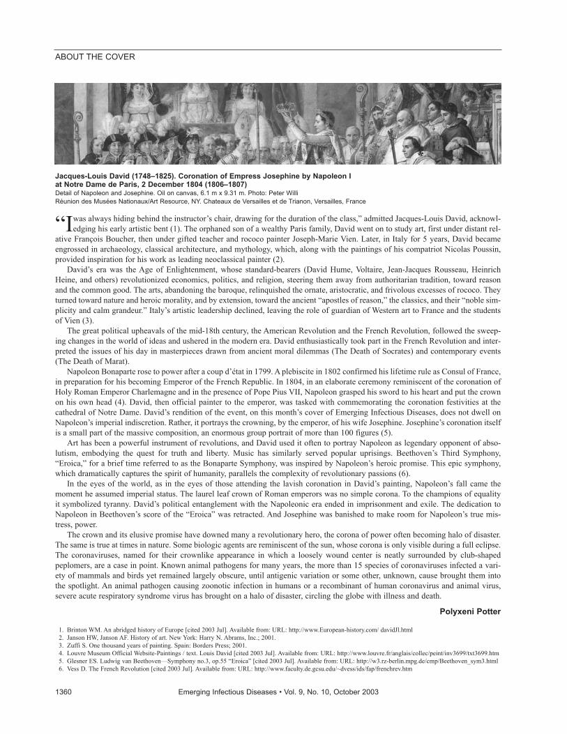
1360 Emerging Infectious Diseases • Vol. 9, No. 10, October 2003
ABOUT THE COVER
Jacques-Louis David (1748–1825). Coronation of Empress Josephine by Napoleon I at Notre Dame de Paris, 2 December 1804 (1806–1807) Detail of Napoleon and Josephine. Oil on canvas, 6.1 m x 9.31 m. Photo: Peter Willi Réunion des Musées Nationaux/Art Resource, NY. Chateaux de Versailles et de Trianon, Versailles, France
“Iwas always hiding behind the instructor’s chair, drawing for the duration of the class,” admitted Jacques-Louis David, acknowl-edging his early artistic bent (1). The orphaned son of a wealthy Paris family, David went on to study art, first under distant rel-
ative François Boucher, then under gifted teacher and rococo painter Joseph-Marie Vien. Later, in Italy for 5 years, David becameengrossed in archaeology, classical architecture, and mythology, which, along with the paintings of his compatriot Nicolas Poussin,provided inspiration for his work as leading neoclassical painter (2).
David’s era was the Age of Enlightenment, whose standard-bearers (David Hume, Voltaire, Jean-Jacques Rousseau, HeinrichHeine, and others) revolutionized economics, politics, and religion, steering them away from authoritarian tradition, toward reasonand the common good. The arts, abandoning the baroque, relinquished the ornate, aristocratic, and frivolous excesses of rococo. Theyturned toward nature and heroic morality, and by extension, toward the ancient “apostles of reason,” the classics, and their “noble sim-plicity and calm grandeur.” Italy’s artistic leadership declined, leaving the role of guardian of Western art to France and the studentsof Vien (3).
The great political upheavals of the mid-18th century, the American Revolution and the French Revolution, followed the sweep-ing changes in the world of ideas and ushered in the modern era. David enthusiastically took part in the French Revolution and inter-preted the issues of his day in masterpieces drawn from ancient moral dilemmas (The Death of Socrates) and contemporary events(The Death of Marat).
Napoleon Bonaparte rose to power after a coup d’état in 1799. A plebiscite in 1802 confirmed his lifetime rule as Consul of France,in preparation for his becoming Emperor of the French Republic. In 1804, in an elaborate ceremony reminiscent of the coronation ofHoly Roman Emperor Charlemagne and in the presence of Pope Pius VII, Napoleon grasped his sword to his heart and put the crownon his own head (4). David, then official painter to the emperor, was tasked with commemorating the coronation festivities at thecathedral of Notre Dame. David’s rendition of the event, on this month’s cover of Emerging Infectious Diseases, does not dwell onNapoleon’s imperial indiscretion. Rather, it portrays the crowning, by the emperor, of his wife Josephine. Josephine’s coronation itselfis a small part of the massive composition, an enormous group portrait of more than 100 figures (5).
Art has been a powerful instrument of revolutions, and David used it often to portray Napoleon as legendary opponent of abso-lutism, embodying the quest for truth and liberty. Music has similarly served popular uprisings. Beethoven’s Third Symphony,“Eroica,” for a brief time referred to as the Bonaparte Symphony, was inspired by Napoleon’s heroic promise. This epic symphony,which dramatically captures the spirit of humanity, parallels the complexity of revolutionary passions (6).
In the eyes of the world, as in the eyes of those attending the lavish coronation in David’s painting, Napoleon’s fall came themoment he assumed imperial status. The laurel leaf crown of Roman emperors was no simple corona. To the champions of equalityit symbolized tyranny. David’s political entanglement with the Napoleonic era ended in imprisonment and exile. The dedication toNapoleon in Beethoven’s score of the “Eroica” was retracted. And Josephine was banished to make room for Napoleon’s true mis-tress, power.
The crown and its elusive promise have downed many a revolutionary hero, the corona of power often becoming halo of disaster.The same is true at times in nature. Some biologic agents are reminiscent of the sun, whose corona is only visible during a full eclipse.The coronaviruses, named for their crownlike appearance in which a loosely wound center is neatly surrounded by club-shapedpeplomers, are a case in point. Known animal pathogens for many years, the more than 15 species of coronaviruses infected a vari-ety of mammals and birds yet remained largely obscure, until antigenic variation or some other, unknown, cause brought them intothe spotlight. An animal pathogen causing zoonotic infection in humans or a recombinant of human coronavirus and animal virus,severe acute respiratory syndrome virus has brought on a halo of disaster, circling the globe with illness and death.
Polyxeni Potter
1. Brinton WM. An abridged history of Europe [cited 2003 Jul]. Available from: URL: http://www.European-history.com/ davidJl.html 2. Janson HW, Janson AF. History of art. New York: Harry N. Abrams, Inc.; 2001. 3. Zuffi S. One thousand years of painting. Spain: Borders Press; 2001. 4. Louvre Museum Official Website-Paintings / text. Louis David [cited 2003 Jul]. Available from: URL: http://www.louvre.fr/anglais/collec/peint/inv3699/txt3699.htm5. Glesner ES. Ludwig van Beethoven—Symphony no.3, op.55 “Eroica” [cited 2003 Jul]. Available from: URL: http://w3.rz-berlin.mpg.de/cmp/Beethoven_sym3.html6. Vess D. The French Revolution [cited 2003 Jul]. Available from: URL: http://www.faculty.de.gcsu.edu/~dvess/ids/fap/frenchrev.htm
Emerging Infectious Diseases • Vol. 9, No. 10, October 2003 1361
NEWS & NOTES
Upcoming InfectiousDisease Conferences
October 7–8, 2003Intensive Update Course in Clinical Tropical Medicine and Travelers’ HealthAmerican Society of TropicalMedicine and HygieneSan Diego, CAContact: ASTMHPhone: 847-480-9592email: [email protected] site: http://www.astmh.org
October 9–12, 2003Infectious Diseases Society ofAmerica (IDSA)San Diego, CAContact: 703-299-0200email: [email protected] site: www.idsociety.org
October 26–29, 2003Symposium on BluetongueOffice International des EpizootiesTaormina, ItalyContact: 33 (0)1 44 15 18 88email: [email protected] site: www.oie.int
November 9–13, 20036th OIE Seminar on Biotechnologyand 11th International Symposium ofthe World Association of VeterinaryLaboratory DiagnosticiansOffice International des EpizootiesBangkok, ThailandContact: 33 (0)1 44 15 18 88email: [email protected] site: www.oie.int
November 15–19, 2003131st Annual Meeting, AmericanPublic Health Association San Francisco, CAContact: APHA Convention Services(202) 78-05600Web site: http://www.apha.org
1362 Emerging Infectious Diseases • Vol. 9, No. 10, October 2003
NEWS & NOTES
A Peer-Reviewed Journal Tracking and Analyzing Disease Trends Vol.9, No.11, November, 2003
Upcoming Issue For a complete list of articles included in the November issue,
and for articles published online ahead of print publication, see http://www.cdc.gov/ncidod/eid/upcoming.htm
Look in the November issue for the following topics:
Risks and Benefits of Pre-and Post-Exposure Smallpox Vaccination
Cardiac Deaths after 6 Million Smallpox Vaccinations, New York City, 1947
Toxoplasma gondii Infection in the United States, 1999-2000
Toxoplasma gondii and Schizophrenia
Coronavirus-positive Nasopharyngeal Aspirate as Predictor for Severe Acute Respiratory Syndrome Mortality
West Nile Virus Infection in Nonhuman Primate Breeding Colony, Concurrent with Human Epidemic, Southern Louisiana
Rapid Antigen Capture Assay To Detect West Nile Virus in Dead Corvids
Severe Acute Respiratory Syndrome-associated Coronavirus Infection
Fluoroquinolone-resistant Salmonella enterica Typhimurium in Humans
Cowpox with Severe Generalized Eruption, Finland
Flow Cytometry and T-Cell Response Monitoring after Smallpox Vaccination
Instructions to Authors
Manuscript Preparation. For word processing, use MS Word. Begineach of the following sections on a new page and in this order: title page,keywords, abstract, text, acknowledgments, biographical sketch, refer-ences, tables, figure legends, appendixes, and figures. Each figure shouldbe in a separate file. Title Page. Give complete information about each author (i.e., full name,graduate degree(s), affiliation, and the name of the institution in whichthe work was done). Clearly identify the corresponding author and pro-vide that author's mailing address (include phone number, fax number,and e-mail address). Include separate word counts for abstract and text. Keywords. Include up to 10 keywords; use terms listed in MedicalSubject Headings Index Medicus.Text. Double-space everything, including the title page, abstract, refer-ences, tables, and figure legends. Printed manuscript should be single-sided, beginning with the title page. Indent paragraphs; leave no extraspace between paragraphs. After a period, leave only one space beforebeginning the next sentence. Use 12-point Times New Roman font andformat with ragged right margins (left align). Italicize (rather than under-line) scientific names when needed. Biographical Sketch. Include a short biographical sketch of the firstauthor—both authors if only two. Include affiliations and the author's pri-mary research interests. References. Follow Uniform Requirements (www.icmje.org/index.html).Do not use endnotes for references. Place reference numbers in parenthe-ses, not superscripts. Number citations in order of appearance (includingin text, figures, and tables). Cite personal communications, unpublisheddata, and manuscripts in preparation or submitted for publication inparentheses in text. Consult List of Journals Indexed in Index Medicus foraccepted journal abbreviations; if a journal is not listed, spell out the jour-nal title. List the first six authors followed by “et al.” Do not cite refer-ences in the abstract.Tables and Figures. Create tables within MS Word’s table tool. Do notformat tables as columns or tabs. Send graphics in native, high-resolution(200 dpi minimum) .TIF (Tagged Image File), or .EPS (EncapsulatedPostscript) format. Graphics should be in a separate electronic file fromthe text file. For graphic files, use Arial font. Convert Macintosh files intothe suggested PC format. Figures, symbols, letters, and numbers shouldbe large enough to remain legible when reduced. Place figure keys with-in the figure. For more information see EID Style Guide (http://www.cdc.gov/ncidod/EID/style_guide.htm). Manuscript Submission. Include a cover letter indicating the proposedcategory of the article (e.g., Research, Dispatch) and verifying that thefinal manuscript has been seen and approved by all authors. To submit amanuscript, access Manuscript Central from the Emerging InfectiousDiseases website (www.cdc.gov/eid).
Manuscript Types
Perspectives. Articles should be under 3,500 words and should includereferences, not to exceed 40. Use of subheadings in the main body of thetext is recommended. Photographs and illustrations are encouraged.Provide a short abstract (150 words) and a brief biographical sketch offirst author. Articles in this section should provide insightful analysis andcommentary about new and reemerging infectious diseases and relatedissues. Perspectives may also address factors known to influence theemergence of diseases, including microbial adaptation and change,human demographics and behavior, technology and industry, economicdevelopment and land use, international travel and commerce, and thebreakdown of public health measures. If detailed methods are included, aseparate section on experimental procedures should immediately followthe body of the text.
Synopses. Articles should be under 3,500 words and should include ref-erences, not to exceed 40. Use of subheadings in the main body of the textis recommended. Photographs and illustrations are encouraged. Provide ashort abstract (150 words) and a brief biographical sketch of firstauthor—both authors if only two. This section comprises concise reviewsof infectious diseases or closely related topics. Preference is given toreviews of new and emerging diseases; however, timely updates of otherdiseases or topics are also welcome. If detailed methods are included, aseparate section on experimental procedures should immediately followthe body of the text. Research Studies. Articles should be under 3,500 words and shouldinclude references, not to exceed 40. Use of subheadings in the main bodyof the text is recommended. Photographs and illustrations are encour-aged. Provide a short abstract (150 words) and a brief biographical sketchof first author—both authors if only two. Report laboratory and epidemi-ologic results within a public health perspective. Although these reportsmay be written in the style of traditional research articles, they shouldexplain the value of the research in public health terms and place the find-ings in a larger perspective (i.e., "Here is what we found, and here is whatthe findings mean").Policy and Historical Reviews. Articles should be under 3,500 wordsand should include references, not to exceed 40. Use of subheadings inthe main body of the text is recommended. Photographs and illustrationsare encouraged. Provide a short abstract (150 words) and a brief biogra-phical sketch. Articles in this section include public health policy or his-torical reports that are based on research and analysis of emerging diseaseissues.Dispatches. Articles should be 1,000–1,500 words and need not be divid-ed into sections. If subheadings are used, they should be general, e.g.,“The Study” and “Conclusions.” Provide a brief abstract (50 words); ref-erences (not to exceed 15); figures or illustrations (not to exceed two);and a brief biographical sketch of first author—both authors if only two.Dispatches are updates on infectious disease trends and research. Thearticles include descriptions of new methods for detecting, characterizing,or subtyping new or reemerging pathogens. Developments in antimicro-bial drugs, vaccines, or infectious disease prevention or elimination pro-grams are appropriate. Case reports are also welcome. Commentaries. Thoughtful discussions (500–1,000 words) of currenttopics. Commentaries may contain references but should not include fig-ures or tables.Another Dimension. Thoughtful essays, short stories, or poems on philo-sophical issues related to science, medical practice, and human health.Topics may include science and the human condition, the unanticipatedside of epidemic investigations, or how people perceive and cope withinfection and illness. This section is intended to evoke compassion forhuman suffering and to expand the science reader's literary scope.Manuscripts are selected for publication as much for their content (theexperiences they describe) as for their literary merit.Letters. This section includes letters that present preliminary data orcomment on published articles. Letters (500–1,000 words) should not bedivided into sections, nor should they contain figures or tables.References (not more than 10) may be included.Book Reviews. Short reviews (250–500 words) of recently publishedbooks on emerging disease issues are welcome. The name of the book,publisher, and number of pages should be included.Announcements. We welcome brief announcements (50–150 words) oftimely events of interest to our readers. (Announcements may be postedon the journal Web page only, depending on the event date.) Conference Summaries. (500–1,000 words) of emerging infectious dis-ease conferences may provide references to a full report of conferenceactivities and should focus on the meeting's content rather than on indi-vidual conference participants.
Editorial Policy and Call for ArticlesEmerging Infectious Diseases is a peer-reviewed journal established expressly to promote the recognition of new and reemerging infectious diseasesaround the world and improve the understanding of factors involved in disease emergence, prevention, and elimination. The journal has an international scope and is intended for professionals in infectious diseases and related sciences. We welcome contributions frominfectious disease specialists in academia, industry, clinical practice, and public health, as well as from specialists in economics, demography, sociol-ogy, and other disciplines. Inquiries about the suitability of proposed articles may be directed to the Editor at 404-371-5329 (tel), 404-371-5449 (fax),or [email protected] (e-mail). Emerging Infectious Diseases is published in English and features the following types of articles: Perspectives, Synopses, Research Studies, Policyand Historical Reviews, Dispatches, Commentaries, Another Dimension, Letters, Book Reviews, and News and Notes. The purpose and requirementsof each type of article are described in detail below. To expedite publication of information, we post journal articles on the Internet as soon as they arecleared and edited. Chinese, French, and Spanish translations of some articles can be accessed through the journal’s home page at http://www.cdc.gov/eid.


