Embed Size (px)
Citation preview
CELLULAR AND SUBCELLULAR LOCALIZATION OF PDE10A, ASTRIATUM-ENRICHED PHOSPHODIESTERASE
Z. XIEa, W. O. ADAMOWICZb, W. D. ELDREDc, A. B. JAKOWSKId, R. J. KLEIMANb, D. G.MORTONd, D. T. STEPHENSONd, C. A. STRICKb, R. D. WILLIAMSb, and F. S. MENNITIb,*aProtein Sciences, Pfizer Global Research and Development, Eastern Point Road, Groton, CT06340, USA
bCNS Discovery, Pfizer Global Research and Development, Eastern Point Road, Groton, CT 06340,USA
cDepartment of Biology, Boston University, Boston, MA 02215, USA
dSafety Sciences, Pfizer Global Research and Development, Eastern Point Road, Groton, CT06340, USA
*Corresponding author. Tel: +1-860-441-5939; fax: +1-860-686-0013. E-mail address: [email protected] (F. S. Menniti).Abbreviations:
cAMP cyclic AMP
cGMP cyclic guanosine mono-phosphate
ChAT choline acetyl transferase
DAPI 4’,6-diamidino-2-phenylindole
DARPP-32 dopamine- and cyclic AMP-regulated phosphoprotein, Mr 32 kDa
n. nucleus
nNOS neuronal nitric oxide synthetase
PB 0.1 M phosphate buffer
PDE phosphodiesterase
PDE10A-ir PDE10A-like immunoreactivity
PSD postsynaptic densities
SPM synaptosomal membrane
SPS synaptic vesicle
SPV synaptosomal vesicle pellet.
NIH Public AccessAuthor ManuscriptNeuroscience. Author manuscript; available in PMC 2006 May 24.
Published in final edited form as:Neuroscience. 2006 May 12; 139(2): 597–607.
NIH
-PA Author Manuscript
NIH
-PA Author Manuscript
NIH
-PA Author Manuscript

AbstractPDE10A is a recently identified phosphodiesterase that is highly expressed by the GABAergicmedium spiny projection neurons of the mammalian striatum. Inhibition of PDE10A results in striatalactivation and behavioral suppression, suggesting that PDE10A inhibitors represent a novel class ofantipsychotic agents. In the present studies we further elucidate the localization of this enzyme instriatum of rat and cynomolgus monkey. We find by confocal microscopy that PDE10A-likeimmunoreactivity is excluded from each class of striatal interneuron. Thus, the enzyme is restrictedto the medium spiny neurons. Subcellular fractionation indicates that PDE10A is primarily membranebound. The protein is present in the synaptosomal fraction but is separated from the postsynapticdensity upon solubilization with 0.4% Triton X-100. Immuno-electron microscopy of striatumconfirms that PDE10A is most often associated with membranes in dendrites and spines. Immuno-gold particles are observed on the edge of the postsynaptic density but not within this structure. Ourstudies indicate that PDE10A is associated with post-synaptic membranes of the medium spinyneurons, suggesting that the specialized compartmentation of PDE10A enables the regulation ofintracellular signaling from glutamatergic and dopaminergic inputs to these neurons.
Keywordsphosphodiesterase; CNS; striatum; medium spiny neurons; immunohistochemistry
Cyclic nucleotides play an important role as second messengers in the CNS. Concentrationsof cyclic AMP (cAMP) and cyclic guanosine monophosphate (cGMP) are determined by therate of synthesis, and of equal importance, by the rate of degradation by the phosphodiesterases(PDEs). The PDEs are a super family of enzymes encoded by 21 genes and subdivided into 11distinct families according to structural, functional and kinetic properties (Soderling andBeavo, 2000). Most of the PDEs are differentially expressed within the CNS and many neuronsappear to express multiple PDEs. This suggests that different isozymes subserve distinctphysiological functions in different neuronal pathways and within individual neurons. Thus,an important step in elucidating these functions is mapping the differential localization of thePDEs within the CNS, both among different neuronal populations and at the subcellular levelwithin identified neuronal types.
PDE10A is the single member of one of the newest PDE gene families. Initial characterization(Fujishige et al., 1999a; Loughney et al., 1999; Soderling et al., 1999) indicates that PDE10Ais a dual-substrate PDE that is highly enriched in brain. Fujishige et al. (1999b) noted highexpression in human caudate nucleus (n.) and putamen. Seeger et al. (2003) subsequentlyreported that in rat, PDE10A mRNA and protein are highly enriched in the GABAergic mediumspiny projection neurons of the striatal complex (caudate n., n. accumbens, and olfactorytubercle), an expression pattern evident in other mammalian species (Coskran et al., 2005;Coskran et al., in preparation). The striatal complex forms the core of the basal ganglia, a systemof interconnected nuclei that process cortical information in the context of dopaminergicsignaling to regulate motoric, appetitive, and cognitive processes. Recently, papaverine wasidentified as a specific inhibitor of PDE10A (Siuciak et al., submitted for publication, 2005).Systemic administration of this compound to mice produces a rapid increase in striatal cGMPand cAMP along with downstream markers of activation of the protein kinase A/protein kinaseG signaling cascades. Papaverine potentiates catalepsy produced by the dopamine D2 receptorantagonist haloperidol in rats and inhibits conditioned avoidance responding in rats and mice.These findings suggest that inhibition of PDE10A may be a novel approach to the treatmentof psychosis (Siuciak et al., submitted for publication, 2005; in press, 2006).
XIE et al. Page 2
Neuroscience. Author manuscript; available in PMC 2006 May 24.
NIH
-PA Author Manuscript
NIH
-PA Author Manuscript
NIH
-PA Author Manuscript

The behavioral effects of papaverine suggest that PDE10A regulates the excitability of mediumspiny neurons (Siuciak et al., submitted for publication, 2005; in press ,2006). However, theexcitability of these neurons is also regulated by several classes of aspiny striatal inter-neurons(Kawaguchi et al., 1995). Expression of PDE10A in one or more of these interneuronpopulations would have implications for understanding the mechanism by which this enzymeregulates striatal output. Thus, the first aim of the present studies was to determine whetherPDE10A is expressed within any striatal interneurons in addition to the medium spinyprojection neurons. It is also becoming increasingly clear that cyclic nucleotide signaling ishighly compartmentalized within cells, indicating a mechanism whereby different PDEisozymes may sub-serve distinct physiological functions within a single cell (Houslay andAdams, 2003; Houslay and Milligan, 1997). Thus, the second aim of the present study was toinvestigate the subcellular distribution of PDE10A in the striatal medium spiny neurons, toprovide further context for elucidating the mechanism(s) whereby the enzyme regulates theexcitability of these neurons.
EXPERIMENTAL PROCEDURESAnimals
Male Sprague-Dawley rats (Charles River Laboratories, Kingston, NY, USA) weighing300-400 g were housed in groups of five to 10 at ambient temperature of 25-27 °C, under a12-h light/dark cycle. Food and water was available ad libitum. For preparation of samples forsubcellular fractionation, rats were killed by decapitation. For preparation of samples forimmunoelectron microscopy, rats were killed by overdose with 100 mg of pentobarbital priorto perfusion fixation. Cynomolgus monkeys (Charles River Primates, BRF, Houston, TX,USA) 2-3 years of age were housed singly in an AAALAC, International accredited animalfacility, fed commercial monkey chow supplemented with fruit, provided unlimited access towater, and used as untreated control animals on pharmacokinetic and toxicology studies.Immediately prior to sample collection, monkeys were sedated with i.m. injections of ketamine,killed with i.v. injections of pentobarbital, and exsanguinated. All procedures relating to animalcare and treatment were conducted according to the guidelines of the Institutional Animal Careand Use Committee at Pfizer, the ILAR Guide for the Care and Use of Laboratory Animals,and all federal and international regulations. All efforts were made to minimize the number ofanimals used and their suffering.
Western blot comparing rat and cynomolgus striatal extractsRat brains were collected after rapid decapitation and striatum was dissected using the atlas ofPaxinos and Watson (1986) as a guide. Tissue was homogenized by sonication in lysis bufferB (50 mM Tris-HCl, pH 7.5, 250 mM NaCl, 5 mM EDTA, pH 8.0, 0.1% nonidet P-40 andcomplete protease inhibitors). The lysates were centrifuged at 11,650×g for 30 min at 4 °C.Pellets were discarded and supernatants stored at -80 °C. Cynomolgus monkey brains werecollected and striatum was dissected using the atlas of Paxinos et al. (2000) as a guide. Tissuewas homogenized in a glass Teflon dounce homogenizer using five volumes to weight B-Perextraction reagent containing complete protease inhibitors. Lysates were centrifuged at11,650×g for 10 min at 4 °C. Pellets were discarded and supernatants stored at -80 °C. Proteinconcentrations in supernatants were determined by the bicinchoninic acid method (PierceChemical Co., Rockford, IL, USA). Equal amounts of striatal homogenates were resolved on4-12% NuPAGE Bis-Tris gel (Invitrogen Corporation, Carlsbad, CA, USA) in MOPS bufferand transferred to nitrocellulose as described below in Subcellular fractionation.
Immunohistochemistry and confocal microscopyDouble immunofluorescence techniques were employed to investigate the localization ofPDE10A relative to defined populations of striatal neurons in striatum of cynomolgus monkey.
XIE et al. Page 3
Neuroscience. Author manuscript; available in PMC 2006 May 24.
NIH
-PA Author Manuscript
NIH
-PA Author Manuscript
NIH
-PA Author Manuscript

The caudate and putamen from immersion fixed (10% neutral buffered formalin, 24 h durationfixation) cynomolgus monkey brain were processed according to routine histologicalprocedures and embedded in paraffin wax. Six micron sections were collected on chargedmicroscope slides, deparaffinized and hydrated to distilled water. Immunohistochemistry wasperformed using co-localization of two antibodies, derived from different species, on the sametissue section by sequential and simultaneous double immunofluorescence techniques.Immunohistochemistry conditions were optimized for each antibody on individual single-stained sections prior to dual staining. Control sections were stained with individual antibodieswhere one of the two antibodies was replaced with isotype control IgG from the appropriatespecies. Individual fluorophores and filter sets were chosen to eliminate the possibility ofoverlap of signal. Masked epitopes were retrieved using Antigen Retrieval Citra Solution(BioGenex, San Ramon, CA, USA) heated to 96 °C with a steamer for 20 min followed bycooling for 20 min at room temperature. Brain sections were incubated with the PDE10A mousemonoclonal antibody 24F3.F11 diluted 1:250 for 60 min at room temperature. PDE10A-likeimmunoreactivity (PDE10A-ir) was detected using a biotinylated secondary antibody, horseanti-mouse IgG (BA-2000; 1:150, Vector Laboratories, Burlingame, CA, USA), and visualizedwith Alexa 488-Streptavidin (Invitrogen Corporation). Sections stained with 24F3.F11 werethen incubated for 24 -48 h at 4 °C with antibodies to dopamine- and cyclic AMP-regulatedphosphoprotein, Mr 32 kDa (DARPP-32) (# 2302, 1:50, Cell Signaling Technology, Inc.,Beverly, MA, USA), calretinin (AB1550, 1:1000; Chemicon International Inc., Temecula, CA,USA), parvalbumin (Ab-1, PC255L; 1:500; Oncogene Science, Cambridge, MA, USA) orcholine acetyltransferase (AB144P; 1:500; Chemicon International Inc.). Interneuronalmarkers were visualized by labeling with goat anti-rabbit antibody directly conjugated to Alexa568 (1:200). Antibody to neuronal nitric oxide synthase (AB5380; 1:1000; ChemiconInternational Inc.) was co-incubated with 24F3.F11 (1:250) overnight at 4 °C and visualizedwith a cocktail of directly labeled secondary antibodies (1:200), goat anti-rabbit-Alexa 568and goat anti-mouse-Alexa 488, respectively. Nuclei were stained with 4’ ,6-diamidino-2-phenylindole (DAPI; Invitrogen Corporation). Images of PDE10A plus parvalbumin-stainedsections were acquired on a Leica TCS-SP laser scanning confocal microscope (LeicaMicrosystems Inc., Exton, PA, USA) equipped with argon, krypton, and UV lasers, whichallowed excitation at 488, 568, and 358 nm wavelengths for the detection of Alexa 488, Alexa568, and DAPI, respectively. The Alexa 488 dye yields a green signal, Alexa 568 is red andDAPI is blue when visualized with the appropriate fluorescent filters. The DARPP-32 plusPDE10A-stained images were photographed with a Zeiss Axioplan microscope equipped withan AxioCam HR digital camera (Carl Zeiss MicroImaging, Inc., Thornwood, NY, USA) andthe PDE10A plus neuronal nitric oxide synthetase (nNOS), choline acetyl transferase (ChAT)and calretinin dual-stained sections were photographed on a Nikon Eclipse E800 microscopeequipped with a SpotRT camera (Maeger Scientific, Dexter, MI, USA). Each fluorescentchannel was acquired sequentially and then merged to create the final image.
Immunoelectron microscopyPentobarbital-overdosed rats were perfused with 200 ml of saline (0.9%) followed by 250 mlof 4% paraformaldehyde with 0.1% glutaraldehyde pH 7.4 in 0.1 M phosphate buffer (PB).The brains were removed and block cut to expose the ventricles, then post-fixed for 30 min inthe same para/glut perfusate, and finally post-fixed for 3h in 4% paraformaldehyde (no glut)pH 10.1 at room temperature on a rotator. Following cryoprotection using an ascending sucroseseries, a sliding microtome was used to cut 60-75 μm thick frozen coronal sections. Followinga descending sucrose series into PB, the sections were treated with fresh 1% sodiumborohydride in PB for 30 min. The sections were then washed in PB and transferred into24F3.F11 diluted 1:2000 in PB for 48 h. Control sections were incubated in the absence of24F3.F11. Following 3×30 min washes in PB, the sections were transferred to goat anti-mouseFAB Nanogold secondary antibody (Nanoprobes, Inc., Yaphank, NY, USA) diluted 1:100 in
XIE et al. Page 4
Neuroscience. Author manuscript; available in PMC 2006 May 24.
NIH
-PA Author Manuscript
NIH
-PA Author Manuscript
NIH
-PA Author Manuscript

PB with 0.5% bovine serum albumin at 4 °C and incubated on a rotator for 72 h. After several15 min PB rinses, the sections were post-fixed with 1% glutaraldehyde in PB for 45 min.Following several changes of PB, the sections were silver intensified and gold toned(Nanoprobes, Inc.) before fixation in 1% OsO4 in PB for 45 min. The sections were thenincubated in 1% uranyl acetate in 30% acetone for 30 min, dehydrated using an acetone series,and embedded in epoxy resin. Thin sections were examined unstained using a JEOL 2010electron microscope.
Subcellular fractionationSubcellular fractionation was carried out as described by Blackstone et al. (1992). Brain tissuefrom six rats was homogenized in 20 volumes of lysis buffer (0.32 M sucrose, 4 mM HEPES/NaOH, pH 7.4, 5 mM DTT, 1 mM EDTA, and Complete protease inhibitor tablets) with aglass-Teflon homogenizer. The homogenate was centrifuged at 800×g to yield P0 and S0. Thepellet, P0, was washed in five volumes of lysis buffer, centrifuged again to yield P1 and S1.P1, containing crude nuclear pellet and unbroken cells, was discarded. S0 and S1 were pooledand centrifuged at 9000×g yielding S2 and a crude synaptosomal fraction (P2). P2 was washedin 10 volumes lysis buffer to yield a washed synaptosomal fraction (P2’) and S2’.P2’ was lysedby hypo-osmotic shock using a 25G needle to resuspend the pellet in three volumes of water.The lysed synaptosomal fraction was adjusted to 1 mM HEPES, pH 7.5 and rocked at 4 °C for30 min. The lysate was centrifuged at 25,000×g for 20 min to yield crude synaptosomalmembranes (SPM) and synaptic vesicles (SPS). The crude SPM were resuspended in sixvolumes lysis buffer and loaded onto a sucrose step gradient containing 0.8 M sucrose 1.0 M,and 1.2 M sucrose in 0.5 mM HEPES (pH 7.4) 5 mM DTT, 1 mM EDTA and complete proteaseinhibitor tables. The gradient was centrifuged for 2h at 65,000×g in a Beckman SW-28 rotor.Fractions were collected from the 0.8 M/1.0 M layer (SPM2), and at the 1.0 M/1.2 M interface(SPM4), with a crude mitochondrial pellet at the bottom of the tube. The SPM2 and crudemitochondrial pellet were resuspended directly in SDS sample buffer (Invitrogen). Accordingto Blackstone et al. (1992) the SPM4 is the synaptosomal plasma membrane fraction, fromwhich postsynaptic densities (PSDs) were isolated. The SPM4 were collected by centrifugationat 100,000×g for 1 h and washed once with lysis buffer. The washed membranes weresolubilized with 0.4% Triton X-100 in 0.5 mM HEPES (pH 7.4) for 1h at4°C and thencentrifuged at 100,000×g for 1 h. The resulting pellet was washed once in 0.4% Tritonextraction buffer and once again in lysis buffer. The pellet containing the PSD and the solublefractions were analyzed separately.
The supernatant S2 was further spun at 100,000×g using the Beckman TLA 100.2 rotor to yieldthe high speed membrane fraction P20. P20 was resuspended into lysis buffer and furtherincubated either with no addition, or with the addition of (final concentrations) 1 M NaCl, 0.4%Triton X-100, or 0.1 M Na2CO3 (pH 10) for 10 min on ice. The suspensions were then spunfor 1 h at 100,000×g using the Beckman TLA 100.2 rotor. The resulting pellets were washed,resuspended in lysis buffer and analyzed by Western blotting along with the supernatantfractions.
Samples were taken at various steps during the subcellular fractionation and loaded by equalvolumes, adjusting for resuspension volumes, for analysis by Western blotting using theInvitrogen NuPage system with detection using a LiCor Odyssey or with ECL DetectionSystem (Amersham Biosciences, Piscataway, NJ, USA). Samples were prepared using NuPageLDS sample buffer and run on 4-12% gels with MOPS or MES buffer and then transferred to0.45 μM pore nitrocellulose membrane. Blots were blocked in Odyssey block followed byincubation in primary and secondary antibody in 50% Odyssey block with 0.1% Tween-20 inPBS. Washes were carried out in 0.1% Tween-20 in PBS. The antibodies and titers employedwere: PDE10A mouse monoclonal antibody 24F3.F11 (1:1000); the plasma membrane marker,
XIE et al. Page 5
Neuroscience. Author manuscript; available in PMC 2006 May 24.
NIH
-PA Author Manuscript
NIH
-PA Author Manuscript
NIH
-PA Author Manuscript
sodium potassium ATPase antibody # ab7671 from Abcam (Cambridge, MA, USA; 1:1000);PSD95 mouse monoclonal antibody MA1-045 from Affinity Bioreagents (Golden, CO, USA;1:2000); NMDAR1 mouse monoclonal antibody 3556308 from BD Pharmingen (San Diego,CA, USA; 1:2000); rabbit polyclonal anti-DARPP-32 antibody from Chemicon (1:1000);synaptophysin mouse monoclonal RDI-PRO61012 from Research Diagnostics (Concord, MA,USA; 1:1000); polyclonal PDE2A antibody PD2A-100P from Fabgennix (Frisco, TX, USA;1:1000); PDE1B1 antibody # AB1655 from Chemicon (1:1000). Alexa 680 Fluorescent anti-mouse and anti-rabbit secondary antibodies were purchased from Molecular Probes (Eugene,OR, USA) and used at a dilution of 1:2000. IRdye 800 secondary antibodies from RocklandImmunochemicals (Gilbertsville, PA, USA) to mouse and rabbit were diluted 1:5000;horseradish peroxidase-conjugated anti-rabbit secondary antibody (Cell Signaling) at 1:5000dilution.
RESULTSPDE10A is expressed only in striatal medium spiny projection neurons
The medium spiny projection neurons that express PDE10A are the major neuronal type instriatum. However, there are several classes of striatal interneurons that play a major role inorganizing the activity of the medium spiny neurons (Kawaguchi et al., 1995). Thus, it is ofinterest to determine whether PDE10A is also expressed in any of the interneuron subtypes.This was investigated using immunofluorescent double staining to compare the disposition ofPDE10A protein with that of a marker for the medium spiny neurons (DARPP-32; Greengardet al., 1999) and each of the interneuron classes. Specifically, interneuron populations werelabeled with antibodies to nNOS, calretinin, ChAT, and parvalbumin.
Double labeling immunohistochemical analyses for PDE10A and the four interneuron markerswere carried out in both rat and cynomolgus monkey striatum. We have observed that thelocalization pattern of PDE10A in the brain is very similar across mammalian species includingmouse, rat, dog, cynomolgus monkey and human (Coskran et al., 2005; Coskran et al., inpreparation). We chose to use cynomolgus monkey brain for presentation of the double labelingin the present study for several reasons. In rat, PDE10A immunoreactivity in striatum is ofvery similar intensity in the cell bodies of the medium spiny neurons and in the neuropil. Incontrast in cynomolgus striatum, the intensity of PDE10A immunoreactivity in the mediumspiny neuron cell bodies is notably greater than that of the surrounding neuropil. This allowsfor an easier visualization of neuronal cell bodies for double labeling with interneuron markers.Nonetheless, the results of the double labeling immunohistochemical analyses for PDE10Aand the four interneuron markers in rats were qualitatively similar to those described here forcynomolgus monkey (unpublished observations). PDE10A-ir was detected with the mousemonoclonal antibody 24F3.F11 (Seeger et al., 2003). The specificity of this antibody forPDE10A in cynoognizes a single band of approximately 89 kDa in cynomolgus monkey striatalextract. The molecular size of this band and the intensity of immunoreactivity are similar tothat observed in an equivalent amount of rat striatal tissue extract. Omitting 24F3.F11 orprimary antibody for each of the interneuron markers abolished staining, also supportingspecificity of staining (data not shown).
In cynomolgus monkey, dense PDE10A-ir is observed throughout the striatum, including thevast majority of the cell bodies and the parenchyma (Figs. 1 A, 1B and 2A), as in rat (Coskranet al., 2005;Seeger et al., 2003; Coskran et al., in preparation). The pattern of PDE10A-ir wasindistinguishable in samples derived from four monkeys, thus, further detailed analysis usingdual labeling was carried out on samples derived from a single animal. There was a completeoverlap between cell bodies expressing DARPP-32-like immunoreactivity and PDE10A-ir(Fig. 1A and 1B). This pattern of labeling with 24F3.F11 is interpreted to indicate that PDE10A
XIE et al. Page 6
Neuroscience. Author manuscript; available in PMC 2006 May 24.
NIH
-PA Author Manuscript
NIH
-PA Author Manuscript
NIH
-PA Author Manuscript
is expressed in the majority of the medium spiny projection neurons, including the dendriticarbors of these neurons (Seeger et al., 2003).
An antibody to nNOS labeled the cell bodies and main dendritic branching of a rare subset ofneurons within cynomolgus monkey striatum (Fig. 2B and 2C). In double stained sections, theneurons labeled with the nNOS antibody were completely devoid of PDE10A-ir (Fig. 2C).Antibodies to calretinin, ChAT, and parvalbumin were used to identify three additionalinterneuron populations. Again, each antibody labeled the cell bodies of small cell populations(Fig. 1D-F). In each case, there was no overlap with PDE10A-ir (Fig. 1D-F). The lack of co-localization between PDE10A-ir and parvalbumin-like immunoreactivity is highlighted in theconfocal image illustrated in Fig. 1F. The Ortho-Slice function in the Leica confocal softwareallowed simultaneous visualization of a point of interest from an image stack in both the y-zplane and x-z plane. This point of interest in Fig. 1F, a single parvalbumin-labeled cell body(red), shows virtually no signal overlap between PDE10A-ir (green) and parvalbumin-likeimmunoreactivity (red), thereby confirming the lack of co-localization. Similar results wereobserved using rat striatum (unpublished observations). These results indicate that PDE10Ais not expressed in the different interneuron populations within the striatum.
Subcellular distribution of PDE10A-biochemical analysisGiven that the majority of the neurons in the striatum are the medium spiny projection neurons,and that these neurons appear to be the only population expressing PDE10A, we nextinvestigated the subcellular distribution of PDE10A in the medium spiny neurons usingclassical differential centrifugation techniques on rat striatal homogenates. The differentialcentrifugation scheme employed is illustrated in Fig. 3. The qualitative distribution of PDE10Ain the fractions isolated using this scheme is indicated (red). Similar results were obtained inthree separate experiments, although not all fractions were analyzed in each experiment.
Striatal homogenate was first centrifuged at 800×g. The supernatant (S1) was then fractionatedby centrifugation at 9000×g to isolate a crude synaptosomal vesicle pellet (SPV). Western blotanalysis with 24F3.F11 indicates that approximately 50% of the PDE10A-ir was recovered inthe SPV and 50% remained in the supernatant (S2; Fig. 4). Further centrifugation of S2 at100,000×g followed by Western blotting indicated that the majority of the PDE10A-ir wasrecovered in the pellet (see Fig. 6). The SPV was lysed by resuspension in hypotonic bufferand then fractionated by centrifugation at 25,000×g. PDE10A-ir was recovered in the SPMfraction but was not detectable in the SPS. The SPM was then resuspended and resolved on asucrose step gradient into three fractions: SPM2 containing myelin sheets, SPM4 containingthe PSD, and a pellet containing mitochondria and other insoluble material (SPMB). PDE10A-ir loaded onto this gradient was recovered in all three fractions (SPM4, Fig. 5; SPM2 andSPMB, data not shown).
To investigate whether PDE10A is tightly associated with the signaling complexes of the PSD,a PSD containing fraction (SPM4) was isolated from the SPM using a sucrose step gradient.Upon solubilization of SPM4 with Triton X-100 and centrifugation, both PSD-95- andNMDAR1-like immunoreactivity were recovered in the pellet (Fig. 5), as previously reported(Kim and Sheng, 2004). In contrast, PDE10A-ir, was recovered exclusively in the solublefraction (SPM4S).
The distribution of PDE10A-ir was also compared with that of a number of proteins used asmarkers for different subcellular compartments (Fig. 4). PDE10A-ir had a distribution similarto that of the plasma membrane associated Na+/K+ ATPase and PSD95. This distributiondiffered from that of DARPP-32, which was detected in the S2 but not in the subsequent SPV.This distribution of DARPP-32 is consistent with the previous reports that the protein iscytosolic (Greengard et al., 1999;Hemmings et al., 1984). The distribution of PDE10A-ir was
XIE et al. Page 7
Neuroscience. Author manuscript; available in PMC 2006 May 24.
NIH
-PA Author Manuscript
NIH
-PA Author Manuscript
NIH
-PA Author Manuscript

also distinct from that of synaptophysin, an integral membrane glycoprotein associated withpresynaptic nerve terminals vesicles (Wiedenmann and Franke, 1985). Like PDE10A-ir,synaptophysin-like immunoreactivity was observed in the SPV fraction. Upon hypotonic lysisof the SPV fraction, synaptophysin-like immunoreactivity was recovered primarily in thesoluble fraction SPS, which presumably contains the presynaptic neurotransmitter vesicles. Incontrast, PDE10A-ir was recovered primarily in the membrane fraction SPM.
There are several PDEs that are also highly expressed in the striatal medium spiny neurons,including PDE1B (Repaske et al., 1993) and PDE2 (Van Staveren et al., 2003) and wecompared the distribution of these enzymes to that of PDE10A. PDE2-like immunoreactivityhad a distribution similar to that of PDE10A with one exception. Like PDE10A-ir, PDE2-likeimmunoreactivity was observed in the SPV, and SPM fractions. However, PDE2-likeimmunoreactivity was also observed in SPS, in contrast to PDE10A-ir. After sucrose gradientdensity fractionation of SPM, PDE2-like immunoreactivity was recovered in SPM4 and, likePDE10A, was nearly completely excluded from the Triton-insoluble PSD fraction. In contrastto PDE10A and PDE2, PDE1B-like immunoreactivity was observed almost exclusively insoluble fractions. Thus, the distribution of PDE1B-like immunoreactivity was very similar tothat of DARPP-32.
The mechanism of membrane association of PDE10A was also investigated. The S2 fractionwas centrifuged at high speed (100,000×g) to obtain the P20 membrane fraction, which wasthen solubilized under different conditions and centrifuged again. As shown in Fig. 6, PDE10A-ir was recovered in the supernatant fraction following incubation of P20 with 0.4% TritonX-100. In contrast, PDE10A-ir remained in the pellet in incubations containing 1 M NaCl or0.1 M Na2CO3 (pH 10). Purified rat recombinant PDE10A incubated with each of thesesolubilization buffers and then centrifuged was recovered in the supernatant. Thus, recoveryof the native PDE10A in pelleted fraction is unlikely due to artifactual protein precipitation.These results suggest that PDE10A associates with membranes through a non-ionic interaction.
Subcellular distribution of PDE10A-immunoelectron microscopyThe results of the biochemical subcellular fractionation studies described above indicate thatPDE10A is primarily membrane associated in the medium spiny neurons. Our previous studyin rat (Seeger et al., 2003) and the results presented in Fig. 1 from cynomolgus monkey indicatethat the enzyme is distributed throughout the dendritic compartment of these neurons.However, it appears that PDE10A is not tightly associated with the PSD. Thus, to gain greaterinsight into the distribution of PDE10A in the dendritic compartment, we evaluated thedistribution of PDE10A in rat striatum by immunoelectron microscopy.
Fig. 7 is representative images of the distribution of gold particles in 24F3.F11-labeled sectionsof rat striatum capturing a number of vesicle-filled nerve terminals (P) of presumedglutamatergic striatal afferents as well as dendrites (D) and spines (S) of presumed mediumspiny neurons. The majority of gold particles are observed in close apposition to membranesof dendrites and spines (blue arrows). Particles are also observed within spines not obviouslyassociated with a membrane (red arrows). Interestingly, particles are observed at the edge ofPSDs (yellow arrows) but not directly apposed to these structures. Particles are not observedassociated with vesicle-filled presynaptic terminals. Finally, in samples from which the primaryantibody was omitted during processing for immunoelectron microscopy, virtually no silvergrains were detected, indicating the specificity of the immunolabeling (data not shown).
DISCUSSIONPDE10A is highly enriched in the mammalian striatum (Coskran et al., 2005; Seeger et al.,2003; Coskran et al., in preparation; and present study) and specifically in the GABAergic
XIE et al. Page 8
Neuroscience. Author manuscript; available in PMC 2006 May 24.
NIH
-PA Author Manuscript
NIH
-PA Author Manuscript
NIH
-PA Author Manuscript

medium spiny projection neurons that are the major neuronal cell type in this brain region. Themedium spiny neurons are the principal input site for the basal ganglia circuit (Wilson,1998). These neurons receive a massive excitatory input from cortical and thalamicglutamatergic neurons and are also the principal recipient of the rostral projection of themidbrain dopaminergic neurons. The medium spiny neurons integrate this cortical/thalamicinformation in the context of dopaminergic signaling, and reflect this output as a single burstor short bursts of action potentials from these typically quiescent neurons. Thus, the regulationof medium spiny neuron excitability is a key element in regulating information processing bythe basal ganglia complex. Recent results utilizing a PDE10A inhibitor and genetic inactivationof PDE10A suggest that this enzyme plays a critical role in regulating the excitability of theseneurons. Papaverine has been identified as a specific inhibitor of PDE10A, enabling aninvestigation of the behavioral effects of inhibiting this enzyme in rodents (Siuciak et al.,submitted for publication, 2005). PDE10A inhibition with papaverine potentiates haloperidol-induced catalepsy in rats, inhibits conditioned avoidance responding in mice (Siuciak et al.,submitted for publication, 2005), and inhibits the locomotor hyperactivity induced bystimulants (Schmidt et al., 2003). The PDE10A knockout mice exhibit reduced locomotoractivity when introduced into a novel environment and also have a reduced locomotorstimulatory response to the NMDA receptor antagonist PCP but no change in response toamphetamine (Siuciak et al., in press, 2006). Thus, the behavioral effects of papaverine andthe behavioral phenotype of the PDE10A knockout mice lead us to hypothesize that PDE10Ainhibition results in an activation of striatal output leading to suppression of behavioralresponsiveness (Siuciak et al., submitted for publication, 2005; in press, 2006). We arecurrently investigating the molecular mechanisms by which inhibition of PDE10A accountsfor the hypothesized changes in striatal output. The present study provides several additionalpieces of information that extends this investigation.
First, it is reported that PDE10A is expressed within the striatum exclusively by the mediumspiny projection neurons and not in the different populations of striatal inter-neurons. Striatalinterneurons play a key role in regulating the activity of medium spiny neurons (Kawaguchi,1997; Kawaguchi et al., 1995). If PDE10A were expressed in one or more of these interneuronpopulations, then inhibition of interneuronal PDE10A might be expected to yield additionallevels of regulation impinging upon medium spiny neuron output via interneuronal synapticactivity. Thus, we investigated whether PDE10A is expressed in the inter-neuron populations.Using double fluorescence immunohistochemistry and confocal microscopy in cynomolgusmonkey striatum, no observable PDE10A expression was detectable in any of the four classesof striatal interneurons that are characterized by expression of nNOS, calretinin, ChAT, orparvalbumin. Instead, the expression of PDE10A in cell bodies overlaps with that ofDARPP-32, which is a signaling molecule also highly expressed in striatal medium spinyneurons across mammalian species (Green-gard et al., 1999). Similar observations were madein rat striatum (unpublished observations). These results indicate that PDE10A is expressedexclusively by the medium spiny projection neurons within the striatum. Therefore, the effectof PDE10A inhibition on striatal output results from effects intrinsic to the medium spinyneurons.
In the medium spiny neurons, PDE10A was found to be highly enriched in membrane fractionsduring classical subcellular fractionation. PDE10A-ir in the cleared homogenate from ratstriatum (S1) was recovered in the SPV along with the plasma membrane-associated Na+/K+
ATPase, and the synapse-associated proteins PSD95 and synaptophysin. In contrast, thecytosolic protein DARPP-32 was excluded from the SPV fraction. Upon hypotonic lysis of thesynaptosomal fraction, both PDE10A and PSD95 remained in association with the membranefraction, whereas synaptophysin was recovered in the supernatant, presumably associated withpresynaptic neurotransmitter vesicles (Wiedenmann and Franke, 1985). A sucrose density stepgradient was then used to isolate a fraction enriched in the PSD complex. This complex contains
XIE et al. Page 9
Neuroscience. Author manuscript; available in PMC 2006 May 24.
NIH
-PA Author Manuscript
NIH
-PA Author Manuscript
NIH
-PA Author Manuscript

a number of tightly associated proteins that are involved in the postsynaptic signaling responseto glutamate (Kim and Sheng, 2004; Sheng and Sala, 2001). Upon Triton X-100 solubilization,PDE10A-ir was completely separated from PSD95 and NMDA receptor-like immuno-reactivity (Kornau et al., 1995). These results indicate that PDE10A is highly associated withmembranes from the medium spiny neurons but is not apparently an integral part of the PSDcomplex of these neurons.
Analysis of the distribution of PDE10A-ir using immunoelectron microscopy confirms andextends the above conclusion. In dendrites of medium spiny neurons, PDE10A-ir was primarilyfound apposed to membranes. In spines, the majority of the PDE10A-ir was also found inapparent association with membranes. However, PDE10A-ir was also observed with no clearmembrane association. Immunoreactivity was not observed within the electron dense regioncharacteristic of the PSD. However, it was not uncommon to observe gold particles at the edgesof these structures. Thus, PDE10A is apparently contained within the spines of the mediumspiny neurons, suggesting that the enzyme is appropriately compartmentalized to regulate post-synaptic cyclic nucleotide signaling and limit cyclic nucleotide accumulation near dendriticmembranes.
The present results corroborate and extend the findings of Kotera et al. (2004). This groupreported that PDE10A is primarily membrane associated in rat brain. Expression of PDE10Aisoforms in PC12 cells suggested that membrane association depends on a unique n-terminalfragment of the PDE10A2 splice variant, the primary isoform of PDE10A expressed in ratstriatum. Whereas in the PC12 cells, PDE10A2 appears to be localized primarily to membranesof the Golgi apparatus, the present results indicate that in medium spiny neurons membraneassociation extends to dendrites and spines. The results of the present study suggest thatPDE10A associates with membrane through a hydrophobic interaction, since the protein issolubilized by treatment of membranes with Triton X-100 but remains membrane associatedduring treatment with high salt (Fig. 6). We also observed that PDE10A remained associatedwith membranes following treatment with Na2CO3. Resistance to solubilization by alkalinecarbonate classifies PDE10A as an ‘integral’ membrane protein (Schwab et al., 2000). A singlepotential transmembrane region for rat PDE10A2, at amino acids 198-214, is predicted by thePSORT program for subcellular localization signals (Nakai and Horton, 1999). This regionmay contribute to the apparently tight association of PDE10A with membranes, but this remainsto be determined.
Kotera et al. (2004) reported compelling evidence that the association of PDE10A withmembranes may be regulated by n-terminal phosphorylation by PKA. Interestingly, we findin the present study through visualization by electron microscopy that the compartment wherePDE10A may not be membrane associated is in spines. An intriguing speculation is that theactivity of PDE10A is regulated by PKA (or PKG) phosphorylation primarily in the spines.The veracity of this speculation and the consequence of such redistribution for the regulationby PDE10A of signaling within the spines await further study.
There are a number of PDEs that are highly expressed in striatum in addition to PDE10A. Theseinclude two other isozymes, PDE1B (Repaske et al., 1993) and PDE2 (Van Staveren et al.,2003), that, like PDE10A, are capable of hydrolyzing both cAMP and cGMP. It is becomingincreasingly apparent that expression of multiple PDE isoforms in a given cell population iscommon and that these different isoforms subserve distinct roles in the regulation ofintracellular signaling cascades. Such segregation of functional activity for enzymes withseemingly identical catalytic activities occurs, at least in part, by differential subcellulardistribution and compartmentation (Conti, 2000; Conti et al., 2003; Houslay et al., 2003). Thus,it was of interest to begin to investigate the possibility of differences in the subcellulardistribution of PDE10A compared with PDE1B and PDE2 in striatum. The subcellular
XIE et al. Page 10
Neuroscience. Author manuscript; available in PMC 2006 May 24.
NIH
-PA Author Manuscript
NIH
-PA Author Manuscript
NIH
-PA Author Manuscript

distribution of PDE1B is clearly distinct from that of PDE10A. PDE1B-like immunoreactivitywas recovered almost exclusively in the soluble fraction of the striatal extracts, similar to whatwas observed for the cytosolic signaling molecule DARPP-32 (Hemmings et al., 1984).Interestingly, the phenotype of PDE1B knockout mice appears to be distinctly different fromthat of the PDE10A knockout mice. Whereas the PDE1B knockout mice have a hyperlocomotorphenotype (Reed et al., 2002), the PDE10A knockout mice are characterized as exhibiting ahypolocomotor phenotype (Siuciak et al., in press, 2006). PDE1B knockout is associated withan increased phosphorylation of DARPP-32 at the PKA-regulated site, leading to thesuggestion that PDE1B plays a role in the regulation of dopamine signaling in the striatum. Itwill be of interest to investigate the effects, if any, of PDE10A inhibition on DARPP-32phosphorylation and signaling through this cascade. The subcellular distribution of PDE2appears to be distinct from that of both PDE10A and PDE1B. Like PDE10A, PDE2 ismembrane associated during subcellular fractionation. However, PDE2-like immunoreactivitywas detected in the soluble fraction following lysis of the synaptosomal pellet, in contrast toPDE10A. The cellular distribution of PDE2 in the CNS appears to be more widespread thanthat of PDE10A, including expression in cortical neurons (Van Staveren et al., 2003). Thus, itis possible that the PDE2-like immuno-reactivity recovered in the synaptosomal supernatantmay have originated in cortical neurons projecting into the striatum.
CONCLUSIONIn summary, the present study indicates that PDE10A is localized exclusively in the mediumspiny projection neurons of the mammalian striatum. In these neurons, the enzyme is associatedwith dendrites and spines but is not tightly associated with the signaling complex of the PSD.Other studies (Siuciak et al., submitted for publication, 2005; in press, 2006) suggest thatPDE10A plays a role in regulating the excitability of the medium spiny neurons. The findingspresented here begin to elucidate the signaling compartment through which PDE10A regulatesthe excitability of these neurons.
REFERENCESBlackstone CD, Moss SJ, Martin LJ, Levey AI, Price DL, Huganir RL. Biochemical characterization and
localization of a non-N-methyl-D-aspartate glutamate receptor in rat brain. J Neurochem1992;58:1118–1126. [PubMed: 1371146]
Conti M. Phosphodiesterases and cyclic nucleotide signaling in endocrine cells. Mol Endocrinol2000;14:1317–1327. [PubMed: 10976911]
Conti M, Richter W, Mehats C, Livera G, Park JY, Jin C. Cyclic AMP-specific PDE4 phosphodiesterasesas critical components of cyclic AMP signaling. J Biol Chem 2003;278:5493–5496. [PubMed:12493749]
Coskran, TM.; Jakowski, AB.; Ramirez, AD.; Morton, DG.; Menniti, FS.; Schmidt, CJ.; Kleiman, RJ.;Ryan, AM.; Stephenson, D. PDE10A: a CNS specific cGMP phosphodiesterase that is expressed inmedium spiny neurons from multiple species. Society for Neuroscience 32nd annual meeting; 2005.
Fujishige K, Kotera J, Michibata H, Yuasa K, Takebayashi S, Okumura K, Omori K. Cloning andcharacterization of a novel human phosphodiesterase that hydrolyzes both cAMP and cGMP(PDE10A). J Biol Chem 1999a;274:18438–18445. [PubMed: 10373451]
Fujishige K, Kotera J, Omori K. Striatum- and testis-specific phosphodiesterase PDE10A isolation andcharacterization of a rat PDE10A. Eur J Biochem 1999b;266:1118–1127. [PubMed: 10583409]
Greengard P, Allen PB, Nairn AC. Beyond the dopamine receptor: the DARPP-32/protein phosphatase-1cascade. Neuron 1999;23:435–447. [PubMed: 10433257]
Hemmings HC Jr, Nairn AC, Aswad DW, Greengard P. DARPP-32, a dopamine- and adenosine 3’:5’-monophosphate-regulated phosphoprotein enriched in dopamine-innervated brain regions. II.Purification and characterization of the phosphoprotein from bovine caudate nucleus. J Neurosci1984;4:99–110. [PubMed: 6319628]
XIE et al. Page 11
Neuroscience. Author manuscript; available in PMC 2006 May 24.
NIH
-PA Author Manuscript
NIH
-PA Author Manuscript
NIH
-PA Author Manuscript

Houslay MD, Adams DR. PDE4 cAMP phosphodiesterases: modular enzymes that orchestrate signallingcross-talk, desensitization and compartmentalization. Biochem J 2003;370:1–18. [PubMed:12444918]
Houslay MD, Milligan G. Tailoring cAMP-signalling responses through isoform multiplicity. TrendsBiochem Sci 1997;22:217–224. [PubMed: 9204709]
Kawaguchi Y. Neostriatal cell subtypes and their functional roles. Neurosci Res 1997;27:1–8. [PubMed:9089693]
Kawaguchi Y, Wilson CJ, Augood SJ, Emson PC. Striatal interneurones: chemical, physiological andmorphological characterization. Trends Neurosci 1995;18:527–535. [PubMed: 8638293]
Kim E, Sheng M. PDZ domain proteins of synapses. Nat Rev Neurosci 2004;5:771–781. [PubMed:15378037]
Kornau HC, Schenker LT, Kennedy MB, Seeburg PH. Domain interaction between NMDA receptorsubunits and the postsynaptic density protein PSD-95. Science 1995;5231:1737–1740. [PubMed:7569905]
Kotera J, Sasaki T, Kobayashi T, Fujishige K, Yamashita Y, Omori K. Subcellular localization of cyclicnucleotide phosphodiesterase type 10A. J Biol Chem 2004;279:4366–4375. [PubMed: 14604994]
Loughney K, Snyder PB, Uher L, Rosman GJ, Ferguson K, Florio VA. Isolation and characterization ofPDE10A, a novel human 3’,5’-cyclic nucleotide phosphodiesterase. Gene 1999;234:109–117.[PubMed: 10393245]
Nakai K, Horton P. PSORT: a program for detecting sorting signals in proteins and predicting theirsubcellular localization. Trends Biochem Sci 1999;24:34–35. [PubMed: 10087920]
Paxinos, G.; Huang, X-F.; Toga, AW. The rhesus monkey brain in stereotaxic coordinates. AcademicPress; San Diego: 2000.
Paxinos, G.; Watson, C. The rat brain in stereotaxic coordinates. Academic Press, Inc; San Diego: 1986.Reed TM, Repaske DR, Snyder GL, Greengard P, Vorhees CV. Phosphodiesterase 1B knock-out mice
exhibit exaggerated loco-motor hyperactivity and DARPP-32 phosphorylation in response todopamine agonists and display impaired spatial learning. J Neurosci 2002;22:5188–5197. [PubMed:12077213]
Repaske DR, Corbin JG, Conti M, Goy MF. A cyclic GMP-stimulated cyclic nucleotidephosphodiesterase gene is highly expressed in the limbic system of the rat brain. Neuroscience1993;56:673–686. [PubMed: 8305078]
Schmidt CJ, Chapin DS, McCarthy SA, Fujiwara RA, Shrikhande A, Chambers L, Wong SF, SiuciakJA. The neurochemical and behavioral effects of papaverine in vivo suggest PDE10 inhibition is“antipsychotic.”. Schizophr Res 2003;60:114.
Schwab RB, Okamoto T, Scherer PE, Lisanti MP. Analysis of the association of proteins with membranes.Curr Protoc Cell Biol 2000;5 4:1–17.
Seeger TF, Bartlett B, Coskran TM, Culp JS, James LC, Krull DL, Lanfear J, Ryan AM, Schmidt CJ,Strick CA, Varghese AH, Williams RD, Wylie PG, Menniti FS. Immunohistochemical localizationof PDE10A in the rat brain. Brain Res 2003;985:113–126. [PubMed: 12967715]
Sheng M, Sala C. PDZ domains and the organization of supramolecular complexes. Annu Rev Neurosci2001;24:1–29. [PubMed: 11283303]
Siuciak JA, Chapin DS, Harms JF, Lebel LA, McCarthy SA, Chambers L, Shrikhande A, Wong SF,Menniti FS, Schmidt CJ. The striatum-enriched phosphodiesterase PDE10A: role in regulation ofbasal ganglia output. J Neurosci. 2005submitted for publication
Siuciak JA, McCarthy SA, Chapin DS, Fujiwara RA, James LC, Williams RD, Stock JL, McNeish JD,Strick CA, Menniti FS, Schmidt CJ. Genetic deletion of the striatum-enriched phosphodiesterasePDE10A: evidence for altered striatal function. Neuropharmacol. 2006in press
Soderling SH, Bayuga SJ, Beavo JA. Isolation and characterization of a dual-substrate phosphodiesterasegene family: PDE10A. Proc Natl Acad Sci U S A 1999;96:7071–7076. [PubMed: 10359840]
Soderling SH, Beavo JA. Regulation of cAMP and cGMP signaling: new phosphodiesterases and newfunctions. Curr Opin Cell Biol 2000;12:174–179. [PubMed: 10712916]
Van Staveren WC, Steinbusch HW, Markerink-van Ittersum M, Repaske DR, Goy MF, Kotera J, OmoriK, Beavo JA, de Vente J. mRNA expression patterns of the cGMP-hydrolyzing phosphodiesterases
XIE et al. Page 12
Neuroscience. Author manuscript; available in PMC 2006 May 24.
NIH
-PA Author Manuscript
NIH
-PA Author Manuscript
NIH
-PA Author Manuscript

types 2, 5, and 9 during development of the rat brain. J Comp Neurol 2003;467:566–580. [PubMed:14624489]
Wiedenmann B, Franke WW. Identification and localization of synaptophysin, an integral membraneglycoprotein of Mr 38,000 characteristic of presynaptic vesicles. Cell 1985;41:1017–1028. [PubMed:3924408]
Wilson, CJ. Basal ganglia. In: Sheperd, GM., editor. The synaptic organization of the brain. 4th edition.Oxford University Press; New York: 1998. p. 329-376.
XIE et al. Page 13
Neuroscience. Author manuscript; available in PMC 2006 May 24.
NIH
-PA Author Manuscript
NIH
-PA Author Manuscript
NIH
-PA Author Manuscript

Fig. 1.(A, B) Localization of PDE10A and DARPP-32 in striatum of cynomolgus monkey. Sectionsof cynomolgus monkey were stained sequentially with antibodies to PDE10A (green) andDARPP-32 (red) as described in Experimental Procedures. Images were merged and overlapbetween PDE10A and DARPP-32 is shown in yellow. Two sections are shown: A,magnification 20×; B, magnification 60×. (C) Western blot of striatal extracts from twocynomolgus monkeys (lanes 2 and 3) and rat (lane 4) stained for PDE10A-ir with 24F3.F11.Equal amounts of striatal extracts were loaded. Rat recombinant PDE10A is shown as acomparator (lane 5) and molecular weight markers are in lane 1.
XIE et al. Page 14
Neuroscience. Author manuscript; available in PMC 2006 May 24.
NIH
-PA Author Manuscript
NIH
-PA Author Manuscript
NIH
-PA Author Manuscript
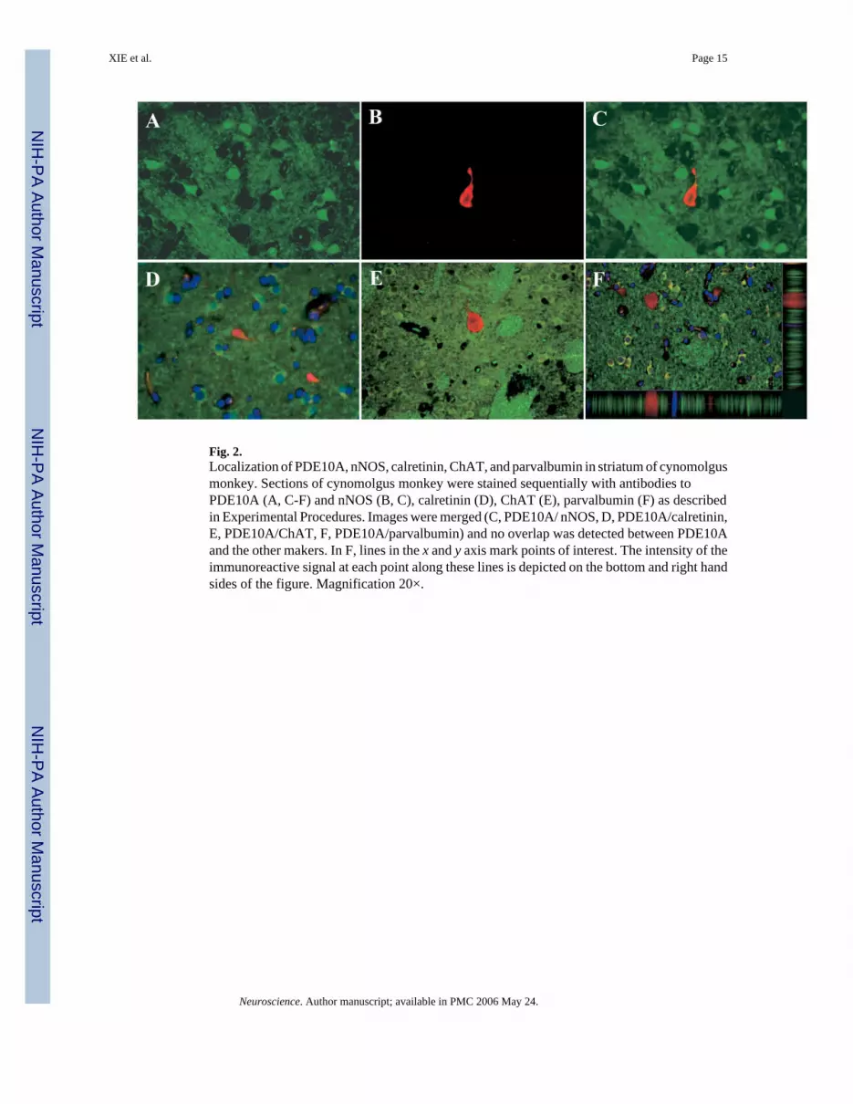
Fig. 2.Localization of PDE10A, nNOS, calretinin, ChAT, and parvalbumin in striatum of cynomolgusmonkey. Sections of cynomolgus monkey were stained sequentially with antibodies toPDE10A (A, C-F) and nNOS (B, C), calretinin (D), ChAT (E), parvalbumin (F) as describedin Experimental Procedures. Images were merged (C, PDE10A/ nNOS, D, PDE10A/calretinin,E, PDE10A/ChAT, F, PDE10A/parvalbumin) and no overlap was detected between PDE10Aand the other makers. In F, lines in the x and y axis mark points of interest. The intensity of theimmunoreactive signal at each point along these lines is depicted on the bottom and right handsides of the figure. Magnification 20×.
XIE et al. Page 15
Neuroscience. Author manuscript; available in PMC 2006 May 24.
NIH
-PA Author Manuscript
NIH
-PA Author Manuscript
NIH
-PA Author Manuscript

Fig. 3.Schematic outline of subcellular fractionation sequence. See Experimental Procedures for fulldescription of different fractions. Red, fractions containing substantial PDE10A-ir, black,fractions with little or no detectable PDE10A-ir.
XIE et al. Page 16
Neuroscience. Author manuscript; available in PMC 2006 May 24.
NIH
-PA Author Manuscript
NIH
-PA Author Manuscript
NIH
-PA Author Manuscript

Fig. 4.Western blot analysis of subcellular fractions from rat striatum with detection using a LiCorOdyssey. Fractions indicated at the top of the figure and antigens indicated on the left aredescribed in Experimental Procedures.
XIE et al. Page 17
Neuroscience. Author manuscript; available in PMC 2006 May 24.
NIH
-PA Author Manuscript
NIH
-PA Author Manuscript
NIH
-PA Author Manuscript

Fig. 5.Western blot analysis of SPM fractions from rat striatum with detection using a LiCor Odyssey.Fractions indicated at the top of the figure and antigens indicated on the left are described inExperimental Procedures.
XIE et al. Page 18
Neuroscience. Author manuscript; available in PMC 2006 May 24.
NIH
-PA Author Manuscript
NIH
-PA Author Manuscript
NIH
-PA Author Manuscript

Fig. 6.Western blot analysis of PDE10A association with the high speed membrane fraction withECL Detection System. P20, the 100,000×g membrane fraction as indicated in Fig. 3; LB, lysisbuffer; Na, 1 M NaCl; T-0.4%, 0.4% Triton X-100; Na2CO3,0.1MNa2CO3; P, pellet; S,supernatant.
XIE et al. Page 19
Neuroscience. Author manuscript; available in PMC 2006 May 24.
NIH
-PA Author Manuscript
NIH
-PA Author Manuscript
NIH
-PA Author Manuscript

Fig. 7.Localization of PDE10A in rat striatum by immuno-electron microscopy. Gold particles (blackdots) represent PDE10A-ir. D, dendrite; P, vesicle-filled presynaptic terminal; S, spine. Bluearrows, PDE10A-ir apposed to dendritic and spine membranes, red arrows, PDE10A-ir withinspines not apparently apposed to membranes, yellow arrows, PDE10A-ir at the edges of thePSD.
XIE et al. Page 20
Neuroscience. Author manuscript; available in PMC 2006 May 24.
NIH
-PA Author Manuscript
NIH
-PA Author Manuscript
NIH
-PA Author Manuscript


