Embed Size (px)
Citation preview

Central arterial hemodynamics in larval bullfrogs (Rma catesbeima): developmental and seasonal influences
B. PELSTER AND W. W. BURGGREN Department of Zoology, University of Massachusetts, Amherst, Massachusetts 01003
PELSTER, B., AND W. W. BURGGREN. Central arterial hemo- dynamics in larval bullfrogs (Rana catesbeiana): developmental and seasonal influences. Am. J. Physiol. 260 (Regulatory Inte- grative Comp. Physiol. 29): R240-R246,1991.-Central arterial hemodynamics (blood pressure and velocity) as a function of ontogeny and season were determined in larval Rana cates- beiana (body mass 0.3-8.7 g, 20-22°C). Ventricular systolic pressure increased from 1.8 mmHg at stage (St) II to 11.9 mmHg at St XIII, while ventricular diastolic pressures usually were cl.0 mmHg. In early stages (up to St V-VII) of fall/winter larvae, the pressure waveform in the conus arteriosus was often biphasic. The first peak was due to weak ventricular contraction (sometimes inadequate to eject blood into the arterial tree), and a stronger second peak resulted from conal contraction. In these young fall/winter larvae, the conus-not the ventricle- was the major circulatory pump. Older larvae (>St X) showed “adultlike” central hemodynamics, with the ventricle ejecting blood through the conus into the central arterial circulation. Systolic blood pressure was considerably higher in young spring/summer (April-June) larvae, and the ventricle rather than the conus was the main circulatory pump at all stages, in contrast to fall/winter larvae. Thus both season and develop- ment have profound influences on the central hemodynamics of bullfrog larvae.
circulation; heart; conus arteriosus; blood pressure; ontogeny
BLOOD PRESSURE represents a crucial measure of cardiac performance in all vertebrates, including embryos. Even in the smallest embryos, once the circulation has taken responsibility for nutrient supply to the tissues and re- moval of waste products, the blood pressure generated must be high enough to assure sufficient perfusion of all capillaries. In bird embryos, which have historically served as the major model for studying vertebrate em- bryonic hemodynamics, both heart rate and blood pres- sure increase significantly with development (5,6). Very little, however, is known about the cardiovascular dy- namics of lower vertebrate embryos and larvae (4).
Of particular interest in this respect are larval am- phibians, because of the extensive morphological and physiological changes that they undergo during meta- morphosis. Almost all information that is available re- lates to heart rate and its nervous and humoral control (for review see Ref. 4). In adult amphibians, central arterial hemodynamics, especially the concerted action of ventricle and conus arteriosus in producing blood pressure and flow, have been carefully analyzed (12, 14, 18, 19; for reviews see Refs. 10, 17). The conus usually plays only a small role in arterial blood flow when com-
pared with the ventricle (19), although it has a marked influence on the preferential distribution of oxygenated and deoxygenated blood to the systemic and the pulmo- cutaneous arch, respectively (see Refs. 1, 17). To what extent the hemodynamics of the metamorphosed adult and the relative roles of the conus and ventricle can be applied to the larval circulation remains completely un- known.
Furthermore, the larval development of many North American anurans (e.g., Rana catesbeiana) is not a con- tinuous but rather a seasonal process. Larvae may over- winter once or even twice at higher latitudes, and growth and development may be confined to <6 mo of the year. Cardiovascular physiology of adult lower vertebrates in general and anurans in particular shows profound daily and seasonal rhythms (7; Burggren, unpublished obser- vations), and seasonal differences may well extend to basic aspects of hemodynamics.
The present study investigates central arterial blood pressure and velocity in larvae of the bullfrog R. cates- beiana. Hemodynamic measurements were made at var- ious developmental stages to determine ontogenetic changes, and seasonal effects were also assessed.
MATERIALS AND METHODS
Experiments were performed at 2042°C on larvae (body mass 0.3-8.7 g) and small adults (body mass l7- 22 g) of the bullfrog R. catesbeiana. Adults were acquired from commercial suppliers, whereas the larvae [stage (St) II-XIII of the Taylor-Kollros staging scheme] were collected in Hampshire County, MA. The animals were maintained in aquaria at 20-23°C on a 12:12 h light-dark photoperiod for at least 1 wk before experimentation and were regularly fed spinach (larvae) or maggots (adults). The first series of experiments was performed with fall/ winter animals during the months October through Jan- uary and a second series with spring/summer animals during the months April, May, and June.
In most experiments, animals were quickly decere- brated, spinally pithed with a fine needle, and then placed in an experimental chamber containing -1 cm of oxy- genated water, which prevented drying of the skin and permitted continued cutaneous gas exchange. The heart and conus arteriosus were exposed by a small incision in the ventral body wall, and the opening was flushed periodically with saline to prevent desiccation of exposed tissues. To test for possible cardiovascular effects of pithing, several animals of different larval stages were
R240 0363-6119/91 $1.50 Copyright 0 1991 the American Physiological Society

HEMODYNAMICS IN LARVAL RANA CATESBEIANA R241
anesthetized with tricaine (MS 222; 0.1-0.15 g/l, pH 7.5), and the results were compared with those of pithed animals.
Blood pressure in the central arterial circulation was recorded with a servo-null micropressure system (model 900, World Precision Instruments, New Haven, CT). The microelectrode (tip diameter 2-5 pm) was filled with 3 M NaCl and mounted in a micromanipulator. With the use of a binocular microscope, the electrode was inserted into the conus arteriosus and left in place to allow for continuous recording. Cardiovascular variables in this preparation usually remained stable for l-2 h, with a maximum decrease in cardiac frequency or arterial pres- sure of <lo% of the values recorded a few minutes after inserting the electrode.
In some experiments, after recording of conus pressure, brief recordings (3-5 min) were made in the ventricle and truncus arteriosus. In such experiments, measure- ments from all three regions were obtained within a single 15-min period. Usually the same electrode was used for all three recording sets. Occasionally, a broken electrode had to be replaced during the experiment, but identical pressures were measured at a single recording site when using different electrodes. To assess hemody- namic events, subsequently measured ventricular, conal, and truncus pressure traces were superimposed on a single graph. The onset of ventricular systole, verified in the original recordings by visual observation of the heart, served to align the time bases of the different recording traces.
In some preparations, arterial blood velocity was also measured noninvasively, using a pulsed Doppler flow- meter (Bioengineering, Iowa City, IA). A l-mm diameter crystal, mounted in a micromanipulator, was maneu- vered into the saline pool overlying the heart and central arteries and positioned -1-2 mm over the anterior end of the conus or, in some preparations, above the origin of the truncus arteriosus. An uncalibrated indication of volume changes in the conus was obtained by looping a thread under the conus and attaching it to a myograph (Narco-Bio-System, Houston, TX). The dorsoventral movements of the conus thus recorded were closely cor- related with visually observed changes in its volume. The signal from the myograph, therefore, was referred to as “volume” in arbitrary units. Ventricular volume was similarly recorded with a 50-mg weight suspended from a thread attached to the myograph and carefully rested on the ventral surface of the ventricle. An increase in volume of the ventricle relieved some of the weight’s strain on the myograph, producing a clear signal associ- ated with volume changes of the ventricle. All signals were continuously recorded on a physiograph chart re- corder (Narco Bio-System).
Statistical analysis was performed using the Biostat I software package of Sigma Soft (Placentia, CA). All data are presented as means t SD.
RESULTS
Central Vascular Anatomy
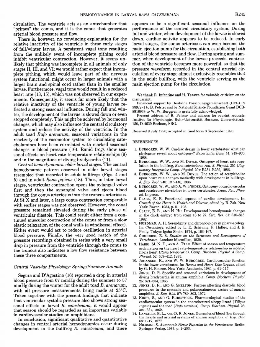
The anatomy of the central circulatory system of larval R. catesbeiana is shown in Fig. 1. All chambers are
Vent1
Conus arteriosus
pie ‘1,
JS
FIG. 1. Schematic drawing of anatomy of central circulatory system of Rana catesbeiana larvae. Appearance of spiral valve in conus arter- iosus is quite variable depending on fraction of the valve that has been removed with the upper part of conus.
separated by cusped valves. The pylangial valve separates the ventricle and the conus arteriosus, whereas the syn- angial valve guards the opening from the conus arteriosus to the truncus arteriosus. A prominent spiral valve at- tached at one edge spirals ~2700 along the length of the larval conus arteriosus. This valve was present in St II larvae, the earliest stage examined. The large left and right atria are located dorsal to the ventricle. Visual observation of dissections and of the intact, beating heart revealed that in early larval stages the conus appeared to have a similar or slightly smaller volume than the ventricle, whereas in later larval stages and adults the ventricle had a much greater volume than the conus.
Central Vascular Physiology: Fall/ Winter Animals
Experiments were performed in two series at different times of the year. Profound seasonal effects on the car- diovascular system were observed. For clarity of presen- tation of the data, we first completely describe the- results obtained with fall/winter animals an .d then, i .n a subse- quent section
9 emphasize the differences observed in the second series performed during spring and summer.
Heart rate. At every developmental stage, tested car- diac frequency and blood pressure of anesthetized ani- mals were not significantly different (P > 0.05) from pithed animals. Therefore, the results of both groups were pooled for subsequent analysis. Heart rate was 62 + 7 beats/min in the youngest larvae (St II) during the fall and winter months, decreasing with development (Fig. 2A). When development is expressed in terms of body mass, heart rate decreased with increasing body mass [heart frequency (fn) = -0.7mass + 61.1, P < 0.051.
BZood pressure. Diastolic pressure in the ventricle was usually Cl.0 mmHg in all developmental stages exam- ined during the fall/winter months. Diastolic pressure recorded in the conus was similar to ventricular diastolic pressure in very young larvae but tended to increase slightly in later stages to -1.5-3.0 mmHg (Fig. 2B). Systolic pressure (P,,,) recorded in both the ventricle and the conus increased markedly with larval develop- ment. We also an .alyzed the relationship between body mass- and arterial blood pressure, because body mass of larval 3). In
R the
catesbeiana increased with development (Fig. fall/winter series, the overall relationship in-

R242 HEMODYNAMICS IN LARVAL RANA CATESBEIANA
0
- A
Developmental Stage
Developmental Stage
FIG. 2. Central vascular function during development in fall and winter months. A: heart rate as a function of developmental stage. Linear regression is represented by equation y = -0.6x + 62.9 (r = 0.8, P < 0.05). B: diastolic and systolic blood pressure recorded in conus. Closed triangles represent conal systolic pressure in preparations where conal systolic pressure exceeded ventricular pressure. Values are means t SD.
Psys = 3.98 Mass o-61
Body Mass (g)
FIG. 3. Relation of systolic blood pressure to increasing body mass in fall/winter animals. Each stage (St) is represented by a separate symbol. Open circle, St II; closed circle, St III; open triangle, St VI; closed triangle,St VII; open square, St X; closed square, St XII; inverted open triangle, St XIII; inverted closed triangle, adult. Dashed lines represent regression lines calculated for each stage; continuous line is overall regression.
eluding all larval stages and small adults is described by the relationship PsYs = 3.98mass”*61 (r = 0.9; P < 0.01).
CentraZ hemodynamics. During the fall and winter months in early larval stages (i.e., many St II, some St III, and few St VI), conal contraction generated a distinct pressure peak that was higher than the mean ventricular pressure in the preparation (triangles in Fig. 2B).
Before St VII, the pressure waveform recorded in the conus showed considerable variability between individ- uals (Fig. 4), but the pattern evident in the youngest larvae became less and less common as development continued. With further larval development, the ventric- ular systolic pressure increased to become the dominant
feature of the waveform, and the contribution of the conal contraction to the pressure waveform recorded in the conus became less obvious (Figs. 4 and 5). By St X and older, the waveform almost invariably showed a dominant ventricular peak with only a slight contribu- tion produced by the conus during early ventricular diastole.
An overlay of serial pressure measurements from the ventricle, conus arteriosus, and truncus arteriosus of animals of different developmental stages is shown in Fig. 5, demonstrating the changing influence of the conus on arterial blood pressure. In the St II larva, shown in Fig. 5, top left, ventricular pressure remained below ar- terial pressure, whereas the conal pressure surpassed it. In later stages, ventricular contraction clearly generated the highest pressure peak.
Figure 6 shows recordings of pressure and volume changes in the conus and in the ventricle of a St VI larva, representative of the hemodynamics in a young fall/ winter larva in which conus systolic pressure was bi- phasic. The pressure in the ventricle, recorded a few minutes after measuring in the conus, exactly matched the first peak of the conus pressure signal. However, while ventricular pressure then fell as the ventricle began to relax, the pressure in the conus increased a second time in the cycle, reflecting a contraction of the conus itself. This overlay of serial recordings often was slightly complicated by small changes in frequency. On a beat- to-beat comparison, however, the pressure profiles from the conus and ventricle were almost always superimpos- able.
The volume changes of the ventricle closely followed pressure changes with filling during the low-pressure phase and emptying during the high-pressure phase of the cycle (Fig. 6). The volume of the conus increased as soon as the ventricle volume decreased, but at the same time the pressure in the conus increased, too. The largest conus volume was observed just after passing the maxi- mum of ventricular pressure where during a slight pres- sure decrease in the conus the maximum extension of the conus was achieved. At this time, the pressure in the conus started to increase again, thus exceeding the ven- tricular pressure, and the conus volume decreased to reach the lowest volume at the beginning of the next systole of the ventricle.
Figure 7 shows simultaneously recorded arterial blood pressure and velocity in a St III and a St XIII fall/winter larva. A short time delay of the blood velocity signal compared with the conus pressure recording was used to confirm that the Doppler flow signal arose downstream of the pressure recording site. Although the conus pro- duced a distinct and significant pressure peak in the St III animal, the velocity trace was hardly influenced, which probably has to be attributed to the smaller volume of the conus compared with the ventricle. Unfortunately, the extremely small dimensions of the central arterial system, the changing diameter of the conus along its length, and possible complicating role of the spiral valve in the conus, all obviated calibration of blood velocity and subsequent calculation of blood flow.

Stage III
HEMODYNAMICS IN LARVAL RANA CATESBEIANA R243
Stage YI Stage X
4
0 L!tA!!L 4
0 [: fyy)
Central Vascular Physiology: Spring/Summer Animals
Profound differences in central hemodynamics were observed between fall/winter and spring/summer larvae (Fig. 8A). Ventricular pressure was much higher in spring/summer larvae from St III to St VII, when com- pared with fall/winter animals over the same develop- mental range (P c 0.05). There was, however, no seasonal difference in ventricular systolic pressure in larvae of St X and St XIII (P > 0.1). The changes become even more obvious when plotting systolic blood pressure vs. body mass (Fig. 8B), where all values for spring/summer lar- vae clearly exceed the mean value obtained in the fall/ winter animals.
In our experiments, no differences were observed in diastolic pressure between fall/winter and spring/sum- mer larvae. The high systolic blood pressures indicate a strong contraction of the ventricle, and accordingly the pressure waveform recorded in the conus was exclusively of the adult type. Heart frequency decreased with devel- opment at the same rate in both spring/summer and fall/ winter groups. Mean values of heart rate were consist-
Stage XIII
4
0 [ m
Stage II C.a. T.a.
--./-\-.-
1 set
FIG. 4. Examples for pressure signals recorded in conus arteriosus of several fall/winter animals of various developmental stages demonstrating variability of pressure waveform, especially in early stages.
ently 6-9 beats/min lower in spring/summer larvae of all stages tested, but because of the individual variation within developmental groups these differences were only significant in two stages.
DISCUSSION
Central Vascular Physiology: Fall/ Winter Animals
Heart rate. Although MS 222 acts as a sympathetic stimulus in anurans (20), there was no difference in heart rate between pithed and anesthetized animals, which probably can be attributed to the low dose of the anes- thetic. Our values for fn are similar to those reported for unrestrained larval R. catesbeiana of similar develop- mental stage (2, 3, 22), suggesting that our methods of immobilization did not bias cardiac performance.
Blood pressure. The present study reveals a significant increase in arterial blood pressure with development in R. catesbeiana, a phenomenon also occurring in larvae of the frog Pseudis paradoxa (W. Burggren, M. Glass, A. Abe, and E. Bicudo, unpublished observations). More- over, an increase in blood pressure with development has
Stage III
1 1
1 set
Adult
FIG. 5. Superimposed pressure recordings ob- tained in series from ventricle (continuous line), conus arteriosus (broken line), and truncus arter- iosus (broken-dotted line) from 3 larval stages and small adult bullfrogs.
I i 1 i
1 set 1 set

R244 HEMODYNAMICS IN LARVAL RANA CAZ’ESBEIANA
1 2 3 I I I sure in R. catesbeiana. Growth, producing new vessels
A -Volume + I-/\\
and enlarged vascular beds, results in both an increase Conus - in total cross-sectional area of the vascular bed (tending +
A-Volume
Ventricle -
I
to decrease vascular resistance) and an increase in total vessel length (tending to increase vascular resistance). If the net effect was an overall increase in vascular resist- ance (i.e., length increase predominated), then an in- crease in blood pressure would be required to assure sufficient tissue perfusion as development progresses. However, this presumes constancy of blood flow per gram tissue as development occurs. Although this appears to be the case in chick embryos (5), there has been no experimental verification of this phenomenon during development in any lower vertebrate.
Conus Pressure
(mm Hg)
Ventricle Pressure 3
(mm Hg)
1 I 1 1 1
1 set
FIG. 6. Original recordings of volume and pressure changes in conus and ventricle of a larva St VI, body mass 1.45 g. Volume changes could not be calibrated, and therefore traces do not allow for volume com- parisons between conus and ventricle. Pressure in ventricle was re- corded separately with a time difference of -5 min. Lines I, 2, and 3 indicate onset of ventricular contraction, end of ventricular, and end of conal contraction, respectively.
Stage III 6
10
Blood Pressure 3 lhh 5 (mm Hg) 0 0
Blood Velocity
0 0
Stage XIII
n A
Central hemodynamics: early larval stages. Dissection of bullfrog larvae demonstrated the early presence of valves separating the different chambers of the central vascular system, and the organization of the spiral valve of the conus appears to be similar to that described for adult ranid frogs (17). The physiological functioning of these valves can be assessed by pressure recordings in the various chambers. If pressure remains elevated distal to the valves compared with proximal, then the valves must be closed and, as a result, separate the chambers completely. The overlay of pressure recordings from dif- ferent sites of the circulatory system (Fig. 5) therefore
VI/“\ demonstrates the presence of functional pylangial and synangial valves early in development as well as the changing importance of the ventricle and conus as con- tractile chambers of the circulation, and both of them have been shown to contain contractile myocytes (8). Examination of Fig. 5, top left, shows that the ventricular systolic pressure remained well below diastolic pressure
. I
’ 1 set ’ I 1
‘1 set ’ FIG. 7. Original recordings of blood pressure in conus and of blood
velocity downstream to micropressure electrode of a St III (0.5 g) and a St XIII (5.8 g) animal.
been documented in vertebrates ranging widely from embryos of chickens (6, 21) to skates (Raja erinacea) (B. Pelster and W. Bemis, unpublished observations) and may be a general developmental pattern in vertebrates.
There could be several explanations for the observed correlation between body mass and arterial blood pres-
16 - 0
s/s I
Ai 0
in the truncus arteriosus at all points in the cardiac cycle. The ventricle generates a sufficiently high pressure to open the pylangial valve and generate flow into the conus in early ventricular systole, and, indeed, this was con- firmed by observing an increase in conus volume at this point in the cardiac cycle. However, the ventricle cannot be responsible for opening the synangial valve and gen- erating ejection of blood through the conus into the truncus arteriosus. In these early larval stages when conal contraction predominates, the conus therefore must renresent the maior contractile chamber of the
A d
B Psys= 10 Mass 0.15 + *-
.- 0 V .-
s/s * 0 00
A c- - a - _ .- *- - . .- -- -. c .- I- .- I- .- -. . -- .- F/W
.- . . .-
II IV VI VIII X XII XIV 0.5 1.0 2.0 5.0
Developmental Stage Body Mass (g)
FIG. 8. Systolic blood pressure of various spring/summer (S/S) animals vs. developmental stage (A) and vs. body mass (B). Open circles in A represent fall/winter (F/W) animals (redrawn from Fig. 2B). Solid line in B represents a linear regression of all data for spring/summer animals. Different stage groups are represented by different symbols (see Fig. 3; open diamonds, St IV; closed diamonds, St V). Dashed line indicates linear regression line obtained for fall/winter animals (redrawn from Fig. 3).
HEMODYNAMICS IN LAI
circulation. The ventricle acts as an antechamber that “primes” the conus, and it is the conus that generates arterial blood pressure and flow.
There is, however, no convincing explanation for the relative inactivity of the ventricle in these early stages of fall/winter larvae. A persistent vagal tone resulting from the unlikely event of incomplete pithing could inhibit ventricular contraction. However, it seems un- likely that pithing was incomplete in all animals of only stages II, III, and VI; we would rather expect that incom- plete pithing, which would leave part of the nervous system functional, might occur in larger animals with a larger brain and spinal cord rather than in the smaller larvae. Furthermore, vagal tone would result in a reduced heart rate (13, 15), which was not observed in our exper- iments. Consequently, it seems far more likely that the relative inactivity of the ventricle of young larvae re- flected a strong seasonal influence. During fall and win- ter, the development of the larvae is slowed down or even stopped completely. This might be achieved by hormonal changes, which may also influence the central circulatory system and reduce the activity of the ventricle. In the adult toad Bufo arenarum, seasonal variations in the reactivity of the vasomotor system to circulating cate- cholamines have been correlated with marked seasonal changes in blood pressure (16). Ranid frogs show sea- sonal effects on heart rate-temperature relationships (9) and in the magnitude of diving bradycardia (11).
Central hemodynamics: older larval stages. The central hemodynamic pattern observed in older larval stages resembled that recorded in adult bullfrogs (Figs. 4 and 5) and in adult Rana pipiens (18). Thus, in later larval stages, ventricular contraction opens the pylangial valve first and then the synangial valve and ejects blood through the conus arteriosus into the truncus arteriosus. At St X and later, a large conus contraction comparable with earlier stages was not observed. However, the conal pressure remained elevated during the early phase of ventricular diastole. This could result either from a con- tinued muscular contraction of the conus or from a slow elastic relaxation of the conal walls (a windkessel effect). Either event would act to reduce oscillation in arterial blood pressure. Finally, the very good match of the pressure recordings obtained in series with a very small drop in pressure from the ventricle through the conus to the truncus also indicates a low flow resistance between these three compartments.
Central Vascular Physiology: Spring/Summer Animals
Segura and D’Agostino (16) reported a drop in arterial blood pressure from 67 mmHg during the summer to 37 mmHg during the winter for the adult toad B. arenarum, with all pressure measurements being made at 25OC. Taken together with the present findings that indicate that ventricular systolic pressure also shows strong sea- sonal effects in larval R. catesbeiana, it would appear that season should be regarded as an important variable in cardiovascular studies on amphibians.
In conclusion, significant qualitative and quantitative changes in central arterial hemodynamics occur during development in the bullfrog R. catesbeiana, and there
AL RANA CATESBEIANA R245
appears to be a significant seasonal influence on the performance of the central circulatory system. During fall and winter, when development of the larvae is slowed down, cardiac activity appears to be reduced. In early larval stages, the conus arteriosus can even become the main ejection pump for the circulation, establishing both arterial blood pressure and flow. During spring and sum- mer, when development of the larvae proceeds, contrac- tion of the ventricle becomes more powerful, so that the pressure waveform recorded in the central arterial cir- culation of every stage almost exclusively resembles that in the adult bullfrog, with the ventricle serving as the main ejection pump for the circulation.
We thank R. Infantino and H. Tazawa for valuable criticism on the manuscript.
Financial support by Deutsche Forschungsgemeinschaft (DFG) Pe 389/2--l to B. Pelster and by National Science Foundation Grant DCB- 8916938 to W. W. Burggren is gratefully acknowledged.
Present address of B. Pelster and address for reprint requests: Institut fur Physiologie, Ruhr-Universitat Bochum, Universitatsstr. 150, D-4630 Bochum, FRG.
Received 9 July 1990; accepted in final form 8 September 1990.
REFERENCES
1.
2.
3.
4.
5.
6.
7.
8.
9.
10.
11.
12.
13.
14.
15.
BURGGREN, W. W. Cardiac design in lower vertebrates: what can phylogeny reveal about ontogeny? Experientia BuseZ44: 919-930, 1988. BURGGREN, W. W., AND M. DOYLE. Ontogeny of heart rate regu- lation in the bullfrog, Rana catesbeiunu. Am. J. Physiol. 251 (Reg- ulatory Integrative Comp. Physiol. 20): R231-R239, 1986. BURGGREN, W. W., AND M. DOYLE. The action of acetylcholine upon heart rate changes markedly with development in bullfrogs. J. Exp. 2001. 240: 137-140, 1986. BURGGREN, W. W., AND A. W. PINDER. Ontogeny of cardiovascular and respiratory physiology in lower vertebrates. Annu. Rev. Phys- iol. In press. CLARK, E. B. Functional aspects of cardiac development. In: Growth of the Heart in Health and Disease, edited by R. Zak. New York: Raven, 1984, p. 81-103. CLARK, E. B., AND N. Hu. Developmental hemodynamic changes in the chick embryo from stage 18 to 27. Circ. Res. 51: 810-815, 1982. FRIEDMAN, A. H. Serendipity and chronobiology in pharmacology. In: Chronology, edited by L. E. Scheving, F. Halber, and J. E. Pauly. Tokyo: Igahu Shoin, 1974, p. 163-167. GOODRICH, E. S. Studies on the Structure and Development of Vertebrates. London: Macmillan, 1930. HARRI, M. N. E., AND A. TALU. Effect of season and temperature acclimation on the heart rate-temperature relationship in isolated frog’s heart (Runu temporuriu). Comp. Biochem. Physiol. A Comp. Physiol. 52: 409-412, 1975. JOHANSEN, K., AND W. W. BURGGREN. Cardiovascular function in the lower vertebrates. In: Hearts and Heart-Like Organs, edited by G. H. Bourne. New York: Academic, 1980, p. 61-117. JONES, D. R. Specific and seasonal variations in development of diving bradycardia in anuran amphibia. Comp. Biochem. Physiol. 25: 821-834,1968. JONES, D. R., AND G. SHELTON. Factors affecting diastolic blood pressures in the systemic and pulmocutaneous arches of anuran amphibia. J. Exp. Biol. 57: 789-803, 1972. KIRBY, S., AND G. BURNSTOCK. Pharmacological studies of the cardiovascular system in the anaesthetized sleepy lizard (Tiliquu rugosu) and the toad (Bufo murinus). Comp. Biochem. Physiol. 28: 321-331,1969. LANGILLE, B. L., AND D. R. JONES. Dynamics of blood flow through the hearts and arterial systems of anuran amphibia. J. Exp. Biol. 68: 1-17, 1977. NILSSON, S. Autonomic Nerve Function in the Vertebrates. Berlin: Springer-Verlag, 1983, p. l-253.

R246 HEMODYNAMICS IN LARVAL RANA CATESBEIANA
16. SEGURA, E. T., AND S. A. D’AGOSTINO. Seasonal variations of blood pressure, vasomotor reactivity and plasmatic catecholamines in the toad. Acta Physiol. Latinoam. 14: 231-237, 1964.
17. SHELTON, G. Gas exchange, pulmonary blood supply, and the partially divided amphibian heart. Perspect. Exp. Biol. 1: 247-259, 1976.
18. SHELTON, G., AND D. R. JONES. Pressure and volume relationships in the ventricle, conus and arterial arches of the frog heart. J. Exp. Biol. 43: 479-488, 1965.
19. SHELTON, G., AND D. R. JONES. A comparative study of central blood pressures in five amphibians. J. Exp. Biol. 49: 631-643,1968.
20. SMITH, D. G. Sympathetic cardiac stimulation in Bufo marinus under MS-222 anesthesia. Am. J. Physiol. 226: 367-370, 1974.
21. TAZAWA, H. Measurement of blood pressure of chick embryo with an implanted needle catheter. J. Appl. Physiol. 51: 1023-1026,198l.
22. WEST, N. H., AND W. W. BURGGREN. Gill and lung ventilatory responses to steady-state aquatic hypoxia and hyperoxia in the bullfrog tadpole. Respir. Physiol. 47: 165-176, 1982.


